Review

Synergy of Body Composition, Exercise Oncology, and Pharmacokinetics: A Narrative Review of Personalizing Paclitaxel Treatment for Breast Cancer

by
Nele Adriaenssens
1,2,*,
Stephanie C. M. Wuyts
3,4,
Stephane Steurbaut
3,5,
Pieter-Jan De Sutter
6,
An Vermeulen
6,
Amy de Haar-Holleman
2,7,
David Beckwée
1,
Steven Provyn
8,
Sofie Vande Casteele
6,
Jinyu Zhou
1,
Katrien Lanckmans
9,
Jan Van Bocxlaer
6 and
Len De Nys
1,2
1
Rehabilitation Research, Vrije Universiteit Brussel (VUB), Laarbeeklaan 121, 1090 Brussels, Belgium
2
Medical Oncology Department, Universitair Ziekenhuis Brussel (UZ Brussel), Laarbeeklaan 101, 1090 Brussels, Belgium
3
Pharmacy Department, Universitair Ziekenhuis Brussel (UZ Brussel), Laarbeeklaan 101, 1090 Brussels, Belgium
4
Research Centre for Digital Medicine, Vrije Universiteit Brussel (VUB), Laarbeeklaan 103, 1090 Brussels, Belgium
5
Vitality Research Group, Vrije Universiteit Brussel (VUB), Laarbeeklaan 103, 1090 Brussels, Belgium
6
Department of Bioanalysis, Faculty of Pharmaceutical Sciences, Universiteit Gent, Ottergemsesteenweg 460, 9000 Gent, Belgium
7
Translational Oncology Research Center, Vrije Universiteit Brussel (VUB), Laarbeeklaan 103, 1090 Brussels, Belgium
8
Human Physiology and Sports Physiotherapy, Vrije Universiteit Brussel (VUB), Pleinlaan 2, 1050 Brussels, Belgium
9
Clinical Biology Department, Universitair Ziekenhuis Brussel (UZ Brussel), Laarbeeklaan 101, 1090 Brussels, Belgium
*
Author to whom correspondence should be addressed.
Cancers 2025, 17(8), 1271; https://doi.org/10.3390/cancers17081271
Submission received: 3 March 2025 / Revised: 28 March 2025 / Accepted: 3 April 2025 / Published: 9 April 2025
(This article belongs to the Section Cancer Therapy)

Simple Summary

Paclitaxel is a chemotherapy drug widely used for breast cancer, but serious side effects, such as nerve damage and low white blood cell counts, often limit its effectiveness. Currently, chemotherapy doses are based on body surface area, but this method does not consider important individual factors like muscle and fat levels, which can affect how the drug is processed. This review explores how body composition and physical activity influence paclitaxel’s effects and side effects. Regular exercise may improve treatment outcomes by supporting muscle health and reducing toxicity risks. By integrating these factors into chemotherapy dosing strategies, we can move toward a more personalized approach, ensuring that each patient receives the most effective and safest treatment. This research highlights the need for further studies to refine chemotherapy dosing, potentially leading to better treatment experiences and outcomes for breast cancer patients.

Abstract

Background/Objectives: Paclitaxel is a type of small molecule chemotherapy widely used for breast cancer, but its clinical efficacy is often hindered by dose-limiting toxicities such as chemotherapy-induced peripheral neuropathy and neutropenia. Traditional dosing based on body surface area does not account for variations in body composition, which may influence paclitaxel metabolism, toxicity, and treatment outcomes. This review explores the interplay between body composition, physical activity, and paclitaxel pharmacokinetics, emphasizing the potential for personalized dosing strategies. Methods: A comprehensive narrative review was conducted by analyzing the literature on body composition, small molecule chemotherapy-related toxicities, pharmacokinetics, and exercise oncology. Studies examining the role of skeletal muscle mass, adipose tissue, and physical activity in modulating paclitaxel metabolism and side effects were included. Results: Evidence suggests that patients with low skeletal muscle mass are at a higher risk of paclitaxel-induced toxicities due to altered drug distribution and clearance. Sarcopenic obesity, characterized by low muscle and high-fat levels, further exacerbates these risks. Exercise, particularly resistance and aerobic training, has been shown to improve muscle mass, mitigate toxicities, and enhance chemotherapy tolerance. However, the precise mechanisms by which exercise influences paclitaxel pharmacokinetics remain underexplored. Conclusions: Personalized chemotherapy dosing, considering body composition and physical activity, may optimize paclitaxel treatment outcomes. Future research should focus on integrating exercise interventions into oncology care and refining dosing models that account for interindividual differences in drug metabolism. These advancements could improve treatment efficacy while minimizing toxicities in breast cancer patients.

1. Introduction

Breast cancer (BC) is the most common cancer among women worldwide, accounting for 11.7% of all cancer cases, with an estimated 2.26 million new diagnoses globally in 2020 [1,2,3]. An estimated 355,000 new cases of BC were diagnosed in the European Union (EU) in 2020, representing approximately 13.3% of all cancer cases in the region. This marks BC as a significant health concern, as it also accounted for 7.3% of cancer-related deaths within the EU in 2020 [4]. Over recent decades, the survival rates and life expectancies of BC patients have significantly improved, mainly due to improvements in early detection through screening and advancements in chemotherapy and targeted therapies [4].
Depending on the stage and type of BC, treatment strategies primarily include local therapies, such as surgery with or without radiation, and systemic therapies, including chemotherapy, hormone therapy, targeted therapies, and/or immunotherapy According to major prospective clinical trials, several standard chemotherapy regimens, including anthracyclines and taxanes, are available for BC treatment [5,6]. Taxane-based regimens, such as paclitaxel (PTX), are among the most effective and commonly used systemic therapies for both early- and late-stage BC [7]. However, the toxicity associated with chemotherapy remains a significant challenge in the treatment of BC [8].
In this narrative review, we explore how interindividual differences in body composition and physical activity levels may influence paclitaxel pharmacokinetics and the risk of chemotherapy-induced toxicities in breast cancer patients. We synthesize current evidence on the relationships between skeletal muscle mass, adipose tissue distribution, physical activity, and paclitaxel metabolism and clearance. Special attention is given to how unfavorable body composition—such as sarcopenia or sarcopenic obesity—can increase the risk of dose-limiting toxicities and how physical activity may mitigate these effects by positively influencing body composition and metabolic function (Figure 1). Finally, we highlight the emerging role of exercise oncology and its potential to contribute to more personalized, safe, and effective chemotherapy strategies.

2. Paclitaxel in the Treatment of Breast Cancer
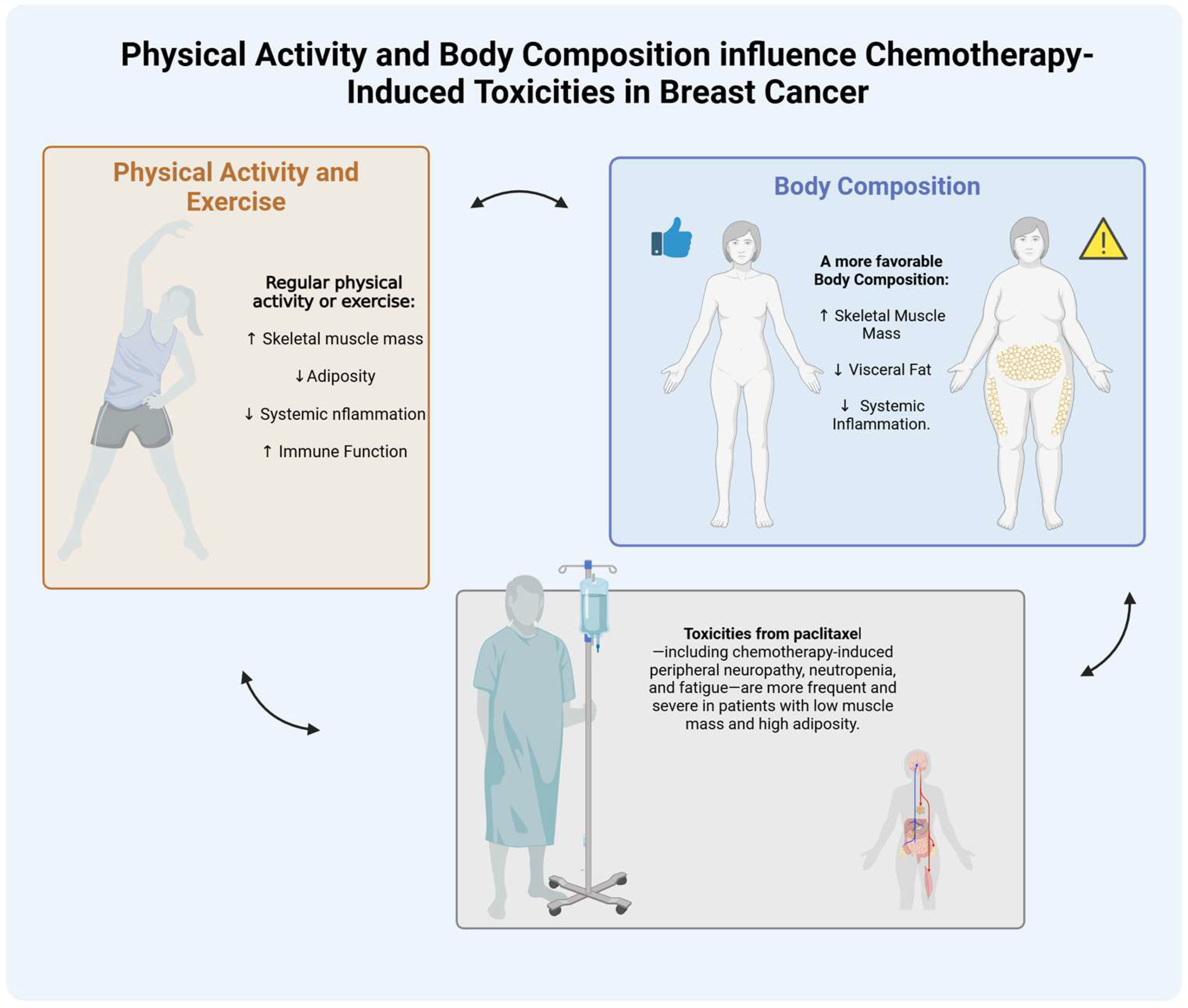
2.1. Toxicities

Paclitaxel induces cancer cell death through microtubule stabilization, leading to a mitotic arrest [9]. However, its mechanism of action can also affect healthy dividing cells, resulting in side effects. Patients with BC undergoing PTX therapy may experience a variety of toxicities, including neurological, hematological, gastrointestinal, and cardiac adverse effects, as well as hypersensitivity reactions [8,10]. The severity and presentation of these side effects can vary among patients and are often influenced by the treatment schedule and dosing protocols [10].
The toxicities associated with PTX continue to limit the effectiveness of treatment regimens based on this drug [10]. For example, while pretreatment with a standard regimen that includes a corticosteroid (such as dexamethasone) and an H1 receptor blocker can help reduce the risk of hypersensitivity reactions before PTX infusions, minor hypersensitivity reactions still occur in a significant number of patients [10,11]. Importantly, a small but critical percentage of individuals still experience life-threatening reactions [12].
The primary hematologic toxicity associated with PTX is neutropenia [13], which can be managed with the administration of granulocyte colony-stimulating factor. This leaves neurotoxicity, particularly chemotherapy-induced peripheral neuropathy (CIPN), as the main dose-limiting toxicity (DLT) [14]. CIPN, which can develop either early or late after PTX administration, affects the majority of patients undergoing treatment [15]. Clinically, it is characterized by numbness and paresthesia in a glove-and-stocking distribution, resulting from the accumulation of PTX in the dorsal root ganglia [16,17]. These DLTs can result in treatment delays, dose reductions, or even premature termination of therapy [18]. Additionally, they significantly impact patients’ health-related quality of life (HRQoL) and functional status in both the short and long term [19].

2.2. PTX Dose Intensity

Relative dose intensity (RDI), the ratio of the actual delivered dose intensity (mg/m2 per week) to the planned dose intensity for a chemotherapy regimen, is a key measure that reflects dose delays or reductions during treatment [20,21,22]; however, its application must be interpreted in the context of evolving evidence highlighting the limitations of BSA-based dosing (see below in Section 3). While an RDI below 85% is considered a clinically significant reduction compared to the standard therapy, these thresholds are typically derived from standard dosing models that do not consider individual patient characteristics, such as body composition. This decreased RDI is associated with worse outcomes, including reduced survival rates in advanced BC. This association has been demonstrated in both randomized clinical trials and retrospective observational studies [20,23,24]. Maintaining an adequate RDI is particularly important for BC patients undergoing PTX therapy, as timely administration of the planned dosage is crucial for achieving optimal prognosis. In most cancers, there is a plateau in the dose–response curve for cytotoxic chemotherapy. This means that increasing the dose towards an asymptotic threshold leads to increased toxicity without providing additional antitumor effects [25]. Therefore, it is essential for patients undergoing chemotherapy to achieve a therapeutic response while minimizing toxicity, ensuring that their drug exposure remains at acceptable levels.
Studies have explored this dose–response relationship, specifically for PTX; however, evidence regarding PTX’s therapeutic and toxic ranges is limited to certain dosing schedules [26]. Key toxicity parameters, such as CIPN, have been associated with elevated maximal PTX plasma concentrations (Cmax) or prolonged exposure durations above the threshold of 0.05 µM [27,28,29] It is crucial to monitor and manage these toxicities to ensure an optimal RDI for patients with BC throughout their chemotherapy treatment.

2.3. Challenges Regarding Body Surface Area Dosing of PTX

Body surface area (BSA) has long been the standard metric for determining chemotherapeutic drug dosage, as described in two reviews from 2016 [30,31]. The normalization of chemotherapy dosing was initially recommended because of the proposed relationship with other physiologic parameters, including resting energy expenditure, plasma volume, and cardiac output [32]. Various BSA formulae have been developed to simplify this process [31]. All recognized formulae are based on easily identifiable patient body variables such as height and weight and in some cases, age and sex [31].
However, the use of BSA as a basis for chemotherapy dosing, including PTX in BC, has long been debated due to its poor correlation with physiological parameters influencing pharmacokinetics and pharmacodynamics (PK/PD), such as differences in skeletal muscle mass (SMM), adipose tissue (AT), and metabolic activity [31]. As Miller et al. (2004) demonstrated two decades ago, BSA is proportional to blood volume, but does not accurately reflect an individual’s ability to metabolize or excrete cytotoxic drugs [33]. Their study found no significant association between BSA and PTX-induced toxicities, such as leukopenia or neutropenia, suggesting that BSA is not a reliable predictor of chemotherapy toxicity.

3. Role of Body Composition and Physical Activity in Breast Cancer Treatment

3.1. Body Composition and Chemotherapy Dosing

Patients exhibit significant biological heterogeneity, which can affect the desired patient outcomes. Even individuals with similar or identical body weight, BSA, or body mass index (BMI) can show considerable variations in the amount and distribution of AT and lean body mass, including SMM and extracellular fluid [34]. Body composition can influence the pharmacokinetics of chemotherapy in several ways. Therefore, it has been suggested that considering body composition parameters may be more accurate than BSA for calculating the appropriate chemotherapy dosage [35].
Changes in body composition, such as reduced SMM, decreased muscle quality, and increased AT, significantly contribute to DLTs across various cancer treatments, irrespective of cancer type, age, sex, BMI, or physical function [35,36,37,38]. While there is a correlation between BMI and both SMM and AT, there is considerable variability in these measurements for any given BMI or BSA value, particularly in women with BC [34,39]. Substantial evidence supports the role of SMM and AT as predictive factors for the occurrence of DLT during chemotherapy [40,41,42,43]. Additionally, studies have shown that toxicities from anthracyclines and taxanes, such as PTX, both commonly used in BC treatment, are more closely related to SMM than to BSA [44]. In patients with metastatic BC, sarcopenia—which is defined as a reduction in SMM—has been associated with an increased risk of chemotherapy toxicities and a shorter median time to tumor progression, irrespective of overall BMI [26,43,44,45]. This association is particularly significant in patients with sarcopenic obesity, characterized by low SMM and high AT. Sarcopenic obesity, along with conditions like myosteatosis (fat infiltration in muscle), can lead to changes in drug distribution, metabolism, and exposure. This may lead to over (or under) dosing, increased systemic inflammation, and decreased physical resilience [36,43,46,47]. If chemotherapy dosing is based on BSA, patients with sarcopenia may receive disproportionately high doses relative to their metabolically active SMM, further increasing toxicity risks [38,48]. Similar findings have been reported in other cancer types, where patients with low SMM are at a higher risk of experiencing treatment-related toxicities [40,49,50,51,52].
Additionally, the body composition of patients with metastatic BC often differs from that of early-stage BC patients due to cancer-related fatigue, sarcopenia and/or cachexia, and treatment-related side effects such as bone demineralization (osteopenia/osteoporosis), reduced muscular strength [53], decreased aerobic capacity [54], and weight gain [55,56]. These observations highlight the need to better understand the role of body composition in chemotherapy tolerance among women with BC [34].
Advances in standard imaging techniques for body composition assessment, such as dual-energy X-ray absorptiometry (DXA) and single-slice computed tomography (CT) scanning, have led to numerous studies exploring the associations between body composition and adverse outcomes in oncology [18,57,58]. Recently, artificial intelligence has been utilized to extract body composition metrics from medical images, providing new opportunities for facilitating personalized treatment approaches [59]. Given the widespread use of CT imaging in cancer diagnosis and monitoring, body composition can be analyzed, without imposing an additional burden on patients, using available diagnostic images [60].

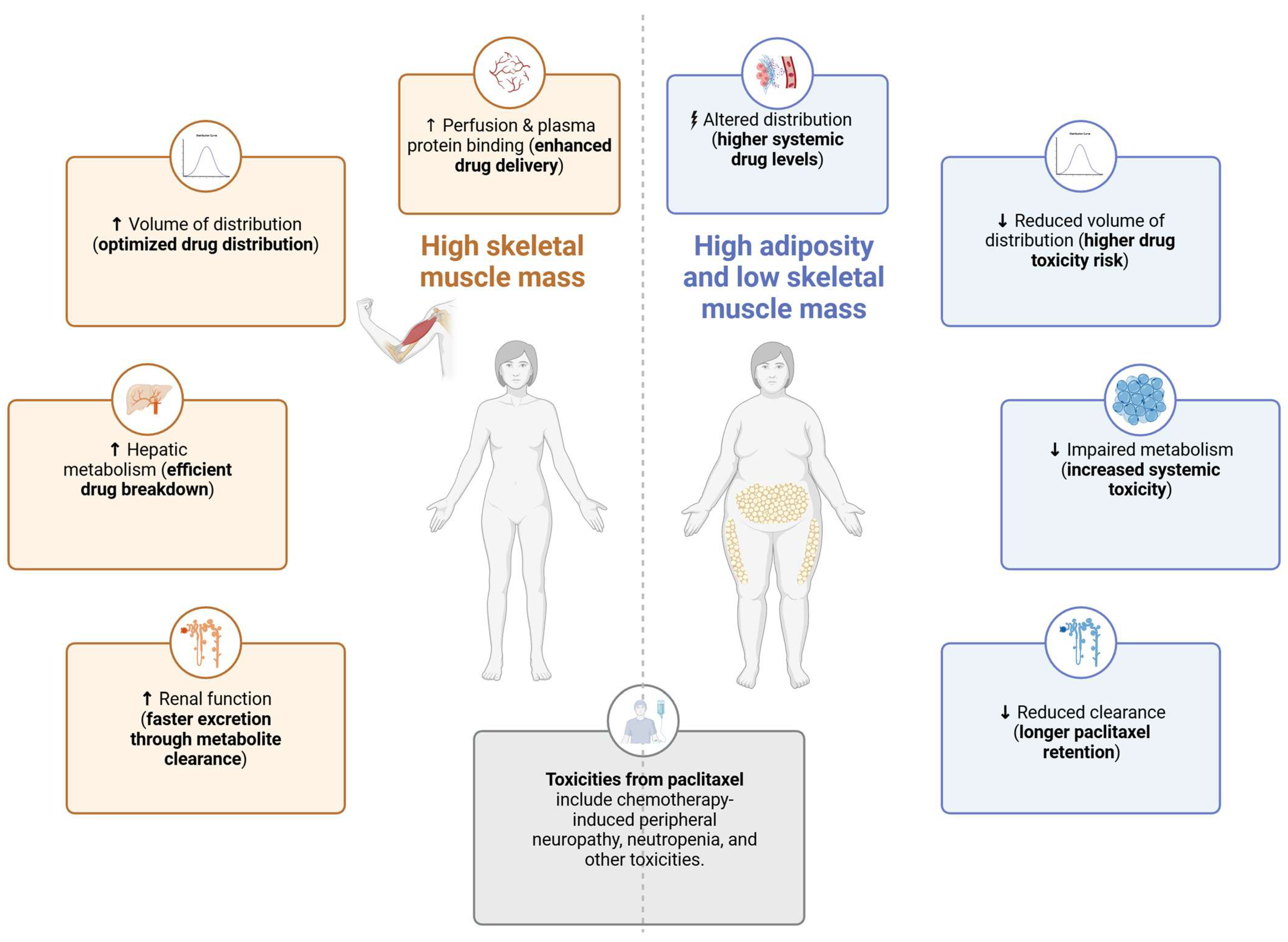
3.2. Body Composition, Paclitaxel Pharmacokinetics and Toxicities

In contrast to the numerous studies that have investigated body composition and cancer outcomes, only a limited number of trials have explored the correlation between body composition and small molecule drug PK [35]. Each drug exhibits intrinsic properties that contribute to variability in its distribution throughout the body. Hydrophilic drugs primarily distribute into lean tissue, while lipophilic drugs accumulate in the AT, affecting their volume of distribution (Vd) and elimination rate from the body [61]. Patients with sarcopenic obesity exhibit a reduced Vd for hydrophilic drugs and an increased Vd for lipophilic drugs. These alterations in drug distribution are not adequately accounted for by BSA-based dosing [38]. Conversely, the PK of large-molecule drugs, such as monoclonal antibodies (mAbs), are expected to be less influenced by body composition, as their elimination is receptor-mediated, and their distribution is primarily limited to the extracellular space [62]. A conceptual model of interactions between body composition and PTX pharmacokinetics is shown in Figure 2.
Several examples were identified for the chemotherapy drugs docetaxel, epirubicin, and oxaliplatin. A recent study confirms that overweight and obese patients receiving adjuvant chemotherapy based on docetaxel, another taxane, exhibit worse disease-free survival and overall survival rates, as well as a higher risk of distant metastases, compared to those of lean patients [61]. In contrast, no significant differences in outcomes were observed across BMI categories for patients treated with non-docetaxel-based chemotherapy. These findings may be attributed to docetaxel’s lipophilic properties, leading to an increased Vd and reduced efficacy in patients with a higher BMI. Although the study discusses BMI rather than body composition, the authors suggested that the risk–benefit ratio of using taxanes in BC should be reassessed based on the patients’ body composition [61]. Research conducted by Prado et al. (2011) demonstrated that variability in SMM was linked to toxicities and differences in the drug clearance of epirubicin in cancer patients, while BSA was not a reliable predictor [63]. More recent research on the PK of oxaliplatin in older adults with gastrointestinal malignancies indicated that patients with low SMM and high total AT exhibited the lowest Vd and drug clearance, the highest maximal drug concentrations, and a significantly increased risk of severe chemotherapy toxicities [64].
The hydrophobic nature of PTX leads to substantial accumulation in AT, high protein binding, and delayed clearance, contributing to its complex PK profile and toxicity risks [10,64]. A decrease in SMM is believed to influence PK by reducing the Vd and affecting protein binding, drug metabolism, and clearance [35]. SMM has been reported to influence the PK of certain chemotherapeutic agents, including PTX [27,35,36,63,65,66]. Specifically, lower SMM, such as in patients with sarcopenia, has been associated with an increase in PTX’s maximal concentration (Cmax), which likely explains the increased risk of CIPN in these patients. PK modeling simulations suggest that extending the infusion of PTX in patients with the lowest SMM may reduce CIPN while maintaining therapeutic efficacy [27]. Furthermore, the active compound and that of the excipient(s) may affect drug exposure. In a study by Smorenburg et al. (2003), older adult patients with BC displayed up to a 50% increase in PTX exposure with age [67]. This increase was attributed to a significantly altered disposition of the formulation vehicle, Cremophor EL, which was markedly decreased in the older patient group. The potential association between changes in body composition, and this decrease in PTX total body clearance with age remains to be clarified [67]. Conversely, in more extensive cohort studies, older age was not identified as an independent risk factor leading to clinically relevant changes in PTX PK [68,69], suggesting that other variables, such as body composition, may be relevant.
Similarly, obesity may alter drug distribution and affect PK by decreasing the plasma concentrations of lipophilic drugs but increasing their storage in adipose tissue. For PTX, Sparreboom et al. found a 46.6% increase in Vd and a 20.4% increase in clearance in obese patients (BMI ≥ 30 kg/m2) compared to the results for lean patients (BMI < 25 kg/m2) [70]. Since PTX is strongly bound to plasma proteins (e.g., albumin), which are found to be reduced in obese patients [71], this may lead to an increase in the free fraction of PTX, which in turn may increase its pharmacological effect, but also the risk of toxicity. PTX is metabolized in the liver by cytochrome P450 enzymes, primarily CYP3A4 and CYP2C8. Obese individuals may exhibit decreased drug metabolism, as individuals with nonalcoholic fatty liver disease can show impaired hepatic drug-metabolizing enzyme activity, resulting in prolonged PTX exposure and increased risk of adverse reactions. Although PTX is primarily metabolized by the liver and excreted in the bile, there is some renal excretion. Therefore, impaired renal function, as observed in some obese patients [72], can also adversely affect the elimination of this drug and its metabolites. In addition, women have been reported to experience greater toxicity than men at equivalent doses of PTX. This may be due to gender-related differences in body composition, such as women tending to carry more body fat than men. The increased susceptibility of women to the adverse effects of PTX has also recently been linked to differences in the functioning of the immune system, although the underlying mechanism remains poorly understood [73,74]. Therefore, it is important to be aware of interpatient variability and to consider differences in body composition when prescribing PTX.
More studies with adequate sample sizes and comprehensive pharmacologic endpoints are needed to examine the effects of body composition on chemotherapy PK, particularly for PTX [32]. Such studies will help validate preliminary findings and support the development of dosing strategies tailored to individual body compositions.

4. Role of Physical Activity and Exercise in Breast Cancer Treatment

In addition to body composition, physical activity (PA), defined as any movement resulting in energy expenditure, including leisure-time activities, and exercise, characterized as planned and structured PA aimed at improving physical capacity or physical fitness, is increasingly recognized as a crucial intervention for patients with BC during and after treatment [75,76]. Engaging in low-to-moderate levels of PA is well known to significantly reduce mortality in patients with various malignancies [77], offering greater health benefits compared to inactivity [78]. Evidence indicates that recreational PA can lower BC mortality risk, enhance physiological and immune functions [78,79], lower stress, and improve sleep disturbances and overall HRQoL [80]. Further, PA helps mitigate cancer- and treatment-related side effects, such as decreased muscular strength, weight gain, and bone demineralization, leading to improved body composition and better clinical outcomes in patients with BC [56,75,79,81,82].
The clinical impact of exercise throughout all phases of cancer care—ranging from prevention to advanced stages and end-of-life care—is well-documented [83,84]. Therefore, physiotherapists play a pivotal role in the interdisciplinary management of cancer, contributing therapies beyond exercise alone [85]. Exercise oncology, a relatively young therapy option discipline, emerged in the 1980s, primarily led by nurses. Early research established that exercise is feasible, safe, acceptable, and effective for individuals living with or beyond cancer [54,86,87,88].
In 2003, the first exercise guidelines for cancer were introduced [89]. In 2007, the Physical Activity Cancer Control framework was established to delineate the different phases of cancer care [90] This framework informed the first roundtable on exercise guidelines in oncology organized by the American College of Sports Medicine in 2010 [91], with subsequent updates and expansions in 2019 [92,93]. In 2022, the American Society of Clinical Oncology (ASCO) published guidelines regarding exercise during cancer treatment [94], as did the Dutch Royal Society for Physical Therapy (KNGF); these were later translated into English in 2023, and are now widely used in clinical practice [95].
Despite the advancements in the field of exercise oncology, two critical gaps persist. First, current guidelines predominantly emphasize rehabilitation after active medical treatment, such as chemotherapy, rather than during treatment. Second, they remain general in scope, primarily recommending PA levels similar to those for healthy populations (i.e., 150 min of moderate-intensity exercise per week or 10,000 steps per day, as per WHO guidelines) [96]. While the long-term effects of exercise on cancer-related side effects are well-documented in these guidelines, the acute and chronic physiological responses to exercise during chemotherapy remain underexplored. Positive changes in the tumor microenvironment might be driven by accumulative effects of repeated acute exercise responses [97,98,99]. These acute but short-lasting changes that exceed the adaptive responses produced by prolonged training might show the potential to provide immediate physiological benefits and in turn, improve DLTs [99].
Addressing these gaps is essential for optimizing exercise interventions tailored to the unique needs of patients undergoing active cancer treatment. Previous studies even suggest that exercise programs can improve DLTs like CIPN in patients undergoing taxane-, platinum-, or vinca alkaloid-based chemotherapy, as concluded in the review by Wirtz et al. (2018) [81] and confirmed in the more recent review by Tanay et al. (2023) [100]. Exercise also has the potential to prevent the onset and slow the progression of CIPN, particularly in patients with BC [81,101]. This reduction in side effects may be related to the multiple PK-related effects of exercise on chemotherapy that warrant further investigation. Further, exercise PK [102,103] and exercise oncology [104] are two young and promising research fields that play an important role in chemotherapy-induced DLTs.
Despite the clear benefits of exercise and PA in managing BC-related side effects, motivating women with BC to engage in regular PA remains a considerable challenge [105]. Post-treatment fatigue and pain related to chemotherapy and other treatments, along with other side effects, often discourage participation in PA [105]. Various studies have demonstrated that interventions such as cognitive behavioral strategies show added value in increasing PA motivation and adherence in women with BC by implementing approaches such as behavior change techniques [106,107,108] and motivational interviewing [109,110]. Additionally, patients with BC often reduce their PA during therapy [111] and tend to remain physically inactive after treatment [112].

5. Exercise-Mediated Improvements in Body Composition and Their Impact on Paclitaxel Tolerance

Chemotherapy often leads to detrimental changes in body composition, including the loss of SMM and increased adiposity, which are associated with poor physical function and adverse treatment outcomes [113,114]. Exercise interventions have shown promising potential in positively influencing body composition in cancer patients, which may be crucial for optimizing chemotherapy dosing and improving treatment outcomes [115].
Exercise, particularly supervised aerobic and resistance exercise, during chemotherapy enhances SMM, muscle strength, and endurance, while reducing AT accumulation and improving overall body composition in patients with BC. This is supported by the meta-analysis of Li et al. (2024) [116] and other recent publications [113,114,117,118,119,120,121,122,123,124,125,126]. Resistance training has emerged as a particularly effective strategy for preserving and even restoring skeletal muscle quality [122,123]. Mijwel et al. (2019) reported that a 16-week exercise intervention during chemotherapy for BC patients significantly improved muscle strength and quality [127]. Additionally, Aires et al. (2024) emphasize the role of exercise in rebuilding skeletal muscle integrity in BC patients post-chemotherapy [117]. They suggest that targeted PA interventions could help mitigate sarcopenia and its associated risks. These functional improvements in muscle tissue may contribute to increased chemotherapy tolerance and reduced incidence of DLTs.
While mechanistic differences exist between aerobic and resistance training, both modalities improve treatment tolerance by positively modulating body composition. Resistance training consistently increases lean mass (e.g., +8.3 kg versus +2.7 kg with aerobic training) [128] and reduces intermuscular fat infiltration, particularly beneficial for preserving skeletal muscle during chemotherapy [92,128]. In contrast, aerobic exercise yields more significant reductions in overall fat mass and percentage of body fat (e.g., −1.8% versus −0.9% for resistance training) [128].
Current ASCO guidelines recommend combining aerobic and resistance exercises to maximize clinical benefits across multiple outcomes [94]. Resistance training is especially emphasized in patients at risk for sarcopenia or who are undergoing dose-dense chemotherapy regimens. However, direct comparisons between aerobic and resistance exercise in regards to PTX pharmacokinetics are currently lacking. Preclinical models suggest that combined exercise may enhance drug delivery through mechanisms such as vascular normalization, but these findings require clinical validation [129]. Therefore, until PTX-specific data become available, exercise prescriptions should be individualized based on patient capacity, safety, and tolerance, with a preference for multimodal programs incorporating aerobic and resistance components. In the context of PTX, SMM is a critical factor influencing treatment outcomes and toxicities. Research by Wopat et al. (2024) and Poltronieri et al. (2022) reveals that lower SMM is associated with higher rates of DLTs, including CIPN and gastrointestinal distress [119,120]. Exercise-based interventions may mitigate these toxicities by maintaining SMM and improving metabolic function [125,126]. The review by Li et al. (2023) [116] further demonstrates that exercise during chemotherapy not only reduces treatment-related weight gain, particularly visceral adiposity, but also enhances chemotherapy tolerance by promoting a healthier body composition profile [116].
Adipose tissue, particularly visceral and subcutaneous fat, plays a nuanced role in chemotherapy outcomes. Excess adiposity has been linked to systemic inflammation and altered drug pharmacokinetics, exacerbating chemotherapy toxicities [121]. Exercise can attenuate these effects by reducing visceral fat and improving the visceral-to-subcutaneous adipose tissue ratio, as highlighted in the studies by Poltronieri et al. (2022) and Godinho-Mota et al. (2021) [113,120]. These improvements are particularly relevant for patients undergoing PTX, in which systemic inflammation is a key driver of DLTs [98,117].
Additionally, the dose and type of exercise matter. An et al. (2020) [125] found that higher doses of resistance exercise led to greater gains in SMM and reductions in toxicities. Meanwhile, Bland et al. (2022) argued for the integration of individualized exercise prescriptions tailored to patient needs and their baseline PA [122]. Such targeted interventions, as evidenced by Altundag (2020), not only improve the skeletal muscle index but also enhance HRQoL and reduce fatigue during chemotherapy [124]. Furthermore, Kudiarasu et al. (2023) report that combining exercise with dietary interventions yields significant improvements in both SMM and AT, supporting a multimodal approach to optimizing body composition [118].

6. Pharmacokinetics in Breast Cancer Treatment

Emerging research highlights the potential of exercise to influence drug pharmacology [32,102,130]. Exercise may reduce chemotherapy-related toxicities both indirectly, through improved physiological resilience, and more directly via modulating systemic drug exposure. In the context of PTX, dose-limiting toxicities, such as peripheral neuropathy, have been linked to higher Cmax and prolonged exposure above the toxicity threshold (Tc > 0.05 μM) [27,131]. Exercise-induced improvements in skeletal muscle mass and reductions in adiposity affect PTX pharmacokinetics, potentially lowering Cmax and shortening Tc > 0.05 due to changes in drug distribution and metabolism [102,119,131]. Additionally, exercise may enhance hepatic and renal perfusion, improve metabolic enzyme activity, and reduce systemic inflammation—factors that can promote drug clearance [102,132,133]. While direct clinical evidence of reduced systemic exposure achieved through exercise remains limited, recent studies, such as the PABTOX study, have highlighted this mechanism as a promising possibility for reducing toxicities and improving chemotherapy adherence [90,134,135]. The review by McLaughlin et al. (2017) provides a detailed overview of the physiological changes associated with acute, subacute, and chronic exercise, along with the respective drug PK variables that are affected [102].
During acute exercise, physiological changes include increased cardiac output; enhanced blood flow to active skeletal muscles, skin, digestive organs, kidneys, liver, and other tissues; and a decreased glomerular filtration rate [136,137]. Chronic effects are typically seen in body composition changes, such as increases in SMM and decreases in AT, along with increased serum albumin concentrations and a reduction in inflammatory cytokines [102]. The possible alteration in drug PK achieved via exercise is likely mediated through its impact on physiological processes that regulate a drug’s absorption, distribution, metabolism, and excretion [32,137,138] (Table 1).
First, regarding absorption, exercising may accelerate the absorption of drugs, e.g., when administered by intramuscular, subcutaneous, and transdermal routes or by inhalation due to improved blood flow [136,139]. However, the expected influence of exercise on drug absorption remains uncertain due to numerous factors, such as the physicochemical and biochemical properties of drugs, as well as individual anatomical and physiological variations [102].
Second, exercise can significantly impact drug distribution [136]. Acute exercise enhances splanchnic and hepatic blood flow, increasing drug delivery rates to targeted receptor sites and facilitating drug absorption and equilibrium between the plasma and tissues [32,102]. Chronic training increases lean body mass, which is well-hydrated, while decreasing AT. On the other hand, reduced blood flow to AT and other inactive regions during exercise may slow the distribution of lipophilic drugs, such as PTX, that rely on these compartments for storage [139]. Exercise can also alter plasma protein levels. An increase in plasma protein concentration during exercise, often due to hemoconcentration from reduced plasma volume, can enhance drug binding and decrease the free, active drug fraction. For highly protein-bound drugs like PTX, this effect may further reduce the free drug concentration during or after intense PA [102]. Additionally, exercise can affect blood pH and core body temperature, leading to minimal changes in drug binding and altering the amount of the free drug available in the bloodstream [102]. The clinical relevance of these changes to the final PK of the chemotherapeutic drug remains to be established.
Third, exercise can influence drug metabolism by modulating physiological factors such as plasma volume, capillarization of skeletal and cardiac muscle, cytochrome P-450 enzyme activity, and mitochondrial density [32]. During exercise, blood flow is redirected from the liver to the muscles, which can reduce the clearance of drugs with a flow-dependent hepatic metabolism, potentially influencing plasma concentrations [139]. An important chronic response to exercise is a reduction in inflammatory cytokines in cancer patients undergoing chemotherapy. A study by Schauer et al. (2021) [98] investigated the effects of high-intensity and low-to-moderate intensity exercise on inflammatory markers in patients with BC [98]. Over six months, patients engaged in combined aerobic and resistance exercise during and after chemotherapy. The study found that, regardless of exercise intensity, levels of interleukin 6 (IL-6), IL-8, IL-10, and tumor necrosis factor alpha (TNF-α) increased post-treatment, but generally declined post-intervention [98]. Notably, high-intensity exercise resulted in a smaller increase in C-reactive protein (CRP) and TNF-α immediately post-treatment compared to the results for low-to-moderate intensity exercise, suggesting that high-intensity exercise may offer better protection against chemotherapy-related inflammation [98]. While cytokines like IL-6 increase during the subacute period following exercise, their preliminary downregulation of the CYP450 genes is eventually outweighed by a more chronic response, leading to potential increases in the CYP450 microsomal content [102]. As the CYP-enzymes CYP3A4 and CYP2C8 are important for PTX metabolism, changes in their activity may influence overall metabolism [140,141].
Finally, exercise may also affect the elimination of water-soluble and renally extracted drugs and metabolites by decreasing their clearance, which in turn, can increase their plasma concentrations [136,139] depending on the exercise intensity [102]. Additionally, exercise can change urine pH, influencing drug ionization and affecting its reabsorption or excretion [102]. In contrast, exercise can increase the clearance of certain lipid-soluble drugs and/or metabolites by enhancing bile production and secretion, boosting biliary excretion, and reducing intestinal reabsorption, ultimately lowering plasma concentrations [102,136].
Currently, there is a limited amount of evidence regarding the impact of exercise on the bioavailability of anticancer drugs, highlighting the necessity for further research [130]. However, existing in silico models, such as the physiologically based PK model published by Guo et al. (2024), offer potential for predicting of the PK behavior of chemotherapeutic drugs during exercise [137].

7. Future Directions

Future research and clinical practice regarding exercise oncology should prioritize integrating body composition data, PA levels, and exercise into chemotherapy dosing decisions, moving beyond traditional BSA-based methods. This approach, supported by emerging evidence, promises to enhance the precision and personalization of chemotherapy, particularly for drugs like PTX with complex pharmacokinetic profiles.
In 2024, the PABTOX trial (NCT06387901) was launched at the Vrije Universiteit Brussel, Brussels, Belgium, in collaboration with the Universitair Ziekenhuis Brussel and the University of Ghent. This two-year translational research project investigates the interactions between PTX, DLTs, body composition, physical activity, and various patient-specific factors. This study aims to develop a predictive PK–PD model, based on these interactions, for DLTs in women with BC treated with PTX, potentially refining chemotherapy dosing and reducing adverse effects in the future [134].
In addition to PA and low to moderate exercise, high-intensity interval training (HIIT) during chemotherapy represents a promising area for further exploration. Limited evidence suggests that HIIT may offer superior benefits in preserving SMM, improving fitness, and reducing AT compared to the results for traditional exercise [91,127]. Given the challenges of therapy adherence and motivation during chemotherapy, HIIT’s time-efficient nature could enhance patient engagement and treatment outcomes (e.g., NCT05786014, NCT04724499, NCT05913713). Future studies should explore the feasibility and efficacy of integrating HIIT into exercise oncology programs, focusing on its impact on body composition and DLT risks in breast cancer patients.
Moreover, the acute responses to exercise and their potential to directly influence DLTs remain underexplored. Understanding these acute responses could provide immediate physiological benefits and improve chemotherapy tolerance [97,98]. Positive changes in the tumor microenvironment driven by repeated acute exercise responses may explain observed benefits such as higher chemotherapy completion rates and improved aerobic capacity [99]. Future research should aim to elucidate these mechanisms and refine exercise prescriptions to maximize therapeutic benefits.
Additionally, integrating nutritional guidelines from the European Society for Clinical Nutrition and Metabolism (ESPEN) into the care of patients with BC during chemotherapy can significantly enhance the benefits of exercise. ESPEN recommends a daily caloric intake of 25–30 kcal/kg body weight and a protein intake of 1.2–1.5 g/kg body weight to support anabolic processes and maintain SMM [142,143]. This nutritional support is crucial for optimizing body composition, as adequate protein intake is essential for muscle repair and growth, particularly when combined with resistance and aerobic exercise [142,143]. By ensuring that patients meet these nutritional targets, healthcare providers can help maximize the effectiveness of exercise interventions, reduce the incidence of DLTs, and improve overall treatment outcomes. This multimodal approach, combining tailored exercise programs with precise nutritional support, offers a comprehensive strategy to enhance HRQoL and treatment efficacy for breast cancer patients undergoing chemotherapy. There are already several examples of (ongoing) studies combining exercise and nutritional interventions in cancer care, with promising results [135,144].
Clinically, low SMI may help identify high-risk patients who could benefit from extended infusion protocols, closer monitoring, or exercise interventions to improve muscle mass and treatment tolerance [27,122,123]. To facilitate this, oncologists could use routinely acquired CT or PET-CT scans to assess e.g., SMI at the L3 level, a suggested proxy for total muscle mass that needs to be further explored [43,59,145]. Automated tools using artificial intelligence can extract SMI from scans without added burden [59,60,145]. As imaging tools become standardized, integrating SMI into pharmacokinetic-informed dosing algorithms represents a promising step toward personalized chemotherapy. Beyond SMI alone, the ongoing PABTOX study (NCT06387901) mentioned above is developing a PK–PD model to integrate additional personalized metrics, i.e., SMM, AT, and PA, to improve early identification of patients at risk for dose-limiting toxicities and low relative dose intensity.

8. Conclusions

This review highlights the limitations of BSA-based dosing for PTX in patients with BC and emphasizes the need to consider individual variations in body composition when optimizing treatment strategies. Differences in body composition influence the risk of toxicities, underscoring the importance of more personalized approaches to chemotherapy dosing.
Exercise and PA have emerged as modifiable factors that could positively impact treatment outcomes by improving overall physical health and enhancing chemotherapy tolerance. However, the specific mechanisms by which exercise influences the PK of chemotherapeutic drugs, including PTX, remain underexplored. Future research is critical for filling this knowledge gap and developing strategies to effectively incorporate exercise into standard treatment protocols.
By integrating body composition assessments and structured exercise (and nutritional) interventions into PTX treatment for BC, therapies can be customized for more personalized and effective outcomes, as these strategies hold significant potential for reducing DLTs, improving PTX efficacy, and enhancing the HRQoL for BC patients.
As the fields of exercise oncology and personalized medicine continue to advance, exercise programs and body composition-based dosing strategies are expected to play an increasingly important role in BC care. Translational research is required to move beyond BSA-based dosing, exploring the potential value of body composition variables in PTX PK. In 2024, the PABTOX trial (Investigating Paclitaxel Toxicity in Breast Cancer: the Roles of Physical Activity and Body Composition—NCT06387901) started patient inclusion, aiming to provide the first preliminary evidence. Future research should focus on developing and validating these approaches to improve the efficacy and tolerability of PTX treatment for BC patients.

Author Contributions

Conceptualization, N.A., S.C.M.W. and L.D.N.; methodology, N.A. and L.D.N.; resources, N.A., S.C.M.W., D.B. and S.P.; data curation, N.A. and L.D.N.; writing—original draft preparation, N.A., J.Z., L.D.N. and S.C.M.W.; writing—review and editing, N.A., A.d.H.-H., S.C.M.W., J.V.B., P.-J.D.S., A.V., S.V.C., K.L. and S.S.; visualization, N.A. and L.D.N.; supervision, N.A. and D.B.; project administration, N.A. and L.D.N.; funding acquisition, N.A. All authors have read and agreed to the published version of the manuscript.

Funding

This research project is funded by Kom op tegen Kanker (Stand up to Cancer), a project of the Flemish Cancer Society (project ID: 13163).

Acknowledgments

The authors would like to acknowledge the contributions of Anita Barzegar Fallah to the initiation of this manuscript, including the provision and integration of sources.

Conflicts of Interest

The authors declare no conflicts of interest.

References

  1. Bray, F.; Laversanne, M.; Sung, H.; Ferlay, J.; Siegel, R.L.; Soerjomataram, I.; Jemal, A. Global cancer statistics 2022: GLOBOCAN estimates of incidence and mortality worldwide for 36 cancers in 185 countries. CA Cancer J. Clin. 2024, 74, 229–263. [Google Scholar] [PubMed]
  2. World Health Organization Cancer. Available online: https://www.who.int/news-room/fact-sheets/detail/cancer (accessed on 15 September 2024).
  3. Siegel, R.L.; Miller, K.D.; Fuchs, H.E.; Jemal, A. Cancer statistics, 2022. CA Cancer J. Clin. 2022, 72, 7–33. [Google Scholar]
  4. Giaquinto, A.N.; Sung, H.; Miller, K.D.; Kramer, J.L.; Newman, L.A.; Minihan, A.; Jemal, A.; Siegel, R.L. Breast cancer statistics, 2022. CA Cancer J. Clin. 2022, 72, 524–541. [Google Scholar] [PubMed]
  5. Waks, A.G.; Winer, E.P. Breast cancer treatment: A review. JAMA 2019, 321, 288–300. [Google Scholar] [PubMed]
  6. Pondé, N.F.; Zardavas, D.; Piccart, M. Progress in adjuvant systemic therapy for breast cancer. Nat. Rev. Clin. Oncol. 2019, 16, 27–44. [Google Scholar]
  7. Gradishar, W. Taxanes for the treatment of metastatic breast cancer. Breast Cancer Basic. Clin. Res. 2012, 6, 159–171. [Google Scholar]
  8. Al-Mahayri, Z.N.; AlAhmad, M.M.; Ali, B.R. Current opinion on the pharmacogenomics of paclitaxel-induced toxicity. Expert Opin. Drug Metab. Toxicol. 2021, 17, 785–801. [Google Scholar] [PubMed]
  9. Klein, I.; Lehmann, H.C. Pathomechanisms of paclitaxel-induced peripheral neuropathy. Toxics 2021, 9, 229. [Google Scholar] [CrossRef]
  10. Marupudi, N.I.; Han, J.E.; Li, K.W.; Renard, V.M.; Tyler, B.M.; Brem, H. Paclitaxel: A review of adverse toxicities and novel delivery strategies. Expert Opin. Drug Saf. 2007, 6, 609–621. [Google Scholar]
  11. Price, K.S.; Castells, M.C. Taxol reactions. In Allergy and Asthma Proceedings; OceanSide Publications: Providence, RI, USA, 2002. [Google Scholar]
  12. Weiss, R.B.; Donehower, R.; Wiernik, P.; Ohnuma, T.; Gralla, R.; Trump, D.; Baker, J., Jr.; Van Echo, D.; Von Hoff, D.; Leyland-Jones, B. Hypersensitivity reactions from taxol. J. Clin. Oncol. 1990, 8, 1263–1268. [Google Scholar]
  13. Rowinsky, E.K.; Donehower, R.C. Paclitaxel (taxol). N. Eng. J. Med. 1995, 332, 1004–1014. [Google Scholar]
  14. Mielke, S.; Sparreboom, A.; Mross, K. Peripheral neuropathy: A persisting challenge in paclitaxel-based regimes. Eur. J. Cancer 2006, 42, 24–30. [Google Scholar] [PubMed]
  15. Seretny, M.; Currie, G.L.; Sena, E.S.; Ramnarine, S.; Grant, R.; MacLeod, M.R.; Colvin, L.A.; Fallon, M. Incidence, prevalence, and predictors of chemotherapy-induced peripheral neuropathy: A systematic review and meta-analysis. Pain 2014, 155, 2461–2470. [Google Scholar] [CrossRef] [PubMed]
  16. Lipton, R.B.; Apfel, S.C.; Dutcher, J.P.; Rosenberg, R.; Kaplan, J.; Berger, A.; Einzig, A.; Wiernik, P.; Schaumburg, H. Taxol produces a predominantly sensory neuropathy. Neurology 1989, 39, 368. [Google Scholar]
  17. Gutiérrez-Gutiérrez, G.; Sereno, M.; Miralles, A.; Casado-Sáenz, E.; Gutiérrez-Rivas, E. Chemotherapy-induced peripheral neuropathy: Clinical features, diagnosis, prevention and treatment strategies. Clin. Transl. Oncol. 2010, 12, 81–91. [Google Scholar]
  18. van den Berg, M.M.; Kok, D.E.; Posthuma, L.; Kamps, L.; Kelfkens, C.S.; Buist, N.; Geenen, M.; Haringhuizen, A.; Heijns, J.B.; van Lieshout, R.H. Body composition is associated with risk of toxicity-induced modifications of treatment in women with stage I–IIIB breast cancer receiving chemotherapy. Breast Cancer Res. Treat. 2019, 173, 475–481. [Google Scholar] [CrossRef]
  19. Jivani, A.; Shinde, R.K. A Comprehensive Review of Taxane Treatment in Breast Cancer: Clinical Perspectives and Toxicity Profiles. Cureus 2024, 16, e59266. [Google Scholar]
  20. Nielson, C.M.; Bylsma, L.C.; Fryzek, J.P.; Saad, H.A.; Crawford, J. Relative Dose Intensity of Chemotherapy and Survival in Patients with Advanced Stage Solid Tumor Cancer: A Systematic Review and Meta-Analysis. Oncol. 2021, 26, e1609–e1618. [Google Scholar]
  21. Denduluri, N.; Patt, D.A.; Wang, Y.; Bhor, M.; Li, X.; Favret, A.M.; Morrow, P.K.; Barron, R.L.; Asmar, L.; Saravanan, S.; et al. Dose Delays, Dose Reductions, and Relative Dose Intensity in Patients With Cancer Who Received Adjuvant or Neoadjuvant Chemotherapy in Community Oncology Practices. J. Natl. Compr. Cancer Netw. 2015, 13, 1383–1393. [Google Scholar] [CrossRef]
  22. Havrilesky, L.J.; Reiner, M.; Morrow, P.K.; Watson, H.; Crawford, J. A review of relative dose intensity and survival in patients with metastatic solid tumors. Crit. Rev. Oncol. Hematol. 2015, 93, 203–210. [Google Scholar] [CrossRef]
  23. Denduluri, N.; Lyman, G.H.; Wang, Y.; Morrow, P.K.; Barron, R.; Patt, D.; Bhowmik, D.; Li, X.; Bhor, M.; Fox, P.; et al. Chemotherapy Dose Intensity and Overall Survival Among Patients with Advanced Breast or Ovarian Cancer. Clin. Breast Cancer 2018, 18, 380–386. [Google Scholar] [CrossRef] [PubMed]
  24. Loibl, S.; Skacel, T.; Nekljudova, V.; Luck, H.J.; Schwenkglenks, M.; Brodowicz, T.; Zielinski, C.; von Minckwitz, G. Evaluating the impact of Relative Total Dose Intensity (RTDI) on patients’ short and long-term outcome in taxane- and anthracycline-based chemotherapy of metastatic breast cancer—A pooled analysis. BMC Cancer 2011, 11, 131. [Google Scholar] [CrossRef] [PubMed]
  25. Gurney, H. How to calculate the dose of chemotherapy. Br. J. Cancer 2002, 86, 1297–1302. [Google Scholar] [CrossRef]
  26. Hertz, D.L.; Joerger, M.; Bang, Y.-J.; Mathijssen, R.H.; Zhou, C.; Zhang, L.; Gandara, D.; Stahl, M.; Monk, B.J.; Jaehde, U.; et al. Paclitaxel therapeutic drug monitoring—International association of therapeutic drug monitoring and clinical toxicology recommendations. Eur. J. Cancer 2024, 202, 114024. [Google Scholar] [CrossRef]
  27. Hertz, D.L.; Chen, L.; Henry, N.L.; Griggs, J.J.; Hayes, D.F.; Derstine, B.A.; Su, G.L.; Wang, S.C.; Pai, M.P. Muscle mass affects paclitaxel systemic exposure and may inform personalized paclitaxel dosing. Br. J. Clin. Pharmacol. 2022, 88, 3222–3229. [Google Scholar] [CrossRef]
  28. Hertz, D.L.; Kidwell, K.M.; Vangipuram, K.; Li, F.; Pai, M.P.; Burness, M.; Griggs, J.J.; Schott, A.F.; Van Poznak, C.; Hayes, D.F. Paclitaxel plasma concentration after the first infusion predicts treatment-limiting peripheral neuropathy. Clin. Cancer Res. 2018, 24, 3602–3610. [Google Scholar] [CrossRef]
  29. Mielke, S.; Sparreboom, A.; Steinberg, S.M.; Gelderblom, H.; Unger, C.; Behringer, D.; Mross, K. Association of paclitaxel pharmacokinetics with the development of peripheral neuropathy in patients with advanced cancer. Clin. Cancer Res. 2005, 11, 4843–4850. [Google Scholar] [CrossRef]
  30. Faisal, W.; Tang, H.-M.; Tiley, S.; Kukard, C. Not all body surface area formulas are the same, but does it matter? J. Glob. Oncol. 2016, 2, 436. [Google Scholar] [CrossRef] [PubMed]
  31. Redlarski, G.; Palkowski, A.; Krawczuk, M. Body surface area formulae: An alarming ambiguity. Sci. Rep. 2016, 6, 27966. [Google Scholar] [CrossRef]
  32. Purcell, S.A.; Kok, D.E.; Ketterl, T.; Garcia, M.B.; Joffe, L.; Brown, J.C.; Dieli-Conwright, C.M.; Williams, G.R. Pharmacokinetics of cancer therapeutics and energy balance: The role of diet intake, energy expenditure, and body composition. JNCI Monogr. 2023, 2023, 3–11. [Google Scholar] [CrossRef]
  33. Miller, A.A.; Rosner, G.L.; Egorin, M.J.; Hollis, D.; Lichtman, S.M.; Ratain, M.J. Prospective Evaluation of Body Surface Area as a Determinant of Paclitaxel Pharmacokinetics and Pharmacodynamics in Women with Solid Tumors: Cancer and Leukemia Group B Study 9763. Clin. Cancer Res. 2004, 10, 8325–8331. [Google Scholar] [CrossRef] [PubMed]
  34. Durkin, K.; Heetun, A.; Ewings, S.; Munday, R.; Wootton, S.A.; Turner, L.; Copson, E.R.; Cutress, R.I.; Group, C.-S. Body composition and chemotherapy toxicity in women with early breast cancer (CANDO-3): Protocol for an observational cohort study. BMJ Open 2022, 12, e054412. [Google Scholar] [CrossRef] [PubMed]
  35. Hopkins, J.J.; Sawyer, M.B. A review of body composition and pharmacokinetics in oncology. Expert. Rev. Clin. Pharmacol. 2017, 10, 947–956. [Google Scholar] [CrossRef]
  36. Bruno, K.d.A.; Sobreira da Silva, M.J.; Chaves, G.V. Association of body composition with toxicity to first-line chemotherapy and three-year survival in women with ovarian adenocarcinoma. Acta Oncol. 2021, 60, 1611–1620. [Google Scholar] [CrossRef]
  37. Ryan, A.M.; Prado, C.M.; Sullivan, E.S.; Power, D.G.; Daly, L.E. Effects of weight loss and sarcopenia on response to chemotherapy, quality of life, and survival. Nutrition 2019, 67–68, 110539. [Google Scholar] [CrossRef] [PubMed]
  38. Hopkins, J.J.; Sawyer, M.B. Interactions of lean soft-tissue and chemotherapy toxicities in patients receiving anti-cancer treatments. Cancer Chemother. Pharmacol. 2018, 82, 1–29. [Google Scholar] [CrossRef]
  39. Romero-Corral, A.; Somers, V.K.; Sierra-Johnson, J.; Thomas, R.J.; Collazo-Clavell, M.; Korinek, J.; Allison, T.G.; Batsis, J.; Sert-Kuniyoshi, F.; Lopez-Jimenez, F. Accuracy of body mass index in diagnosing obesity in the adult general population. Int. J. Obes. 2008, 32, 959–966. [Google Scholar] [CrossRef]
  40. Barret, M.; Antoun, S.; Dalban, C.; Malka, D.; Mansourbakht, T.; Zaanan, A.; Latko, E.; Taieb, J. Sarcopenia is linked to treatment toxicity in patients with metastatic colorectal cancer. Nutr. Cancer 2014, 66, 583–589. [Google Scholar] [CrossRef] [PubMed]
  41. Huillard, O.; Mir, O.; Peyromaure, M.; Tlemsani, C.; Giroux, J.; Boudou-Rouquette, P.; Ropert, S.; Delongchamps, N.B.; Zerbib, M.; Goldwasser, F.; et al. Sarcopenia and body mass index predict sunitinib-induced early dose-limiting toxicities in renal cancer patients. Br. J. Cancer 2013, 108, 1034–1041. [Google Scholar] [CrossRef]
  42. Tan, B.H.L.; Brammer, K.; Randhawa, N.; Welch, N.T.; Parsons, S.L.; James, E.J.; Catton, J.A. Sarcopenia is associated with toxicity in patients undergoing neo-adjuvant chemotherapy for oesophago-gastric cancer. Eur. J. Surg. Oncol. 2015, 41, 333–338. [Google Scholar] [CrossRef]
  43. Prado, C.M.; Baracos, V.E.; McCargar, L.J.; Reiman, T.; Mourtzakis, M.; Tonkin, K.; Mackey, J.R.; Koski, S.; Pituskin, E.; Sawyer, M.B. Sarcopenia as a Determinant of Chemotherapy Toxicity and Time to Tumor Progression in Metastatic Breast Cancer Patients Receiving Capecitabine Treatment. Clin. Cancer Res. 2009, 15, 2920–2926. [Google Scholar] [PubMed]
  44. Shachar, S.S.; Deal, A.M.; Weinberg, M.; Nyrop, K.A.; Williams, G.R.; Nishijima, T.F.; Benbow, J.M.; Muss, H.B. Skeletal Muscle Measures as Predictors of Toxicity, Hospitalization, and Survival in Patients with Metastatic Breast Cancer Receiving Taxane-Based Chemotherapy. Clin. Cancer Res. 2017, 23, 658–665. [Google Scholar] [CrossRef] [PubMed]
  45. Shachar, S.S.; Williams, G.R.; Muss, H.B.; Nishijima, T.F. Prognostic value of sarcopenia in adults with solid tumours: A meta-analysis and systematic review. Eur. J. Cancer 2016, 57, 58–67. [Google Scholar]
  46. Baracos, V.E.; Arribas, L. Sarcopenic obesity: Hidden muscle wasting and its impact for survival and complications of cancer therapy. Ann. Oncol. 2018, 29, ii1–ii9. [Google Scholar]
  47. Aleixo, G.; Shachar, S.; Nyrop, K.; Muss, H.; Malpica, L.; Williams, G. Myosteatosis and prognosis in cancer: Systematic review and meta-analysis. Crit. Rev. Oncol. Hematol. 2020, 145, 102839. [Google Scholar]
  48. Cousin, S.; Hollebecque, A.; Koscielny, S.; Mir, O.; Varga, A.; Baracos, V.; Soria, J.; Antoun, S. Low skeletal muscle is associated with toxicity in patients included in phase I trials. Investig. New Drugs 2014, 32, 382–387. [Google Scholar]
  49. Jung, H.-W.; Kim, J.W.; Kim, J.-Y.; Kim, S.-W.; Yang, H.K.; Lee, J.W.; Lee, K.-W.; Kim, D.-W.; Kang, S.-B.; Kim, K.-i. Effect of muscle mass on toxicity and survival in patients with colon cancer undergoing adjuvant chemotherapy. Support. Care Cancer 2015, 23, 687–694. [Google Scholar]
  50. Prado, C.M.; Baracos, V.E.; McCargar, L.J.; Mourtzakis, M.; Mulder, K.E.; Reiman, T.; Butts, C.A.; Scarfe, A.G.; Sawyer, M.B. Body composition as an independent determinant of 5-fluorouracil–based chemotherapy toxicity. Clin. Cancer Res. 2007, 13, 3264–3268. [Google Scholar]
  51. Ali, R.; Baracos, V.E.; Sawyer, M.B.; Bianchi, L.; Roberts, S.; Assenat, E.; Mollevi, C.; Senesse, P. Lean body mass as an independent determinant of dose-limiting toxicity and neuropathy in patients with colon cancer treated with FOLFOX regimens. Cancer Med. 2016, 5, 607–616. [Google Scholar]
  52. Anandavadivelan, P.; Brismar, T.B.; Nilsson, M.; Johar, A.M.; Martin, L. Sarcopenic obesity: A probable risk factor for dose limiting toxicity during neo-adjuvant chemotherapy in oesophageal cancer patients. Clin. Nutr. 2016, 35, 724–730. [Google Scholar]
  53. Ott, C.D.; Twiss, J.J.; Waltman, N.L.; Gross, G.J.; Lindsey, A.M. Challenges of recruitment of breast cancer survivors to a randomized clinical trial for osteoporosis prevention. Cancer Nurs. 2006, 29, 21–31. [Google Scholar]
  54. MACVICAR, M.G.; Winningham, M.L.; NICKEL, J.L. Effects of aerobic interval training on cancer patients’ functional capacity. Nurs. Res. 1989, 38, 348–353. [Google Scholar] [PubMed]
  55. Demark-Wahnefried, W.; Peterson, B.L.; Winer, E.P.; Marks, L.; Aziz, N.; Marcom, P.K.; Blackwell, K.; Rimer, B.K. Changes in weight, body composition, and factors influencing energy balance among premenopausal breast cancer patients receiving adjuvant chemotherapy. J. Clin. Oncol. 2001, 19, 2381–2389. [Google Scholar]
  56. Loprinzi, P.D.; Cardinal, B.J. Effects of physical activity on common side effects of breast cancer treatment. Breast Cancer 2012, 19, 4–10. [Google Scholar] [PubMed]
  57. Kazemi-Bajestani, S.M.R.; Mazurak, V.C.; Baracos, V. Computed tomography-defined muscle and fat wasting are associated with cancer clinical outcomes. In Seminars in Cell & Developmental Biology; Elsevier: Amsterdam, The Netherlands, 2016. [Google Scholar]
  58. Brown, J.C.; Cespedes Feliciano, E.M.; Caan, B.J. The Evolution of Body Composition in Oncology—Epidemiology, Clinical Trials, and the Future of Patient Care: Facts and Numbers; Wiley Online Library: Hoboken, NJ, USA, 2018; pp. 1200–1208. [Google Scholar]
  59. Attanasio, S.; Forte, S.M.; Restante, G.; Gabelloni, M.; Guglielmi, G.; Neri, E. Artificial intelligence, radiomics and other horizons in body composition assessment. Quant. Imagining Med. Surg. 2020, 10, 1650–1660. [Google Scholar] [CrossRef]
  60. Bi, W.L.; Hosny, A.; Schabath, M.B.; Giger, M.L.; Birkbak, N.J.; Mehrtash, A.; Allison, T.; Arnaout, O.; Abbosh, C.; Dunn, I.F.; et al. Artificial intelligence in cancer imaging: Clinical challenges and applications. CA A Cancer J. Clin. 2019, 69, 127–157. [Google Scholar]
  61. Desmedt, C.; Fornili, M.; Clatot, F.; Demicheli, R.; Bortoli, D.D.; Leo, A.D.; Viale, G.; Azambuja, E.d.; Crown, J.; Francis, P.A.; et al. Differential Benefit of Adjuvant Docetaxel-Based Chemotherapy in Patients With Early Breast Cancer According to Baseline Body Mass Index. J. Clin. Oncol. 2020, 38, 2883–2891. [Google Scholar] [PubMed]
  62. Hendrikx, J.; Haanen, J.; Voest, E.E.; Schellens, J.H.M.; Huitema, A.D.R.; Beijnen, J.H. Fixed Dosing of Monoclonal Antibodies in Oncology. Oncologist 2017, 22, 1212–1221. [Google Scholar]
  63. Prado, C.M.; Lima, I.S.; Baracos, V.E.; Bies, R.R.; McCargar, L.J.; Reiman, T.; Mackey, J.R.; Kuzma, M.; Damaraju, V.L.; Sawyer, M.B. An exploratory study of body composition as a determinant of epirubicin pharmacokinetics and toxicity. Cancer Chemother. Pharmacol. 2011, 67, 93–101. [Google Scholar]
  64. Williams, G.R.; Al-Obaidi, M.; Rower, J.; Harmon, C.; Dai, C.; Acosta, E.; Giri, S.; Zamboni, W.; Lucas, A.T.; Shachar, S.S. Does Oxaliplatin Pharmacokinetics (PKs) Explain Associations Between Body Composition and Chemotherapy Toxicity Risk in Older Adults with Gastrointestinal (GI) Cancers? Wolters Kluwer Health: Philadelphia, PA, USA, 2021. [Google Scholar]
  65. Wong, A.; Seng, K.; Ong, E.; Wang, L.; Oscar, H.; Cordero, M.; Copones, R.; Fan, L.; Tan, S.; Goh, B. Body fat composition impacts the hematologic toxicities and pharmacokinetics of doxorubicin in Asian breast cancer patients. Breast Cancer Res. Treat. 2014, 144, 143–152. [Google Scholar]
  66. Massicotte, M.H.; Borget, I.; Broutin, S.; Baracos, V.E.; Leboulleux, S.; Baudin, E.; Paci, A.; Deroussent, A.; Schlumberger, M.; Antoun, S. Body composition variation and impact of low skeletal muscle mass in patients with advanced medullary thyroid carcinoma treated with vandetanib: Results from a placebo-controlled study. J. Clin. Endocrinol. Metab. 2013, 98, 2401–2408. [Google Scholar] [CrossRef]
  67. Smorenburg, C.H.; ten Tije, A.J.; Verweij, J.; Bontenbal, M.; Mross, K.; van Zomeren, D.M.; Seynaeve, C.; Sparreboom, A. Altered clearance of unbound paclitaxel in elderly patients with metastatic breast cancer. Eur. J. Cancer 2003, 39, 196–202. [Google Scholar] [CrossRef]
  68. Crombag, M.B.S.; de Vries Schultink, A.H.M.; Koolen, S.L.W.; Wijngaard, S.; Joerger, M.; Schellens, J.H.M.; Dorlo, T.P.C.; van Erp, N.P.; Mathijssen, R.H.J.; Beijnen, J.H.; et al. Impact of Older Age on the Exposure of Paclitaxel: A Population Pharmacokinetic Study. Pharm. Res. 2019, 36, 33. [Google Scholar]
  69. Barginear, M.; Dueck, A.C.; Allred, J.B.; Bunnell, C.; Cohen, H.J.; Freedman, R.A.; Hurria, A.; Kimmick, G.; Le-Rademacher, J.G.; Lichtman, S.; et al. Age and the Risk of Paclitaxel-Induced Neuropathy in Women with Early-Stage Breast Cancer (Alliance A151411): Results from 1,881 Patients from Cancer and Leukemia Group B (CALGB) 40101. Oncologist 2019, 24, 617–623. [Google Scholar]
  70. Sparreboom, A.; Wolff, A.C.; Mathijssen, R.H.; Chatelut, E.; Rowinsky, E.K.; Verweij, J.; Baker, S.D. Evaluation of alternate size descriptors for dose calculation of anticancer drugs in the obese. J. Clin. Oncol. 2007, 25, 4707–4713. [Google Scholar] [PubMed]
  71. Mosli, R.H.; Mosli, H.H. Obesity and morbid obesity associated with higher odds of hypoalbuminemia in adults without liver disease or renal failure. Diabetes Metab. Syndr. Obes. 2017, 10, 467–472. [Google Scholar]
  72. Kjaergaard, A.D.; Teumer, A.; Witte, D.R.; Stanzick, K.J.; Winkler, T.W.; Burgess, S.; Ellervik, C. Obesity and Kidney Function: A Two-Sample Mendelian Randomization Study. Clin. Chem. 2022, 68, 461–472. [Google Scholar] [PubMed]
  73. Zamboni, W.C.; Charlab, R.; Burckart, G.J.; Stewart, C.F. Effect of Obesity on the Pharmacokinetics and Pharmacodynamics of Anticancer Agents. J. Clin. Pharmacol. 2023, 63 (Suppl. 2), S85–S102. [Google Scholar]
  74. Chmielewski, N.N.; Limoli, C.L. Sex Differences in Taxane Toxicities. Cancers 2022, 14, 3325. [Google Scholar] [CrossRef]
  75. Mizrahi, D.; Lai, J.K.L.; Wareing, H.; Ren, Y.; Li, T.; Swain, C.T.; Smith, D.P.; Adams, D.; Martiniuk, A.; David, M. Effect of exercise interventions on hospital length of stay and admissions during cancer treatment: A systematic review and meta-analysis. Br. J. Sports Med. 2024, 58, 97–109. [Google Scholar]
  76. Watson, G.; Coyne, Z.; Houlihan, E.; Leonard, G. Exercise oncology: An emerging discipline in the cancer care continuum. Postgrad. Med. 2022, 134, 26–36. [Google Scholar]
  77. Cannioto, R.A.; Dighe, S.; Mahoney, M.C.; Moysich, K.B.; Sen, A.; Hulme, K.; McCann, S.E.; Ambrosone, C.B. Habitual recreational physical activity is associated with significantly improved survival in cancer patients: Evidence from the Roswell Park Data Bank and BioRepository. Cancer Causes Control. 2019, 30, 1–12. [Google Scholar] [PubMed]
  78. Kim, J.; Choi, W.J.; Jeong, S.H. The effects of physical activity on breast cancer survivors after diagnosis. J. Cancer Prev. 2013, 18, 193. [Google Scholar]
  79. Cannioto, R.A.; Hutson, A.; Dighe, S.; McCann, W.; McCann, S.E.; Zirpoli, G.R.; Barlow, W.; Kelly, K.M.; DeNysschen, C.A.; Hershman, D.L. Physical activity before, during, and after chemotherapy for high-risk breast cancer: Relationships with survival. JNCI J. Natl. Cancer Inst. 2021, 113, 54–63. [Google Scholar]
  80. De Nys, L.; Anderson, K.; Ofosu, E.F.; Ryde, G.C.; Connelly, J.; Whittaker, A.C. The effects of physical activity on cortisol and sleep: A systematic review and meta-analysis. Psychoneuroendocrinology 2022, 143, 105843. [Google Scholar] [PubMed]
  81. Wirtz, P.; Baumann, F.T. Physical activity, exercise and breast cancer-what is the evidence for rehabilitation, aftercare, and survival a review. Breast Care 2018, 13, 92–100. [Google Scholar]
  82. Spence, R.R.; Heesch, K.C.; Brown, W.J. Exercise and cancer rehabilitation: A systematic review. Cancer Treat. Rev. 2010, 36, 185–194. [Google Scholar] [PubMed]
  83. Hojman, P.; Gehl, J.; Christensen, J.F.; Pedersen, B.K. Molecular Mechanisms Linking Exercise to Cancer Prevention and Treatment. Cell Metab. 2018, 27, 10–21. [Google Scholar]
  84. Gauchez, L.; Boyle, S.L.L.; Eekman, S.S.; Harnie, S.; Decoster, L.; Van Ginderdeuren, F.; De Nys, L.; Adriaenssens, N. Recommended Physiotherapy Modalities for Oncology Patients with Palliative Needs and Its Influence on Patient-Reported Outcome Measures: A Systematic Review. Cancers 2024, 16, 3371. [Google Scholar] [CrossRef]
  85. Adriaenssens, N.; Strimpakos, N.; Rotem, N.; Sheill, G.; Cannone, M.; Gigli, L.; Tiesnese, L.; Descloux, A.; MacKenzie, A.; Pérez Navarro, M.; et al. The Role of Physiotherapy in Cancer Care in the Europe Region: A Position Paper of the Cancer Working Group of Europe Region World Physiotherapy. J. Cancer Rehabil. 2023, 6, 70–79. [Google Scholar]
  86. Mock, V.; Burke, M.B.; Sheehan, P.; Creaton, E.M.; Winningham, M.L.; McKenney-Tedder, S.; Schwager, L.P.; Liebman, M. A nursing rehabilitation program for women with breast cancer receiving adjuvant chemotherapy. Oncol. Nurs. Forum 1994, 21, 899–907. [Google Scholar]
  87. Winningham, M.L.; MacVicar, M.G.; Bondoc, M.; Anderson, J.I.; Minton, J.P. Effect of aerobic exercise on body weight and composition in patients with breast cancer on adjuvant chemotherapy. Oncol. Nurs. Forum 1989, 16, 683–689. [Google Scholar] [PubMed]
  88. Winningham, M.L.; MacVicar, M.G.; Burke, C.A. Exercise for Cancer Patients: Guidelines and Precautions. Phys. Sport. 1986, 14, 125–134. [Google Scholar]
  89. Brown, J.K.; Byers, T.; Doyle, C.; Coumeya, K.S.; Demark-Wahnefried, W.; Kushi, L.H.; McTieman, A.; Rock, C.L.; Aziz, N.; Bloch, A.S.; et al. Nutrition and physical activity during and after cancer treatment: An American Cancer Society guide for informed choices. CA Cancer J. Clin. 2003, 53, 268–291. [Google Scholar] [PubMed]
  90. Courneya, K.S.; Segal, R.J.; Mackey, J.R.; Gelmon, K.; Reid, R.D.; Friedenreich, C.M.; Ladha, A.B.; Proulx, C.; Vallance, J.K.H.; Lane, K.; et al. Effects of Aerobic and Resistance Exercise in Breast Cancer Patients Receiving Adjuvant Chemotherapy: A Multicenter Randomized Controlled Trial. J. Clin. Oncol. 2007, 25, 4396–4404. [Google Scholar]
  91. Schmitz, K.H.; Courneya, K.S.; Matthews, C.; Demark-Wahnefried, W.; Galvão, D.A.; Pinto, B.M.; Irwin, M.L.; Wolin, K.Y.; Segal, R.J.; Lucia, A.; et al. American College of Sports Medicine Roundtable on Exercise Guidelines for Cancer Survivors. Med. Sci. Sports Exerc. 2010, 42, 1409–1426. [Google Scholar]
  92. Campbell, K.L.; Winters-Stone, K.M.; Wiskemann, J.; May, A.M.; Schwartz, A.L.; Courneya, K.S.; Zucker, D.S.; Matthews, C.E.; Ligibel, J.A.; Gerber, L.H.; et al. Exercise Guidelines for Cancer Survivors: Consensus Statement from International Multidisciplinary Roundtable. Med. Sci. Sports Exerc. 2019, 51, 2375–2390. [Google Scholar]
  93. Patel, A.V.; Friedenreich, C.M.; Moore, S.C.; Hayes, S.C.; Silver, J.K.; Campbell, K.L.; Winters-Stone, K.; Gerber, L.H.; George, S.M.; Fulton, J.E.; et al. American College of Sports Medicine Roundtable Report on Physical Activity, Sedentary Behavior, and Cancer Prevention and Control. Med. Sci. Sports Exerc. 2019, 51, 2391–2402. [Google Scholar]
  94. Ligibel, J.A.; Bohlke, K.; May, A.M.; Clinton, S.K.; Demark-Wahnefried, W.; Gilchrist, S.C.; Irwin, M.L.; Late, M.; Mansfield, S.; Marshall, T.F.; et al. Exercise, Diet, and Weight Management During Cancer Treatment: ASCO Guideline. J. Clin. Oncol. 2022, 40, 2491–2507. [Google Scholar]
  95. Sweegers, M.G.; van Doormaal, M.C.M.; Conijn, D.; Stuiver, M.M. KNGF Guideline on Oncology. Available online: https://www.kennisplatformfysiotherapie.nl/app/uploads/sites/2/2024/10/kngf-guideline-on-oncology.pdf (accessed on 2 April 2025).
  96. Strain, T.; Strain, T.; Flaxman, S.; Flaxman, S.; Guthold, R.; Guthold, R.; Semenova, E.; Semenova, E.; Cowan, M.; Cowan, M.; et al. National, regional, and global trends in insufficient physical activity among adults from 2000 to 2022: A pooled analysis of 507 population-based surveys with 5·7 million participants. Lancet Glob. Health 2024, 12, e1232–e1243. [Google Scholar]
  97. Hanson, E.D.; Sakkal, S.; Evans, W.S.; Violet, J.A.; Battaglini, C.L.; McConell, G.K.; Hayes, A. Altered stress hormone response following acute exercise during prostate cancer treatment. Scand. J. Med. Sci. Sports 2018, 28, 1925–1933. [Google Scholar] [CrossRef] [PubMed]
  98. Schauer, T.; Mazzoni, A.S.; Henriksson, A.; Demmelmaier, I.; Berntsen, S.; Raastad, T.; Nordin, K.; Pedersen, B.K.; Christensen, J.F. Exercise intensity and markers of inflammation during and after (neo-) adjuvant cancer treatment. Endocr. Relat. Cancer 2021, 28, 191–201. [Google Scholar] [CrossRef] [PubMed]
  99. Dethlefsen, C.; Lillelund, C.; Midtgaard, J.; Andersen, C.; Pedersen, B.K.; Christensen, J.F.; Hojman, P. Exercise regulates breast cancer cell viability: Systemic training adaptations versus acute exercise responses. Breast Cancer Res. Treat. 2016, 159, 469–479. [Google Scholar] [CrossRef]
  100. Tanay, M.A.L.; Armes, J.; Moss-Morris, R.; Rafferty, A.M.; Robert, G. A systematic review of behavioural and exercise interventions for the prevention and management of chemotherapy-induced peripheral neuropathy symptoms. J. Cancer Surviv. 2023, 17, 254–277. [Google Scholar] [CrossRef] [PubMed]
  101. Streckmann, F.; Zopf, E.M.; Lehmann, H.C.; May, K.; Rizza, J.; Zimmer, P.; Gollhofer, A.; Bloch, W.; Baumann, F.T. Exercise intervention studies in patients with peripheral neuropathy: A systematic review. Sports Med. 2014, 44, 1289–1304. [Google Scholar]
  102. McLaughlin, M.; Jacobs, I. Exercise Is Medicine, But Does It Interfere With Medicine? Exerc. Sport Sci. Rev. 2017, 45, 127–135. [Google Scholar] [CrossRef]
  103. Persky, A.M.; Eddington, N.D.; Derendorf, H. A review of the effects of chronic exercise and physical fitness level on resting pharmacokinetics. Int. J. Clin. Pharmacol. Ther. 2003, 41, 504–516. [Google Scholar] [CrossRef]
  104. Sasso, J.P.; Eves, N.D.; Christensen, J.F.; Koelwyn, G.J.; Scott, J.; Jones, L.W. A framework for prescription in exercise-oncology research. J. Cachexia Sarcopenia Muscle 2015, 6, 115–124. [Google Scholar]
  105. Jacobsen, P.B.; Hann, D.M.; Azzarello, L.M.; Horton, J.; Balducci, L.; Lyman, G.H. Fatigue in women receiving adjuvant chemotherapy for breast cancer: Characteristics, course, and correlates. J. Pain. Symptom Manag. 1999, 18, 233–242. [Google Scholar] [CrossRef]
  106. Finne, E.; Glausch, M.; Exner, A.-K.; Sauzet, O.; Stölzel, F.; Seidel, N. Behavior change techniques for increasing physical activity in cancer survivors: A systematic review and meta-analysis of randomized controlled trials. Cancer Manag. Res. 2018, 10, 5125–5143. [Google Scholar] [CrossRef]
  107. Hailey, V.; Rojas-Garcia, A.; Kassianos, A.P. A systematic review of behaviour change techniques used in interventions to increase physical activity among breast cancer survivors. Breast Cancer 2022, 29, 193–208. [Google Scholar] [CrossRef] [PubMed]
  108. Stacey, F.G.; James, E.L.; Chapman, K.; Courneya, K.S.; Lubans, D.R. A systematic review and meta-analysis of social cognitive theory-based physical activity and/or nutrition behavior change interventions for cancer survivors. J. Cancer Surviv. 2015, 9, 305–338. [Google Scholar]
  109. Pudkasam, S.; Polman, R.; Pitcher, M.; Fisher, M.; Chinlumprasert, N.; Stojanovska, L.; Apostolopoulos, V. Physical activity and breast cancer survivors: Importance of adherence, motivational interviewing and psychological health. Maturitas 2018, 116, 66–72. [Google Scholar]
  110. Seven, M.; Reid, A.; Abban, S.; Madziar, C.; Faro, J.M. Motivational interviewing interventions aiming to improve health behaviors among cancer survivors: A systematic scoping review. J. Cancer Surviv. 2023, 17, 795–804. [Google Scholar] [PubMed]
  111. Huy, C.; Schmidt, M.E.; Vrieling, A.; Chang-Claude, J.; Steindorf, K. Physical activity in a German breast cancer patient cohort: One-year trends and characteristics associated with change in activity level. Eur. J. Cancer 2012, 48, 297–304. [Google Scholar]
  112. Lucas, A.R.; Levine, B.J.; Avis, N.E. Posttreatment trajectories of physical activity in breast cancer survivors. Cancer 2017, 123, 2773–2780. [Google Scholar] [PubMed]
  113. Godinho-Mota, J.C.M.; Mota, J.F.; Goncalves, L.V.; Soares, L.R.; Schincaglia, R.M.; Prado, C.M.; Martins, K.A.; Freitas-Junior, R. Chemotherapy negatively impacts body composition, physical function and metabolic profile in patients with breast cancer. Clin. Nutr. 2021, 40, 3421–3428. [Google Scholar]
  114. Jung, G.H.; Kim, J.H.; Chung, M.S. Changes in weight, body composition, and physical activity among patients with breast cancer under adjuvant chemotherapy. Eur. J. Oncol. Nurs. 2020, 44, 101680. [Google Scholar]
  115. Visovsky, C. Muscle strength, body composition, and physical activity in women receiving chemotherapy for breast cancer. Integr. Cancer Ther. 2006, 5, 183–191. [Google Scholar]
  116. Li, X.; Wang, J.; Zhang, J.; Zhang, N.; Wu, C.; Geng, Z.; Zhou, J.; Dong, L. The Effect of Exercise on Weight and Body Composition of Breast Cancer Patients Undergoing Chemotherapy: A Systematic Review. Cancer Nurs. 2023, 47, 207–220. [Google Scholar]
  117. Aires, I.; Duarte, J.A.; Vitorino, R.; Moreira-Goncalves, D.; Oliveira, P.; Ferreira, R. Restoring Skeletal Muscle Health through Exercise in Breast Cancer Patients and after Receiving Chemotherapy. Int. J. Mol. Sci. 2024, 25, 7533. [Google Scholar] [CrossRef] [PubMed]
  118. Kudiarasu, C.; Lopez, P.; Galvao, D.A.; Newton, R.U.; Taaffe, D.R.; Mansell, L.; Fleay, B.; Saunders, C.; Fox-Harding, C.; Singh, F. What are the most effective exercise, physical activity and dietary interventions to improve body composition in women diagnosed with or at high-risk of breast cancer? A systematic review and network meta-analysis. Cancer 2023, 129, 3697–3712. [Google Scholar] [PubMed]
  119. Wopat, H.; Harrod, T.; Brem, R.F.; Kaltman, R.; Anderson, K.; Robien, K. Body composition and chemotherapy toxicity among women treated for breast cancer: A systematic review. J. Cancer Surviv. 2024, 18, 1356–1369. [Google Scholar] [CrossRef]
  120. Poltronieri, T.S.; Persico, R.S.; Falcetta, F.S.; Viana, L.V. Changes in Body Adiposity in Women Undergoing Breast Cancer Treatment: A Scoping Review. Nutr. Cancer 2022, 74, 3431–3445. [Google Scholar] [CrossRef] [PubMed]
  121. Barnes, O.; Wilson, R.L.; Gonzalo-Encabo, P.; Kang, D.W.; Christopher, C.N.; Bentley, T.; Dieli-Conwright, C.M. The Effect of Exercise and Nutritional Interventions on Body Composition in Patients with Advanced or Metastatic Cancer: A Systematic Review. Nutrients 2022, 14, 2110. [Google Scholar] [CrossRef]
  122. Bland, K.A.; Kouw, I.W.K.; van Loon, L.J.C.; Zopf, E.M.; Fairman, C.M. Exercise-Based Interventions to Counteract Skeletal Muscle Mass Loss in People with Cancer: Can We Overcome the Odds? Sports Med. 2022, 52, 1009–1027. [Google Scholar]
  123. Gerland, L.; Baumann, F.T.; Niels, T. Resistance Exercise for Breast Cancer Patients? Evidence from the Last Decade. Breast Care 2021, 16, 657–663. [Google Scholar] [CrossRef]
  124. Altundag, K. Correlation between exercise and skeletal muscle index in early breast cancer patients: Is it worth mentioning? J. BUON 2020, 25, 1268. [Google Scholar]
  125. An, K.Y.; Morielli, A.R.; Kang, D.W.; Friedenreich, C.M.; McKenzie, D.C.; Gelmon, K.; Mackey, J.R.; Reid, R.D.; Courneya, K.S. Effects of exercise dose and type during breast cancer chemotherapy on longer-term patient-reported outcomes and health-related fitness: A randomized controlled trial. Int. J. Cancer 2020, 146, 150–160. [Google Scholar] [CrossRef]
  126. Rosenberg, J.; Hyde, P.N.; Yancy, W.S., Jr.; Ford, K.M.; Champ, C.E. Quantity of Resistance Exercise for Breast Cancer Patients: Does the Dose Match the Objective? J. Strength. Cond. Res. 2021, 35, 1467–1476. [Google Scholar]
  127. Mijwel, S.; Jervaeus, A.; Bolam, K.A.; Norrbom, J.; Bergh, J.; Rundqvist, H.; Wengstrom, Y. High-intensity exercise during chemotherapy induces beneficial effects 12 months into breast cancer survivorship. J. Cancer Surviv. 2019, 13, 244–256. [Google Scholar] [PubMed]
  128. Winters-Stone, K.M.; Torgrimson-Ojerio, B.; Dieckmann, N.F.; Stoyles, S.; Mitri, Z.; Luoh, S.W. A randomized-controlled trial comparing supervised aerobic training to resistance training followed by unsupervised exercise on physical functioning in older breast cancer survivors. J. Geriatr. Oncol. 2022, 13, 152–160. [Google Scholar]
  129. Feng, Y.; Feng, X.; Wan, R.; Luo, Z.; Qu, L.; Wang, Q. Impact of exercise on cancer: Mechanistic perspectives and new insights. Front. Immunol. 2024, 15, 1474770. [Google Scholar]
  130. Caru, M.; Curnier, D. The Potential Role of Exercise on the Bioavailability of Cancer Treatments. Acad. J. Pediatr. Neonatol. 2019, 7, 555775. [Google Scholar]
  131. Chen, L.; Chen, C.S.; Sun, Y.; Henry, N.L.; Stringer, K.A.; Hertz, D.L. Feasibility of pharmacometabolomics to identify potential predictors of paclitaxel pharmacokinetic variability. Cancer Chemother. Pharmacol. 2021, 88, 475–483. [Google Scholar]
  132. Bagheri, R.; Shakibaee, A.; Camera, D.M.; Sobhani, V.; Ghobadi, H.; Nazar, E.; Fakhari, H.; Dutheil, F. Effects of 8 weeks of resistance training in combination with a high protein diet on body composition, muscular performance, and markers of liver and kidney function in untrained older ex-military men. Front. Nutr. 2023, 10, 1205310. [Google Scholar]
  133. Bagheri, R.; Kargarfard, M.; Sadeghi, R.; Scott, D.; Camera, D.M. Effects of 16 weeks of two different high-protein diets with either resistance or concurrent training on body composition, muscular strength and performance, and markers of liver and kidney function in resistance-trained males. J. Int. Soc. Sports Nutr. 2023, 20, 2236053. [Google Scholar]
  134. De Nys, L.; Barzegar-Fallah, A.; Lanckmans, K.; Steurbaut, S.; Beckwee, D.; de Haar-Holleman, A.; Provyn, S.; Gasthuys, E.; Vande Casteele, S.; De Sutter, P.J.; et al. Dose-Limiting Toxicities of Paclitaxel in Breast Cancer Patients: Studying Interactions Between Pharmacokinetics, Physical Activity, and Body Composition-A Protocol for an Observational Cohort Study. Cancers 2024, 17, 50. [Google Scholar] [CrossRef] [PubMed]
  135. Schmitz, K.H.; Brown, J.C.; Irwin, M.L.; Robien, K.; Scott, J.M.; Berger, N.A.; Caan, B.; Cercek, A.; Crane, T.E.; Evans, S.R.; et al. Exercise and Nutrition to Improve Cancer Treatment-Related Outcomes (ENICTO). J. Natl. Cancer Inst. 2025, 117, 9–19. [Google Scholar]
  136. Khazaeinia, T.; Ramsey, A.A.; Tam, Y.K. The effects of exercise on the pharmacokinetics of drugs. J. Pharm. Pharm. Sci. 2000, 3, 292–302. [Google Scholar]
  137. Guo, Z.; Gao, J.; Liu, L.; Liu, X. Quantitatively Predicting Effects of Exercise on Pharmacokinetics of Drugs Using a Physiologically Based Pharmacokinetic Model. Drug Metab. Dispos. 2024, 52, 1271–1287. [Google Scholar] [PubMed]
  138. Lenz, T.L.; Lenz, N.J.; Faulkner, M.A. Potential interactions between exercise and drug therapy. Sports Med. 2004, 34, 293–306. [Google Scholar]
  139. Ylitalo, P. Effect of exercise on pharmacokinetics. Ann. Med. 1991, 23, 289–294. [Google Scholar] [PubMed]
  140. Dunvald, A.D.; Jarvinen, E.; Mortensen, C.; Stage, T.B. Clinical and Molecular Perspectives on Inflammation-Mediated Regulation of Drug Metabolism and Transport. Clin. Pharmacol. Ther. 2022, 112, 277–290. [Google Scholar]
  141. Stage, T.B.; Bergmann, T.K.; Kroetz, D.L. Clinical pharmacokinetics of paclitaxel monotherapy: An updated literature review. Clin. Pharmacokinet. 2018, 57, 7–19. [Google Scholar]
  142. Kipouros, M.; Vamvakari, K.; Kalafati, I.P.; Evangelou, I.; Kasti, A.N.; Kosti, R.I.; Androutsos, O. The Level of Adherence to the ESPEN Guidelines for Energy and Protein Intake Prospectively Influences Weight Loss and Nutritional Status in Patients with Cancer. Nutrients 2023, 15, 4232. [Google Scholar] [CrossRef] [PubMed]
  143. Muscaritoli, M.; Arends, J.; Bachmann, P.; Baracos, V.; Barthelemy, N.; Bertz, H.; Bozzetti, F.; Hutterer, E.; Isenring, E.; Kaasa, S.; et al. ESPEN practical guideline: Clinical Nutrition in cancer. Clin. Nutr. 2021, 40, 2898–2913. [Google Scholar]
  144. Baldessari, C.; Guaitoli, G.; Valoriani, F.; Bonacini, R.; Marcheselli, R.; Reverberi, L.; Pecchi, A.; Menozzi, R.; Torricelli, P.; Bertolini, F.; et al. Impact of body composition, nutritional and inflammatory status on outcome of non-small cell lung cancer patients treated with immunotherapy. Clin. Nutr. ESPEN 2021, 43, 64–75. [Google Scholar]
  145. Shachar, S.S.; Deal, A.M.; Weinberg, M.; Williams, G.R.; Nyrop, K.A.; Popuri, K.; Choi, S.K.; Muss, H.B. Body Composition as a Predictor of Toxicity in Patients Receiving Anthracycline and Taxane–Based Chemotherapy for Early-Stage Breast Cancer. Clin. Cancer Res. 2017, 23, 3537–3543. [Google Scholar]
Figure 1. Illustration showing how physical activity and body composition influence chemotherapy-induced toxicities in breast cancer patients. A more favorable body composition—characterized by higher skeletal muscle mass, lower visceral fat, and reduced systemic inflammation—is associated with a lower risk of chemotherapy-induced toxicities such as peripheral neuropathy, neutropenia, and fatigue.In contrast, patients with low muscle mass and high adiposity are more susceptible to treatment-related toxicities due to altered drug metabolism, distribution, and increased systemic inflammation.Curved arrows represent cyclical and interdependent relationships between physical activity, body composition, and treatment tolerance. Improving physical activity can positively shift body composition and reduce toxicities, which may in turn support continued or enhanced engagement in exercise. Created in BioRender, https://BioRender.com.
Cancers 17 01271 g001
Figure 2. Conceptual model of interactions between body composition and PTX pharmacokinetics. Upward arrows (↑) indicate increases or enhancements in physiological or pharmacokinetic processes (e.g., increased hepatic metabolism or renal clearance), which are generally associated with improved drug handling and reduced toxicity risk.Downward arrows (↓) indicate reduced function or capacity, such as decreased volume of distribution or impaired metabolism, typically associated with higher systemic drug levels and increased toxicity risk.Lightning bolts (⚡) indicate altered or dysregulated processes, such as disrupted drug distribution, which may contribute to unpredictable paclitaxel exposure. Created in BioRender, https://BioRender.com.
Cancers 17 01271 g002
Table 1. Potential mechanisms by which exercise and body composition modulate paclitaxel pharmacokinetics.
Pharmacokinetic Process Exercise Effect Proposed Mechanism Relationship to Body Composition Clinical Implications
AbsorptionMinimal direct effect (for IV drugs); potential changes in initial plasma levels. Increased cardiovascular output and blood flow may influence early drug distribution rather than absorption. High adiposity alters plasma protein binding, potentially increasing free PTX concentrations. Low muscle mass (sarcopenia) may lead to higher peak plasma drug levels. Exercise may help optimize early PTX distribution by enhancing vascular function. Patients with sarcopenia or high adiposity may experience altered plasma drug levels, requiring personalized monitoring.
DistributionGreater volume of distribution (Vd) with higher lean mass. Higher skeletal muscle mass enhances tissue perfusion and drug dispersion. Reduced adipose tissue prevents excessive retention in circulation. Sarcopenia reduces Vd, leading to higher PTX plasma concentrations and increased toxicity risk. High adiposity alters PTX storage and plasma exposure. Patients with low muscle mass may require lower PTX doses to prevent toxicity. Exercise that increases lean mass may improve PTX distribution and reduce side effects.
MetabolismEnhanced CYP3A4 and CYP2C8 activity. Exercise reduces systemic inflammation, indirectly improving hepatic metabolism via CYP enzymes. Higher skeletal muscle mass is associated with increased hepatic enzyme activity, while adiposity-related inflammation suppresses metabolism. Exercise may help optimize PTX metabolism by reducing inflammation. Obese patients may exhibit a slower metabolism and higher toxicity risk.
ExcretionImproved hepatic elimination and renal function. Exercise reduces glomerular filtration transiently, but long-term exercise improves renal function. Obesity and sarcopenia may alter hepatic elimination, affecting PTX elimination and toxicity risk. Exercise may help regulate overall metabolic function, potentially lowering systemic toxicity in high-risk patients.
Disclaimer/Publisher’s Note: The statements, opinions and data contained in all publications are solely those of the individual author(s) and contributor(s) and not of MDPI and/or the editor(s). MDPI and/or the editor(s) disclaim responsibility for any injury to people or property resulting from any ideas, methods, instructions or products referred to in the content.

