Review

The Unfolded Protein Response in Sarcomas: From Proteostasis to Therapy Resistance

1
Department of Biomedical and Neuromotor Sciences, Alma Mater Studiorum, Università di Bologna, 40138 Bologna, Italy
2
Biomedical Science, Technology and Nanobiotechnology Laboratory, IRCCS Istituto Ortopedico Rizzoli, 40136 Bologna, Italy
*
Author to whom correspondence should be addressed.
These authors contributed equally to this work.
Cancers 2025, 17(21), 3489; https://doi.org/10.3390/cancers17213489
Submission received: 2 October 2025 / Revised: 27 October 2025 / Accepted: 29 October 2025 / Published: 30 October 2025

Simple Summary

Sarcomas are aggressive cancers that develop from mesenchymal tissues. They are highly diverse and often difficult to treat, with limited progress in therapy and poor outcomes for many patients, especially those with advanced or metastatic disease. To improve treatment outcomes, it is crucial to uncover details on how these tumors grow and survive. One possible weakness is the unfolded protein response (UPR), a cellular stress-response system mediated by three main proteins—IRE1, PERK, and ATF6—which help cells adapt but can also promote tumor growth, survival, and resistance to therapy. This review summarizes current knowledge about the role of the UPR in sarcomas, with particular attention to osteosarcoma. It also discusses how altering this pathway, via IRE1, PERK and ATF6 inhibition, might open new opportunities for treatment. By pulling together recent evidence, the review aims to inspire future research that could lead to more effective strategies against sarcomas.

Abstract

Sarcomas are a rare and heterogeneous group of malignant tumors that pose significant clinical challenges, including delayed diagnosis, therapeutic resistance, and lack of reliable biomarkers. Despite advances in surgery and chemotherapy, effective treatment options for advanced disease remain limited, underscoring the urgent need to identify novel therapeutic vulnerabilities. The unfolded protein response (UPR), a conserved cellular stress pathway that maintains proteostasis under conditions of endoplasmic reticulum stress, has emerged as a critical modulator of cancer cell fate. By regulating protein folding, redox balance, and survival pathways, the UPR exerts a dual role in tumor biology, supporting tumor growth under stress while triggering apoptosis when stress becomes sustained or severe. In sarcomas, accumulating evidence indicates that UPR activation contributes to metabolic adaptation, angiogenesis, immune evasion, and chemoresistance. Drawing on the current literature encompassing preclinical models, recent translational research (PubMed from 2000 to 2025), and registered clinical trials, this narrative review synthesizes current knowledge on the multifaceted role of the UPR in sarcoma pathogenesis, with a particular focus on osteosarcoma. Furthermore, it explores the feasibility of UPR-targeted strategies as adjuvant or combinatorial approaches. In conclusion, this review provides an integrated and in-depth analysis of UPR-mediated mechanisms in sarcomas, offering perspectives on how targeting this pathway could accelerate the development of more effective and personalized treatments.

1. Introduction

Sarcomas represent a heterogeneous group of rare mesenchymal malignancies, constituting 1% of all adult solid tumors [1], with over 100 distinct histological subtypes, affecting skeletal (bone and cartilage sarcomas) or soft tissues like fat, muscles, nerves, fibrous tissues, blood vessels, or deep skin tissues (soft tissue sarcomas). Each subtype is characterized by a unique genetic and molecular landscape [2,3]. Despite advances in surgery, chemotherapy, and radiotherapy, outcomes for patients with advanced or metastatic sarcoma remain poor. This is largely due to intrinsic or acquired resistance to conventional therapies and a paucity of effective targeted treatments [3,4].
The Unfolded Protein Response (UPR), originally described as a mechanism to maintain protein folding, is now recognized as a broader regulator of cellular adaptation and stress response. It actively supports malignant progression, immune evasion, and therapy resistance [5]. While its role is well established in other tumor types [6,7,8,9,10,11], its contribution to sarcoma biology has only recently begun to emerge. Although the role of the UPR in sarcomas has yet to be fully elucidated, emerging evidence points to an association of UPR markers upregulation with poorer prognosis and increased disease aggressiveness in patients [12,13,14]. In addition, experimental findings indicate that UPR signaling contributes to key aspects of sarcoma progression, including proliferation [13,14,15,16], resistance to chemotherapy [12,17], immune evasion [13,18], and the promotion of angiogenesis [19]. Given its central role in sustaining multiple tumor-promoting processes, UPR signaling represents a promising target for therapeutic intervention in sarcomas. Pharmacologic inhibitors of the UPR are currently under investigation in other malignancies and may offer therapeutic potential for sarcoma subtypes with demonstrated UPR dependence.
This review explores current insights into UPR signaling in sarcomas, with a particular emphasis on osteosarcoma (OS), the subtype in which the role of the UPR has been most extensively studied. A detailed discussion of other stress-related pathways beyond the canonical UPR, or of endoplasmic reticulum (ER) stress responses independent of UPR activation, is beyond the scope of this work. Instead, we aim to highlight potential vulnerabilities and discuss the translational relevance of UPR targeting in these tumors. By integrating molecular data, preclinical findings, and emerging clinical perspectives, this review seeks to define the role of the UPR in sarcoma pathophysiology and its potential as a therapeutic avenue.

2. Unfolded Protein Response

The UPR is a highly conserved signaling pathway that includes three signaling cascades initiated by ER transmembrane proteins: IRE1 (inositol-requiring enzyme 1), PERK (double-stranded RNA-activated protein kinase (PKR)—like ER kinase) and ATF6 (activating transcription factor 6). From an evolutionary point of view, it originated with the IRE1 branch in yeast (Saccharomyces cerevisiae), followed by the appearance of PERK in Caenorhabditis elegans, and culminated in the evolution of ATF6 in mammals [20]. In mammals, this complexity is further emphasized by the presence of two ATF6 isoforms (ATF6α and ATF6β, both ubiquitously expressed, though ATF6α is far better studied) [21] and two paralogs of IRE1 (IRE1α, widely distributed, and IRE1β, primarily expressed in the respiratory and gastrointestinal tracts) [22]. Together, these variants point to a level of tissue- and cell-type-specific regulation that is still not fully understood. UPR sensors consist of three domains: a luminal domain that recognizes misfolded proteins in the ER, a transmembrane domain, and a cytosolic signal-transducing domain [23]. Together, they monitor ER homeostasis, detecting inefficacy in its protein-folding capabilities, and coordinating responses to prevent and manage the accumulation of unfolded proteins. Activation of the UPR significantly influences nearly all facets of the secretory pathway, modulating protein synthesis and entry into the ER, influencing protein folding, maturation, and quality control. Additionally, it affects protein trafficking and the clearance of misfolded proteins via the ER-associated protein degradation (ERAD) and autophagy pathways. Each UPR branch ultimately activates bZIP transcription factors, which bind DNA and foster the expression of target genes [24] (Figure 1).
ATF6α is a type II transmembrane glycoprotein with two Golgi localization signals within its ER luminal domain, where it is normally bound to the chaperone binding immunoglobulin protein (also known as glucose-regulated protein 78, GRP78, herein BiP) [25]. Upon ER stress, BiP dissociates and ATF6α is exported from the ER to the Golgi apparatus in COP-II vesicles [26], where it undergoes two sequential cleavages by S1P (site-1 protease) and S2P (site-2 protease) [27]. The resulting N-terminal fragment (ATF6α(N)) is released into the cytosol, translocates to the nucleus and binds to ER stress-response elements, stimulating the transcription of ER chaperones (e.g., BiP), XBP1 (X-box binding protein 1), enzymes involved in protein translocation, folding, maturation, and secretion, as well as enzymes responsible for degrading misfolded proteins [28].
PERK is a type I transmembrane serine/threonine kinase, which oligomerizes and trans-autophosphorylates when activated. Unlike ATF6α, PERK can be either activated by the dissociation of BiP [29,30] or by direct binding to unfolded proteins [31]. Once active, PERK phosphorylates eIF2α (α-subunit of eukaryotic initiation factor 2) [32], that on one hand, inhibits mRNAs translation to reduce protein influx into the ER, while on the other, triggers the activation of ATF4 (activating transcription factor 4). ATF4, in turn, induces the expression of genes responsible for amino acid uptake, glutathione synthesis, and oxidative stress resistance [33]. It also stimulates expression of GADD34 (growth arrest and DNA damage–inducible gene 34), which forms a complex with the catalytic subunit of PP1c (protein phosphatase 1) to dephosphorylate eIF2α, thereby establishing a negative feedback loop [34].
IRE1α is the most evolutionarily conserved branch of the UPR and has the most complex signaling pathway. It is a type I transmembrane kinase/endonuclease that, like PERK, oligomerizes and trans-autophosphorylates when activated by unfolded proteins [35] or by BiP dissociation [29,30]. Activation of its RNase activity triggers the alternative cytosolic splicing of XBP1 mRNA, removing a 26-nucleotide intron [36] and generating a transcript that encodes for XBP1s (XBP1 spliced), a transcription factor that cooperates with ATF6α(N) [37], to induce genes encoding molecular chaperones, ERAD components, as well as genes involved in ER expansion, protein import into the ER and protein folding [38]. Moreover, IRE1α can degrade selected mRNAs and miRNAs in a process known as regulated IRE1-dependent decay (RIDD), thereby lowering the protein load within the ER [39,40]. IRE1α endoribonuclease preferentially targets stem-loop structures containing a CUGCAG consensus present both in XBP1 mRNA and in RIDD target RNAs. However, recent studies reveal that RIDD can also cleave substrates lacking these motifs [41]. In all, the UPR comprises interconnected signaling pathways that dynamically modulate ER folding capacity to preserve proteostasis and support cellular function during ER stress.
When adaptive signaling is successful, PERK, ATF6α and IRE1α sustain survival by restoring ER homeostasis. However, if the stress responses persist, the UPR evolves into a “terminal UPR” that promotes apoptosis [42]. PERK, via ATF4, activates the transcription factor CHOP (C/EBP homologous protein), which suppresses anti-apoptotic BCL-2 (BCL2 apoptosis regulator) while inducing pro-apoptotic proteins like BIM (BCL-2 interacting mediator of cell death), NOXA (phorbol-12-myristate-13-acetate-induced protein 1) and PUMA (p53 upregulated modulator of apoptosis) [43]. IRE1α engages TRAF2 (tumor necrosis factor receptor-associated factor 2), activating the ASK1 (apoptosis signal-regulating kinase 1)/JNK (c-Jun N-terminal kinase) pathway and promoting BAK (BCL2 Antagonist/Killer 1)/BAX (BCL2 associated X protein)-mediated mitochondrial apoptosis. Chronic IRE1α activation also enhances RIDD-mediated mRNA decay, depleting essential proteins such as ER chaperones (e.g., BiP) and exacerbating ER stress [44]. Additionally, it induces the degradation of specific miRNAs involved in the regulation of caspase-2 expression, facilitating mitochondrial outer membrane permeabilization, while increasing pro-inflammatory proteins, such as TXNIP (thioredoxin interacting protein), which activates the NLRP3 (NLR family pyrin domain containing 3) inflammasome and cell death [45].
Beyond its role in proteostasis, UPR contributes to a wide range of biological processes [46]. Through molecular interactions, UPR proteins facilitate inter-organelle communication and signal transmission, influencing mitochondrial energy balance [47,48], cytoskeletal organization [49,50], and membrane dynamics [51]. Moreover, UPR activation plays a crucial role in the differentiation of various cell types (osteoclasts, osteoblasts, chondrocytes [52], plasma cells [53], and exocrine cells [54]), metabolic regulation [55,56], neuronal plasticity [57], and angiogenesis [58], highlighting its broader significance in cellular function and adaptation.

3. UPR in Cancer

Given the wide range of processes in which the UPR is involved, it is unsurprising that it is now an acknowledged hallmark of cancer [59]. Several studies have demonstrated increased expression of IRE1α, PERK and ATF6α in various tumors, including those of the lung, breast, kidney, pancreas, colon, prostate and liver. Similarly, the chaperone BiP has been found overexpressed in many tumor tissues [60]. Furthermore, somatic mutations in UPR sensor genes display distinct mutation patterns across different cancer types [61].

3.1. UPR and Tumor Progression

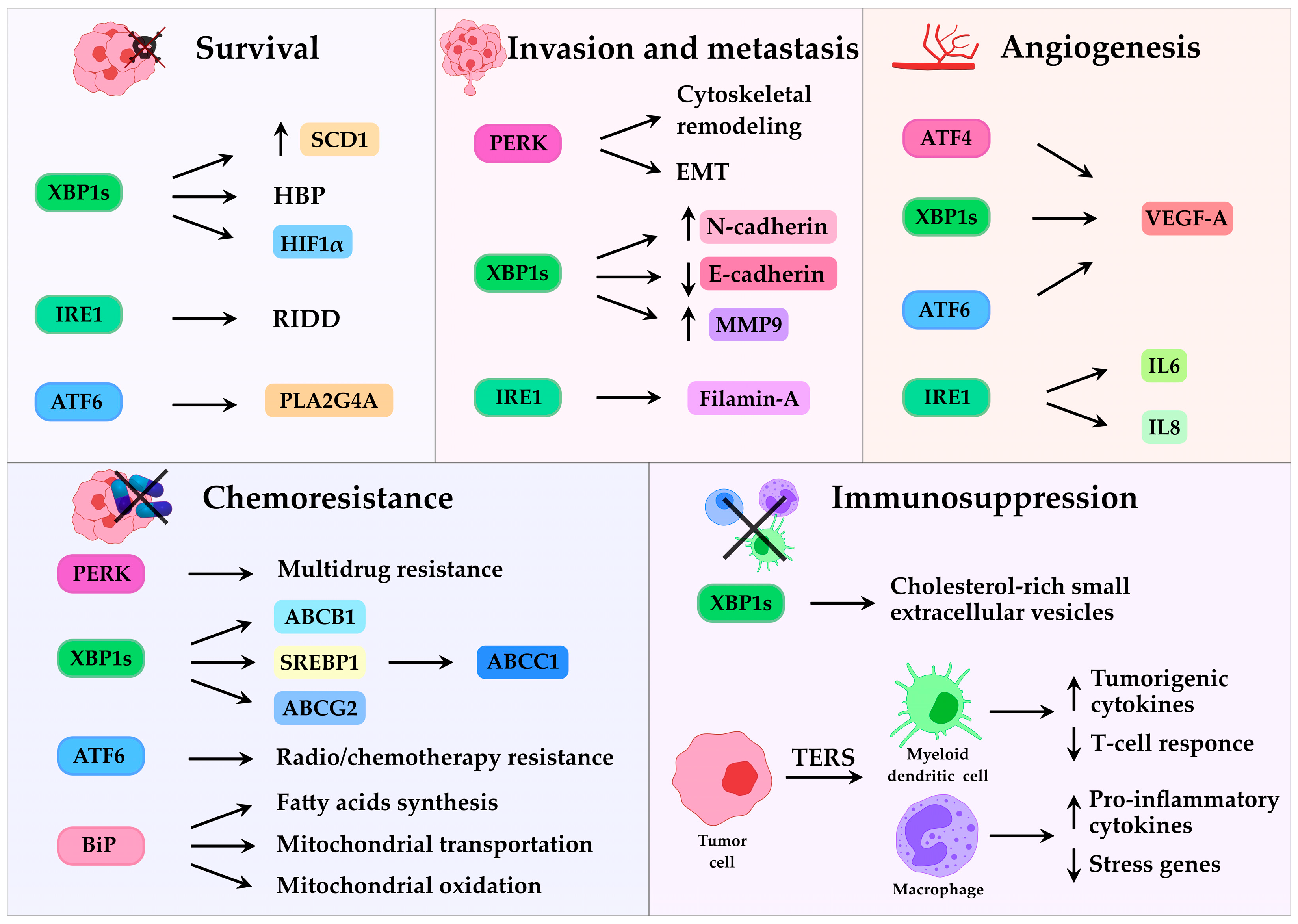
The UPR appears to play a role in every phase of tumor development. It contributes to tumor onset and progression, including neoplastic transformation, tumor growth, invasion and metastasis. Oncogenic transformation, the initial phase of tumor development, imposes a high metabolic demand on cancer cells, resulting in increased protein synthesis and translocation into the ER [62]. This process is further amplified by oncogene hyperactivation, such as MYC, RAS, or the loss of function in tumor suppressors, like p53 (tumor protein P53), PTEN (phosphatase and tensin homolog), all of which converge on UPR activation [63]. Following oncogenic transformation, UPR also promotes tumor progression by inducing epithelial–mesenchymal transition (EMT), invasion, and metastasis. PERK activation is essential for EMT, enabling cells to acquire invasive and metastatic traits through cytoskeletal remodeling and adaptation to cellular stress [64]. Similarly, IRE1α–XBP1 signaling facilitates EMT by downregulating E-cadherin and upregulating N-cadherin in breast cancer [65] and colorectal carcinoma cells [66]. In addition, IRE1α directly interacts with filamin A, an actin-binding protein, to regulate cytoskeleton remodeling and enhance cell invasiveness [49]. Furthermore, ATF6 has been shown to promote brain metastasis in breast cancer [67] while XBP1 supports invasion and proliferation by inducing MMP9 (matrix metalloproteinase-9) expression and remodelling of the extracellular matrix in human esophageal squamous cell carcinoma [68].
Beyond these cell-intrinsic effects, the UPR also influences tumor-stroma interactions, modulating the immune response, angiogenesis, and chemoresistance [69]. Depending on the intensity and duration of ER stress, as well as the tissue context, the UPR can exert both pro-tumorigenic and anti-tumorigenic effects, either promoting cancer cell survival or triggering apoptosis.

3.2. UPR and Lipid Metabolism

Altered lipid metabolism, including fatty acid synthesis, uptake, and oxidation, [70,71], is a well-studied feature of cancer. Recent evidence indicates that the UPR also plays a critical role in regulating metabolic and lipid homeostasis [56,72,73]. Among the UPR branches, ATF6α has been shown to promote prostate cancer progression by upregulating PLA2G4A (cytosolic phospholipase A2)-mediated arachidonic acid metabolism, leading to elevated prostaglandin production and resistance to ferroptotic cell death in tumor cells [74]. Instead, the IRE1α/XBP1s signaling axis directly supports tumor growth and survival by linking oncogenic signaling to lipid metabolic reprogramming. For example, in Myc-transformed cancer cells, XBP1s upregulates SCD1 (stearoyl-CoA desaturase 1), which catalyzes the conversion of saturated fatty acids into monounsaturated fatty acids. This process maintains membrane fluidity, supporting rapid cell proliferation, and protects cells from lipotoxic stress, thereby integrating oncogenic Myc activity with lipid metabolism. Moreover, IRE1α-dependent RIDD activity contributes to lipid metabolic reprogramming in triple-negative breast cancer (TNBC) by directly cleaving DGAT2 (diacylglycerol O-acyltransferase 2) mRNA, which encodes an enzyme critical for triacylglycerides biosynthesis and lipid droplet formation. This mechanism helps cancer cells adapt to nutrient deprivation stress [75]. Interestingly, in non-small cell lung cancer cells the IRE1α/XBP1 axis upregulates SREBP1 (sterol regulatory element-binding protein 1), which binds directly to the promoter of the MRP1 (multidrug resistance-associated protein 1) and drives its transcription. Elevated MRP1 expression facilitates drug efflux and confers resistance to cytotoxic chemotherapy, thereby establishing a link between UPR signaling, lipid metabolism, and drug resistance [76]. Consistently, pharmacological inhibition of IRE1α RNase activity with 4μ8C in hepatocellular carcinoma was found to alter lipid turnover and improve therapeutic response, sensitizing cells to doxorubicin (DOX) [77]. Additionally, BiP was found to drive chemoresistance in breast cancer by regulating fatty acids synthesis, mitochondrial transportation and oxidation [78].

3.3. UPR and Resistance to Therapy

Chemotherapy resistance remains one of the most significant challenges in oncology, severely limiting the long-term efficacy of anticancer treatments. This issue is exacerbated not only by the heterogeneity observed across different tumor types and among patients, but also by the substantial intratumoral heterogeneity, where distinct cellular subpopulations display variable responses to therapy. Uncovering and characterizing the molecular mechanisms underlying chemoresistance is therefore imperative for improving the prognosis and therapeutic outcomes of cancer patients. The UPR seems to play a key role in helping cancer cells survive chemotherapy, with all three UPR branches contributing to resistance [69]. For instance, in colon cancer, PERK has been shown to mediate 5-fluorouracil [79] and multidrug [80] resistance. Similarly, activation of the IRE1α–XBP1 pathway by 5- fluorouracil induces the expression of ATP-binding cassette (ABC) family drug efflux transporters such as ABCB1 (ABC subfamily B member 1), ABCC1 (alias MRP1), and ABCG2 (ABC subfamily G member 2) [81]. In ovarian cancer, inhibition of IRE1α signaling restores sensitivity to cisplatin both in vitro and in vivo [82]. Likewise, ATF6 signaling has also been implicated in promoting resistance in glioblastoma [83] and lymphoma [84], where its knockdown sensitizes tumor cells to radiotherapy or chemotherapy, respectively. Moreover, the chaperone BiP has emerged as a critical regulator of chemoresistance in pancreatic ductal adenocarcinoma [85].

3.4. UPR and Tumor Microenvironment

The tumor microenvironment (TME) is characterized by hostile conditions, such as acidosis, hypoxia, nutrients deficiency and oxidative stress. These stressors exacerbate protein misfolding, disrupt intracellular calcium and reactive oxygen species homeostasis, and promote persistent activation of UPR signaling [86].
Among these factors, hypoxia has a particularly profound effect on ER homeostasis, as it impairs oxygen-dependent processes such as disulfide bond formation and lipid desaturation, both necessary for proper protein folding and ER expansion [87]. To adapt, cancer cells rely heavily on the IRE1α and PERK arms of the UPR. XBP1-deficient cells display impaired survival in hypoxic conditions, indicating the need for XBP1s signaling in adaptation [88]. Mechanistically, XBP1s has been suggested to cooperate with HIF1α (hypoxia inducible-factor 1 alpha), a key mediator of hypoxic adaptation, to support cell survival under hypoxia. HIF1α is stabilized in low oxygen and activates genes involved in growth, metabolism, and angiogenesis. In TNBC, XBP1s is essential for efficient transcription of HIF1α target genes, thereby promoting tumor survival [89]. The PERK pathway is also crucial for survival under hypoxia, as PERK-deficient cells exhibit markedly reduced viability under oxygen limited conditions [90]. In addition to hypoxia, nutrient deprivation is another major stressor within the TME that perturbs ER function and sustains UPR activation. Glucose and glutamine shortage disrupt critical metabolic pathways, like the hexosamine biosynthetic pathway (HBP), which generates uridine diphosphate-N-acetylglucosamine (UDP-GlcNAc) for N-linked glycosylation (O-GlcNAcylation) and protein folding [91]. XBP1 overexpression or UPR induction enhances HBP enzyme activity, whereas XBP1 knockdown suppresses starvation-induced O-GlcNAcylation [92]. To overcome oxygen and nutrient limitation, tumors also stimulate angiogenesis, primarily through VEGF-A (vascular endothelial growth factor-A), as well as FGF (fibroblast growth factor) and PDGF (platelet-derived growth factor) [93]. In TNBC, XBP1s is required for efficient HIF1α-mediated VEGF-A expression and angiogenesis [89] and IRE1α deficiency reduces VEGF-A production and cytokines such as IL-6 (interleukin 6) and IL-8 (interleukin 8) [94]. Similarly, PERK promotes angiogenesis via ATF4, which binds the VEGF-A promoter and induces additional pro-angiogenic mediators [95]. ATF6 may also contribute, possibly through indirect regulation of XBP1 [96]. All together, these findings underscore the importance of UPR signaling in driving tumor vascularization.
While angiogenesis is a key adaptive mechanism for tumor expansion, an equally critical strategy is immune evasion. Emerging evidence suggests that UPR signaling enables cancer cells to suppress or modulate immune responses. For example, in colon adenocarcinoma and melanoma, XBP1 drives the production and secretion of cholesterol-rich small extracellular vesicles, which in turn promote the expansion and activation of myeloid-derived suppressor cells. These immunosuppressive cells inhibit T-cell activity and foster a tolerogenic TME [97]. Furthermore, ER-stressed cancer cells can secrete factors that transmit ER stress to immune cells, in a process called transmissible ER stress (TERS). Myeloid dendritic cells exposed to TERS produce tumorigenic cytokines and suppress T-cell responses. In vivo, TERS-primed dendritic cells accelerate tumor growth and reduce CD8+ T-cell infiltration. Similar effects have been observed in macrophages, where ER stress induced by cancer cells increases the expression of pro-inflammatory cytokines and stress response genes [98]. These findings highlight how UPR activation not only enables cancer cells to adapt to microenvironmental stress but also reprograms immune responses to support tumor progression (Figure 2).

4. UPR in Sarcomas

The UPR has been extensively characterized in various mesenchymal-derived malignancies, especially in multiple myeloma, where the IRE1α/XBP1 axis is central to plasma cell survival under proteotoxic stress [34]. Over the past decade, there has been growing interest in exploring UPR mechanisms in sarcomas, also of mesenchymal origin, raising the possibility that molecular insights from other mesenchymal tumors may be translationally relevant to understanding and targeting sarcoma biology.
Sarcomas are highly aggressive tumors characterized by rapid growth, local invasiveness, and an early tendency for metastatic dissemination. A hallmark of sarcomas is their high cellular plasticity, shaped by genetic and epigenetic alterations, adaptation to the TME, and therapeutic selection pressures. Together, these factors drive tumor progression and relapse. Importantly, sarcomas represent a miscellaneous group of neoplasms with distinct genetic abnormalities, depending on the subtype and patient. Some, such as Ewing’s sarcoma (ES) [99], rhabdomyosarcoma (RMS) [100] and liposarcoma [101], harbor chromosomal translocations and fusion genes, while others, such as OS [102], liposarcoma [103] and leiomyosarcoma [104], display genome complexity and aneuploidy. Clinically, sarcomas are further complicated by their poor response to chemotherapy and radiotherapy, and frequent multidrug resistance. The absence of highly specific biomarkers continues to limit early detection and the development of effective targeted therapies, underscoring the urgent need for improved strategies in their management.
Interestingly, UPR activation can be triggered by genomic alterations, including aneuploidy [105] and fusion oncogenes [14], both of which disrupt protein homeostasis and induce cellular stress. Given that many sarcomas display high levels of genomic instability, chromosomal aneuploidy, and recurrent gene fusions, it is plausible that UPR activation represent a biologically relevant process in these tumors. This may occur either as a direct consequence of defined genetic abnormalities or as a broader adaptive mechanism in subtypes lacking clear molecular drivers, further emphasizing the potential role of the UPR in sarcomas biology.

4.1. UPR in Bone Sarcomas

4.1.1. UPR in Osteosarcoma

OS is the most prevalent bone cancer and is characterized by an aggressive clinical course, with a strong tendency to metastasize, especially to the lungs. Despite ongoing research and therapeutic advancements, patient prognosis has remained largely unchanged over the past four decades [106]. Many features of the TME discussed previously are also highly relevant to OS, supporting the hypothesis that the UPR may play a critical role in sustaining OS aggressiveness and therapeutic resistance.
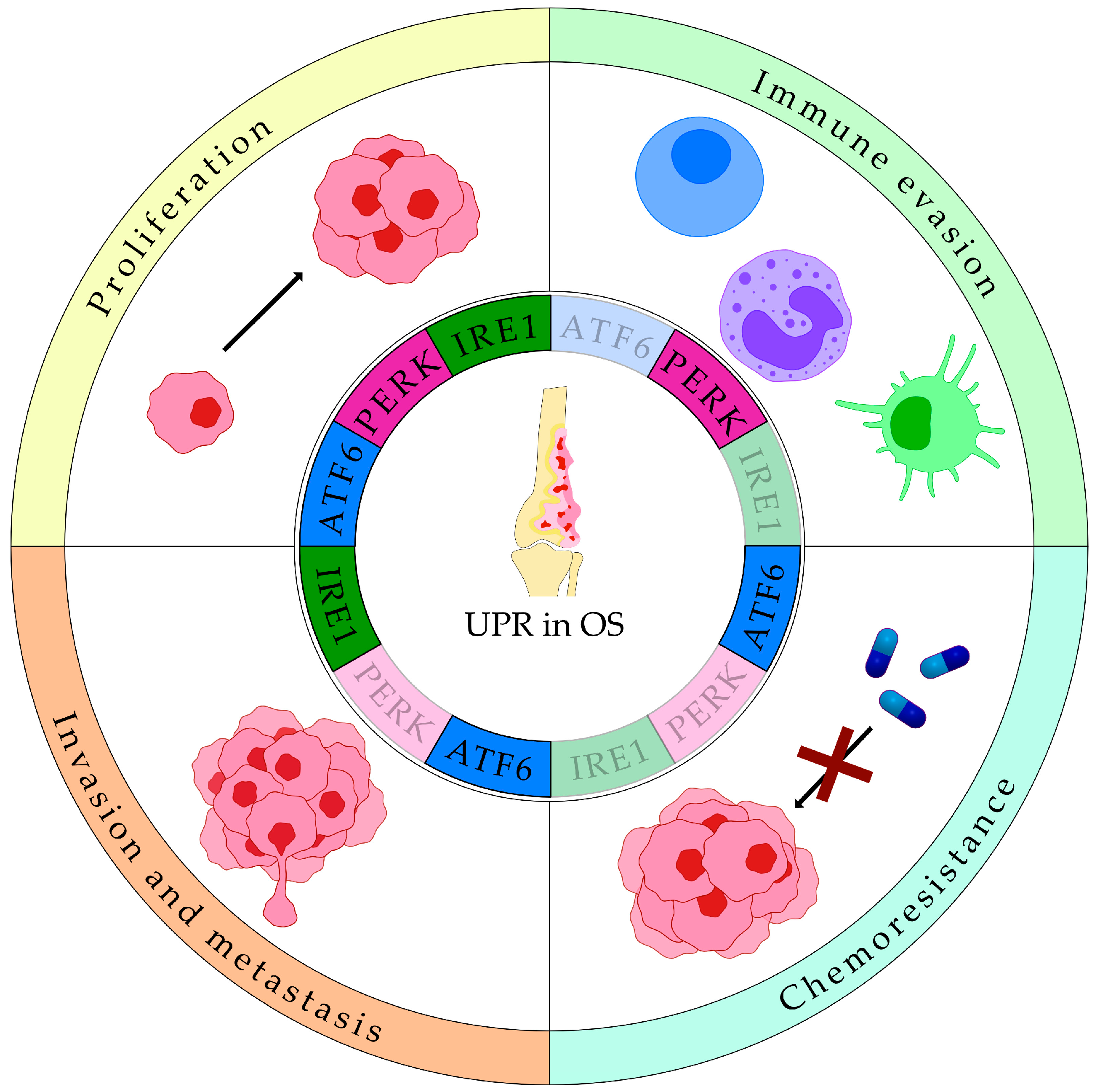
Recent studies have shown that UPR markers are upregulated in OS and may serve as prognostic indicators, as they are associated with more aggressive disease phenotypes (Table 1). In particular, Shi et al. reported enhanced expression of BiP, XBP1s, ATF4 and ATF6α in OS samples compared to normal tissues [13]. Clustering of patients revealed that increased UPR activity associates with high tumor cell proliferation, low immune cell infiltration, reduced immunotherapy response, and poor survival [13]. Similarly, Zhang et al. demonstrated that expression patterns of UPR-related genes could predict tumor-associated immune infiltration and prognosis of OS patients [107]. At the single-cell level, transcriptomic analysis revealed that ATF6α and its downstream targets are enriched in OS cells and correlate with WNT and TGF-β (transforming growth factor beta) pathways, both known to promote OS progression [108,109], and with poor prognosis [15]. Functionally, ATF6 has emerged as a critical mediator of OS aggressiveness. Its inhibition, either by genetic silencing or pharmacological repression (with the small molecule ceapin-7) reduced HOS cell viability, proliferation and colony formation [15]. Proteomic analysis further revealed that ATF6α-dependent upregulation of ER chaperones, like BiP, HSP90B1 (heat shock protein 90 beta family member 1) and calreticulin in OS tissues, compared to the soft tissue callus used as a non-cancerous control. Among these, BiP was associated to resistance to chemotherapeutic agents like DOX and platinum-based drugs [12]. Additionally, BiP loss sensitized OS cells to the proteasome inhibitor bortezomib (BTZ) by upregulating ATF4 and CHOP [110]. In a retrospective study on OS patients treated with cisplatin, DOX, and methotrexate, high ATF6α(N) expression correlated with PDI (Protein disulfide isomerase) and BiP, metastatic disease, and poor response to chemotherapy [17]. Supporting these findings, in vitro experiments demonstrated that inhibition of ATF6α, but not IRE1α and PERK, sensitized OS cells to chemotherapy-induced death, highlighting its potential role in therapeutic resistance [17].
Beyond ATF6, the IRE1 branch of the UPR also contributes to OS progression. XBP1s is overexpressed in OS cells compared to osteoblasts [12] and has been linked to disease progression and low tumor necrosis rate [111]. Knockdown of XBP1 in MG63 and U2OS cells delayed cell cycle progression and decreased viability via PIK3R3 (phosphoinositide-3-kinase regulatory subunit 3)/mTOR (mechanistic target of rapamycin kinase) pathway, particularly under hypoxic conditions [111]. Moreover, XBP1 promoted tumor growth and metastasis by binding to the GNL3 (G protein nucleolar 3—a nucleolar protein involved in cell proliferation, cell cycle, and invasion) promoter and fostering its expression [112].
PERK signaling also plays a dual role in OS, contributing to both tumor progression and modulation of the immune infiltrate within the TME. PERK inhibition reduces tumor growth and immunosuppression [18], while one of it downstream targets, STC2 (stanniocalcin 2), is upregulated in OS tissues and correlates with poorer survival outcomes [113].
Table 1. Clinical correlation between UPR markers and OS progression.
UPR MarkersDatasetsNormal Tissue ControlClinical CorrelationTotal Number of
Patients/Samples
Reference
BiP, XBP1s, ATF4 and ATF6αGSE99671, GSE126209, GSE21257, TARGET, Zhengzhou, TCGA sarcoma datasetYesTumor growth, low immune infiltration, reduced immunotherapy response, poor survival512[13]
UPR related genes (STC2, PREB, TSPYL2, and ATP6V0D1)GSE21257, TARGET Yesimmune infiltration and prognosis138[107]
ATF6αGSE152048 and GSE162454YesOS progression and poor prognosis110,000 single cells from 17 samples[15]
BiPMaharaj Nakorn Chiang Mai HospitalYesChemoresistance20[12]
ATF6α(N)Phoenix Children’s HospitalYesMetastatic disease and poor response to chemotherapy40[17]
XBP1sShanghai Jiao Tong University Affiliated Sixth People’s HospitalYesDisease progression and low tumor necrosis rate40[111]
STC2GSE21257, GSE33382 and TARGETYesPoor survival225[113]
In addition to its direct effects, the UPR operates within a dynamic regulatory network, interacting with multiple intracellular signaling pathways. For example, ER stress induces the long non-coding RNA LINC00629, which promotes OS progression via the KLF4 (Kruppel-like factor 4)-LAMA4 (laminin subunit alpha 4) axis [114]. Similarly, upregulation of GADD45GIP1 (growth arrest and DNA damage-inducible gamma interacting protein 1) activates the PERK-eIF2α axis, promoting ER stress and OS progression [115]. Furthermore, SREBP1 overexpression enhances PERK-mediated ER stress and apoptosis, suggesting a synergistic interaction between metabolic and stress-response pathways in OS [116]. Interestingly, in U2OS cells, the UPR directly impacts circadian rhythms and cell survival. Activation of the UPR, primarily through the PERK- inducible miR-211 axis, causes a phase shift in circadian oscillations and suppresses core regulators like BMAL1 (basic helix-loop-helix ARNT like 1) and CLOCK (Clock Circadian Regulator). This suppression of BMAL1 is essential for UPR-dependent inhibition of protein synthesis, enabling OS cells to adapt to ER stress [117].
Reflecting this complex interplay, transcriptomic analyses have identified a strong increase in UPR-related genes in high-risk OS groups. For instance, a risk score model based on sixteen glutamine metabolism-related genes, revealed significant UPR enrichment in the high-risk subgroup [118]. Similarly, another investigation employing m6A-related lncRNA signatures revealed a strong association with increased UPR activity in high-risk patients [119].
Altogether, these findings underscore a pro-tumorigenic role of the UPR in OS, promoting cellular proliferation, invasion, metastasis, immune evasion, and resistance to therapy (Figure 3).

4.1.2. UPR in Ewing’s Sarcoma

ES is a prevalent pediatric bone tumor characterized by the EWS (Ewing sarcoma breakpoint region 1)/FLI1 (friend leukemia integration 1 transcription factor) gene fusion. In contrast to OS, the role of the UPR in ES remains poorly characterized. To date, research in this area is extremely limited, with only a single study directly investigating how UPR signaling influences ES biology. Tanabe et al. identified over 2000 proteins regulated by this fusion gene, among which the XBP1 pathway was highlighted as critical for tumor viability. High mRNA expression of XBP1 was confirmed in ES cell lines and patient surgical samples, and silencing XBP1 significantly suppressed ES cell proliferation and viability [14]. Among the IRE1-XBP1 inhibitors tested, toyocamycin was the most effective. In vivo experiments showed that toyocamycin significantly reduced tumor volume and weight in mouse xenograft models and increased cellular apoptosis [14]. Future studies should explore the mechanisms behind XBP1 activation. Special focus should be given to the regulatory role of the EWS/FLI1 fusion gene, which is central to this cancer, since current data reveal a major gap in our understanding. Additionally, more research is needed to elucidate whether the UPR exerts a pro-tumorigenic, anti-tumorigenic, or context-dependent role in ES, as well as to clarify its potential therapeutic relevance.

4.2. UPR in Soft-Tissue Sarcomas

Soft tissue sarcomas originate in the soft tissues of the body and can develop in virtually any anatomical site. Their rarity and biological heterogeneity complicate early detection and treatment, emphasizing the urgent need to identify new therapeutic targets [120].
RMS, the most common soft tissue pediatric cancer, exhibits basal activation of the IRE1 and PERK branches of the UPR. Pharmacological inhibition of these pathways with MKC8866 (an IRE1α inhibitor, used at 10 μM, 20 μM and 40 μM) and AMGEN44 (a PERK inhibitor, used at 2 μM, 5 μM and 10 μM) resulted in a marked reduction in RMS cell viability, proliferation, and colony-forming ability, with the two highest concentrations eliciting the most pronounced effects. Notably, combined inhibition (MKC8866 used at 20 μM and AMGEN44 used at 2 μM) produced stronger anti-proliferative effects than either agent alone, suggesting a potential synergistic impact on RMS cells. Subtype-specific differences in sensitivity were observed. Alveolar RMS cells exhibited higher responsiveness to IRE1α inhibition, whereas embryonal RMS cells were more susceptible to PERK inhibition. Mechanistically, UPR blockade induced cellular senescence, contributing to decreased proliferation. Importantly, both inhibitors showed minimal toxicity in non-malignant cells, underscoring their potential as selective therapeutic agents for RMS [16]. Further confirmation of these results in physiologically relevant 3D and in vivo models will be essential to establish their translational significance.
Another study reported that the expression of key UPR components (BiP, XBP1s, and IRE1α) was significantly associated with RMS. Specifically, BiP expression correlated with lymph node involvement, while both BiP and XBP1s were linked to all four RMS subtypes, including alveolar, embryonal, pleomorphic, and sclerosing/spindle cell RMS. In contrast, IRE1α expression was associated with alveolar, pleomorphic, and embryonal RMS, with the highest levels observed in alveolar subtype. Furthermore, XBP1s expression was correlated positively with distant metastasis in alveolar RMS and with tumor size in pleomorphic RMS. These findings suggest that activation of the IRE1α/XBP1s axis may contribute to disease aggressiveness and subtype-specific progression [121].
Overall, current evidence points toward a predominantly pro-tumorigenic role for the UPR in RMS, with the IRE1α/XBP1s and PERK branches contributing to proliferation, metastasis, and subtype-specific tumor progression. Nonetheless, these findings remain preliminary, and further investigations are needed to comprehensively define the functional and molecular mechanisms through which the UPR influences RMS pathophysiology. Such investigations will be crucial not only for clarifying the functional role of the UPR in RMS but also for assessing the therapeutic potential of UPR-targeted strategies in this malignancy.
Whereas the UPR appears to sustain proliferation and subtype-specific progression in RMS, its role in angiosarcoma extends to the regulation of angiogenesis, accentuating the context-dependent nature of UPR signaling across different sarcomas. Angiosarcomas exhibit a distinctive UPR signature, characterized by elevated BiP and PERK expression alongside reduced IRE1α protein levels. BiP is essential for maintaining VEGFR2 (VEGF receptor 2) stability and endothelial cell functions; its depletion severely impairs angiogenesis and can even cause regression of vascular structures. Elevated PERK expression, in turn, promotes the accumulation of unspliced XBP1, which has been linked to increased proliferation and malignancy. Although IRE1α protein levels are attenuated, its kinase activity remains functionally relevant, as it governs the splicing of XBP1, a central regulator of angiogenic signaling. Consistently, pharmacological inhibition of IRE1α kinase suppresses in vitro angiogenesis [19].
Beyond angiosarcoma, limited evidence suggests that UPR signaling may also play a role is Kaposi’s sarcoma, primarily through interactions with HHV-8 (human herpesvirus 8), the causative agent of this cancer. Preliminary studies have explored the involvement of XBP1 signaling in this context [122], suggesting a potential, though yet unexplored, link between the UPR activation and Kaposi’s sarcoma pathogenesis. Moreover, malignant peripheral nerve sheath tumors (MPNSTs) display elevated UPR markers, including BiP, eIF2α and XBP1s, compared with normal nerve tissue [123].
Overall, while current understanding of the role of the UPR in soft tissue sarcomas remains incomplete, emerging data suggest that it may represent a promising therapeutic target. Given the context-specific nature of UPR signaling, future research should aim to dissect its functional roles within individual sarcoma subtypes to better define its contribution to tumor progression and treatment response.
In other sarcoma subtypes, like leiomyosarcoma, liposarcoma, synovial sarcoma and chondrosarcoma, the role of the UPR remains unexplored. Notably, in chondrosarcoma, only a single study reports that PRP-1 (proline-rich polypeptide), a toll-like receptor ligand, increases the expression of PERK, eIF2α, ATF4, CHOP, ATF6, IRE1α, and XBP1 [124]; however, no studies have yet investigated the functional role of the UPR in this tumor.

5. Targeting the UPR in Cancer

The evidence discussed thus far underscores a predominantly pro-tumorigenic role of the UPR in sarcomas. Nevertheless, it is essential to acknowledge the dual nature of the UPR: under conditions of sustained ER stress, its activation can shift toward the “terminal UPR,” which drives apoptosis in malignant cells.
Several agents, such as MO-OH-Nap, an α-substituted tropolone, induce UPR-mediated apoptotic death in several OS cell lines [125], while psoralen, a natural active component of Psoralea corylifolia, triggers ATF6α/CHOP-dependent apoptosis [126]. Sdox [127], a DOX analog, and clemastine [128], an antagonist of histamine H1 receptor, also activate ER stress-dependent apoptotic pathways in OS cells. Furthermore, compounds like CB-5083 activate both PERK and IRE1α to induce apoptosis in OS cells [129], and traditional medicines such as tongguanteng [130], or active peptides like mellitin [131] activate IRE1α/XBP1/CHOP axis to promote OS cell death. EUR, an active compound in Eurycoma longifolia Jack root, inhibits OS growth by destabilizing BiP mRNA and blocking its transcription [132]. Moreover, propofol, a common anesthetic, suppresses OS proliferation and invasion through ER stress induction and synergizes with DOX [133]. Similarly, HMnO2@CaO2, a manganese dioxide nanoplatform with incorporated calcium peroxide, induces both ER stress and ferroptosis, showing promising antitumor activity [134]. Knockout of the DNMT2/TRDMT1 (tRNA aspartic acid methyltransferase 1) gene sensitizes cells to ER stress-induced apoptosis by impairing UPR activation upon DOX treatment [135]. In addition, β-elemonic acid, an active component extracted from Boswellia carterii bird, activates the PERK/eIF2α/ATF4 branch, stimulating CHOP-regulated apoptosis and Ca2+-mediated caspase activation [136], while inhibition of GGDPS (geranylgeranyl diphosphate synthase) with RAM2061 induces UPR markers and apoptosis in both OS and ES cells [137].
Similar vulnerabilities are observed in other sarcomas: the IRE1α inhibitor MKC3946 sensitizes leiomyosarcoma cells to apoptosis [138], and proteasome inhibition with carfilzomib enhances nutlin-3-induced apoptosis in liposarcoma by activating the ATF4/CHOP/NOXA axis [139]. Moreover, nelfinavir, a clinically approved HIV (human immunodeficiency virus) protease inhibitor, blocks S2P activity, leading to inhibition of SREBP1 and ATF6 signaling and subsequent apoptosis [140]. In MPNST, BTZ-driven UPR activation enhances the synergistic antitumor efficacy observed when combined with oHSV (oncolytic herpes simplex virus-1) [141], while ER-stress-inducing agents such as thapsigargin, tunicamycin or the HSP90 inhibitor IPI-504 cause UPR overexpression leading to cell death [123]. In RMS, both the HSP70 (heat shock 70 kDa protein) inhibitor MAL3-101 and the p97 (valosin containing protein) ATPase inhibitor CB-5083 trigger robust UPR activation, marked by increased eIF2α phosphorylation, ATF4 and CHOP upregulation, and XBP1 splicing, thereby promoting tumor cell death [142]. Recently, nanostructures and nanoformulated drugs have emerged as promising tools to modulate or intercept the UPR in cancer cells. In glioblastoma, the PDI inhibitor CCF642, especially when delivered via albumin-based nanoparticles, was shown to activate apoptosis-inducing UPR, downregulate PERK signaling, and restore sensitivity to temozolomide [143]. In lung-carcinoma cells, the UPR is triggered by denatured proteins in the nanoparticle corona, which recruits the chaperone Hsp90ab1 to the particle surface and up-regulates downstream pathways that promote EMT. Blocking Hsp90ab1 with geldanamycin prevents UPR-induced activation and EMT, pointing to the UPR-Hsp90ab1 axis as a driver of tumor cell reprogramming [144].
In addition, it is noteworthy that preclinical studies in cancers other than sarcomas have provided compelling evidence that pharmacological inhibition of the UPR can exert significant antitumor effects, particularly when combined with standard therapies. Inhibiting the IRE1α/XBP1 axis with Z4P enhances temozolomide efficacy in glioblastoma [145], while 4µ8C and STF-083010 potentiate 5-fluorouracil in colorectal cancer [146] and show strong anti-myeloma activity [147]. Local MKC8866 delivery in glioblastoma prolongs survival when combined with radiochemotherapy [148]. Targeting PERK has yielded similarly promising results: GSK2606414 potentiates oridonin in small-cell lung cancer [149], enhances sorafenib activity in hepatocellular carcinoma [150], and, notably, boosts PD-1 immunotherapy in murine sarcoma [151]. GSK2656157 synergizes with 5-fluorouracil in colon cancer [79], reduces hepatocellular carcinoma growth [152], and, in melanoma, improves responses to mRNA vaccination [153]. More recently, PERK inhibition with HC-5404 enhanced VEGFR-targeted therapy in renal carcinoma, achieving durable tumor control [154]. Collectively, these findings underscore the translational relevance of UPR inhibitors as partners to established therapies, immunotherapies, and resistance-modulating approaches.
Several clinical trials (https://clinicaltrials.gov/) are currently investigating therapeutic strategies targeting different branches of the UPR and related stress pathways across a variety of malignancies, even though clinical trials in sarcomas remain scarce (Table 2). Compounds such as BOLD-100 and NKP-1339 [155], BiP inhibitors, are being tested both as single agents and in combination with standard chemotherapeutic regimens (e.g., FOLFOX, DOX) in gastrointestinal cancers, soft tissue sarcomas, and other solid tumors. Inhibition of PERK is being explored with agents such as HC 5404-FU and NMS-03597812, the latter evaluated in combination with dexamethasone for multiple myeloma and as monotherapy for acute myeloid leukemia. Targeting the IRE1α RNase domain by MKC8866 is currently in trials in breast cancer and solid tumors, either in combination with Abraxane or with standard-of-care regimens [156]. In parallel, proteasome inhibition with IXAZOMIB is under investigation in non-hematologic malignancies, including soft tissue sarcomas.
Collectively, these studies reflect the growing clinical interest in modulating ER stress and UPR-related pathways as potential therapeutic avenues in both hematologic and solid tumors.

6. Conclusions and Future Perspectives

The growing evidence for therapeutic targeting of UPR across various tumor types supports the extension of these strategies to sarcomas. In OS, where scientific data are more robust, investigating UPR modulation could address critical clinical challenges including relapse, therapy resistance, and metastatic dissemination, and integrating UPR-targeted approaches with standard-of-care treatments, like DOX and cisplatin, may enhance therapeutic efficacy and improve patient outcomes. The UPR dual capacity to promote both cell survival and apoptosis necessitates precise interventions that shift this balance toward tumor suppression. Inhibiting adaptive UPR branches may limit tumor growth and resistance, while exacerbating ER stress to trigger terminal UPR activation offers a complementary approach to induce tumor apoptosis. The association between elevated UPR marker expression and poor prognosis in OS patients further highlights the potential for precision medicine approaches, where stratifying patients based on UPR activity could identify those most likely to benefit targeted therapies, while minimizing unnecessary toxicity. However, translating these insights into clinically effective interventions requires further elucidation of the underlying molecular mechanisms. Moreover, comprehensive investigations are required to elucidate the functional relevance of the UPR within specific sarcoma subtypes. Future studies should begin by systematically characterizing standardized panels of UPR biomarkers, including IRE1, ATF6, PERK, BiP, and XBP1, to define their expression patterns and activation states across different sarcoma models. Subsequent efforts should aim to clarify how these signaling components contribute to distinct aspects of tumor biology, such as proliferation, survival, invasion, and therapeutic resistance, and to uncover the molecular pathways mediating these effects. Ultimately, in vivo studies will be essential to validate these findings and to assess the therapeutic potential of combinational strategies that target the UPR alongside conventional or novel anticancer treatments.

Author Contributions

Conceptualization, E.I., M.C., S.A. and N.B.; writing—original draft preparation, E.I. and M.C.; writing—review and editing, M.C., S.A. and N.B.; supervision, M.C.; project administration, N.B., S.A., and M.C.; funding acquisition, N.B. All authors have read and agreed to the published version of the manuscript.

Funding

This research received funding from AIRC under the IG 2018—project ID 21403—to N.B. (“Altered lipid metabolism as a stress reaction to acid tumor microenvironment and a driver of metastasis in osteosarcoma”) and from the National Center for Gene Therapy and Drugs Based on RNA Technology, funded in the framework of the National Recovery and Resilience Plan (NRRP), Mission 4 Investment 1.4 funded by the European Union—Next Generation EU, Project CN00000041, CUP B93D21010860004, Spoke 2.

Data Availability Statement

No new data were created or analyzed in this study. Data sharing is not applicable to this article.

Conflicts of Interest

The authors declare no conflicts of interest.

Abbreviations

The following abbreviations are used in this manuscript:
ABCATP-binding cassette
ABCB1ABC subfamily B member 1
ABCG2ABC subfamily G member 2
ATF4activating transcription factor 4
ATF6activating transcription factor 6
ASK1apoptosis signal-regulating kinase 1
BMAL1basic helix-loop-helix ARNT like 1
BAKBCL2 antagonist/killer 1
BCL-2BCL2 apoptosis regulator
BAXBCL2 associated X protein
BIMBCL-2 interacting mediator of cell death
BiPbinding immunoglobulin protein
BTZbortezomib
CHOPC/EBP homologous protein
JNKc-Jun N-terminal kinase
CLOCKclock circadian regulator
PLA2G4Acytosolic phospholipase A2
DGAT2diacylglycerol O-acyltransferase 2
PKRdouble-stranded RNA-activated protein kinase
DOXdoxorubicin
eIF2αα-subunit of eukaryotic initiation factor 2
ERendoplasmic reticulum
EMTepithelial–mesenchymal transition
ERADER-associated protein degradation
ESEwing’s sarcoma
EWSEwing sarcoma breakpoint region 1
FGFfibroblast growth factor
FLI1friend leukemia integration 1 transcription factor
GNL3G protein nucleolar 3
GGDPSgeranylgeranyl diphosphate synthase
GRP78glucose-regulated protein 78
GADD45GIP1growth arrest and DNA damage-inducible gamma interacting protein 1
GADD34growth arrest and DNA damage–inducible gene 34
HSP70heat shock 70 kDa protein
HSP90B1heat shock protein 90 beta family member 1
HBPhexosamine biosynthetic pathway
HHV-8human herpesvirus 8
HIVhuman immunodeficiency virus
HIF1αhypoxia-inducible factor 1 alpha
IRE1inositol-requiring enzyme 1
IL-6interleukin 6
IL-8interleukin 8
KLF4Kruppel-like factor 4
MPNSTsmalignant peripheral nerve sheath tumors
MMP9matrix metalloproteinase-9
LAMA4laminin subunit alpha 4
mTORmechanistic target of rapamycin kinase
MRP1multidrug resistance-associated protein 1
O-GlcNAcylationN-linked glycosylation
NLRP3NLR family pyrin domain containing 3
ATF6(N)N-terminal ATF6 fragment
oHSVoncolytic herpes simplex virus-1
OSosteosarcoma
PUMAp53 upregulated modulator of apoptosis
NOXAphorbol-12-myristate-13-acetate-induced protein 1
PTENphosphatase and tensin homolog
PIK3R3phosphoinositide-3-kinase regulatory subunit 3
PERKPKR-like ER kinase
PDGFplatelet-derived growth factor
PRP-1proline-rich polypeptide
PDIprotein disulfide isomerase
PP1Cprotein phosphatase 1
RIDDregulated IRE1-dependent decay
RMSrhabdomyosarcoma
S1Psite-1 protease
S2Psite-2 protease
STC2stanniocalcin 2
SCD1stearoyl-CoA desaturase 1
SREBP1sterol regulatory element-binding protein 1
TXNIPthioredoxin interacting protein
TGF-βtransforming growth factor beta
TERStransmissible ER stress
TNBCtriple-negative breast cancer
TRDMT1tRNA aspartic acid methyltransferase 1
TRAF2tumor necrosis factor receptor-associated factor 2
TMEtumor microenvironment
p53tumor protein P53
UPRunfolded protein response
UDP-GlcNAcuridine diphosphate-N-acetylglucosamine
p97valosin containing protein
VEGF-Avascular endothelial growth factor-A
VEGFR2VEGF receptor 2
XBP1X-box binding protein 1

References

  1. Burningham, Z.; Hashibe, M.; Spector, L.; Schiffman, J.D. The Epidemiology of Sarcoma. Clin. Sarcoma Res. 2012, 2, 14. [Google Scholar] [CrossRef]
  2. Al Shihabi, A.; Tebon, P.J.; Nguyen, H.T.L.; Chantharasamee, J.; Sartini, S.; Davarifar, A.; Jensen, A.Y.; Diaz-Infante, M.; Cox, H.; Gonzalez, A.E.; et al. The landscape of drug sensitivity and resistance in sarcoma. Cell Stem Cell 2024, 31, 1524–1542.e4. [Google Scholar] [CrossRef] [PubMed]
  3. Damerell, V.; Pepper, M.S.; Prince, S. Molecular mechanisms underpinning sarcomas and implications for current and future therapy. Signal Transduct. Target. Ther. 2021, 6, 246. [Google Scholar] [CrossRef] [PubMed]
  4. Dancsok, A.R.; Asleh-Aburaya, K.; Nielsen, T.O. Advances in sarcoma diagnostics and treatment. Oncotarget 2016, 8, 7068–7093. [Google Scholar] [CrossRef]
  5. Urra, H.; Dufey, E.; Avril, T.; Chevet, E.; Hetz, C. Endoplasmic Reticulum Stress and the Hallmarks of Cancer. Trends Cancer 2016, 2, 252–262. [Google Scholar] [CrossRef]
  6. Le Reste, P.-J.; Avril, T.; Quillien, V.; Morandi, X.; Chevet, E. Signaling the Unfolded Protein Response in primary brain cancers. Brain Res. 2016, 1642, 59–69. [Google Scholar] [CrossRef]
  7. Rajapaksa, G.; Thomas, C.; Gustafsson, J. Estrogen signaling and unfolded protein response in breast cancer. J. Steroid Biochem. Mol. Biol. 2016, 163, 45–50. [Google Scholar] [CrossRef]
  8. Rufo, N.; Yang, Y.; De Vleeschouwer, S.; Agostinis, P. The “Yin and Yang” of Unfolded Protein Response in Cancer and Immunogenic Cell Death. Cells 2022, 11, 2899. [Google Scholar] [CrossRef] [PubMed]
  9. Soumya, V.V.; Jisna, B.; Anu, D.; Binoy, C.F.; Babu, T.D. IRE1α-mediated UPR activation in gastrointestinal cancers: Adaptive mechanisms and therapeutic potential. Drug Discov. Today 2025, 30, 104335. [Google Scholar] [CrossRef]
  10. Storm, M.; Sheng, X.; Arnoldussen, Y.J.; Saatcioglu, F. Prostate cancer and the unfolded protein response. Oncotarget 2016, 7, 54051–54066. [Google Scholar] [CrossRef]
  11. Vincenz, L.; Jäger, R.; O’Dwyer, M.; Samali, A. Endoplasmic Reticulum Stress and the Unfolded Protein Response: Targeting the Achilles Heel of Multiple Myeloma. Mol. Cancer Ther. 2013, 12, 831–843. [Google Scholar] [CrossRef] [PubMed]
  12. Chaiyawat, P.; Sungngam, P.; Teeyakasem, P.; Sirikaew, N.; Klangjorhor, J.; Settakorn, J.; Diskul-Na-Ayudthaya, P.; Chokchaichamnankit, D.; Srisomsap, C.; Svasti, J.; et al. Protein profiling of osteosarcoma tissue and soft callus unveils activation of the unfolded protein response pathway. Int. J. Oncol. 2019, 54, 1704–1718. [Google Scholar] [CrossRef]
  13. Shi, C.; Zhao, F.; Zhang, T.; Xu, D.; Hao, Z.; Cui, F.; Shi, J.; Jin, Y.; Li, N.; Yang, C.; et al. A novel prognostic signature in osteosarcoma characterised from the perspective of unfolded protein response. Clin. Transl. Med. 2022, 12, e750. [Google Scholar] [CrossRef]
  14. Tanabe, Y.; Suehara, Y.; Kohsaka, S.; Hayashi, T.; Akaike, K.; Mukaihara, K.; Kurihara, T.; Kim, Y.; Okubo, T.; Ishii, M.; et al. IRE1α-XBP1 inhibitors exerted anti-tumor activities in Ewing’s sarcoma. Oncotarget 2018, 9, 14428–14443. [Google Scholar] [CrossRef]
  15. Liu, F.; Zhang, T.; Yang, Y.; Wang, K.; Wei, J.; Shi, J.-H.; Zhang, D.; Sheng, X.; Zhang, Y.; Zhou, J.; et al. Integrated analysis of single-cell and bulk transcriptomics reveals cellular subtypes and molecular features associated with osteosarcoma prognosis. BMC Cancer 2025, 25, 280. [Google Scholar] [CrossRef]
  16. McCarthy, N.; Dolgikh, N.; Logue, S.; Patterson, J.B.; Zeng, Q.; Gorman, A.M.; Samali, A.; Fulda, S. The IRE1 and PERK arms of the unfolded protein response promote survival of rhabdomyosarcoma cells. Cancer Lett. 2020, 490, 76–88. [Google Scholar] [CrossRef]
  17. Yarapureddy, S.; Abril, J.; Foote, J.; Kumar, S.; Asad, O.; Sharath, V.; Faraj, J.; Daniel, D.; Dickman, P.; White-Collins, A.; et al. ATF6α Activation Enhances Survival against Chemotherapy and Serves as a Prognostic Indicator in Osteosarcoma. Neoplasia 2019, 21, 516–532. [Google Scholar] [CrossRef] [PubMed]
  18. Li, X.; Guo, X.; Wang, W.; Liu, P. Inhibition of PERK pathway promotes TAMs M1 polarization and ER stress in Osteosarcoma cells hindering tumor progression. Int. Immunopharmacol. 2025, 163, 115293. [Google Scholar] [CrossRef]
  19. Anspach, L.; Tsaryk, R.; Seidmann, L.; Unger, R.E.; Jayasinghe, C.; Simiantonaki, N.; Kirkpatrick, C.J.; Pröls, F. Function and mutual interaction of BiP-, PERK-, and IRE1α-dependent signalling pathways in vascular tumours. J. Pathol. 2020, 251, 123–134. [Google Scholar] [CrossRef]
  20. Mori, K. Signalling Pathways in the Unfolded Protein Response: Development from Yeast to Mammals. J. Biochem. 2009, 146, 743–750. [Google Scholar] [CrossRef] [PubMed]
  21. Hien, L.T.; Back, S.H. Establishment of a reporter system for monitoring activation of the ER stress transducer ATF6β. Biochem. Biophys. Res. Commun. 2021, 558, 1–7. [Google Scholar] [CrossRef]
  22. Luo, H.; Gong, W.-Y.; Zhang, Y.-Y.; Liu, Y.-Y.; Chen, Z.; Feng, X.-L.; Jiao, Q.-B.; Zhang, X.-W. IRE1β evolves to be a guardian of respiratory and gastrointestinal mucosa. Heliyon 2024, 10, e39011. [Google Scholar] [CrossRef]
  23. Worby, C.A.; Dixon, J.E. Unpacking the Unfolded Protein Response. Cell 2014, 158, 1221–1224. [Google Scholar] [CrossRef]
  24. Walter, P.; Ron, D. The Unfolded Protein Response: From Stress Pathway to Homeostatic Regulation. Science 2011, 334, 1081–1086. [Google Scholar] [CrossRef]
  25. Shen, J.; Chen, X.; Hendershot, L.; Prywes, R. ER Stress Regulation of ATF6 Localization by Dissociation of BiP/GRP78 Binding and Unmasking of Golgi Localization Signals. Dev. Cell 2002, 3, 99–111. [Google Scholar] [CrossRef]
  26. Schindler, A.J.; Schekman, R. In vitro reconstitution of ER-stress induced ATF6 transport in COPII vesicles. Proc. Natl. Acad. Sci. USA 2009, 106, 17775–17780. [Google Scholar] [CrossRef]
  27. Ye, J.; Rawson, R.B.; Komuro, R.; Chen, X.; Davé, U.P.; Prywes, R.; Brown, M.S.; Goldstein, J.L. ER Stress Induces Cleavage of Membrane-Bound ATF6 by the Same Proteases that Process SREBPs. Mol. Cell 2000, 6, 1355–1364. [Google Scholar] [CrossRef]
  28. Wu, J.; Rutkowski, D.T.; Dubois, M.; Swathirajan, J.; Saunders, T.; Wang, J.; Song, B.; Yau, G.D.-Y.; Kaufman, R.J. ATF6α Optimizes Long-Term Endoplasmic Reticulum Function to Protect Cells from Chronic Stress. Dev. Cell 2007, 13, 351–364. [Google Scholar] [CrossRef] [PubMed]
  29. Bertolotti, A.; Zhang, Y.; Hendershot, L.M.; Harding, H.P.; Ron, D. Dynamic interaction of BiP and ER stress transducers in the unfolded-protein response. Nat. Cell Biol. 2000, 2, 326–332. [Google Scholar] [CrossRef]
  30. Carrara, M.; Prischi, F.; Nowak, P.R.; Kopp, M.C.; Ali, M.M. Noncanonical binding of BiP ATPase domain to Ire1 and Perk is dissociated by unfolded protein CH1 to initiate ER stress signaling. eLife 2015, 4, e03522. [Google Scholar] [CrossRef] [PubMed]
  31. Wang, P.; Li, J.; Tao, J.; Sha, B. The luminal domain of the ER stress sensor protein PERK binds misfolded proteins and thereby triggers PERK oligomerization. J. Biol. Chem. 2018, 293, 4110–4121. [Google Scholar] [CrossRef]
  32. Harding, H.P.; Zhang, Y.; Bertolotti, A.; Zeng, H.; Ron, D. Perk Is Essential for Translational Regulation and Cell Survival during the Unfolded Protein Response. Mol. Cell 2000, 5, 897–904. [Google Scholar] [CrossRef]
  33. Harding, H.P.; Zhang, Y.; Zeng, H.; Novoa, I.; Lu, P.D.; Calfon, M.; Sadri, N.; Yun, C.; Popko, B.; Paules, R.S.; et al. An Integrated Stress Response Regulates Amino Acid Metabolism and Resistance to Oxidative Stress. Mol. Cell 2003, 11, 619–633. [Google Scholar] [CrossRef]
  34. Novoa, I.; Zeng, H.; Harding, H.P.; Ron, D. Feedback Inhibition of the Unfolded Protein Response by GADD34-Mediated Dephosphorylation of eIF2α. J. Cell Biol. 2001, 153, 1011–1022. [Google Scholar] [CrossRef]
  35. Gardner, B.M.; Walter, P. Unfolded Proteins Are Ire1-Activating Ligands That Directly Induce the Unfolded Protein Response. Science 2011, 333, 1891–1894. [Google Scholar] [CrossRef]
  36. Calfon, M.; Zeng, H.; Urano, F.; Till, J.H.; Hubbard, S.R.; Harding, H.P.; Clark, S.G.; Ron, D. IRE1 couples endoplasmic reticulum load to secretory capacity by processing the XBP-1 mRNA. Nature 2002, 415, 92–96. [Google Scholar] [CrossRef] [PubMed]
  37. Shoulders, M.D.; Ryno, L.M.; Genereux, J.C.; Moresco, J.J.; Tu, P.G.; Wu, C.; Yates, J.R., III; Su, A.I.; Kelly, J.W.; Wiseman, R.L. Stress-Independent Activation of XBP1s and/or ATF6 Reveals Three Functionally Diverse ER Proteostasis Environments. Cell Rep. 2013, 3, 1279–1292. [Google Scholar] [CrossRef] [PubMed]
  38. Park, S.-M.; Kang, T.-I.; So, J.-S. Roles of XBP1s in Transcriptional Regulation of Target Genes. Biomedicines 2021, 9, 791. [Google Scholar] [CrossRef] [PubMed]
  39. Hollien, J.; Lin, J.H.; Li, H.; Stevens, N.; Walter, P.; Weissman, J.S. Regulated Ire1-dependent decay of messenger RNAs in mammalian cells. J. Cell Biol. 2009, 186, 323–331. [Google Scholar] [CrossRef]
  40. Upton, J.-P.; Wang, L.; Han, D.; Wang, E.S.; Huskey, N.E.; Lim, L.; Truitt, M.; McManus, M.T.; Ruggero, D.; Goga, A.; et al. IRE1α Cleaves Select microRNAs During ER Stress to Derepress Translation of Proapoptotic Caspase-2. Science 2012, 338, 818–822. [Google Scholar] [CrossRef]
  41. Le Thomas, A.; Ferri, E.; Marsters, S.; Harnoss, J.M.; Lawrence, D.A.; Zuazo-Gaztelu, I.; Modrusan, Z.; Chan, S.; Solon, M.; Chalouni, C.; et al. Decoding non-canonical mRNA decay by the endoplasmic-reticulum stress sensor IRE1α. Nat. Commun. 2021, 12, 7310. [Google Scholar] [CrossRef]
  42. Hetz, C.; Papa, F.R. The Unfolded Protein Response and Cell Fate Control. Mol. Cell 2018, 69, 169–181. [Google Scholar] [CrossRef]
  43. Urra, H.; Dufey, E.; Lisbona, F.; Rojas-Rivera, D.; Hetz, C. When ER stress reaches a dead end. Biochim. Biophys. Acta (BBA)—Mol. Cell Res. 2013, 1833, 3507–3517. [Google Scholar] [CrossRef]
  44. Han, D.; Lerner, A.G.; Walle, L.V.; Upton, J.-P.; Xu, W.; Hagen, A.; Backes, B.J.; Oakes, S.A.; Papa, F.R. IRE1α Kinase Activation Modes Control Alternate Endoribonuclease Outputs to Determine Divergent Cell Fates. Cell 2009, 138, 562–575. [Google Scholar] [CrossRef]
  45. Bronner, D.N.; Abuaita, B.H.; Chen, X.; Fitzgerald, K.A.; Nuñez, G.; He, Y.; Yin, X.-M.; O’riordan, M.X. Endoplasmic Reticulum Stress Activates the Inflammasome via NLRP3- and Caspase-2-Driven Mitochondrial Damage. Immunity 2015, 43, 451–462. [Google Scholar] [CrossRef]
  46. Hetz, C.; Zhang, K.; Kaufman, R.J. Mechanisms, regulation and functions of the unfolded protein response. Nat. Rev. Mol. Cell Biol. 2020, 21, 421–438. [Google Scholar] [CrossRef] [PubMed]
  47. Carreras-Sureda, A.; Pihán, P.; Hetz, C. The Unfolded Protein Response: At the Intersection between Endoplasmic Reticulum Function and Mitochondrial Bioenergetics. Front. Oncol. 2017, 7, 55. [Google Scholar] [CrossRef] [PubMed]
  48. Senft, D.; Ronai, Z.A. UPR, autophagy, and mitochondria crosstalk underlies the ER stress response. Trends Biochem. Sci. 2015, 40, 141–148. [Google Scholar] [CrossRef]
  49. Urra, H.; Henriquez, D.R.; Cánovas, J.; Villarroel-Campos, D.; Carreras-Sureda, A.; Pulgar, E.; Molina, E.; Hazari, Y.M.; Limia, C.M.; Alvarez-Rojas, S.; et al. IRE1α governs cytoskeleton remodelling and cell migration through a direct interaction with filamin A. Nat. Cell Biol. 2018, 20, 942–953. [Google Scholar] [CrossRef]
  50. van Vliet, A.R.; Agostinis, P. PERK and filamin A in actin cytoskeleton remodeling at ER-plasma membrane contact sites. Mol. Cell. Oncol. 2017, 4, e1340105. [Google Scholar] [CrossRef] [PubMed]
  51. Chua, C.E.L.; Tang, B.L. Linking membrane dynamics and trafficking to autophagy and the unfolded protein response. J. Cell. Physiol. 2013, 228, 1638–1640. [Google Scholar] [CrossRef]
  52. Horiuchi, K.; Tohmonda, T.; Morioka, H. The unfolded protein response in skeletal development and homeostasis. Cell. Mol. Life Sci. 2016, 73, 2851–2869. [Google Scholar] [CrossRef]
  53. Aronov, M.; Tirosh, B. Metabolic Control of Plasma Cell Differentiation- What We Know and What We Don’t Know. J. Clin. Immunol. 2016, 36, 12–17. [Google Scholar] [CrossRef]
  54. Lee, A.-H.; Chu, G.C.; Iwakoshi, N.N.; Glimcher, L.H. XBP-1 is required for biogenesis of cellular secretory machinery of exocrine glands. EMBO J. 2005, 24, 4368–4380. [Google Scholar] [CrossRef]
  55. Hotamisligil, G.S. Endoplasmic Reticulum Stress and the Inflammatory Basis of Metabolic Disease. Cell 2010, 140, 900–917. [Google Scholar] [CrossRef]
  56. Moncan, M.; Mnich, K.; Blomme, A.; Almanza, A.; Samali, A.; Gorman, A.M. Regulation of lipid metabolism by the unfolded protein response. J. Cell. Mol. Med. 2021, 25, 1359–1370. [Google Scholar] [CrossRef] [PubMed]
  57. Martínez, G.; Khatiwada, S.; Costa-Mattioli, M.; Hetz, C. ER Proteostasis Control of Neuronal Physiology and Synaptic Function. Trends Neurosci. 2018, 41, 610–624. [Google Scholar] [CrossRef] [PubMed]
  58. Karali, E.; Bellou, S.; Stellas, D.; Klinakis, A.; Murphy, C.; Fotsis, T. VEGF Signals through ATF6 and PERK to Promote Endothelial Cell Survival and Angiogenesis in the Absence of ER Stress. Mol. Cell 2014, 54, 559–572. [Google Scholar] [CrossRef]
  59. Urra, H.; Aravena, R.; González-Johnson, L.; Hetz, C. The UPRising connection between endoplasmic reticulum stress and the tumor microenvironment. Trends Cancer 2024, 10, 1161–1173. [Google Scholar] [CrossRef]
  60. Obacz, J.; Avril, T.; Rubio-Patiño, C.; Bossowski, J.P.; Igbaria, A.; Ricci, J.; Chevet, E. Regulation of tumor–stroma interactions by the unfolded protein response. FEBS J. 2017, 286, 279–296. [Google Scholar] [CrossRef] [PubMed]
  61. Chevet, E.; Hetz, C.; Samali, A. Endoplasmic Reticulum Stress–Activated Cell Reprogramming in Oncogenesis. Cancer Discov. 2015, 5, 586–597. [Google Scholar] [CrossRef]
  62. Oakes, S.A. Endoplasmic Reticulum Stress Signaling in Cancer Cells. Am. J. Pathol. 2020, 190, 934–946. [Google Scholar] [CrossRef]
  63. Wang, M.; Kaufman, R.J. The impact of the endoplasmic reticulum protein-folding environment on cancer development. Nat. Rev. Cancer 2014, 14, 581–597. [Google Scholar] [CrossRef] [PubMed]
  64. Feng, Y.-X.; Sokol, E.S.; Del Vecchio, C.A.; Sanduja, S.; Claessen, J.H.; Proia, T.A.; Jin, D.X.; Reinhardt, F.; Ploegh, H.L.; Wang, Q.; et al. Epithelial-to-Mesenchymal Transition Activates PERK–eIF2α and Sensitizes Cells to Endoplasmic Reticulum Stress. Cancer Discov. 2014, 4, 702–715. [Google Scholar] [CrossRef] [PubMed]
  65. Li, H.; Chen, X.; Gao, Y.; Wu, J.; Zeng, F.; Song, F. XBP1 induces snail expression to promote epithelial- to-mesenchymal transition and invasion of breast cancer cells. Cell. Signal. 2015, 27, 82–89. [Google Scholar] [CrossRef]
  66. Jin, C.; Jin, Z.; Chen, N.-Z.; Lu, M.; Liu, C.-B.; Hu, W.-L.; Zheng, C.-G. Activation of IRE1α-XBP1 pathway induces cell proliferation and invasion in colorectal carcinoma. Biochem. Biophys. Res. Commun. 2016, 470, 75–81. [Google Scholar] [CrossRef] [PubMed]
  67. Wang, S.; Gu, G.; Xian, X.; Li, J.; Zhang, D.; Guo, J.; Zhang, A.; Chen, S.; Yan, D.; Yang, B.; et al. Cancer cell-derived migrasomes harboring ATF6 promote breast cancer brain metastasis via endoplasmic reticulum stress-mediated disruption of the blood-brain barrier. Cancer Biol. Med. 2025, 22, 1–24. [Google Scholar] [CrossRef]
  68. Xia, T.; Tong, S.; Fan, K.; Zhai, W.; Fang, B.; Wang, S.-H.; Wang, J.-J. XBP1 induces MMP-9 expression to promote proliferation and invasion in human esophageal squamous cell carcinoma. Am. J. Cancer Res. 2016, 6, 2031–2040. [Google Scholar]
  69. Madden, E.; Logue, S.E.; Healy, S.J.; Manie, S.; Samali, A. The role of the unfolded protein response in cancer progression: From oncogenesis to chemoresistance. Biol. Cell 2019, 111, 1–17. [Google Scholar] [CrossRef]
  70. Broadfield, L.A.; Pane, A.A.; Talebi, A.; Swinnen, J.V.; Fendt, S.-M. Lipid metabolism in cancer: New perspectives and emerging mechanisms. Dev. Cell 2021, 56, 1363–1393. [Google Scholar] [CrossRef]
  71. Martin-Perez, M.; Urdiroz-Urricelqui, U.; Bigas, C.; Benitah, S.A. The role of lipids in cancer progression and metastasis. Cell Metab. 2022, 34, 1675–1699. [Google Scholar] [CrossRef] [PubMed]
  72. Han, J.; Kaufman, R.J. The role of ER stress in lipid metabolism and lipotoxicity. J. Lipid Res. 2016, 57, 1329–1338. [Google Scholar] [CrossRef]
  73. Pinto, B.A.S.; França, L.M.; Laurindo, F.R.M.; Paes, A.M.d.A. Unfolded Protein Response: Cause or Consequence of Lipid and Lipoprotein Metabolism Disturbances? Adv. Exp. Med. Biol. 2019, 1127, 67–82. [Google Scholar] [CrossRef]
  74. Zhao, R.; Lv, Y.; Feng, T.; Zhang, R.; Ge, L.; Pan, J.; Han, B.; Song, G.; Wang, L. ATF6α promotes prostate cancer progression by enhancing PLA2G4A-mediated arachidonic acid metabolism and protecting tumor cells against ferroptosis. Prostate 2022, 82, 617–629. [Google Scholar] [CrossRef]
  75. Almanza, A.; Mnich, K.; Blomme, A.; Robinson, C.M.; Rodriguez-Blanco, G.; Kierszniowska, S.; McGrath, E.P.; Le Gallo, M.; Pilalis, E.; Swinnen, J.V.; et al. Regulated IRE1α-dependent decay (RIDD)-mediated reprograming of lipid metabolism in cancer. Nat. Commun. 2022, 13, 2493. [Google Scholar] [CrossRef]
  76. Xu, Y.; Gui, F.; Zhang, Z.; Chen, Z.; Zhang, T.; Hu, Y.; Wei, H.; Fu, Y.; Chen, X.; Wu, Z. IRE1α-XBP1s axis regulates SREBP1-dependent MRP1 expression to promote chemoresistance in non-small cell lung cancer cells. Thorac. Cancer 2024, 15, 2116–2127. [Google Scholar] [CrossRef]
  77. Kopsida, M.; Clavero, A.L.; Khaled, J.; Balgoma, D.; Luna-Marco, C.; Chowdhury, A.; Nyman, S.S.; Rorsman, F.; Barbier, C.E.; Bergsten, P.; et al. Inhibiting the endoplasmic reticulum stress response enhances the effect of doxorubicin by altering the lipid metabolism of liver cancer cells. Mol. Metab. 2023, 79, 101846. [Google Scholar] [CrossRef]
  78. Cook, K.L.; Soto-Pantoja, D.R.; Clarke, P.A.; Cruz, M.I.; Zwart, A.; Wärri, A.; Hilakivi-Clarke, L.; Roberts, D.D.; Clarke, R. Endoplasmic Reticulum Stress Protein GRP78 Modulates Lipid Metabolism to Control Drug Sensitivity and Antitumor Immunity in Breast Cancer. Cancer Res. 2016, 76, 5657–5670. [Google Scholar] [CrossRef]
  79. Shi, Z.; Yu, X.; Yuan, M.; Lv, W.; Feng, T.; Bai, R.; Zhong, H. Activation of the PERK-ATF4 pathway promotes chemo-resistance in colon cancer cells. Sci. Rep. 2019, 9, 3210. [Google Scholar] [CrossRef] [PubMed]
  80. Salaroglio, I.C.; Panada, E.; Moiso, E.; Buondonno, I.; Provero, P.; Rubinstein, M.; Kopecka, J.; Riganti, C. PERK induces resistance to cell death elicited by endoplasmic reticulum stress and chemotherapy. Mol. Cancer 2017, 16, 1–13. [Google Scholar] [CrossRef] [PubMed]
  81. Gao, Q.; Li, X.-X.; Xu, Y.-M.; Zhang, J.-Z.; Rong, S.-D.; Qin, Y.-Q.; Fang, J. IRE1α-targeting downregulates ABC transporters and overcomes drug resistance of colon cancer cells. Cancer Lett. 2020, 476, 67–74. [Google Scholar] [CrossRef]
  82. Lv, S.; Zhang, L.; Wu, M.; Zhu, S.; Wang, Y.; Liu, L.; Li, Y.; Zhang, T.; Wu, Y.; Chen, H.; et al. IRE1α inhibitor reduces cisplatin resistance in ovarian cancer by modulating IRE1α/XBP1 pathway. Cell. Oncol. 2024, 47, 2233–2246. [Google Scholar] [CrossRef] [PubMed]
  83. Dadey, D.Y.; Kapoor, V.; Khudanyan, A.; Urano, F.; Kim, A.H.; Thotala, D.; Hallahan, D.E. The ATF6 pathway of the ER stress response contributes to enhanced viability in glioblastoma. Oncotarget 2015, 7, 2080–2092. [Google Scholar] [CrossRef]
  84. Su, S.; Wu, L.; Huang, C.; He, C.; Wang, L.; Li, W.; Liu, W.; Liu, L. ATF6 activation promotes tumorigenesis and drug resistance in diffuse large B-cell lymphoma (DLBCL) by regulating the mTOR/S6K signaling pathway. Discov. Oncol. 2025, 16, 499. [Google Scholar] [CrossRef]
  85. Gifford, J.B.; Huang, W.; Zeleniak, A.E.; Hindoyan, A.; Wu, H.; Donahue, T.R.; Hill, R. Expression of GRP78, Master Regulator of the Unfolded Protein Response, Increases Chemoresistance in Pancreatic Ductal Adenocarcinoma. Mol. Cancer Ther. 2016, 15, 1043–1052. [Google Scholar] [CrossRef] [PubMed]
  86. Chen, X.; Cubillos-Ruiz, J.R. Endoplasmic reticulum stress signals in the tumour and its microenvironment. Nat. Rev. Cancer 2020, 21, 71–88. [Google Scholar] [CrossRef]
  87. Koritzinsky, M.; Levitin, F.; van den Beucken, T.; Rumantir, R.A.; Harding, N.J.; Chu, K.C.; Boutros, P.C.; Braakman, I.; Wouters, B.G. Two phases of disulfide bond formation have differing requirements for oxygen. J. Cell Biol. 2013, 203, 615–627. [Google Scholar] [CrossRef] [PubMed]
  88. Romero-Ramirez, L.; Cao, H.; Nelson, D.; Hammond, E.; Lee, A.-H.; Yoshida, H.; Mori, K.; Glimcher, L.H.; Denko, N.C.; Giaccia, A.J.; et al. XBP1 Is Essential for Survival under Hypoxic Conditions and Is Required for Tumor Growth. Cancer Res. 2004, 64, 5943–5947. [Google Scholar] [CrossRef]
  89. Chen, X.; Iliopoulos, D.; Zhang, Q.; Tang, Q.; Greenblatt, M.B.; Hatziapostolou, M.; Lim, E.; Tam, W.L.; Ni, M.; Chen, Y.; et al. XBP1 promotes triple-negative breast cancer by controlling the HIF1α pathway. Nature 2014, 508, 103–107. [Google Scholar] [CrossRef]
  90. Koumenis, C.; Naczki, C.; Koritzinsky, M.; Rastani, S.; Diehl, A.; Sonenberg, N.; Koromilas, A.; Wouters, B.G. Regulation of Protein Synthesis by Hypoxia via Activation of the Endoplasmic Reticulum Kinase PERK and Phosphorylation of the Translation Initiation Factor eIF2α. Mol. Cell. Biol. 2002, 22, 7405–7416. [Google Scholar] [CrossRef]
  91. Denzel, M.S.; Antebi, A. Hexosamine pathway and (ER) protein quality control. Curr. Opin. Cell Biol. 2015, 33, 14–18. [Google Scholar] [CrossRef]
  92. Wang, Z.V.; Deng, Y.; Gao, N.; Pedrozo, Z.; Li, D.L.; Morales, C.R.; Criollo, A.; Luo, X.; Tan, W.; Jiang, N.; et al. Spliced X-Box Binding Protein 1 Couples the Unfolded Protein Response to Hexosamine Biosynthetic Pathway. Cell 2014, 156, 1179–1192. [Google Scholar] [CrossRef]
  93. Kaur, G.; Roy, B. Decoding Tumor Angiogenesis for Therapeutic Advancements: Mechanistic Insights. Biomedicines 2024, 12, 827. [Google Scholar] [CrossRef] [PubMed]
  94. Auf, G.; Jabouille, A.; Guérit, S.; Pineau, R.; Delugin, M.; Bouchecareilh, M.; Magnin, N.; Favereaux, A.; Maitre, M.; Gaiser, T.; et al. Inositol-requiring enzyme 1α is a key regulator of angiogenesis and invasion in malignant glioma. Proc. Natl. Acad. Sci. 2010, 107, 15553–15558. [Google Scholar] [CrossRef]
  95. Wang, Y.; Alam, G.N.; Ning, Y.; Visioli, F.; Dong, Z.; Nör, J.E.; Polverini, P.J. The Unfolded Protein Response Induces the Angiogenic Switch in Human Tumor Cells through the PERK/ATF4 Pathway. Cancer Res. 2012, 72, 5396–5406. [Google Scholar] [CrossRef]
  96. McGrath, E.P.; Logue, S.E.; Mnich, K.; Deegan, S.; Jäger, R.; Gorman, A.M.; Samali, A. The Unfolded Protein Response in Breast Cancer. Cancers 2018, 10, 344. [Google Scholar] [CrossRef] [PubMed]
  97. Yang, Z.; Huo, Y.; Zhou, S.; Guo, J.; Ma, X.; Li, T.; Fan, C.; Wang, L. Cancer cell-intrinsic XBP1 drives immunosuppressive reprogramming of intratumoral myeloid cells by promoting cholesterol production. Cell Metab. 2022, 34, 2018–2035.e8. [Google Scholar] [CrossRef]
  98. Mahadevan, N.R.; Anufreichik, V.; Rodvold, J.J.; Chiu, K.T.; Sepulveda, H.; Zanetti, M. Cell-Extrinsic Effects of Tumor ER Stress Imprint Myeloid Dendritic Cells and Impair CD8+ T Cell Priming. PLoS ONE 2012, 7, e51845. [Google Scholar] [CrossRef]
  99. Orth, M.F.; Surdez, D.; Faehling, T.; Ehlers, A.C.; Marchetto, A.; Grossetête, S.; Volckmann, R.; Zwijnenburg, D.A.; Gerke, J.S.; Zaidi, S.; et al. Systematic multi-omics cell line profiling uncovers principles of Ewing sarcoma fusion oncogene-mediated gene regulation. Cell Rep. 2022, 41, 111761. [Google Scholar] [CrossRef]
  100. Hsieh, J.; Danis, E.P.; Owens, C.R.; Parrish, J.K.; Nowling, N.L.; Wolin, A.R.; Purdy, S.C.; Rosenbaum, S.R.; Ivancevic, A.M.; Chuong, E.B.; et al. Dependence of PAX3-FOXO1 chromatin occupancy on ETS1 at important disease-promoting genes exposes new targetable vulnerability in Fusion-Positive Rhabdomyosarcoma. Oncogene 2024, 44, 19–29. [Google Scholar] [CrossRef]
  101. Lee, A.T.J.; Thway, K.; Huang, P.H.; Jones, R.L. Clinical and Molecular Spectrum of Liposarcoma. J. Clin. Oncol. 2018, 36, 151–159. [Google Scholar] [CrossRef]
  102. de Azevedo, J.W.V.; Fernandes, T.A.A.D.M.; Fernandes, J.V.; de Azevedo, J.C.V.; Lanza, D.C.F.; Bezerra, C.M.; Andrade, V.S.; de Araujo, J.M.G. Biology and pathogenesis of human osteosarcoma. Oncol. Lett. 2020, 19, 1099–1116. [Google Scholar] [CrossRef]
  103. Conyers, R.; Young, S.; Thomas, D.M. Liposarcoma: Molecular Genetics and Therapeutics. Sarcoma 2010, 2011, 1–13. [Google Scholar] [CrossRef] [PubMed]
  104. Falcao, R.M.; de Souza, J.E.S.; Gonzalez-Molina, J.; Mathieson, W.; Carlson, J.W.; Petta, T.B. Deep multi-omics integration approach reveals new molecular features of uterine leiomyosarcoma. Biochim. Biophys. Acta (BBA)—Mol. Basis Dis. 2024, 1871, 167632. [Google Scholar] [CrossRef]
  105. Xian, S.; Dosset, M.; Almanza, G.; Searles, S.; Sahani, P.; Waller, T.C.; Jepsen, K.; Carter, H.; Zanetti, M. The unfolded protein response links tumor aneuploidy to local immune dysregulation. Embo Rep. 2021, 22, e52509. [Google Scholar] [CrossRef] [PubMed]
  106. Beird, H.C.; Bielack, S.S.; Flanagan, A.M.; Gill, J.; Heymann, D.; Janeway, K.A.; Livingston, J.A.; Roberts, R.D.; Strauss, S.J.; Gorlick, R. Osteosarcoma. Nat. Rev. Dis. Prim. 2022, 8, 77. [Google Scholar] [CrossRef]
  107. Zhang, Z.; Liu, X.; Cheng, D.; Dang, J.; Mi, Z.; Shi, Y.; Wang, L.; Fan, H. Unfolded Protein Response–Related Signature Associates With the Immune Microenvironment and Prognostic Prediction in Osteosarcoma. Front. Genet. 2022, 13, 911346. [Google Scholar] [CrossRef] [PubMed]
  108. Bugter, J.M.; Fenderico, N.; Maurice, M.M. Mutations and mechanisms of WNT pathway tumour suppressors in cancer. Nat. Rev. Cancer 2020, 21, 5–21. [Google Scholar] [CrossRef]
  109. Ge, R.; Huang, G.M. Targeting transforming growth factor beta signaling in metastatic osteosarcoma. J. Bone Oncol. 2023, 43, 100513. [Google Scholar] [CrossRef]
  110. Luo, J.; Xia, Y.; Luo, J.; Li, J.; Zhang, C.; Zhang, H.; Ma, T.; Yang, L.; Kong, L. GRP78 inhibition enhances ATF4-induced cell death by the deubiquitination and stabilization of CHOP in human osteosarcoma. Cancer Lett. 2017, 410, 112–123. [Google Scholar] [CrossRef]
  111. Yang, J.; Cheng, D.; Zhou, S.; Zhu, B.; Hu, T.; Yang, Q. Overexpression of X-Box Binding Protein 1 (XBP1) Correlates to Poor Prognosis and Up-Regulation of PI3K/mTOR in Human Osteosarcoma. Int. J. Mol. Sci. 2015, 16, 28635–28646. [Google Scholar] [CrossRef]
  112. Li, T.; Li, L.; Wu, X.; Tian, K.; Wang, Y. The oncogenic role of GNL3 in the progression and metastasis of osteosarcoma. Cancer Manag. Res. 2019, 11, 2179–2188. [Google Scholar] [CrossRef]
  113. Chen, W.; Liao, Y.; Sun, P.; Tu, J.; Zou, Y.; Fang, J.; Chen, Z.; Li, H.; Chen, J.; Peng, Y.; et al. Construction of an ER stress-related prognostic signature for predicting prognosis and screening the effective anti-tumor drug in osteosarcoma. J. Transl. Med. 2024, 22, 66. [Google Scholar] [CrossRef]
  114. Wang, Y.; Zheng, S.; Han, J.; Li, N.; Ji, R.; Li, X.; Han, C.; Zhao, W.; Zhang, L. LINC00629 protects osteosarcoma cell from ER stress-induced apoptosis and facilitates tumour progression by elevating KLF4 stability. J. Exp. Clin. Cancer Res. 2022, 41, 354. [Google Scholar] [CrossRef] [PubMed]
  115. Li, Z.; Yu, X.; Xu, R.; Zhang, X.; Shen, J.; Xu, W. GADD45GIP1 promotes osteosarcoma progression by modulating RPL35 ubiquitination and alleviating endoplasmic reticulum stress via the PERK/eIF2α pathway. Cancer Cell Int. 2025, 25, 242. [Google Scholar] [CrossRef]
  116. Hu, Q.; Mao, Y.; Liu, M.; Luo, R.; Jiang, R.; Guo, F. The active nuclear form of SREBP1 amplifies ER stress and autophagy via regulation of PERK. FEBS J. 2019, 287, 2348–2366. [Google Scholar] [CrossRef] [PubMed]
  117. Bu, Y.; Yoshida, A.; Chitnis, N.; Altman, B.J.; Tameire, F.; Oran, A.; Gennaro, V.; Armeson, K.E.; McMahon, S.B.; Wertheim, G.B.; et al. A PERK–miR-211 axis suppresses circadian regulators and protein synthesis to promote cancer cell survival. Nat. Cell Biol. 2017, 20, 104–115. [Google Scholar] [CrossRef] [PubMed]
  118. Wan, L.; Zhang, W.; Liu, Z.; Yang, Z.; Tu, C.; Li, Z. A Novel Glutamine Metabolism-Related Gene Signature in Prognostic Prediction of Osteosarcoma. Int. J. Gen. Med. 2022, 15, 997–1011. [Google Scholar] [CrossRef]
  119. Bi, Y.; Meng, D.; Wan, M.; Xu, N.; Xu, Y.; Yuan, K.; Liu, P.; Fang, H.; Hu, H.; Lan, S. m6A-Related lncRNAs Predict Overall Survival of Patients and Regulate the Tumor Immune Microenvironment in Osteosarcoma. Comput. Intell. Neurosci. 2022, 2022, 1–16. [Google Scholar] [CrossRef]
  120. Ardakani, A.H.G.; Woollard, A.; Ware, H.; Gikas, P. Soft tissue sarcoma: Recognizing a rare disease. Clevel. Clin. J. Med. 2022, 89, 73–80. [Google Scholar] [CrossRef]
  121. Aghaei, M.; Nasimian, A.; Rahmati, M.; Kawalec, P.; Machaj, F.; Rosik, J.; Bhushan, B.; Bathaie, S.Z.; Azarpira, N.; Łos, M.J.; et al. The Role of BiP and the IRE1α–XBP1 Axis in Rhabdomyosarcoma Pathology. Cancers 2021, 13, 4927. [Google Scholar] [CrossRef]
  122. Johnston, B.P.; McCormick, C. Herpesviruses and the Unfolded Protein Response. Viruses 2019, 12, 17. [Google Scholar] [CrossRef]
  123. De Raedt, T.; Walton, Z.; Yecies, J.L.; Li, D.; Chen, Y.; Malone, C.F.; Maertens, O.; Jeong, S.M.; Bronson, R.T.; Lebleu, V.; et al. Exploiting Cancer Cell Vulnerabilities to Develop a Combination Therapy for Ras-Driven Tumors. Cancer Cell 2011, 20, 400–413. [Google Scholar] [CrossRef]
  124. Galoian, K.; Dahl, V.; Perez, A.; Denny, C.; Becker, B.; Sedani, A.; Moran, A.; Martinez, D.; Hoyt, A.; Brown, J. PRP-1, a toll-like receptor ligand, upregulates the unfolded protein response in human chondrosarcoma cells. Cancer Treat. Res. Commun. 2022, 33, 100644. [Google Scholar] [CrossRef] [PubMed]
  125. Haney, S.L.; Feng, D.; Kollala, S.S.; Chhonker, Y.S.; Varney, M.L.; Williams, J.T.; Ford, J.B.; Murry, D.J.; Holstein, S.A. Investigation of the activity of a novel tropolone in osteosarcoma. Drug Dev. Res. 2023, 85, e22129. [Google Scholar] [CrossRef] [PubMed]
  126. Li, S.; Tu, H. Psoralen inhibits the proliferation and promotes apoptosis through endoplasmic reticulum stress in human osteosarcoma cells. Folia Histochem. et Cytobiol. 2022, 60, 101–109. [Google Scholar] [CrossRef]
  127. Buondonno, I.; Gazzano, E.; Tavanti, E.; Chegaev, K.; Kopecka, J.; Fanelli, M.; Rolando, B.; Fruttero, R.; Gasco, A.; Hattinger, C.; et al. Endoplasmic reticulum-targeting doxorubicin: A new tool effective against doxorubicin-resistant osteosarcoma. Cell. Mol. Life Sci. 2018, 76, 609–625. [Google Scholar] [CrossRef]
  128. Obu, S.; Niture, S.; Hoang, H.; Gadi, S.; Vandana; He, Y.; Kumar, D. Clemastine and hyperthermia enhance sensitization of osteosarcoma cells for apoptosis. Mol. Cell. Oncol. 2024, 11, 2351622. [Google Scholar] [CrossRef]
  129. Zhao, Z.; Wu, M.; Zhang, X.; Jin, Q.; Wang, Y.; Zou, C.; Huang, G.; Yin, J.; Xie, X.; Shen, J. CB-5083, an inhibitor of P97, suppresses osteosarcoma growth and stem cell properties by altering protein homeostasis. Am. J. Transl. Res. 2020, 12, 2956–2967. [Google Scholar]
  130. Xue, X.-C.; Zhou, Y.-Y.; Xu, L.-Y.; Wei, L.-Y.; Hu, Y.-J.; Yang, J.; Zhang, X.-Q.; Wang, M.-Y.; Han, Y.-L.; Chen, J.-J. Tongguanteng injection exerts anti-osteosarcoma effects through the ER stress-associated IRE1/CHOP pathway. BMC Complement. Med. Ther. 2024, 24, 400. [Google Scholar] [CrossRef]
  131. Fan, Q.; Hu, Y.; Pang, H.; Sun, J.; Wang, Z.; Li, J. Melittin protein inhibits the proliferation of MG63 cells by activating inositol-requiring protein-1α and X-box binding protein 1-mediated apoptosis. Mol. Med. Rep. 2014, 9, 1365–1370. [Google Scholar] [CrossRef]
  132. Xu, W.; Wang, Z.; Liu, T.; Ma, X.; Jiao, M.; Zhao, W.; Yu, L.; Hua, Y.; Cai, Z.; Li, J.; et al. Eurycomanone inhibits osteosarcoma growth and metastasis by suppressing GRP78 expression. J. Ethnopharmacol. 2024, 335, 118709. [Google Scholar] [CrossRef] [PubMed]
  133. Wei, H.; Du, X.; Zhao, H.; Sun, P.; Yang, J. Propofol Regulates ER Stress to Inhibit Tumour Growth and Sensitize Osteosarcoma to Doxorubicin. Int. J. Clin. Pract. 2023, 2023, 1–8. [Google Scholar] [CrossRef]
  134. Zhang, M.; Wang, Q.; Li, C.; Chen, M.; Wang, C.; Wang, Z.; Xia, T.; Yi, C.; Shi, S. Ion interference induced by Ca-Mn nanoplatform enhances ferroptosis and promotes immune response for osteosarcoma treatment. J. Adv. Res. 2025. [Google Scholar] [CrossRef] [PubMed]
  135. Adamczyk-Grochala, J.; Bloniarz, D.; Zielinska, K.; Lewinska, A.; Wnuk, M. DNMT2/TRDMT1 gene knockout compromises doxorubicin-induced unfolded protein response and sensitizes cancer cells to ER stress-induced apoptosis. Apoptosis 2022, 28, 166–185. [Google Scholar] [CrossRef]
  136. Zhao, A.; Zhang, Z.; Zhou, Y.; Li, X.; Li, X.; Ma, B.; Zhang, Q. β-Elemonic acid inhibits the growth of human Osteosarcoma through endoplasmic reticulum (ER) stress-mediated PERK/eIF2α/ATF4/CHOP activation and Wnt/β-catenin signal suppression. Phytomedicine 2020, 69, 153183. [Google Scholar] [CrossRef]
  137. Haney, S.L.; Feng, D.; Chhonker, Y.S.; Varney, M.L.; Williams, J.T.; Smith, L.M.; Ford, J.B.; Murry, D.J.; Holstein, S.A. Evaluation of geranylgeranyl diphosphate synthase inhibition as a novel strategy for the treatment of osteosarcoma and Ewing sarcoma. Drug Dev. Res. 2022, 84, 62–74. [Google Scholar] [CrossRef]
  138. Iuliano, L.; Dalla, E.; Picco, R.; Mallavarapu, S.; Minisini, M.; Malavasi, E.; Brancolini, C. Proteotoxic stress-induced apoptosis in cancer cells: Understanding the susceptibility and enhancing the potency. Cell Death Discov. 2022, 8, 407. [Google Scholar] [CrossRef]
  139. Ludwig, M.P.; Galbraith, M.D.; Eduthan, N.P.; Hill, A.A.; Clay, M.R.; Tellez, C.M.; Wilky, B.A.; Elias, A.; Espinosa, J.M.; Sullivan, K.D. Proteasome Inhibition Sensitizes Liposarcoma to MDM2 Inhibition with Nutlin-3 by Activating the ATF4/CHOP Stress Response Pathway. Cancer Res. 2023, 83, 2543–2556. [Google Scholar] [CrossRef]
  140. Guan, M.; Fousek, K.; Jiang, C.; Guo, S.; Synold, T.; Xi, B.; Shih, C.-C.; Chow, W.A. Nelfinavir Induces Liposarcoma Apoptosis through Inhibition of Regulated Intramembrane Proteolysis of SREBP-1 and ATF6. Clin. Cancer Res. 2011, 17, 1796–1806. [Google Scholar] [CrossRef]
  141. Yoo, J.Y.; Hurwitz, B.S.; Bolyard, C.; Yu, J.-G.; Zhang, J.; Selvendiran, K.; Rath, K.S.; He, S.; Bailey, Z.; Eaves, D.; et al. Bortezomib-Induced Unfolded Protein Response Increases Oncolytic HSV-1 Replication Resulting in Synergistic Antitumor Effects. Clin. Cancer Res. 2014, 20, 3787–3798. [Google Scholar] [CrossRef]
  142. Kwong, K.; Pan, Y.; Morales, J.; Watson, M.; Allegakoen, D.V.; Lee, A.G.; Bivona, T.G.; Wipf, P.; Guerriero, C.J.; Brodsky, J.L.; et al. In vivo manipulation of the protein homeostasis network in rhabdomyosarcoma. Oncotarget 2025, 16, 681–696. [Google Scholar] [CrossRef]
  143. Kiang, K.M.-Y.; Tang, W.; Song, Q.; Liu, J.; Li, N.; Lam, T.-L.; Shum, H.C.; Zhu, Z.; Leung, G.K.-K. Targeting unfolded protein response using albumin-encapsulated nanoparticles attenuates temozolomide resistance in glioblastoma. Br. J. Cancer 2023, 128, 1955–1963. [Google Scholar] [CrossRef] [PubMed]
  144. Liu, S.; Wang, Z.; Jiang, X.; Gan, J.; Tian, X.; Xing, Z.; Yan, Y.; Chen, J.; Zhang, J.; Wang, C.; et al. Denatured corona proteins mediate the intracellular bioactivities of nanoparticles via the unfolded protein response. Biomaterials 2021, 265, 120452. [Google Scholar] [CrossRef]
  145. Pelizzari-Raymundo, D.; Doultsinos, D.; Pineau, R.; Sauzay, C.; Koutsandreas, T.; Langlais, T.; Carlesso, A.; Gkotsi, E.; Negroni, L.; Avril, T.; et al. A novel IRE1 kinase inhibitor for adjuvant glioblastoma treatment. iScience 2023, 26, 106687. [Google Scholar] [CrossRef]
  146. Abbasi, S.; Rivand, H.; Eshaghi, F.; Moosavi, M.A.; Amanpour, S.; McDermott, M.F.; Rahmati, M. Inhibition of IRE1 RNase activity modulates tumor cell progression and enhances the response to chemotherapy in colorectal cancer. Med Oncol. 2023, 40, 247. [Google Scholar] [CrossRef] [PubMed]
  147. Papandreou, I.; Denko, N.C.; Olson, M.; Van Melckebeke, H.; Lust, S.; Tam, A.; Solow-Cordero, D.E.; Bouley, D.M.; Offner, F.; Niwa, M.; et al. Identification of an Ire1alpha endonuclease specific inhibitor with cytotoxic activity against human multiple myeloma. Blood 2011, 117, 1311–1314. [Google Scholar] [CrossRef] [PubMed]
  148. Le Reste, P.J.; Pineau, R.; Voutetakis, K.; Samal, J.; Jégou, G.; Lhomond, S.; Gorman, A.M.; Samali, A.; Patterson, J.B.; Zeng, Q.; et al. Local intracerebral inhibition of IRE1 by MKC8866 sensitizes glioblastoma to irradiation/chemotherapy in vivo. Cancer Lett. 2020, 494, 73–83. [Google Scholar] [CrossRef]
  149. Xu, L.; Jiang, Y.; Bi, Y.; Zheng, S.; Wu, Y.; Wu, Y.; Xu, Y.; Chen, J. Suppression of PERK/eIF2α/CHOP pathway enhances oridonin-induced apoptosis by inhibiting autophagy in Small-Cell lung cancer cells. Biomed. Pharmacother. 2024, 175, 116684. [Google Scholar] [CrossRef]
  150. Li, X.; Lu, F.; Zhou, J.; Li, X.; Li, Y.; Ye, W.; Li, J.; Yang, L.; Tang, S.; Zhou, Y.; et al. IFNγ augments TKI efficacy by alleviating protein unfolding stress to promote GSDME-mediated pyroptosis in hepatocellular carcinoma. Cell Death Dis. 2025, 16, 512. [Google Scholar] [CrossRef]
  151. Hurst, K.E.; Lawrence, K.A.; Essman, M.T.; Walton, Z.J.; Leddy, L.R.; Thaxton, J.E. Endoplasmic Reticulum Stress Contributes to Mitochondrial Exhaustion of CD8+ T Cells. Cancer Immunol. Res. 2019, 7, 476–486. [Google Scholar] [CrossRef]
  152. Vandewynckel, Y.-P.; Laukens, D.; Bogaerts, E.; Paridaens, A.; Bussche, A.V.D.; Verhelst, X.; Van Steenkiste, C.; Descamps, B.; Vanhove, C.; Libbrecht, L.; et al. Modulation of the unfolded protein response impedes tumor cell adaptation to proteotoxic stress: A PERK for hepatocellular carcinoma therapy. Hepatol. Int. 2014, 9, 93–104. [Google Scholar] [CrossRef]
  153. Li, X.; Ma, L.; Guo, J.; Wei, Y.; Ma, S.; Mai, Y.; Gou, G.; Zuo, W.; Yang, J. Synergistic anti-tumor effects of mRNA vaccine and PERK inhibitor combination in melanoma treatment. Colloids Surfaces B Biointerfaces 2025, 254, 114808. [Google Scholar] [CrossRef] [PubMed]
  154. Stokes, M.E.; Calvo, V.; Fujisawa, S.; Dudgeon, C.; Huang, S.; Ballal, N.; Shen, L.; Gasparek, J.; Betzenhauser, M.; Taylor, S.J.; et al. PERK Inhibition by HC-5404 Sensitizes Renal Cell Carcinoma Tumor Models to Antiangiogenic Tyrosine Kinase Inhibitors. Clin. Cancer Res. 2023, 29, 4870–4882. [Google Scholar] [CrossRef] [PubMed]
  155. Burris, H.A.; Bakewell, S.; Bendell, J.C.; Infante, J.; Jones, S.F.; Spigel, D.R.; Weiss, G.J.; Ramanathan, R.K.; Ogden, A.; Von Hoff, D. Safety and activity of IT-139, a ruthenium-based compound, in patients with advanced solid tumours: A first-in-human, open-label, dose-escalation phase I study with expansion cohort. ESMO Open 2016, 1, e000154. [Google Scholar] [CrossRef] [PubMed]
  156. Li, X.; Bo, Y.; Zeng, Q.; Diao, L.; Greene, S.; Patterson, J.; Liu, L.; Yang, F. Population pharmacokinetic model for oral ORIN1001 in Chinese patients with advanced solid tumors. Front. Pharmacol. 2024, 15, 1322557. [Google Scholar] [CrossRef]
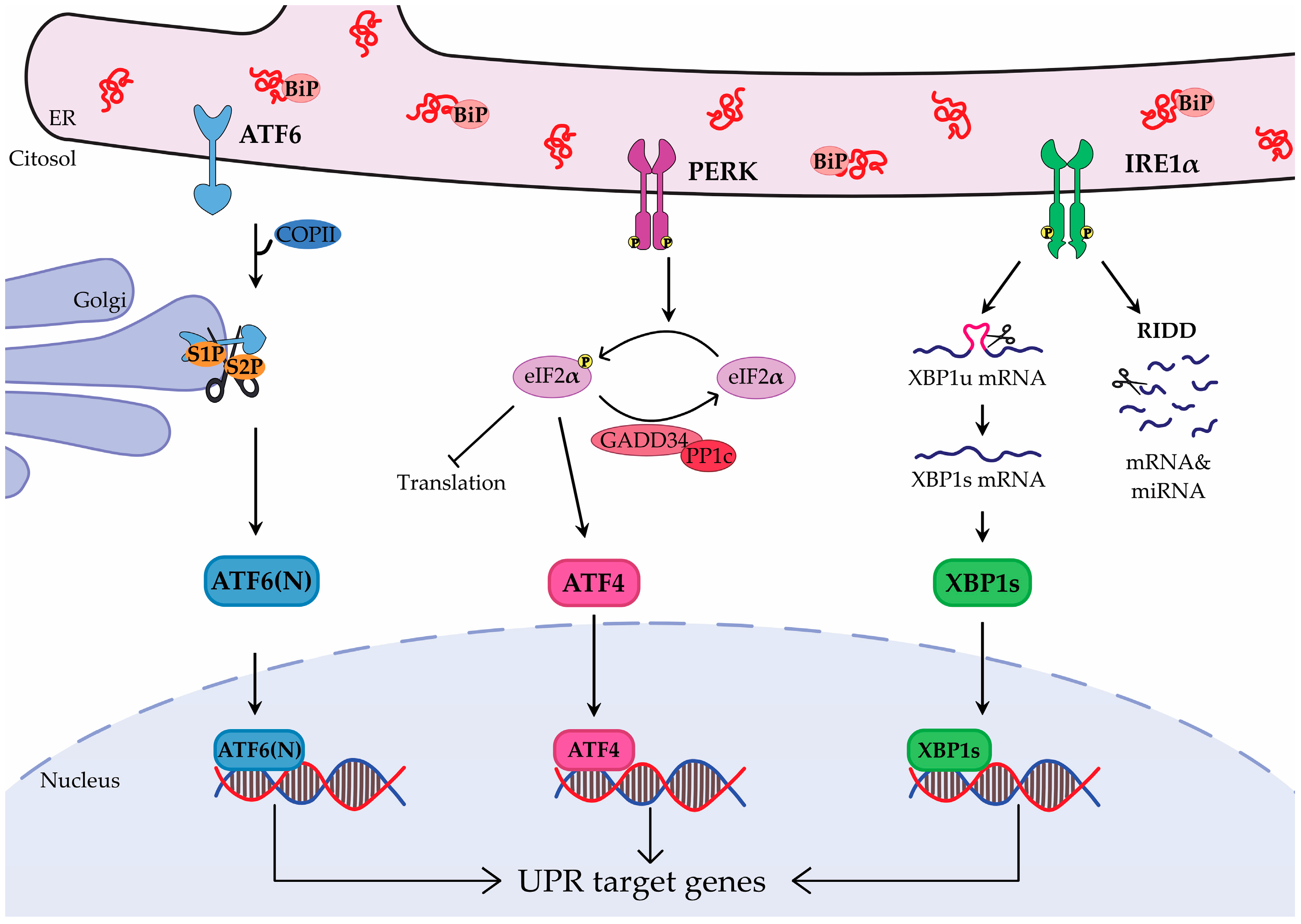
Figure 1. Schematic representation of the Unfolded Protein Response (UPR) signaling. Unfolded or misfolded proteins in the endoplasmic reticulum (ER) activate the three branches of the UPR: ATF6 (activating transcription factor 6), PERK (double-stranded RNA-activated protein kinase (PKR)–like ER kinase) and IRE1 (inositol-requiring enzyme 1). ATF6 is activated when BiP (binding immunoglobulin protein) dissociates from its ER luminal domain and is transported to the Golgi for proteolytic cleavage by S1P (site-1 protease) and S2P (site-2 protease), which generates ATF6(N) (ATF6 N-terminal fragment). PERK is activated either by BiP dissociation or by direct binding of unfolded proteins. It phosphorylates eIF2α (α-subunit of eukaryotic initiation factor 2), which reduces protein translation and activates ATF4 (activating transcription factor 4). GADD34 (growth arrest and DNA damage–inducible gene 34) and PP1c (protein phosphatase 1) form a complex to dephosphorylate eIF2α. IRE1, activated by unfolded proteins or by BiP dissociation, cleaves XBP1 (X-box binding protein 1) mRNA producing XBP1s (XBP1 spliced). It also mediates the selective breakdown of mRNAs and miRNAs via regulated IRE1-dependent decay (RIDD). ATF6(N), ATF4 and XBP1s translocate to the nucleus, where they trigger the expression of target genes.
Cancers 17 03489 g001
Figure 2. Diagram of Unfolded Protein Response (UPR)-mediated signaling in tumor cells, highlighting how IRE1 (inositol-requiring enzyme 1), PERK (double-stranded RNA-activated protein kinase (PKR)–like ER kinase), and ATF6 (activating transcription factor 6) pathways facilitate cancer survival, progression and resistance to treatment. XBP1s (X-box binding protein 1 spliced) upregulates SCD1 (stearoyl-CoA desaturase 1), to prevent lipotoxic stress, stimulates the hexosamine biosynthetic pathway (HBP) to support survival under starvation, and cooperates with HIF1α (hypoxia inducible-factor 1 alpha) to sustain cell survival under hypoxia. Regulated IRE1-dependent decay (RIDD) aids adaptation to nutrient deprivation, while ATF6 upregulates PLA2G4A (cytosolic phospholipase A2)-mediated arachidonic acid metabolism, to promote resistance to ferroptotic cell death. During invasion and metastasis, PERK drives epithelial–mesenchymal transition (EMT) and metastasis through cytoskeletal remodeling, while XBP1s induces EMT by downregulating E-cadherin and upregulating N-cadherin and upregulates MMP9 to support extracellular matrix remodeling and invasion. Moreover, IRE1 directly interacts with filamin A to enhance invasiveness. To promote angiogenesis, ATF4 (activating transcription factor 4), XBP1s and ATF6 upregulate VEGF-A, while IRE1 augments IL6 (interleukin 6) and IL8 (interleukin 8) expression. Chemoresistance involves all three branches: PERK promotes multidrug resistance, while XBP1s enhances drug efflux via ABCB1 (ABC subfamily B member 1), ABCC1 (alias, MRP1, multidrug resistance-associated protein 1—through SREBP1 (sterol regulatory element-binding protein 1)) and ABCG2 (ABC subfamily G member 2). AFT6 contributes to radio- and chemoresistance, while BiP (binding immunoglobulin protein) drives chemoresistance by regulating fatty acid synthesis, mitochondrial transportation and oxidation. Finally, UPR activation fosters immune evasion, as XBP1s drives a tolerogenic tumor microenvironment via cholesterol-rich small extracellular vesicles, while transmissible ER stress (TERS) reprograms myeloid dendritic cells and macrophages to produce tumorigenic and pro-inflammatory cytokines and suppress T-cell responses.
Cancers 17 03489 g002
Figure 3. Roles of the Unfolded Protein Response (UPR) in osteosarcoma (OS) survival and progression. All three UPR branches, ATF6 (activating transcription factor 6), PERK (double-stranded RNA-activated protein kinase (PKR)–like ER kinase), and IRE1 (inositol-requiring enzyme 1), contribute to the regulation of tumor proliferation. Beyond this shared role, PERK also mediates immune evasion, IRE1 and ATF6 promote invasion and metastasis, and ATF6 further drives chemoresistance.
Cancers 17 03489 g003
Table 2. Drug targeting the UPR currently in clinical trials.
DrugCombination Drug TherapyTargetTumorClinical Trial IDClinical Trial Phase
BOLD-100FOLFOX ChemotherapyBiPGastric, Pancreatic, Colorectal Cancer and CholangiocarcinomaNCT04421820Phase 1 & 2
BOLD-100DOXBiPSoft tissue sarcomasNCT07027423Phase 1
NKP-1339NoneBiPSolid tumorsNCT01415297Phase 1
HC 5404-FUNonePERKRenal cell carcinoma, gastric cancer, metastatic breast cancer, small cell lung cancer, and other solid tumors (e.g., non-small cell lung cancer, colorectal cancer, carcinoma of unknown primary)NCT04834778Phase 1
NMS-03597812DexamethasonePERKMultiple MyelomaNCT05027594Phase 1
NMS-03597812NonePERKAcute Myeloid LeukemiaNCT06549790Phase 1
ORIN 1001 (MKC8866)AbraxaneIRE1 RNaseBreast cancer and other solid tumorsNCT03950570Phase 1 & 2
ORIN 1001 (MKC8866)Standard of CareIRE1 RNaseSolid tumorsNCT05154201Phase 1 & 2
IXAZOMIBNoneProteasomeNonhematologic Malignancies including Soft Tissue SarcomasNCT00830869Phase 1
Disclaimer/Publisher’s Note: The statements, opinions and data contained in all publications are solely those of the individual author(s) and contributor(s) and not of MDPI and/or the editor(s). MDPI and/or the editor(s) disclaim responsibility for any injury to people or property resulting from any ideas, methods, instructions or products referred to in the content.