Article

Ovarian Cancer in the Era of Precision Surgery and Targeted Therapies

by
Yagmur Sisman
1,
Tim Svenstrup Poulsen
2,
Tine Henrichsen Schnack
3,
Claus Høgdall
1 and
Estrid Høgdall
2,*
1
Department of Gynecology, Copenhagen University Hospital, Rigshospitalet, 2100 Copenhagen, Denmark
2
Department of Pathology, Copenhagen University Hospital, Herlev University Hospital, 2730 Herlev, Denmark
3
Department of Gynecology, Odense University Hospital, 5000 Odense, Denmark
*
Author to whom correspondence should be addressed.
Cancers 2025, 17(20), 3371; https://doi.org/10.3390/cancers17203371
Submission received: 10 September 2025 / Revised: 10 October 2025 / Accepted: 16 October 2025 / Published: 18 October 2025
(This article belongs to the Special Issue Novel Approaches in the Management of Gynecological Cancers)

Simple Summary

High-grade serous ovarian cancer is the most frequent and aggressive form of ovarian cancer. Although most patients respond to initial chemotherapy, relapses are almost inevitable. At relapse, surgery to remove recurrent disease can be considered in selected patients. In this study, we examined whether the genetic profile of the tumor changes from diagnosis to relapse. By analyzing paired tumor samples from 16 patients, we found that the mutational landscape remained highly stable, with only one case showing an additional mutation at relapse. These results suggest that the main genetic drivers are established early and persist throughout disease progression. Our findings emphasize the role of surgery in carefully chosen patients and indicate that mechanisms beyond genetic mutations should be explored to understand resistance and guide future treatment strategies.

Abstract

Background: High-grade serous ovarian cancer (HGSC) is the most common and aggressive subtype of ovarian cancer. Despite initial response to platinum-based chemotherapy, most patients relapse. Cytoreductive surgery at relapse has been shown to improve survival in selected patients, but the biological mechanisms underlying recurrence and resistance remain unclear. This study aimed to investigate whether the mutational profile of HGSC changes from diagnosis to relapse, and to evaluate treatment patterns and survival outcomes in a cohort undergoing cytoreductive surgery. Methods: Sixteen patients with HGSC who underwent cytoreductive surgery at both diagnosis and relapse were included. Matched tumor tissue samples (n = 32) were collected and sequenced using a 501-gene cancer panel. Only pathogenic or likely pathogenic variants were registered. Clinical data, treatment history, and survival outcomes were obtained from medical records, with a median follow-up of 63 months. Results: All patients harbored pathogenic or likely pathogenic mutations, most frequently in TP53 (88%) and BRCA1/2 (38%). The mutational landscape was largely stable, with 15 of 16 patients (94%) showing no mutational changes between diagnosis and relapse. One patient acquired a NOTCH2 mutation at relapse. Complete resection was achieved in 88% of relapse surgeries. Median time to first relapse was 32 months, and overall survival was prolonged, with 87.5% of patients alive at last follow-up. BRCA mutated patients showed longer time to relapse, and overall follow-up compared to BRCA wild-type cases. Conclusions: The somatic mutational profile of HGSC remains remarkably stable from diagnosis to relapse. Clinically, this stability suggests that repeat mutational sequencing at relapse is unlikely to yield new actionable findings and may have limited value in guiding treatment decisions. Instead, resistance mechanisms likely arise from epigenetic or non-genetic changes, underscoring the need for future research in these areas and the continued importance of optimal surgical management in selected patients.

1. Introduction

Managing relapsed high-grade serous ovarian cancer (HGSC) presents significant challenges. Historically, systemic platinum-based chemotherapy has been the cornerstone of treatment. Despite initial response, most of the patients with advanced-stage disease ultimately experience disease recurrence. Relapses can be diagnosed through radiographic progression, an increase in cancer antigen 125 (CA125), symptoms or clinical findings on physical examination.
The DESKTOP III trial demonstrated a significant survival benefit from cytoreductive surgery at relapse in selected patients with recurrence [1]. Patients with a positive Arbeitsgemeinschaft Gynäkologische Onkologie (AGO) score—defined by an Eastern Cooperative Oncology Group (ECOG) performance status 0, ascites < 500 mL, and complete resection at initial surgery were randomized to receive either cytoreductive surgery at relapse followed by chemotherapy or chemotherapy alone. The study demonstrated a significantly improved overall survival (OS) in the surgery arm (54 months vs. 46 months, HR 0.75, CI 0.59–0.96), with the most favorable outcome observed in patients who achieved complete resection (OS 62 months), while patients who underwent surgery with incomplete resection had a lower survival (OS 28 months) [1]. Two additional studies have also demonstrated a survival advantage in appropriately selected patients who achieved no residual disease following surgery. In the SOC1 study, cytoreductive surgery was associated with a significant improvement in progression-free survival (PFS), while in the GOG 213 trial, PFS improved from 16 to 22 months, although no OS benefit was observed [2,3]. All studies included patients with platinum-sensitive disease. Eligibility was based on a high likelihood of achieving complete cytoreduction—either assessed through validated predictive models or based on surgeon discretion. Current international guidelines now recommend that patients with a first relapse occurring more than six months after completion of platinum therapy should be referred to specialized gynecologic oncology centers for evaluation of potential cytoreductive surgery at relapse [4,5,6].
In parallel with advances in surgical strategies, recent years have seen a growing emphasis on targeted treatment approaches. Poly (ADP-ribose) polymerase (PARP) inhibitors have shown improved PFS and survival in recurrent platinum-sensitive, PARP inhibitor-naïve patients [7]. PARP inhibitor maintenance is currently recommended for PARP-naive patients with relapsed HGSC and a BRCA1/2 mutation and/or HRD, following response to platinum-based chemotherapy [4,5,8]. Similarly, bevacizumab has shown benefit in prolonging PFS when combined with chemotherapy [9]. It should be offered to bevacizumab-naive patients with recurrent disease as part of a combined regimen followed by maintenance therapy [4,5,8].
To date, only a few biological drivers of recurrence and resistance have been identified, such as BRCA1/2 reversion mutations or loss of BRCA1 promoter methylation [10,11,12]. However, findings from the 2023 BriTROC-1 study, a UK-based translational effort, revealed striking genomic stability between diagnosis and relapse in HGSC. Among 276 women with relapsed HGSC, 134 had matched samples from both time points. Panel sequencing demonstrated high concordance, with only four discordant cases and no identified BRCA1/2 reversion mutations [13].
In this study, we aimed to assess differences in molecular driver mutations and therapeutic targets in tissue samples from diagnosis and relapse in 16 patients with HGSC undergoing cytoreductive surgery, alongside an evaluation of treatment patterns and survival outcomes. Our findings should be interpreted with caution due to the small sample size and warrant confirmation in larger patient cohorts.

2. Materials and Methods

2.1. Patients and Tissue Samples

Initially, 23 patients with HGSC were enrolled. Four were excluded due to insufficient tumor material, and three were excluded following quality assessment for sequencing due to low tumor content, resulting in a final cohort of 16 patients who underwent cytoreductive surgery at relapse. The included and excluded patients differed in the type of surgery. Thus, 86% of excluded vs. 19% of included cases underwent interval debulking surgery. No other clinical differences were identified between the groups (Table S1). DNA sequencing was performed on all patients using tissue samples obtained during cytoreductive surgery at both diagnosis and relapse. Clinical data were obtained from each patient’s medical record. Platinum sensitivity was defined as the absence of relapse or progressive disease within 12 months after completion of first-line platinum-based chemotherapy. Partial platinum sensitivity was defined as relapse occurring between 6 and 12 months after treatment completion. Relapse was defined based on the best clinical evaluation, including CT or PET-CT imaging, serum CA125 levels, and patient symptoms. Overall survival was not reported, as only two patients had died at the time of data cut-off. Instead, time to first relapse and total follow-up time were chosen as clinically meaningful endpoints. All patients were enrolled in the Pelvic Mass Study/GOVEC Study and followed from March 2014 until May 2025. No patients were lost to follow-up. All tissue samples were registered and stored in the Danish Cancer Biobank. The diagnosis of HGSC was confirmed by an experienced gynecologic oncology pathologist through a review of the original tissue samples.

2.2. DNA Extraction, Library Preparation and Sequencing

A total of 32 samples were collected. A representative area of cancerous tissue was identified and collected as fresh-frozen tissue in 17 samples. In 15 samples, a representative area within FFPE blocks was identified and extracted to ensure a high tumor cell content. All assessments were performed by an experienced gynecologic oncology pathologist. Genomic DNA was extracted using the Maxwell® RSC DNA FFPE Kit (Promega, Madison, WI, USA). DNA concentration was measured using the Qubit™ dsDNA High Sensitivity Assay Kit (Thermo Fisher Scientific, Waltham, MA, USA) on the Qubit Fluorometer (Thermo Fisher Scientific, Waltham, MA, USA). Library preparation for the OCA- Plus panel, covering 501 cancer-related genes, was performed manually. Sequencing was performed on the Ion S5™ XL Sequencer (Thermo Fisher Scientific, Waltham, MA, USA), and data were analyzed using Ion Reporter™ Software (v. 5.14) and the Oncomine Comprehensive Plus–w2.0–DNA–Single Sample workflow [14]. Only pathogenic and likely pathogenic variants, defined by ClinVar and classified according to AMCG criteria, were included for further analyses [15]. All performed molecular analyzes are somatic and not germline.

2.3. Data Analyses

All data analyses were carried out using Ion Reporter Software version 5.20 (Thermo Fisher Scientific, Waltham, MA, USA). The analyses were conducted using the human hg19 as the standard reference genome. The workflow Oncomine Comprehensive Plus-w2.3-DNA-Single Sample was utilized to identify somatic variants [single nucleotide variation (SNV), multi nucleotide variation (MNV), insertion-deletion (indel), and copy number variation (CNV)]. The Ion reporter’s coverage analysis reports were also used to evaluate the quality of the sequencing reactions by measuring mapping reads, mean depth, uniformity, and alignment over a target region. The genetic variants were clinically annotated using the database provided at VarSome using the implemented ClinVar database. Only variants classified as pathogenic and likely pathogenic (class 4 or 5) according to ACMG variant classification were included in the analysis [15].

2.4. Statistical Analyses

All statistical analyses were performed with Rstudio version 2025.05.1-513. Survival outcomes were summarized descriptively using medians and ranges. Formal subgroup comparisons (e.g., BRCA-mutated vs. wild-type) were not performed, as the study was not powered to detect statistically significant differences. Reported subgroup outcomes should therefore be considered exploratory and descriptive. No formal power calculation was performed prior to this study. The analyses were exploratory in nature, and the sample size was determined by the availability of patients meeting the inclusion criteria.

3. Results

3.1. Patients

Clinical characteristics of the 16 included HGSC patients at the time of cytoreductive surgery, both at diagnosis and relapse, are summarized in Table 1. All patients underwent cytoreductive surgery at diagnosis. At first relapse, 14 patients (87.5%) underwent cytoreductive surgery, and 2 patients (12.5%) underwent surgery at second relapse (Figure 1). The median age at diagnosis was 64 years, increasing to 67 years at follow-up. At the time of cytoreductive surgery at relapse, 14 patients (87.5%) had performance status 0, while the remaining 2 patients (12.5%) had a performance status of 1. Fourteen patients (87.5%) were platinum-sensitive, and two patients (12.5%) were partial platinum-sensitive. Complete resection was achieved in all patients during surgery at diagnosis. At cytoreductive surgery for relapsed disease, 14 patients (87.5%) had complete resection, whereas 2 patients (12.5%) were unresectable. The median follow-up was 63 months. The median time from diagnosis to first relapse was 32 months. The median follow-up for patients with complete resection was 64 months, compared to 33 months for those deemed unresectable. At time of follow-up, 14 patients (87.5%) were alive (Figure 1).

3.2. Treatments

At diagnosis, primary cytoreductive surgery was performed in 13 patients (81%), and interval debulking surgery in 3 patients (19%). Four patients (25%) had a third cytoreductive surgery, and one patient underwent a fourth cytoreductive surgery at the time of follow up (Figure 1).
All patients received platinum-based chemotherapy at both initial diagnosis and after surgical treatment of their first or second relapse. Chemotherapy regimens administered at subsequent relapses are detailed in Table S2.
Seven patients (44%) received bevacizumab as part of their treatment plan. The timing and duration of the individual bevacizumab treatments are presented in Table S3.
PARP inhibitors were part of the treatment plan for ten patients (63%). The timing and duration of the individual PARP inhibitor treatments is presented in Table S3.

3.3. Pathogenic and Likely Pathogenic Mutations

All patients harbored pathogenic or likely pathogenic mutations (Table 2). TP53 mutations were identified in 14 patients (88%), and BRCA1/2 mutations were present in 6 patients (38%). Only one patient (6%) showed a difference in the mutational profile between the samples obtained at diagnosis and cytoreductive surgery at relapse. In this case, a newly acquired mutation in the NOTCH2 gene was detected in tissue from relapse (Table 2).
At cytoreductive surgery at diagnosis, carcinomatosis was present in all BRCA1/2 mutated patients and in 6 (60%) of BRCA1/2 wild-type patients (Table S4). Conversely, at cytoreductive surgery at relapse, carcinomatosis was observed in only 1 (17%) of BRCA1/2-mutated patients compared to 6 (60%) of BRCA1/2 wild-type cases. Of the four patients without carcinomatosis at diagnosis (patient ID 9, 10, 13, and 19), only one (patient ID 19) remained free of carcinomatosis at the time of cytoreductive surgery for relapse. Apart from the observation that the majority of BRCA1/2 mutated patients (83%) did not present with carcinomatosis at relapse, no other associations between mutations and the presence of carcinomatosis could be identified. Complete resection at cytoreductive surgery for relapse was achieved in all BRCA1/2 mutated patients, whereas two (20%) of the BRCA1/2 wild-type patients were unresectable. Median time to first relapse was prolonged in the BRCA1/2 mutated group (41 months; range 9–66) as compared with the BRCA1/2 wild-type group (26 months; range 14–55). Overall follow-up was likewise longer for BRCA1/2 mutated patients (median 96 months; range 63–121) than for BRCA1/2 wild-type patients (median 46 months; range 33–75).

4. Discussion

In this study with 16 patients with HGSC who underwent cytoreductive surgery at diagnosis and relapse, 32 paired tissue samples were analyzed. The mutational landscape remained largely stable over time. All patients harbored pathogenic or likely pathogenic mutations at diagnosis, and in 15 of 16 cases (94%) the mutational profile was unchanged at relapse. Only one patient acquired an additional mutation, indicating that mutational alterations remained remarkably consistent despite disease progression and treatment.
The drivers of recurrence and resistance in HGSC remain poorly understood, and no biomarker has yet entered clinical practice. We collected tumor biopsies at diagnosis and relapse and observed no significant changes in mutations over time. Our findings contribute to the discussion about whether routine biopsies for molecular analyses in the relapse setting should be implemented in clinical practice, as no new actionable mutations emerge over time [13].
Similarly, the BriTROC-1 study, which included tumor biopsies from 276 patients with relapsed HGSC, demonstrated high concordance between primary and relapse samples, with only four discordant cases [13]. These included mutations unique to either diagnosis or relapse, but no consistent acquisition of new driver events. The study also reported stability in copy number alterations, suggesting that genetic evolution is not the main driver of relapse. While BriTROC-1 included both platinum-resistant and platinum-sensitive patients and used both surgical and image-guided biopsies, our study focused exclusively on a surgically treated, platinum-sensitive cohort, offering a clinically well-defined comparison.
The OCTIPS consortium analyzed 31 paired samples of platinum-sensitive HGSC using whole-exome sequencing and SNP array profiling [16]. Although differences were observed between primary and relapse tumors, no recurrent genomic alterations were consistently identified across pairs. Taken together, our findings, along with those from BriTROC-1 and OCTIPS, indicate that acquired resistance in HGSC is not driven by new mutational events. Instead, other mechanisms—such as epigenetic modulation, transcriptional reprogramming, or microenvironmental factors—are likely to underlie treatment resistance [13,16]. Notably, previous research has demonstrated that loss of BRCA1 promoter methylation can contribute to resistance to both platinum-based chemotherapy and PARP inhibitors [11,12].
Systemic therapy has traditionally been the standard for recurrent HGSC, but the DESKTOP III trial demonstrated that cytoreductive surgery at relapse in addition to platinum-based chemotherapy improves OS, particularly when complete resection is achieved [1]. Although OS could not be estimated in our cohort as most patients were still alive, the median follow-up of 63 months and a complete resection rate of 88% support the survival benefit observed in DESKTOP III. Moreover, they found that patients who achieved complete resection had the most favorable outcomes. These findings are consistent with ours, where the median follow-up time for patients with complete resection was 64 months, compared to 33 months for those deemed unresectable [1].
The novelty of our study lies in the specific clinical context: we investigated a surgically treated, platinum-sensitive cohort with high rates of complete resection at relapse and prolonged follow-up. This design allowed us to analyze paired high-quality tumor samples from both diagnosis and relapse, within a population characterized by extended disease control and favorable overall survival. By showing that mutational stability persists, our study highlights that mechanisms of recurrence and resistance are unlikely to be explained by new somatic mutations. These findings have important clinical implications. The observed mutational stability suggests that repeated mutational profiling at relapse may offer limited additional value in this setting. Instead, research and clinical efforts could be more effectively directed toward understanding alternative mechanisms of treatment resistance, including epigenetic modulation, transcriptional reprogramming, and changes in the tumor microenvironment.
We observed a trend toward less carcinomatosis at relapse among BRCA mutated patients (17% vs. 60%), although this finding is based on very small numbers and should be regarded as hypothesis-generating only. Previous studies have not demonstrated such an association, and in our cohort all BRCA mutated patients were completely resected, compared with 80% in the BRCA wild-type group. One study, which aimed to examine whether BRCA mutated patients were more likely candidates for cytoreductive surgery at relapse, found no difference in the number of lesions at recurrence between BRCA mutated and BRCA wild-type patients [17]. Furthermore, they did not find a higher rate of complete resection among BRCA mutated patients, whereas in our study, all BRCA mutated patients achieved complete resection compared to 80% in the BRCA wild-type group. Notably, the two BRCA wild-type patients who were unresectable both presented with carcinomatosis. Carcinomatosis has previously been identified as a negative predictive of complete resection [18,19,20]. In the study by Harter et al., only 26% of patients with carcinomatosis achieved complete resection compared with 74% without, although survival did not differ if resection was complete [18]. The role of tumor biology remains to be clarified in the recurrent setting. It has long been unclear whether improved resectability—and consequently, better survival—reflects a more favorable tumor biology, or whether poor surgical outcomes are merely a consequence of a more aggressive disease less amenable to complete resection [21,22]. Our findings raise the question of whether BRCA status may indirectly influence surgical outcomes through underlying tumor biology. Future research integrating genomic, radiologic, and clinical variables could help elucidate whether BRCA status contributes to predictive models for resectability or treatment response in recurrent HGSC.
Our study has several methodological and clinical strengths. By combining longitudinal clinical data with sequencing of paired tumor samples from diagnosis and relapse, we could track mutational changes over and confirm that key driver mutations remain stable. The cohort represents a clinically relevant group —patients selected for cytoreductive relapse surgery—in whom molecular data are sparse. Rigorous pathological review ensured high tumor content, and variant interpretation followed ACMG and ClinVar criteria, supporting reproducibility [15]. With a median of 63 months, a high complete resection rate (87.5%), and integration in national biobanking programs, the study provides robust and well-annotated material for translational research.
This study has several limitations. The small and highly selected cohort limits statistical power and generalizability. The limited sample size also constrains the ability to detect differences and to draw firm conclusions from subgroup analyses, such as those comparing BRCA-mutated and BRCA wild-type patients. Moreover, as all patients in this study underwent cytoreductive surgery at relapse, the findings reflect a surgically favorable and platinum-sensitive population and should not be extrapolated to patients with unresectable or platinum-resistant disease. Therefore, these findings should be interpreted as hypothesis-generating and validated in larger, more diverse cohorts. Furthermore, the cohort represents a highly selected group of patients who underwent cytoreductive relapse surgery, which does not reflect the broader HGSC population, particularly those with unresectable disease. Accordingly, our conclusions should be viewed with caution and primarily as exploratory observations that warrant confirmation in prospective studies including less selected patient groups. As all patients were treated at a single national institute, local clinical practice may also influence generalizability, although standardized procedures can be considered a strength. Moreover, treatment algorithms and criteria for secondary cytoreductive surgery may differ across countries, which could affect patient selection and outcomes. However, as Denmark follows international ESMO–ESGO guidelines for recurrent ovarian cancer, we consider our findings broadly comparable to other Western healthcare settings.
Many excluded patients had undergone interval debulking surgery, where neoadjuvant chemotherapy often reduces tumor content, making molecular analyses difficult [23,24]. This challenge has also been reported in other studies, and while we observed a higher proportion of interval surgery among excluded patients, no other systematic clinical differences were seen [23]. Previous studies have demonstrated that neoadjuvant chemotherapy followed by interval debulking surgery and completion of chemotherapy is not inferior to primary cytoreductive surgery followed by chemotherapy in patients with FIGO stage IIIC or IV disease, where complete resection at primary surgery is unlikely or where the patient is unfit for extensive surgery [25,26]. Given these findings, it is unlikely that our exclusion of a higher proportion of interval debulking surgery patients introduced a survival-related selection bias [27]. However, we acknowledge that excluding cases with insufficient tumor tissue may have introduced a potential selection bias, as these patients could differ biologically from those with adequate tissue for analysis.
Additionally, we lack data on patients who were offered but not included for relapse surgery, which prevents us from fully assessing selection bias [27]. Thus, our findings may not be representative of all patients with relapsed HGSC, especially if those included had disease that was more accessible for resection.
Finally, we applied a 501-gene cancer panel, as it covers the clinically relevant and currently targetable variants, and allowed us to obtain higher sequencing depth in paired tumor samples [28,29]. Nevertheless, we acknowledge that broader sequencing strategies, such as WES or WGS, may reveal additional alterations of potential relevance. However, since some of the tumor samples were FFPE, these approaches were not feasible. Future studies with larger patient cohorts would therefore benefit from applying more comprehensive approaches to further explore potential mechanisms of platinum resistance.
Our findings provide insights into a selected group of patients undergoing cytoreductive surgery for relapsed HGSC, but validation in larger, prospective cohorts is needed. Future studies should confirm the observed mutational stability, assess molecular predictors of surgical outcome, and test whether BRCA status influences disease spread at relapse. Such work should include both resectable and unresectable patients, apply standardized tissue-collection protocols, and incorporate multi-omic profiling to capture genomic, epigenetic, and transcriptomic mechanisms of resistance. Deep clinical annotation, long-term follow-up, and patient involvement in study design will be essential to maximize translational relevance [11,12,30]. In summary, our study underscores that HGSC recurrence occurs without major mutational evolution, highlighting the importance of integrating molecular stability into future treatment strategies and trial design.

5. Conclusions

The somatic mutational landscape remains largely unchanged in patients with relapsed HGSC undergoing cytoreductive surgery at relapse, supporting the notion that key driver mutations emerge early and persist throughout disease progression. Clinically, high rates of complete resection and long-term survival were demonstrated, reinforcing the benefit of cytoreductive surgery at relapse in well-selected patients. Future prospective, multi-institutional studies are warranted to validate these observations and further refine patient selection.

Supplementary Materials

The following supporting information can be downloaded at https://www.mdpi.com/article/10.3390/cancers17203371/s1, Table S1: Clinical characteristics of excluded platinum-sensitive relapsed HGSC patients undergoing cytoreductive relapse surgery; Table S2: Chemotherapy regimens administered over time; Table S3: Treatment duration of targeted therapies in months; Table S4: Presence of carcinomatosis at diagnosis and relapse.

Author Contributions

Conceptualization, Y.S., T.S.P., T.H.S., C.H. and E.H.; methodology, Y.S., T.S.P., T.H.S., C.H. and E.H.; software, T.S.P. and E.H.; validation, T.S.P., T.H.S., C.H. and E.H.; formal analysis, Y.S., T.S.P. and E.H.; investigation, Y.S., T.S.P., T.H.S., C.H. and E.H.; resources, Y.S., T.S.P., T.H.S., C.H. and E.H.; data curation, Y.S., T.S.P. and E.H.; writing—original draft preparation, Y.S.; writing—review and editing, T.S.P., T.H.S., C.H. and E.H.; visualization, Y.S., T.S.P., T.H.S., C.H. and E.H.; supervision, T.S.P., T.H.S., C.H. and E.H.; project administration, Y.S.; funding acquisition, Y.S. All authors have read and agreed to the published version of the manuscript.

Funding

This research received a grant from The Research Council at Herlev University Hospital and Danish Medical Women’s Association.

Institutional Review Board Statement

The study was conducted according to the guidelines of the Declaration of Helsinki, including written informed consent from all subjects. The Danish National Committee has approved the study for research ethics, Capital Region (approval codes; KF01-227/03 and KF01-143/04, H15020061. Approval date: 22 June 2016). All performed molecular analyzes are somatic and not germline.

Informed Consent Statement

Informed consent was obtained from all subjects involved in the study. Written informed consent has been obtained from the patients to publish this paper.

Data Availability Statement

Data are contained within the article.

Acknowledgments

During the preparation of this manuscript/study, the author used OpenAI GPT-5 language model for the purposes of text refinement. The authors have reviewed and edited the output and take full responsibility for the content of this publication.

Conflicts of Interest

The authors declare no conflicts of interest.

Abbreviations

The following abbreviations are used in this manuscript:
ACMGAmerican College of Medical Genetics
AGOArbeitsgemeinschaft Gynäkologische Onkologie
AIArtificial Intelligence
BARD1BRCA1 Associated RING Domain 1
BRCA1/2Breast Cancer Gene 1/2
CA125Cancer Antigen 125
CIConfidence Interval
CNVCopy Number Variations
CTComputed Tomography
DNADeoxyribonucleic Acid
ECOGEastern Cooperative Oncology Group
FFPEFormalin-Fixed Paraffin-Embedded
FIGOInternational Federation of Gynecology and Obstetrics
GOGGynecologic Oncology Group
GOVECGynecologic Oncology and Pelvis Mass Study
HGSCHigh-grade Serous Carcinoma
HRHazard Ratio
HRDHomologous Recombination Deficiency
IndelInsertion-Deletion
MAMassachusetts
MNVMulti Nucleotide Variation
NF1Neurofibromin 1
NOTCH2Neurogenic Locus Notch Homolog Protein 2
OCA-PlusOncomine Comprehensive Assay Plus
OSOverall Survival
PARPPoly (ADP-ribose) Polymerase
PFSProgression Free Survival
PDXPatient-Derived Xenograft
PET-CTPositron Emission Tomography-Computed Tomography
RAD51DRAD51 Paralog D
RSCResearch Simplified Chemistry
SNVSingle Nucleotide Variation
SNPSingle Nucleotide Polymorphism
UKUnited Kingdom

References

  1. Harter, P.; Sehouli, J.; Vergote, I.; Ferron, G.; Reuss, A.; Meier, W.; Greggi, S.; Mosgaard, B.J.; Selle, F.; Guyon, F.; et al. Randomized Trial of Cytoreductive Surgery for Relapsed Ovarian Cancer. N. Engl. J. Med. 2021, 385, 2123–2131. [Google Scholar] [CrossRef]
  2. Coleman, R.L.; Spirtos, N.M.; Enserro, D.; Herzog, T.J.; Sabbatini, P.; Armstrong, D.K.; Kim, J.W.; Park, S.Y.; Kim, B.G.; Nam, J.H.; et al. Secondary Surgical Cytoreduction for Recurrent Ovarian Cancer. N. Engl. J. Med. 2019, 381, 1929–1939. [Google Scholar] [CrossRef]
  3. Shi, T.; Zhu, J.; Feng, Y.; Tu, D.; Zhang, Y.; Zhang, P.; Jia, H.; Huang, X.; Cai, Y.; Yin, S.; et al. Secondary cytoreduction followed by chemotherapy versus chemotherapy alone in platinum-sensitive relapsed ovarian cancer (SOC-1): A multicentre, open-label, randomised, phase 3 trial. Lancet Oncol. 2021, 22, 439–449. [Google Scholar] [CrossRef] [PubMed]
  4. González-Martín, A.; Harter, P.; Leary, A.; Lorusso, D.; Miller, R.; Pothuri, B.; Ray-Coquard, I.; Tan, D.; Bellet, E.; Oaknin, A.; et al. Newly diagnosed and relapsed epithelial ovarian cancer: ESMO Clinical Practice Guideline for diagnosis, treatment and follow-up. Ann. Oncol. 2023, 34, 833–848. [Google Scholar] [CrossRef] [PubMed]
  5. National Comprehensive Cancer Network. NCCN Clinical Practice Guidelines in Oncology (NCCN Guidelines®) Ovarian Cancer Continue Including Fallopian Tube Cancer and Primary Peritoneal Cancer; National Comprehensive Cancer Network: Philadelphia, PA, USA, 2024. [Google Scholar]
  6. Dansk Gynækologisk Cancer Gruppe (DGCG). Ovariecancer-Kirurgi Ved Recidiv; Dansk Gynækologisk Cancer Gruppe: Copenhagen, Denmark, 2022. [Google Scholar]
  7. Mirza, M.R.; Monk, B.J.; Herrstedt, J.; Oza, A.M.; Mahner, S.; Redondo, A.; Fabbro, M.; Ledermann, J.A.; Lorusso, D.; Vergote, I.; et al. Niraparib Maintenance Therapy in Platinum-Sensitive, Recurrent Ovarian Cancer. N. Engl. J. Med. 2016, 375, 2154–2164. [Google Scholar] [CrossRef] [PubMed]
  8. Dansk Gynækologisk Cancer Gruppe (DGCG). Ovariecancer-Edicinsk Recidivbehandling; Dansk Gynækologisk Cancer Gruppe: Copenhagen, Denmark, 2023. [Google Scholar]
  9. Aghajanian, C.; Blank, S.V.; Goff, B.A.; Judson, P.L.; Teneriello, M.G.; Husain, A.; Sovak, M.A.; Yi, J.; Nycum, L.R. OCEANS: A randomized, double-blind, placebo-controlled phase III trial of chemotherapy with or without bevacizumab in patients with platinum-sensitive recurrent epithelial ovarian, primary peritoneal, or fallopian tube cancer. J. Clin. Oncol. 2012, 30, 2039–2045. [Google Scholar] [CrossRef]
  10. Cancer Genome Atlas Research Network. Integrated genomic analyses of ovarian carcinoma. Nature 2011, 474, 609–615. [Google Scholar] [CrossRef]
  11. Swisher, E.M.; Kwan, T.T.; Oza, A.M.; Tinker, A.V.; Ray-Coquard, I.; Oaknin, A.; Coleman, R.L.; Aghajanian, C.; Konecny, G.E.; O’mAlley, D.M.; et al. Molecular and clinical determinants of response and resistance to rucaparib for recurrent ovarian cancer treatment in ARIEL2 (Parts 1 and 2). Nat. Commun. 2021, 12, 1–13. [Google Scholar] [CrossRef]
  12. Kondrashova, O.; Topp, M.; Nesic, K.; Lieschke, E.; Ho, G.Y.; Harrell, M.I.; Zapparoli, G.V.; Hadley, A.; Holian, R.; Boehm, E.; et al. Methylation of all BRCA1 copies predicts response to the PARP inhibitor rucaparib in ovarian carcinoma. Nat. Commun. 2018, 9, 1–16. [Google Scholar] [CrossRef]
  13. Smith, P.; Bradley, T.; Gavarró, L.M.; Goranova, T.; Ennis, D.P.; Mirza, H.B.; De Silva, D.; Piskorz, A.M.; Sauer, C.M.; Al-Khalidi, S.; et al. The copy number and mutational landscape of recurrent ovarian high-grade serous carcinoma. Nat. Commun. 2023, 14, 1–15. [Google Scholar] [CrossRef]
  14. Vestergaard, L.K.; Oliveira, D.N.P.; Poulsen, T.S.; Høgdall, C.K.; Høgdall, E.V. Oncomine TM Comprehensive Assay v3 vs. Oncomine TM Comprehensive Assay Plus. Cancers 2021, 13, 5230. [Google Scholar] [CrossRef] [PubMed]
  15. Richards, S.; Aziz, N.; Bale, S.; Bick, D.; Das, S.; Gastier-Foster, J.; Grody, W.W.; Hegde, M.; Lyon, E.; Spector, E.; et al. Standards and guidelines for the interpretation of sequence variants: A joint consensus recommendation of the American College of Medical Genetics and Genomics and the Association for Molecular Pathology. Genet. Med. 2015, 17, 405–424. [Google Scholar] [CrossRef] [PubMed]
  16. Lambrechts, S.; Smeets, D.; Moisse, M.; Braicu, E.I.; Vanderstichele, A.; Zhao, H.; Van Nieuwenhuysen, E.; Berns, E.; Sehouli, J.; Zeillinger, R.; et al. Genetic heterogeneity after first-line chemotherapy in high-grade serous ovarian cancer. Eur. J. Cancer 2016, 53, 51–64. [Google Scholar] [CrossRef] [PubMed]
  17. Yfat, K.; Mariam, K.; Mario, B.; Hal, H.; Dana, J.; Lina, S.; Ilan, B.; Gregory, P.; Limor, H. Germline BRCA mutation carriers are more likely to undergo cytoreductive surgery for relapsed, platinum sensitive, ovarian cancer. Gynecol. Oncol. 2022, 167, 256–260. [Google Scholar] [CrossRef]
  18. Harter, P.; Hahmann, M.; Lueck, H.J.; Poelcher, M.; Wimberger, P.; Ortmann, O.; Canzler, U.; Richter, B.; Wagner, U.; Hasenburg, A.; et al. Surgery for recurrent ovarian cancer: Role of peritoneal carcinomatosis: Exploratory analysis of the DESKTOP i trial about risk factors, surgical implications, and prognostic value of peritoneal carcinomatosis. Ann. Surg. Oncol. 2009, 16, 1324–1330. [Google Scholar] [CrossRef]
  19. Eisenkop, S.M.; Friedman, R.L.; Spirtos, N.M. The role of secondary cytoreductive surgery in the treatment of patients with recurrent epithelial ovarian carcinoma. Cancer 2000, 88, 144–153. [Google Scholar] [CrossRef]
  20. Chi, D.S.; McCaughty, K.; Diaz, J.P.; Huh, J.; Schwabenbauer, S.; Hummer, A.J.; Venkatraman, E.S.; Aghajanian, C.; Sonoda, Y.; Abu-Rustum, N.R.; et al. Guidelines and selection criteria for secondary cytoreductive surgery in patients with recurrent, platinum-sensitive epithelial ovarian carcinoma. Cancer 2006, 106, 1933–1939. [Google Scholar] [CrossRef]
  21. Fotopoulou, C.; Eriksson, A.G.; Yagel, I.; Chang, S.J.; Lim, M.C. Surgery for Recurrent Epithelial Ovarian Cancer. Curr. Oncol. Rep. 2024, 26, 46–54. [Google Scholar] [CrossRef]
  22. Hall, M.; Savvatis, K.; Nixon, K.; Kyrgiou, M.; Hariharan, K.; Padwick, M.; Owens, O.; Cunnea, P.; Campbell, J.; Farthing, A.; et al. Maximal-Effort Cytoreductive Surgery for Ovarian Cancer Patients with a High Tumor Burden: Variations in Practice and Impact on Outcome. Ann. Surg. Oncol. 2019, 26, 2943–2951. [Google Scholar] [CrossRef]
  23. Nikolaidi, A.; Papadopoulou, E.; Haidopoulos, D.; Liontos, M.; Fountzilas, E.; Tsaousis, G.; Goula, K.; Tsolaki, E.; Christopoulou, A.; Binas, I.; et al. Molecular Alterations in Paired Epithelial Ovarian Tumors in Patients Treated with Neoadjuvant Chemotherapy. Cancers 2024, 16, 3580. [Google Scholar] [CrossRef]
  24. Zalaznick, H.; Clegg, B.; Cogan, E.; Perry, M.; Trost, J.; Mancini-DiNardo, D.; Gutin, A.; Lanchbury, J.; Timms, K. Rates of homologous recombination deficiency across different subtypes of ovarian cancer and in pre- and post-neoadjuvant chemotherapy tumor samples (139.5). Gynecol. Oncol. 2022, 166, S86–S87. [Google Scholar] [CrossRef]
  25. Vergote, I.; Tropé, C.G.; Amant, F.; Kristensen, G.B.; Ehlen, T.; Johnson, N.; Verheijen, R.H.M.; van der Burg, M.E.L.; Lacave, A.J.; Panici, P.B.; et al. Neoadjuvant Chemotherapy or Primary Surgery in Stage IIIC or IV Ovarian Cancer. N. Engl. J. Med. 2010, 363, 943–953. [Google Scholar] [CrossRef]
  26. Kehoe, S.; Hook, J.; Nankivell, M.; Jayson, G.C.; Kitchener, H.; Lopes, T.; Luesley, D.; Perren, T.; Bannoo, S.; Mascarenhas, M.; et al. Primary chemotherapy versus primary surgery for newly diagnosed advanced ovarian cancer (CHORUS): An open-label, randomised, controlled, non-inferiority trial. Lancet 2015, 386, 249–257. [Google Scholar] [CrossRef]
  27. Phillips, M.R.; Kaiser, P.; Thabane, L.; Bhandari, M.; Chaudhary, V. Retina Evidence Trials International Alliance Study Group. Risk of bias: Why measure it, and how? Eye 2022, 36, 346–348. [Google Scholar] [CrossRef]
  28. Muzzey, D.; Evans, E.A.; Lieber, C. Understanding the Basics of NGS: From Mechanism to Variant Calling. Curr. Genet. Med. Rep. 2015, 3, 158–165. [Google Scholar] [CrossRef]
  29. Altmann, A.; Weber, P.; Bader, D.; Preuss, M.; Binder, E.B.; Müller-Myhsok, B. A beginners guide to SNP calling from high-Throughput DNA-sequencing data. Hum. Genet. 2012, 131, 1541–1554. [Google Scholar] [CrossRef] [PubMed]
  30. Kordowitzki, P.; Lange, B.; Elias, K.M.; Haigis, M.C.; Mechsner, S.; Braicu, I.E.; Sehouli, J. Transforming treatment paradigms: Focus on personalized medicine for high-grade serous ovarian cancer. CA Cancer J. Clin. 2025, 75, 436–460. [Google Scholar] [CrossRef] [PubMed]
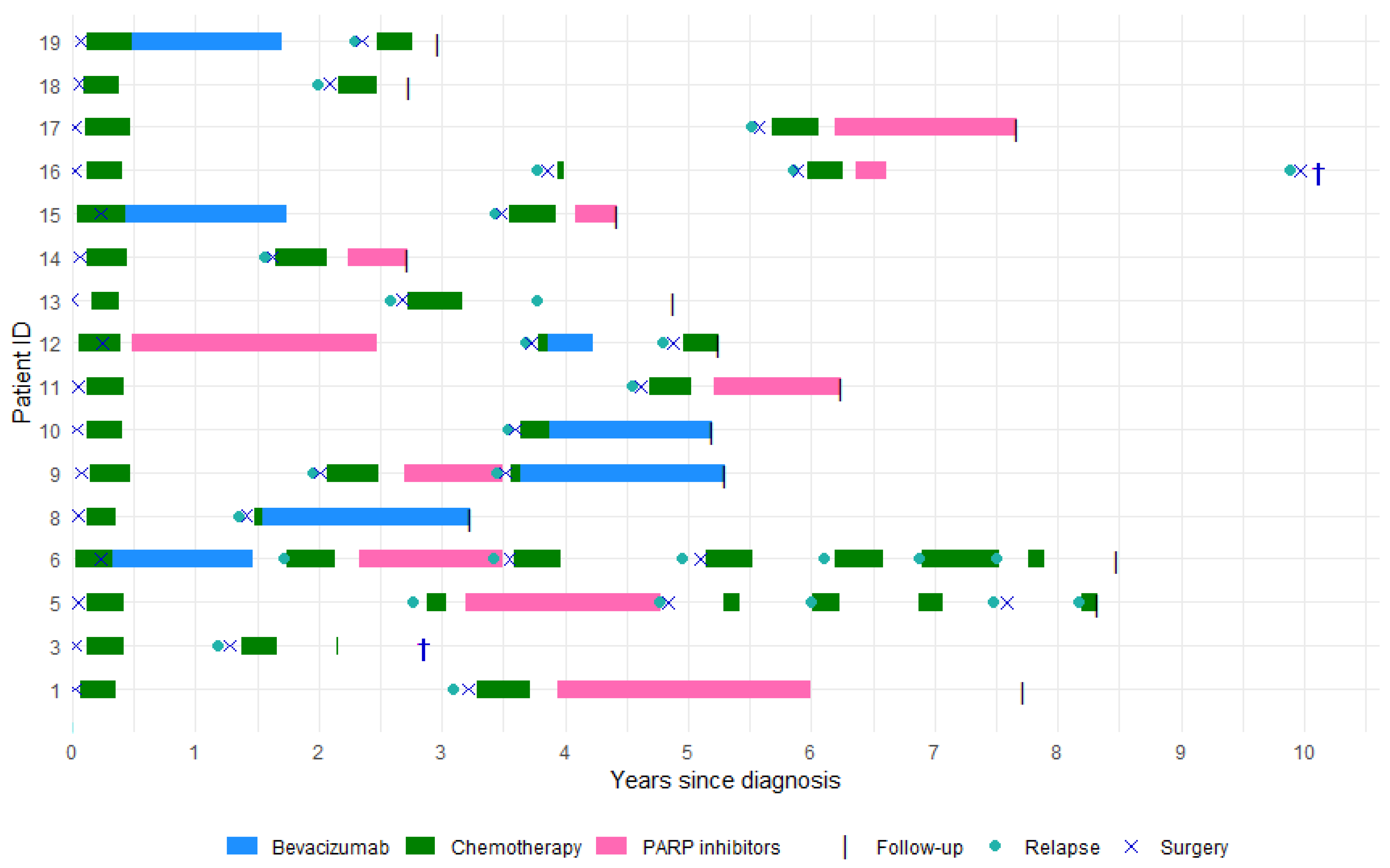
Figure 1. Overview of treatment timelines and clinical events in 16 HGSC patients.
Cancers 17 03371 g001
Table 1. Clinical characteristics of 16 platinum-sensitive relapsed HGSC patients from a cohort of EOC patients undergoing relapse surgery at the University Hospital of Copenhagen.
DiagnosisSecondary Cytoreductive Surgery
Age in years median (range)64 (41–74)67 (46–77)
CA125 median (range)195 (14–8890)35 (6–490)
BMI median (range)24 (18–34)24 (18–35)
Performance score  
09 (56%)14 (87.5%)
16 (38%)2 (12.5%)
21 (6%)-
FIGO stage  
I-II4 (25%)-
III-IV12 (75%)-
Surgery  
Primary debulking13 (81%)-
Interval debulking3 (19%)-
Residual tumor after surgery  
016 (100%)14 (87.5%)
Unresectable02 (12.5%)
Platinum response  
>12 months (sensitive)14 (87.5%)-
6 – ≤ 12 months (partial sensitive)2 (12.5%)-
Follow-up in months  
Time to first relapse median (range)-32 (14–66)
Total follow-up time median (range)-63 (33–122)
BMI: Body mass index, CA125: Cancer Antigen 125, EOC: Epithelial ovarian cancer, FIGO: International Federation of Gynecology and Obstetrics, HGSC: High grade serous carcinoma.
Table 2. Pathogenic and likely pathogenic mutations at diagnosis and cytoreductive relapse surgery.
Patient IDDiagnosisRelapse
1
BRCA2 (c.7876T>C p.Trp2626Arg exon 17)
TP53 (c.422G>A p.Cys141Tyr exon 5)
BRCA2
TP53
3
TP53 (c.810T>G p.Phe270Leu exon 8)
TP53
5
BRCA2(c. 37G>T p.Glu13Ter exon 2)
BRCA2
6
BRCA1 (c.115T>G p.Cys39Gly exon 3)
TP53 (c.1028delA p.Glu343GlyfsTer2 exon 10)
BRCA1
TP53
8FANCD2 (c.935delT p.Leu312CysfsTer10 exon 12)FANCD2
9
ILR7 (c.636T>A p.Tyr212Ter exon 5)
TP53 (c.659A>G p.Tyr220Cys exon 6)
ILR7
TP53
NOTCH2 (c.6575_6605del p.Pro2192LeufsTer20 exon 34)
10TP53 (c.916C>T p.Arg306Ter exon 8)TP53
11
TP53 (c.839G>A p.Arg280Lys exon 8)
TP53
12
BRCA2 (c.5903C>G p.Ser1968Ter exon 11)
TP53 (c.150_151insT p.Glu51Ter exon 4)
BRCA2
TP53
13TP53 (c.536A>G p.His179Arg exon 5)TP53
14
TP53 (c.742C>T p.Arg248Trp exon7)
TP53
15
TP53 (c.734G>A p.Gly245Asp exon 7)
TP53
16BRCA2 (c.5346G>A p.Trp1782Ter exon 21)
RB1 (c.37_40delGCCG p.Ala13ProfsTer51 exon1)
TP53 (c.339_341delCTT p.Phe113del exon 4)
BRCA2
RB1
TP53
17
BRCA2 (c.2454delT p.Gln819LysfsTer6 exon 11)
TP53 (c.731G>A p.Gly244Asp exon 7)
BRCA2
TP53
18
TP53 (c.659A>C p.Tyr220Ser exon 6)
TP53
19TP53 (c.1109C>T p.Arg337Cys exon 10)TP53
BRCA1: Breast cancer gene 1, BRCA2: Breast cancer gene 2, FANCD1: Fanconi Anemia Group D2, ILR7: Interleukin-7 receptor, RB1: Retinoblastoma 1, TP53: Tumor Protein P53.
Disclaimer/Publisher’s Note: The statements, opinions and data contained in all publications are solely those of the individual author(s) and contributor(s) and not of MDPI and/or the editor(s). MDPI and/or the editor(s) disclaim responsibility for any injury to people or property resulting from any ideas, methods, instructions or products referred to in the content.