Review

The Immune–Genomics of Cholangiocarcinoma: A Biological Footprint to Develop Novel Immunotherapies

1
Hepatobiliary Immunopathology Laboratory, IRCCS Humanitas Research Hospital, 20089 Rozzano, Italy
2
Department of Biomedical Sciences, Humanitas University, 20090 Pieve Emanuele, Italy
3
Medical Oncology and Hematology Unit, Humanitas Cancer Center, IRCCS Humanitas Research Hospital, 20089 Rozzano, Italy
4
Division of Internal Medicine and Hepatology, Department of Gastroenterology, IRCCS Humanitas Research Hospital, 20089 Rozzano, Italy
*
Author to whom correspondence should be addressed.
Cancers 2025, 17(2), 272; https://doi.org/10.3390/cancers17020272
Submission received: 29 November 2024 / Revised: 11 January 2025 / Accepted: 13 January 2025 / Published: 15 January 2025
(This article belongs to the Special Issue Targeting the Tumor Microenvironment (Volume II))

Simple Summary

Cholangiocarcinoma (CCA), a rare but aggressive bile duct cancer, is on the rise globally, especially a type called intrahepatic CCA. Recent research has revealed that CCA is highly diverse at the genetic and immune levels, and creates an environment that weakens the immune system, making it harder for the body to fight the cancer. A big step forward has been combining chemotherapy with immune checkpoint inhibitors (ICIs), a type of immunotherapy, which is now the standard for advanced CCA. Although these treatments extend survival, the improvement is still limited. Thus, scientists are exploring new strategies, like targeting multiple immune pathways and focusing on specific genetic vulnerabilities to boost treatment response. This work summarizes advancements in our understanding of CCA at the molecular level, which have enabled the development of personalized therapies, and highlights ongoing efforts to make CCA a more treatable disease through innovative and targeted approaches.

Abstract

Cholangiocarcinoma (CCA) represents approximately 3% of all gastrointestinal cancers and is a highly heterogeneous and aggressive malignancy originating from the epithelial cells of the biliary tree. CCA is classified by anatomical location into intrahepatic (iCCA), extrahepatic (eCCA), gallbladder cancer (GBC), and ampullary cancers. Although considered a rare tumor, CCA incidence has risen globally, particularly due to the increased diagnosis of iCCA. Genomic and immune profiling studies have revealed significant heterogeneity within CCA, leading to the identification of molecular subtypes and actionable genetic alterations in 40–60% of cases, particularly in iCCA. Among these, FGFR2 rearrangements or fusions (7–15%) and IDH1 mutations (10–20%) are common in iCCA, while HER2 amplifications/overexpression are more frequent in eCCA and GBC. The tumor-immune microenvironment (TIME) of CCAs plays an active role in the pathogenesis and progression of the disease, creating a complex and plastic environment dominated by immune-suppressive populations. Among these, cancer-associated fibroblasts (CAFs) are a key component of the TIME and are associated with worse survival due to their role in maintaining a poorly immunogenic landscape through the deposition of stiff extracellular matrix and release of pro-tumor soluble factors. Improved understanding of CCA tumor biology has driven the development of novel treatments. Combination therapies of cisplatin and gemcitabine with immune checkpoint inhibitors (ICIs) have replaced the decade-long standard doublet chemotherapy, becoming the new standard of care in patients with advanced CCA. However, the survival improvements remain modest prompting research into more effective ways to target the TIME of CCAs. As key mechanisms of immune evasion in CCA are uncovered, novel immune molecules emerge as potential therapeutic targets. Current studies are exploring strategies targeting multiple immune checkpoints, angiogenesis, and tumor-specific antigens that contribute to immune escape. Additionally, the success of ICIs in advanced CCA has led to interest in their application in earlier stages of the disease, such as in adjuvant and neoadjuvant settings. This review offers a comprehensive overview of the immune biology of CCAs and examines how this knowledge has guided clinical drug development, with a focus on both approved and emergent treatment strategies.

1. Introduction

Cholangiocarcinoma (CCA) accounts for approximately 3% of all gastrointestinal cancers and belongs to a heterogeneous group of aggressive cancers arising from the epithelial component of the biliary tree system [1]. It is classified according to the anatomical location of origin as intrahepatic (iCCA) or extrahepatic (eCCA), including perihilar (pCCA) and distal tumors (dCCA), gallbladder cancer (GBC), and ampullary cancers. Overall considered a rare tumor entity, CCA incidence has been rising over the past decades in most countries, mainly due to an increased diagnosis of iCCA [2]. Risk factors vary worldwide and encompass viral (hepatitis B and C) and non-viral (primary sclerosing cholangitis) chronic liver disease, coledocolithiasis, structural abnormalities of the biliary tree, and, especially in Asian countries, liver flukes (Opisthorchis viverrini and Clonorchis sinensis). Rising trends in Western countries have been linked to the increase in metabolic syndrome and its associated liver chronic diseases (metabolic dysfunction-associated steatotic liver disease [MASLD] and steatohepatitis [MASH]) [3]. However, causative links with metabolic syndrome remain elusive, and, in many cases, CCA arises on a seemingly “healthy” liver [4]. The lack of identifiable risk factors in a large proportion of patients has limited the implementation of dedicated early detection programs, affecting the prognosis of CCA, which remains dismal due to late-stage diagnosis and resistance to conventional therapies [1,2].
Translational research has shown that CCA is a molecularly heterogeneous disease in which the tumor-immune microenvironment (TIME) plays an active role in the pathogenesis and progression [5]. The TIME of CCAs hosts a variety of immune cells, endothelial cells, and stromal components, many of which promote immune-suppressive programs [6,7,8,9,10,11,12,13]. Cancer-associated fibroblasts (CAFs) are a key population within the TIME, contributing to the immunosuppressive landscape and correlating with worse survival [12,13,14].
Advances in the study of CCA tumor biology have been a major driver toward the implementation of novel treatments. First-line combinations with anti-programmed death-(ligand)-1 (PD-(L)1) antibodies plus cisplatin and gemcitabine have set a new standard of care in the management of patients with unresectable or metastatic CCA [15,16]. Moreover, the identification of druggable genetic alterations in nearly half of CCAs has established a paradigm change, offering pathways for personalized therapies in a sizeable proportion of patients [17,18,19,20,21,22]. However, benefits from systemic treatments remain overall modest spurring interest in exploiting novel immune molecules as therapeutic targets.
This review aims to provide a detailed overview of the immune biology of CCAs and explores its therapeutic implications in the context of both established and emerging treatment strategies.

2. Biology of CCA

2.1. Tumor Phenotypes

The genomic landscape of CCA is highly heterogeneous. A study by the Cancer Genome Atlas (TCGA) revealed that the disease has an intermediate mutation burden with overlapping, low-penetrance driver mutations in several signaling pathways [23]. This integrated analysis of somatic mutations, RNA expression, copy number aberrations (CNAs), and DNA methylation distinguished four molecular subclasses leveraging a small cohort of viral-negative and fluke-negative CCAs. Cluster 1, which included mostly eCCA, comprised molecularly atypical tumors characterized by hypomethylation and lack of CNAs or recurrent driver mutations previously described in CCA. Cluster 2 was enriched in genomically unstable tumors with multiple chromosomal deletions. Of note, isocitrate dehydrogenase (IDH) hotspot mutations were only found in this group. Other features were high representation of mitochondrial genes and low expression of chromatin modifier genes, pointing to a possible regulatory role of these pathways by (R)-2-hydroxyglutarate (2HG), the oncometabolite that accumulates in IDH1-mutated CCAs. Cluster 3 contained CCA-common CNAs (e.g., 1p loss and 1q gain) and was enriched in fibroblast growth factor receptors 2 (FGFR2) alterations, while cluster 4 had high-level amplification of Cyclin D1 (CCND1) and the highest methylation profile.
Differences in the anatomic site of origin and etiologies further contribute to the genomic heterogeneity seen in CCAs [24,25,26]. A recent analysis conducted on 412 patients with CCAs from Italy and Japan showed that the majority of iCCAs had mutations in epigenetic genes, whereas eCCA and gallbladder tumors had mutations in TP53 and cell cycle genes [27]. Accordingly, more than half of the cases in a cohort of Western patients with idiopathic pCCA harbored TP53 alterations, along with other oncogenic signaling pathways encompassing Rat sarcoma-mitogen-activated protein kinase (RAS/MAPK) (59%), phosphatidylinositol-3-kinase-mammalian target of rapamycin (PI3K/mTOR) (29%), and neurogenic locus notch homolog protein (NOTCH) (14%) [28]. When related to liver flukes or viral hepatitis, CCAs showed unique mutation and DNA hypermethylation profiles which included a higher incidence of TP53 mutations and lower rates of IDH mutations [29]. These findings are in keeping with the low prevalence of IDH (5%), Protein polybromo-1 (PBRM1) (1%), and BRCA1-associated protein-1 (BAP1) (1%) mutations reported in a Chinese iCCA cohort [30].
Tumor profiling has allowed the definition of distinct molecular phenotypes of CCA with implications for patient prognosis and response to therapy [31,32,33]. Based on gene expression, single-nucleotide polymorphisms, and mutational profiles, 149 iCCA were categorized into an inflammation or proliferation class depending on whether or not they had predominantly activated inflammatory (e.g., Signal Transducer and Activator of Transcription 3 [STAT3]) or oncogenic (RAS/MAPK and mesenchymal-epithelial transition factor [MET]) signaling pathways, respectively [32]. Of note, the inflammation class had a more indolent clinical course. Combined genomic and transcriptomic analyses have also introduced similar classifications in eCCA. A study of 189 eCCA revealed four distinct clusters: mesenchymal (47%), proliferation (23%), metabolic (19%), and immune (11%) [33]. The mesenchymal cluster, which was associated with poor prognosis, was characterized by increased epithelial-to-mesenchymal transition (EMT), activation of tumor growth factor beta (TGF-β) signaling, and an extensive desmoplastic reaction. The proliferation cluster harbored over-represented cell cycle and DNA repair pathways, HER2 alterations, and MYC targets, and was enriched in dCCA. The metabolic cluster presented with an HNF4A-driven hepatocyte-like phenotype, deregulated pathways involved in the metabolism of bile acids, fatty acids, and xenobiotics, and infiltration of gamma-delta T cells. Lastly, the immune cluster had high levels of lymphocyte infiltration and PD-1/PD-L1 expression.
While these biologically defined classes of CCA have been an elegant attempt to tackle disease heterogeneity, their clinical use has remained limited. However, these studies had the value of showing that CCAs are studded with potential therapeutic vulnerabilities. Tumor profiling in a clinically relevant window has reported druggable genetic alterations in 40–60% of the patients with CCAs, particularly among iCCA [17,34,35]. These include FGFR2 rearrangements or fusions (7–15%) and IDH1 mutations (10–20%), which are found almost exclusively in iCCA. Less frequent targetable alterations are B-Raf proto-oncogene serine/threonine kinase (BRAF) V600E (5%), RET (1%), Neurotrophic tyrosine receptor kinase (NTRK) (1%), and microsatellite instability (MSI-H) (1%). Gallbladder cancers and eCCA are more target-devoid, with HER2 aberrations (15–20% amplifications/overexpression and 1–2% mutations) being the most frequently reported. These molecular subsets seem to harbor a unique genomic landscape. Tumors harboring FGFR2 alterations have been associated with BAP1 co-mutations, while IDH1-mutant tumors exhibit downregulation of chromatin remodeling genes and an IDH-specific hypermethylation profile, which is interestingly maintained across multiple histologies [23,34]. Lastly, these molecular subtypes may be correlated with distinct survival outcomes, with FGFR2-altered tumors showing improved prognosis in clinical cohort studies [36,37].

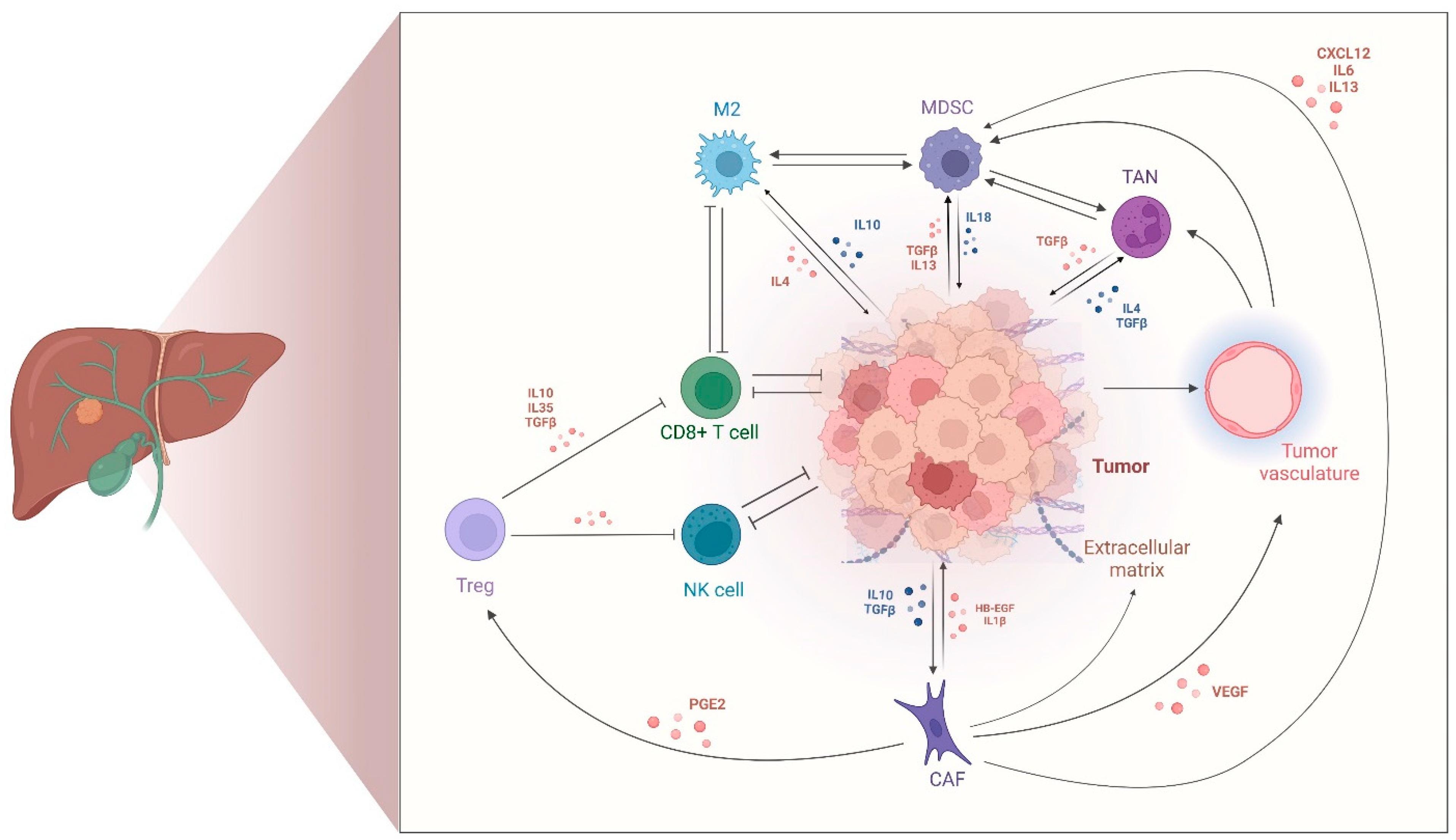
2.2. Tumor-Immune Microenvironment in CCA

The TIME has an integral role in tumor growth and progression in CCA as in other solid tumors [5]. CCAs are largely characterized by an immune-suppressive landscape resulting from the predominance of regulatory or dysfunctional immune populations, reduced infiltration by cytotoxic immune cells, and extensive desmoplasia. Immune profiling studies have described an intricate crosstalk between tumor and different TIME cell compartments, including myeloid-derived suppressor cells (MDSCs), tumor-associated macrophages (TAMs) and neutrophils (TANs), regulatory T cells (T regs), and CAFs that sustain these immune-suppressive programs in CCA [6,7,8,9,10,11,12,13]. This paragraph focuses on the innate and adaptive immune populations composing the TIME of CCAs, while CAFs are treated in greater detail in Section 2.3.
MDSCs and TAMs are abundant pro-tumorigenic and immune-suppressive populations in the TIME of CCAs [38,39]. MDSCs are organized into two major subsets of monocytic (M-MDSC) and polymorphonuclear (PMN) MDSCs, whereas TAMs have mostly a STAT3-mediated M2 polarization (CD163+) [40,41,42]. Recently, TANs have also been demonstrated to be a functionally relevant TIME cell type, with active contributions to cancer growth and immune escape in CCA [8,9]. Mostly derived from peripheral blood neutrophils, TANs are a highly plastic population recruited in the liver following chemokine release by tumor and resident immune and stromal cells. In CCAs, the abundance of all these immune-suppressive cell types has been correlated with tumorigenesis, disease progression, increased infiltration and differentiation of regulatory cells, and ultimately worse clinical outcomes [43,44,45,46]. However, the contribution and interdependency of each cell population in creating the poorly immunogenic TIME of CCAs remain largely unknown, posing major challenges to the implementation of therapeutic strategies that could overcome resistance to first-generation immunotherapies. These populations establish a series of delicately intertwined communications in the TIME of CCAs. For instance, TAMs and TANs were proven to have additive pro-tumor effects in vitro, and depletion of TAMs in mice did not improve survival due to a compensatory increase in MDSCs, requiring dual blockade of TAMs and MDSCs to obtain immune-sensitive CCA preclinical models [6,9].
The TIME of CCAs is also enriched with T regs supporting the immune-suppressive programs that are pathognomonic of the disease [10]. Under physiological conditions, the transcriptional factor forkhead box protein P3 (FOXP3) determines the specific functional phenotype of T regs [47,48]. These regulatory immune cells restrict the activity of antigen-presenting, natural killer (NK), and CD8+ cytotoxic T cells via multiple mechanisms. These include the release of anti-inflammatory molecules, such as interleukin (IL)-10, IL-35, and TGF-β1, the consumption of IL-2, and the expression of immune checkpoints. In CCA, it has been suggested that multiple oncogenic pathways, such as PI3K/Akt and Dickkopf-1 (DKK1)-Wnt, have pivotal roles in the recruitment and differentiation of T regs [49,50]. Further, a sc-RNA-seq study identified a phenotype of intratumoral T regs with enhanced immune-suppressive roles resulting from alterations in the network of transcription factors between tumor-infiltrating and peritumoral T cells [10].
Lastly, the spatial distribution of these populations further contributes to the TIME polarization and retains prognostic significance [51,52,53,54,55,56]. Immunohistochemical analyses of immune markers on 74 iCCA resected samples showed that T cell and immune checkpoint markers are mainly enriched at the tumor margins in CCA [57]. Here, their relative abundance and spatial organization exert distinct prognostic effects. In detail, tumor-infiltrating CD4+ or CD8+ T cells, along with a high density of B cells, particularly when clustered into tertiary lymphoid structures (TLSs), have been linked with more favorable survival, whereas FOXP3+ T reg cells and CD163+ macrophages with poorer patient outcomes [53,58,59,60]. By contrast, the clinical relevance of the quantification of regulatory immune checkpoints in CCA remains controversial. For instance, PD-L1, a renowned biomarker of response to immunotherapy in other solid tumors, has been reported in 9–70% of the cases in CCA but its association with patient-relevant outcomes remains unclear [61,62,63,64,65]. Figure 1 provides an overview of the main cell types composing the TIME of CCAs and their respective functional roles.

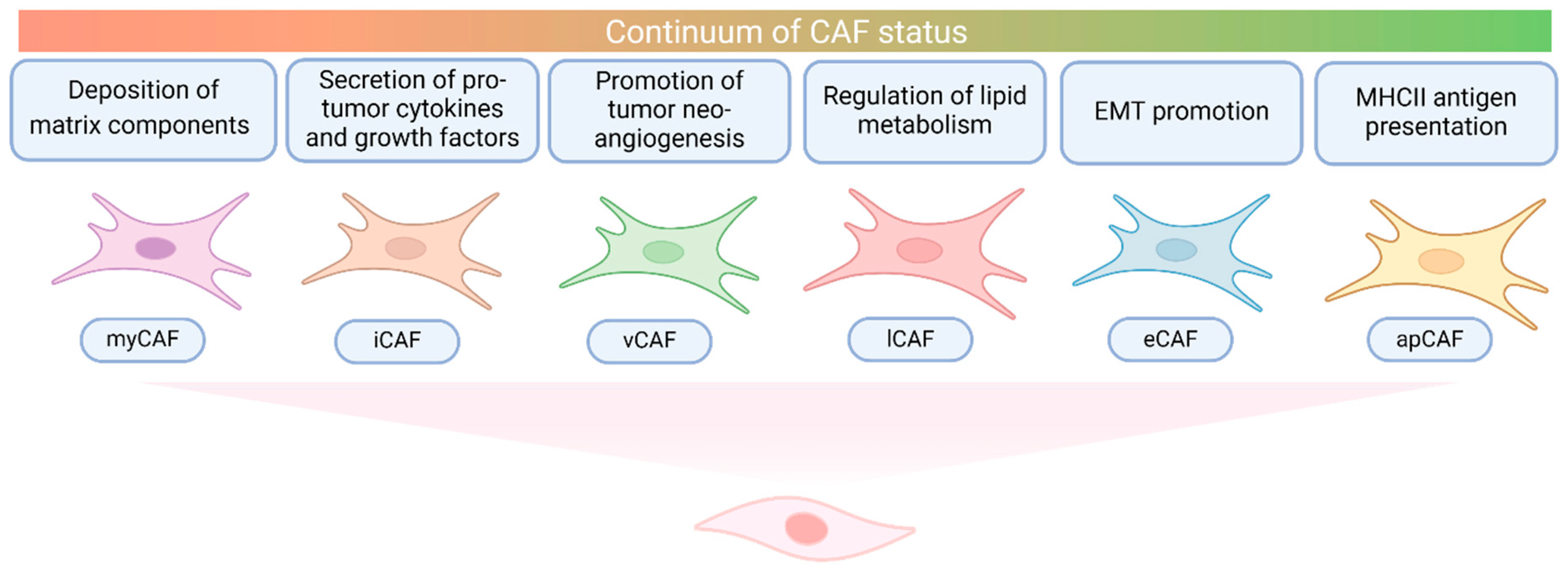
2.3. Cancer-Associated Fibroblasts in CCA

In desmoplastic tumors like CCA, CAFs are a well-represented population in the TIME. In iCCA, CAFs have been shown to originate mainly from hepatic stellate cells (HSCs) and portal fibroblasts, whereas their origin is less clear in eCCA [13,66]. Upon malignant transformation, these cells activate, proliferate, produce extracellular matrix (ECM), and assume a pro-inflammatory secretory profile [13,67]. As in other tumors, CAFs in CCAs have largely been identified by the expression of alpha-smooth muscle actin (α-SMA). However, collagen type I alpha-1 (COL1A1), vimentin, fibroblast activation protein (FAP), platelet-derived growth factor receptor-alpha (PDGFR-α), and platelet-derived growth factor receptor-beta (PDGFR-β) are also commonly expressed markers [68]. In recent years, studies with single-cell resolution have enabled the identification of a pan-CAF signature that recapitulates the different shades of CAFs in CCAs. Patients whose tumors have abundant CAFs have been found to have a worse prognosis [13,69].
CAFs have broadly recognized tumor-promoting roles in solid tumors, including CCAs, whereas their potential tumor-suppressive activity remains to be better defined. High-resolution studies have described multifaceted interactions of CAFs with the tumor, immune, and endothelial compartments of CCAs resulting in the activation of multiple pathways involved in tumor stiffness, tumorigenesis, immune suppression, and neo-angiogenesis [12,13].
A pivotal study in mice with validation in human cohorts revealed that CCAs have a heterogeneous and highly plastic population of CAFs [13]. The two predominant subtypes have been defined as myofibroblastic CAFs (myCAFs) due to the deposition of matrix components and inflammatory CAFs (iCAFs) due to the secretion of cytokines and growth factors. Although different analyses have adopted different classifications, there is an overall agreement about these two subtypes. Other clusters identified in CCAs include antigen-presenting CAFs (apCAFs) enriched in major histocompatibility complex II, vascular CAFs (vCAFs) involved in tumor neo-angiogenesis, epithelial to mesenchymal-like CAFs (eCAFs) expressing EMT markers, and lipofibroblasts (lCAFs) implicated in the regulation of lipid metabolism [12].
The deposition of ECM components is one of the most studied roles of CAFs and myCAFs are the key subpopulation with this function [12,13]. In CCAs, ECM proteins promote tumorigenesis and invasion via self-sustaining stroma-tumor interactions as increased tumor stiffness activates CAFs in return. The expression of many of these ECM components, such as laminin, osteopontin, and several matrix metalloproteinases (MMPs), has been associated with more advanced tumors and poor patient prognosis [14,70,71]. Notably, the fibrillar collagen COL1A1, which is one of the most abundant ECM proteins secreted during tumorigenesis, cooperates to increase tumor stiffness, alter TIME organization, and enhance MMP activity [13]. However, despite beliefs that COL1A1 would also contribute to tumor progression, this has not been confirmed in iCCA where this role has been reported for hyaluronic acid but not COL1A1.
Another recognized function of CAFs is the release of growth factors implicated in cancer growth and progression. For example, myCAFs produce heparin-binding epithelial growth factor (HB-EGF) which induces EGFR activation in CCA cells, ultimately promoting migration and invasiveness [72]. In return, on HB-EGF stimulation, CCA cells produce TGF-β1, which stimulates HB-EGF expression in myCAF, resulting in a constant positive feedback loop sustaining CCA progression. Moreover, the EGFR and TGF-β1 signaling pathways induce EMT transition and immune suppression in CCAs [73]. Another pro-tumorigenic growth factor produced by CAFs is the hepatocyte growth factor (HGF), which binds to c-MET on tumor cells and activates the downstream MAPK, extracellular signal-related kinase (ERK), and PI3K pathways [13,74]. PDGF-β, which activates the Hedgehog signaling (Hh) pathway, has a similar role [74,75]. CAFs also release chemokines and cytokines, such as C-X-C motif chemokine 12 (CXCL12) and interleukin (IL)-1β, that promote CCA proliferation, migration, and invasion through ERK1/2 and PI3K/Akt signaling cascades [76,77,78]. Notably, high expression of the CXCL12 binding receptor CXCR4 was correlated with features of aggressive disease and shorter survivals in patients with iCCA [79].
Furthermore, CAFs have immunomodulatory functions and emergent roles in resistance to immunotherapy in solid tumors, including CCAs [68,80,81,82]. CAF-secreted prostaglandin E2 (PGE2) and TGF-β contribute to the establishment of an immune-suppressive niche populated by abundant and activated T regs and scarce and dysfunctional effector cells [68]. For instance, myCAFs, which have been reported to have the highest expression of TGF-β ligands, were associated with a signature of resistance to immunotherapy (IPRES) in CCAs [82]. Through the release of IL-13, IL-6, CXCL12, and macrophage colony-stimulating factor, CAFs have also been shown to recruit the immune-suppressive populations of MDSCs and TAMs, and support M2 polarization [83,84]. The activation of STAT3 and the production of chemokine ligand 2 (CCL2) following FAP expression by CAFs enhances the recruitment of MDSCs. Further, the ECM compounds secreted by CAFs create a physical barrier to immune cell recruitment and may further favor the selection of immune-suppressive populations [85]. In pancreatic cancer, MMPs are chemotactic for leukocytes and COL1A1 increases MDSC accumulation and suppression of CD8+ T cells; this remains yet to be confirmed in CCA [86].
Additionally, CAFs contribute to tumor vascular and lymphatic neo-angiogenesis [87]. In CCAs, CAFs are a vast source of vascular endothelial growth factor (VEGF) in the tumor stroma. A sc-RNA-seq study highlighted a specific CD146+ vCAF subpopulation that is highly abundant in CCAs and fosters neo-angiogenesis through the activation of the IL-6/IL-6R axis among other pathways [12]. Notably, HSCs can also secrete pro-angiogenic factors, including PDGF, FGF, EGF, and angiopoietin-1 and -2, but their role in CCA neo-angiogenesis remains to be further explored [88].
Preliminary evidence has suggested CAF involvement in other tumor-promoting roles, including metabolic reprogramming, senescence, and dysfunctional protein post-translational modifications, which remain a matter of study in CCAs [68].
In summary, CAFs exert pro-tumorigenic and pro-inflammatory functions in the TIME of CCAs through a series of complementary mechanisms. While these functions may be preferentially executed by different CAF subpopulations, the existence of clusters with mixed features also suggests that CAF subpopulations are not strictly separate. In studies conducted in pancreatic tumors, the Janus Kinase/Signal Transducer and Activator of Transcription (JAK/STAT) signaling pathway may be involved in myCAF-iCAF plasticity programs [89]. Thus, CAFs possibly exist in a continuum of states adapting to tumor and TIME changes. However, as functional studies remain limited, the exact mechanisms driving these dynamic changes in CCA deserve further investigation. Figure 2 summarizes the main roles of CAFs in CCA.

2.4. Immune-Based Classifications of CCA

Immune checkpoint blockade has transformed cancer therapy, but its efficacy in unselected populations, including CCAs, remains limited [15,16]. Sensitivity to anti-PD-(L)1 antibodies is largely dependent on the pre-existing cellular makeup and functional state of the TIME. Thus, the advent of high-resolution techniques has provided an unprecedented opportunity to identify TIME phenotypes that predict responsiveness or resistance to these therapies [90].
Foundational studies of the immune infiltrate contexture in solid tumors are mainly based on low-to-moderate resolution analyses (e.g., bulk sequencing and immunostainings). These studies divide the TIME into three main classes, namely infiltrated–excluded (i.e., poorly immunogenic or immunologically “cold”), infiltrated–inflamed, and infiltrated–TLS [91]. These classes, which are likely to evolve along a spectrum of sub-statuses, are characterized by the organization of immune cells at the invasive tumor margin, inside the tumor area, or in aggregates that organize similarly to lymph nodes, respectively. Despite TIME classes assuming various names across different studies, their main composition and behavior are maintained, making this broad classification handy for interpreting existing evidence in CCA.
Using transcriptomics from a large cohort of iCCAs, a study identified four immune subclasses associated with distinct immune escape mechanisms and patient outcomes: immune-desert, myeloid, mesenchymal, and immunogenomic [92]. The immune-desert subset, characterized by low expression of TIME gene signatures, was the main subclass (45%) supporting the notion that CCA is an immunologically “cold” tumor. The myeloid and mesenchymal clusters were enriched with monocyte-derived and fibroblast gene signatures, respectively. Lastly, the immunogenomic cluster was the least represented (about 11% of CCAs). It featured infiltration by innate and adaptive immune cells with highly activated inflammatory and immune checkpoint pathways and was associated with prolonged survival.
Another recent study proposed a novel Stroma, Tumor and Immune Microenvironment (STIM) classification of iCCAs from the analysis of 122 samples with further validation in five independent datasets [82]. This classification identified five immune classes of iCCA, overall split between non-inflamed (65%) and inflamed (35%) tumors. The non-inflamed tumors comprised three major classes: a “desert-like” class (20%) with scarce immune cells but enriched in T regs, a “hepatic stem-like” class (35%) rich in M2 macrophages and tumors with IDH, BAP1, or FGFR2 alterations, and a “tumor classical” class (10%) characterized by the activation of cell cycle pathways and worse patient survivals. The inflamed tumors were further divided into an “immune classical” (10%) group, which was infiltrated by adaptive immune cells like CD8+ T cells, and an “inflammatory stroma” (25%) group, which presented extensive desmoplastic reaction, T cell exhaustion, and KRAS mutations.
Further, a comprehensive analysis using whole-exome, RNA, and T cell receptor (TCR) sequencing together with multiplexed immunofluorescence clustered iCCAs into sparsely, heterogeneously, and highly infiltrated subgroups with distinct immunogenomic features [93]. Sparsely infiltrated tumors had active copy-number loss of clonal neoantigens and heterogeneous immune infiltration which played an important role in the subclonal evolution across tumor regions. Highly infiltrated tumors presented with wide immune activation and a homogeneous TCR repertoire across tumor regions. However, these features were offset by T cell exhaustion and impaired antigen presentation. Interestingly, FGFR2 mutations and fusions correlated with low mutation burden and reduced immune infiltration. In keeping with this, other recent analyses exploring immune biomarkers in molecularly defined subgroups found low PD-L1 expression in the FGFR2-altered cluster [94,95].
Lastly, a Chinese study provided a four-tiered immune classification of iCCAs based on the spatial distribution and abundance of TLSs [96]. Overall, tumors sat on a continuum with edges represented by class I (28%) iCCAs with TLSs located in the peri-tumoral region and class IV (12%) iCCAs with TLSs mainly found in the tumor region. In between, classes II (31%) and III (28%) had heterogeneous distribution patterns. Of note, the maturation stage of TLSs also varied depending on their localization. Intra-tumoral TLSs had features of more mature lymphoid structures with germinal centers and more intact tumor–host immune response, and were associated with improved survival.
In summary, the integration of genomic, transcriptomic, and immunological data has enabled the identification of distinct patterns describing the TIME of CCAs, particularly in iCCAs. While there is no current consensus favoring one classification system over another, these approaches collectively highlight that CCAs predominantly harbor an immunosuppressive TIME. However, even though these tools may be helpful in stratifying patients into immunologically and prognostically different subtypes, their confinement to resected iCCAs and lack of prospective validation limit their implementation in routine patient care.

3. Targeting the TIME of Clinics

Immunotherapy leverages the intrinsic ability of the immune system to recognize and eliminate “non-self” entities, including tumors [97]. Having revolutionized the treatment of many cancers, immunotherapy also warranted investigation in CCA, where standard-of-care first-line chemotherapy with cisplatin and gemcitabine resulted in a dismal median OS below one year [98]. In keeping with the finding that CCA largely lacks cytotoxic immune infiltration, single-agent immune checkpoint inhibitors (ICIs), which rely on pre-existent anti-tumor immunity, showed a limited activity (overall response rate [ORR] 5.8–13%) for advanced CCA [99]. However, the efficacy improved by combining ICIs with chemotherapy, an approach used to establish a more proficient anti-tumor immune infiltrate. In fact, although cisplatin and gemcitabine, the cornerstone treatment for unresectable CCA, have immune-suppressive effects on peripheral blood cell counts, their cytotoxic activity in the tumor bed has been proven to be immunogenic, fostering antigen presentation, depleting the TIME of immune-suppressive populations, and promoting cytotoxic T cells infiltration [100,101]. In early-phase clinical trials testing ICIs with chemotherapy in patients with unresectable CCA, ORRs ranged from 36 to 72%, acknowledging differences in study design and dosing regimens [102,103,104].
The following global randomized phase 3 studies TOPAZ-1 and KEYNOTE-966 changed practice establishing combination treatment with anti-PD-(L)1 and chemotherapy as the recommended first-line choice for patients with advanced CCA [15,16]. The two studies had an overall similar design testing standard chemotherapy with cisplatin and gemcitabine combined with an anti-PD-(L)1 agent or placebo followed by a maintenance phase with the anti-PD-(L)1 or placebo alone in the TOPAZ-1 trial and the anti-PD-(L)1 or placebo combined with gemcitabine in the KEYNOTE-966 trial. Both trials met their primary endpoint, showing improved OS in the chemo-immunotherapy arm. Updated results from the TOPAZ-1 trial reported a median OS of 12.0 months (95% CI, 11.6–14.1) versus 11.3 months (95% CI, 10.1–12.5) and a 3-year OS rate of 14.6% (95% CI, 11.0–18.6%) versus 6.9% (95% CI, 4.5–10.0%) with cisplatin plus gemcitabine and the anti-PD-L1 antibody durvalumab compared to chemotherapy-placebo [65]. Similarly, pembrolizumab-chemotherapy achieved longer median OS (12.7 months, 95% CI, 11.5–13.6) versus chemotherapy-placebo (10.9 months, 95% CI, 9.9–11.6) in the KEYNOTE-966 trial, with a maintained benefit after 3 years of follow-up [16,105]. Reassuringly, the performance of chemo-immunotherapy in real-world registries mirrored trial results [106]. To better reflect global treatment patterns and confirm reproducibility in broader patient populations, the efficacy of durvalumab combined with various gemcitabine-based regimens and its use in patients with Eastern Cooperative Oncology Group (ECOG) Performance Status (PS) 2, a group excluded from the registrational trials, is being further investigated in phase 3b studies (NCT05771480, NCT05924880). While validating the role of ICIs in the treatment of CCA, these studies also showed that the added value of these agents was rather modest in an unselected population, fostering the exploration of novel avenues to accomplish the challenging task of targeting the TIME of CCAs.
One such venture has focused on the development of strategies targeting multiple immune checkpoint inhibitors at a time rather than the PD-1/PD-L1 axis alone. Several studies of novel agents targeting PD-(L)1 together with Cytotoxic T-Lymphocyte Antigen 4 (CTLA-4), T cell immunoreceptor with Ig and ITIM domains (TIGIT), or Lymphocyte-activation gene 3 (LAG-3), among others, are ongoing. An example is the phase 2 GEMINI-Hepatobiliary (NCT05775159) trial evaluating volrustomig, an anti-PD-1/CTLA-4 bispecific antibody, or rilvegostomig, an anti-PD-1/TIGIT bispecific antibody, in combination with cisplatin and gemcitabine as front-line treatment in advanced CCA. Of note, rilvegostomig is also being developed in combination with triplet chemotherapy regimens (NCT06569225) or with the antibody-drug conjugate (ADC) AZD8205 targeting B7-H4, a downregulator of T cell function that has been found overexpressed in CCA (NCT05123482). Another negative regulator of the immune response being exploited is lectin-like transcript 1 (LLT1), which reduces NK and cytotoxic T cell activation via CD161 and has been found at high levels in the TIME of renowned immunologically cold tumors like CCAs [107]. A dose-escalation phase 1 trial is studying ZM008, a first-in-class anti-LLT1 antibody, as a single agent followed by combination with pembrolizumab in patients with advanced solid tumors, including CCA (NCT06451497). Boosting the adaptive immune response while orthogonally targeting tumor-associated antigens is a complementary approach. In this space, MDX2001 is a tetraspecific antibody engaging with TROP2 and c-MET on tumor cells and CD3 and CD28 on cytotoxic T cells, improving their activation and proliferation. A phase 1/2a multicenter trial is investigating this molecule in CCA among other solid tumors (NCT06239194). TROP2 and c-MET are also being targeted individually by ADC in early-phase trials open to patients with CCAs (NCT06084481). Additional efforts are developing the use of adoptive T cell therapies. Chimeric antigen receptor (CAR)-T cells targeting CD133 [108] or anti-mucin 1 (MUC1) [109], both highly expressed antigens in CCA, are in preclinical stages, while CAR-T treatments against mesothelin (NCT06256055) and CEA have entered early clinical testing (NCT06043466).
Other paths include targeting tumor angiogenesis, which may represent a strategy to tackle the abundant tumor stroma of CCAs and CAFs, given they largely contribute to VEGF production [87]. While tyrosine-kinase inhibitors (TKIs) [110] and anti-VEGF(R-2) antibodies with [111,112] or without anti-PD-(L)1 [113,114] treatments had proven limited efficacy in an unselected population with refractory CCA, novel studies are evaluating the concomitant dual blockade of VEGF/PD-1 (NCT06529718) and VEGF-A/DLL4 (NCT04492033, NCT05506943) with bispecific antibodies. Both agents are being planned for further study in combination with first-line chemotherapy with (NCT06548412) or without durvalumab (NCT06591520). Lastly, other strategies are testing therapies targeting genomic vulnerabilities deemed to cooperate with the tumor immune escape. Among the known actionable alterations, both IDH1 mutations and FGFR2 aberrations have been associated with an immune-suppressive TIME [82] and studies are ongoing to establish if the addition of an anti-PD-(L)1 agent to the IDH1 inhibitor ivosidenib (NCT06501625, NCT05921760) and the FGFR2 inhibitor pemigatinib (NCT06530823) could revert this effect and improve outcomes in refractory CCA. An emergent target is MDM2, which inactivates TP53 under physiologic conditions. MDM2 amplifications have been found in 5–16% of CCAs, particularly in ampullary cancers, and are generally mutually exclusive with the other actionable aberrations [115]. A pooled analysis of two phase 1a/1b trials testing brigimadlin (BI 907828), a highly potent, oral MDM2TP53 antagonist, with or without the PD-1 inhibitor ezabenlimab and the anti-LAG-3 BI 754111, revealed an encouraging ORR of 50% in patients with refractory MDM2-amplified CCA [116]. The subsequent phase 2a/2b Brightline-2 trial (NCT05512377) is ongoing to further study the safety and efficacy of BI 907828 monotherapy in patients with locally advanced or metastatic MDM2-amplified, TP53 wild-type CCA.
Importantly, the establishment of ICIs in advanced CCA has created opportunities to test these agents in earlier disease stages, where outcomes are severely impacted by high relapse rates in patients who undergo curative-intent surgery and by the access to the same limited palliative therapies available for patients with metastatic disease in those with unresectable primary tumors. For patients with resected disease, the multicenter, randomized, double-blind, placebo-controlled phase 3 ARTEMIDE-Biliary01 trial (NCT06109779) is assessing the efficacy of the anti-PD-1/TIGIT bispecific antibody rilvegostomig compared to investigator’s choice of adjuvant chemotherapy. Other ongoing efforts include testing the combination of the anti-PD-L1 tislelizumab and TKI levantinib with capecitabine, the standard backbone adjuvant treatment (NCT05254847). The interim results of this Asian phase 2 study reported a good safety profile and a median disease-free survival (DFS) of 20.23 months (95% CI, 10.84–29.62), while OS is not yet mature [117]. Another open-label, multicenter, phase 2 study (NCT05239169) is currently exploring the association of the anti-CTLA-4 tremelimumab plus durvalumab with or without capecitabine. In the neoadjuvant setting, the phase 2 DEBATE trial (NCT04308174) randomized patients with localized CCA to receive 2:1 neoadjuvant cisplatin and gemcitabine with or without durvalumab, followed by six cycles of durvalumab after surgery. Notably, patients in the chemoimmunotherapy arm achieved almost doubled rates of curative-intent surgery (61% versus 36%) and prolonged progression-free survival (15.1 versus 3.6 months) [118]. Table 1 provides a snapshot of the clinical trials ongoing or planned with immunotherapies in CCA.

4. Conclusions and Future Directions

The integration of immunotherapy into the treatment landscape of CCA has marked a significant advancement in a disease with traditionally poor outcomes. The use of ICIs combined with chemotherapy has established a new standard of care for patients with advanced and unresectable CCA, demonstrating OS improvements and opening the door to the exploration of such a strategy in earlier disease settings. Biomarker analyses from early-phase studies revealed associations between response to ICIs and high baseline tumor-infiltrating lymphocytes (TILs) or early on-treatment increase in PD-L1 expression, particularly on immune rather than tumor cells, supporting the concept that immune infiltration is also pivotal for response to immunotherapy in CCA [103,119]. However, baseline PD-L1 expression failed to capture long-term clinical benefits in the larger, registrational-intent phase 3 trials [16,65], making chemoimmunotherapy the currently recommended front-line treatment in all-comers [120]. Given the overall benefit from these combinations remains modest, there is a pressing need to identify robust biomarkers of response and resistance and additional therapeutic targets.
Translational research in CCA has largely suffered from the rarity of the disease and often limited accessibility of tumor lesions, resulting in inadequate quantity and quality of the tissue harvested, particularly from patients with unresectable disease. Thus, most of the studies conducted relied on surgical specimens from primary resected tumors, especially from iCCAs, limiting their generalizability to the broader spectrum of CCAs across sites and stages. While more recent technologies have allowed us to take a more comprehensive look at CCA integrating genomic, transcriptomic, and image analyses, essential work is ongoing to develop tissue-free platforms [121,122,123]. Tumor profiling at this unprecedented resolution led to the identification of distinct molecular phenotypes of CCA beyond conventional anatomic classifications. Additionally, numerous multidimensional CCA datasets with matched genomic and clinical information have been made available on open-source platforms, providing the broader research community with opportunities for further analysis [124]. However, the genomic and immune CCA clusters identified, even those with some utility for patient risk stratification, remain primarily confined to research settings, due to the lack of reproducibility in a routine clinical scenario.
Nonetheless, these studies had the value of showing that druggable genetic alterations are found in a relevant proportion of patients with advanced CCA, allowing the development of targeted treatments and the implementation of tumor profiling as a guideline-recommended clinical practice [120]. Moreover, these studies offered an in-depth characterization of the CCA TIME and demonstrated its crucial role in tumor progression and resistance to therapies, particularly to ICIs. Hosting abundant CAFs, T regs, TAMs, MDSCs, and TANs, among other pro-tumor cell types, the CCA TIME is largely unbalanced toward immune suppression. The intricate, interdependent pro-tumor networks formed by these populations ultimately limit the effectiveness of first-generation immunotherapies. This may also explain why conventional tissue biomarkers of response to ICIs, such as PD-L1 expression, have little to no predictive value in CCA and are therefore not recommended for selecting patients for chemoimmunotherapy in clinical practice. The intrinsically immune tolerogenic liver niche may favor the establishment of a multitude of tumor escape mechanisms beyond the upregulation of the PD-(L)1 axis requiring the study of tumor-specific biomarkers and therapeutic strategies in CCAs.
To address these barriers, drug development efforts are focusing on novel immunomodulatory strategies to enhance cytotoxic immune cell infiltration and activation. Promising approaches involve the simultaneous targeting of multiple immune checkpoints or newly identified molecules with immune-suppressive roles such as LLT1. Others include the synthetical induction of a more proficient anti-tumor immunity, such as with bispecific antibodies rescuing activated cytotoxic T cells or with adoptive cell therapy such as CAR-T cells. Tumor angiogenesis and targetable genomic vulnerabilities are also being exploited as additional drivers of immune evasion with or without immune checkpoint inhibition.
Looking forward, favoring enrolment in clinical trials, establishing international collaborative consortia, and standardizing diagnostic tumor profiling procedures would be key to helping translate basic discoveries into clinical applications. Some of these ambitious programs are the pan-European ENS-CCA and COST Action Precision-BTC-Network which aim to improve the understanding and management of CCA through interdisciplinary collaborations [125,126]. With continued research, the hope is that biomarker-informed and tailored immunotherapies could change the storyline of CCA into an increasingly manageable disease.

Author Contributions

Conceptualization, A.C., L.R. and A.L.; methodology, A.C., L.R. and A.L.; resources, A.C., R.B., T.P., S.B., L.R. and A.L.; data curation, A.C., R.B., T.P., S.B., L.R. and A.L.; writing—original draft preparation, A.C., R.B., T.P. and S.B.; writing—review and editing, A.C., R.B., T.P., S.B., L.R. and A.L.; visualization, A.C. and R.B.; supervision, L.R. and A.L.; project administration, A.C., L.R. and A.L. All authors have read and agreed to the published version of the manuscript.

Funding

This research received no external funding.

Data Availability Statement

All the data presented within the manuscript is available online through the given references.

Conflicts of Interest

A.C. is supported by an AIRC Foundation for Cancer Research post-doctoral fellowship. R.B. reports lecture fees from AstraZeneca; travel expenses from Roche. T.P. reports consulting fees from Bayer, Ipsen and AstraZeneca; institutional research funding from Roche, Bayer, Astra Zeneca; travel expenses from Roche. A.L. reports consulting fees from Advanz Pharma, AlfaSigma, Takeda and Albireo Pharma, and speaker fees from Gilead, AbbVie, MSD, Intercept Pharma, AlfaSigma, GSK and Incyte. LR reports consulting fees from AbbVie, AstraZeneca, Basilea, Bayer, BMS, Eisai, Elevar Therapeutics, Exelixis, Genenta, Hengrui, Incyte, Ipsen, IQVIA, Jazz Pharmaceuticals, MSD, Nerviano Medical Sciences, Roche, Servier, Taiho Oncology, Zymeworks; lecture fees from AstraZeneca, Bayer, BMS, Guerbet, Incyte, Ipsen, Roche, Servier; travel expenses from AstraZeneca; Institutional research funding from AbbVie, Agios, AstraZeneca, BeiGene, Eisai, Exelixis, Fibrogen, Incyte, Ipsen, Jazz Pharmaceuticals, Lilly, MSD, Nerviano Medical Sciences, Roche, Servier, Taiho Oncology, TransThera Sciences, Zymeworks. S.B. declares no conflicts of interest.

Abbreviations

2HG, 2-hydroxyglutarate; ADC, antibody-drug conjugate; BAP1, BRCA1 associated protein-1; BRAF, B-Raf proto-oncogene serine/threonine kinase; CAFs, cancer-associated fibroblasts; CAR-T, Chimeric antigen receptor-T; CCA, cholangiocarcinoma; CCL2, chemokine ligand 2; CCND1, Cyclin D1; CNAs, copy number aberrations; COL1A1, collagen type I alpha-1; CTLA-4, Cytotoxic T-Lymphocyte Antigen 4; CXCL12, C-X-C motif chemokine 12; DFS, disease-free survival; ECM, extracellular matrix; ECOG PS, Eastern Cooperative Oncology Group Performance Status; EMT, epithelial-to-mesenchymal transition; ERK, extracellular signal-related kinase; FAP, fibroblast activation protein, PDGFR-α/β, platelet-derived growth factor receptor-alpha/beta; FGFR2, fibroblast growth factor receptors 2; FOXP3, forkhead box protein P3; GBC, gallbladder cancer; HB-EGF, epithelial growth factor; HGF, hepatocyte growth factor; HSC, hepatic stellate cell; Hh, Hedgehog signaling pathway; ICIs, immune checkpoint inhibitors; IDH, isocitrate dehydrogenase; IL-1β, interleukin-1beta; JAK/STAT, Janus Kinase/Signal Transducer and Activator of Transcription; LAG-3, Lymphocyte-activation gene 3; LLT1, lectin-like transcript 1; M-MDSCs, monocytic myeloid-derived suppressor cells; MASH, metabolic dysfunction-associated steatohepatitis; MASLD, metabolic dysfunction-associated steatotic liver disease; MDSCs, myeloid-derived suppressor cells; MET, mesenchymal-epithelial transition factor; MMPs, metalloproteinases; MSI-H, microsatellite instability; MUC1, mucin 1; NK, natural killer; NOTCH, neurogenic locus notch homolog protein 1; NTRK, Neurotrophic tyrosine receptor kinase; ORR, overall response rate; OS, overall survival; PBRM1, Protein polybromo-1; PD-(L)1, programmed death-(ligand)-1; PGE2, prostaglandin E2; PI3K/mTOR, phosphatidylinositol-3-kinase-mammalian target of rapamycin; PMN-MDSCs, polymorphonuclear myeloid-derived suppressor cells; RAS/MAPK, Rat sarcoma-mitogen-activated protein kinase; STAT3, Signal Transducer and Activator of Transcription 3; T regs, regulatory T cells; TAMs, tumor-associated macrophages; TANs, tumor-associated neutrophils; TCR, T cell receptor; TGF-β, tumor growth factor beta; TIGIT, T cell immunoreceptor with Ig and ITIM domains; TILs, tumor-infiltrating lymphocytes; TIME, tumor-immune microenvironment; TKIs, tyrosine-kinase inhibitors; TLS, tertiary lymphoid structure; VEGF(R), vascular endothelial growth factor (receptor); apCAFs, antigen-presenting cancer-associated fibroblasts; dCCA, distal cholangiocarcinoma; eCAFs, epithelial to mesenchymal-like cancer-associated fibroblasts; eCCA, extrahepatic cholangiocarcinoma; iCAFs, inflammatory cancer-associated fibroblasts; iCCA, intrahepatic cholangiocarcinoma; lCAFs, lipofibroblasts; myCAF, myofibroblastic cancer-associated fibroblasts; pCCA, perihilar cholangiocarcinoma; vCAFs, vascular cancer-associated fibroblasts; α-SMA, alpha-smooth muscle actin.

References

  1. Banales, J.M.; Marin, J.J.G.; Lamarca, A.; Rodrigues, P.M.; Khan, S.A.; Roberts, L.R.; Cardinale, V.; Carpino, G.; Andersen, J.B.; Braconi, C.; et al. Cholangiocarcinoma 2020: The next horizon in mechanisms and management. Nat. Rev. Gastroenterol. Hepatol. 2020, 17, 557–588. [Google Scholar] [CrossRef]
  2. Valle, J.W.; Kelley, R.K.; Nervi, B.; Oh, D.-Y.; Zhu, A.X. Biliary tract cancer. Lancet 2021, 397, 428–444. [Google Scholar] [CrossRef]
  3. Welzel, T.M.; Graubard, B.I.; Zeuzem, S.; El-Serag, H.B.; Davila, J.A.; McGlynn, K.A. Metabolic syndrome increases the risk of primary liver cancer in the United States: A study in the SEER-Medicare database. Hepatology 2011, 54, 463–471. [Google Scholar] [CrossRef]
  4. Michelotti, G.A.; Machado, M.V.; Diehl, A.M. NAFLD, NASH and liver cancer. Nat. Rev. Gastroenterol. Hepatol. 2013, 10, 656–665. [Google Scholar] [CrossRef]
  5. Greten, T.F.; Schwabe, R.; Bardeesy, N.; Ma, L.; Goyal, L.; Kelley, R.K.; Wang, X.W. Immunology and immunotherapy of cholangiocarcinoma. Nat. Rev. Gastroenterol. Hepatol. 2023, 20, 349–365. [Google Scholar] [CrossRef]
  6. Loeuillard, E.; Yang, J.; Buckarma, E.; Wang, J.; Liu, Y.; Conboy, C.; Pavelko, K.D.; Li, Y.; O’Brien, D.; Wang, C.; et al. Targeting tumor-associated macrophages and granulocytic myeloid-derived suppressor cells augments PD-1 blockade in cholangiocarcinoma. J. Clin. Investig. 2020, 130, 5380–5396. [Google Scholar] [CrossRef] [PubMed]
  7. Ma, C.; Zhang, Q.; Greten, T.F. MDSCs in liver cancer: A critical tumor-promoting player and a potential therapeutic target. Cell. Immunol. 2021, 361, 104295. [Google Scholar] [CrossRef] [PubMed]
  8. Xue, R.; Zhang, Q.; Cao, Q.; Kong, R.; Xiang, X.; Liu, H.; Feng, M.; Wang, F.; Cheng, J.; Li, Z.; et al. Liver tumour immune microenvironment subtypes and neutrophil heterogeneity. Nature 2022, 612, 141–147. [Google Scholar] [CrossRef]
  9. Zhou, Z.; Wang, P.; Sun, R.; Li, J.; Hu, Z.; Xin, H.; Luo, C.; Zhou, J.; Fan, J.; Zhou, S. Tumor-associated neutrophils and macrophages interaction contributes to intrahepatic cholangiocarcinoma progression by activating STAT3. J. Immunother. Cancer 2021, 9, e001946. [Google Scholar] [CrossRef]
  10. Alvisi, G.; Termanini, A.; Soldani, C.; Portale, F.; Carriero, R.; Pilipow, K.; Costa, G.; Polidoro, M.; Franceschini, B.; Malenica, I.; et al. Multimodal single-cell profiling of intrahepatic cholangiocarcinoma defines hyperactivated Tregs as a potential therapeutic target. J. Hepatol. 2022, 77, 1359–1372. [Google Scholar] [CrossRef]
  11. Bao, X.; Li, Q.; Chen, J.; Chen, D.; Ye, C.; Dai, X.; Wang, Y.; Li, X.; Rong, X.; Cheng, F.; et al. Molecular Subgroups of Intrahepatic Cholangiocarcinoma Discovered by Single-Cell RNA Sequencing-Assisted Multiomics Analysis. Cancer Immunol. Res. 2022, 10, 811–828. [Google Scholar] [CrossRef] [PubMed]
  12. Zhang, M.; Yang, H.; Wan, L.; Wang, Z.; Wang, H.; Ge, C.; Liu, Y.; Hao, Y.; Zhang, D.; Shi, G.; et al. Single-cell transcriptomic architecture and intercellular crosstalk of human intrahepatic cholangiocarcinoma. J. Hepatol. 2020, 73, 1118–1130. [Google Scholar] [CrossRef]
  13. Affo, S.; Nair, A.; Brundu, F.; Ravichandra, A.; Bhattacharjee, S.; Matsuda, M.; Chin, L.; Filliol, A.; Wen, W.; Song, X.; et al. Promotion of cholangiocarcinoma growth by diverse cancer-associated fibroblast subpopulations. Cancer Cell 2021, 39, 866–882.e11. [Google Scholar] [CrossRef]
  14. Sulpice, L.; Rayar, M.; Desille, M.; Turlin, B.; Fautrel, A.; Boucher, E.; Llamas-Gutierrez, F.; Meunier, B.; Boudjema, K.; Clément, B.; et al. Molecular profiling of stroma identifies osteopontin as an independent predictor of poor prognosis in intrahepatic cholangiocarcinoma. Hepatology 2013, 58, 1992–2000. [Google Scholar] [CrossRef] [PubMed]
  15. Oh, D.-Y.; He, A.R.; Qin, S.; Chen, L.T.; Okusaka, T.; Vogel, A.; Kim, J.W.; Suksombooncharoen, T.; Ah Lee, M.; Kitano, M.; et al. Durvalumab plus Gemcitabine and Cisplatin in Advanced Biliary Tract Cancer. NEJM Evid. 2022, 1, EVIDoa2200015. [Google Scholar] [CrossRef]
  16. Kelley, R.K.; Ueno, M.; Finn, R.S.; Furuse, J.; Ren, Z.; Yau, T.; Klümpen, H.J.; Chan, S.L.; Ozaka, M.; Verslype, C.; et al. Pembrolizumab in combination with gemcitabine and cisplatin compared with gemcitabine and cisplatin alone for patients with advanced biliary tract cancer (KEYNOTE-966): A randomised, double-blind, placebo-controlled, phase 3 trial. Lancet 2023, 401, 1853–1865. [Google Scholar] [CrossRef]
  17. Verlingue, L.; Malka, D.; Allorant, A.; Massard, C.; Ferté, C.; Lacroix, L.; Rouleau, E.; Auger, N.; Ngo, M.; Nicotra, C.; et al. Precision medicine for patients with advanced biliary tract cancers: An effective strategy within the prospective MOSCATO-01 trial. Eur. J. Cancer 2017, 87, 122–130. [Google Scholar] [CrossRef] [PubMed]
  18. Abou-Alfa, G.K.; Sahai, V.; Hollebecque, A.; Vaccaro, G.; Melisi, D.; Al-Rajabi, R.; Paulson, A.S.; Borad, M.J.; Gallinson, D.; Murphy, A.G.; et al. Pemigatinib for previously treated, locally advanced or metastatic cholangiocarcinoma: A multicentre, open-label, phase 2 study. Lancet Oncol. 2020, 21, 671–684. [Google Scholar] [CrossRef]
  19. Goyal, L.; Meric-Bernstam, F.; Hollebecque, A.; Valle, J.W.; Morizane, C.; Karasic, T.B.; Abrams, T.A.; Furuse, J.; Kelley, R.K.; Cassier, P.A.; et al. Futibatinib for FGFR2-Rearranged Intrahepatic Cholangiocarcinoma. N. Engl. J. Med. 2023, 388, 228–239. [Google Scholar] [CrossRef] [PubMed]
  20. Abou-Alfa, G.K.; Macarulla, T.; Javle, M.M.; Kelley, R.K.; Lubner, S.J.; Adeva, J.; Cleary, J.M.; Catenacci, D.V.; Borad, M.J.; Bridgewater, J.; et al. Ivosidenib in IDH1-mutant, chemotherapy-refractory cholangiocarcinoma (ClarIDHy): A multicentre, randomised, double-blind, placebo-controlled, phase 3 study. Lancet Oncol. 2020, 21, 796–807. [Google Scholar] [CrossRef] [PubMed]
  21. Javle, M.; Borad, M.J.; Azad, N.S.; Kurzrock, R.; Abou-Alfa, G.K.; George, B.; Hainsworth, J.; Meric-Bernstam, F.; Swanton, C.; Sweeney, C.J.; et al. Pertuzumab and trastuzumab for HER2-positive, metastatic biliary tract cancer (MyPathway): A multicentre, open-label, phase 2a, multiple basket study. Lancet Oncol. 2021, 22, 1290–1300. [Google Scholar] [CrossRef]
  22. Subbiah, V.; Lassen, U.; Élez, E.; Italiano, A.; Curigliano, G.; Javle, M.; de Braud, F.; Prager, G.W.; Greil, R.; Stein, A.; et al. Dabrafenib plus trametinib in patients with BRAF(V600E)-mutated biliary tract cancer (ROAR): A phase 2, open-label, single-arm, multicentre basket trial. Lancet Oncol. 2020, 21, 1234–1243. [Google Scholar] [CrossRef]
  23. Farshidfar, F.; Zheng, S.; Gingras, M.-C.; Newton, Y.; Shih, J.; Robertson, A.G.; Hinoue, T.; Hoadley, K.A.; Gibb, E.A.; Roszik, J.; et al. Integrative Genomic Analysis of Cholangiocarcinoma Identifies Distinct IDH-Mutant Molecular Profiles. Cell Rep. 2017, 18, 2780–2794. [Google Scholar] [CrossRef]
  24. Churi, C.R.; Shroff, R.; Wang, Y.; Rashid, A.; Kang, H.C.; Weatherly, J.; Zuo, M.; Zinner, R.; Hong, D.; Meric-Bernstam, F.; et al. Mutation profiling in cholangiocarcinoma: Prognostic and therapeutic implications. PLoS ONE. 2014, 9, e115383. [Google Scholar] [CrossRef]
  25. Jusakul, A.; Cutcutache, I.; Yong, C.H.; Lim, J.Q.; Huang, M.N.; Padmanabhan, N.; Nellore, V.; Kongpetch, S.; Ng, A.W.T.; Ng, L.M.; et al. Whole-Genome and Epigenomic Landscapes of Etiologically Distinct Subtypes of Cholangiocarcinoma. Cancer Discov. 2017, 7, 1116–1135. [Google Scholar] [CrossRef] [PubMed]
  26. Zhang, Y.; Ma, Z.; Li, C.; Wang, C.; Jiang, W.; Chang, J.; Han, S.; Lu, Z.; Shao, Z.; Wang, Y.; et al. The genomic landscape of cholangiocarcinoma reveals the disruption of post-transcriptional modifiers. Nat. Commun. 2022, 13, 3061. [Google Scholar] [CrossRef] [PubMed]
  27. Wardell, C.P.; Fujita, M.; Yamada, T.; Simbolo, M.; Fassan, M.; Karlic, R.; Polak, P.; Kim, J.; Hatanaka, Y.; Maejima, K.; et al. Genomic characterization of biliary tract cancers identifies driver genes and predisposing mutations. J. Hepatol. 2018, 68, 959–969. [Google Scholar] [CrossRef]
  28. Quinn, L.M.; Haldenby, S.; Antzcak, P.; Fowler, A.; Bullock, K.; Kenny, J.; Gilbert, T.; Andrews, T.; Diaz-Nieto, R.; Fenwick, S.; et al. Genomic profiling of idiopathic peri-hilar cholangiocarcinoma reveals new targets and mutational pathways. Sci. Rep. 2023, 13, 6681. [Google Scholar] [CrossRef]
  29. Ong, C.K.; Subimerb, C.; Pairojkul, C.; Wongkham, S.; Cutcutache, I.; Yu, W.; McPherson, J.R.; Allen, G.E.; Ng, C.C.; Wong, B.H.; et al. Exome sequencing of liver fluke-associated cholangiocarcinoma. Nat. Genet. 2012, 44, 690–693. [Google Scholar] [CrossRef]
  30. Zou, S.; Li, J.; Zhou, H.; Hou, Y.R.; Liu, K.X.; Sun, R.Q.; Wang, P.C.; Luo, C.B.; Li, J.; Zou, J.X.; et al. Mutational landscape of intrahepatic cholangiocarcinoma. Nat. Commun. 2014, 5, 5696. [Google Scholar] [CrossRef]
  31. Andersen, J.B.; Spee, B.; Blechacz, B.R.; Avital, I.; Komuta, M.; Barbour, A.; Conner, E.A.; Gillen, M.C.; Roskams, T.; Roberts, L.R.; et al. Genomic and genetic characterization of cholangiocarcinoma identifies therapeutic targets for tyrosine kinase inhibitors. Gastroenterology 2012, 142, 1021–1031.e15. [Google Scholar] [CrossRef] [PubMed]
  32. Sia, D.; Hoshida, Y.; Villanueva, A.; Roayaie, S.; Ferrer, J.; Tabak, B.; Peix, J.; Sole, M.; Tovar, V.; Alsinet, C.; et al. Integrative molecular analysis of intrahepatic cholangiocarcinoma reveals 2 classes that have different outcomes. Gastroenterology 2013, 144, 829–840. [Google Scholar] [CrossRef]
  33. Montal, R.; Sia, D.; Montironi, C.; Leow, W.Q.; Esteban-Fabró, R.; Pinyol, R.; Torres-Martin, M.; Bassaganyas, L.; Moeini, A.; Peix, J.; et al. Molecular classification and therapeutic targets in extrahepatic cholangiocarcinoma. J. Hepatol. 2020, 73, 315–327. [Google Scholar] [CrossRef] [PubMed]
  34. Lowery, M.A.; Ptashkin, R.; Jordan, E.; Berger, M.F.; Zehir, A.; Capanu, M.; Kemeny, N.E.; O’Reilly, E.M.; El-Dika, I.; Jarnagin, W.R.; et al. Comprehensive Molecular Profiling of Intrahepatic and Extrahepatic Cholangiocarcinomas: Potential Targets for Intervention. Clin. Cancer Res. 2018, 24, 4154–4161. [Google Scholar] [CrossRef] [PubMed]
  35. Silverman, I.M.; Murugesan, K.; Lihou, C.F.; Féliz, L.; Frampton, G.M.; Newton, R.C.; Tada, H.; Albacker, L.A.; Burn, T.C. Comprehensive genomic profiling in FIGHT-202 reveals the landscape of actionable alterations in advanced cholangiocarcinoma. J. Clin. Oncol. 2019, 37 (Suppl. S15), 4080. [Google Scholar] [CrossRef]
  36. Javle, M.; Bekaii-Saab, T.; Jain, A.; Wang, Y.; Kelley, R.K.; Wang, K.; Kang, H.C.; Catenacci, D.; Ali, S.; Krishnan, S.; et al. Biliary cancer: Utility of next-generation sequencing for clinical management. Cancer 2016, 122, 3838–3847. [Google Scholar] [CrossRef] [PubMed]
  37. Abou-Alfa, G.K.; Bibeau, K.; Schultz, N.; Yaqubie, A.; Millang, B.; Ren, H.; Féliz, L. Effect of FGFR2 Alterations on Overall and Progression-Free Survival in Patients Receiving Systemic Therapy for Intrahepatic Cholangiocarcinoma. Target Oncol. 2022, 17, 517–527. [Google Scholar] [CrossRef] [PubMed]
  38. Noy, R.; Pollard, J.W. Tumor-associated macrophages: From mechanisms to therapy. Immunity 2014, 41, 49–61. [Google Scholar] [CrossRef] [PubMed]
  39. Veglia, F.; Perego, M.; Gabrilovich, D. Myeloid-derived suppressor cells coming of age. Nat. Immunol. 2018, 19, 108–119. [Google Scholar] [CrossRef]
  40. Subimerb, C.; Pinlaor, S.; Khuntikeo, N.; Leelayuwat, C.; Morris, A.; McGrath, M.S.; Wongkham, S. Tissue invasive macrophage density is correlated with prognosis in cholangiocarcinoma. Mol. Med. Rep. 2010, 3, 597–605. [Google Scholar] [CrossRef]
  41. Charbel, A.; Tavernar, L.; Albrecht, T.; Brinkmann, F.; Verheij, J.; Roos, E.; Vogel, M.N.; Köhler, B.; Springfeld, C.; Brobeil, A.; et al. Spatiotemporal analysis of tumour-infiltrating immune cells in biliary carcinogenesis. Br. J. Cancer 2022, 127, 1603–1614. [Google Scholar] [CrossRef] [PubMed]
  42. Sun, D.; Luo, T.; Dong, P.; Brinkmann, F.; Verheij, J.; Roos, E.; Vogel, M.N.; Köhler, B.; Springfeld, C.; Brobeil, A.; et al. M2-polarized tumor-associated macrophages promote epithelial-mesenchymal transition via activation of the AKT3/PRAS40 signaling pathway in intrahepatic cholangiocarcinoma. J. Cell Biochem. 2020, 121, 2828–2838. [Google Scholar] [CrossRef]
  43. Hasita, H.; Komohara, Y.; Okabe, H.; Masuda, T.; Ohnishi, K.; Lei, X.F.; Beppu, T.; Baba, H.; Takeya, M. Significance of alternatively activated macrophages in patients with intrahepatic cholangiocarcinoma. Cancer Sci. 2010, 101, 1913–1919. [Google Scholar] [CrossRef]
  44. Miura, T.; Yoshizawa, T.; Hirai, H.; Seino, H.; Morohashi, S.; Wu, Y.; Wakiya, T.; Kimura, N.; Kudo, D.; Ishido, K.; et al. Prognostic Impact of CD163+ Macrophages in Tumor Stroma and CD8+ T-Cells in Cancer Cell Nests in Invasive Extrahepatic Bile Duct Cancer. Anticancer Res. 2017, 37, 183–190. [Google Scholar] [CrossRef]
  45. Zhang, Q.; Ma, C.; Duan, Y.; Heinrich, B.; Rosato, U.; Diggs, L.P.; Ma, L.; Roy, S.; Fu, Q.; Brown, Z.J.; et al. Gut Microbiome Directs Hepatocytes to Recruit MDSCs and Promote Cholangiocarcinoma. Cancer Discov. 2021, 11, 1248–1267. [Google Scholar] [CrossRef] [PubMed]
  46. Liu, H.; Zeng, X.; Ren, X.; Zhang, Y.; Huang, M.; Tan, L.; Dai, Z.; Lai, J.; Xie, W.; Chen, Z.; et al. Targeting tumour-intrinsic N7-methylguanosine tRNA modification inhibits MDSC recruitment and improves anti-PD-1 efficacy. Gut 2023, 72, 1555–1567. [Google Scholar] [CrossRef] [PubMed]
  47. Hori, S.; Nomura, T.; Sakaguchi, S. Control of regulatory T cell development by the transcription factor Foxp3. Science 2003, 299, 1057–1061. [Google Scholar] [CrossRef]
  48. Fontenot, J.D.; Gavin, M.A.; Rudensky, A.Y. Foxp3 programs the development and function of CD4+CD25+ regulatory T cells. Nat. Immunol. 2003, 4, 330–336. [Google Scholar] [CrossRef]
  49. Zhang, G.; Zheng, G.; Zhang, H.; Qiu, L. MUC1 induces the accumulation of Foxp3(+) Treg cells in the tumor microenvironment to promote the growth and metastasis of cholangiocarcinoma through the EGFR/PI3K/Akt signaling pathway. Int. Immunopharmacol. 2023, 118, 110091. [Google Scholar] [CrossRef] [PubMed]
  50. Jarman, E.J.; Horcas-Lopez, M.; Waddell, S.H.; MacMaster, S.; Gournopanos, K.; Soong, D.Y.H.; Musialik, K.I.; Tsokkou, P.; Ng, M.E.; Cambridge, W.A.; et al. DKK1 drives immune suppressive phenotypes in intrahepatic cholangiocarcinoma and can be targeted with anti-DKK1 therapeutic DKN-01. Liver Int. 2023, 43, 208–220. [Google Scholar] [CrossRef] [PubMed]
  51. Lim, Y.J.; Koh, J.; Kim, K.; Chie, E.K.; Kim, B.; Lee, K.B.; Jang, J.Y.; Kim, S.W.; Oh, D.Y.; Bang, Y.J.; et al. High ratio of programmed cell death protein 1 (PD-1)(+)/CD8(+) tumor-infiltrating lymphocytes identifies a poor prognostic subset of extrahepatic bile duct cancer undergoing surgery plus adjuvant chemoradiotherapy. Radiother Oncol. 2015, 117, 165–170. [Google Scholar] [CrossRef] [PubMed]
  52. Cao, L.; Prithviraj, P.; Shrestha, R.; Sharma, R.; Anaka, M.; Bridle, K.R.; Kannourakis, G.; Crawford, D.H.G.; Jayachandran, A. Prognostic Role of Immune Checkpoint Regulators in Cholangiocarcinoma: A Pilot Study. J. Clin. Med. 2021, 10, 2191. [Google Scholar] [CrossRef] [PubMed]
  53. Nakamura, H.; Arai, Y.; Totoki, Y.; Shirota, T.; Elzawahry, A.; Kato, M.; Hama, N.; Hosoda, F.; Urushidate, T.; Ohashi, S.; et al. Genomic spectra of biliary tract cancer. Nat. Genet. 2015, 47, 1003–1010. [Google Scholar] [CrossRef] [PubMed]
  54. Wirta, E.V.; Szeto, S.; Koppatz, H.; Nordin, A.; Mäkisalo, H.; Arola, J.; Sirén, J.; Ahtiainen, M.; Böhm, J.; Mecklin, J.P.; et al. High immune cell infiltration predicts improved survival in cholangiocarcinoma. Front. Oncol. 2024, 14, 1333926. [Google Scholar] [CrossRef]
  55. Tian, L.; Ma, J.; Ma, L.; Zheng, B.; Liu, L.; Song, D.; Wang, Y.; Zhang, Z.; Gao, Q.; Song, K.; et al. PD-1/PD-L1 expression profiles within intrahepatic cholangiocarcinoma predict clinical outcome. World J. Surg. Oncol. 2020, 18, 303. [Google Scholar] [CrossRef] [PubMed]
  56. Lan, C.; Kitano, Y.; Yamashita, Y.I.; Yamao, T.; Kajiyama, K.; Yoshizumi, T.; Fukuzawa, K.; Sugimachi, K.; Ikeda, Y.; Takamori, H.; et al. Cancer-associated fibroblast senescence and its relation with tumour-infiltrating lymphocytes and PD-L1 expressions in intrahepatic cholangiocarcinoma. Br. J. Cancer 2022, 126, 219–227. [Google Scholar] [CrossRef]
  57. Carapeto, F.; Bozorgui, B.; Shroff, R.T.; Chagani, S.; Solis Soto, L.; Foo, W.C.; Wistuba, I.; Meric-Bernstam, F.; Shalaby, A.; Javle, M.; et al. The immunogenomic landscape of resected intrahepatic cholangiocarcinoma. Hepatology 2022, 75, 297–308. [Google Scholar] [CrossRef]
  58. Vigano, L.; Soldani, C.; Franceschini, B.; Cimino, M.; Lleo, A.; Donadon, M.; Roncalli, M.; Aghemo, A.; Di Tommaso, L.; Torzilli, G. Tumor-Infiltrating Lymphocytes and Macrophages in Intrahepatic Cholangiocellular Carcinoma. Impact on Prognosis after Complete Surgery. J. Gastrointest Surg. 2019, 23, 2216–2224. [Google Scholar] [CrossRef] [PubMed]
  59. Guo, X.J.; Lu, J.C.; Zeng, H.Y.; Zhou, R.; Sun, Q.M.; Yang, G.H.; Pei, Y.Z.; Meng, X.L.; Shen, Y.H.; Zhang, P.F.; et al. CTLA-4 Synergizes With PD1/PD-L1 in the Inhibitory Tumor Microenvironment of Intrahepatic Cholangiocarcinoma. Front Immunol. 2021, 12, 705378. [Google Scholar] [CrossRef]
  60. Zhang, Q.W.; Zhu, M.X.; Liu, W.F.; Rui, W.W.; Chen, Y.; Ding, X.Y.; Jiang, Y.S.; Wu, Z.Y.; Liu, B.B. Identification of clinically relevant subsets CD39(+)PD-1(+)CD8(+) T cells and CD39(+) regulatory T cells in intrahepatic cholangiocarcinoma using single-cell CyTOF. Transl. Oncol. 2024, 44, 101954. [Google Scholar] [CrossRef] [PubMed]
  61. Mocan, L.P.; Craciun, R.; Grapa, C.; Melincovici, C.S.; Rusu, I.; Al Hajjar, N.; Sparchez, Z.; Leucuta, D.; Ilies, M.; Sparchez, M.; et al. PD-L1 expression on immune cells, but not on tumor cells, is a favorable prognostic factor for patients with intrahepatic cholangiocarcinoma. Cancer Immunol. Immunother. 2023, 72, 1003–1014. [Google Scholar] [CrossRef]
  62. Fontugne, J.; Augustin, J.; Pujals, A.; Compagnon, P.; Rousseau, B.; Luciani, A.; Tournigand, C.; Cherqui, D.; Azoulay, D.; Pawlotsky, J.M.; et al. PD-L1 expression in perihilar and intrahepatic cholangiocarcinoma. Oncotarget 2017, 8, 24644–24651. [Google Scholar] [CrossRef] [PubMed]
  63. Walter, D.; Herrmann, E.; Schnitzbauer, A.A.; Zeuzem, S.; Hansmann, M.L.; Peveling-Oberhag, J.; Hartmann, S. PD-L1 expression in extrahepatic cholangiocarcinoma. Histopathology 2017, 71, 383–392. [Google Scholar] [CrossRef] [PubMed]
  64. Ahn, S.; Lee, J.-C.; Shin, D.W.; Kim, J.; Hwang, J.-H. High PD-L1 expression is associated with therapeutic response to pembrolizumab in patients with advanced biliary tract cancer. Sci. Rep. 2020, 10, 12348. [Google Scholar] [CrossRef]
  65. Oh, D.-Y.; He, A.R.; Bouattour, M.; Okusaka, T.; Qin, S.; Chen, L.T.; Kitano, M.; Lee, C.K.; Kim, J.W.; Chen, M.H.; et al. Durvalumab or placebo plus gemcitabine and cisplatin in participants with advanced biliary tract cancer (TOPAZ-1): Updated overall survival from a randomised phase 3 study. Lancet Gastroenterol. Hepatol. 2024, 9, 694–704. [Google Scholar] [CrossRef]
  66. Filliol, A.; Saito, Y.; Nair, A.; Dapito, D.H.; Yu, L.X.; Ravichandra, A.; Bhattacharjee, S.; Affo, S.; Fujiwara, N.; Su, H.; et al. Opposing roles of hepatic stellate cell subpopulations in hepatocarcinogenesis. Nature 2022, 610, 356–365. [Google Scholar] [CrossRef]
  67. Vaquero, J.; Aoudjehane, L.; Fouassier, L. Cancer-associated fibroblasts in cholangiocarcinoma. Curr. Opin. Gastroenterol. 2020, 36, 63–69. [Google Scholar] [CrossRef] [PubMed]
  68. Cantallops Vilà, P.; Ravichandra, A.; Agirre Lizaso, A.; Perugorria, M.J.; Affò, S. Heterogeneity, crosstalk, and targeting of cancer-associated fibroblasts in cholangiocarcinoma. Hepatology 2024, 79, 941–958. [Google Scholar] [CrossRef]
  69. Chuaysri, C.; Thuwajit, P.; Paupairoj, A.; Chau-In, S.; Suthiphongchai, T.; Thuwajit, C. Alpha-smooth muscle actin-positive fibroblasts promote biliary cell proliferation and correlate with poor survival in cholangiocarcinoma. Oncol. Rep. 2009, 21, 957–969. [Google Scholar] [CrossRef] [PubMed]
  70. Sirica, A.E.; Dumur, C.I.; Campbell, D.J.; Almenara, J.A.; Ogunwobi, O.O.; Dewitt, J.L. Intrahepatic cholangiocarcinoma progression: Prognostic factors and basic mechanisms. Clin. Gastroenterol. Hepatol. 2009, 7 (Suppl. 11), S68–S78. [Google Scholar] [CrossRef]
  71. Loosen, S.H.; Roderburg, C.; Kauertz, K.L.; Pombeiro, I.; Leyh, C.; Benz, F.; Vucur, M.; Longerich, T.; Koch, A.; Braunschweig, T.; et al. Elevated levels of circulating osteopontin are associated with a poor survival after resection of cholangiocarcinoma. J. Hepatol. 2017, 67, 749–757. [Google Scholar] [CrossRef] [PubMed]
  72. Clapéron, A.; Mergey, M.; Aoudjehane, L.; Ho-Bouldoires, T.H.; Wendum, D.; Prignon, A.; Merabtene, F.; Firrincieli, D.; Desbois-Mouthon, C.; Scatton, O.; et al. Hepatic myofibroblasts promote the progression of human cholangiocarcinoma through activation of epidermal growth factor receptor. Hepatology 2013, 58, 2001–2011. [Google Scholar] [CrossRef] [PubMed]
  73. Clapéron, A.; Mergey, M.; Nguyen Ho-Bouldoires, T.H.; Vignjevic, D.; Wendum, D.; Chrétien, Y.; Merabtene, F.; Frazao, A.; Paradis, V.; Housset, C.; et al. EGF/EGFR axis contributes to the progression of cholangiocarcinoma through the induction of an epithelial-mesenchymal transition. J. Hepatol. 2014, 61, 325–332. [Google Scholar] [CrossRef] [PubMed]
  74. Lai, G.H.; Radaeva, S.; Nakamura, T.; Sirica, A.E. Unique epithelial cell production of hepatocyte growth factor/scatter factor by putative precancerous intestinal metaplasias and associated “intestinal-type” biliary cancer chemically induced in rat liver. Hepatology 2000, 31, 1257–1265. [Google Scholar] [CrossRef]
  75. Fingas, C.D.; Bronk, S.F.; Werneburg, N.W.; Mott, J.L.; Guicciardi, M.E.; Cazanave, S.C.; Mertens, J.C.; Sirica, A.E.; Gores, G.J. Myofibroblast-derived PDGF-BB promotes Hedgehog survival signaling in cholangiocarcinoma cells. Hepatology 2011, 54, 2076–2088. [Google Scholar] [CrossRef] [PubMed]
  76. Gentilini, A.; Rombouts, K.; Galastri, S.; Caligiuri, A.; Mingarelli, E.; Mello, T.; Marra, F.; Mantero, S.; Roncalli, M.; Invernizzi, P.; et al. Role of the stromal-derived factor-1 (SDF-1)-CXCR4 axis in the interaction between hepatic stellate cells and cholangiocarcinoma. J. Hepatol. 2012, 57, 813–820. [Google Scholar] [CrossRef]
  77. Okabe, H.; Beppu, T.; Ueda, M.; Hayashi, H.; Ishiko, T.; Masuda, T.; Otao, R.; Horlad, H.; Mima, K.; Miyake, K.; et al. Identification of CXCL5/ENA-78 as a factor involved in the interaction between cholangiocarcinoma cells and cancer-associated fibroblasts. Int. J. Cancer 2012, 131, 2234–2241. [Google Scholar] [CrossRef]
  78. Okamoto, K.; Tajima, H.; Nakanuma, S.; Sakai, S.; Makino, I.; Kinoshita, J.; Hayashi, H.; Nakamura, K.; Oyama, K.; Nakagawara, H.; et al. Angiotensin II enhances epithelial-to-mesenchymal transition through the interaction between activated hepatic stellate cells and the stromal cell-derived factor-1/CXCR4 axis in intrahepatic cholangiocarcinoma. Int. J. Oncol. 2012, 41, 573–582. [Google Scholar] [CrossRef]
  79. Zhao, S.; Wang, J.; Qin, C. Blockade of CXCL12/CXCR4 signaling inhibits intrahepatic cholangiocarcinoma progression and metastasis via inactivation of canonical Wnt pathway. J. Exp. Clin. Cancer Res. 2014, 33, 103. [Google Scholar] [CrossRef]
  80. Kieffer, Y.; Hocine, H.R.; Gentric, G.; Pelon, F.; Bernard, C.; Bourachot, B.; Lameiras, S.; Albergante, L.; Bonneau, C.; Guyard, A.; et al. Single-Cell Analysis Reveals Fibroblast Clusters Linked to Immunotherapy Resistance in Cancer. Cancer Discov. 2020, 10, 1330–1351. [Google Scholar] [CrossRef] [PubMed]
  81. Yao, Y.; Yang, K.; Wang, Q.; Zhu, Z.; Li, S.; Li, B.; Feng, B.; Tang, C. Prediction of CAF-related genes in immunotherapy and drug sensitivity in hepatocellular carcinoma: A multi-database analysis. Genes Immun. 2024, 25, 55–65. [Google Scholar] [CrossRef] [PubMed]
  82. Martin-Serrano, M.A.; Kepecs, B.; Torres-Martin, M.; Bramel, E.R.; Haber, P.K.; Merritt, E.; Rialdi, A.; Param, N.J.; Maeda, M.; Lindblad, K.E.; et al. Novel microenvironment-based classification of intrahepatic cholangiocarcinoma with therapeutic implications. Gut 2023, 72, 736–748. [Google Scholar] [CrossRef]
  83. Raggi, C.; Correnti, M.; Sica, A.; Andersen, J.B.; Cardinale, V.; Alvaro, D.; Chiorino, G.; Forti, E.; Glaser, S.; Alpini, G.; et al. Cholangiocarcinoma stem-like subset shapes tumor-initiating niche by educating associated macrophages. J. Hepatol. 2017, 66, 102–115. [Google Scholar] [CrossRef] [PubMed]
  84. Lin, Y.; Li, B.; Yang, X.; Cai, Q.; Liu, W.; Tian, M.; Luo, H.; Yin, W.; Song, Y.; Shi, Y.; et al. Fibroblastic FAP promotes intrahepatic cholangiocarcinoma growth via MDSCs recruitment. Neoplasia 2019, 21, 1133–1142. [Google Scholar] [CrossRef] [PubMed]
  85. Gentilini, A.; Pastore, M.; Marra, F.; Raggi, C. The Role of Stroma in Cholangiocarcinoma: The Intriguing Interplay between Fibroblastic Component, Immune Cell Subsets and Tumor Epithelium. Int. J. Mol. Sci. 2018, 19, 2885. [Google Scholar] [CrossRef] [PubMed]
  86. Chen, Y.; Yang, S.; Tavormina, J.; Tampe, D.; Zeisberg, M.; Wang, H.; Mahadevan, K.K.; Wu, C.J.; Sugimoto, H.; Chang, C.C.; et al. Oncogenic collagen I homotrimers from cancer cells bind to α3β1 integrin and impact tumor microbiome and immunity to promote pancreatic cancer. Cancer Cell. 2022, 40, 818–834.e9. [Google Scholar] [CrossRef]
  87. Cadamuro, M.; Brivio, S.; Mertens, J.; Vismara, M.; Moncsek, A.; Milani, C.; Fingas, C.; Cristina Malerba, M.; Nardo, G.; Dall’Olmo, L.; et al. Platelet-derived growth factor-D enables liver myofibroblasts to promote tumor lymphangiogenesis in cholangiocarcinoma. J. Hepatol. 2019, 70, 700–709. [Google Scholar] [CrossRef] [PubMed]
  88. Aoki, S.; Inoue, K.; Klein, S.; Halvorsen, S.; Chen, J.; Matsui, A.; Nikmaneshi, M.R.; Kitahara, S.; Hato, T.; Chen, X.; et al. Placental growth factor promotes tumour desmoplasia and treatment resistance in intrahepatic cholangiocarcinoma. Gut 2022, 71, 185–193. [Google Scholar] [CrossRef]
  89. Biffi, G.; Oni, T.E.; Spielman, B.; Hao, Y.; Elyada, E.; Park, Y.; Preall, J.; Tuveson, D.A. IL1-Induced JAK/STAT Signaling Is Antagonized by TGFβ to Shape CAF Heterogeneity in Pancreatic Ductal Adenocarcinoma. Cancer Discov. 2019, 9, 282–301. [Google Scholar] [CrossRef]
  90. Nowicki, T.S.; Hu-Lieskovan, S.; Ribas, A. Mechanisms of Resistance to PD-1 and PD-L1 Blockade. Cancer J. 2018, 24, 47–53. [Google Scholar] [CrossRef] [PubMed]
  91. Binnewies, M.; Roberts, E.W.; Kersten, K.; Chan, V.; Fearon, D.F.; Merad, M.; Coussens, L.M.; Gabrilovich, D.I.; Ostrand-Rosenberg, S.; Hedrick, C.C.; et al. Understanding the tumor immune microenvironment (TIME) for effective therapy. Nat. Med. 2018, 24, 541–550. [Google Scholar] [CrossRef] [PubMed]
  92. Job, S.; Rapoud, D.; Dos Santos, A.; Gonzalez, P.; Desterke, C.; Pascal, G.; Elarouci, N.; Ayadi, M.; Adam, R.; Azoulay, D.; et al. Identification of Four Immune Subtypes Characterized by Distinct Composition and Functions of Tumor Microenvironment in Intrahepatic Cholangiocarcinoma. Hepatology 2020, 72, 965–981. [Google Scholar] [CrossRef]
  93. Lin, Y.; Peng, L.; Dong, L.; Liu, D.; Ma, J.; Lin, J.; Chen, X.; Lin, P.; Song, G.; Zhang, M.; et al. Geospatial Immune Heterogeneity Reflects the Diverse Tumor–Immune Interactions in Intrahepatic Cholangiocarcinoma. Cancer Discov. 2022, 12, 2350–2371. [Google Scholar] [CrossRef]
  94. Mody, K.; Jain, P.; El-Refai, S.M.; Azad, N.S.; Zabransky, D.J.; Baretti, M.; Shroff, R.T.; Kelley, R.K.; El-Khouiery, A.B.; Hockenberry, A.J.; et al. Clinical, Genomic, and Transcriptomic Data Profiling of Biliary Tract Cancer Reveals Subtype-Specific Immune Signatures. JCO Precis. Oncol. 2022, 6, e2100510. [Google Scholar] [CrossRef]
  95. Sridharan, V.; Neyaz, A.; Chogule, A.; Baiev, I.; Reyes, S.; Barr Fritcher, E.G.; Lennerz, J.K.; Sukov, W.; Kipp, B.; Ting, D.T.; et al. FGFR mRNA Expression in Cholangiocarcinoma and Its Correlation with FGFR2 Fusion Status and Immune Signatures. Clin. Cancer Res. 2022, 28, 5431–5439. [Google Scholar] [CrossRef]
  96. Ding, G.Y.; Ma, J.Q.; Yun, J.P.; Chen, X.; Ling, Y.; Zhang, S.; Shi, J.Y.; Chang, Y.Q.; Ji, Y.; Wang, X.Y.; et al. Distribution and density of tertiary lymphoid structures predict clinical outcome in intrahepatic cholangiocarcinoma. J. Hepatol. 2022, 76, 608–618. [Google Scholar] [CrossRef] [PubMed]
  97. Riley, R.S.; June, C.H.; Langer, R.; Mitchell, M.J. Delivery technologies for cancer immunotherapy. Nat. Rev. Drug Discov. 2019, 18, 175–196. [Google Scholar] [CrossRef] [PubMed]
  98. Valle, J.; Wasan, H.; Palmer, D.H.; Cunningham, D.; Anthoney, A.; Maraveyas, A.; Madhusudan, S.; Iveson, T.; Hughes, S.; Pereira, S.P.; et al. Cisplatin plus gemcitabine versus gemcitabine for biliary tract cancer. N. Engl. J. Med. 2010, 362, 1273–1281. [Google Scholar] [CrossRef]
  99. Piha-Paul, S.A.; Oh, D.Y.; Ueno, M.; Malka, D.; Chung, H.C.; Nagrial, A.; Kelley, R.K.; Ros, W.; Italiano, A.; Nakagawa, K.; et al. Efficacy and safety of pembrolizumab for the treatment of advanced biliary cancer: Results from the KEYNOTE-158 and KEYNOTE-028 studies. Int. J. Cancer 2020, 147, 2190–2198. [Google Scholar] [CrossRef]
  100. de Biasi, A.R.; Villena-Vargas, J.; Adusumilli, P.S. Cisplatin-induced antitumor immunomodulation: A review of preclinical and clinical evidence. Clin. Cancer Res. 2014, 20, 5384–5391. [Google Scholar] [CrossRef]
  101. Wang, Y.-J.; Fletcher, R.; Yu, J.; Zhang, L. Immunogenic effects of chemotherapy-induced tumor cell death. Genes Dis. 2018, 5, 194–203. [Google Scholar] [CrossRef]
  102. Ueno, M.; Ikeda, M.; Morizane, C.; Kobayashi, S.; Ohno, I.; Kondo, S.; Okano, N.; Kimura, K.; Asada, S.; Namba, Y.; et al. Nivolumab alone or in combination with cisplatin plus gemcitabine in Japanese patients with unresectable or recurrent biliary tract cancer: A non-randomised, multicentre, open-label, phase 1 study. Lancet Gastroenterol. Hepatol. 2019, 4, 611–621. [Google Scholar] [CrossRef]
  103. Oh, D.Y.; Lee, K.H.; Lee, D.W.; Yoon, J.; Kim, T.Y.; Bang, J.H.; Nam, A.R.; Oh, K.S.; Kim, J.M.; Lee, Y.; et al. Gemcitabine and cisplatin plus durvalumab with or without tremelimumab in chemotherapy-naive patients with advanced biliary tract cancer: An open-label, single-centre, phase 2 study. Lancet Gastroenterol. Hepatol. 2022, 7, 522–532. [Google Scholar] [CrossRef]
  104. Feng, K.; Liu, Y.; Zhao, Y.; Yang, Q.; Dong, L.; Liu, J.; Li, X.; Zhao, Z.; Mei, Q.; Han, W.; et al. Efficacy and biomarker analysis of nivolumab plus gemcitabine and cisplatin in patients with unresectable or metastatic biliary tract cancers: Results from a phase II study. J. Immunother. Cancer 2020, 8, e000367. [Google Scholar] [CrossRef] [PubMed]
  105. Finn, R.S.; Ueno, M.; Yoo, C.; Ren, Z.; Furuse, J.; Kelley, R.K.; Chan, S.L.; Edeline, J.; Klümpen, H.-J.; Yau, T.; et al. Three-year follow-up data from KEYNOTE-966: Pembrolizumab (pembro) plus gemcitabine and cisplatin (gem/cis) compared with gem/cis alone for patients (pts) with advanced biliary tract cancer (BTC). J. Clin. Oncol. 2024, 42 (Suppl. S16), 4093. [Google Scholar] [CrossRef]
  106. Rimini, M.; Fornaro, L.; Rizzato, M.D.; Antonuzzo, L.; Rossari, F.; Satake, T.; Vandeputte, H.; Vivaldi, C.; Pressiani, T.; Lucchetti, J.; et al. Durvalumab plus gemcitabine and cisplatin in advanced biliary tract cancer: A large real-life worldwide population. Eur. J. Cancer 2024, 208, 114199. [Google Scholar] [CrossRef]
  107. Ghosh, M.; Tiwari, A.; Dubey, A.K.; Bhattacharjee, S.; Manjunath, Y.; Kashipathi, S.; Krishna, S.; Mandal, T.; Madhusudhan, M.S.; Rodrigues, G. Abstract 7535: First-in-human phase 1 clinical trial of ZM008, a monoclonal IgG1 targeting LLT1, monotherapy and in combination with pembrolizumab in advanced solid tumors. Cancer Res. 2024, 84 (Suppl. S6), 7535. [Google Scholar] [CrossRef]
  108. Sangsuwannukul, T.; Supimon, K.; Sujjitjoon, J.; Phanthaphol, N.; Chieochansin, T.; Poungvarin, N.; Wongkham, S.; Junking, M.; Yenchitsomanus, P.T. Anti-tumour effect of the fourth-generation chimeric antigen receptor T cells targeting CD133 against cholangiocarcinoma cells. Int. Immunopharmacol. 2020, 89, 107069. [Google Scholar] [CrossRef] [PubMed]
  109. Supimon, K.; Sangsuwannukul, T.; Sujjitjoon, J.; Phanthaphol, N.; Chieochansin, T.; Poungvarin, N.; Wongkham, S.; Junking, M.; Yenchitsomanus, P.T. Anti-mucin 1 chimeric antigen receptor T cells for adoptive T cell therapy of cholangiocarcinoma. Sci. Rep. 2021, 11, 6276. [Google Scholar] [CrossRef] [PubMed]
  110. Villanueva, L.; Lwin, Z.; Chung, H.C.; Gomez-Roca, C.; Longo, F.; Yanez, E.; Senellart, H.; Doherty, H.; García-Corbacho, J.; Hendifar, A.E.; et al. Lenvatinib plus pembrolizumab for patients with previously treated biliary tract cancers in the multicohort phase II LEAP-005 study. J. Clin. Oncol. 2021, 39 (Suppl. S3), 321. [Google Scholar] [CrossRef]
  111. Arkenau, H.T.; Martin-Liberal, J.; Calvo, E.; Penel, N.; Krebs, M.G.; Herbst, R.S.; Walgren, R.A.; Widau, R.C.; Mi, G.; Jin, J.; et al. Ramucirumab Plus Pembrolizumab in Patients with Previously Treated Advanced or Metastatic Biliary Tract Cancer: Nonrandomized, Open-Label, Phase I Trial (JVDF). Oncologist 2018, 23, 1407-e136. [Google Scholar] [CrossRef] [PubMed]
  112. El-Khoueiry, A.B.; Ren, Z.; Chon, H.J.; Park, J.O.; Kim, J.W.; Pressiani, T.; Li, D.; Zhukova, L.; Zhu, A.X.; Chen, M.-H.; et al. Atezolizumab plus chemotherapy with or without bevacizumab in advanced biliary tract cancer: Results from a randomized proof-of-concept phase II trial (IMbrave151). J. Clin. Oncol. 2024, 42 (Suppl. S3), 435. [Google Scholar] [CrossRef]
  113. Iyer, R.V.; Pokuri, V.K.; Groman, A.; Ma, W.W.; Malhotra, U.; Iancu, D.M.; Grande, C.; Saab, T.B. A Multicenter Phase II Study of Gemcitabine, Capecitabine, and Bevacizumab for Locally Advanced or Metastatic Biliary Tract Cancer. Am. J. Clin. Oncol. 2018, 41, 649–655. [Google Scholar] [CrossRef]
  114. Lee, S.; Shroff, R.T.; Makawita, S.; Xiao, L.; Danner De Armas, A.; Bhosale, P.; Reddy, K.; Shalaby, A.; Raghav, K.; Pant, S.; et al. Phase II Study of Ramucirumab in Advanced Biliary Tract Cancer Previously Treated By Gemcitabine-Based Chemotherapy. Clin. Cancer Res. 2022, 28, 2229–2236. [Google Scholar] [CrossRef]
  115. LoRusso, P.; Yamamoto, N.; Patel, M.R.; Laurie, S.A.; Bauer, T.M.; Geng, J.; Davenport, T.; Teufel, M.; Li, J.; Lahmar, M.; et al. The MDM2-p53 Antagonist Brigimadlin (BI 907828) in Patients with Advanced or Metastatic Solid Tumors: Results of a Phase Ia, First-in-Human, Dose-Escalation Study. Cancer Discov. 2023, 13, 1802–1813. [Google Scholar] [CrossRef] [PubMed]
  116. Yamamoto, N.; Tolcher, A.; Hafez, N.; Lugowska, I.; Ramlau, R.; Macarulla, T.; Geng, J.; Li, J.; Teufel, M.; Märten, A.; et al. Efficacy and Safety of the MDM2-p53 Antagonist Brigimadlin (BI 907828) in Patients with Advanced Biliary Tract Cancer: A Case Series. Oncol. Targets Ther. 2024, 17, 267–280. [Google Scholar] [CrossRef]
  117. Feng, Y.; Zhang, N.; Zhao, Y.; Pan, Q.; Mao, A.; Zhu, W.; Lin, Z.; Wang, L.; Wang, Y.L.; Zhou, J.; et al. 283P Postoperative adjuvant tislelizumab plus lenvatinib and capecitabine for biliary tract cancer: Interim results of a prospective, single-arm, phase II study. Ann. Oncol. 2024, 35, S119. [Google Scholar] [CrossRef]
  118. Yoo, C.; Park, J.O.; Kim, K.P.; Hyung, J.; Ryoo, B.Y.; Hong, J.Y.; Shin, S.H.; Song, T.J.; Oh, D.; Lee, W.; et al. 97P Neoadjuvant durvalumab plus gemcitabine and cisplatin (D+GemCis) versus gemcis alone for localized biliary tract cancer (BTC): Results of a randomized, multicenter, open-label, phase II trial (DEBATE). Ann. Oncol. 2023, 34, S216. [Google Scholar] [CrossRef]
  119. Bang, Y.H.; Lee, C.-K.; Bang, K.; Kim, H.D.; Kim, K.P.; Jeong, J.H.; Park, I.; Ryoo, B.Y.; Lee, D.K.; Choi, H.J.; et al. Artificial Intelligence-Powered Spatial Analysis of Tumor-Infiltrating Lymphocytes as a Potential Biomarker for Immune Checkpoint Inhibitors in Patients with Biliary Tract Cancer. Clin. Cancer Res. 2024, 30, 4635–4643. [Google Scholar] [CrossRef] [PubMed]
  120. Vogel, A.; Bridgewater, J.; Edeline, J.; Kelley, R.K.; Klümpen, H.J.; Malka, D.; Primrose, J.N.; Rimassa, L.; Stenzinger, A.; Valle, J.W.; et al. Biliary tract cancer: ESMO Clinical Practice Guideline for diagnosis, treatment and follow-up. Ann. Oncol. 2023, 34, 127–140. [Google Scholar] [CrossRef] [PubMed]
  121. Mahipal, A.; Clemens, K.M.; Liao, J.; Weipert, C.M.; Bucheit, L.; Khushman, M.D.M. Real-world testing, treatment patterns, and outcomes following liquid biopsy in advanced cholangiocarcinoma. J. Clin. Oncol. 2024, 42 (Suppl. S3), 455. [Google Scholar] [CrossRef]
  122. Lapitz, A.; Azkargorta, M.; Milkiewicz, P.; Olaizola, P.; Zhuravleva, E.; Grimsrud, M.M.; Schramm, C.; Arbelaiz, A.; O’Rourke, C.J.; La Casta, A.; et al. Liquid biopsy-based protein biomarkers for risk prediction, early diagnosis, and prognostication of cholangiocarcinoma. J. Hepatol. 2023, 79, 93–108. [Google Scholar] [CrossRef] [PubMed]
  123. González-Medina, A.; Vila-Casadesús, M.; Gomez-Rey, M.; Fabregat-Franco, C.; Sierra, A.; Tian, T.V.; Castet, F.; Castillo, G.; Matito, J.; Martinez, P.; et al. Clinical Value of Liquid Biopsy in Patients with FGFR2 Fusion–Positive Cholangiocarcinoma During Targeted Therapy. Clin. Cancer Res. 2024, 30, 4491–4504. [Google Scholar] [CrossRef]
  124. Cerami, E.; Gao, J.; Dogrusoz, U.; Gross, B.E.; Sumer, S.O.; Aksoy, B.A.; Jacobsen, A.; Byrne, C.J.; Heuer, M.L.; Larsson, E.; et al. The cBio cancer genomics portal: An open platform for exploring multidimensional cancer genomics data. Cancer Discov. 2012, 2, 401–404. [Google Scholar] [CrossRef]
  125. European Network for the Study of Cholangiocarcinoma. Available online: https://eurocholangionet.eu/european-network-for-the-study-of-cholangiocarcinoma-2/ (accessed on 23 November 2024).
  126. Precision BTC Network. Available online: https://precision-btc.eu/ (accessed on 4 October 2024).
Figure 1. Overview of the main cell types composing the tumor-immune microenvironment of CCA and their functional roles. This figure illustrates the tumor immune ecosystem of CCA with its predominant cell types and their main functional roles. TAMs, MDSCs, TANs, T regs, and CAFs engage in immunosuppressive signaling with tumor and neighboring immune cells, forming a positive feedback loop that drives immune exhaustion, extracellular matrix deposition, neoangiogenesis, and tumor progression. The figure depicts cell–cell interactions, with arrows representing stimulatory signals and T-shaped lines denoting inhibitory signals. CCA, cholangiocarcinoma; TAN, tumor-associated neutrophils; M2, M2-polarized macrophages; MDSC, myeloid-derived suppressor cells; T reg, T regulatory cells; CAF, cancer-associated fibroblasts; CD8+, cytotoxic T cells; NK, natural killers; IL, interleukin; TGFβ, transforming growth factor β; CXCL12, C-X-C motif chemokine 12; VEGF, vascular endothelial growth factor; PGE2, prostaglandin E2.
Cancers 17 00272 g001
Figure 2. Populations and roles of cancer-associated fibroblasts in cholangiocarcinoma. Main clusters and roles of cancer-associated fibroblasts in cholangiocarcinoma. myCAF, myofibroblastic cancer-associated fibroblast; iCAF, inflammatory cancer-associated fibroblast; vCAF, vascular cancer-associated fibroblast; lCAF, lipofibroblast; eCAF, epithelial to mesenchymal-like cancer-associated fibroblast; apCAF, antigen-presenting cancer-associated fibroblast; EMT, epithelial to mesenchymal transition; MHCII, major histocompatibility complex II.
Cancers 17 00272 g002
Table 1. Ongoing and planned clinical trials with immunotherapies in CCA.
Study Name/IDPhaseStatusExperimental RegimenTargetClassSetting
NCT064514971RecruitingZM008 as single agent followed by combination with pembrolizumabLLT1Monoclonal antibodyMetastatic
NCT062560551RecruitingUCMYM802MesothelinCellular therapy (CAR-T)Metastatic
NCT060434661RecruitingC-13-60CEACellular therapy (CAR-T)Metastatic
NCT062391941/2RecruitingMDX2001CD3/CD28 on T cells and TROP2/c-MET on tumor cellsTetraspecific T cell engager antibodyMetastatic
NCT065484121/2Not yet recruitingCTX-009 + cisplatin, gemcitabine, durvalumabVEGF-A/DLL4Bispecific antibodyMetastatic
NCT065016251/2Not yet recruitingIvosidenib + cisplatin, gemcitabine, durvalumabIDH1Small molecule inhibitorMetastatic
NCT059217601/2Not yet recruitingIvosidenib + nivolumab + ipilimumabIDH1 + PD-1 + CTLA-4Small molecule inhibitor + monoclonal antibodiesMetastatic
NCT067086631/2Not yet recruitingHX009 + IN10018PD-1/CD47 + FAKBispecific antibody + small molecule inhibitorMetastatic
NCT044920331b/2Active,
not recruiting
CTX-009 + irinotecan or paclitaxelVEGF-A/DLL4Bispecific antibodyMetastatic
BLUESTAR
(NCT05123482)
1/2aRecruitingRilvegostomig + AZD8205PD-1/TIGIT + B7-H4Bispecific antibody + ADCMetastatic
GEMINI-Hepatobiliary
(NCT05775159)
2RecruitingVolrustomig or rilvegostomig + cisplatin and gemcitabinePD-1/CTLA-4 or PD-1/TIGIT Bispecific antibodyMetastatic
NCT065692252Not yet recruitingRilvegostomig + cisplatin, gemcitabine, nab-paclitaxelPD-1/TIGITBispecific antibodyMetastatic
SEVILLA
(NCT06529718)
2Not yet recruitingIvonescimabPD-1/VEGFBispecific antibodyMetastatic
NCT065915202Not yet recruitingIvonescimab + cisplatin and gemcitabinePD-1/VEGFBispecific antibodyMetastatic
NCT065308232Not yet recruitingPemigatinib + durvalumabFGFR1-3Small molecule inhibitor + monoclonal antibodyMetastatic
Brightline-2
(NCT05512377)
2Recruiting BrigimadlinMDM2–TP53Small molecule inhibitorMetastatic
NCT066549472Not yet recruitingSurufatinib + toripalimab + GEMOXVEGFR1-3, FGFR1, CSF1R + PD-1Small molecule inhibitor + Monoclonal antibody Metastatic
NCT055069432/3Active,
not recruiting
CTX-009 + paclitaxelVEGF-A/DLL4Bispecific antibodyMetastatic
NCT065915203Not yet recruitingGemcitabine, cisplatin + AK112 or durvalumabPD-1/VEGF
Bispecific antibodyMetastatic
TOURMALINE
(NCT05771480)
3bRecruitingDurvalumab + gemcitabine-based chemotherapyPD-L1Monoclonal antibodyMetastatic
TopDouble
(NCT05924880)
3bActive,
not recruiting
Durvalumab + gemcitabine-based chemotherapyPD-L1Monoclonal antibodyMetastatic
DEBATE
(NCT04308174)
2Active,
not recruiting
Cisplatin, gemcitabine with or without durvalumabPD-L1Monoclonal antibodyNeoadjuvant
NCT052548472RecruitingTislelizumab + lenvatinib + capecitabinePD-L1 + VEGFR1-3, FGFR1-4, PDGFRα, KIT, and RET Monoclonal antibody + multi-TKIAdjuvant
NCT052391692Active,
not recruiting
Durvalumab + tremelimumab with or without capecitabinePD-L1 + CTLA-4Monoclonal antibodiesAdjuvant
ARTEMIDE-Biliary01
(NCT06109779)
3RecruitingRilvegostomig/placebo + investigator’s choice of chemotherapyPD-1/TIGITBispecific antibodyAdjuvant
The table provides an overview of the clinical trials ongoing or planned testing immunotherapies for patients with cholangiocarcinoma, as listed on clinicaltrial.gov (accessed on 9 January 2025). Target and class columns summarize drug targets and pharmacological classes for investigational therapies. Abbreviations: LLT1, Lectin-like transcript 1; CAR-T, Chimeric antigen receptor T cells; TROP2, Trophoblastic cell-surface antigen 2; c-MET, Mesenchymal-epithelial transition factor; VEGF(-A), Vascular endothelial growth factor (A); DLL4, Delta-Like Canonical Notch Ligand 4; IDH1, Isocitrate dehydrogenase 1; PD-(L)1, Programmed death-(ligand) 1; CTLA-4, Cytotoxic T-lymphocyte associated protein 4; FAK, focal adhesion kinase; TIGIT, T cell immunoreceptor with Ig and ITIM domains; B7-H4, V-set domain containing T cell activation inhibitor 1; ADC, antibody-drug conjugate; FGFR, fibroblast growth factor receptor; MDM2, Mouse double minute 2 homolog; TP53, Tumor Protein P53; VEGFR, Vascular endothelial growth factor receptor; CSF1R, macrophage colony-stimulating factor 1 receptor; PDGFRα, platelet-derived growth factor receptor-α; KIT, KIT proto-oncogene; and RET, RET proto-oncogene.
Disclaimer/Publisher’s Note: The statements, opinions and data contained in all publications are solely those of the individual author(s) and contributor(s) and not of MDPI and/or the editor(s). MDPI and/or the editor(s) disclaim responsibility for any injury to people or property resulting from any ideas, methods, instructions or products referred to in the content.

