Article

Co-Stimulatory and Immune Checkpoint Molecule Expression on Peripheral Immune Cells Differs Age Dependently Between Healthy Donors and Patients with Head and Neck Squamous Cell Carcinoma

1
Department of Otorhinolaryngology, Head & Neck Surgery, Ulm University Medical Center, Frauensteige 12, 89075 Ulm, Germany
2
Core Facility Immune Monitoring, Ulm University, 89075 Ulm, Germany
*
Authors to whom correspondence should be addressed.
Cancers 2025, 17(19), 3215; https://doi.org/10.3390/cancers17193215
Submission received: 23 August 2025 / Revised: 21 September 2025 / Accepted: 29 September 2025 / Published: 2 October 2025
(This article belongs to the Section Cancer Immunology and Immunotherapy)

Simple Summary

In this study, we examined how aging affects the immune system in individuals with and without head and neck cancer. In healthy individuals, certain immune markers declined with age, whereas in cancer patients these markers increased. The most pronounced changes involved molecules that regulate immune activation and inhibition. Our findings suggest that age is an important factor to consider when designing immunotherapy strategies for cancer patients.

Abstract

Introduction: The analysis of exhaustion marker expression, like immune checkpoint molecules (ICMs) on tumor-infiltrating lymphocytes as well as peripheral immune cells of patients with head and neck squamous cell carcinoma (HNSCC), is of high interest since it reveals information about the patient’s immune status and the effectiveness of immune modulation. However, data regarding age-dependent expression are lacking. Here, we demonstrate an increase in most ICMs associated with age and disease, suggesting immune checkpoint modulation as an evidence-based option for the treatment of aged HNSCC patients. Material and Methods: Peripheral blood mononuclear cells (PBMCs) (healthy: n = 30 with an age range from 21 to 84 years/HNSCC: n = 37 with an age range from 37 to 94 years) were analyzed via flow cytometry following (density gradient separation) standard protocol. Flow cytometry was performed using CD4 and CD8 as backbone markers as well as co-stimulatory molecules like CD137, OX40, GITR, and CD27 or ICM like PD-1, CTLA4, BTLA, LAG3, or TIM3 using a Gallios flow cytometer (Beckman Coulter, Brea, CA, USA). Results: We found a statistically significant decreased ICM expression on the PBMCs of healthy donors with increasing age. However, the expression of ICMs in HNSCC was significantly increased (PD1 on CD4+ T cells: p = 0.001), while we found a decreased co-stimulatory molecule expression (e.g., CD27 on CD4+ T cells and CD39+ T cells: p = 0.003 and p = 0.009, respectively). Immune cells of HPVneg HNSCC have a significant age-dependent decrease in CD27 expression on CD8+, CD4+, and CD39+ T cells (p = 0.0426, p = 0.0078, and p = 0.0078, respectively). Conclusions: This study sheds light on the changing co-stimulatory molecule/ICM expression regarding the patient’s age and reveals an increase in most immune checkpoint molecules for HNSCC patients according to their age. This new evidence is valuable in ensuring individualized therapeutic approaches in the increasingly relevant field of checkpoint inhibition, even in old age.

1. Introduction

Head and neck squamous cell carcinoma (HNSCC) affects more than 2.5 million people and results in 379,000 deaths per year, making it the sixth most common type of cancer worldwide [1]. HNSCC accounts for more than 90% of all malignant tumors in the head and neck region. This entity includes oropharyngeal squamous cell carcinoma (OPSCC) as well as squamous cell carcinomas (SCCs) of the oral cavity, larynx, and hypopharynx. The main risk factors are tobacco and alcohol use, while in OPSCC, infection with human papillomavirus (HPV) represents an additional causative factor [2,3,4].
Inhibitory antibodies targeting immune checkpoint molecules (ICMs), particularly PD-1, have become an established standard of care in recurrent or metastatic (R/M) HNSCC. They are applied either as monotherapy or, in platinum-naïve patients, in combination with cisplatin and 5-fluorouracil, while in platinum-refractory disease monotherapy is the preferred option [5,6,7,8].
However, the immune checkpoint expression on peripheral blood cells is also an important factor for understanding the regulation of the patient’s immune system and their response to an antibody cancer treatment. When these checkpoints are dysregulated systemically, it can lead to inappropriate immune responses, which can contribute to the development of autoimmune diseases, allergies, and most importantly cancer.
The assessment of the tumor-infiltrating lymphocytes (TIL) status in solid tumors has gained more attraction and can be evaluated inter alia by CD8, CD4, or CD3 immunohistochemical (IHC) staining. Additionally, examining the expression of exhaustion markers in TIL is of great interest regarding their activation status and response to therapeutic antibodies in cancer treatment. Well-characterized co-stimulatory molecules include CD137, OX40, glucocorticoid-induced TNF receptor family-related protein (GITR), and CD27, whereas immune checkpoint molecules (ICM) comprise programmed death-1 (PD-1), cytotoxic T-lymphocyte–associated protein 4 (CTLA-4), B- and T-lymphocyte attenuator (BTLA), lymphocyte activation gene 3 (LAG-3), and T-cell immunoglobulin and mucin domain-containing protein 3 (TIM-3).
The immune status of an individual person is dependent on their age. The immune system undergoes significant changes throughout life, which can affect the ability to respond to infections and tumor cells [9]. There exists a controversial phenomenon called immunosenescence, characterized by immunological decline, attributed to a multitude of factors encompassing alterations in the thymus gland, a vital organ responsible for T cell maturation, diminished generation of novel immune cells, and modifications in the functionality of pre-existing immune cells. This can lead to increased susceptibility to infections and a decreased ability to respond to vaccination. Therefore, with age humans have an increased risk of developing certain diseases, such as cancer and autoimmune disorders, which is relevant as the mean age of HNSCC patients is increasing [10].
Summarized, there is a need to reveal the immune checkpoint molecule expression on peripheral blood cells as an important factor in the regulation of the immune system, which can give superior information about the patient’s immune status, especially if compared between tumor and healthy patients. We aimed to investigate how ICM expression differs between HNSCC patients and healthy donors depending on age and to identify potential prognostic ICM markers for cancer immunotherapy in the elderly.

2. Material and Methods

2.1. Case Selection

This retrospective study was approved by the ethics committee of the University of Ulm (#90/15). Peripheral blood samples were collected from 30 healthy donors and 37 patients with HNSCC treated at the local ENT department (Table 1). All tumor patients were treatment-naïve at the time of sampling and subsequently received therapy according to their respective tumor stage. The HPV status was analyzed in routine diagnostics through pathology. The inclusion criteria for tumor patients included histopathological diagnosed HNSCC, no prior cancer diagnosis, and no prior cancer treatment. Healthy patients were characterized as patients without cancer diagnosis prior to a non-cancer related surgery in the ENT department (detailed characteristics in Supplementary Materials Table S1). All patients signed the informed consent form approved by the local ethical review committee.

2.2. Isolation of Peripheral Blood Mononuclear Cells (PBMC)

Blood samples (40–50 mL) from both cancer patients and healthy donors were collected in citrate anticoagulation tubes. Peripheral blood mononuclear cells (PBMCs) were isolated using the Leucosep System (Greiner Bio-One, Frickenhausen, Germany) following the manufacturer’s instructions. For preparation, Leucosep tubes were filled with 15 mL Biocoll separation medium (Biochrom, Berlin, Germany) and centrifuged at 1000× g for 1 min. Whole blood samples were then layered directly onto the prepared tubes and centrifuged at 800× g for 15 min. After centrifugation, the PBMC-containing interphase was carefully collected. The cell fraction was washed once with phosphate-buffered saline (PBS) by centrifugation at 400× g for 10 min. Following removal of the supernatant, PBMCs were resuspended in 10 mL PBS and quantified using an automated cell counter (Bio-Rad, Hercules, CA, USA).

2.3. Flow Cytometry

Up to 1 × 10⁷ cells were incubated with monoclonal antibodies (mAbs) at room temperature for 30 min in the dark, then washed and resuspended in PBS containing 5% BSA for flow cytometry analysis. Flow cytometry was carried out using a Gallios flow cytometer with Kaluza software version 2.1 (Beckman Coulter, Brea, CA, USA).
The following anti-human mAbs were used for staining: CD8-FITC, CD137-PE, CD39-PE-Cy7, CD4-AF700, LAG3-PE, PD1-PE (all eBioscience, Waltham, MA, USA), BLTA-BV421, GITR-BV421, OX40-PE-Cy5, TIM3-PB (all BioLegend, London, UK), and CD45-AMCyan (BD Biosciences, San Jose, CA, USA). Detailed staining panels and antibody information are provided in Supplementary Table S2. All antibodies were pre-titrated on both stimulated and unstimulated peripheral blood lymphocytes to determine optimal staining concentrations.

2.4. Gating Strategy

The lymphocyte population was gated based on characteristic forward and side scatter properties. Cytotoxic T cells were identified as CD8+ cells, as previously described by our group [11]. T helper cells were defined as CD4+ cells, and regulatory T cells (Tregs) were defined as CD4 + CD39+ cells, following previous studies that characterized this subset as highly immunosuppressive [12]. The complete gating strategy is shown in Supplementary Figure S1.

2.5. Analyses and Statistics

The data were analyzed using Microsoft Office Excel for Mac (vers. 16.45; Redmond, Washington, DC, USA) and GraphPad Prism (vers. 9; San Diego, CA, USA). Statistical analyses were performed using correlation analyses with Pearson’s r value. In the analyses p-values with lower than 0.05 are considered statistically significant.

3. Results

3.1. Patient Characteristics

The patient cohort used for analysis consisted of 30 healthy controls (female: n = 18; male: n = 12) and 37 HNSCC patients (female: n = 12; male: n = 25). The median age of the control group was 49 years (range: 21–84 years); for the HNSCC patients it was higher at 62 years (range 37–94 years). Of the included 11 oropharyngeal cases, 7 were classified as HPVpos. The remaining oropharyngeal cases and the tumors from the other locations were classified as HPVneg (n = 20). The detailed characteristics are provided in Supplementary Materials Table S1. A summary of the clinicopathological features of the healthy donors is provided in Supplementary Materials Table S1.

3.2. ICM and Co-Stimulatory Molecule Expression on CD8+ T Cells Differs Between Healthy Donors and HNSCC Patients Depending on Their Age

In healthy donor PBMCs, ICM expression on CD8+ T cells shows a statistically significant decrease with increasing age for LAG3, PD1, and BTLA (LAG3: p = 0.0282; PD1: p = 0.0072; BTLA: p = 0.0027) while for TIM3 and CTLA4 no statistically significant changes were detected. In PBMCs from HNSCC patients, there is a tendency of increasing ICM expression depending on their age (Figure 1A). For the co-stimulatory molecules, we could find a statistically significant decrease in CD137 (p = 0.0470) on the healthy PBMCs while there was no difference in the expression of the HNSCC immune cells. Interestingly, CD27 was the only co-stimulatory molecule that was equally reduced expressed on PMBCs of healthy as well as HNSCC patients (healthy: p < 0.0001; HNSCC: p = 0.0168; Figure 1B; non-statistically significant pairs are provided in the Supplementary Materials Figure S2).

3.3. ICM and Co-Stimulatory Molecule Expression on CD4+ T Cells Varies Between Healthy Donors and HNSCC Patients Depending on Their Age

Consistent with the CD8+ T cell data, the ICM expression on CD4+ T cells has a statistically significant decrease with increasing age in the healthy donor PBMCs for LAG3, BTLA, and—different to CD8+—CTLA4 (LAG3: p = 0.0349; BTLA: p = 0.0230; CTLA4: p = 0.0470), while for PD1 and TIM3 no statistically significant changes were detected. In contrast, the expression of the ICM PD1, LAG3, and CTLA4 (PD1: p = 0.0005; LAG3: p = 0.0321; CTLA4: p = ns) revealed a positive correlation with increasing age in HNSCC patients (Figure 2A). Intriguingly, the BTLA expression showed, similar to the healthy donors, a significant decrease in expression with increasing age (p = 0.0019; Figure 2A).
The co-stimulatory molecule expression of GITR (p = 0.0404) on CD4+ T cells was statistically significantly decreased on the healthy PBMCs, while there was no difference in the expression of the HNSCC immune cells. Corresponding to the CD8+ T cell data, CD27 was again the single co-stimulatory molecule that was equally reduced, expressed on PBMCs of healthy as well as on PMBCs of HNSCC patients (healthy: p = 0.0219; HNSCC: p = 0.0018; Figure 2B; non-statistically significant pairs are provided in the Supplementary Materials Figure S2).

3.4. Immunosuppressive CD39+ T Cells Exhibit a Different ICM and Co-Stimulatory MOLECULE Expression Pattern

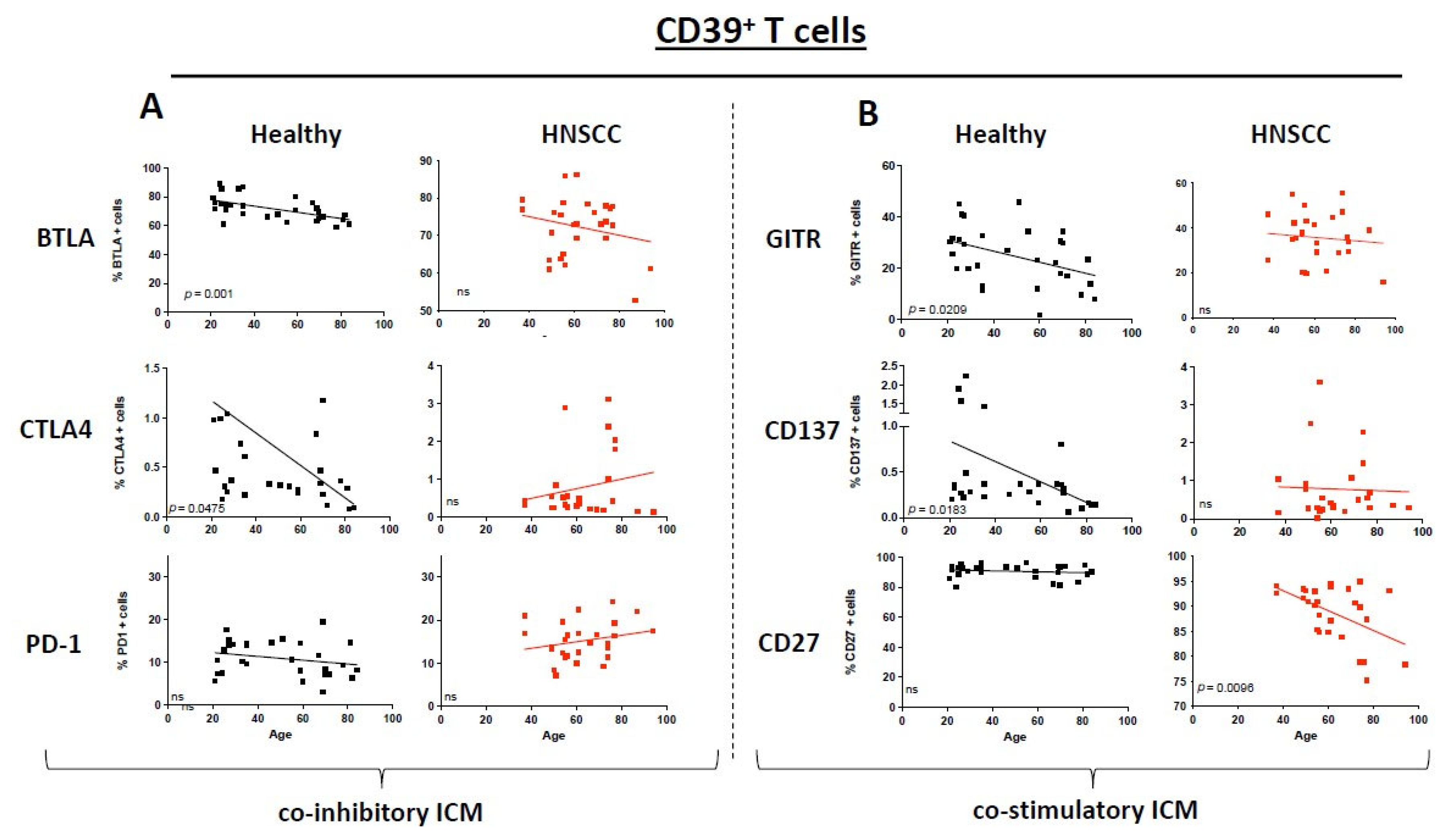
CD39+ T cells in HNSCC patients as well as healthy donors have a reduction in BTLA expression with increasing age. Nevertheless, this tendency was proven to be significant only for the healthy donors (p = 0.001; Figure 3A). CTLA4 expression revealed a reverse correlation between expression and age (healthy CTLA4: p = 0.0475; Figure 3A). No statistically significant changes are seen for PD1—for comparison it is presented in Figure 3. The co-stimulatory molecule expression on CD39+ T cells revealed a statistically significant decrease in GITR and CD137 (p = 0.0209, p = 0.0183, respectively; Figure 3B) on healthy PBMCs while there was a significant decrease on HNSCC PMBCs for CD27 expression with increasing age (p = 0.0205). The expression of CD27 in CD39+ T cells increases in both healthy individuals and tumor patients, whereas the CD8+ and CD4+ T-cell populations exhibit an opposite trend between the two groups (Figure 3B; non-statistically significant pairs are provided in the Supplementary Materials Figure S2).
We provided a result summary of all p-values and correlation coefficients in a table format for each analysis in Supplementary Materials Table S3.

3.5. HPV Status Has a Significant Influence on ICM and Co-Stimulatory Molecule Expression

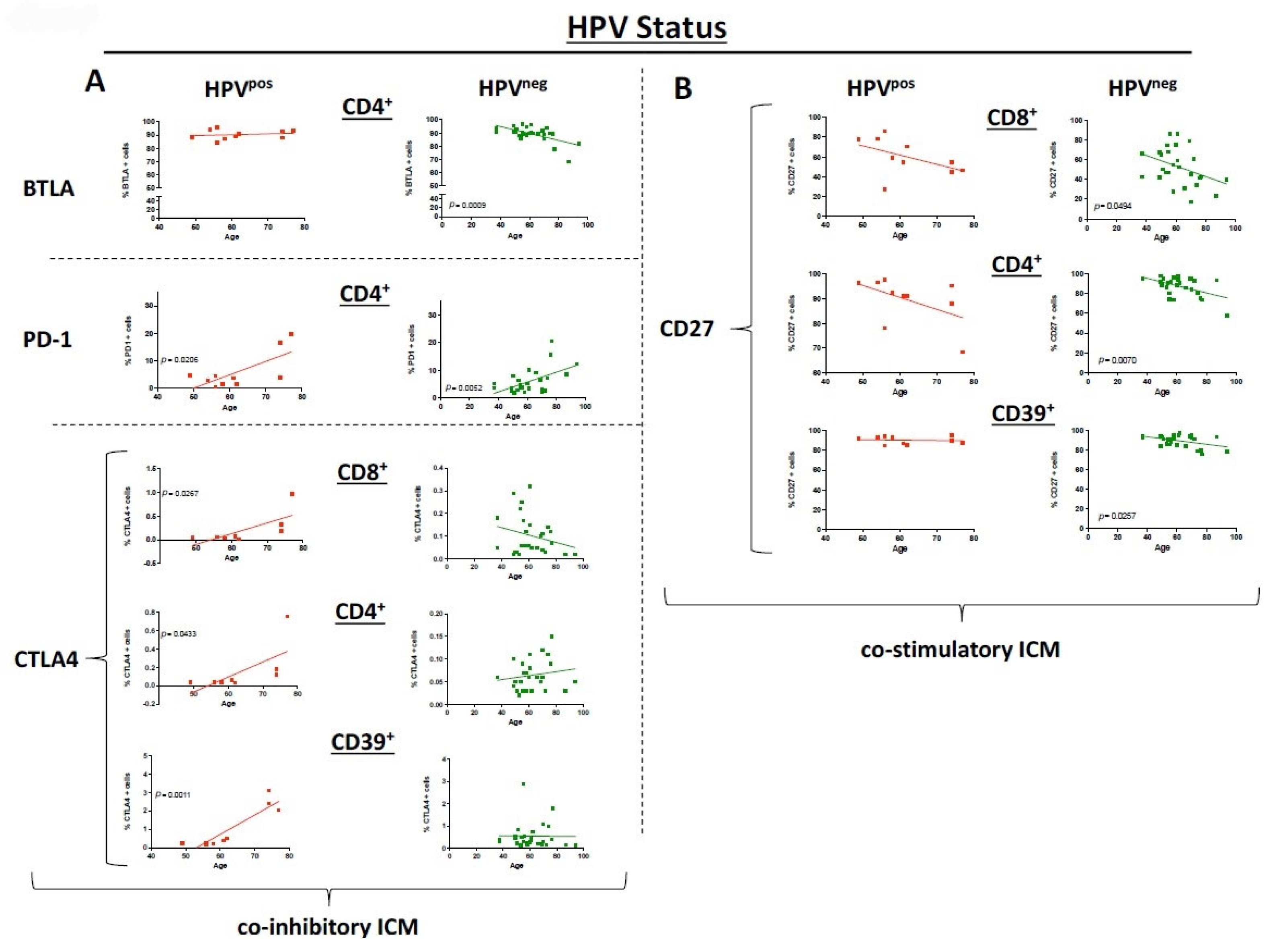
The cohort of HNSCC cases was dichotomized in HPVpos OPSCC patients and HPVneg HNSCC patients (HPVneg OPSCC and all other locations). We were able to demonstrate a statistically significant decrease in BTLA in HPVneg PBMC (p = 0.0009) while there was no change in HPVpos PBMC for ICM on CD4+ T cells. Interestingly, CTLA4 as an ICM was significantly increased on CD8+, CD4+, and CD39+ T cells of the HPVpos but not for the HPVneg group (Figure 4A). Both HPVpos and HPVneg HNSCC PBMC showed a significant increase in PD1 expression (p = 0.0206, p = 0.0052; Figure 4A). OX40 expression was also significantly increased with age in HPVpos cases, while HPVneg had no change in expression.
There was an equal expression of the co-stimulatory molecule CD27 on CD8+, CD4+, and CD39+ T cells for HPVpos OPSCC. However, the expression was decreasing with higher age in HPVneg HNSCC PBMC (p = 0.0494, p = 0.0070, and p = 0.0258, respectively; Figure 4B). No differences between the HPV status were seen for CD137, GITR, LAG3, or TIM3.
We provide a summary of all expression analysis results including the respective statistics in Supplementary Materials Table S3.

3.6. ICM and Co-Stimulatory Molecule Expression Differs Between the Sexes

We conducted an additional sex-specific analysis of both cohorts to evaluate potential differences in ICM and co-stimulatory molecule expression. Regarding the ICM PD-1, LAG-3, and CTLA-4, the increase in expression in the tumor group was more pronounced in male participants (Supplementary Materials Figure S3). We barely saw expression differences between females and males in the healthy donors, whereas the expression of co-stimulatory molecules, such as CD27, showed the same trends in both cohorts in the sex-specific analysis as in the overall evaluation (Figure 5). An separate analysis of the CD4/CD8 ratio was consistently 3:1 in both women and men, with no differences observed between the control group and the tumor patients.

4. Discussion

In this study, we aimed to investigate the expression of co-stimulatory and immune checkpoint molecules (ICMs) on peripheral blood cells in healthy individuals and cancer patients across different age groups. Our findings highlight significant age-related shifts in immune regulation, particularly in the expression of CTLA-4, CD27, and BTLA, which could influence treatment strategies for older HNSCC patients.

4.1. Age-Related Differences in Co-Stimulatory Molecules/ICM Expression and Implications

Aging is associated with a decline in adaptive immunity, facilitating tumor immune evasion [9]. The weakened immune response to cancer in the elderly may also stem from various alterations in immune cell populations. Age-related immune senescence, characterized by diminished T cell function and fewer tumor-infiltrating immune cells, further reduces the effectiveness of immune checkpoint blockade in HNSCC [13]. Additionally, the expression patterns of immune checkpoint molecules and co-stimulatory signals impair T cell activation, weakening the anti-tumor response. Age also influences the tolerability and toxicity of immune checkpoint inhibitors, as older patients—who often have more comorbidities and a lower physiological reserve—are more prone to treatment-related adverse events, particularly skin-related toxicity, while younger patients are more likely to experience endocrine side effects [14].
In our study, we examined the expression of the ICM CTLA-4, which is constitutively expressed on regulatory T cells (Tregs) and upregulated in conventional T cells upon activation, with its dynamic regulation attracting significant interest in cancer immunology [15]. We found a significant decrease in CTLA-4 expression on CD4+ T cells in healthy donors and an increase in cancer patients with age. The upregulation of CTLA-4 on CD4+ T cells which may reflect a tendency towards immune suppression and could be relevant for tumor progression [16]. We observed a significantly higher expression of CTLA-4 on CD39+ T cells in HPVpos patients with age. These T cells, known for their strong suppressive activity, have an enhanced ability to suppress immune responses due to the increased CTLA-4 expression [12]. The age-associated increase in CTLA-4 expression on CD4+ T cells highlight potential implications for cancer immunotherapy and treatment strategies. Immunotherapeutic approaches targeting CTLA-4, such as monoclonal antibodies that block its inhibitory function, have shown promising potential in enhancing anti-tumor immune responses [17,18]. However, the age-related increase in CTLA-4 expression warrants further investigation to assess the potential benefits of such interventions in older individuals.
Our results further highlight the impact of aging on immune function, particularly the loss of co-stimulatory molecules like CD27, which diminishes the capacity to respond to antigens and contributes to resistance to immunotherapy [19]. This reduced CD27 expression with increasing age was observed in both CD8+ and CD4+ T cells of HNSCC and healthy patients. Interestingly, in HNSCC patients, CD27 expression on CD39+ T cells remained stable compared to healthy donors. The co-expression of CD27 and CD39 on Tregs, which has been linked to a highly suppressive phenotype, suggests that this combined expression may enhance the regulatory functions of Tregs, presenting an additional immunosuppressive mechanism in aged HNSCC patients.
In our study, we also investigated the role of BTLA, an immune checkpoint molecule primarily expressed by B and T cells that inhibits activation and signal transduction. BTLA shares similarities with other ICMs like PD1 and CTLA-4, which are current targets of immunotherapies. Our results show that in HNSCC patients, the expression of BTLA on CD8+ T cells remains stable at around 60%, whereas in healthy individuals, BTLA expression on PBMCs decreases significantly with age. This might suggests that older HNSCC patients have a more exhausted and dysfunctional cytotoxic T cell population, as published before [20], highlighting potential therapeutic opportunities. Notably, ongoing clinical trials are exploring the effects of anti-BTLA antibodies, both as monotherapy and in combination with anti-PD1 antibodies, for advanced solid malignancies (NCT04137900).
Importantly, these findings might also be transferable to other solid tumors, such as lung cancer or melanoma, where published data already suggest that the efficacy of immune checkpoint inhibitors may be reduced in older patients [21,22,23].

4.2. HPV Status and Immune Landscape Differences

Regarding HPV-status and ICM expression, we found that HPVpos HNSCC patients exhibited a more stable or increasing expression of co-stimulatory molecules with age, whereas HPVneg patients showed a decrease. Additionally, HPVneg HNSCC patients had higher expression of co-inhibitory ICMs, such as PD1, which may contribute to immune suppression, which was shown by our group in a sequencing analysis [24]. A positive HPV-status is known to significantly influence tumor immune cell infiltration, enhancing the immune response due to viral antigens, which correlates with improved survival in this group [25,26,27]. These immunological differences explain the significantly better prognosis of HPVpos patients compared to HPVneg patients, as HPVpos tumors tend to have a more favorable immune profile and therefore a better response to immune-based therapies. The increasing incidence of oropharyngeal cancer also among elderly patients, largely attributed to HPV-induced cancers [28,29], highlights the need for further investigation into the age-specific immune landscape in HPVpos HNSCC patients, an area where data are currently lacking.

4.3. Sex Differences in Immune Aging

The sex-specific analysis revealed notable differences in ICM expression, particularly PD-1, within the tumor group, with higher expression levels observed in males. It is known that immunological parameters such as the percentage of CD4+ naïve T cells, memory CD4+ T cells, and NK cells, decline more gradually in women than in men, along with a more favorable CD4+/CD8+ ratio in women [30]. This pattern seems to extend to ICM expression, potentially leading to a weaker immune response in male HNSCC patients. Since there were fewer women than men in our tumor cohort, this can only be considered a trend. A matched analysis with an equal proportion of men and women would be necessary to confirm this with certainty.

4.4. Limitations

As further limitations of this study, we note the sample size of both groups. While we acknowledge that a larger sample could provide more robust data, we believe that our sample size of 30 healthy individuals and 37 cancer patients is adequate for an initial study aimed at identifying key trends and drawing meaningful preliminary conclusions. A potential limitation is that three healthy individuals had chronic inflammatory conditions. However, since these conditions were primarily localized, they are unlikely to significantly affect systemic inflammation or our comparisons. Another important limitation we need to mention is the gating strategy which focused mainly on identifying cytotoxic and immunosuppressive T cells. We did not include other subpopulations like NK-T cells, which might influence our findings. The ICM and co-stimulatory molecule expression in these subpopulations could be an interesting areal for future research.

5. Conclusions

In summary, our data show age-related changes in ICM expression across the lymphocyte populations we investigated in tumor patients compared to healthy donors, pointing to alterations in immune regulation that could influence the tumor microenvironment. These observations may provide a rationale for considering immune checkpoint inhibitors in older HNSCC patients, although we emphasize that our findings do not warrant immediate changes to current treatment. Importantly, immune checkpoint inhibitors are not specifically approved for age-related indications, and current guidelines do not offer age-specific recommendations. In our study, we identified ICMs such as CTLA-4, TIM3, and BTLA as promising targets for further investigation, particularly in elderly patients, and we highlight the need for additional research to better understand their predictive and therapeutic potential in HNSCC.

Supplementary Materials

The following supporting information can be downloaded at https://www.mdpi.com/article/10.3390/cancers17193215/s1, Figure S1: Flow cytometry gating strategy used for selection of the lymphocytes (A) the CD8+ (B), CD4+ (C) and CD39+ (D) T cells; Figure S2: Dot plots depict the expression of immune checkpoint molecules (ICMs) on CD8+, CD4+ and CD39+ T cells for healthy donors (black) and HNSCC samples (red). The straight line represents Pearson’s correlation analysis along with the corresponding p-values. A: all co-inhibitory ICM B: all co-stimulatory molecules. ns = not significant; Figure S3: Dot plots depict the expression of immune checkpoint molecules (ICMs) on CD39+ T cells and CD4+ T cells for healthy donors (black) and HNSCC samples (red). The straight line rep-resents Pearson’s correlation analysis along with the corresponding p-values. A: PD1-Expression on CD39+ Treg cells B: PD1 Expression on CD4+ T cells. ns = not significant; Table S1: Clinicopathological features of healthy donors; Table S2: Staining panels and detailed antibody information; Table S3: Summary of all expression analysis results including the respective statistics and trends.

Author Contributions

Experiments: L.K.P., A.G. and F.O.; Analysis: L.K.P. and A.V.W.; Writing Manuscript: L.K.P. and A.V.W.; Supervision: A.V.W., S.L., P.J.S., C.B. and T.K.H.; Proof of Manuscript: J.M.V., M.B., J.G., M.N.T., T.K.H., P.J.S. and S.L. All authors have read and agreed to the published version of the manuscript.

Funding

German Research Foundation (DFG, SCHU 2563/9-1; BR 2891/11-1), Ulm University (Promotionsprogramm Experimentelle Medizin, M.Sc. Lisa K. Puntigam). This paper is funded by the Deutsche Forschungsgemeinschaft (DFG, German Research Foundation)—Projektnummer 514808265.

Institutional Review Board Statement

This retrospective study was approved by the ethics committee of the University of Ulm (#90/15). All patients signed the informed consent form approved by the local ethical review committee including the consent for publication.

Informed Consent Statement

All patients signed the informed consent form approved by the local ethical review committee including the consent for publication.

Data Availability Statement

The data are available upon request from the authors.

Acknowledgments

We thank Katja Hasch, Gabriela Cudek, and Sandra Jeske for their support in performing the experiments. Additionally, we would like to thank the Core Facility Immune Monitoring of the Medical Faculty at Ulm University for providing support and instrumentation.

Conflicts of Interest

Simon Laban: Advisory Boards: Merck Sharp & Dohme, Bristol Myers Squibb, Astra Zeneca, Sanofi Genzyme. Honoraria: Merck Sharp & Drohme, Bristol Myers Squibb, Astra Zeneca, Merck Serono. The other authors declare no potential conflicts of interest.

References

  1. GBD 2016 Disease and Injury Incidence and Prevalence Collaborators. Global, regional, and national incidence, prevalence, and years lived with disability for 328 diseases and injuries for 195 countries, 1990–2016: A systematic analysis for the Global Burden of Disease Study 2016. Lancet 2017, 390, 1211–1259. [Google Scholar] [CrossRef]
  2. Syrjanen, S.; Waterboer, T.; Kero, K.; Rautava, J.; Syrjanen, K.; Grenman, S.; Pawlita, M. Oral human papillomavirus infection in men might contribute to HPV serology. Eur. J. Clin. Microbiol. Infect. Dis. 2015, 34, 237–245. [Google Scholar] [CrossRef]
  3. Andre, K.; Schraub, S.; Mercier, M.; Bontemps, P. Role of alcohol and tobacco in the aetiology of head and neck cancer: A case-control study in the Doubs region of France. Eur. J. Cancer B Oral Oncol. 1995, 31B, 301–309. [Google Scholar] [CrossRef]
  4. von Witzleben, A.; Currall, E.; Wood, O.; Chudley, L.; Akinyegun, O.; Thomas, J.; Bendjama, K.; Thomas, G.J.; Friedmann, P.S.; King, E.V.; et al. Correlation of HPV16 Gene Status and Gene Expression with Antibody Seropositivity and TIL Status in OPSCC. Front. Oncol. 2020, 10, 591063. [Google Scholar] [CrossRef] [PubMed]
  5. Burtness, B.; Harrington, K.J.; Greil, R.; Soulieres, D.; Tahara, M.; de Castro, G., Jr.; Psyrri, A.; Baste, N.; Neupane, P.; Bratland, A.; et al. Pembrolizumab alone or with chemotherapy versus cetuximab with chemotherapy for recurrent or metastatic squamous cell carcinoma of the head and neck (KEYNOTE-048): A randomised, open-label, phase 3 study. Lancet 2019, 394, 1915–1928. [Google Scholar] [CrossRef] [PubMed]
  6. Cohen, E.E.W.; Soulieres, D.; Le Tourneau, C.; Dinis, J.; Licitra, L.; Ahn, M.J.; Soria, A.; Machiels, J.P.; Mach, N.; Mehra, R.; et al. Pembrolizumab versus methotrexate, docetaxel, or cetuximab for recurrent or metastatic head-and-neck squamous cell carcinoma (KEYNOTE-040): A randomised, open-label, phase 3 study. Lancet 2019, 393, 156–167. [Google Scholar] [CrossRef] [PubMed]
  7. Ferris, R.L.; Blumenschein, G., Jr.; Fayette, J.; Guigay, J.; Colevas, A.D.; Licitra, L.; Harrington, K.; Kasper, S.; Vokes, E.E.; Even, C.; et al. Nivolumab for Recurrent Squamous-Cell Carcinoma of the Head and Neck. N. Engl. J. Med. 2016, 375, 1856–1867. [Google Scholar] [CrossRef]
  8. von Witzleben, A.; Fehn, A.; Grages, A.; Ezić, J.; Jeske, S.S.; Puntigam, L.K.; Brunner, C.; Kraus, J.M.; Kestler, H.A.; Doescher, J.; et al. Prospective longitudinal study of immune checkpoint molecule (ICM) expression in immune cell subsets during curative conventional therapy of head and neck squamous cell carcinoma (HNSCC). Int. J. Cancer 2021, 148, 2023–2035. [Google Scholar] [CrossRef]
  9. Sadighi Akha, A.A. Aging and the immune system: An overview. J. Immunol. Methods 2018, 463, 21–26. [Google Scholar] [CrossRef]
  10. Vahl, J.M.; Wigand, M.C.; Denkinger, M.; Dallmeier, D.; Steiger, C.; Welke, C.; Kuhn, P.; Idel, C.; Doescher, J.; von Witzleben, A.; et al. Increasing Mean Age of Head and Neck Cancer Patients at a German Tertiary Referral Center. Cancers 2021, 13, 832. [Google Scholar] [CrossRef]
  11. Puntigam, L.K.; Jeske, S.S.; Götz, M.; Greiner, J.; Laban, S.; Theodoraki, M.N.; Doescher, J.; Weissinger, S.E.; Brunner, C.; Hoffmann, T.K.; et al. Immune Checkpoint Expression on Immune Cells of HNSCC Patients and Modulation by Chemo- and Immunotherapy. Int. J. Mol. Sci. 2020, 21, 5181. [Google Scholar] [CrossRef]
  12. Schuler, P.J.; Harasymczuk, M.; Schilling, B.; Lang, S.; Whiteside, T.L. Separation of human CD4+CD39+ T cells by magnetic beads reveals two phenotypically and functionally different subsets. J. Immunol. Methods 2011, 369, 59–68. [Google Scholar] [CrossRef]
  13. López-Otín, C.; Blasco, M.A.; Partridge, L.; Serrano, M.; Kroemer, G. The hallmarks of aging. Cell 2013, 153, 1194–1217. [Google Scholar] [CrossRef] [PubMed]
  14. Paderi, A.; Fancelli, S.; Caliman, E.; Pillozzi, S.; Gambale, E.; Mela, M.M.; Doni, L.; Mazzoni, F.; Antonuzzo, L. Safety of Immune Checkpoint Inhibitors in Elderly Patients: An Observational Study. Curr. Oncol. 2021, 28, 3259–3267. [Google Scholar] [CrossRef] [PubMed]
  15. Jeske, S.S.; Schuler, P.J.; Doescher, J.; Theodoraki, M.N.; Laban, S.; Brunner, C.; Hoffmann, T.K.; Wigand, M.C. Age-related changes in T lymphocytes of patients with head and neck squamous cell carcinoma. Immun. Ageing 2020, 17, 3. [Google Scholar] [CrossRef] [PubMed]
  16. Syn, N.L.; Teng, M.W.L.; Mok, T.S.K.; Soo, R.A. De-novo and acquired resistance to immune checkpoint targeting. Lancet Oncol. 2017, 18, e731–e741. [Google Scholar] [CrossRef]
  17. Kawczak, P.; Feszak, I.J.; Bączek, T. Comparative Mechanistic Insights and Therapeutic Potential of Pembrolizumab, Durvalumab, and Ipilimumab as Immune Checkpoint Inhibitors in the Targeted Management of Oral and Head and Neck Squamous Cell Carcinoma. Cancers 2025, 17, 2805. [Google Scholar] [CrossRef]
  18. Hodi, F.S.; O’Day, S.J.; McDermott, D.F.; Weber, R.W.; Sosman, J.A.; Haanen, J.B.; Gonzalez, R.; Robert, C.; Schadendorf, D.; Hassel, J.C.; et al. Improved survival with ipilimumab in patients with metastatic melanoma. N. Engl. J. Med. 2010, 363, 711–723. [Google Scholar] [CrossRef]
  19. Moreira, A.; Gross, S.; Kirchberger, M.C.; Erdmann, M.; Schuler, G.; Heinzerling, L. Senescence markers: Predictive for response to checkpoint inhibitors. Int. J. Cancer 2019, 144, 1147–1150. [Google Scholar] [CrossRef]
  20. Demerlé, C.; Gorvel, L.; Olive, D. BTLA-HVEM Couple in Health and Diseases: Insights for Immunotherapy in Lung Cancer. Front. Oncol. 2021, 11, 682007. [Google Scholar] [CrossRef]
  21. Voruganti, T.; Soulos, P.R.; Mamtani, R.; Presley, C.J.; Gross, C.P. Association Between Age and Survival Trends in Advanced Non–Small Cell Lung Cancer After Adoption of Immunotherapy. JAMA Oncol. 2023, 9, 334–341. [Google Scholar] [CrossRef]
  22. Kugel, C.H., III; Douglass, S.M.; Webster, M.R.; Kaur, A.; Liu, Q.; Yin, X.; Weiss, S.A.; Darvishian, F.; Al-Rohil, R.N.; Ndoye, A.; et al. Age Correlates with Response to Anti-PD1, Reflecting Age-Related Differences in Intratumoral Effector and Regulatory T-Cell Populations. Clin. Cancer Res. 2018, 24, 5347–5356. [Google Scholar] [CrossRef] [PubMed]
  23. Yin, J.; Song, Y.; Fu, Y.; Jun, W.; Tang, J.; Zhang, Z.; Song, Q.; Zhang, B. The efficacy of immune checkpoint inhibitors is limited in elderly NSCLC: A retrospective efficacy study and meta-analysis. Aging 2023, 15, 15025–15049. [Google Scholar] [CrossRef] [PubMed]
  24. von Witzleben, A.; Grages, A.; Thomas, J.; Ezić, J.; Brunner, C.; Schuler, P.J.; Kraus, J.M.; Kestler, H.A.; Vahl, J.M.; Doescher, J.; et al. Immune checkpoint expression on tumor-infiltrating lymphocytes (TIL) is dependent on HPV status in oropharyngeal carcinoma (OPSCC)—A single-cell RNA sequencing analysis. Oral Oncol. 2024, 159, 107107. [Google Scholar] [CrossRef] [PubMed]
  25. Ward, M.J.; Thirdborough, S.M.; Mellows, T.; Riley, C.; Harris, S.; Suchak, K.; Webb, A.; Hampton, C.; Patel, N.N.; Randall, C.J.; et al. Tumour-infiltrating lymphocytes predict for outcome in HPV-positive oropharyngeal cancer. Br. J. Cancer 2014, 110, 489–500. [Google Scholar] [CrossRef]
  26. Solomon, B.; Young, R.J.; Bressel, M.; Urban, D.; Hendry, S.; Thai, A.; Angel, C.; Haddad, A.; Kowanetz, M.; Fua, T.; et al. Prognostic Significance of PD-L1+ and CD8+ Immune Cells in HPV+ Oropharyngeal Squamous Cell Carcinoma. Cancer Immunol. Res. 2018, 6, 295–304. [Google Scholar] [CrossRef]
  27. Wang, J.; Sun, H.; Zeng, Q.; Guo, X.-J.; Wang, H.; Liu, H.-H.; Dong, Z.-Y. HPV-positive status associated with inflamed immune microenvironment and improved response to anti-PD-1 therapy in head and neck squamous cell carcinoma. Sci. Rep. 2019, 9, 13404. [Google Scholar] [CrossRef]
  28. Lassen, P.; Alsner, J.; Primdahl, H.; Plaschke, C.C.; Maare, C.; Johansen, J.; Andersen, M.; Farhadi, M.; Overgaard, J. Subsite variation of HPV-related p16-expression in oropharynx cancer: Incidence and prognostic impact in a population-based DAHANCA cohort 1986–2020. Acta Oncol. 2025, 64, 1071–1078. [Google Scholar] [CrossRef]
  29. Zumsteg, Z.S.; Cook-Wiens, G.; Yoshida, E.; Shiao, S.L.; Lee, N.Y.; Mita, A.; Jeon, C.; Goodman, M.T.; Ho, A.S. Incidence of Oropharyngeal Cancer Among Elderly Patients in the United States. JAMA Oncol. 2016, 2, 1617–1623. [Google Scholar] [CrossRef]
  30. Hirokawa, K.; Utsuyama, M.; Hayashi, Y.; Kitagawa, M.; Makinodan, T.; Fulop, T. Slower immune system aging in women versus men in the Japanese population. Immun. Ageing 2013, 10, 19. [Google Scholar] [CrossRef]
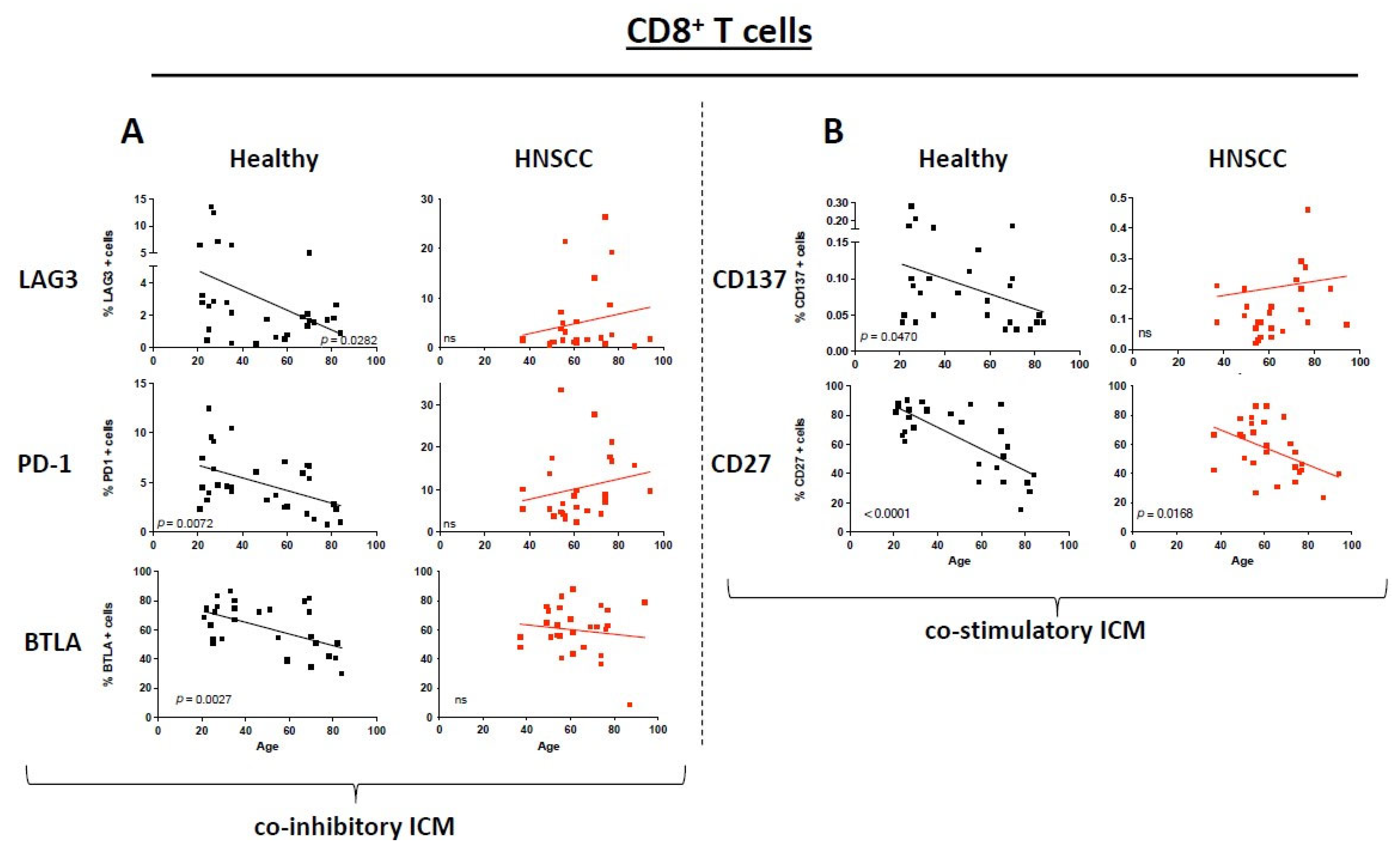
Figure 1. Dot plots depict the expression of immune checkpoint molecules and co-stimulatory molecules on CD8+ T cells for healthy donors (black) and HNSCC samples (red). The straight line represents Pearson’s correlation analysis along with the corresponding p-values. (A): Selected immune checkpoint molecules; (B): selected co-stimulatory molecules. ns = not significant.
Cancers 17 03215 g001
Figure 2. Dot plots depict the expression of immune checkpoint molecules and co-stimulatory molecules on CD4+ T cells for healthy donors (black) and HNSCC samples (red). The straight line represents Pearson’s correlation analysis along with the corresponding p-values. (A): Selected immune checkpoint molecules; (B): selected co-stimulatory molecules. ns = not significant.
Cancers 17 03215 g002
Figure 3. Dot plots depict the expression of immune checkpoint molecules and co-stimulatory molecules on CD39+ T cells for healthy donors (black) and HNSCC samples (red). The straight line represents Pearson’s correlation analysis along with the corresponding p-values. (A): Selected immune checkpoint molecules; (B): selected co-stimulatory molecules. ns = not significant.
Cancers 17 03215 g003
Figure 4. Dot plots depict the expression of selected immune checkpoint molecules and co-stimulatory molecules on different T cells for HNSCC samples grouped by HPV status: HPVpos: red, HPVneg: green. The straight line represents Pearson’s correlation analysis along with the corresponding p-values. (A): Selected immune checkpoint molecules; (B): selected co-stimulatory molecules. ns = not significant.
Cancers 17 03215 g004
Figure 5. Dot plots depict the expression of the costimulatory molecule CD27 on CD8+ T cells for healthy donors (black) and HNSCC samples (red). The straight line represents Pearson’s correlation analysis along with the corresponding p-values. Female and male patients are compared. ns = not significant.
Cancers 17 03215 g005
Table 1. Clinicopathological characteristics of HNSCC patients and the control group. Summary of the patient’s characteristics. HPV = human papillomavirus, y = years, HNSCC = head and neck squamous cell carcinoma, OPSCC = oropharyngeal squamous cell carcinoma.
Control GroupHNSCC Patients
N (female/male)30 (18/12)37 (12/25)
Age (±SD) range (y)49 ± 22 (21–84)62 ± 12 (37–94)
Stage n=
T1 8
T2 14
T3 7
T4 8
Nodal status n=
N0 10
N1 12
N2 6
N3 9
Location n=
Oral cavity 11
Oropharynx 15
Hypopharynx 5
Larynx 6
HPV status (of OPSCC cases) n=
Positive 10
Negative 5
Disclaimer/Publisher’s Note: The statements, opinions and data contained in all publications are solely those of the individual author(s) and contributor(s) and not of MDPI and/or the editor(s). MDPI and/or the editor(s) disclaim responsibility for any injury to people or property resulting from any ideas, methods, instructions or products referred to in the content.