Article

Clinical and Molecular Differences Suggest Different Responses to Immune Checkpoint Inhibitors in Microsatellite-Stable Solid Tumors with High Tumor Mutational Burden †

1
Robert H. Lurie Comprehensive Cancer Center, Feinberg School of Medicine, Northwestern University, Chicago, IL 60611, USA
2
Division of Hematology and Oncology, Developmental Therapeutics Institute, Northwestern University, Chicago, IL 60611, USA
3
Preventive Medicine, Feinberg School of Medicine, Northwestern University, Chicago, IL 60611, USA
*
Author to whom correspondence should be addressed.
This paper is an extended version of our paper published in Nizamuddin, I.; Doukas, P.; de Viveiros, P.A.H.; Mi, X.; Katam, N.; Chae, Y.K.; Behdad, A.; Wehbe, F.H.; Mahalingam, D. Microsatellite-stable tumors with high tumor mutational burden in association with tumor response to immune checkpoint inhibitor therapy across solid tumors and correlation with specific oncogenic alterations. In Proceedings of the Poster Presentation at ASCO 2022 Annual Meeting, Online, 3–7 June 2022.
These authors contributed equally to this work.
Cancers 2025, 17(16), 2673; https://doi.org/10.3390/cancers17162673 (registering DOI)
Submission received: 30 June 2025 / Revised: 7 August 2025 / Accepted: 13 August 2025 / Published: 16 August 2025

Simple Summary

This study investigated the predictors of response to immune checkpoint inhibitors (ICIs) in 117 patients diagnosed with advanced solid tumors exhibiting a high tumor mutational burden (TMB) of ≥10 mutations per megabase (mut/Mb). Enhanced treatment responses were correlated with the absence of liver metastasis, a lack of prior systemic therapy, and the presence of mutations in the TERT gene. It is noteworthy that a TMB of ≥15 mut/Mb specifically correlated with improved outcomes in patients whose tumor types are not typically sensitive to ICIs. In contrast, mutations in the MYC pathway and the MLL2 gene were associated with diminished treatment responses. Furthermore, demographic factors such as age, sex, and PD-L1 status did not exhibit significant predictive value. The findings underscore the validity of TMB as a biomarker, indicating that its effectiveness is influenced by tumor type, the presence of liver metastases, and specific genetic mutations.

Abstract

Background/Objectives: We aim to identify predictors of response to ICIs in patients with advanced solid tumors that exhibiting a TMB ≥ 10 mut/Mb. Methods: Patients treated with ICIs alone at Northwestern University between 1 January 2015 and 31 December 2020 were identified. Progression-free survival (PFS) and overall survival (OS) were calculated using the Kaplan–Meier method, and groups were compared using the log-rank test. Wilcoxon rank sum tests, chi-squared tests, and Fisher’s exact tests were used for univariable analyses evaluating the impact of clinical and genetic variables on response, with significance defined as p < 0.05. Results: A total of 117 patients were classified as ICI-sensitive (n = 88) or non-ICI-sensitive (n = 29). Among evaluable patients (n = 105), the overall response rate was 34% with 14% achieving a complete response. Median PFS and OS were 8.05 months and 26.8 months, respectively. Higher PFS rates were significantly linked to the ICI-sensitive tumor group (p = 0.009), absence of liver metastasis (p = 0.015), and no prior systemic treatment (p = 0.001) in both cohorts. In non-ICI-sensitive patients, a TMB of ≥15 mut/Mb correlated with improved outcomes (p = 0.012). Mutations in the MYC pathway (p = 0.03) and the MLL2 gene (p = 0.014) were associated with poorer responses, while mutations in the TERT gene were linked to better responses (p = 0.031). Conclusions: Patients without liver metastasis, mutations in TERT, and TMB ≥ 15 mut/Mb are associated with superior response, while mutations in the MYC pathway and MLL2 are associated with worse responses.

1. Introduction

Over a decade ago, the Food and Drug Administration (FDA) approved the first immune checkpoint inhibitor (ICI), ipilimumab [1]. Since then, ICIs have transformed the treatment landscape for various cancers, with the capacity to elicit durable responses in tumors previously deemed untreatable [2]. Currently, the FDA has approved eleven ICIs for the treatment of over 20 different types of cancer, with ongoing research into additional indications [3].
A critical consideration is the identification of biomarkers to predict patients who would benefit from these therapies [4,5]. Increased programmed cell death ligand 1 (PD-L1) expression has been shown to result in increased response rates to ICIs depending on tumor type and expression level [6]. Tumors with high microsatellite instability (MSI-H), indicative of a deficient mismatch repair (dMMR) system, have also demonstrated sensitivity to ICIs [7]. In May 2017, the FDA granted accelerated approval of pembrolizumab for treatment of MSI-H/dMMR solid tumors, regardless of primary site, based on data from patients enrolled across five clinical trials [8]. This marked the first tumor-agnostic predictive biomarker approval for ICIs.
Recent interest has emerged in TMB as a predictive biomarker. TMB is defined as the total number of somatic mutations per coding area of a tumor genome [9]. It is hypothesized that highly mutated tumors, such as those with high TMB, may generate immunogenic neoantigens, thereby increasing T-cell reactivity [10]. While most MSI-H/dMMR tumors are TMB-high, not all TMB-high tumors exhibit MSI-H/dMMR [6]. Consequently, TMB has been investigated as a predictive biomarker even in the absence of MSI-H/dMMR.
Numerous retrospective studies have demonstrated a correlation between high tumor mutational burden (TMB) and enhanced response rates to specific ICIs, including nivolumab, pembrolizumab, and ipilimumab [11,12,13]. Large-scale meta-analyses have further substantiated these findings, indicating that patients with high TMB exhibit improved overall survival (OS) and progression-free survival (PFS) when treated with ICIs compared to those receiving chemotherapy alone [14,15]. A prospective exploratory analysis of the KEYNOTE-158 study investigated the association between TMB and response to pembrolizumab, revealing that patients with TMB ≥ 10 mut/Mb had a significantly higher likelihood response, irrespective of tumor type, with an overall response rate (ORR) of 29% [16]. Consequently, the FDA granted accelerated approval of pembrolizumab for the treatment of unresectable or metastatic tumors with high TMB that have progressed on prior therapies [17]. Nevertheless, this approval has sparked considerable debate within the oncology community [18,19]. While some view it as a means to provide additional therapeutic options for a population lacking effective alternatives, others have criticized it based on arbitrary TMB cutoffs, absence of clear improvement in OS, limited sample sizes in rare tumors, and questionable cost-effectiveness [20,21,22].
Subsequent studies have continued to explore the role of TMB in predicting response to ICIs. For instance, a study in advanced colorectal cancer found that only patients with MSI-H/dMMR or specific mutations exhibited improved OS when treated with ICIs [23]. Moreover, a retrospective study involving over 10,000 patients across various cancer types failed to identify a consistent TMB threshold predictive of response to ICIs in a tumor-agnostic manner [24].
Given these developments, there is interest in reassessing the current FDA approval criteria based on TMB and identifying specific patient populations and molecular phenotypes that may substantially benefit from ICIs.

2. Materials and Methods

2.1. Patients

Patients treated at the Robert H. Lurie Comprehensive Cancer Center of Northwestern University (Chicago, IL, USA) between 1 January 2015 and 31 December 2020 were identified using an Enterprise Data Warehouse grant. Eligible participants were individuals aged ≥18 years with biopsy-proven advanced (unresectable or metastatic) tumors treated with ICIs alone and high TMB (defined as ≥10 mut/Mb) identified from an evaluable tissue sample for biomarker analysis. ICIs included those targeting the CTLA-4, PD-1, and PD-L1 pathways. The study excluded tumors other than carcinomas and those with MSI-H, as well as patients with active autoimmune diseases requiring systemic treatment or who had received investigational therapy within four weeks prior to the administration of ICIs. Demographic and clinical characteristics, disease outcomes, and toxicity outcomes were retrospectively collected. PD-L1 expression was evaluated differently depending on type of cancer, with some cancers, such as non-small cell lung cancer, utilizing tumor proportion score (TPS) testing, and others, such as gastrointestinal cancers, utilizing combined positive score (CPS) testing. For both TPS and CPS, PD-L1 positivity was defined as expression level ≥ 1%.
Patients were categorized into two distinct cohorts based on their sensitivity to ICI treatments and FDA-approved indications. Group 1 represented tumors for which ICI treatments have FDA approval for standalone use, including melanoma, non-small cell lung cancer (NSCLC), adrenal corticoid carcinoma (ACC), squamous cell carcinoma (SCC) (anal, esophageal, skin, and head and neck), renal cell carcinoma, and urothelial carcinoma. Group 2 included patients with non-ICI-sensitive tumors (those for whom ICI treatments have not been approved by the FDA for standalone use), including breast cancer (BC), colorectal cancer (CRC), non-SCC gynecologic cancers (GC), pancreaticobiliary cancers (PBC), small cell lung cancer (SCLC), and upper gastrointestinal cancers (UGC) (esophageal, stomach, and duodenal adenocarcinomas).

2.2. Outcome Evaluation

The primary efficacy endpoint was ORR. Radiologic response to ICIs was assessed using iRECIST criteria [25] through independent central review based on computed tomography or magnetic resonance imaging. Target lesions and non-target lesions were defined at baseline to ascertain the overall tumor burden. Subsequent response evaluations were carried out through standard radiologic assessments at regular intervals, typically every 6–12 weeks. This process involved the quantitative measurement of target lesions and the qualitative assessment of non-target lesions. Patients were classified as responders (complete response (CR), partial response (PR), or stable disease (SD) ≥ 6 months) or non-responders (SD < 6 months or progression of disease (PD)). Additional endpoints included PFS and OS. PFS was defined as the time from ICI initiation to disease progression, recurrence, or death from any cause. Surviving patients without progression or recurrence were censored at time of last follow-up. OS was defined as the time from ICI initiation to death or last follow-up.

2.3. Safety

The assessment of toxicity outcomes was based on the presence of immune-related adverse events (irAEs) affecting various organs, including endocrine organs (thyroid, adrenal, pancreas, and pituitary glands), colon, lungs, skin, liver, kidneys, and heart. The severity of each irAE was graded according to the Common Terminology Criteria for Adverse Events (CTCAE) v5.0 classification [26]. Data were gathered through chart reviews, clinical notes, and radiological and laboratory reports.

2.4. Next-Generation Sequencing (NGS)

The assessment of tissue TMB was performed on archived or newly obtained formalin-fixed paraffin-embedded tumor samples using FDA-approved platforms, including the FoundationOne CDx assay (Foundation Medicine, Boston, MA, USA) and Tempus xT Gene Panel (Tempus, Chicago, IL, USA). The Guardant360 CDx assay (GuardantHealth, Palo Alto, CA, USA) also reported blood-based TMB. High TMB was defined as ≥10 mut/Mb, based on the cutoff established by the KEYNOTE-158 study and the FDA [16]. These platforms also provided tumor mutational profiling using NGS to detect genetic alterations present in tumor tissue. NGS platforms varied in sample type, gene coverage, and fusion detection. FoundationOne CDx utilized hybrid-capture targeted DNA sequencing on formalin-fixed paraffin-embedded (FFPE) tumor tissue. In addition to hybrid-capture targeted DNA sequencing, Tempus xT also included RNA sequencing for broader coverage. Guardant360 CDx utilized plasma samples to detect somatic mutations in circulating tumor DNA via hybrid-capture targeted DNA sequencing. Platform choice was left to clinician discretion. Mutations deemed pathogenic or likely pathogenic were used in the analysis.

2.5. Ethics Approval

The Northwestern University institutional review board approved the study protocol, which was conducted in compliance with Good Clinical Practice and the Declaration of Helsinki.

2.6. Statistical Analysis

The efficacy analysis population comprised all patients who received at least one dose of an ICI with evaluable TMB data and follow-up imaging for evaluation. The safety analysis population included all patients who received at least one dose of an ICI. A patient was considered not evaluable if a baseline assessment was obtained, but no post-baseline assessment had been performed. Descriptive statistics were used to summarize patient characteristics. Continuous variables were summarized using median and interquartile range (IQR) and compared between groups using the Wilcoxon rank sum test. Kaplan–Meier curves were used to estimate PFS and OS, and groups were compared using the log-rank test. Cox proportional hazard models were used to estimate hazard ratios (HR), and the proportional hazards assumption was checked [27]. Mutations identified by NGS were grouped into common signaling pathways responsible for carcinogenesis [28], and an oncoplot [29] was generated based on available co-mutation data. Unadjusted p-values are reported, with α < 0.05 considered statistically significant. All analyses were conducted in R Version 4.3.1.

3. Results

3.1. Patient Characteristics

Baseline characteristics, treatments administered, and immune biomarkers are summarized in Table 1.
In total, 117 patients were enrolled, including 105 (95%) patients evaluable by iRECIST criteria. The median age was 68, and 61% were male. The study population comprised 88% White, 6% African American, and 3.4% Asian participants. The cohort encompassed 14 different primary malignancies, including melanoma (33%; 39/117), NSCLC (28%; 33/117), SCLC (6%; 7/117), CRC (5.1%; 6/117), PBC (4.3%; 5/117), and others. Of the patients, 47% (55/117) were treatment-naïve, while the remaining 53% (62/117) had received prior systemic treatment. ICIs included pembrolizumab (37%), nivolumab (16%), ipilimumab plus nivolumab (16%), atezolizumab (11%), ipilimumab (1.7%), durvalumab (0.9%), and other (6%). A total of 60% of patients had TMB ≥ 15 mut/Mb. Median TMB level by tumor type is shown in Table 2.
A total of 88 patients (75%) were considered ICI-sensitive (Group 1), while 29 (25%) were considered non-ICI-sensitive (Group 2). Median TMB value for ICI-sensitive patients was 21.1 mut/Mb (IQR: 13.4–42.2 mut/Mb), compared to 15.0 mut/Mb (IQR: 11.4–22.8 mut/Mb) for non-ICI-sensitive patients. PD-L1 CPS expression level was positive in 46% and negative in 39% of patients.
At baseline, 31 patients (27%) had liver metastasis, while 83 patients (71%) did not. Data for three patients were missing or unknown. Of those in the ICI-sensitive group and the non-ICI-sensitive group, 67 (76%) and 16 (55%), respectively, did not present with liver metastasis.
irAEs were infrequent, with colitis (8%; 9/117), hepatitis (8%; 9/117), and skin reactions (6%; 7/117) being the most commonly reported (Table 3).

3.2. Gene Data

A total of 105 patients had NGS data, revealing 3899 different types of mutations. In descending order, frequency of mutations included missense mutations (80%, n = 2942), nonsense mutations (7.9%, n = 292), splice mutations (3.5%, n = 128), frameshift mutations (3.3%, n = 120), other mutations (2.5%, n = 931), substitutions (1.5, n = 54), and promoter/regulatory mutations (1.3%, n = 47).
A co-mutation plot, which summarizes all mutations according to response status, TMB, and type of mutation, is illustrated in Figure 1. The most common mutations included TP53 (66.6%), LRP1B (40.9%), TERT (40%), CDKN2A (38%), NF1 (36.1%), MLL2 (35.2%), and ROS1 (27.6%).
TERT and CDKN2A mutations were more frequently observed in ICI-sensitive tumors (p < 0.001 and p = 0.022, respectively), while CDK12 mutations were more often found in non-ICI-sensitive tumors (p = 0.025). TP53 mutations were significantly more prevalent in lung cancer compared to melanoma or other types of cancer (p < 0.001). BRAF mutations were more commonly identified in melanoma (p < 0.001).

3.3. Efficacy

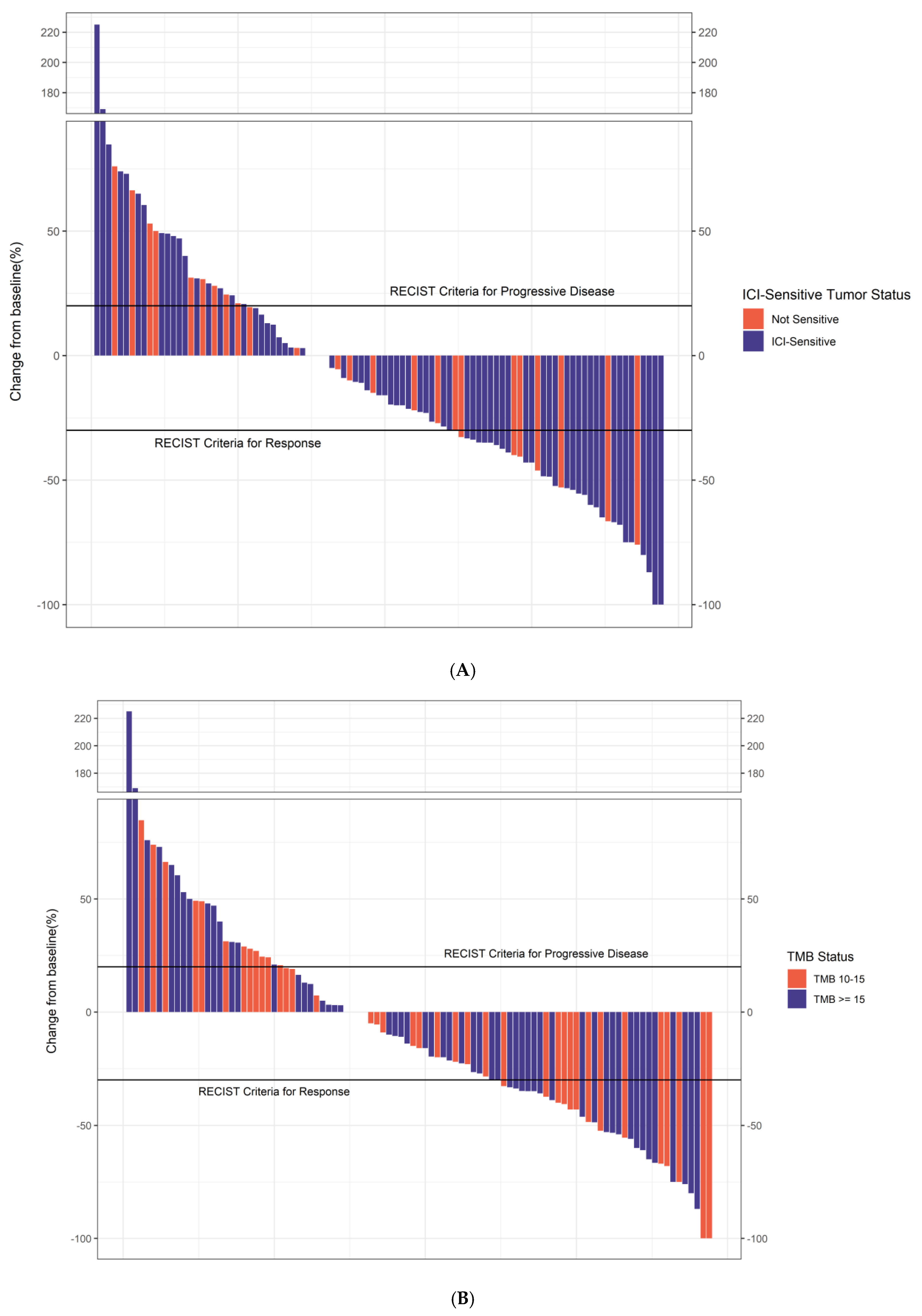
Among all patients, 51% were responders, while 49% were non-responders. Of 105 evaluable patients, ORR was 34% across all malignancies (CR 14%). Within the ICI-sensitive subgroup, the ORR was 37%, compared to 27% in the non-ICI-sensitive group. Waterfall plots show the percentage change in tumor size from baseline for TMB and ICI-sensitive status (Figure 2).
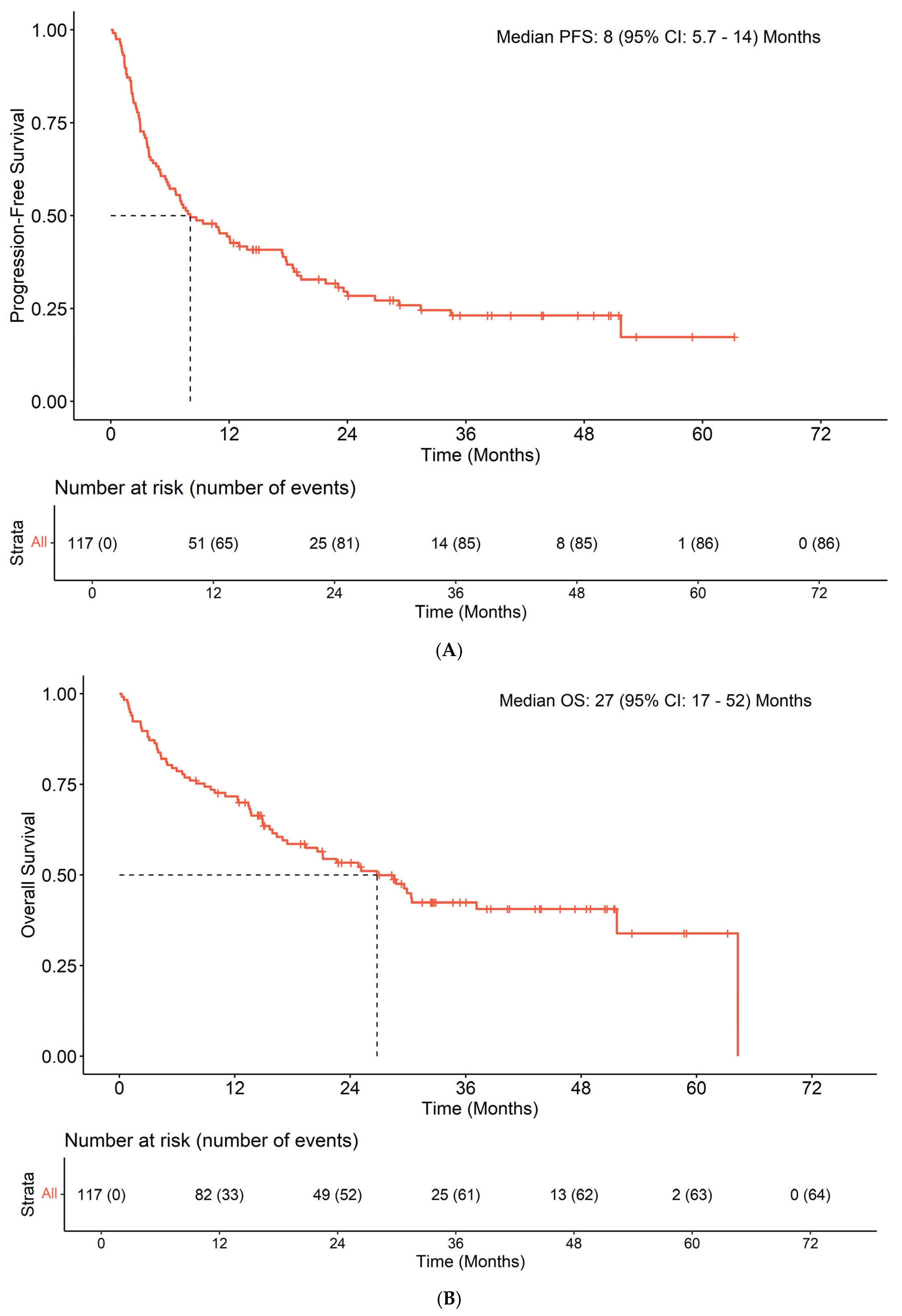
The median PFS and OS were 8.05 months and 26.8 months, respectively (Figure 3). Median follow-up among patients who were censored for OS was 33 (IQR: 21–44) months.
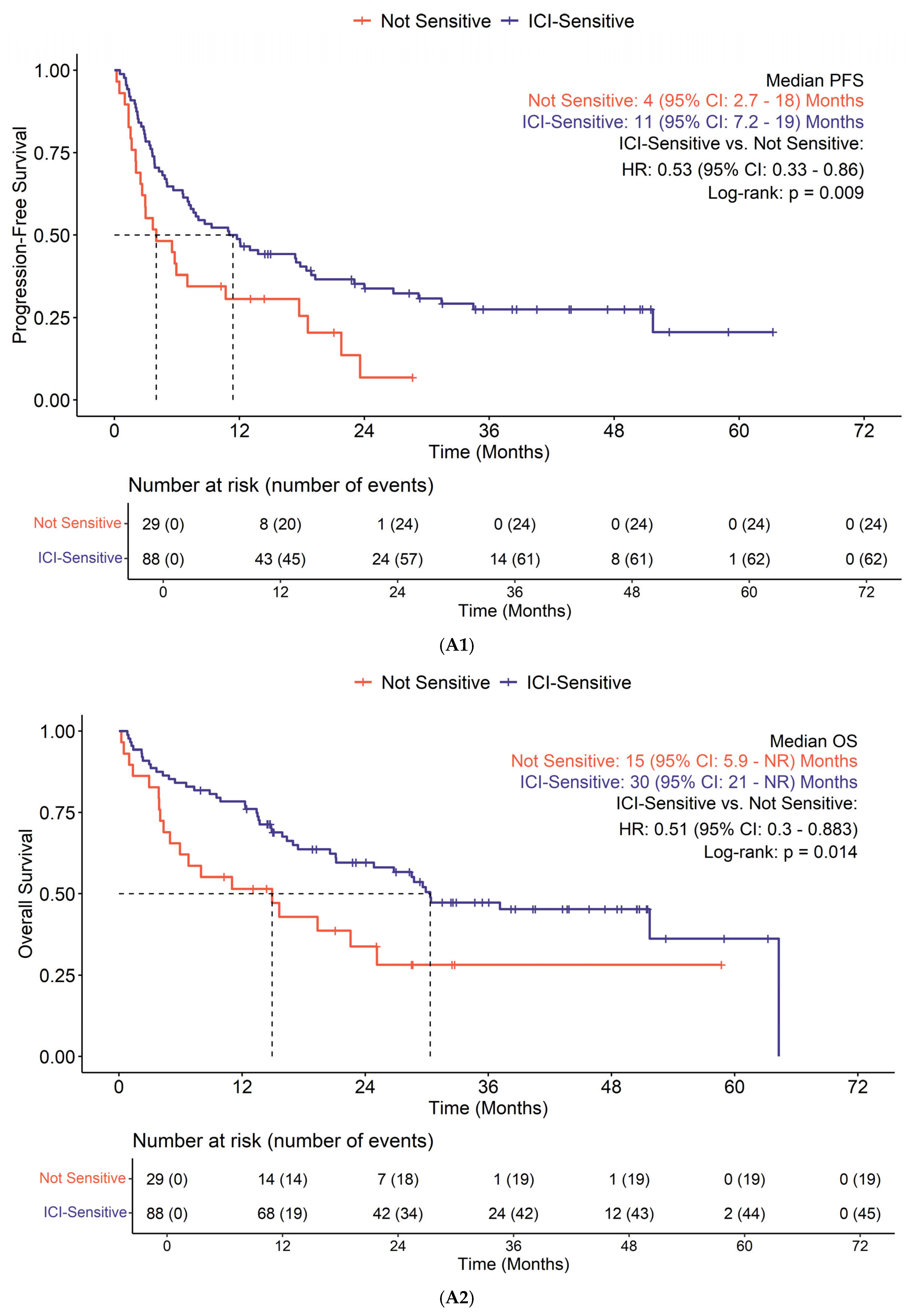
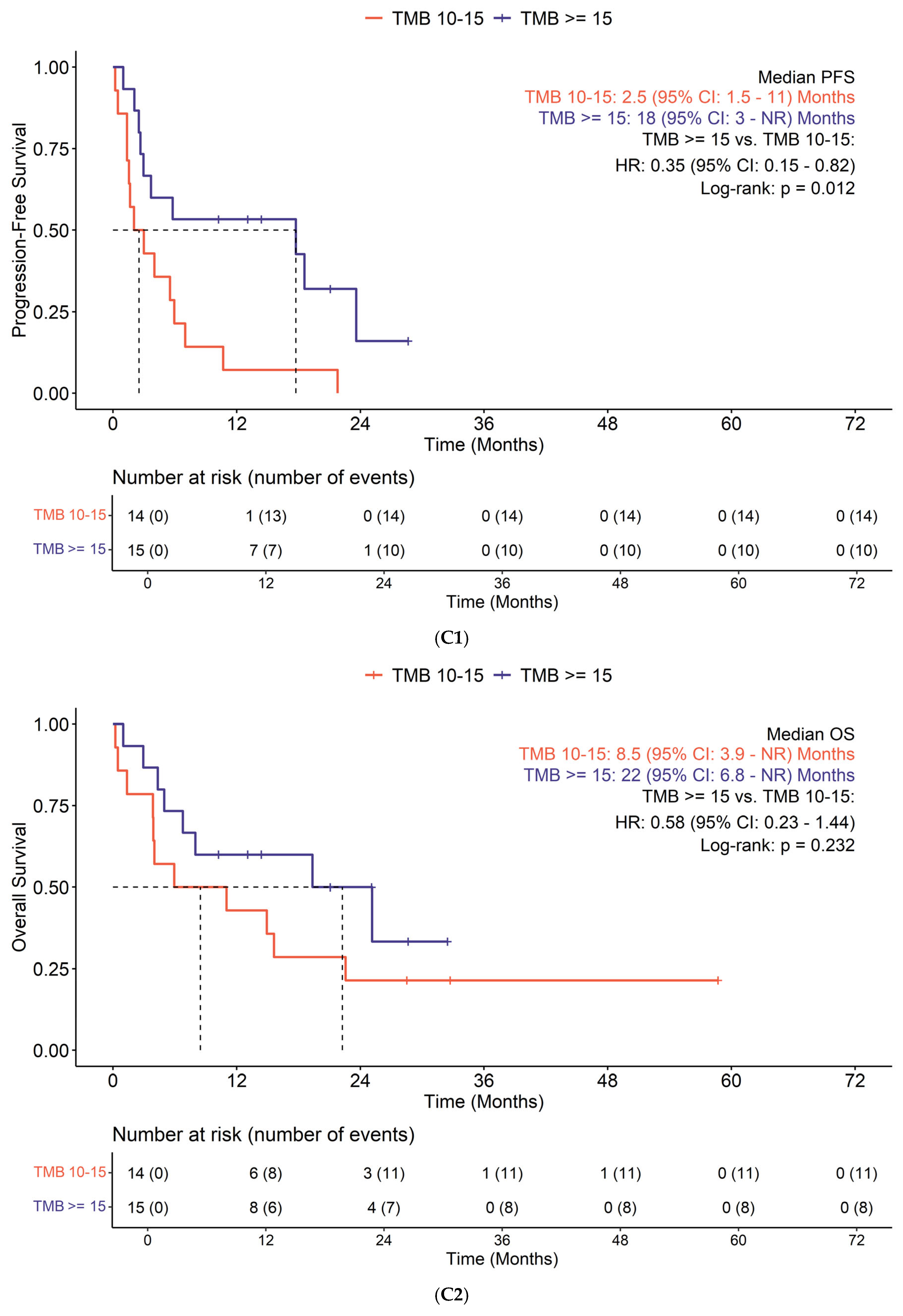
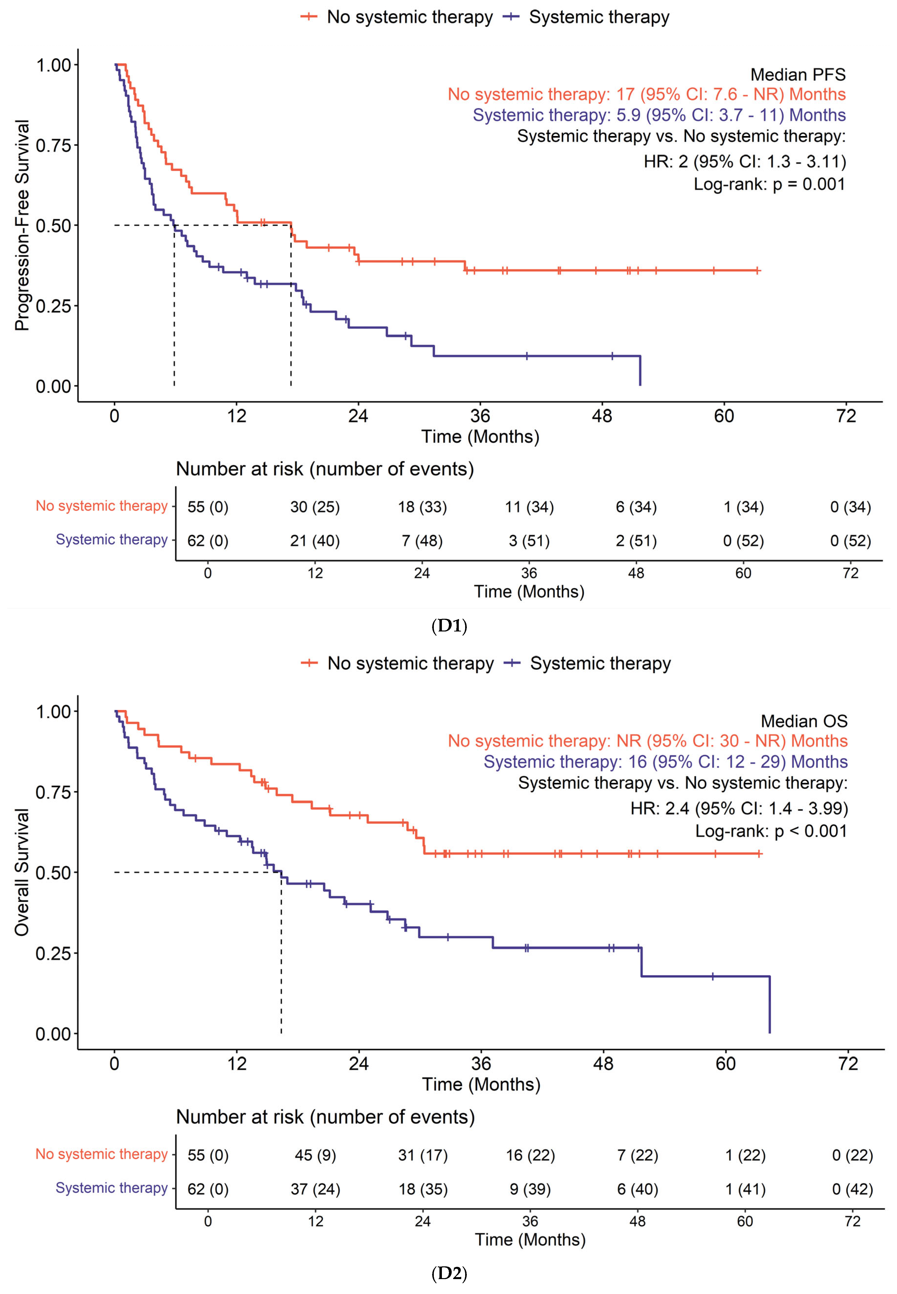
Survival based on subgroup is shown in Figure 4. Those with ICI-sensitive tumors demonstrated improved PFS (p = 0.009) and OS (p = 0.014) compared to non-ICI-sensitive tumors (Figure 4A). Liver metastasis was associated with worse PFS (p = 0.015) and OS (p = 0.006) (Figure 4B). A cutoff of TMB ≥ 15 mut/Mb did not show significant difference in PFS (p = 0.95) or OS (p = 0.79) in the ICI-sensitive tumor subset; however, it was linked to improved PFS (p = 0.012) in patients with non-ICI-sensitive tumors (Figure 4C). Lack of previous systemic therapy also demonstrated improved PFS (p = 0.001) and OS (p < 0.001) (Figure 4D). Other factors such as age (p = 0.8), sex (p = 0.9), and PD-L1 status (p > 0.9) did not exhibit prognostic significance. Cox proportional hazards assumptions were satisfied for all predictors.
Mutations in the MYC pathway (p = 0.03) and MLL2 (p = 0.014) were linked to a poorer response, whereas mutations in the TERT gene (p = 0.031) were associated with improved response. The following pathways did not show significant prognostic or predictive value for survival: TP53 (p = 0.3), RTK/RAS (p > 0.9), PIK3 (p = 0.4), NOTCH (p = 0.5), WNT (p = 0.9), NRF2 (p = 0.2), TGFβ (p = 0.8), and HIPPO (p > 0.9) (Supplementary Tables S1 and S2).

4. Discussion

In this work, the study presented in [30], is expended upon. We found that patients with high TMB had similar response rates to ICIs as reported in the KEYNOTE-158 trial. Type of tumor, absence of liver metastasis, lack of prior systemic therapy, and presence of TERT mutation were associated with improved responses to ICIs. Additionally, TMB ≥ 15 mut/Mb correlated with improved responses among patients categorized as non-ICI-sensitive. Conversely, liver metastasis and specific mutations in MYC and MLL2 genes were associated with a poor response to ICIs across all patient groups. Factors such as PD-L1 status, age, and gender did not demonstrate significant prognostic value.
TMB has both limited sensitivity and specificity for the prediction of benefit from ICIs [31]. Previous studies examining the association of TMB with ICI responses in specific tumor types often failed to confirm the positive associations observed in more diverse cohorts [24,32]. Additionally, recent immunoscore trials suggest that a high TMB alone may not be a reliable predictor of responses to ICIs in solid tumors, suggesting potential limitations in the clinical utility of TMB [33]. Our study reinforces earlier studies highlighting the significance of tumor type when utilizing TMB-H as a predictive biomarker. We categorized patients with TMB-H into two groups based on their eligibility for standalone ICI treatment approved by the FDA: ICI-sensitive and non-ICI-sensitive [34]. The non-ICI-sensitive group primarily consisted of patients with CRC, PBC, and SCLC. In contrast, the ICI-sensitive group included patients with melanoma, NSCLC, urothelial cancer, and SCC. Notably, patients in the ICI-sensitive group experienced significantly better survival rates with ICI treatments.
The influence of tumor type on survival underscores the importance of various clinical and molecular factors. One significant clinical factor that may affect the efficacy of ICIs in solid tumors is the presence of liver metastases. Recent findings indicate that the immunosuppressive environment of the liver may contribute to resistance to ICIs among patients with liver metastases [35,36,37,38,39]. In our study, subgroup analysis revealed that 20% of patients in the ICI-sensitive group had liver metastases. In comparison, 45% of patients in the non-ICI-sensitive group had liver metastases. This higher incidence of liver metastases in the non-ICI-sensitive group may explain their reduced response to ICIs. Moreover, our study demonstrated a significant difference in median PFS and OS between patients with and without liver metastases, which aligns with recent data.
Numerous studies indicate that incorporating underlying mutational processes alongside traditional TMB may provide more accurate guidance for applying ICIs [40,41]. For instance, clinical studies involving the same tumor type have yielded differing results regarding patients classified as having high TMB. The phase I/II non-randomized trial, CheckMate 032, reported improvements in ORR and one-year OS rates in SCLC patients with high TMB, particularly among those receiving a combination of nivolumab and ipilimumab [42]. In contrast, the phase III trial, IMpower133, did not demonstrate an enhancement in OS for patients with high TMB who received atezolizumab combined with platinum-based chemotherapy compared to those with low TMB subjected to the same treatment regimen [43]. The high prevalence of the MYC oncogene in SCLC may account for this discrepancy [44], as elevated MYC expression has been associated with reduced survival following anti-PD-L1 treatment in solid tumors [45,46,47,48]. Our study has identified a correlation between MYC mutations and a low response rate to immunotherapy in solid tumors characterized by high TMB. MYC gene mutations have been associated with promoting tumor immune evasion through multiple mechanisms, including suppression of interferon signaling, downregulation of antigen presentation (such as MHC class I), upregulation of immune checkpoint molecules, and remodeling of the tumor microenvironment. These changes collectively result in a “cold” tumor immune phenotype with low T-cell infiltration, plausibly resulting in diminished response to immune checkpoint blockade [49].
MLL2 (also known as KMT2B) is a histone lysine methyltransferase that plays a crucial role as an epigenetic regulator of transcription [50]. MLL2 alterations are present in some immunotherapy-responsive cancer types, such as up to 29% of melanoma [51]. In some cancers, such as early-stage lung SCC, cervical cancer, and gastrointestinal diffuse large B cell lymphoma, MLL2 has been linked to poor prognosis [52,53,54,55]. The role of MLL2 gene alterations as a biomarker for cancer immunotherapy is still being explored. A study involving various solid tumors found that in a small group of patients with MLL2 deleterious alterations, there was an association with higher TMB and an improved survival after ICI therapy, though MLL2 was not independently predictive of response to ICI [56]. A study assessing 11 long-term survivors with extensive-stage SCLC suggests that alterations in MLL2 may predict better survival outcomes for patients undergoing first-line chemoimmunotherapy [57]. In this same study, in order to validate findings, a large pan-cancer immunotherapy cohort was assessed, and KMT2B mutations correlated with worse OS (p = 0.007). Moreover, KMT2B had divergent effects on immunotherapy response, with higher KMT2B expression associated with response in melanoma patients (p = 0.043) and lower KMT2B expression linked to response in stomach adenocarcinoma patients (p = 0.001). In our study, alterations in MLL2 were overall associated with a poor response to ICIs in TMB-H solid tumors, suggesting that the response to ICI in the presence of MLL2 alterations varies according to tumor type or MLL2 alteration type. While the exact mechanism for this discrepancy is not known, the role of MLL2 in the tumor microenvironment continues to be elucidated. In lymphoma models, loss of function of MLL2 is known to disrupt enhancer-mediated transcriptional regulation, leading to reduced expression of genes involved in antigen presentation and interferon signaling [58,59]. While this may not be applicable to carcinomas, it is possible that the resultant altered tumor immune microenvironment may promote immune evasion and explain suboptimal response to immune checkpoint inhibitor therapy in certain tumor types.
TERT mutations are significantly associated with increased TMB and a higher neoantigen load, which can lead to improved responses to ICIs [60]. Numerous studies have demonstrated that TERT mutations are linked to more favorable responses to ICIs in solid tumors [61]. However, a recent study found that TERT mutations are associated with poorer PFS in biliary tract carcinoma [62]. Additionally, co-mutations of TP53 and TERT have been linked to worse survival outcomes in patients with hepatocellular carcinoma [63]. Unfortunately, we were unable to evaluate these co-mutation correlations in our cohort due to limited sample size. However, these findings emphasize that mutational profiles alone are insufficient for predicting ICI response, highlighting the importance of cancer type and co-occurring mutations.
In the Keynote-158 trial, response rates for tumors varied by TMB: 6.7% for TMBs < 10 mut/Mb, 12.5% for 10–13 mut/Mb, and 37% for >13 mut/Mb. This suggests the 10 mut/Mb cutoff is ineffective for distinguishing responders [16]. This threshold was established based on earlier retrospective studies involving patients with NSCLC [64]. However, numerous studies conducted since then suggest that a reevaluation of appropriate, potentially cancer-type-specific TMB cutoffs is necessary to improve patient selection for those most likely to benefit from ICIs [24,65]. In our study, 60% of patients demonstrated a TMB ≥ 15 mut/Mb. However, evaluating the entire patient population, this elevated TMB was not linked to enhanced survival outcomes. Conversely, within the subgroup of patients who were non-sensitive to ICIs, those with a TMB ≥ 15 mut/Mb exhibited significantly improved survival compared to individuals with TMB values ranging from 10 to 15 mut/Mb. The optimal TMB threshold remains unclear, though establishing one optimal cutoff in heterogenous tumor types with variable tumor microenvironments may not be possible. Further studies to identify optimal cutoffs, particularly focusing on specific tumor types, are warranted.
The limitations of this study include a single center, a small sample size, and its retrospective nature. As with other retrospective cohort studies, immortal time bias is a limitation, though statistical considerations were employed to attempt to mitigate this. Additionally, most patients had likely received ICIs for a different FDA-approved indication, which introduces the potential for confounding factors.

5. Conclusions

Our study highlights the predictive validity of TMB as a critical biomarker for assessing the effectiveness of ICIs across various cancer types. The benefits of TMB are especially significant in patients with TERT mutations and who do not have liver metastases. Additionally, TMB of ≥15 mut/Mb were associated with better outcomes for non-ICI-sensitive tumor types. On the other hand, we found that specific mutations, such as those in the MYC, and MLL2 genes, can negatively impact the effectiveness of these treatments.

Supplementary Materials

The following supporting information can be downloaded at: https://www.mdpi.com/article/10.3390/cancers17162673/s1, Table S1: Pathways; Table S2: Genes.

Author Contributions

Conceptualization, I.N., T.D., K.D., R.C., M.K., N.K. and D.M.; methodology, I.N., T.D., K.D., R.C., P.D., N.K., C.M. and D.M.; software, I.N., T.D., K.D., R.C., M.K., N.K. and D.M.; validation, I.N., T.D., K.D., R.C., M.K., P.D., N.K., C.M. and D.M.; formal analysis, I.N., T.D., K.D., R.C., M.K., N.K. and D.M.; investigation, I.N., T.D., K.D., R.C., M.K., P.D., N.K., C.M. and D.M.; resources, I.N., T.D., K.D., R.C., M.K., P.D., N.K., C.M. and D.M.; data curation, I.N., T.D., K.D., R.C., M.K., P.D., N.K., C.M. and D.M.; writing—original draft preparation, I.N., T.D., K.D., R.C., M.K., P.D., N.K., C.M. and D.M.; writing—review and editing, I.N., T.D., K.D., R.C., M.K., P.D., N.K., C.M. and D.M.; visualization, I.N., T.D., K.D., R.C., M.K., P.D., N.K., C.M. and D.M.; supervision, I.N., T.D., R.C., M.K. and D.M.; project administration, I.N., T.D., K.D., R.C., M.K., P.D., N.K., C.M. and D.M.; funding acquisition, I.N., T.D., K.D., R.C., M.K., P.D., N.K., C.M. and D.M. All authors have read and agreed to the published version of the manuscript.

Funding

This work is supported in part by the NIH/NCI Cancer Center Support Grant R35CA197450.

Institutional Review Board Statement

The Northwestern University institutional review board approved the study protocol, which was conducted in compliance with Good Clinical Practice and the Declaration of Helsinki (IRB Number: STU00207984, date of approval: 7/30/2018).

Informed Consent Statement

Not applicable. Informed consent was not necessary due to the retrospective collection of demographic and clinical characteristics, disease outcomes, and toxicity outcomes.

Data Availability Statement

The data presented in this study are available on request from the corresponding author.

Conflicts of Interest

D.M. has received research funding from Amgen, Merck, Oncolytics, and Rafael; is on the scientific advisory board for Actuate and Qurient; acts as an advisory/speaker bureau for Amgen, BMS, Eisai, and Exelixis; has received funding paid to their institution from Acepodia, Actuate Therapeutics, ADC Therapeutics, Amgen, AVEO, Bayer, Blueprint Medicines, BMS, BioNTech, Dialectic Therapeutics, Epizyme, Fujifilm, ImmuneSensor, Immune-Onc Therapeutics, Leap Therapeutics, Lycera Corp, Merck, Millennium, MiNA Alpha, NGM Biopharmaceuticals, Novartis, Oncolytics, Orano Med, Puma, Qurient, Rafael, Repare Therapeutics, Triumvira Immunologics, Vigeo Therapeutics, and Warewolf Therapeutics. All other authors report no conflicts of interest.

Abbreviations

ACCAdrenal corticoid carcinoma
BCBreast cancer
CRComplete response
CRCColorectal cancer
CTCAECommon Terminology Criteria for Adverse Events
DMDiabetes mellitus
dMMRDeficient mismatch repair
FDAFood and drug administration
GCGynecologic cancers
HRHazard ratios
ICIImmune checkpoint inhibitor
IQRInterquartile range
irAEsImmune-related adverse events
MSI-HHigh microsatellite instability
mut/MbMutations per megabase
NNumber
N/ANot assessed
NENot evaluable
NGSNext-generation sequencing
NSCLCNon-small cell lung cancer
ORROverall response rate
OSOverall survival
PBCPancreaticobiliary cancers
PDProgression of disease
PD-L1Programmed cell death ligand 1
PFSProgression-free survival
PR PArtial response
SCCSquamous cell carcinoma
SCLCSmall cell lung cancer
SDStable disease 
TMBTumor mutational burden
UGCUpper gastrointestinal cancers

References

  1. Hodi, F.S.; O’Day, S.J.; McDermott, D.F.; Weber, R.W.; Sosman, J.A.; Haanen, J.B.; Gonzalez, R.; Robert, C.; Schadendorf, D.; Hassel, J.C.; et al. Improved Survival with Ipilimumab in Patients with Metastatic Melanoma. N. Engl. J. Med. 2010, 363, 711–723. [Google Scholar] [CrossRef]
  2. Vaddepally, R.K.; Kharel, P.; Pandey, R.; Garje, R.; Chandra, A.B. Review of Indications of FDA-Approved Immune Checkpoint Inhibitors per NCCN Guidelines with the Level of Evidence. Cancers 2020, 12, 738. [Google Scholar] [CrossRef]
  3. Prasad, V.; Haslam, A.; Olivier, T. Updated Estimates of Eligibility and Response: Immune Checkpoint Inhibitors. J. Clin. Oncol. 2024, 42, e14613. [Google Scholar] [CrossRef]
  4. Mohamed, Y.I.; Lee, S.S.; Demir, T.; Chamseddine, S.; Hu, Z.I.; Xiao, L.; Elsayes, K.; Morris, J.S.; Wolff, R.A.; Hiatia, R.; et al. Circulating Tumor DNA (CtDNA) as a Biomarker of Response to Therapy in Advanced Hepatocellular Carcinoma Treated with Nivolumab. Cancer Biomark. 2024, 41, 83–91. [Google Scholar] [CrossRef]
  5. Akula, V.; Chen, L.; Acikgoz, Y.; Klein, K.; Yavuz, B.G.; Cevik, L.; Demir, T.; Manne, A.; Sahin, I.; Kaseb, A.; et al. Neoadjuvant Immune Checkpoint Inhibitors for Hepatocellular Carcinoma. NPJ Precis. Oncol. 2025, 9, 60. [Google Scholar] [CrossRef]
  6. Cortes, J.; Cescon, D.W.; Rugo, H.S.; Nowecki, Z.; Im, S.A.; Yusof, M.M.; Gallardo, C.; Lipatov, O.; Barrios, C.H.; Holgado, E.; et al. Pembrolizumab plus Chemotherapy versus Placebo plus Chemotherapy for Previously Untreated Locally Recurrent Inoperable or Metastatic Triple-Negative Breast Cancer (KEYNOTE-355): A Randomised, Placebo-Controlled, Double-Blind, Phase 3 Clinical Trial. Lancet 2020, 396, 1817–1828. [Google Scholar] [CrossRef]
  7. Le, D.T.; Durham, J.N.; Smith, K.N.; Wang, H.; Bartlett, B.R.; Aulakh, L.K.; Lu, S.; Kemberling, H.; Wilt, C.; Luber, B.S.; et al. Mismatch Repair Deficiency Predicts Response of Solid Tumors to PD-1 Blockade. Science 2017, 357, 409–413. [Google Scholar] [CrossRef] [PubMed]
  8. Prasad, V.; Kaestner, V.; Mailankody, S. Cancer Drugs Approved Based on Biomarkers and Not Tumor Type—FDA Approval of Pembrolizumab for Mismatch Repair-Deficient Solid Cancers. JAMA Oncol. 2018, 4, 157–158. [Google Scholar] [CrossRef] [PubMed]
  9. Büttner, R.; Longshore, J.W.; López-Ríos, F.; Merkelbach-Bruse, S.; Normanno, N.; Rouleau, E.; Penault-Llorca, F. Implementing TMB Measurement in Clinical Practice: Considerations on Assay Requirements. ESMO Open 2019, 4, e000442. [Google Scholar] [CrossRef] [PubMed]
  10. Gubin, M.M.; Artyomov, M.N.; Mardis, E.R.; Schreiber, R.D. Tumor Neoantigens: Building a Framework for Personalized Cancer Immunotherapy. J. Clin. Investig. 2015, 125, 3413–3421. [Google Scholar] [CrossRef] [PubMed]
  11. Goodman, A.M.; Kato, S.; Bazhenova, L.; Patel, S.P.; Frampton, G.M.; Miller, V.; Stephens, P.J.; Daniels, G.A.; Kurzrock, R. Tumor Mutational Burden as an Independent Predictor of Response to Immunotherapy in Diverse Cancers. Mol. Cancer Ther. 2017, 16, 2598–2608. [Google Scholar] [CrossRef]
  12. Richard, C.; Fumet, J.D.; Chevrier, S.; Derangere, V.; Ledys, F.; Lagrange, A.; Favier, L.; Coudert, B.; Arnould, L.; Truntzer, C.; et al. Exome Analysis Reveals Genomic Markers Associated with Better Efficacy of Nivolumab in Lung Cancer Patients. Clin. Cancer Res. 2019, 25, 957–966. [Google Scholar] [CrossRef]
  13. Snyder, A.; Makarov, V.; Merghoub, T.; Yuan, J.; Zaretsky, J.M.; Desrichard, A.; Walsh, L.A.; Postow, M.A.; Wong, P.; Ho, T.S.; et al. Genetic Basis for Clinical Response to CTLA-4 Blockade in Melanoma. N. Engl. J. Med. 2014, 371, 2189–2199. [Google Scholar] [CrossRef] [PubMed]
  14. Kim, J.Y.; Kronbichler, A.; Eisenhut, M.; Hong, S.H.; van der Vliet, H.J.; Kang, J.; Shin, J., II.; Gamerith, G. Tumor Mutational Burden and Efficacy of Immune Checkpoint Inhibitors: A Systematic Review and Meta-Analysis. Cancers 2019, 11, 1798. [Google Scholar] [CrossRef] [PubMed]
  15. Yarchoan, M.; Hopkins, A.; Jaffee, E.M. Tumor Mutational Burden and Response Rate to PD-1 Inhibition. N. Engl. J. Med. 2017, 377, 2500–2501. [Google Scholar] [CrossRef]
  16. Marabelle, A.; Fakih, M.; Lopez, J.; Shah, M.; Shapira-Frommer, R.; Nakagawa, K.; Chung, H.C.; Kindler, H.L.; Lopez-Martin, J.A.; Miller, W.H.; et al. Association of Tumour Mutational Burden with Outcomes in Patients with Advanced Solid Tumours Treated with Pembrolizumab: Prospective Biomarker Analysis of the Multicohort, Open-Label, Phase 2 KEYNOTE-158 Study. Lancet Oncol. 2020, 21, 1353–1365. [Google Scholar] [CrossRef] [PubMed]
  17. Marcus, L.; Fashoyin-Aje, L.A.; Donoghue, M.; Yuan, M.; Rodriguez, L.; Gallagher, P.S.; Philip, R.; Ghosh, S.; Theoret, M.R.; Beaver, J.A.; et al. FDA Approval Summary: Pembrolizumab for the Treatment of Tumor Mutational Burden-High Solid Tumors. Clin. Cancer Res. 2021, 27, 4685–4689. [Google Scholar] [CrossRef]
  18. Demir, T.; Moloney, C.; Mahalingam, D. Threading the Needle: Navigating Novel Immunotherapeutics in Pancreatic Ductal Adenocarcinoma. Cancers 2025, 17, 715. [Google Scholar] [CrossRef]
  19. Demir, T.; Moloney, C.; Mahalingam, D. Emerging Targeted Therapies and Strategies to Overcome Resistance in Biliary Tract Cancers. Crit. Rev. Oncol. Hematol. 2024, 199, 104388. [Google Scholar] [CrossRef]
  20. Subbiah, V.; Solit, D.B.; Chan, T.A.; Kurzrock, R. The FDA Approval of Pembrolizumab for Adult and Pediatric Patients with Tumor Mutational Burden (TMB) ≥10: A Decision Centered on Empowering Patients and Their Physicians. Ann. Oncol. 2020, 31, 1115–1118. [Google Scholar] [CrossRef]
  21. Prasad, V.; Addeo, A. The FDA Approval of Pembrolizumab for Patients with TMB >10 Mut/Mb: Was It a Wise Decision? No. Ann. Oncol. 2020, 31, 1112–1114. [Google Scholar] [CrossRef]
  22. Chan, T.A.; Yarchoan, M.; Jaffee, E.; Swanton, C.; Quezada, S.A.; Stenzinger, A.; Peters, S. Development of Tumor Mutation Burden as an Immunotherapy Biomarker: Utility for the Oncology Clinic. Ann. Oncol. 2019, 30, 44–56. [Google Scholar] [CrossRef]
  23. Rousseau, B.; Foote, M.B.; Maron, S.B.; Diplas, B.H.; Lu, S.; Argilés, G.; Cercek, A.; Diaz, L.A. The Spectrum of Benefit from Checkpoint Blockade in Hypermutated Tumors. N. Engl. J. Med. 2021, 384, 1168–1170. [Google Scholar] [CrossRef]
  24. McGrail, D.J.; Pilié, P.G.; Rashid, N.U.; Voorwerk, L.; Slagter, M.; Kok, M.; Jonasch, E.; Khasraw, M.; Heimberger, A.B.; Lim, B.; et al. High Tumor Mutation Burden Fails to Predict Immune Checkpoint Blockade Response across All Cancer Types. Ann. Oncol. 2021, 32, 661–672. [Google Scholar] [CrossRef]
  25. Seymour, L.; Bogaerts, J.; Perrone, A.; Ford, R.; Schwartz, L.H.; Mandrekar, S.; Lin, N.U.; Litière, S.; Dancey, J.; Chen, A.; et al. IRECIST: Guidelines for Response Criteria for Use in Trials Testing Immunotherapeutics. Lancet Oncol. 2017, 18, e143–e152. [Google Scholar] [CrossRef]
  26. U.S. Department of Health and Human Services; National Institutes of Health; National Cancer Institute. Common Terminology Criteria for Adverse Events (CTCAE) Version 5.0; U.S. Department of Health and Human Services: Bethesda, MD, USA, 2017. Available online: https://ctep.cancer.gov/protocoldevelopment/electronic_applications/docs/CTCAE_v5_Quick_Reference_8.5x11.pdf (accessed on 14 August 2025).
  27. Patricia Grambsch, B.M.; Therneau, T.M. Proportional Hazards Tests and Diagnostics Based on Weighted Residuals. Biometrika 1994, 81, 515–526. [Google Scholar] [CrossRef]
  28. Sanchez-Vega, F.; Mina, M.; Armenia, J.; Chatila, W.K.; Luna, A.; La, K.C.; Dimitriadoy, S.; Liu, D.L.; Kantheti, H.S.; Saghafinia, S.; et al. Oncogenic Signaling Pathways in The Cancer Genome Atlas. Cell 2018, 173, 321–337.e10. [Google Scholar] [CrossRef]
  29. Duan, Q.; Wang, W.; Feng, F.; Jiang, X.; Chen, H.; Zhang, D.; Zhang, T. Comut-Viz: Efficiently Creating and Browsing Comutation Plots Online. BMC Bioinform. 2023, 24, 226. [Google Scholar] [CrossRef]
  30. Nizamuddin, I.; Doukas, P.; de Viveiros, P.A.H.; Mi, X.; Katam, N.; Chae, Y.K.; Behdad, A.; Wehbe, F.H.; Ma-halingam, D. Microsatellite-stable tumors with high tumor mutational burden in association with tumor re-sponse to immune checkpoint inhibitor therapy across solid tumors and correlation with specific oncogenic alterations. In Proceedings of the Poster Presentation at ASCO 2022 Annual Meeting, Online, 3–7 June 2022. [Google Scholar]
  31. Budczies, J.; Kazdal, D.; Menzel, M.; Beck, S.; Kluck, K.; Altbürger, C.; Schwab, C.; Allgäuer, M.; Ahadova, A.; Kloor, M.; et al. Tumour Mutational Burden: Clinical Utility, Challenges and Emerging Improvements. Nat. Rev. Clin. Oncol. 2024, 21, 725–742. [Google Scholar] [CrossRef] [PubMed]
  32. Cao, D.; Xu, H.; Xu, X.; Guo, T.; Ge, W. High Tumor Mutation Burden Predicts Better Efficacy of Immunotherapy: A Pooled Analysis of 103078 Cancer Patients. Oncoimmunology 2019, 8, e1629258. [Google Scholar] [CrossRef] [PubMed]
  33. Ghiringhelli, F.; Bibeau, F.; Greillier, L.; Fumet, J.D.; Ilie, A.; Monville, F.; Laugé, C.; Catteau, A.; Boquet, I.; Majdi, A.; et al. Immunoscore Immune Checkpoint Using Spatial Quantitative Analysis of CD8 and PD-L1 Markers Is Predictive of the Efficacy of Anti- PD1/PD-L1 Immunotherapy in Non-Small Cell Lung Cancer. EBioMedicine 2023, 92, 104633. [Google Scholar] [CrossRef]
  34. Bai, R.; Chen, N.; Li, L.; Du, N.; Bai, L.; Lv, Z.; Tian, H.; Cui, J. Mechanisms of Cancer Resistance to Immunotherapy. Front. Oncol. 2020, 10, 1290. [Google Scholar] [CrossRef]
  35. Tumeh, P.C.; Hellmann, M.D.; Hamid, O.; Tsai, K.K.; Loo, K.L.; Gubens, M.A.; Rosenblum, M.; Harview, C.L.; Taube, J.M.; Handley, N.; et al. Liver Metastasis and Treatment Outcome with Anti-PD-1 Monoclonal Antibody in Patients with Melanoma and NSCLC. Cancer Immunol. Res. 2017, 5, 417–424. [Google Scholar] [CrossRef]
  36. Yu, J.; Green, M.D.; Li, S.; Sun, Y.; Journey, S.N.; Choi, J.E.; Rizvi, S.M.; Qin, A.; Waninger, J.J.; Lang, X.; et al. Liver Metastasis Restrains Immunotherapy Efficacy via Macrophage-Mediated T Cell Elimination. Nat. Med. 2021, 27, 152–164. [Google Scholar] [CrossRef]
  37. Cohen, R.; Raeisi, M.; Chibaudel, B.; Shi, Q.; Yoshino, T.; Zalcberg, J.R.; Adams, R.; Cremolini, C.; Van Cutsem, E.; Heinemann, V.; et al. Prognostic Value of Liver Metastases in Colorectal Cancer Treated by Systemic Therapy: An ARCAD Pooled Analysis. Eur. J. Cancer 2024, 207, 114160. [Google Scholar] [CrossRef]
  38. Wang, C.; Sandhu, J.; Ouyang, C.; Ye, J.; Lee, P.P.; Fakih, M. Clinical Response to Immunotherapy Targeting Programmed Cell Death Receptor 1/Programmed Cell Death Ligand 1 in Patients With Treatment-Resistant Microsatellite Stable Colorectal Cancer With and Without Liver Metastases. JAMA Netw. Open 2021, 4, e2118416. [Google Scholar] [CrossRef]
  39. Bullock, A.J.; Schlechter, B.L.; Fakih, M.G.; Tsimberidou, A.M.; Grossman, J.E.; Gordon, M.S.; Wilky, B.A.; Pimentel, A.; Mahadevan, D.; Balmanoukian, A.S.; et al. Botensilimab plus Balstilimab in Relapsed/Refractory Microsatellite Stable Metastatic Colorectal Cancer: A Phase 1 Trial. Nat. Med. 2024, 30, 2558–2567. [Google Scholar] [CrossRef] [PubMed]
  40. Newell, F.; Pires da Silva, I.; Johansson, P.A.; Menzies, A.M.; Wilmott, J.S.; Addala, V.; Carlino, M.S.; Rizos, H.; Nones, K.; Edwards, J.J.; et al. Multiomic Profiling of Checkpoint Inhibitor-Treated Melanoma: Identifying Predictors of Response and Resistance, and Markers of Biological Discordance. Cancer Cell 2022, 40, 88–102.e7. [Google Scholar] [CrossRef] [PubMed]
  41. Anagnostou, V.; Niknafs, N.; Marrone, K.; Bruhm, D.C.; White, J.R.; Naidoo, J.; Hummelink, K.; Monkhorst, K.; Lalezari, F.; Lanis, M. Multimodal genomic features predict outcome of immune checkpoint blockade in non-small-cell lung cancer. Nat. Cancer 2020, 1, 99–111. [Google Scholar] [CrossRef]
  42. Hellmann, M.D.; Callahan, M.K.; Awad, M.M.; Calvo, E.; Ascierto, P.A.; Atmaca, A.; Rizvi, N.A.; Hirsch, F.R.; Selvaggi, G.; Szustakowski, J.D.; et al. Tumor Mutational Burden and Efficacy of Nivolumab Monotherapy and in Combination with Ipilimumab in Small-Cell Lung Cancer. Cancer Cell 2018, 33, 853–861.e4. [Google Scholar] [CrossRef] [PubMed]
  43. Horn, L.; Mansfield, A.S.; Szczęsna, A.; Havel, L.; Krzakowski, M.; Hochmair, M.J.; Huemer, F.; Losonczy, G.; Johnson, M.L.; Nishio, M.; et al. First-Line Atezolizumab plus Chemotherapy in Extensive-Stage Small-Cell Lung Cancer. N. Engl. J. Med. 2018, 379, 2220–2229. [Google Scholar] [CrossRef]
  44. Park, S.; Lee, H.; Lee, B.; Lee, S.H.; Sun, J.M.; Park, W.Y.; Ahn, J.S.; Ahn, M.J.; Park, K. DNA Damage Response and Repair Pathway Alteration and Its Association With Tumor Mutation Burden and Platinum-Based Chemotherapy in SCLC. J. Thorac. Oncol. 2019, 14, 1640–1650. [Google Scholar] [CrossRef] [PubMed]
  45. Mariathasan, S.; Turley, S.J.; Nickles, D.; Castiglioni, A.; Yuen, K.; Wang, Y.; Kadel, E.E.; Koeppen, H.; Astarita, J.L.; Cubas, R.; et al. TGFβ Attenuates Tumour Response to PD-L1 Blockade by Contributing to Exclusion of T Cells. Nature 2018, 554, 544–548. [Google Scholar] [CrossRef]
  46. Lee, J.V.; Housley, F.; Yau, C.; Nakagawa, R.; Winkler, J.; Anttila, J.M.; Munne, P.M.; Savelius, M.; Houlahan, K.E.; Van de Mark, D.; et al. Combinatorial Immunotherapies Overcome MYC-Driven Immune Evasion in Triple Negative Breast Cancer. Nat. Commun. 2022, 13, 3671. [Google Scholar] [CrossRef]
  47. Li, J.; Dong, T.; Wu, Z.; Zhu, D.; Gu, H. The Effects of MYC on Tumor Immunity and Immunotherapy. Cell Death Discov. 2023, 9, 103. [Google Scholar] [CrossRef]
  48. Dhanasekaran, R.; Deutzmann, A.; Mahauad-Fernandez, W.D.; Hansen, A.S.; Gouw, A.M.; Felsher, D.W. The MYC Oncogene—The Grand Orchestrator of Cancer Growth and Immune Evasion. Nat. Rev. Clin. Oncol. 2022, 19, 23–36. [Google Scholar] [CrossRef]
  49. Casey, S.C.; Baylot, V.; Felsher, D.W. The MYC Oncogene Is a Global Regulator of the Immune Response. Blood 2018, 131, 2007–2015. [Google Scholar] [CrossRef]
  50. Wang, H.; Helin, K. Roles of H3K4 Methylation in Biology and Disease. Trends Cell Biol. 2025, 35, 115–128. [Google Scholar] [CrossRef]
  51. Lee, J.J.; Sholl, L.M.; Lindeman, N.I.; Granter, S.R.; Laga, A.C.; Shivdasani, P.; Chin, G.; Luke, J.J.; Ott, P.A.; Hodi, F.S.; et al. Targeted Next-Generation Sequencing Reveals High Frequency of Mutations in Epigenetic Regulators across Treatment-Naïve Patient Melanomas. Clin. Epigenetics 2015, 7, 59. [Google Scholar] [CrossRef] [PubMed]
  52. Ye, H.; Lu, L.; Ge, B.; Gao, S.; Ma, Y.; Liang, B.; Yu, K.; Yang, K. MLL2 protein is a prognostic marker for gastrointestinal diffuse large B-cell lymphoma. Int. J. Clin. Exp. Pathol. 2015, 8, 13043–13050. [Google Scholar]
  53. Ge, S.; Li, B.; Li, Y.; Li, Z.; Liu, Z.; Chen, Z.; Wu, J.; Gao, J.; Shen, L. Genomic Alterations in Advanced Gastric Cancer Endoscopic Biopsy Samples Using Targeted Next-Generation Sequencing. Am. J. Cancer Res. 2017, 7, 1540–1553. [Google Scholar] [PubMed]
  54. Choi, M.; Kadara, H.; Zhang, J.; Parra, E.R.; Rodriguez-Canales, J.; Gaffney, S.G.; Zhao, Z.; Behrens, C.; Fujimoto, J.; Chow, C.; et al. Mutation Profiles in Early-Stage Lung Squamous Cell Carcinoma with Clinical Follow-up and Correlation with Markers of Immune Function. Ann. Oncol. 2017, 28, 83–89. [Google Scholar] [CrossRef]
  55. Li, X. Emerging Role of Mutations in Epigenetic Regulators Including MLL2 Derived from The Cancer Genome Atlas for Cervical Cancer. BMC Cancer 2017, 17, 252. [Google Scholar] [CrossRef]
  56. Liang, N.; Niu, Y.; Zhang, X.; Ma, T. The predictive values of loss-of-function variants in histone methyltransferases for response to immune checkpoint inhibitors in solid tumors. J. Clin. Oncol. 2021, 39, 2586. [Google Scholar]
  57. Jiang, Y.; Xie, J.; Cheng, Q.; Cai, Z.; Xu, K.; Lu, W.; Wang, F.; Wu, X.; Song, Y.; Lv, T.; et al. Comprehensive Genomic and Spatial Immune Infiltration Analysis of Survival Outliers in Extensive-Stage Small Cell Lung Cancer Receiving First-Line Chemoimmunotherapy. Int. Immunopharmacol. 2024, 141, 112901. [Google Scholar] [CrossRef]
  58. Li, J.; Chin, C.R.; Ying, H.Y.; Meydan, C.; Teater, M.R.; Xia, M.; Farinha, P.; Takata, K.; Chu, C.S.; Jiang, Y.; et al. Loss of CREBBP and KMT2D Cooperate to Accelerate Lymphomagenesis and Shape the Lymphoma Immune Microenvironment. Nat. Commun. 2024, 15, 2879. [Google Scholar] [CrossRef] [PubMed]
  59. Liu, Q.X.; Zhu, Y.; Yi, H.M.; Shen, Y.G.; Wang, L.; Cheng, S.; Xu, P.P.; Xu, H.M.; Zhou, L.T.; Huang, Y.H.; et al. KMT2D Mutations Promoted Tumor Progression in Diffuse Large B-Cell Lymphoma through Altering Tumor-Induced Regulatory T Cell Trafficking via FBXW7-NOTCH-MYC/TGF-Β1 Axis. Int. J. Biol. Sci. 2024, 20, 3972–3985. [Google Scholar] [CrossRef]
  60. Li, H.; Li, J.; Zhang, C.; Zhang, C.; Wang, H. TERT Mutations Correlate with Higher TMB Value and Unique Tumor Microenvironment and May Be a Potential Biomarker for Anti-CTLA4 Treatment. Cancer Med. 2020, 9, 7151–7160. [Google Scholar] [CrossRef]
  61. Du, J.; Zhang, E.; Huang, Z. The Predictive Value of next Generation Sequencing for Matching Advanced Hepatocellular Carcinoma Patients to Targeted and Immunotherapy. Front. Immunol. 2024, 15, 1358306. [Google Scholar] [CrossRef]
  62. Hwang, I.; Kang, S.Y.; Kim, D.G.; Jang, K.T.; Kim, K.M. Clinicopathologic and Genomic Characteristics of Biliary Tract Carcinomas with TERT Promoter Mutations among East Asian Population. Pathol. Res. Pract. 2025, 266, 155806. [Google Scholar] [CrossRef]
  63. Ou, Q.; Yu, Y.; Li, A.; Chen, J.; Yu, T.; Xu, X.; Xie, X.; Chen, Y.; Lin, D.; Zeng, Q.; et al. Association of Survival and Genomic Mutation Signature with Immunotherapy in Patients with Hepatocellular Carcinoma. Ann. Transl. Med. 2020, 8, 230. [Google Scholar] [CrossRef] [PubMed]
  64. Hellmann, M.D.; Ciuleanu, T.-E.; Pluzanski, A.; Lee, J.S.; Otterson, G.A.; Audigier-Valette, C.; Minenza, E.; Linardou, H.; Burgers, S.; Salman, P.; et al. Nivolumab plus Ipilimumab in Lung Cancer with a High Tumor Mutational Burden. N. Engl. J. Med. 2018, 378, 2093–2104. [Google Scholar] [CrossRef] [PubMed]
  65. Samstein, R.M.; Lee, C.H.; Shoushtari, A.N.; Hellmann, M.D.; Shen, R.; Janjigian, Y.Y.; Barron, D.A.; Zehir, A.; Jordan, E.J.; Omuro, A.; et al. Tumor Mutational Load Predicts Survival after Immunotherapy across Multiple Cancer Types. Nat. Genet. 2019, 51, 202–206. [Google Scholar] [CrossRef]
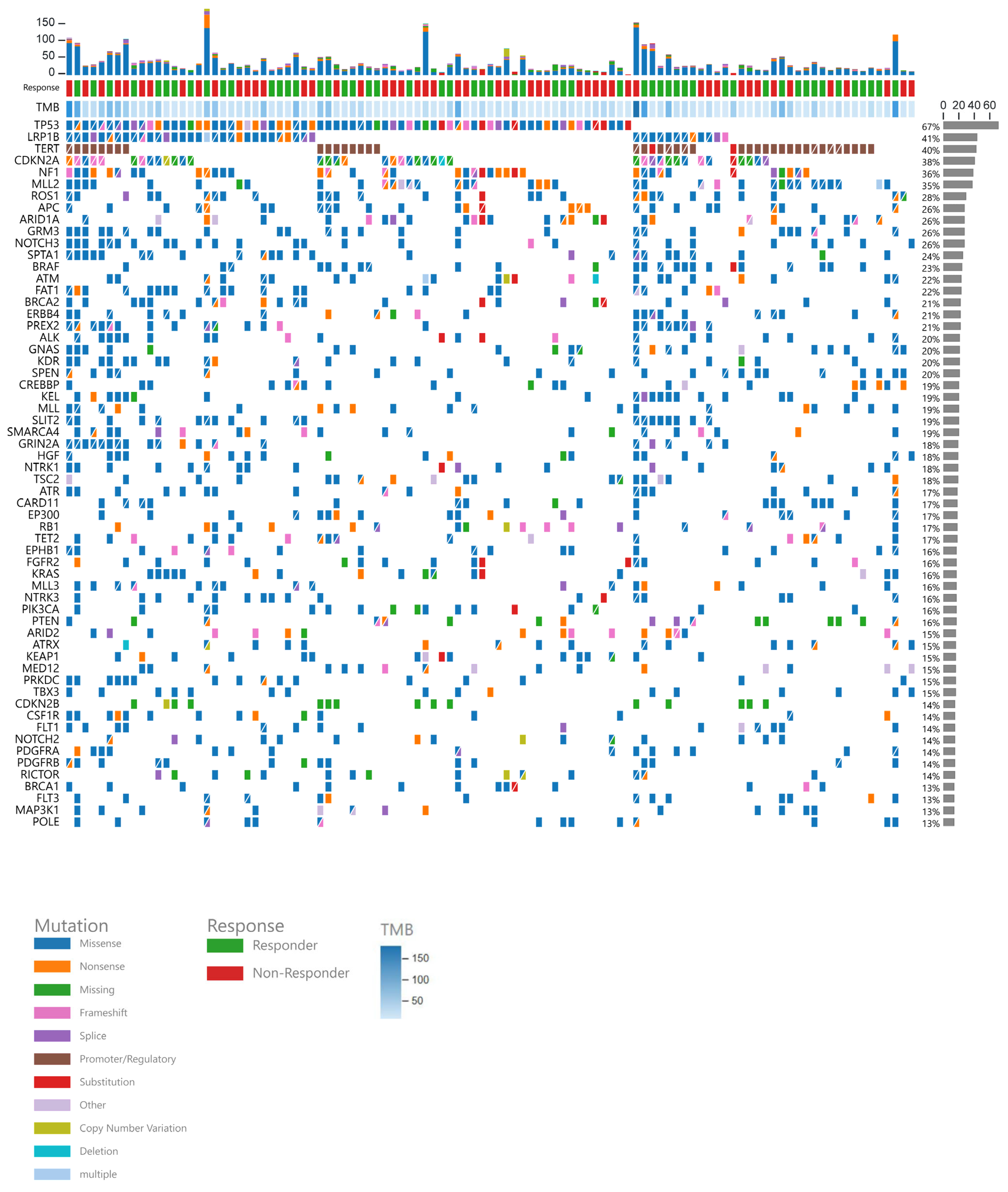
Figure 1. Co-mutation plot displaying mutations per patient (N = 105), with each patient’s mutations listed in individual columns. The stacked bar plot at the top shows the total number of mutations for each patient, followed by their response status, TMB level, and specific detected mutations. The bar graph on the right displays the percentage of patients with each mutation.
Cancers 17 02673 g001
Figure 2. Waterfall plot showing the percentage change in tumor size from baseline based on ICI-sensitive status (A) and TMB status (B) (N = 117).
Cancers 17 02673 g002
Figure 3. Kaplan–Meier estimates and risk tables for PFS (A) and OS (B) for the overall population (N = 117).
Cancers 17 02673 g003
Figure 4. (A1): ICI-sensitive tumors demonstrated improved PFS compared to non-ICI-sensitive tumors. (A2): ICI-sensitive tumors demonstrated improved OS compared to non-ICI-sensitive tumors. (B1): Liver metastasis was associated with worse PFS. (B2): Liver metastasis was associated with worse OS. (C1): A cutoff of TMB ≥ 15 mut/Mb showed a significant difference in PFS in patients with non-ICI-sensitive tumors. (C2): A cutoff of TMB ≥ 15 mut/Mb did show significant difference in OS in patients with non-ICI-sensitive tumors. (D1): Lack of previous systemic therapy demonstrated improved PFS. (D2): Lack of previous systemic therapy demonstrated improved OS.
Cancers 17 02673 g004aCancers 17 02673 g004bCancers 17 02673 g004cCancers 17 02673 g004d
Table 1. Baseline characteristics in the overall group and based on treatment response.
CharacteristicOverall, n = 117 1[SD, <6 m]/PD, n = 51 1CR/PR/[SD, >6 m], n = 54 1p-Value 2
Age 68 (62, 76) 68 (62, 77)69 (62, 76)0.8
Sex   0.9
Female 46 (39%) 20 (39%)22 (41%) 
Male 71 (61%) 31 (61%)32 (59%) 
Race   0.2
Asian 4 (3.4%) 3 (5.9%)1 (1.9%) 
Black/African American 7 (6.0%) 1 (2.0%)4 (7.4%) 
Unknown 3 (2.6%) 2 (3.9%)0 (0%) 
White 103 (88%) 45 (88%)49 (91%) 
PD-L1 status   >0.9
Positive 36 (46%) 16 (46%)18 (49%) 
Negative 31 (39%) 13 (37%)13 (35%) 
NE 12 (15%) 6 (17%)6 (16%) 
Unknown 38 1617 
Previous therapies   0.039
No systemic therapy 55 (47%) 19 (37%)31 (57%) 
Adjuvant/neoadjuvant/definitive therapy 62 (53%) 32 (63%)23 (43%) 
Previous therapy lines   0.10
One line 29 (48%) 11 (35%)14 (61%) 
Two lines 17 (28%) 10 (32%)5 (22%) 
Three lines 5 (8.2%) 5 (16%)0 (0%) 
Four or more lines 10 (16%) 5 (16%)4 (17%) 
Unknown 56 2031 
All responses    <0.001
Complete response 15 (13%) 0 (0%)15 (28%) 
Partial response 21 (18%) 0 (0%)21 (39%) 
Stable disease 22 (19%) 4 (7.8%)18 (33%) 
Progressive disease 47 (40%) 47 (92%)0 (0%) 
NE 8 (6.8%) 0 (0%)0 (0%) 
N/A 4 (3.4%) 0 (0%)0 (0%) 
First line immunotherapy    0.048
Ipilimumab 2 (1.7%) 2 (3.9%)0 (0%) 
Nivolumab 32 (27%) 15 (29%)14 (26%) 
Ipilimumab/Nivolumab 19 (16%) 5 (9.8%)11 (20%) 
Pembrolizumab 43 (37%) 16 (31%)23 (43%) 
Atezolizumab 13 (11%) 10 (20%)2 (3.7%) 
Durvalumab 1 (0.9%) 0 (0%)1 (1.9%) 
Other 7 (6.0%) 3 (5.9%)3 (5.6%) 
Known ICI-sensitive tumor    0.13
Non-ICI-sensitive 29 (25%) 16 (31%)10 (19%) 
ICI-sensitive 88 (75%) 35 (69%)44 (81%) 
Tumor type    0.2
Adrenocortical carcinoma 2 (1.7%) 1 (2.0%)1 (1.9%) 
Anal squamous cell carcinoma 2 (1.7%) 1 (2.0%)0 (0%) 
Breast cancer 2 (1.7%) 0 (0%)1 (1.9%) 
Colorectal carcinoma 6 (5.1%) 4 (7.8%)2 (3.7%) 
Gynecologic carcinoma 4 (3.4%) 2 (3.9%)2 (3.7%) 
Melanoma 39 (33%) 12 (24%)24 (44%) 
Non-small cell lung carcinoma 33 (28%) 18 (35%)12 (22%) 
Other skin 6 (5.1%) 1 (2.0%)4 (7.4%) 
Pancreatobiliary 5 (4.3%) 2 (3.9%)2 (3.7%) 
Small cell lung carcinoma 7 (6.0%) 3 (5.9%)3 (5.6%) 
Unknown primary 4 (3.4%) 4 (7.8%)0 (0%) 
Upper GI 2 (1.7%) 1 (2.0%)1 (1.9%) 
Urothelial carcinoma 4 (3.4%) 2 (3.9%)2 (3.7%) 
Head and neck SCC 1 (0.9%) 0(0%)1(1.9%) 
MSI status    0.5
MSI stable 113 (96.6%) 49 (96%)52 (96.3%) 
N/A 4 (3.4%) 2 (4%)2 (3.7%) 
TMB status    0.5
TMB 10–15 47 (40%) 22 (43%)20 (37%) 
TMB ≥ 15 70 (60%) 29 (57%)34 (63%) 
1 Median (IQR); n (%). 2 Wilcoxon rank sum test; Pearson’s chi-squared test; Fisher’s exact test.
Table 2. Summary of TMB levels by tumor type.
GroupTumor TypeMedian TMB Level (mut/Mb)IQR (mut/Mb)
ICI-sensitiveUnknown primary (SCC)27.821.1, 49.5
Melanoma26.515.1, 56.7
Non-small cell lung cancer14.913.5, 26.5
Adrenal corticoid carcinoma35.829.8, 41.9
Anal SCC10.110.1, 10.1
Head and neck SCC16.716.7, 16.7
Genitourinary carcinoma ¥15.813.9, 19.5
Other skin cancers *27.713.9, 48.2
Non-ICI-sensitiveBreast cancer11.310.9, 11.7
Colorectal cancer15.212.0, 27.8
Gynecologic cancers (non-SCC)11.310.3, 24.1
Pancreaticobiliary cancers15.115.0, 19.3
Small cell lung cancer14.911.0, 17.1
Upper gastrointestinal cancers Ɨ (non-SCC)15.313.3, 17.3
¥ Renal cell carcinomas and urothelial carcinomas; * basal cell carcinomas and squamous cell carcinomas; Ɨ esophageal, stomach, and duodenal adenocarcinomas.
Table 3. Immune-related adverse events classified by severity per CTCAE v5.0.
Adverse EventGrade 1 Grade 2 Grade 3 Grade 4 Grade 5 Total (n = 48/117) (41%)
Thyroid disease3 (2.5%)2 (1.7%)0005 (4.2%)
Adrenal insufficiency1 (0.8%)1 (0.8%)0002 (1.7%)
Colitis2 (1.7%)1 (0.8%)4 (3.4%)2 (1.7%)09 (7.6%)
Pneumonitis01 (0.8%)3 (2.5%)1 (0.8%)1 (0.8%)6 (5%)
Infusion reactions01 (0.8%)0001 (0.8%)
Severe skin reactions5 (4.2%)1 (0.8%)1 (0.8%)007 (5.9%)
Hepatitis2 (1.7%)1 (0.8%)3 (2.5%)3 (2.5%)09 (7.6%)
Nephritis01 (0.8%)1 (0.8%)002 (1.7%)
Hypophysitis05 (4.2%)0005 (4.2%)
Myocarditis002 (1.7%)002 (1.7%)
Type 1 DM000000
Pancreatitis000000
Disclaimer/Publisher’s Note: The statements, opinions and data contained in all publications are solely those of the individual author(s) and contributor(s) and not of MDPI and/or the editor(s). MDPI and/or the editor(s) disclaim responsibility for any injury to people or property resulting from any ideas, methods, instructions or products referred to in the content.