Article

Prognostic Impact of the AML60+ Score for Elderly Patients with Acute Myeloid Leukemia Treated with Hypomethylating Agents: A Retrospective Multicentric Analysis

1
Clinic for Medical Oncology and Hematology, Cantonal Hospital St. Gallen, 9007 St. Gallen, Switzerland
2
Department of Internal Medicine, Cantonal Hospital Graubünden, 7000 Chur, Switzerland
3
Institute of Pathology, Cantonal Hospital St. Gallen, 9007 St. Gallen, Switzerland
4
Swiss Cancer Institute, Statistics Unit, 3008 Bern, Switzerland
5
Center for Laboratory Medicine, St. Gallen, 9007 St. Gallen, Switzerland
6
Medical Oncology and Hematology, Cantonal Hospital Winterthur, 8400 Winterthur, Switzerland
7
Department of Medical Oncology and Hematology, Cantonal Hospital Graubünden, 7000 Chur, Switzerland
*
Author to whom correspondence should be addressed.
These authors contributed equally to this work.
Cancers 2025, 17(16), 2658; https://doi.org/10.3390/cancers17162658
Submission received: 29 June 2025 / Revised: 3 August 2025 / Accepted: 11 August 2025 / Published: 14 August 2025

Simple Summary

The treatment of acute myeloid leukemia (AML) in elderly patients has changed significantly with the introduction of venetoclax in combination with hypomethylating agents (HMA). The AML60+ score was developed to identify elderly patients who would benefit from intensive chemotherapy and allogeneic stem cell transplantation. The aim of our retrospective and multicenter study was to determine whether the AML60+ is also helpful in assessing the prognosis of elderly AML patients receiving HMA-based therapy. Patients with lower risk according to AML60+ lived significantly longer than those within the higher risk categories. The C-index, as a comparative measure of prognostic significance, was higher for AML60+ than for the two indices used for comparison, the molecular Prognostic Scoring System and the European Leukemia Net 2022 classification. Given the small sample size, analyses of larger cohorts are needed to confirm our observation.

Abstract

Background: The AML60+ score has been proposed for risk stratification in intensively treated elderly patients with acute myeloid leukemia (AML) or high-risk myelodysplastic neoplasms (MDS). Its prognostic impact in patients treated with hypomethylating agents (HMA) is unknown. Methods: Patients ≥ 60 years of age diagnosed with AML or MDS/AML according to ICC2022 were eligible for this retrospective and multicenter chart review if they had received at least one cycle of HMA-based treatment. Results: A cohort of 142 patients was analyzed. During follow-up (median 8 months), 114 patients died. The molecular Prognostic Score (mPRS) was available for 121 patients, the European Leukemia Net (ELN) 2022 classification for 117 patients, and the AML60+ for 105 patients. According to AML60+, 33 patients (31.4%) were classified as very poor risk, 36 (34.3%) as poor risk, and 34 (32.4%) as intermediate risk. Two patients (1.9%) were classified as favorable. Median overall survival (OS) was 21.7 months (mo) for the combined intermediate/favorable group, 7 mo for the poor risk group and 3 mo for the very poor risk group (p < 0.0001). Cox regression analysis (reference category: very poor) showed a significantly lower risk of death for both intermediate/favorable risk patients (HR 0.17, 95% CI 0.10–0.31, p < 0.001) and poor risk patients (HR 0.47, 95% CI 0.28–0.78, p = 0.004). The concordance score was 0.67 for AML60+, 0.60 for mPRS, and 0.58 for ELN2022. Conclusions: The AML60+ may represent a useful prognostic tool for elderly AML patients treated with HMA-based therapies. In particular, it could help to identify a group with a relatively favorable prognosis that is not clearly identified by the ELN2022 or the mPRS risk classification. However, analyses of larger cohorts are necessary to confirm our findings.

Graphical Abstract

1. Introduction

Acute myeloid leukemia (AML) and AML/Myelodysplastic neoplasms (AML/MDS) are heterogeneous clonal disorders of myeloid progenitor cells [1]. With a median age at diagnosis of about 70 years [2], they are primarily diseases of the elderly. In this patient group, comorbidities and frailty often lead to ineligibility for intensive treatments such as induction chemotherapy or allogeneic stem-cell transplantation.
Instead, these patients receive less intensive regimens. A current standard of care is the combination of hypomethylating agents (HMAs), such as azacitidine (Aza) or decitabine (Dec), with the BCL2 inhibitor venetoclax (Ven) [3,4]. While these treatments may not be as effective in the real world as they were in the trials that led to their approval, they provide a clinically meaningful benefit to this difficult-to-treat population [5,6]. Despite a significant risk of tumor lysis syndrome and infection in the early phase of treatment and myelosuppression later, HMA-based regimens can be safely used even in octogenarians and nonagenarians [7] when proper precautions are taken [8]. Long-term responses appear increasingly possible [9,10], even after stopping treatment [11,12].
The European Leukemia Net (ELN) 2022 classification [3] was developed using data from patients treated with intensive chemotherapy and proved to be of less value when applied to patients treated with HMA ± Ven. Therefore, several prognostic scores have been proposed for non-intensively treated patients. Some of them are based on the mutational profile alone, such as the molecular Prognostic Scoring System (mPRS) [13,14] or the “Genetic Risk Classification for Adults with AML Receiving Less Intensive Treatments” proposed by the ELN in 2024 (ELN2024) [15] while other systems still consider the cytogenetic profile in terms of adverse cytogenetic risk together with the mutational profile, such as the BEAT AML2024 or the MAYO risk model [16,17].
In contrast, the AML60+ score considers age and sex as patient-related factors and the white blood cell count together with cytogenetics (monosomal karyotype according to ELN2022) and mutation profile (TP53, FLT3-ITD, ASXL1, DNMT3A, and RUNX1) as disease-related factors [18]. It is designed to identify elderly patients who may benefit from intensive treatment, including conventional induction and allogeneic stem cell transplantation. Its prognostic value in the context of less intensive regimens, including HMA +/− Ven, is currently unknown.
Therefore, we aimed to evaluate its prognostic significance in the context of HMA-based therapies in comparison to the ELN2022 classification and the mPRS [13,14], as this score was developed specifically for patients on HMA + Ven combinations.

2. Materials and Methods

2.1. Study Cohort

Patients aged 60 years or older were included in this retrospective and multicentric chart review, if they were diagnosed with either acute myeloid leukemia according to the 2022 International Consensus Classification (ICC2022) [19] or the 2022 World Health Organization (WHO) classification [20], or with MDS/AML according to ICC2022, at one of the participating centers (Cantonal Hospital Graubünden, Cantonal Hospital St. Gallen and Cantonal Hospital Winterthur, all in Switzerland) between 2017 and 2024. Patients were eligible if they were ineligible for intensive treatment (including induction chemotherapy with either the conventional “7 + 3” regimen or CPX-351 and allogeneic stem cell transplantation) in the opinion of the treating physician and if they had received at least one cycle of an HMA-based regimen as first-line treatment. Patients with acute promyelocytic leukemia and with relapsed/refractory disease were excluded. All cases were individually reviewed prior to inclusion in the dataset to ensure correct classification.
We retrospectively collected clinical and laboratory data from medical records as documented at the time of diagnosis (±30 days) and before the start of treatment, including cytogenetic and molecular data.

2.2. Molecular Profiling

For patients whose diagnostic work-up did not include next-generation sequencing (NGS), molecular profiling was performed retrospectively if DNA was available from the respective samples obtained at diagnosis, as described in the Supplementary Materials (Supplementary Methods).

2.3. Prognostic Scoring Systems

Patients were classified according to ELN2022 [3] and according to mPRS [13,14]. The AML60+ score was calculated as described by Versluis et al. [18]. Components and risk groups of the AML60+ score are shown in the Supplementary Table S1A,B.

2.4. Statistical Analysis

Categorical variables were described using frequencies and percentages and compared using the χ2 test. Continuous variables were described by median and interquartile range (IQR) or range, and compared using the Mann–Whitney U test. Overall survival was calculated in months from the date of diagnosis to the respective event date, i.e., death or censoring. Survival functions were estimated using the Kaplan–Meier approach and survival curves were compared using the log-rank (if not otherwise specified) or Breslow test. For pairwise comparisons, the Benjamini–Hochberg procedure was applied to the p-values to correct for multiple testing. Median survival times with 95% confidence intervals (CIs) are reported. Hazard ratios (HRs) with 95% CIs were estimated using univariable Cox proportional hazards (PH) regression models. Continuous explanatory variables were log-transformed prior to inclusion in the Cox models. The PH assumption was assessed visually and, when appropriate, using Schoenfeld residuals. In addition, the concordance probability estimate (C-index) was calculated for the Cox models as a metric of model discrimination. All analyses were performed using R Statistical Software (v4.5; R Core Team, R Foundation for Statistical Computing, Vienna, Austria) or IPSS (Version 25.0, IBM Corp., Armonk, NY, USA).

3. Results

3.1. Patient Population

A total of 142 patients were identified (female n = 68 [47.9%], male n = 74 [52.1%]). Median age was 77 years (range 61–90). A total of 122 patients (86%) were diagnosed with AML and 20 (14%) with MDS/AML. Detailed patient characteristics, including the distribution of disease entities according to ICC2022, are shown in Table 1.
During follow-up (median 8 months, range 0–68; database closure on 4 June 2025), 114 patients (80%) died (25/114 [22%] within 30 days of diagnosis) and 1 patient was lost to follow-up.
The majority of patients received a combination of an HMA and Ven (Aza and Ven n = 80 [56.3%], Dec and Ven n = 16 [11.3%]). A total of 32 patients (22.5%) received Aza and 11 patients (7.7%) received Dec alone. A total of 3 patients (2.1%) received Dec and ibrutinib in the HOVON135/SAKK30/15 trial [21]. No patient received an IDH1/2 inhibitor as part of the first-line treatment.
Complete information on cytogenetic alterations (conventional metaphase cytogenetics or comparative genomic hybridization combined with a fluorescence in situ hybridization panel allowing detection of translocations according to ELN2022) was available for 122 patients (86%). NGS data were available for 129 patients (89.4%), including 6 patients for whom NGS was performed retrospectively.
Overall, the available data allowed risk stratification according to mPRS in 121 patients (85.2%) and according to ELN2022 in 117 patients (82.4%). The AML60+ score was available for 105 patients (73.9%). All three scores were available for 102 patients (71.8%).

3.2. Distribution of Risk Groups According to the Different Scores

As shown in Figure 1A, according to ELN2022, 10/117 (8.5%) patients were classified as favorable, 16/117 (13.7%) as intermediate, and 91/117 (77.8%) as adverse.
A monosomal karyotype was present in 28 patients (23.9%) and a complex karyotype in 11 patients (7.7%). The majority of patients with a monosomal or complex karyotype (28/39, 72%) had a TP53 mutation (VAF > 1%). A TP53 mutation without complex or monosomal karyotype was found in 6/117 patients (4.2%). Two patients with a complex karyotype and six patients with a monosomal karyotype were TP53 wild-type. In three patients with a monosomal or complex karyotype, TP53-status was unknown. A detailed overview of the individual frequencies of the ELN2022 classification factors can be found in the Supplementary Table S2A,B.
A total of 121 patients could be stratified according to the mPRS. The distribution of the risk groups according to mPRS (higher benefit n = 68 [56.2%], intermediate benefit n = 18 [14.9%], and lower benefit n = 35 [28.9%]) is shown in Figure 1B and the distribution of risk factors is shown in detail in Supplementary Table S3.
The AML60+ was available for 105 patients (73.9%). A total of 33 patients (31.4%) were classified as very poor risk, 36 (34.3%) as poor risk, and 34 (32.4%) as intermediate risk. Only two patients (1.9%) were classified as favorable (see Figure 1C). Therefore, favorable and intermediate risk patients were analyzed together for survival analyses. The distribution of the individual risk factors in the single risk groups according to AML60+ is shown in the Supplementary Table S4.

3.3. Risk Stratification of Patients Stratified According ELN2022 by AML60+

Both AML60+ and ELN2022 were available for 102 patients. The majority (7/9, 77.8%) of patients stratified as favorable by ELN2022 had an intermediate risk profile according to AML60+. The remaining two patients (22.2%) were classified as poor risk. The intermediate risk group according to ELN2022 (n = 15) consisted of two patients (13.3%) with favorable risk, eight patients (53.3%) with intermediate risk, four patients (26.7%) with poor risk, and one patient with very poor risk according to AML60+. The ELN2022 adverse risk group (n = 78) consisted of 17 (21.8%) intermediate risk patients, 29 (37.2%) poor risk patients, and 32 very poor risk patients according to AML60+. See Figure 2.

3.4. Risk Stratification of Patients Stratified According ELN2022 by mPRS

Both mPRS and ELN2022 were available for 108 patients. Within the favorable risk group, according to ELN2022 (n = 9), eight patients (88.9%) were classified as higher benefit according to mPRS and the remaining patient was classified as intermediate benefit. Of the 15 ELN2022 intermediate risk patients, 10 patients (66.7%) were classified as higher benefit and 5 (33.3%) as intermediate benefit. The adverse risk category according to ELN2022 (n = 84) was divided into 40 patients with higher benefit (47.6%), 9 patients with intermediate benefit (10.7%), and 35 patients with lower benefit (41.7%). See Figure 2.

3.5. Prognostic Impact of the Single Risk Factors According to the AML60+ in Univariate Analysis

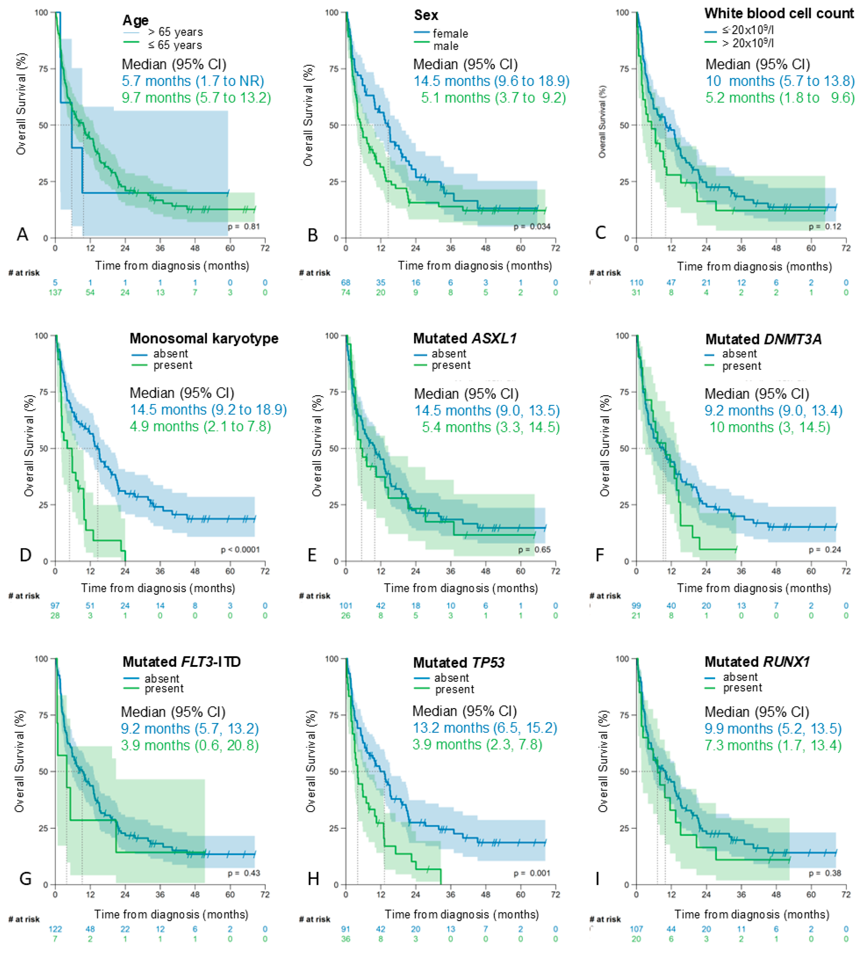
As shown in Figure 3, both a monosomal karyotype and a TP53 mutation were associated with a significantly shorter median OS (Figure 3D,H).
Mutations in ASXL1, FLT3-ITD, or RUNX1 were also associated with a shorter median OS, but the difference was not significant (Figure 3E,G,I). No statistically significant association was observed between DNMT3A mutations and survival (Figure 3F).
Male patients (n = 74, 52.1%) had a significantly shorter OS than female patients (median 5 months [95% CI 3–7] versus 15 months [95% CI 10–19], p = 0.034; Figure 3B). For age as a continuous variable, there was no evidence of an association with OS after log-transformation of age (HR 0.88; 95% CI 0.06–13.8, p > 0.9). However, the Schoenfeld test indicated a violation of the proportional hazards assumption (see Figure S1 in the Supplementary Materials), so this result should be interpreted with caution. The OS of patients aged 65 years or older was shorter than that of patients aged <65 years, but the difference was not significant (Figure 3A). Given the small sample size, we did not attempt to identify an age-related cut-off that might be more appropriate for prognostication.
For the log-transformed white blood cell (WBC) count, Cox regression analysis provided strong evidence of an association with OS (HR 1.18, 95% CI 1.05–1.34, p = 0.008), without evidence of a violation of the proportional hazards assumption (Supplementary Figure S2). Correspondingly, a white blood cell count > 20 × 109/L was associated with a shorter median overall survival (10 months [95%CI 6–14] versus 5 months [95%CI 2–10], p = 0.130 [log rank] and p = 0.046 [Breslow]; see Figure 3C).
Patients with K/N-RAS- or FLT3-ITD mutations had higher WBCs at diagnosis (median 9.6 × 10⁹/L [IQR 2–43.7] versus 2.8 × 10⁹/L [IQR 1.69–12.0], p = 0.026) and patients with a WBC > 20 × 109/L were more frequent within K/N-RAS- or FLT3-ITD mutated cases (no K/N-RAS—or FLT3-ITD mutation: 20/104 [19%] versus 8/19 [42%], p = 0.034).

3.6. Frequency and Prognostic Impact of NPM1 and IDH2 Mutations in the Risk Groups According to AML60+

An IDH2 mutation without negative prognostic factors according to mPRS (FLT3-ITD, NRAS, KRAS, or TP53 mutations) was present in 15/105 patients (14%). IDH2 mutations were significantly more frequent in patients with favorable/intermediate risk than in those with poor/very poor risk (10/36 [28%] versus 5/69 [7%], p = 0.007) and were associated with a favorable OS (median 19 months [95% CI 14.5-NR] versus 7 months [95% CI 5–13], p = 0.038; see Supplementary Figure S3).
NPM1 mutations without NRAS, KRAS, TP53, or FLT3-ITD mutations were present in 9/105 patients (9%) and were significantly more frequent in the intermediate/favorable risk group than in the poor/very poor risk groups (7/36, [19%] versus 2/69 [3%], p = 0.007). These patients had a longer median OS (19 months [95% CI 15–23] versus 9 months [95% CI 1.5-NR]). However, this difference was not statistically significant (p = 0.150). See Supplementary Figure S4.
Taken together, nearly 50% of patients in the favorable/intermediate category according to AML60+ (17/36, [47%]) had an IDH2 or NPM1 mutation without an additional molecular risk factor according to mPRS.

3.7. Prognosis According to ELN2022, mPRS, and AML60+

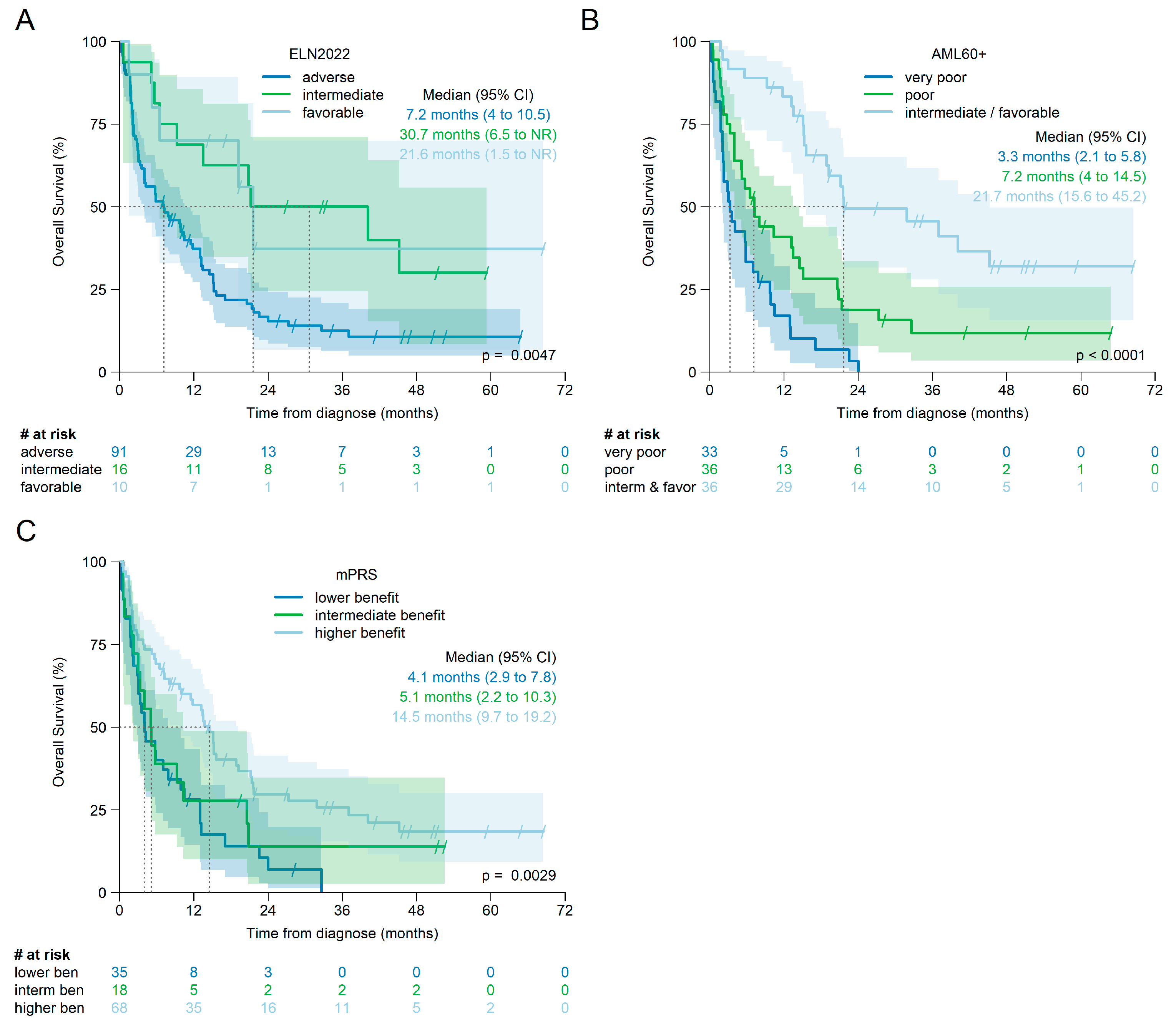
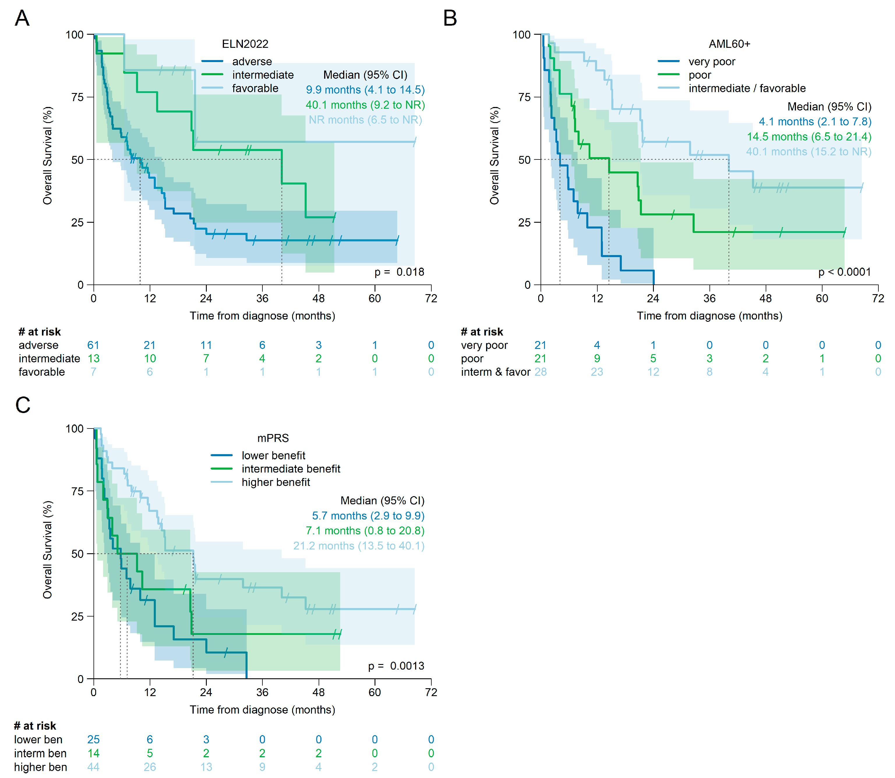
The overall survival of the individual risk groups according to ELN2022, mPRS, and AML60+ for all patients is shown in Figure 4 and for patients treated with HMA + Ven in Figure 5. For each score, there is evidence that at least one of the individual risk groups has a statistically different survival curve.
However, for the overall cohort, pairwise comparisons of Kaplan–Meier estimates using the log-rank test, and corrected for multiple testing, showed a significant difference in OS only between intermediate and adverse risk according to ELN2022 (corrected log-rank p = 0.021), but not between favorable and adverse (corrected log-rank p = 0.062) or favorable and intermediate (corrected log-rank p = 0.95).
For mPRS, OS was significantly different between higher and lower benefit (corrected log-rank p = 0.001), but not between lower and intermediate benefit (corrected log-rank p = 0.449) or intermediate and higher benefit (corrected log-rank p = 0.192).
OS by AML60+ was significantly different between all risk groups: favorable/intermediate risk versus poor (corrected log-rank p < 0.001) or versus very poor (corrected log-rank p < 0.001) and between poor and very poor (corrected log-rank p = 0.007).
When only patients treated with HMA + Ven were analyzed (see Figure 5), no difference was observed between the individual risk groups according to ELN2022 (favorable vs. adverse: log-rank p = 0.069; favorable vs. intermediate: p = 0.45 and intermediate vs. adverse: p = 0.069; all p-values corrected for multiple testing).
OS difference, according to mPRS, was significant between higher and lower benefit (corrected log-rank p < 0.001), but not between lower and intermediate benefit (corrected log-rank p = 0.397) or between intermediate and higher benefit (corrected log-rank p = 0.11).
Again, OS was significantly different between all risk groups according to AML60+: intermediate/favorable risk versus poor: corrected log-rank p = 0.023; intermediate/favorable versus very poor risk: corrected log-rank p < 0.001; and poor versus very poor risk corrected log-rank p = 0.005.
An analysis of patients treated with HMA without Ven revealed a significant survival difference only for the risk groups according to AML60+ (intermediate/favorable versus poor: corrected log-rank p = 0.02 and versus very poor: corrected log-rank p = 0.02), but not for the risk groups according to ELN2022 or mPRS (see Supplementary Table S5 and Supplementary Figure S5 for further details).
Univariable Cox regression models with, for each score, the highest risk category as reference category revealed the lowest hazard ratio for AML60+ intermediate/favorable patients compared to very poor risk (all patients: HR 0.17 [95% CI 0.10, 0.31], p < 0.001; HMA + Ven treated patients: HR 0.14 [95% CI 0.07, 0.30], p < 0.001; HMA without Ven HR 0.25 [95% CI 0.09–0.70], p = 0.008).
Detailed results of the Cox regression analyses for all three scores are shown in Table 2 (all patients), in Table 3 (patients treated with HMA + Ven), and in the Supplementary Table S6 for patients treated with HMA without Ven.
Because the sample sizes are small and the proportional hazards assumption appears to be violated for ELN2022 and mPRS (see Figure 4A,C and Figure 5A,C) these results must be interpreted with caution.

3.8. Comparison of the C-Indices

The concordance score was calculated for patients for whom all three scores were available (patients treated with HMA alone or HMA + Ven, n = 102). It was highest for AML60+ (0.67), followed by mPRS (0.60) and ELN2022 (0.58). When only patients receiving HMA + Ven (n = 69) or HMA without Ven (n = 33) were analyzed, similar results were obtained (HMA + Ven: concordance score AML60+ 0.69, mPRS 0.64, ELN 2022 0.60; HMA without Ven: concordance score AML60+ 0.62, mPRS 0.56, and ELN2022 0.54).

4. Discussion

Our analysis of elderly AML patients treated with HMA with or without Ven suggests that the AML60+ score may be a valuable prognostic tool for this population, although it was originally developed to identify elderly patients who would benefit from intensive treatment, including allogeneic stem cell transplantation [18].
The combined favorable/intermediate group according to AML60+ showed a median overall survival of 40 months for patients treated with HMA + Ven and, as shown by the C-indices, the AML60+ was superior to both ELN2022 and mPRS. Therefore, the AML60+ may help to identify a group of patients with a favorable prognosis or even long-term responders that are missed by the ELN2022 classification and not as clearly captured by the mPRS.
NPM1 and IDH2 mutations are among the mutations most associated with favorable outcomes to treatment with HMA +/− Ven, when they occur in the absence of signaling mutations and together with TP53 wild-type (for review [22,23]). In our cohort, both constellations are significantly enriched in the intermediate/favorable group according to AML60+. This implies that the AML60+ score indirectly captures a substantial proportion of patients with a favorable molecular risk profile without specifically testing for the respective mutations.
On the other hand, approximately 50% of patients in the intermediate/favorable group of AML60+ did not have NPM1 or IDH2 mutations. Thus, the unique combination of patient-related factors (age and sex), with conventional disease-related factors (WBC), and oncogenomic markers provided by the AML60+ identifies additional patients who can expect a favorable outcome following HMA-based treatment.
Regarding sex, a significantly better response to HMA has been reported in female patients with high-risk MDS [24], and the inclusion of sex added prognostic information to all standard prognostic tools for MDS [25]. In addition, male patients with high-risk MDS or AML showed an inferior outcome after treatment with Dec in the prospective HOVON135 study [26]. With regard to HMA + Ven treatment, male sex was an independent negative prognostic marker for overall and relapse-free survival in the MAYO prognostic score cohort [17], and female patients were overrepresented in the favorable risk group according to BEAT-AML2024 [16]. In addition, sex-related differences in the prognostic impact of co-occurrence patterns have been described for selected mutations [27]. On the other hand, no negative prognostic effect of sex was observed in a National Health Service (NHS) analysis of 587 patients treated with HMA + Ven [28]. Despite the latter observation, the consideration of sex may be one point that explains the better performance of AML60+ in our cohort. Recently, it has been suggested to include sex as a prognostic marker in AML prognostication [29] and our observations imply that this is useful in the context of HMA-based treatment as well.
Another advantage of the AML60 + may be the inclusion of age as a second patient-related factor. According to real-world data from the NHS on the use of HMA + Ven, increasing age proved to be an independent risk factor independent of AML subtype (secondary and/or therapy-related AML) and oncogenomic profile. [28] In secondary AML, age added prognostic information independent of the mutational profile in patients treated with HMA + Ven. [30]. Advanced age has also shown to be an independent negative prognostic marker, independent of mutational profile, in a Japanese AML cohort treated with Ven-based regimens [31], and was also prognostic in a large series of elderly AML patients from the MAYO Clinic [32].
AML patients with an elevated WBC count are at higher risk for complications such as tumor lysis syndrome, coagulopathies, or infections, especially in the early phase of treatment [33]. In patients treated with HMA and low-dose cytosine arabinoside, a higher WBC count was associated with a shorter OS in an analysis of the PETHEMA group [34] and a negative prognostic impact of WBC > 10 × 109/L was described in a Chinese cohort [35]. A higher WBC is often associated with FLT3-ITD or K/N-RAS mutations [36,37], as it was the case in our cohort. Therefore, a WBC > 20 × 109/L may additionally represent a surrogate marker for the presence of RAS-mutations, which are not directly considered by the AML60+.
Furthermore, it may be relevant that according to AML60+, both TP53 mutations and a monosomal karyotype are considered as individual risk factors, each with a high weight. TP53 mutations are a marker of very poor prognosis with low response rates and dismal survival [38,39] even with the combination of HMA + Ven [40]. They show a high association with a complex or monosomal karyotype [41,42] and the majority of patients with the latter abnormalities are identified by the presence of a TP53 mutation. However, some patients with a monosomal or complex karyotype have a TP53 wild-type. A pooled analysis of two pivotal trials of HMA + Ven suggested that in this situation, the adverse prognostic impact of poor-risk cytogenetics may not apply when they are treated with Aza + Ven [43].
However, in an analysis of a prospective phase II trial of Dec + Ven [44], a complex karyotype and especially monosomies of chromosomes 5, 7, and 17 were associated with worse survival. In another analysis of 301 patients treated with HMA + Ven, an unfavorable karyotype, according to ELN2022, remained an independent negative prognostic marker for both the achievement of a complete remission and for overall survival, independent of the TP53 mutation status [45]. A complex karyotype was associated with reduced overall survival according to real-world data from the NHS [28]. Therefore, identifying patients with an unsatisfactory response to HMA-based therapies is likely to be aided by considering both TP53 mutations and adverse karyotypes.
In addition to FLT3-ITD and TP53 mutations, mutations in ASXL1, DNMT3A, and RUNX1 are risk factors according to AML60+. Their prognostic role in the context of HMA-based treatments is not as well defined and they are not specifically considered by other currently proposed prognostic scoring systems. AML patients with mutated ASXL1 showed high response rates to HMA + Ven, but with lower rates of MRD-negativity and, therefore, higher risk of relapse [45]. Accordingly, ASXL1 mutations represent an independent negative prognostic marker in an analysis of the NHS cohort [28]. In case of relapse after HMA-based treatment, ASXL1 mutations are associated with a worse prognosis independent of mutations in TP53 and RAS [46].
DNMT3A mutations were associated with better response rates and longer overall survival in the MAYO cohort, but did not retain independent prognostic value in multivariable models [17]. In an analysis of Chinese AML patients with myelodysplasia-related changes receiving low-intensity Ven-based treatments, mutated DNMT3A was associated with a higher risk of relapse [47]. When occurring as a co-mutation in IDH-mutated cases, mutated DNMT3A is likely to confer an unfavorable prognosis [48]. This finding may be particularly relevant because IDH2-R172 mutations often co-occur with mutated DNMT3A [49]. A similar negative prognostic effect of DNMT3A mutations co-occurring with NPM1 mutations has been suggested in the context of various induction therapies, including Ven-based low-intensity treatments [50].
RUNX1 mutations were found with a high frequency of approximately 40% in primary Ven-refractory cases and were mutually exclusive with TP53 mutations [51]. When present as the only adverse factor according to ELN2022, they were associated with lower OS when intensive chemotherapy or HMA were used, but not with HMA + Ven as treatment [52]. Within the MAYO cohort [17], RUNX1 mutations were associated with lower response rates and shorter survival in both univariate and multivariate analyses, but were not included in the final model with censoring at the time of allogeneic stem cell transplantation.
Taken together, these data suggest that the mutational status with respect to ASXL1, DNMT3A, and RUNX1 may add prognostic value at least in combination with the other variables in the AML60+ score.
The main limitations of our study primarily include its retrospective nature and the limited sample size, which precluded assessing the prognostic impact of most of the individual AML60+ factors or identifying alternative cut-offs for age and WBC as continuous variables. As a retrospective study based on data collected during clinical routine, not all parameters necessary to calculate the individual scores were available for all patients, implying a risk of selection bias. In addition, the treatments were heterogeneous, and no patient received a combination of Ven with an IDH1/2 inhibitor or low-dose cytosine arabinoside. However, all key observations were confirmed in patients treated with HMA + Ven, which is the current standard of care. Neither DDX41 nor MLL were covered by the NGS panels used in the routine diagnostic laboratories collaborating with the participating centers. Therefore, we were not able to compare the AML60+ score with the ELN2024 classification [15] or the BEAT-AML 2024 score [16], which provide alternative options for prognostication in daily clinical practice.

5. Conclusions

Our data show for the first time that the AML60+ can be used as a prognostic tool in elderly patients treated with HMA alone or in combination with Ven. The AML60+ may even be more accurate than the mPRS, which was developed specifically for patients treated with HMA + Ven. However, further studies analyzing larger datasets, ideally derived from prospective registry data, are needed to confirm our observations. In addition, it is likely that some modifications of the AML60+ would improve its prognostic accuracy, such as defining an age cut-off that is more appropriate for a target population that often includes octogenarians or considering adverse cytogenetics according to ELN2022 instead of only a monosomal karyotype. Due to the small sample size, we were not able to answer these questions. Despite its limitations, our analysis underscores that conventional metaphase cytogenetics and simple patient- and disease-related factors such as WBC, sex, and age can help refine the prognosis in AML apart from the mutational profile.

Supplementary Materials

The following supporting information can be downloaded at the following addess: https://www.mdpi.com/article/10.3390/cancers17162658/s1. Next Generation Sequencing, Table S1A: Components of the AML60+; Table S1B: Risk groups according to AML60+; Table S2A: Distribution of risk factors according to ELN2022; Table S2B: Cytogenetic and/or molecular abnormalities not classified as favorable or adverse according to ELN2022 found in patients with intermediate risk according to ELN2022; Table S3: Distribution of risk factors according to mPRS; Table S4: Frequency of risk factors in the single risk groups according to AML60+; Table S5: p-values for the pairwise comparisons between the categories according to ELN2002 using the log-rank test for patients treated with HMA without venetoclax; Table S6: Univariable Cox regression models for OS by ELN2022, mPRS, and AML60+ for patients treated with HMA without venetoclax; Figure S1: A, Boxplot of age; B, Univariable Cox regression model for OS by log (Age); and C, Schoenfeld residuals for the Cox model for OS by log (Age); Figure S2: A, Boxplot of White blood cell count at diagnosis; B, Univariable Cox regression model for OS by log (White blood cell count at diagnosis); and C, Schoenfeld residuals for the Cox model for OS by log (White blood cell count at diagnosis); Figure S3: Kaplan–Meier estimates for overall survival for patients with available AML60+ according to the mutational status of IDH2; Figure S4: Kaplan–Meier estimates for overall survival for patients with available AML60+ according to the mutational status of NPM1; and Figure S5: Kaplan–Meier estimates for overall survival for patients treated with HMA without Ven by ELN 2022, n = 36 (A), AML60+, n = 35 (B), and mPRS, n = 38 (C).

Author Contributions

Conceptualization, T.S.; formal analysis, V.P., S.F., K.G., C.M. and T.S.; funding acquisition, C.D. and R.C.; investigation, V.P. and S.F.; methodology, K.G. and C.M.; project administration, V.P. and T.S.; resources, M.W., L.G., Y.G., J.S.G., T.L., C.D., U.J.M.M., R.C., S.C. and T.S.; supervision, T.S.; visualization, V.P., S.F., K.G., C.M. and T.S.; writing—original draft, V.P. and S.F.; writing—review and editing, M.W., K.G., C.M., L.G., Y.G., J.S.G., T.L., C.D., U.J.M.M., R.C., S.C. and T.S. All authors have read and agreed to the published version of the manuscript.

Funding

This project was supported by the research funds of the Clinic for Medical Oncology and Hematology, Cantonal Hospital St. Gallen and of the Department for Medical Oncology and Hematology, Cantonal Hospital Graubünden, Chur, and received no external funding.

Institutional Review Board Statement

The study was conducted according to the guidelines of the Declaration of Helsinki and was approved by the Ethics Committee of Eastern Switzerland (Ethikkommission Ostschweiz, Project ID: EKOS 22/102, date of approval 30 June 2022) and listed in the Registry of All Projects in Switzerland (https://raps.swissethics.ch, BASEC ID 2022-01134).

Informed Consent Statement

Written informed consent was obtained from each patient alive before inclusion into the study. In accordance with the vote of the ethics committee, data of patients deceased or lost to follow-up were included into the dataset, in accordance with Paragraph 34 of the Swiss Law regulating research with human subjects (“Humanforschungsgesetz”).

Data Availability Statement

The data presented in this study are available on request from the corresponding author.

Conflicts of Interest

The authors declare no conflicts of interest.

Abbreviations

The following abbreviations are used in this manuscript:
AMLAcute myeloid leukemia
ASXLAdditional Sex Combs-Like 1
CIConfidence interval
DNMT3ADNA-Metyhltransferase 3A
DecDecitabine
ELNEuropean Leukemia Net
FLT3FMS-like Tyrosinkinase 3
ICCInternational Consensus Classification
HRHazard ratio
HMAHypomethylating agent
IDHIsocitrate Dehydrogenase
ITDInternal Tandem
IQRInter Quartile range
mPRSMolecular Prognostic Score
NHSNational Health Service
NPMNucleophosmin 1
OSOverall survival
RASRat Sarcoma
TP53Tumor Protein p53
RUNX1Runt-related transcription factor 1
VenVenetoclax
WBCWhite blood cell count
WHOWorld Health Organization

References

  1. DiNardo, C.D.; Erba, H.P.; Freeman, S.D.; Wei, A.H. Acute Myeloid Leukaemia. Lancet 2023, 401, 2073–2086. [Google Scholar] [CrossRef] [PubMed]
  2. Roman, E.; Smith, A.; Appleton, S.; Crouch, S.; Kelly, R.; Kinsey, S.; Cargo, C.; Patmore, R. Myeloid Malignancies in the Real-World: Occurrence, Progression and Survival in the UK’s Population-Based Haematological Malignancy Research Network 2004–15. Cancer Epidemiol. 2016, 42, 186–198. [Google Scholar] [CrossRef]
  3. Döhner, H.; Wei, A.H.; Appelbaum, F.R.; Craddock, C.; DiNardo, C.D.; Dombret, H.; Ebert, B.L.; Fenaux, P.; Godley, L.A.; Hasserjian, R.P.; et al. Diagnosis and Management of AML in Adults: 2022 Recommendations from an International Expert Panel on Behalf of the ELN. Blood 2022, 140, 1345–1377. [Google Scholar] [CrossRef] [PubMed]
  4. Shimony, S.; Stahl, M.; Stone, R.M. Acute Myeloid Leukemia: 2025 Update on Diagnosis, Risk-Stratification, and Management. Am. J. Hematol. 2025, 100, 860–891. [Google Scholar] [CrossRef]
  5. Ucciero, A.; Pagnoni, F.; Scotti, L.; Pisterna, A.; Barone-Adesi, F.; Gaidano, G.; Patriarca, A.; Lunghi, M. Venetoclax with Hypomethylating Agents in Newly Diagnosed Acute Myeloid Leukemia: A Systematic Review and Meta-Analysis of Survival Data from Real-World Studies. Cancers 2023, 15, 4618. [Google Scholar] [CrossRef]
  6. Wang, Y.; Chen, Y.; Ji, D.; Ge, L.; Zhang, Y.; Liu, L.; Jiang, L.; Jin, F.; Xia, L. Meta-Analysis on the Effectiveness and Safety of Venetoclax-Based Combination Therapy with Hypomethylation in Acute Myeloid Leukemia. Eur. J. Med. Res. 2025, 30, 330. [Google Scholar] [CrossRef] [PubMed]
  7. Madarang, E.; Lykon, J.; Zhao, W.; Sekeres, M.A.; Bradley, T.; Chandhok, N.S.; Taylor, J.; Venugopal, S.; Koru-Sengul, T.; Iyer, S.G.; et al. Venetoclax and Hypomethylating Agents in Octogenarians and Nonagenarians with Acute Myeloid Leukemia. Blood Neoplasia 2024, 1, 100016. [Google Scholar] [CrossRef]
  8. Wei, A.H.; Loo, S.; Daver, N. How I Treat Patients with AML Using Azacitidine and Venetoclax. Blood 2025, 145, 1237–1250. [Google Scholar] [CrossRef]
  9. Pratz, K.W.; Jonas, B.A.; Pullarkat, V.; Thirman, M.J.; Garcia, J.S.; Döhner, H.; Récher, C.; Fiedler, W.; Yamamoto, K.; Wang, J.; et al. Long-Term Follow-up of VIALE-A: Venetoclax and Azacitidine in Chemotherapy-Ineligible Untreated Acute Myeloid Leukemia. Am. J. Hematol. 2024, 99, 615–624. [Google Scholar] [CrossRef]
  10. Niscola, P.; Gianfelici, V.; Giovannini, M.; Piccioni, D.; Mazzone, C.; de Fabritiis, P. Very Long-Term Efficacy of Venetoclax Combined with Hypomethylating Agents in Two AML Elderly: Is It the Time for Treatment Discontinuation Strategies? Ann. Hematol. 2024, 103, 1787–1788. [Google Scholar] [CrossRef]
  11. Chua, C.C.; Hammond, D.; Kent, A.; Tiong, I.S.; Konopleva, M.Y.; Pollyea, D.A.; DiNardo, C.D.; Wei, A.H. Treatment-Free Remission after Ceasing Venetoclax-Based Therapy in Patients with Acute Myeloid Leukemia. Blood Adv. 2022, 6, 3879–3883. [Google Scholar] [CrossRef]
  12. Garciaz, S.; Dumas, P.-Y.; Bertoli, S.; Sallman, D.A.; Decroocq, J.; Belhabri, A.; Orvain, C.; Aspas Requena, G.; Simand, C.; Laribi, K.; et al. Outcomes of Acute Myeloid Leukemia Patients Who Responded to Venetoclax and Azacitidine and Stopped Treatment. Am. J. Hematol. 2024, 99, 1870–1876. [Google Scholar] [CrossRef]
  13. Döhner, H.; Pratz, K.W.; DiNardo, C.D.; Wei, A.H.; Jonas, B.A.; Pullarkat, V.A.; Thirman, M.J.; Récher, C.; Schuh, A.C.; Babu, S.; et al. Genetic Risk Stratification and Outcomes among Treatment-Naive Patients with AML Treated with Venetoclax and Azacitidine. Blood 2024, 144, 2211–2222. [Google Scholar] [CrossRef]
  14. Bataller, A.; Bazinet, A.; DiNardo, C.D.; Maiti, A.; Borthakur, G.; Daver, N.G.; Short, N.J.; Jabbour, E.J.; Issa, G.C.; Pemmaraju, N.; et al. Prognostic Risk Signature in Patients with Acute Myeloid Leukemia Treated with Hypomethylating Agents and Venetoclax. Blood Adv. 2024, 8, 927–935. [Google Scholar] [CrossRef]
  15. Döhner, H.; DiNardo, C.D.; Appelbaum, F.R.; Craddock, C.; Dombret, H.; Ebert, B.L.; Fenaux, P.; Godley, L.A.; Hasserjian, R.P.; Larson, R.A.; et al. Genetic Risk Classification for Adults with AML Receiving Less-Intensive Therapies: The 2024 ELN Recommendations. Blood 2024, 144, 2169–2173. [Google Scholar] [CrossRef]
  16. Hoff, F.W.; Blum, W.G.; Huang, Y.; Welkie, R.L.; Swords, R.T.; Traer, E.; Stein, E.M.; Lin, T.L.; Archer, K.J.; Patel, P.A.; et al. Beat-AML 2024 ELN–Refined Risk Stratification for Older Adults with Newly Diagnosed AML given Lower-Intensity Therapy. Blood Adv. 2024, 8, 5297–5305. [Google Scholar] [CrossRef] [PubMed]
  17. Gangat, N.; Elbeih, A.; Ghosoun, N.; McCullough, K.; Aperna, F.; Johnson, I.M.; Abdelmagid, M.; Al-Kali, A.; Alkhateeb, H.B.; Begna, K.H.; et al. Mayo Genetic Risk Models for Newly Diagnosed Acute Myeloid Leukemia Treated with Venetoclax + Hypomethylating Agent. Am. J. Hematol. 2025, 100, 260–271. [Google Scholar] [CrossRef] [PubMed]
  18. Versluis, J.; Metzner, M.; Wang, A.; Gradowska, P.; Thomas, A.; Jakobsen, N.A.; Kennedy, A.; Moore, R.; Boertjes, E.; Vonk, C.M.; et al. Risk Stratification in Older Intensively Treated Patients With AML. J. Clin. Oncol. 2024, 42, 4084–4094. [Google Scholar] [CrossRef] [PubMed]
  19. Arber, D.A.; Orazi, A.; Hasserjian, R.P.; Borowitz, M.J.; Calvo, K.R.; Kvasnicka, H.-M.; Wang, S.A.; Bagg, A.; Barbui, T.; Branford, S.; et al. International Consensus Classification of Myeloid Neoplasms and Acute Leukemias: Integrating Morphologic, Clinical, and Genomic Data. Blood 2022, 140, 1200–1228. [Google Scholar] [CrossRef] [PubMed]
  20. Khoury, J.D.; Solary, E.; Abla, O.; Akkari, Y.; Alaggio, R.; Apperley, J.F.; Bejar, R.; Berti, E.; Busque, L.; Chan, J.K.C.; et al. The 5th Edition of the World Health Organization Classification of Haematolymphoid Tumours: Myeloid and Histiocytic/Dendritic Neoplasms. Leukemia 2022, 36, 1703–1719. [Google Scholar] [CrossRef]
  21. Huls, G.; Chitu, D.A.; Pabst, T.; Klein, S.K.; Stussi, G.; Griskevicius, L.; Valk, P.J.M.; Cloos, J.; van de Loosdrecht, A.A.; Breems, D.; et al. Ibrutinib Added to 10-Day Decitabine for Older Patients with AML and Higher Risk MDS. Blood Adv. 2020, 4, 4267–4277. [Google Scholar] [CrossRef]
  22. Griffioen, M.S.; de Leeuw, D.C.; Janssen, J.J.W.M.; Smit, L. Targeting Acute Myeloid Leukemia with Venetoclax; Biomarkers for Sensitivity and Rationale for Venetoclax-Based Combination Therapies. Cancers 2022, 14, 3456. [Google Scholar] [CrossRef]
  23. Lachowiez, C.A.; DiNardo, C.D. Mutation- and MRD-Informed Treatments for Transplant-Ineligible Patients. Hematol. Am. Soc. Hematol. Educ. Program. 2024, 2024, 168–177. [Google Scholar] [CrossRef]
  24. DeZern, A.E.; Zeidan, A.M.; Barnard, J.; Hand, W.; Al Ali, N.; Brown, F.; Zimmerman, C.; Roboz, G.J.; Garcia-Manero, G.; Steensma, D.P.; et al. Differential Response to Hypomethylating Agents Based on Sex: A Report on Behalf of the MDS Clinical Research Consortium (MDS CRC). Leuk. Lymphoma 2017, 58, 1325–1331. [Google Scholar] [CrossRef]
  25. Maggioni, G.; Bersanelli, M.; Travaglino, E.; Piérola, A.A.; Kasprzak, A.; Montserrat, A.S.; Sauta, E.; Sala, C.; Matteuzzi, T.; Meggendorfer, M.; et al. A Sex-Informed Approach to Improve the Personalised Decision Making Process in Myelodysplastic Syndromes: A Multicentre, Observational Cohort Study. Lancet Haematol. 2023, 10, e117–e128. [Google Scholar] [CrossRef] [PubMed]
  26. Hilberink, J.R.; van Zeventer, I.A.; Chitu, D.A.; Pabst, T.; Klein, S.K.; Stussi, G.; Griskevicius, L.; Valk, P.J.M.; Cloos, J.; van de Loosdrecht, A.A.; et al. Age and Sex Associate with Outcome in Older AML and High Risk MDS Patients Treated with 10-Day Decitabine. Blood Cancer J. 2023, 13, 93. [Google Scholar] [CrossRef] [PubMed]
  27. Ozga, M.; Nicolet, D.; Mrózek, K.; Yilmaz, A.S.; Kohlschmidt, J.; Larkin, K.T.; Blachly, J.S.; Oakes, C.C.; Buss, J.; Walker, C.J.; et al. Sex-Associated Differences in Frequencies and Prognostic Impact of Recurrent Genetic Alterations in Adult Acute Myeloid Leukemia (Alliance, AMLCG). Leukemia 2024, 38, 45–57. [Google Scholar] [CrossRef] [PubMed]
  28. Othman, J.; Lam, H.P.J.; Leong, S.; Basheer, F.; Abdallah, I.; Fleming, K.; Mehta, P.; Yassin, H.; Laurie, J.; Austin, M.; et al. Real-World Outcomes of Newly Diagnosed AML Treated with Venetoclax and Azacitidine or Low-Dose Cytarabine in the UK NHS. Blood Neoplasia 2024, 1, 100017. [Google Scholar] [CrossRef]
  29. Tettero, J.M.; Cloos, J.; Bullinger, L. Acute Myeloid Leukemia: Does Sex Matter? Leukemia 2024, 38, 2329–2331. [Google Scholar] [CrossRef]
  30. Shimony, S.; Bewersdorf, J.P.; Shallis, R.M.; Liu, Y.; Schaefer, E.J.; Zeidan, A.M.; Goldberg, A.D.; Stein, E.M.; Marcucci, G.; Lindsley, R.C.; et al. Hypomethylating Agents plus Venetoclax Compared with Intensive Induction Chemotherapy Regimens in Molecularly Defined Secondary AML. Leukemia 2024, 38, 762–768. [Google Scholar] [CrossRef]
  31. Ikoma, Y.; Nakamura, N.; Kaneda, Y.; Takamori, H.; Seki, T.; Hiramoto, N.; Kitagawa, J.; Kanda, J.; Fujita, K.; Morishita, T.; et al. Impact of Myelodysplasia-Related Gene Mutations and Residual Mutations at Remission in Venetoclax/Azacitidine for AML. Leukemia 2025, 39, 1–6. [Google Scholar] [CrossRef]
  32. Sasaki, K.; Ravandi, F.; Kadia, T.M.; Borthakur, G.; Short, N.J.; Jain, N.; Daver, N.G.; Jabbour, E.J.; Garcia-Manero, G.; Loghavi, S.; et al. Prediction of Survival with Lower Intensity Therapy among Older Patients with Acute Myeloid Leukemia. Cancer 2023, 129, 1017–1029. [Google Scholar] [CrossRef]
  33. Bewersdorf, J.P.; Zeidan, A.M. Hyperleukocytosis and Leukostasis in Acute Myeloid Leukemia: Can a Better Understanding of the Underlying Molecular Pathophysiology Lead to Novel Treatments? Cells 2020, 9, 2310. [Google Scholar] [CrossRef]
  34. Suárez, E.U.; Boluda, B.; Lavilla, E.; Tormo, M.; Botella, C.; Gil, C.; Vives, S.; Rodríguez, C.; Serrano, J.; Sayas, M.J.; et al. Do NPM1 and FLT3-ITD Mutations Modify Prognosis in Patients Treated with Non-Intensive Regimens? Ann. Hematol. 2024, 103, 2845–2851. [Google Scholar] [CrossRef] [PubMed]
  35. Chen, Y.; Cao, J.; Ye, Y.; Luo, L.; Zheng, X.; Yang, X.; Zheng, Z.; Zheng, J.; Yang, T.; Hu, J. Hypomethylating Agents Combined with Low-Dose Chemotherapy for Elderly Patients with Acute Myeloid Leukaemia Unfit for Intensive Chemotherapy: A Real-World Clinical Experience. J. Chemother. 2023, 35, 322–329. [Google Scholar] [CrossRef] [PubMed]
  36. Kiyoi, H.; Naoe, T.; Nakano, Y.; Yokota, S.; Minami, S.; Miyawaki, S.; Asou, N.; Kuriyama, K.; Jinnai, I.; Shimazaki, C.; et al. Prognostic Implication of FLT3 and N-RAS Gene Mutations in Acute Myeloid Leukemia. Blood 1999, 93, 3074–3080. [Google Scholar]
  37. Kottaridis, P.D.; Gale, R.E.; Frew, M.E.; Harrison, G.; Langabeer, S.E.; Belton, A.A.; Walker, H.; Wheatley, K.; Bowen, D.T.; Burnett, A.K.; et al. The Presence of a FLT3 Internal Tandem Duplication in Patients with Acute Myeloid Leukemia (AML) Adds Important Prognostic Information to Cytogenetic Risk Group and Response to the First Cycle of Chemotherapy: Analysis of 854 Patients from the United Kingdom Medical Research Council AML 10 and 12 Trials. Blood 2001, 98, 1752–1759. [Google Scholar] [CrossRef]
  38. Shahzad, M.; Amin, M.K.; Daver, N.G.; Shah, M.V.; Hiwase, D.; Arber, D.A.; Kharfan-Dabaja, M.A.; Badar, T. What Have We Learned about TP53-Mutated Acute Myeloid Leukemia? Blood Cancer J. 2024, 14, 202. [Google Scholar] [CrossRef]
  39. Senapati, J.; Loghavi, S.; Garcia-Manero, G.; Tang, G.; Kadia, T.; Short, N.J.; Abbas, H.A.; Arani, N.; DiNardo, C.D.; Borthakur, G.; et al. Clinical Interrogation of TP53 Aberrations and Its Impact on Survival in Patients with Myeloid Neoplasms. Haematologica 2025, 110, 1304–1315. [Google Scholar] [CrossRef]
  40. Badar, T.; Nanaa, A.; Atallah, E.; Shallis, R.M.; Guilherme, S.d.C.C.; Goldberg, A.D.; Saliba, A.N.; Patel, A.; Bewersdorf, J.P.; DuVall, A.S.; et al. Comparing Venetoclax in Combination with Hypomethylating Agents to Hypomethylating Agent-Based Therapies for Treatment Naive TP53-Mutated Acute Myeloid Leukemia: Results from the Consortium on Myeloid Malignancies and Neoplastic Diseases (COMMAND). Blood Cancer J. 2024, 14, 32. [Google Scholar] [CrossRef]
  41. Rücker, F.G.; Schlenk, R.F.; Bullinger, L.; Kayser, S.; Teleanu, V.; Kett, H.; Habdank, M.; Kugler, C.-M.; Holzmann, K.; Gaidzik, V.I.; et al. TP53 Alterations in Acute Myeloid Leukemia with Complex Karyotype Correlate with Specific Copy Number Alterations, Monosomal Karyotype, and Dismal Outcome. Blood 2012, 119, 2114–2121. [Google Scholar] [CrossRef]
  42. Kadia, T.M.; Jain, P.; Ravandi, F.; Garcia-Manero, G.; Andreef, M.; Takahashi, K.; Borthakur, G.; Jabbour, E.; Konopleva, M.; Daver, N.G.; et al. TP53 Mutations in Newly Diagnosed Acute Myeloid Leukemia: Clinicomolecular Characteristics, Response to Therapy, and Outcomes. Cancer 2016, 122, 3484–3491. [Google Scholar] [CrossRef] [PubMed]
  43. Pollyea, D.A.; Pratz, K.W.; Wei, A.H.; Pullarkat, V.; Jonas, B.A.; Recher, C.; Babu, S.; Schuh, A.C.; Dail, M.; Sun, Y.; et al. Outcomes in Patients with Poor-Risk Cytogenetics with or without TP53 Mutations Treated with Venetoclax and Azacitidine. Clin. Cancer Res. 2022, 28, 5272–5279. [Google Scholar] [CrossRef] [PubMed]
  44. Venugopal, S.; Maiti, A.; DiNardo, C.D.; Qiao, W.; Ning, J.; Loghavi, S.; Daver, N.G.; Kadia, T.M.; Rausch, C.R.; Alvarado, Y.; et al. Prognostic Impact of Conventional Cytogenetics in Acute Myeloid Leukemia Treated with Venetoclax and Decitabine. Leuk. Lymphoma 2021, 62, 3501–3505. [Google Scholar] [CrossRef]
  45. Gangat, N.; Johnson, I.; McCullough, K.; Farrukh, F.; Al-Kali, A.; Alkhateeb, H.; Begna, K.; Mangaonkar, A.; Litzow, M.; Hogan, W.; et al. Molecular Predictors of Response to Venetoclax plus Hypomethylating Agent in Treatment-Naïve Acute Myeloid Leukemia. Haematologica 2022, 107, 2501–2505. [Google Scholar] [CrossRef]
  46. Gangat, N.; Ilyas, R.; Johnson, I.M.; McCullough, K.; Al-Kali, A.; Alkhateeb, H.B.; Begna, K.H.; Mangaonkar, A.; Litzow, M.R.; Hogan, W.; et al. Outcome of Patients with Acute Myeloid Leukemia Following Failure of Frontline Venetoclax plus Hypomethylating Agent Therapy. Haematologica 2023, 108, 3170–3174. [Google Scholar] [CrossRef]
  47. Lin, J.; Liu, X.; Ying, S.; Zhu, Y.; Huang, W.; Zheng, W.; Ye, X.; Shi, J.; Luo, Y.; He, J.; et al. Selective Efficacy of Venetoclax in Unfit Patients with Acute Myeloid Leukemia with Myelodysplasia-Related Gene Mutations under Low-Intensity Therapy. Ann. Hematol. 2025, 104, 2717–2729. [Google Scholar] [CrossRef]
  48. Lai, A.; Liu, W.; Zhou, C.; Li, Y.; Wei, S.; Liu, K.; Gong, B.; Gong, X.; Liu, Y.; Zhang, G.; et al. Prognostic Impact of Co-Mutations in Adults with IDH1/2-Mutated Acute Myeloid Leukemia. Blood Sci. 2025, 7, e00231. [Google Scholar] [CrossRef]
  49. Hoff, F.W.; Huang, Y.; Welkie, R.L.; Swords, R.T.; Traer, E.; Stein, E.M.; Lin, T.L.; Patel, P.A.; Robert H Collins, J.; Baer, M.R.; et al. Molecular Characterization of Newly Diagnosed Acute Myeloid Leukemia Patients Aged 60 Years or Older: A Report from the Beat AML Clinical Trial. Blood Cancer J. 2025, 15, 55. [Google Scholar] [CrossRef]
  50. Yao, Y.; Zhou, Y.; Zhuo, N.; Xie, W.; Meng, H.; Lou, Y.; Mao, L.; Tong, H.; Qian, J.; Yang, M.; et al. Co-mutation landscape and its prognostic impact on newly diagnosed adult patients with NPM1-mutated de novo acute myeloid leukemia. Blood Cancer J. 2024, 14, 118. [Google Scholar] [CrossRef]
  51. DiNardo, C.D.; Tiong, I.S.; Quaglieri, A.; MacRaild, S.; Loghavi, S.; Brown, F.C.; Thijssen, R.; Pomilio, G.; Ivey, A.; Salmon, J.M.; et al. Molecular Patterns of Response and Treatment Failure after Frontline Venetoclax Combinations in Older Patients with AML. Blood 2020, 135, 791–803. [Google Scholar] [CrossRef]
  52. Venugopal, S.; DiNardo, C.D.; Loghavi, S.; Qiao, W.; Ravandi, F.; Konopleva, M.; Kadia, T.; Bhalla, K.; Jabbour, E.; Issa, G.C.; et al. Differential Prognostic Impact of RUNX1 Mutations According to Frontline Therapy in Patients with Acute Myeloid Leukemia. Am. J. Hematol. 2022, 97, 1560–1567. [Google Scholar] [CrossRef]
Figure 1. Distribution of the risk groups according to ELN2022 (A), mPRS (B), and AML60+ (C).
Cancers 17 02658 g001
Figure 2. River plot of patients for whom all three scores were available (n = 102).
Cancers 17 02658 g002
Figure 3. Kaplan–Meier estimates of overall survival by the single factors according to AML60+. (A) age, (B) sex, (C) white blood cell count, (D) monosomal karyotype, (E) mutated ASXL1, (F) mutated DNMT3A, (G) FLT3-ITD mutation, (H) mutated TP53 and (I) mutated RUNX1.
Cancers 17 02658 g003
Figure 4. Kaplan–Meier estimates of overall survival for patients treated with either HMA or HMA + Ven by ELN2022, n = 117 (A), AML60+, n = 105 (B), and mPRS, n = 121 (C).
Cancers 17 02658 g004
Figure 5. Kaplan–Meier estimates for overall survival for patients treated with HMA + Ven by ELN2022, n = 81 (A), AML60+, n = 70 (B), and mPRS, n = 83 (C).
Cancers 17 02658 g005
Table 1. Patient characteristics.
CharacteristicWhole Population
n142
Age [years], median, (IQR)77 (74–81)
Female n, (%)68 (48)
Male n, (%)74 (52)
AML according to ICC2022 n, (%)122 * (86)
AML with recurrent genetic abnormality n, (%)21 * (14)
AML with Mutated NPM117 (11.5)
AML with KMT2A Rearrangement3 (2)
AML with inv(3)(q21.3q26.2) 1 (0.5)
AML with mutated TP53 n, (%)26 (18)
AML with myelodysplasia-related gene mutation n, (%)45 (32)
AML with myelodysplasia-related cytogenetic abnormality n, (%)4 (2.8)
AML not otherwise specified n, (%)13 (9)
Subtype unknown due to insufficient work-up n, (%)13 (9)
MDS/AML according to ICC2022 n, (%)20 (14)
MDS/AML with mutated TP53 n, (%)8 (5.6)
MDS/AML with myelodysplasia-related gene mutation n, (%)9 (6.3)
MDS/AML not otherwise specified n, (%)1 (0.7)
Subtype unknown due to insufficient work-up n, (%)2 (1.4)
Hemoglobin
available141/142
[g/L], median (IQR)88 (76–102)
Leukocytes
available141/142
(×109/L), median (IQR)3.3 (1.8–15)
Platelet count
available141/142
(×109/L), median (IQR)66 (36–111)
Neutrophils
available139/142
(×109/L), median (IQR)0.93 (0.36–3.1)
Blasts peripheral blood
available140/142
(%), median, IQR8.3 (0.5–23)
Blasts bone marrow
available141/142
(%) median, IQR35 (25–63)
* Including one patient with 6% bone marrow blasts and NPM1 mutation (AML according to WHO2022) IQR, Inter Quartile Range.
Table 2. Univariable Cox regression models for OS by ELN2022, mPRS, and AML60+ for patients treated with either HMA or HMA + Ven.
HR95% CIp
ELN2022
Adverse *----
Intermediate0.410.21, 0.790.008
Favorable0.400.16, 0.990.047
mPRS
Lower benefit *----
Intermediate benefit0.730.39, 1.360.3
Higher benefit0.460.29, 0.73<0.001
AML60+
Very poor *------
Poor0.470.28, 0.780.004
Intermediate/favorable0.170.10, 0.31<0.001
* Reference category; Abbreviations: CI = Confidence Interval, and HR = Hazard Ratio.
Table 3. Univariable Cox regression models for OS by ELN2022, mPRS, and AML60+ for patients treated with HMA + Ven only.
HR95% CIp
ELN2022
Adverse *----
Intermediate0.470.22, 1.000.049
Favorable0.240.06, 0.970.045
mPRS
Lower benefit *----
Intermediate benefit0.670.32, 1.400.3
Higher benefit0.360.20, 0.64<0.001
AML60+
Very poor *------
Poor0.320.16, 0.650.002
Intermediate/favorable0.140.07, 0.30<0.001
* Reference category; Abbreviations: CI = Confidence Interval, and HR = Hazard Ratio.
Disclaimer/Publisher’s Note: The statements, opinions and data contained in all publications are solely those of the individual author(s) and contributor(s) and not of MDPI and/or the editor(s). MDPI and/or the editor(s) disclaim responsibility for any injury to people or property resulting from any ideas, methods, instructions or products referred to in the content.