Review

IFN γ and the IFN γ Signaling Pathways in Merkel Cell Carcinoma

1
Department of Dermatology, Shanghai Children’s Medical Center, School of Medicine, Shanghai Jiao Tong University, Shanghai 200127, China
2
German Cancer Consortium (DKTK), Deutsches Krebsforschung Institut, 69120 Heidelberg, Germany
*
Authors to whom correspondence should be addressed.
These authors contributed equally to this work.
Cancers 2025, 17(15), 2547; https://doi.org/10.3390/cancers17152547 (registering DOI)
Submission received: 18 June 2025 / Revised: 21 July 2025 / Accepted: 26 July 2025 / Published: 1 August 2025
(This article belongs to the Special Issue Histopathology and Pathogenesis of Skin Cancer)

Simple Summary

Interferons are crucial for the host immune response against malignant cells. However, the role of IFN γ and its associated signaling pathways in Merkel cell carcinoma (MCC) remains unclear. This review summarizes recent epigenetic and functional insights into IFN γ in MCC, along with genetic alterations in its associated signaling pathway. We also discuss the potential therapeutic applications of these insights to improve MCC treatment.

Abstract

Recent preclinical and clinical studies have confirmed the essential role of interferons in the host’s immune response against malignant cells. Merkel cell carcinoma (MCC) is a rare, aggressive skin cancer strongly associated with Merkel cell polyomavirus (MCPyV). Despite progress in understanding MCC pathogenesis, the role of innate immune signaling, particularly interferon-γ (IFN γ) and its downstream pathways, remains underexplored. This review summarizes recent findings on IFN-γ in MCC, highlighting its dual role in promoting both antitumor immunity and immune evasion. IFN-γ enhances cytotoxic T cell responses, upregulates MHC class I/II expression, and induces tumor cell apoptosis. Transcriptomic studies have shown that IFN-γ treatment upregulates immune-regulatory genes including PD-L1, HLA-A/B/C, and IDO1 by over threefold; it also activates APOBEC3B and 3G, contributing to antiviral defense and tumor editing. Clinically, immune checkpoint inhibitors (ICIs) such as pembrolizumab and avelumab yield objective response rates of 30–56% and two-year overall survival rates exceeding 60% in advanced MCC. However, approximately 50% of patients do not respond, in part due to IFN-γ signaling deficiencies. This review further discusses IFN-γ’s crosstalk with the STAT1/3/5 pathways and emerging combination strategies aimed at restoring immune sensitivity. Understanding these mechanisms may inform personalized immunotherapeutic approaches and guide the development of IFN-γ–based interventions in MCC.

1. Introduction

Significant progress has been made in understanding the relationships between the host immune system, oncogenesis, and tumor progression. The tumor microenvironment (TME) is increasingly gaining attention, with a growing number of cells and molecules involved in immunotherapy being identified [1]. Interferons (IFNs) are cytokines with pleiotropic biological effects, including the induction of interferon-stimulated gene (ISG) expression, which is essential in immune responses [2]. Among these, type II IFNs act as key mediators of tumor–immune interactions, directly influencing tumor cells in antitumor immune responses. Mechanistically, IFN γ enhances immune recognition of tumor cells by promoting antigen presentation and activating the major histocompatibility complex (MHC) class I pathway, highlighting its critical role in early antigen presentation [3]. Notably, Grasso et al. suggested that antitumor T-cell responses activated by the IFN γ signaling pathway are the primary effectors in melanoma immune checkpoint inhibitor (ICI) therapy [4].
The TME is composed of tumor cells, stromal cells, immune cells, and the extracellular matrix containing various cytokines, all of which are potentially involved in tumor progression and immune evasion [5]. Enhancing the immune system‘s ability to recognize and eleminate tumor cells is currently the most effective method for controlling tumor growth, with IFN γ playing a particularly important role [6]. This cytokine regulates various immune functions through IFN γ, including the activation of cytotoxic T lymphocytes (CTLs), natural killer (NK) cells, and macrophages, which are crucial for tumor immune responses [6].
Merkel cell carcinoma (MCC) is an immunogenic, but still highly aggressive skin cancer characterized by a high recurrence rate associated with pronounced mortality [7]. In recent years, ICI therapy has shown promising results in MCC patients [8]. However, primary and secondary resistance are common, and at least half of advanced MCC patients do not benefit from ICI therapy. One contributing factor is the downregulation of MHC I expression, which can be restored by histone deacetylase (HDAC) inhibitors [9]. Furthermore, emerging studies indicate that loss-of-function mutations and genomic alterations in antigen presentation and IFN signaling pathways contribute to tumor resistance to ICI [10,11]. Recent transcriptomic studies provide additional insights into the role of IFN γ in immune escape mechanisms. Tatjana et al. employed single-molecule RNA sequencing to investigate how IFN γ influences gene expression in Merkel cell polyomavirus (MCPyV)-positive MCC cell lines. Their results revealed that IFN γ treatment significantly upregulates a set of genes involved in immune evasion, including members of the suppressor of cytokine signaling and interferon regulatory factor families, suggesting the dual role of IFN γ in both enhancing immune activation and promoting tumor immune escape [12].
Given the pivotal role of IFN γ signaling in anti-tumor responses, impaired IFN γ signaling is increasingly acknowledged as a mechanism underlying both cancer immune escape and resistance to ICI [13]. Therefore, it is crucial to investigate the function and integrity of the IFN γ signaling pathway in MCC for a deeper understanding of its initiation and progression, as well as for the development of novel therapeutic strategies for MCC. Notably, recent preclinical efforts have explored vaccine-based immunotherapy approaches. For example, Kerstin et al. demonstrated that dendritic cells engineered to express the truncated large T antigen of MCPyV, combined with constitutive nuclear factor kappa B (NF-κB) activation to enhance immunogenicity, successfully elicited virus-specific T-cell responses in healthy donors and some MCC patients [14]. These findings highlight the therapeutic potential of dendritic-cell-based vaccination in virus-driven cancers such as MCC.
This review aims to offer a comprehensive examination of IFN γ and its associated signaling pathways in the context of MCC, highlighting their intricate role in immune regulation and their therapeutic potential. Furthermore, we summarize the exploration of the role of the IFN γ signaling pathway in MCC and address the challenges and future prospects of IFN-γ-based immunotherapy, focusing on how these insights can be translated into viable therapeutic options.

2. MCC and IFN γ

MCC is an immunogenic but highly aggressive skin cancer with neuroendocrine differentiation [15]. It is marked by rapid growth and typically presents as firm, red, or purple nodules on sun-exposed areas such as the head, neck, and limbs, primarily in elderly individuals. MCC has a high propensity for metastasis [16]. Although relatively rare with a low incidence rate, the incidence of MCC has been increasing in recent decades, particularly in the aging populations. Notably, MCC is more common among individuals with compromised immune function, in whom it also exhibits more aggressive behavior, thus confirming the relevance of immune surveillance for MCC. The pathogenesis of MCC is complex, involving various factors, i.e., UV-associated gene mutations or clonal integration of MCPyV during viral oncogenesis (Figure 1) [17,18].
MCPyV is a small, non-enveloped, double-stranded DNA virus that contains a genome with three regions: a noncoding control region (NCCR), an early coding region (ECR), and a late coding region (LCR). In MCC pathogenesis, MCPyV plays a pivotal role through its two early genes: large transforming (LT) and small transforming antigens (sT) [19]. The LT antigen interacts with the retinoblastoma protein (Rb) via its N-terminal LXCXE motif, which releases Rb‘s inhibition of the E2F transcription factor, thereby promoting the progression of the cell cycle [20]. The sT antigen, through its unique C-terminal domain, binds to various host proteins, including cyclin-dependent kinase 20 (CDC20), F-box and WD-repeat-domain-containing protein 7 (FBXW7), and protein phosphatase 2A (PP2A) [21]. This binding of sT to CDC20 enhances the phosphorylation of the eukaryotic translation initiation factor 4E-binding protein 1 (4E-BP1), leading to the overproduction of cellular mRNA and promoting cell proliferation [22]. Additionally, sT forms complexes with MYCL and EP400 proteins, activating MDM2 and MDM4 expression while inhibiting p53 activity, thus facilitating cell transformation [23]. The integration of MCPyV DNA into the host cell genome is a critical step in MCC development, resulting in the persistent expression of T antigens that drive malignant transformation [24].
The treatment approaches for MCC include surgery, radiation therapy, chemotherapy and immunotherapy [25]. Surgical resection, either alone or combined with adjuvant radiation therapy, is the standard first-line treatment for localized tumors without metastatic spread [25]. In the advanced setting, it is important to note that MCC shows a high response rate to cytotoxic agents even if used as monotherapy; unfortunately, however, MCC rapidly develops secondary resistances, significantly limiting the effectiveness of conventional chemotherapy regimens [26]. While early localized MCC can often be managed with surgery and adjuvant radiation, approximately 30–40% of patients develop metastatic or locally advanced tumors that are unresectable or refractory to conventional therapies [27], highlighting the urgent need for novel therapeutic strategies.
The discovery of MCPyV integration in MCCs and the identification of ultraviolet-induced mutagenesis in virus-negative tumors provided critical insights into tumor immunogenicity [28]. These findings, coupled with the success of immune checkpoint inhibitors (ICI) in other immunogenic cancers, positioned MCC as a prime candidate for PD-1/PD-L1 blockade. Landmark clinical trials, including KEYNOTE-017 [29] and JAVELIN Merkel 200 [30], demonstrated the efficacy of pembrolizumab and avelumab, respectively, with objective response rates (ORR) of 30–56%, durable complete remissions, and two-year OS rates exceeding 60%. These breakthroughs established ICI as the cornerstone of first-line therapy for advanced MCC, as endorsed by current NCCN (2024) and ESMO guidelines. Thus, the advent of immunotherapy, particularly ICI targeting the PD-1/PD-L1 axis (e.g., pembrolizumab and avelumab), has transformed the treatment landscape for advanced-stage MCC [31,32]. These agents have shown significant and lasting antitumor effects, establishing them as key therapies for metastatic or recurrent MCC. Ongoing research is focused on evaluating the role of immunotherapy in neoadjuvant and adjuvant settings and optimizing combination strategies to improve therapeutic outcomes and delay resistance development [26,33].
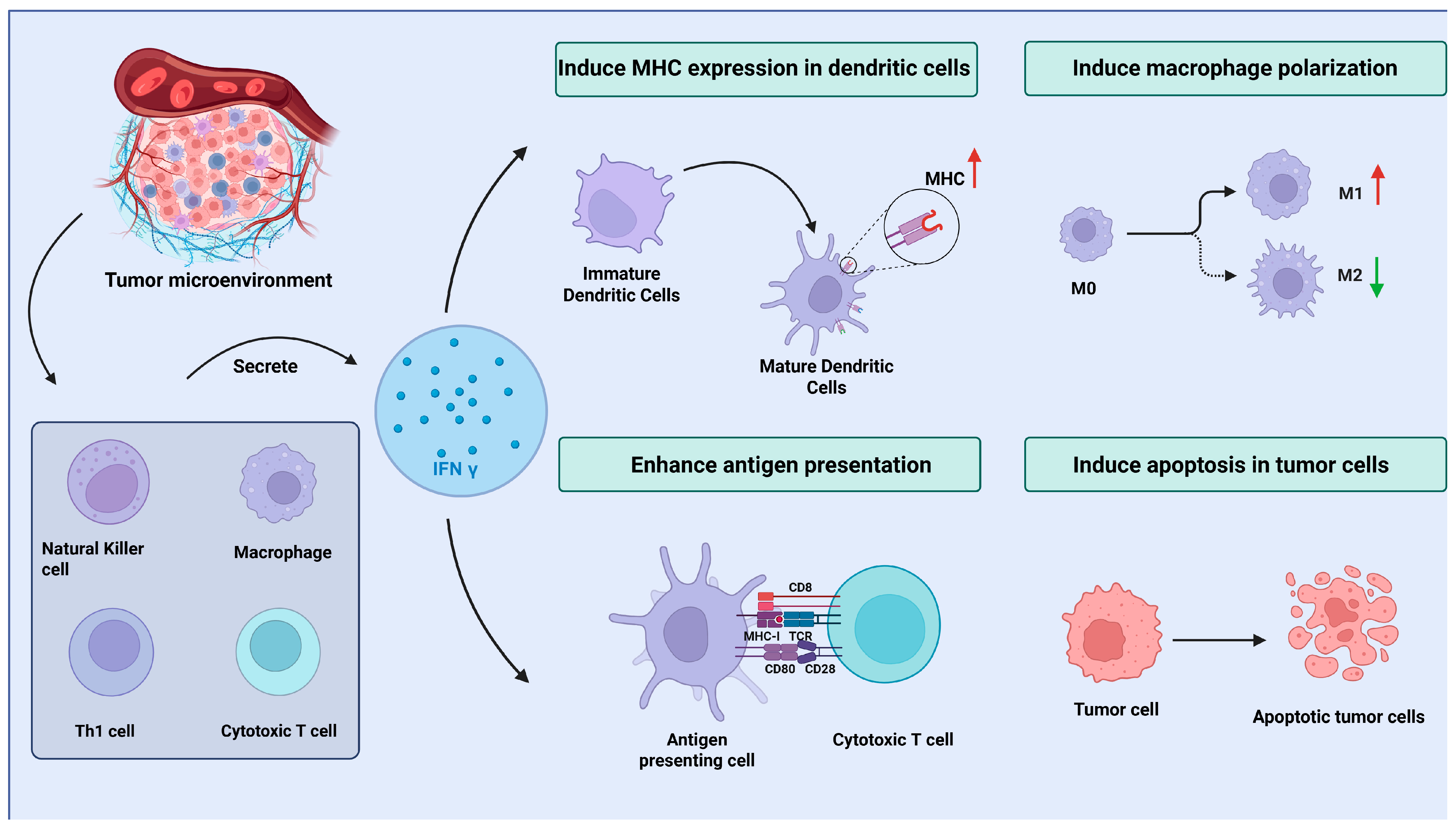
Despite significant advancements in immunotherapy, intrinsic or acquired resistance to ICI affects nearly half of patients, and subsets with immunosuppression or MCPyV-negative status exhibit attenuated responses, highlighting the urgent need for further exploration of novel targeted treatment strategies [34]. In recent years, emerging strategies to overcome these barriers—including combination therapies, novel immune targets, and personalized approaches—are now at the forefront of clinical investigation [33]. As a member of the type II interferon family, IFN γ is primarily secreted by immune cells, such as CD4+ Th1 cells, CD8+ cytotoxic T cells, NK cells, and some macrophages [35]. As a key mediator of the immune response, IFN γ plays a crucial role in enhancing the cytotoxic activity of NK cells and CTLs, which is essential for recognizing and eliminating tumor cells, including those in MCC [36,37]. Additionally, IFN γ regulates macrophage polarization, promoting their shift toward the proinflammatory M1 phenotype, which is associated with increased antigen presentation and the activation of antitumor immunity [38]. This process improves the immune system’s ability to detect and target tumor cells, which is critical for controlling cancer growth. Moreover, IFN γ upregulates the expression of MHC class I and II molecules on dendritic cells, leading to the induction of effective cytotoxic T-cell responses and further enhancing the immune system’s ability to recognize tumor-associated antigens [39]. Figure 2 illustrates the sources of IFN γ and its role in activating the antitumor immune response within the TME.
Approximately 80% of MCC cases harbor a clonally integrated truncated MCPyV genome coding for sT and a truncated form of LTA expressed from the viral promoter [40,41]. IFN γ, a type II interferon primarily produced by activated T cells and NKcells, exerts potent antiviral effects not only by directly suppressing viral replication but predominantly through immune-mediated cytotoxicity against virus-infected cells [42]. Emerging studies suggest that IFN γ may exhibit antitumor activity against MCC through both direct immunomodulatory effects and indirect mechanisms targeting the tumor microenvironment (TME) [43]. Specifically, IFN γ enhances antigen presentation by upregulating MHC class I/II molecules on tumor cells and dendritic cells (DCs), thereby promoting cytotoxic T lymphocyte (CTL)-mediated tumor cell killing [44]. In MCC, which is strongly associated with MCPyV, IFN γ may also suppress viral oncoprotein-driven tumorigenesis by restoring p53 pathway activity [45,46]. Preclinical models further indicate that IFN γ induces tumor vascular regression via endothelial cell-specific signaling, leading to ischemia-mediated tumor necrosis—a mechanism analogous to its effects in other solid tumors [47]. Additionally, IFN-γ-mediated activation of Janus Kinase/Signal Transducer and Activator of Transcription (JAK/STAT) pathways enhances the recruitment of immune cells (e.g., NK cells and macrophages) to the TME [48], which may synergize with ICIs to overcome resistance in refractory MCC. These findings position IFN γ as a promising therapeutic adjunct in MCC management, particularly for tumors that are resistant to conventional therapies [49]. Recent research has shown that neoantigen-specific CD4+ T cells in MCC tumors produce substantial amounts of IFN γ when activated by tumor-derived neoantigen peptides, further supporting the importance of IFN γ in antitumor immune responses [50].
Indeed, the molecular mechanisms underlying IFN γ signaling remain complex and are not fully elucidated in MCC. The canonical IFN γ signaling pathway, which involves the JAK-STAT pathway, is likely to be involved. With the binding of IFN γ to its receptor on MCC cells, JAK1 and JAK2 are activated, leading to the phosphorylation of signal transducer and activator of transcription 1 (STAT1). The phosphorylated STAT1 then translocates to the nucleus and regulates the transcription of genes responsive to IFN γ [38]. However, additional mechanisms specific to MCC may exist. For example, the stimulator of interferon genes (STING) pathway, which senses cytoplasmic DNA, may interact with IFN γ signaling [51]. In some cases, STING agonists have been shown to enhance antitumor immunity in MCC by indirectly signaling through immune and stromal cells in the TME, potentially modulating IFN γ-related responses [39]. Additionally, MCC cells may exhibit unique epigenetic modifications that influence IFN γ signaling. For instance, recent studies have demonstrated that histone demethylases such as KDM7A (also known as JHDM10) can modulate the JAK2/STAT1 pathway by altering chromatin structure and affecting protein methylation status [52]. Given the functional parallels in immune evasion strategies between viruses and tumors, it is plausible that similar epigenetic mechanisms may operate in MCC, contributing to immune escape. Understanding these regulatory processes could provide new insights into the development of targeted therapies that modulate IFN γ signaling in MCC.
Despite its potent antitumor effects, the role of IFN γ in MCC is intricate and may be influenced by the TME, which often contains immunosuppressive factors that limit the efficacy of IFN γ [53]. Like many other cancers, the TME of MCC may harbor various immunosuppressive components, such as regulatory T cells (Tregs), myeloid-derived suppressor cells (MDSCs), and numerous soluble immune checkpoints, all of which collectively suppress immune responses and hinder effective tumor elimination [54]. These immunosuppressive factors can diminish the action of IFN γ, restricting its ability to activate immune cells or induce tumor cell apoptosis. To overcome these challenges, strategies to enhance the efficacy of IFN γ in MCC are being explored. Combining IFN γ therapy with ICI or other immunotherapies may help counteract the immune evasion mechanisms present in the TME [55].

3. The Role of IFN γ in the MCC Microenvironment: Antitumor and Immune Evasion Effects

The role of IFN γ in the MCC microenvironment is multifaceted, contributing both to significant antitumor effects and to immune evasion and drug resistance mechanisms. Figure 3 illustrates this dual role of IFN γ.
Specifically, IFN γ exerts anti-tumor effects by inducing tumor cell apoptosis, enhancing antigen presentation, and promoting the cytotoxic activity of immune cells against tumors [56]. Conversely, it facilitates immune evasion by upregulating PD-L1 expression on tumor cells, inducing the secretion of immunosuppressive factors, and promoting EMT, which aids tumor progression [57]. This figure emphasizes the complex role of IFN γ in the TME, highlighting its capacity to enhance both antitumor immunity and immune evasion.
In MCC, IFN γ has been shown to upregulate the expression of cytidine deaminase in the apolipoprotein B mRNA-editing catalytic polypeptide-like (APOBEC) family, particularly APOBEC3B and APOBEC3G, in MCPyV-positive cell lines [58]. This induction is associated with cytosine mutations that preferentially occur in TpC dinucleotide contexts, a mutation signature consistent with APOBEC-mediated activity. Studies have shown that IFN γ plays a central role in shaping the MCPyV genome through the induction of APOBEC family cytidine deaminases, such as APOBEC3A, 3B, and 3G. Specifically, IFN γ treatment significantly upregulates APOBEC3B and APOBEC3G expression in MCPyV-positive MCC cell lines, leading to cytosine mutations with a strong TpC dinucleotide preference within the viral genome [58]. Although classical kataegis—defined as localized hypermutation clusters—was not explicitly reported in this study, the mutation pattern is consistent with APOBEC-mediated mutagenesis, which is known to underlie kataegis in other cancer types. Thus, IFN-γ-induced APOBEC expression may contribute to viral genome editing and the accumulation of tumor-specific mutations in MCPyV-positive MCC. In addition, IFN γ has been shown to inhibit tumor cell proliferation and induce apoptosis in MCC cells [59,60,61], potentially through mechanisms such as the modulation of oxidative stress [12,62] and the activation of downstream pathways including the canonical JAK–STAT axis, as previously described.
IFN γ can also enhance immune cell function by regulating immune cell infiltration in the TME. For example, IFN γ can promote the maturation and antigen-presenting capacity of dendritic cells (DCs) [63]. Other studies have reported that IFN γ can promote the transmission of inflammatory signals, attracting more immune cells to the tumor site. In the IFN γRKO tumor model, IFN γ accumulation enhances inflammatory signaling within the tumor, particularly increasing the recruitment of monocytes and inflammatory macrophages [64]. Thus, even IFN-γ-insensitive tumor cells can reshape the tumor immune microenvironment by accumulating IFN γ, especially by promoting the recruitment and differentiation of monocytes. Monocytes synergize with CD8+ T cells around tumor blood vessels, boosting CD8+ T cell activity and improving their ability to recognize and kill tumor cells [64].
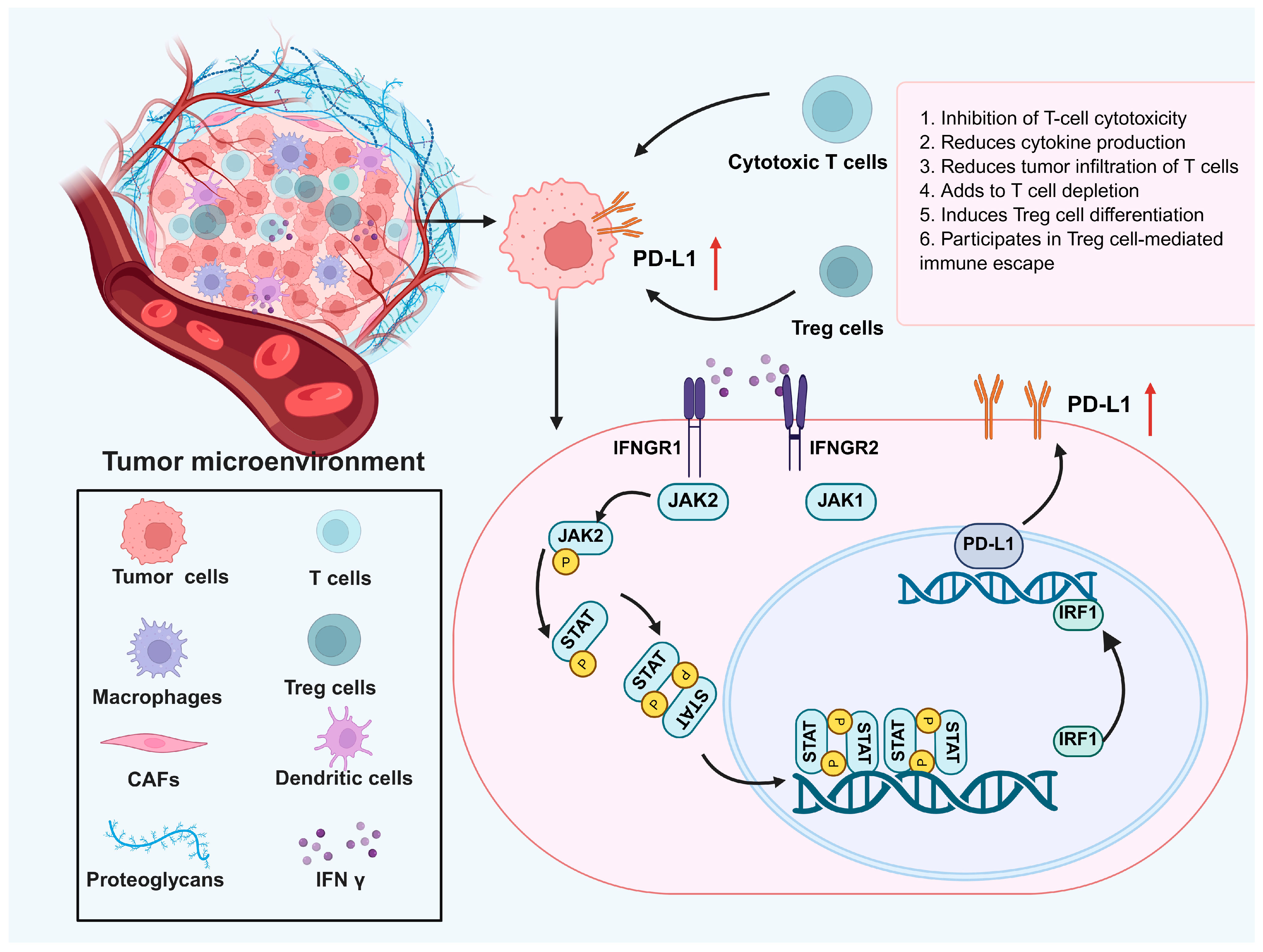
Upon binding to its receptor on tumor cells, IFN γ activates JAK1 and JAK2, which in turn phosphorylate STAT1. This induces STAT1 dimerization and nuclear translocation, where it binds to the PD-L1 gene promoter, enhancing PD-L1 transcription and expression. The figure illustrates how IFN γ upregulates PD-L1 in tumor cells, facilitating immune evasion and contributing to resistance against immune-mediated destruction.
Despite its potent antitumor functions, IFN γ can paradoxically promote immune evasion and tumor progression through several mechanisms. One such mechanism involves the epigenetic suppression of the expression of the major histocompatibility complex class II [65]. In colorectal cancer cells, for instance, promoter methylation of the class II transactivator (CIITA) gene impairs IFN-γ-induced CIITA expression, thereby blocking MHC class II upregulation and reducing CD4+T cell recognition [65]. IFN γ also modulates the tumor immune microenvironment by inducing the expression of immune checkpoint and immunosuppressive molecules. It promotes programmed death-ligand 1 (PD-L1) expression via the Janus Kinase/signal transducer and activator of transcription 1 (JAK–STAT1) pathway in both tumor and immune cells, contributing to immune evasion and reduced cytotoxic T cell activity [57,66]. As illustrated in Figure 4, defects in this pathway may result in resistance to ICI therapy. In addition, IFN γ upregulates indoleamine 2,3-dioxygenase 1 (IDO1), an immunoregulatory enzyme involved in tryptophan catabolism. Mechanistically, IFN γ activates STAT1, which cooperates with CREB-binding protein (CBP)/p300 to remodel chromatin and enhance accessibility of the IDO1 promoter. This facilitates the transcriptional activation of IDO1, resulting in elevated kynurenine levels that suppress CD8+ T cell function and promote regulatory T cell expansion [67]. Furthermore, IFN γ signaling has been implicated in the induction of epithelial-to-mesenchymal transition (EMT), thereby increasing tumor cell motility and metastatic potential. This process is believed to be mediated through the sustained activation of STAT1 and the downstream transcription factor interferon regulatory factor 1 (IRF1), which can modulate EMT-related gene networks including SNAIL, ZEB1, and TWIST. Additionally, IFN γ may synergize with transforming growth factor-beta (TGF-β) signaling to reinforce EMT programs, particularly in the presence of chronic inflammation [57].
While STAT1 is the canonical IFN-γ signaling mediator, other STAT members such as STAT3 and STAT5 also influence immune responses. STAT3, often aberrantly activated in tumors, drives immunosuppression by inducing IL-10 and TGF-β and expanding MDSCs. It can antagonize STAT1 by competing for upstream kinases and DNA binding and may be indirectly activated via cytokine crosstalk, promoting tumor progression [68,69]. STAT5, primarily activated by γ-chain cytokines such as IL-2 and IL-7, regulates T cell differentiation and survival, contributing to regulatory T cell expansion and immune evasion [70]. These findings highlight the complexity of STAT signaling crosstalk and suggest that targeting multiple STAT pathways may improve IFN-γ-based therapies, especially in MCC [71].
In summary, the role of IFN γ in the MCC microenvironment is inherently dualistic, with its antitumor effects intertwined with its capacity to promote immune evasion and drug resistance. On one hand, IFN γ significantly boosts the antitumor immune response by inducing apoptosis, enhancing immune cell activity, promoting antigen presentation, and upregulating the expression of immune-related genes, thus inhibiting tumor cell growth and metastasis. On the other hand, IFN γ may also drive tumor cells to express immunosuppressive molecules (such as PD-L1) and recruit immunosuppressive cells (such as MDSCs and TAMs), thereby diminishing the immune system’s ability to target tumor cells and facilitating immune evasion and resistance to treatment. This dual role emphasizes the need to consider both the beneficial and detrimental effects of IFN γ in the treatment of MCC, in order to optimize immunotherapy strategies.

4. IFN γ and Immunotherapy for MCC

Since 2018, ICI agents targeting PD-1 and PD-L1 have been approved for the treatment of advanced MCC, demonstrating remarkable clinical success [29,72]. Approximately 50% of MCC patients respond to ICI therapy, which has become the standard treatment for metastatic MCC [73]. However, the other half of patients still exhibit resistance to ICI therapy [8,9].
Resistance to anti-PD1 and anti-CTLA-4 therapies has recently been linked to IFN γ resistance, which protects tumors from cytokine-induced apoptosis, cell cycle arrest, and sustained IFN γ signaling that upregulates ligands for inhibitory T-cell receptors [74,75,76]. Additionally, previous studies have shown that defects in the IFN γ signaling pathway are associated with resistance to ICI therapy [4]. In melanoma, the IFN γ signaling signature correlates closely with clinical responses to immunotherapy, acting as a primary driver of either response or resistance to treatment [4]. Loss-of-function mutations and genomic alterations in the IFN γ signaling pathway lead to immune evasion, causing tumor resistance to ICI therapy [74,75,77]. However, in most tumors, such as colorectal cancer as well as MCC, genetic mutations in the IFN γ signaling pathway are either rare or have not been previously described [78].
Given these observations, combining IFN γ with checkpoint inhibitors may provide a synergistic effect, as IFN γ enhances tumor immunogenicity and may augment the efficacy of checkpoint blockade [79]. Although IFN γ is not currently approved as a standard cancer therapy, several preclinical studies and early-phase clinical trials have explored its potential. For instance, IFN γ has been investigated in combination with anti-PD-1 or anti-CTLA-4 therapies in melanoma, renal cell carcinoma, and ovarian cancer, where it showed enhanced T cell infiltration and improved antitumor responses [80,81,82]. These findings suggest that IFN γ could serve as an immunomodulatory adjuvant in combination immunotherapy strategies. Additionally, IFN-γ-based therapies may synergize with established treatments such as chemotherapy or radiotherapy, both of which can induce immunogenic cell death and promote the release of tumor-associated antigens recognized by the immune system [83,84]. By enhancing antigen presentation and T cell activation, these conventional treatments may create a more favorable immune contexture in which IFN γ can exert its imuunomodulatory effects. Therefore, elucidating the mechanisms that compromise the integrity of IFN γ signaling in MCCs is an attractive area for further research.

5. Enhancing IFN γ Therapy for MCCs: Overcoming Immune Evasion and Resistance

Despite the substantial potential of IFN γ in MCC treatment, its clinical application faces significant challenges, primarily due to immune evasion, which can markedly reduce the efficacy of IFN γ [85]. The immunosuppressive effects induced by MCPyV play a crucial role in regulating immune responses, impairing the ability of immune cells to effectively target and eliminate tumor cells [86]. MCPyV-encoded oncoproteins, particularly the large T (LT) and small T (sT) antigens, can downregulate immune recognition by interfering with MHC class I expression, suppressing antigen presentation, and modulating interferon signaling pathways [7]. These viral proteins may also alter the tumor microenvironment by recruiting immunosuppressive cells such as regulatory T cells and myeloid-derived suppressor cells, thereby facilitating immune evasion [87,88]. This suppressive milieu impairs the activation and function of key cytotoxic immune cells, such as CD8+ T cells and natural killer (NK) cells, thereby limiting the antitumor efficacy of IFN γ.
Moreover, despite its efficacy in some settings, such as the prevention of severe infections in chronic granulomatous disease and the treatment of malignant osteopetrosis and some hematologic malignancies, the clinical use of IFN γ is often associated with side effects, such as flu-like symptoms, fatigue, and hematologic toxicity [89,90]. These adverse effects, common to many immune-based therapies, can significantly reduce patient tolerance and may limit the overall benefits of IFN γ in clinical practice. Additionally, prolonged IFN γ therapy may lead to the development of resistance mechanisms that undermine its effectiveness in treating MCC. Notably, mutations in the IFN γ receptor (IFNGR1 and IFNGR2) and downstream signaling molecules, such as STAT1, have been identified as key factors in resistance, as they impair the signaling cascade necessary for IFN-γ-mediated tumor cell death [91]. Such mutations can result in immune evasion, with tumor cells becoming unresponsive to IFN γ, thereby limiting its clinical success.
Personalized treatment strategies will also be vital for optimizing IFN γ therapy. Identifying biomarkers to predict patient responses to IFN γ and other immunotherapies, along with genomic and proteomic analyses to reveal molecular characteristics of immune resistance, will enable clinicians to tailor treatment plans based on individual patient profiles. One study used single-molecule RNA sequencing to analyze the transcriptomic effects of IFN γ on MCC cell lines (WaGa, MKL-1, and MKL-2), reporting significant changes in gene expression following IFN γ treatment [12]. In particular, the study observed the upregulation of multiple immune evasion-related genes, including PD-L1 (CD274), HLA class I molecules (HLA-A/B/C), and β2-microglobulin (B2M), suggesting that IFN γ may influence its antitumor effects by modulating tumor cell immune escape mechanisms [12].
Clinical trials related to IFN γ modulation in MCC are still relatively limited. However, some studies in other cancers have provided insights. For instance, in patients with solid tumors treated with low-dose DNA-demethylating agents, an increase in IFN γ+ T cells was correlated with improved therapeutic responses and survival. This suggests that strategies aimed at enhancing IFN-γ-related immune responses could be beneficial in MCC treatment [71]. In recent years, the combination of IFN γ with other immunotherapeutic agents has garnered increasing attention. For example, preclinical studies have shown that combining IFN γ with anti-PD-1/PD-L1 antibodies results in synergistic effects, such as enhanced T cell infiltration, upregulation of MHC class I molecules, increased expression of chemokines such as CXCL10, and improved dendritic cell activation, leading to more robust antitumor immunity [92,93]. This combination therapy holds promise for improving treatment outcomes in MCC patients, particularly those with advanced or metastatic disease. Furthermore, combining IFN γ with other immune modulators, such as other ICIs (e.g., LAG3 or TIGIT directed antibodies), cytokine therapies, or oncolytic viruses, may help overcome the limitations of monotherapy [55,94]. These strategies are expected to enhance therapeutic outcomes by amplifying immune responses and overcoming immune resistance mechanisms in MCC. To further improve IFN-γ-based immunotherapy, targeting key immune evasion nodes within the IFN γ pathway has emerged as a promising strategy. For instance, IDO1 inhibitors such as epacadostat or linrodostat may counteract IFN-γ-induced immunosuppression by reducing kynurenine-mediated T cell dysfunction [95]. Epigenetic modulators, including DNA methyltransferase inhibitors and HDAC inhibitors, may restore MHC class II expression by reversing CIITA promoter silencing, thereby enhancing tumor immunogenicity and responsiveness to IFN γ [96]. Moreover, IFN-γ-driven signaling often interacts with the STAT3 pathway, which suppresses immune activation via IL-10 and TGF-β and recruits MDSCs. Small-molecule STAT3 inhibitors, such as napabucasin, are being explored to reinforce STAT1-mediated antitumor immunity and reduce immunosuppressive feedback [97]. These therapeutic targets align with the immunosuppressive mechanisms illustrated in Figure 4, particularly those involving IFN-γ-induced PD-L1 upregulation. By combining IFN γ with pathway-specific inhibitors or epigenetic drugs, it may be possible to reprogram the tumor microenvironment, alleviate immune suppression, and enhance the therapeutic efficacy of IFN-γ-based regimens in MCC.
Although IFN γ therapy has only been approved for chronic granulomatous disease and severe malignant osteopetrosis, several early-phase and translational studies have explored its potential role in treating MCC [93]. Notably, pilot efforts combing IFN γ with immune checkpoint blockade or adoptive T-cell therapy have demonstrated acceptable safety profiles and signs of clinical activity, even in patients refractory to standard immunotherapy [98]. These exploratory investigations [99] lay important groundwork for future clinical trials and support the rationale for incorporating IFN-γ-based strategies into MCC treatment paradigms.

6. Conclusions

IFN γ has significant potential for treating MCC. As a key mediator of the immune response, IFN γ plays an essential role in enhancing antitumor immunity by promoting cytotoxic activity, antigen presentation, and the activation of immune cells such as NK cells and T lymphocytes. However, its therapeutic efficacy in MCC is hampered by immune evasion mechanisms, immunosuppressive factors in the TME, and resistance to IFN γ signaling. These challenges could be further exacerbated by genetic mutations within the IFN γ signaling pathway. Future research should focus on overcoming these obstacles by targeting immune evasion mechanisms, enhancing our understanding of the TME, and identifying biomarkers for personalized treatment strategies. Combination therapies involving IFN γ and other immune modulators, such as checkpoint inhibitors, may help counteract the suppressive effects of the TME and improve treatment outcomes. Personalized approaches based on genomics and proteomics may also increase the likelihood of successful treatment. Ultimately, continued research into IFN-γ-based therapies is vital to improving clinical outcomes of MCC patients, offering hope for more effective and personalized treatments.

Author Contributions

Conceptualization: L.S., J.G., Q.Z., W.L., J.C.B. and D.D.; methodology: L.S. and D.D.; investigation: L.S., J.G., Q.Z., W.L., J.C.B. and D.D.; data curation: L.S., J.G., J.C.B. and D.D.; writing—original draft preparation: L.S., J.G., Q.Z., W.L., J.C.B. and D.D.; writing—review and editing: L.S., J.G., Q.Z., W.L., J.C.B. and D.D.; supervision: J.C.B. and D.D. All authors have read and agreed to the published version of the manuscript.

Funding

This research was funded by the Shanghai Sailing Program (23YF1405500) and the National Natural Science Foundation of China (82173396).

Acknowledgments

The authors would like to thank Shanghai Jiao Tong University School of Medicine for providing training and support. The diagrams in the figures were created using BioRender.com (https://www.biorender.com/). We express our gratitude for the invaluable assistance provided by BioRender.

Conflicts of Interest

J.C. Becker received speaker’s bureau honoraria from Amgen, Incyte, MerckSerono, Pfizer, and Sun Pharma and is a paid consultant/advisory board member/DSMB member for Almirall, Boehringer Ingelheim, ICON, and Regeneron. His group receives research grants from Merck Serono/IQVIA, Regeneron and Alcedis. The authors declare no conflicts of interest.

Abbreviations

The following abbreviations are used in this manuscript:
APOBECApolipoprotein B mRNA-editing catalytic polypeptide-like
CBPCREB-binding protein
CDC20Cell division cycle 20
CIITAClass II transactivator
CTLA-4Cytotoxic T-lymphocyte associated protein 4
CTLCytotoxic T lymphocyte
CXCL10C-X-C motif chemokine ligand 10
DNADeoxyribonucleic acid
EMTEpithelial-to-mesenchymal transition
ESMOEuropean Society for Medical Oncology
FBXW7F-box and WD repeat-domain-containing 7
HDACHistone deacetylase
ICIImmune checkpoint inhibitor
IDO1Indoleamine 2,3-Dioxygenase 1
IFNInterferon
IFN γInterferon gamma
IFNGR1/2Interferon gamma receptor 1/2
IL-2/10/15Interleukin-2/10/15
JAK1/2Janus kinase 1/2
KDM7ALysine demethylase 7A
LAG3Lymphocyte-activation gene 3
LTLarge T antigen
MCCMerkel cell carcinoma
MCPyVMerkel cell polyomavirus
MDSCsMyeloid-derived suppressor cells
MHCMajor histocompatibility complex
MYCLMyelocytomatosis oncogene-like
NF-κBNuclear factor Kappa B
NK cellsNatural killer cells
NCCRNoncoding control region
NCCNNational Comprehensive Cancer Network
PD-1Programmed death 1
PD-L1Programmed death-ligand 1
PP2AProtein phosphatase 2A
RbRetinoblastoma protein
ROSReactive oxygen species
sTSmall T antigen
STAT1/3/5Signal transducer and activator of transcription 1/3/5
STINGStimulator of interferon genes
TAMsTumor-associated macrophages
TGF-βTransforming growth factor beta
TMETumor microenvironment
TregsRegulatory T Cells
UVUltraviolet

References

  1. Xu, L.; Zou, C.; Zhang, S.; Chu, T.S.M.; Zhang, Y.; Chen, W.; Zhao, C.; Yang, L.; Xu, Z.; Dong, S.; et al. Reshaping the systemic tumor immune environment (STIE) and tumor immune microenvironment (TIME) to enhance immunotherapy efficacy in solid tumors. J. Hematol. Oncol. 2022, 15, 87. [Google Scholar] [CrossRef]
  2. Fenton, S.E.; Saleiro, D.; Platanias, L.C. Type I and II Interferons in the Anti-Tumor Immune Response. Cancers 2021, 13, 1037. [Google Scholar] [CrossRef]
  3. Benci, J.L.; Johnson, L.R.; Choa, R.; Xu, Y.; Qiu, J.; Zhou, Z.; Xu, B.; Ye, D.; Nathanson, K.L.; June, C.H.; et al. Opposing Functions of Interferon Coordinate Adaptive and Innate Immune Responses to Cancer Immune Checkpoint Blockade. Cell 2019, 178, 933–948.e914. [Google Scholar] [CrossRef] [PubMed]
  4. Grasso, C.S.; Tsoi, J.; Onyshchenko, M.; Abril-Rodriguez, G.; Ross-Macdonald, P.; Wind-Rotolo, M.; Champhekar, A.; Medina, E.; Torrejon, D.Y.; Shin, D.S.; et al. Conserved Interferon-γ Signaling Drives Clinical Response to Immune Checkpoint Blockade Therapy in Melanoma. Cancer Cell 2020, 38, 500–515.e3. [Google Scholar] [CrossRef] [PubMed]
  5. Hanahan, D.; Weinberg, R.A. Hallmarks of cancer: The next generation. Cell 2011, 144, 646–674. [Google Scholar] [CrossRef]
  6. Deniset, J.F.; Belke, D.; Lee, W.Y.; Jorch, S.K.; Deppermann, C.; Hassanabad, A.F.; Turnbull, J.D.; Teng, G.; Rozich, I.; Hudspeth, K.; et al. Gata6(+) Pericardial Cavity Macrophages Relocate to the Injured Heart and Prevent Cardiac Fibrosis. Immunity 2019, 51, 131–140.e135. [Google Scholar] [CrossRef]
  7. Becker, J.C.; Stang, A.; DeCaprio, J.A.; Cerroni, L.; Lebbé, C.; Veness, M.; Nghiem, P. Merkel cell carcinoma. Nat. Rev. Dis. Primers 2017, 3, 17077. [Google Scholar] [CrossRef] [PubMed]
  8. Spassova, I.; Ugurel, S.; Terheyden, P.; Sucker, A.; Hassel, J.C.; Ritter, C.; Kubat, L.; Habermann, D.; Farahpour, F.; Saeedghalati, M.; et al. Predominance of Central Memory T Cells with High T-Cell Receptor Repertoire Diversity is Associated with Response to PD-1/PD-L1 Inhibition in Merkel Cell Carcinoma. Clin. Cancer Res. 2020, 26, 2257–2267. [Google Scholar] [CrossRef]
  9. Song, L.; Bretz, A.C.; Gravemeyer, J.; Spassova, I.; Muminova, S.; Gambichler, T.; Sriram, A.; Ferrone, S.; Becker, J.C. The HDAC Inhibitor Domatinostat Promotes Cell-Cycle Arrest, Induces Apoptosis, and Increases Immunogenicity of Merkel Cell Carcinoma Cells. J. Investig. Dermatol. 2021, 141, 903–912.e904. [Google Scholar] [CrossRef]
  10. Jhunjhunwala, S.; Hammer, C.; Delamarre, L. Antigen presentation in cancer: Insights into tumour immunogenicity and immune evasion. Nat. Rev. Cancer. 2021, 21, 298–312. [Google Scholar] [CrossRef]
  11. Kalbasi, A.; Tariveranmoshabad, M.; Hakimi, K.; Kremer, S.; Campbell, K.M.; Funes, J.M.; Vega-Crespo, A.; Parisi, G.; Champekar, A.; Nguyen, C.; et al. Uncoupling interferon signaling and antigen presentation to overcome immunotherapy resistance due to JAK1 loss in melanoma. Sci. Transl. Med. 2020, 12, eabb0152. [Google Scholar] [CrossRef]
  12. Sauerer, T.; Lischer, C.; Weich, A.; Berking, C.; Vera, J.; Dörrie, J. Single-Molecule RNA Sequencing Reveals IFNγ-Induced Differential Expression of Immune Escape Genes in Merkel Cell Polyomavirus-Positive MCC Cell Lines. Front. Microbiol. 2021, 12, 785662. [Google Scholar] [CrossRef]
  13. Patel, S.A.; Nilsson, M.B.; Yang, Y.; Le, X.; Tran, H.T.; Elamin, Y.Y.; Yu, X.; Zhang, F.; Poteete, A.; Ren, X.; et al. IL6 Mediates Suppression of T- and NK-cell Function in EMT-associated TKI-resistant EGFR-mutant NSCLC. Clin. Cancer Res. 2023, 29, 1292–1304. [Google Scholar] [CrossRef] [PubMed]
  14. Gerer, K.F.; Erdmann, M.; Hadrup, S.R.; Lyngaa, R.; Martin, L.M.; Voll, R.E.; Schuler-Thurner, B.; Schuler, G.; Schaft, N.; Hoyer, S.; et al. Preclinical evaluation of NF-κB-triggered dendritic cells expressing the viral oncogenic driver of Merkel cell carcinoma for therapeutic vaccination. Ther. Adv. Med. Oncol. 2017, 9, 451–464. [Google Scholar] [CrossRef]
  15. Alves, A.S.; Scampa, M.; Martineau, J.; Giordano, S.; Kalbermatten, D.F.; Oranges, C.M. Merkel Cell Carcinoma of the External Ear: Population-Based Analysis and Survival Outcomes. Cancers 2022, 14, 5653. [Google Scholar] [CrossRef] [PubMed]
  16. Aicher, A.; Sindrilaru, A.; Crisan, D.; Thaiss, W.; Steinacker, J.; Beer, M.; Wiegel, T.; Scharffetter-Kochanek, K.; Beer, A.J.; Prasad, V. Short-Interval, Low-Dose Peptide Receptor Radionuclide Therapy in Combination with PD-1 Checkpoint Immunotherapy Induces Remission in Immunocompromised Patients with Metastatic Merkel Cell Carcinoma. Pharmaceutics 2022, 14, 1466. [Google Scholar] [CrossRef]
  17. Spassova, I.; Ugurel, S.; Kubat, L.; Zimmer, L.; Terheyden, P.; Mohr, A.; Björn Andtback, H.; Villabona, L.; Leiter, U.; Eigentler, T.; et al. Clinical and molecular characteristics associated with response to therapeutic PD-1/PD-L1 inhibition in advanced Merkel cell carcinoma. J. Immunother. Cancer 2022, 10, e003198. [Google Scholar] [CrossRef] [PubMed]
  18. Harms, K.L.; Zhao, L.; Johnson, B.; Wang, X.; Carskadon, S.; Palanisamy, N.; Rhodes, D.R.; Mannan, R.; Vo, J.N.; Choi, J.E.; et al. Virus-positive Merkel Cell Carcinoma Is an Independent Prognostic Group with Distinct Predictive Biomarkers. Clin. Cancer Res. 2021, 27, 2494–2504. [Google Scholar] [CrossRef]
  19. Liu, W.; You, J. Molecular Mechanisms of Merkel Cell Polyomavirus Transformation and Replication. Annu. Rev. Virol. 2020, 7, 289–307. [Google Scholar] [CrossRef]
  20. Spurgeon, M.E.; Cheng, J.; Ward-Shaw, E.; Dick, F.A.; DeCaprio, J.A.; Lambert, P.F. Merkel cell polyomavirus large T antigen binding to pRb promotes skin hyperplasia and tumor development. PLoS Pathog. 2022, 18, e1010551. [Google Scholar] [CrossRef]
  21. Li, Z.; Ji, W.; Hu, Q.; Zhu, P.; Jin, Y.; Duan, G. Current status of Merkel cell carcinoma: Epidemiology, pathogenesis and prognostic factors. Virology 2024, 599, 110186. [Google Scholar] [CrossRef]
  22. Shuda, M.; Kwun, H.J.; Feng, H.; Chang, Y.; Moore, P.S. Human Merkel cell polyomavirus small T antigen is an oncoprotein targeting the 4E-BP1 translation regulator. J. Clin. Investig. 2011, 121, 3623–3634. [Google Scholar] [CrossRef] [PubMed]
  23. Cheng, J.; Park, D.E.; Berrios, C.; White, E.A.; Arora, R.; Yoon, R.; Branigan, T.; Xiao, T.; Westerling, T.; Federation, A.; et al. Merkel cell polyomavirus recruits MYCL to the EP400 complex to promote oncogenesis. PLoS Pathog. 2017, 13, e1006668. [Google Scholar] [CrossRef] [PubMed]
  24. Dimitraki, M.G.; Sourvinos, G. Merkel Cell Polyomavirus (MCPyV) and Cancers: Emergency Bell or False Alarm? Cancers 2022, 14, 5548. [Google Scholar] [CrossRef]
  25. Tello, T.L.; Coggshall, K.; Yom, S.S.; Yu, S.S. Merkel cell carcinoma: An update and review: Current and future therapy. J. Am. Acad. Dermatol. 2018, 78, 445–454. [Google Scholar] [CrossRef]
  26. Angeles, C.V.; Sabel, M.S. Immunotherapy for Merkel cell carcinoma. J. Surg. Oncol. 2021, 123, 775–781. [Google Scholar] [CrossRef]
  27. Truong, K.; Goldinger, S.M.; Chou, S.; Howle, J.R.; Veness, M.J.; Fernandez-Peñas, P.; Varey, A.H.R. Merkel cell carcinoma in situ: A systematic review of prognosis and management. Australas. J. Dermatol. 2022, 63, e6–e12. [Google Scholar] [CrossRef]
  28. Donizy, P.; Wróblewska, J.P.; Dias-Santagata, D.; Woznica, K.; Biecek, P.; Mochel, M.C.; Wu, C.L.; Kopczynski, J.; Pieniazek, M.; Ryś, J.; et al. Merkel Cell Carcinoma of Unknown Primary: Immunohistochemical and Molecular Analyses Reveal Distinct UV-Signature/MCPyV-Negative and High Immunogenicity/MCPyV-Positive Profiles. Cancers 2021, 13, 1621. [Google Scholar] [CrossRef] [PubMed]
  29. Nghiem, P.T.; Bhatia, S.; Lipson, E.J.; Kudchadkar, R.R.; Miller, N.J.; Annamalai, L.; Berry, S.; Chartash, E.K.; Daud, A.; Fling, S.P.; et al. PD-1 Blockade with Pembrolizumab in Advanced Merkel-Cell Carcinoma. N. Engl. J. Med. 2016, 374, 2542–2552. [Google Scholar] [CrossRef]
  30. Pernas, S.; Martin, M.; Kaufman, P.A.; Gil-Martin, M.; Gomez Pardo, P.; Lopez-Tarruella, S.; Manso, L.; Ciruelos, E.; Perez-Fidalgo, J.A.; Hernando, C.; et al. Balixafortide plus eribulin in HER2-negative metastatic breast cancer: A phase 1, single-arm, dose-escalation trial. Lancet Oncol. 2018, 19, 812–824. [Google Scholar] [CrossRef]
  31. Sergi, M.C.; Lauricella, E.; Porta, C.; Tucci, M.; Cives, M.J. An update on Merkel cell carcinoma. Biochim. Biophys. Acta Rev. Cancer 2023, 1878, 188880. [Google Scholar] [CrossRef] [PubMed]
  32. Dellambra, E.; Carbone, M.L.; Ricci, F.; Ricci, F.; Di Pietro, F.R.; Moretta, G.; Verkoskaia, S.; Feudi, E.; Failla, C.M.; Abeni, D.; et al. Merkel Cell Carcinoma. Biomedicines 2021, 9, 718. [Google Scholar] [CrossRef]
  33. Becker, J.C.; Ugurel, S.; Leiter, U.; Meier, F.; Gutzmer, R.; Haferkamp, S.; Zimmer, L.; Livingstone, E.; Eigentler, T.K.; Hauschild, A.; et al. Adjuvant immunotherapy with nivolumab versus observation in completely resected Merkel cell carcinoma (ADMEC-O): Disease-free survival results from a randomised, open-label, phase 2 trial. Lancet 2023, 402, 798–808. [Google Scholar] [CrossRef] [PubMed]
  34. Turshudzhyan, A.; Hadfield, M.; Grant-Kels, J. Updates on the diagnosis, current and future therapeutic options in Merkel-cell carcinoma. Melanoma Res. 2021, 31, 421–425. [Google Scholar] [CrossRef]
  35. Xie, J.; Huppa, J.B.; Newell, E.W.; Huang, J.; Ebert, P.J.; Li, Q.J.; Davis, M.M. Photocrosslinkable pMHC monomers stain T cells specifically and cause ligand-bound TCRs to be ‘preferentially’ transported to the cSMAC. Nat. Immunol. 2012, 13, 674–680. [Google Scholar] [CrossRef]
  36. Tani, T.; Mathsyaraja, H.; Campisi, M.; Li, Z.H.; Haratani, K.; Fahey, C.G.; Ota, K.; Mahadevan, N.R.; Shi, Y.; Saito, S.; et al. TREX1 Inactivation Unleashes Cancer Cell STING-Interferon Signaling and Promotes Antitumor Immunity. Cancer Discov. 2024, 14, 752–765. [Google Scholar] [CrossRef]
  37. Bhat, P.; Leggatt, G.; Waterhouse, N.; Frazer, I.H. Interferon-γ derived from cytotoxic lymphocytes directly enhances their motility and cytotoxicity. Cell Death Dis. 2017, 8, e2836. [Google Scholar] [CrossRef]
  38. Xie, C.; Liu, C.; Wu, B.; Lin, Y.; Ma, T.; Xiong, H.; Wang, Q.; Li, Z.; Ma, C.; Tu, Z. Effects of IRF1 and IFN-β interaction on the M1 polarization of macrophages and its antitumor function. Int. J. Mol. Med. 2016, 38, 148–160. [Google Scholar] [CrossRef]
  39. He, T.; Tang, C.; Xu, S.; Moyana, T.; Xiang, J. Interferon gamma stimulates cellular maturation of dendritic cell line DC2.4 leading to induction of efficient cytotoxic T cell responses and antitumor immunity. Cell. Mol. Immunol. 2007, 4, 105–111. [Google Scholar]
  40. Feng, H.; Shuda, M.; Chang, Y.; Moore, P.S. Clonal integration of a polyomavirus in human Merkel cell carcinoma. Science 2008, 319, 1096–1100. [Google Scholar] [CrossRef] [PubMed]
  41. Houben, R.; Shuda, M.; Weinkam, R.; Schrama, D.; Feng, H.; Chang, Y.; Moore, P.S.; Becker, J.C. Merkel cell polyomavirus-infected Merkel cell carcinoma cells require expression of viral T antigens. J. Virol. 2010, 84, 7064–7072. [Google Scholar] [CrossRef]
  42. Kumar, A.; Chen, T.; Pakkanen, S.; Kantele, A.; Söderlund-Venermo, M.; Hedman, K.; Franssila, R. T-helper cell-mediated proliferation and cytokine responses against recombinant Merkel cell polyomavirus-like particles. PLoS ONE 2011, 6, e25751. [Google Scholar] [CrossRef] [PubMed]
  43. Wahl, R.U.; Braunschweig, T.; Ghassemi, A.; Rübben, A. Immunotherapy with imiquimod and interferon alfa for metastasized Merkel cell carcinoma. Curr. Oncol. 2016, 23, e150–e153. [Google Scholar] [CrossRef] [PubMed]
  44. Bonavita, E.; Bromley, C.P.; Jonsson, G.; Pelly, V.S.; Sahoo, S.; Walwyn-Brown, K.; Mensurado, S.; Moeini, A.; Flanagan, E.; Bell, C.R.; et al. Antagonistic Inflammatory Phenotypes Dictate Tumor Fate and Response to Immune Checkpoint Blockade. Immunity 2020, 53, 1215–1229.e8. [Google Scholar] [CrossRef]
  45. Bhatia, S.; Afanasiev, O.; Nghiem, P. Immunobiology of Merkel cell carcinoma: Implications for immunotherapy of a polyomavirus-associated cancer. Curr. Oncol. Rep. 2011, 13, 488–497. [Google Scholar] [CrossRef]
  46. Rivas, C.; Aaronson, S.A.; Munoz-Fontela, C. Dual Role of p53 in Innate Antiviral Immunity. Viruses 2010, 2, 298–313. [Google Scholar] [CrossRef]
  47. Kichev, A.; Rousset, C.I.; Baburamani, A.A.; Levison, S.W.; Wood, T.L.; Gressens, P.; Thornton, C.; Hagberg, H. Tumor necrosis factor-related apoptosis-inducing ligand (TRAIL) signaling and cell death in the immature central nervous system after hypoxia-ischemia and inflammation. J. Biol. Chem. 2014, 289, 9430–9439. [Google Scholar] [CrossRef]
  48. Kandhaya-Pillai, R.; Yang, X.; Tchkonia, T.; Martin, G.M.; Kirkland, J.L.; Oshima, J. TNF-α/IFN-γ synergy amplifies senescence-associated inflammation and SARS-CoV-2 receptor expression via hyper-activated JAK/STAT1. Aging Cell 2022, 21, e13646. [Google Scholar] [CrossRef] [PubMed]
  49. Bhat, A.A.; Goyal, A.; Thapa, R.; Almalki, W.H.; Kazmi, I.; Alzarea, S.I.; Singh, M.; Rohilla, S.; Saini, T.K.; Kukreti, N.; et al. Uncovering the complex role of interferon-gamma in suppressing type 2 immunity to cancer. Cytokine 2023, 171, 156376. [Google Scholar] [CrossRef]
  50. Church, C.; Pulliam, T.; Longino, N.; Park, S.Y.; Smythe, K.S.; Makarov, V.; Riaz, N.; Jing, L.; Amezquita, R.; Campbell, J.S.; et al. Transcriptional and functional analyses of neoantigen-specific CD4 T cells during a profound response to anti-PD-L1 in metastatic Merkel cell carcinoma. J. Immunother. Cancer 2022, 10, e005328. [Google Scholar] [CrossRef]
  51. Nakamura, T.; Sato, T.; Endo, R.; Sasaki, S.; Takahashi, N.; Sato, Y.; Hyodo, M.; Hayakawa, Y.; Harashima, H. STING agonist loaded lipid nanoparticles overcome anti-PD-1 resistance in melanoma lung metastasis via NK cell activation. J. Immunother. Cancer 2021, 9, e002852. [Google Scholar] [CrossRef]
  52. Yang, D.; Tian, R.; Deng, R.; Xue, B.; Liu, S.; Wang, L.; Li, H.; Liu, Q.; Wan, M.; Tang, S.; et al. The dual functions of KDM7A in HBV replication and immune microenvironment. Microbiol. Spectr. 2023, 11, e0164123. [Google Scholar] [CrossRef] [PubMed]
  53. Hornburg, M.; Desbois, M.; Lu, S.; Guan, Y.; Lo, A.A.; Kaufman, S.; Elrod, A.; Lotstein, A.; DesRochers, T.M.; Munoz-Rodriguez, J.L.; et al. Single-cell dissection of cellular components and interactions shaping the tumor immune phenotypes in ovarian cancer. Cancer Cell 2021, 39, 928–944.e6. [Google Scholar] [CrossRef]
  54. Miller, N.J.; Church, C.D.; Fling, S.P.; Kulikauskas, R.; Ramchurren, N.; Shinohara, M.M.; Kluger, H.M.; Bhatia, S.; Lundgren, L.; Cheever, M.A.; et al. Merkel cell polyomavirus-specific immune responses in patients with Merkel cell carcinoma receiving anti-PD-1 therapy. J. Immunother. Cancer 2018, 6, 131. [Google Scholar] [CrossRef]
  55. Lei, X.; Xiao, R.; Chen, Z.; Ren, J.; Zhao, W.; Tang, W.; Wen, K.; Zhu, Y.; Li, X.; Ouyang, S.; et al. Augmenting antitumor efficacy of Th17-derived Th1 cells through IFN-γ-induced type I interferon response network via IRF7. Proc. Natl. Acad. Sci. USA 2024, 121, e2412120121. [Google Scholar] [CrossRef]
  56. Du, J.; Zhou, L.; Chen, X.; Yan, S.; Ke, M.; Lu, X.; Wang, Z.; Yu, W.; Xiang, A.P. IFN-γ-primed human bone marrow mesenchymal stem cells induce tumor cell apoptosis in vitro via tumor necrosis factor-related apoptosis-inducing ligand. Int. J. Biochem. Cell Biol. 2012, 44, 1305–1314. [Google Scholar] [CrossRef]
  57. Imai, D.; Yoshizumi, T.; Okano, S.; Itoh, S.; Ikegami, T.; Harada, N.; Aishima, S.; Oda, Y.; Maehara, Y. IFN-γ Promotes Epithelial-Mesenchymal Transition and the Expression of PD-L1 in Pancreatic Cancer. J. Surg. Res. 2019, 240, 115–123. [Google Scholar] [CrossRef]
  58. Que, L.; Li, Y.; Dainichi, T.; Kukimoto, I.; Nishiyama, T.; Nakano, Y.; Shima, K.; Suzuki, T.; Sato, Y.; Horike, S.; et al. IFN-γ-Induced APOBEC3B Contributes to Merkel Cell Polyomavirus Genome Mutagenesis in Merkel Cell Carcinoma. J. Investig. Dermatol. 2022, 142, 1793–1803.e11. [Google Scholar] [CrossRef]
  59. Willmes, C.; Adam, C.; Alb, M.; Völkert, L.; Houben, R.; Becker, J.C.; Schrama, D. Type I and II IFNs inhibit Merkel cell carcinoma via modulation of the Merkel cell polyomavirus T antigens. Cancer Res. 2012, 72, 2120–2128. [Google Scholar] [CrossRef] [PubMed]
  60. Angelicola, S.; Giunchi, F.; Ruzzi, F.; Frascino, M.; Pitzalis, M.; Scalambra, L.; Semprini, M.S.; Pittino, O.M.; Cappello, C.; Siracusa, I.; et al. PD-L1 and IFN-γ modulate Non-Small Cell Lung Cancer (NSCLC) cell plasticity associated to immune checkpoint inhibitor (ICI)-mediated hyperprogressive disease (HPD). J. Transl. Med. 2025, 23, 2. [Google Scholar] [CrossRef] [PubMed]
  61. Xiong, F.; Wang, Q.; Wu, G.H.; Liu, W.Z.; Wang, B.; Chen, Y.J. Direct and indirect effects of IFN-α2b in malignancy treatment: Not only an archer but also an arrow. Biomark. Res. 2022, 10, 69. [Google Scholar] [CrossRef]
  62. Wang, M.; Yu, F.; Zhang, Y. Present and future of cancer nano-immunotherapy: Opportunities, obstacles and challenges. Mol. Cancer 2025, 24, 26. [Google Scholar] [CrossRef]
  63. Galassi, C.; Galluzzi, L. Cancer stem cell immunoediting by IFNγ. Cell Death Dis. 2023, 14, 538. [Google Scholar] [CrossRef]
  64. Lau, V.W.C.; Mead, G.J.; Varyova, Z.; Mazet, J.M.; Krishnan, A.; Roberts, E.W.; Prota, G.; Gileadi, U.; Midwood, K.S.; Cerundolo, V.; et al. Remodelling of the immune landscape by IFNγ counteracts IFNγ-dependent tumour escape in mouse tumour models. Nat. Commun. 2025, 16, 2. [Google Scholar] [CrossRef] [PubMed]
  65. Han, J.; Wu, M.; Liu, Z. Dysregulation in IFN-γ signaling and response: The barricade to tumor immunotherapy. Front. Immunol. 2023, 14, 1190333. [Google Scholar] [CrossRef]
  66. Knopf, P.; Stowbur, D.; Hoffmann, S.H.L.; Hermann, N.; Maurer, A.; Bucher, V.; Poxleitner, M.; Tako, B.; Sonanini, D.; Krishnamachary, B.; et al. Acidosis-mediated increase in IFN-γ-induced PD-L1 expression on cancer cells as an immune escape mechanism in solid tumors. Mol. Cancer 2023, 22, 207. [Google Scholar] [CrossRef] [PubMed]
  67. Chen, E.; Wu, J.; Huang, J.; Zhu, W.; Sun, H.; Wang, X.; Lin, D.; Li, X.; Shi, D.; Liu, Z.; et al. FLI1 promotes IFN-γ-induced kynurenine production to impair anti-tumor immunity. Nat. Commun. 2024, 15, 4590. [Google Scholar] [CrossRef]
  68. Schelker, R.C.; Fioravanti, J.A.-O.; Mastrogiovanni, F.; Baldwin, J.G.; Rana, N.; Li, P.; Chen, P.; Vadász, T.; Spolski, R.; Heuser-Loy, C.A.-O.; et al. LIM-domain-only 4 (LMO4) enhances CD8(+) T-cell stemness and tumor rejection by boosting IL-21-STAT3 signaling. Signal Transduct. Target. Ther. 2024, 9, 199. [Google Scholar] [CrossRef] [PubMed]
  69. Deng, B.; Yang, B.; Chen, J.; Wang, S.; Zhang, W.; Guo, Y.A.-O.; Han, Y.; Li, H.; Dang, Y.; Yuan, Y.; et al. Gallic acid induces T-helper-1-like T(reg) cells and strengthens immune checkpoint blockade efficacy. J. Immunother. Cancer 2022, 10, e004037. [Google Scholar] [CrossRef] [PubMed]
  70. Sumaria, N.; Fiala, G.A.-O.; Inácio, D.; Curado-Avelar, M.; Cachucho, A.; Pinheiro, R.; Wiesheu, R.A.-O.; Kimura, S.A.-O.X.; Courtois, L.; Blankenhaus, B.; et al. Perinatal thymic-derived CD8αβ-expressing γδ T cells are innate IFN-γ producers that expand in IL-7R-STAT5B-driven neoplasms. Nat. Immunol. 2024, 25, 1207–1217. [Google Scholar] [CrossRef]
  71. Tolomeo, M.; Cascio, A. STAT4 and STAT6, their role in cellular and humoral immunity and in diverse human diseases. Int. Rev. Immunol. 2024, 43, 394–418. [Google Scholar] [CrossRef] [PubMed]
  72. Kaufman, H.L.; Russell, J.; Hamid, O.; Bhatia, S.; Terheyden, P.; D’Angelo, S.P.; Shih, K.C.; Lebbé, C.; Linette, G.P.; Milella, M.; et al. Avelumab in patients with chemotherapy-refractory metastatic Merkel cell carcinoma: A multicentre, single-group, open-label, phase 2 trial. Lancet Oncol. 2016, 17, 1374–1385. [Google Scholar] [CrossRef] [PubMed]
  73. Garza-Davila, V.F.; Valdespino-Valdes, J.; Barrera, F.J.; Ocampo-Candiani, J.; Garza-Rodríguez, V. Clinical impact of immunotherapy in Merkel cell carcinoma patients: A systematic review and meta-analysis. J. Am. Acad. Dermatol. 2022, 87, 121–130. [Google Scholar] [CrossRef] [PubMed]
  74. Zaretsky, J.M.; Garcia-Diaz, A.; Shin, D.S.; Escuin-Ordinas, H.; Hugo, W.; Hu-Lieskovan, S.; Torrejon, D.Y.; Abril-Rodriguez, G.; Sandoval, S.; Barthly, L.; et al. Mutations Associated with Acquired Resistance to PD-1 Blockade in Melanoma. N. Engl. J. Med. 2016, 375, 819–829. [Google Scholar] [CrossRef] [PubMed]
  75. Shin, D.S.; Zaretsky, J.M.; Escuin-Ordinas, H.; Garcia-Diaz, A.; Hu-Lieskovan, S.; Kalbasi, A.; Grasso, C.S.; Hugo, W.; Sandoval, S.; Torrejon, D.Y.; et al. Primary Resistance to PD-1 Blockade Mediated by JAK1/2 Mutations. Cancer Discov. 2017, 7, 188–201. [Google Scholar] [CrossRef]
  76. Gao, J.; Shi, L.Z.; Zhao, H.; Chen, J.; Xiong, L.; He, Q.; Chen, T.; Roszik, J.; Bernatchez, C.; Woodman, S.E.; et al. Loss of IFN-γ Pathway Genes in Tumor Cells as a Mechanism of Resistance to Anti-CTLA-4 Therapy. Cell 2016, 167, 397–404.e9. [Google Scholar] [CrossRef]
  77. Sade-Feldman, M.; Jiao, Y.J.; Chen, J.H.; Rooney, M.S.; Barzily-Rokni, M.; Eliane, J.P.; Bjorgaard, S.L.; Hammond, M.R.; Vitzthum, H.; Blackmon, S.M.; et al. Resistance to checkpoint blockade therapy through inactivation of antigen presentation. Nat. Commun. 2017, 8, 1136. [Google Scholar] [CrossRef]
  78. Haoyue, W.; Kexiang, S.; Shan, T.W.; Jiamin, G.; Luyun, Y.; Junkai, W.; Wanli, D. Icariin promoted ferroptosis by activating mitochondrial dysfunction to inhibit colorectal cancer and synergistically enhanced the efficacy of PD-1 inhibitors. Phytomedicine 2025, 136, 156224. [Google Scholar] [CrossRef]
  79. Wimberly, H.; Brown, J.R.; Schalper, K.; Haack, H.; Silver, M.R.; Nixon, C.; Bossuyt, V.; Pusztai, L.; Lannin, D.R.; Rimm, D.L. PD-L1 Expression Correlates with Tumor-Infiltrating Lymphocytes and Response to Neoadjuvant Chemotherapy in Breast Cancer. Cancer Immunol. Res. 2015, 3, 326–332. [Google Scholar] [CrossRef]
  80. Castro, F.; Cardoso, A.P.; Gonçalves, R.M.; Serre, K.; Oliveira, M.J. Interferon-Gamma at the Crossroads of Tumor Immune Surveillance or Evasion. Front. Immunol. 2018, 9, 847. [Google Scholar] [CrossRef]
  81. Zaidi, M.R.; Merlino, G. The two faces of interferon-γ in cancer. Clin. Cancer Res. 2011, 17, 6118–6124. [Google Scholar] [CrossRef] [PubMed]
  82. Slingluff, C.L.; Zarour, H.M.; Tawbi, H.A.; Kirkwood, J.M.; Postow, M.A.; Friedlander, P.; Devoe, C.E.; Gaughan, E.M.; Mauldin, I.S.; Olson, W.C.; et al. A phase 1 study of NY-ESO-1 vaccine + anti-CTLA4 antibody Ipilimumab (IPI) in patients with unresectable or metastatic melanoma. Oncoimmunology 2021, 10, 1898105. [Google Scholar] [CrossRef]
  83. Wang, Y.; Radfar, S.; Khong, H.T. Activated CD4+ T cells enhance radiation effect through the cooperation of interferon-gamma and TNF-alpha. BMC Cancer 2010, 10, 60. [Google Scholar] [CrossRef]
  84. Gao, Z.; Zhao, Q.; Xu, Y.; Wang, L. Improving the efficacy of combined radiotherapy and immunotherapy: Focusing on the effects of radiosensitivity. Radiat. Oncol. 2023, 18, 89. [Google Scholar] [CrossRef]
  85. Kong, F.; Sun, T.; Kong, X.; Xie, D.; Li, Z.; Xie, K. Krüppel-like Factor 4 Suppresses Serine/Threonine Kinase 33 Activation and Metastasis of Gastric Cancer through Reversing Epithelial-Mesenchymal Transition. Clin. Cancer Res. 2018, 24, 2440–2451. [Google Scholar] [CrossRef]
  86. Harms, P.W.; Harms, K.L.; Moore, P.S.; DeCaprio, J.A.; Nghiem, P.; Wong, M.K.K.; Brownell, I. The biology and treatment of Merkel cell carcinoma: Current understanding and research priorities. Nat. Rev. Clin. Oncol. 2018, 15, 763–776. [Google Scholar] [CrossRef]
  87. Kervarrec, T.; Gaboriaud, P.; Berthon, P.; Zaragoza, J.; Schrama, D.; Houben, R.; Le Corre, Y.; Hainaut-Wierzbicka, E.; Aubin, F.; Bens, G.; et al. Merkel cell carcinomas infiltrated with CD33(+) myeloid cells and CD8(+) T cells are associated with improved outcome. J. Am. Acad. Dermatol. 2018, 78, 973–982.e8. [Google Scholar] [CrossRef] [PubMed]
  88. Mitteldorf, C.; Berisha, A.; Tronnier, M.; Pfaltz, M.C.; Kempf, W. PD-1 and PD-L1 in neoplastic cells and the tumor microenvironment of Merkel cell carcinoma. J. Cutan. Pathol. 2017, 44, 740–746. [Google Scholar] [CrossRef]
  89. Jorgovanovic, D.; Song, M.; Wang, L.; Zhang, Y. Roles of IFN-γ in tumor progression and regression: A review. Biomark. Res. 2020, 8, 49. [Google Scholar] [CrossRef]
  90. Nunoi, H.; Nakamura, H.; Nishimura, T.; Matsukura, M. Recent topics and advanced therapies in chronic granulomatous disease. Hum. Cell 2023, 36, 515–527. [Google Scholar] [CrossRef] [PubMed]
  91. Crispe, I.N.; Pierce, R.H. Killer T cells find meaningful encounters through iMATEs. Nat. Immunol. 2013, 14, 533–534. [Google Scholar] [CrossRef]
  92. Schadendorf, D.; Lebbé, C.; Zur Hausen, A.; Avril, M.F.; Hariharan, S.; Bharmal, M.; Becker, J.C. Merkel cell carcinoma: Epidemiology, prognosis, therapy and unmet medical needs. Eur. J. Cancer 2017, 71, 53–69. [Google Scholar] [CrossRef]
  93. Razaghi, A.; Durand-Dubief, M.; Brusselaers, N.; Björnstedt, M. Combining PD-1/PD-L1 blockade with type I interferon in cancer therapy. Front. Immunol. 2023, 14, 1249330. [Google Scholar] [CrossRef]
  94. Li, Y.; Wang, F.; Zhou, J.; Li, L.; Song, C.; Chen, E. Optimal Treatment Based on Interferon No Longer Makes Clinical Cure of Chronic Hepatitis B Far Away: An Evidence-Based Review on Emerging Clinical Data. Clin. Pharmacol. Ther. 2024, 116, 295–303. [Google Scholar] [CrossRef]
  95. Jochems, C.; Fantini, M.; Fernando, R.I.; Kwilas, A.R.; Donahue, R.N.; Lepone, L.M.; Grenga, I.; Kim, Y.S.; Brechbiel, M.W.; Gulley, J.L.; et al. The IDO1 selective inhibitor epacadostat enhances dendritic cell immunogenicity and lytic ability of tumor antigen-specific T cells. Oncotarget 2016, 7, 37762–37772. [Google Scholar] [CrossRef]
  96. Chen, X.; Pan, X.; Zhang, W.; Guo, H.; Cheng, S.; He, Q.; Yang, B.; Ding, L. Epigenetic strategies synergize with PD-L1/PD-1 targeted cancer immunotherapies to enhance antitumor responses. Acta Pharm. Sin. B 2020, 10, 723–733. [Google Scholar] [CrossRef] [PubMed]
  97. Zhang, C.; Yang, L.; Yang, X.; Gao, Q.; Qu, Y.; Wu, L. Design, synthesis, and biological evaluation of novel napabucasin-melatonin hybrids as potent STAT3 inhibitors. Bioorg. Chem. 2023, 136, 106541. [Google Scholar] [CrossRef] [PubMed]
  98. Davies, S.I.; Barrett, J.; Wong, S.; Chang, M.J.; Muranski, P.J.; Brownell, I. Robust Production of Merkel Cell Polyomavirus Oncogene Specific T Cells From Healthy Donors for Adoptive Transfer. Front. Immunol. 2020, 11, 592721. [Google Scholar] [CrossRef] [PubMed]
  99. de Moraes, F.C.A.; Kreuz, M.; de Lara, I.C.A.; Lôbo, A.O.M.; Burbano, R.M.R. Efficacy and safety of PD-1/PD-L1 inhibitors in patients with Merkel Cell Carcinoma: A systematic review and Meta-analysis. BMC Cancer 2024, 24, 1357. [Google Scholar] [CrossRef]
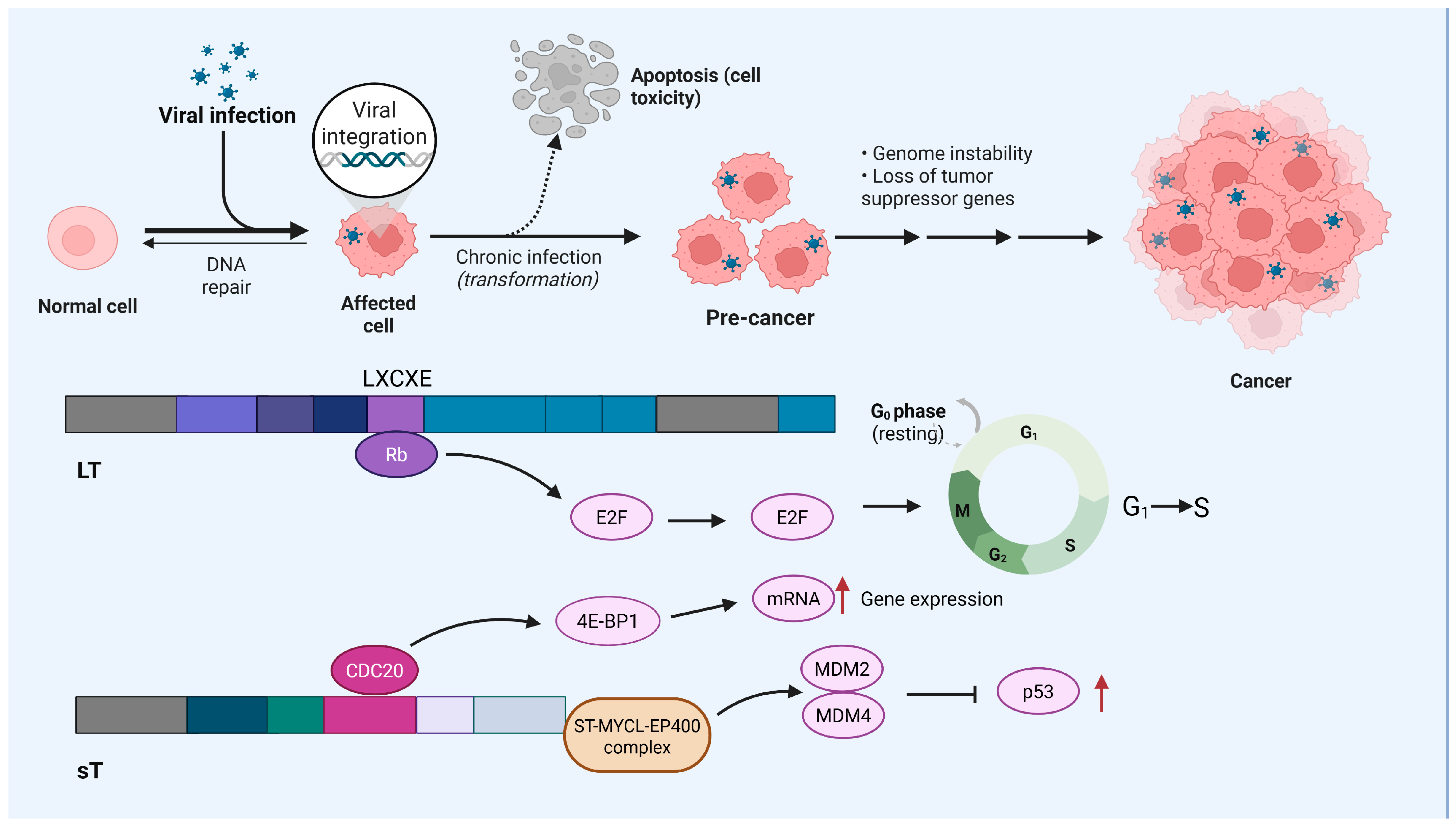
Figure 1. Schematic diagram of the pathogenic mechanism of MCC caused by MCPyV infection.
Cancers 17 02547 g001
Figure 2. The sources of IFN γ and its role in activating the antitumor immune response within the TME. Created using BioRender.com.
Cancers 17 02547 g002
Figure 3. Dual effects of IFN γ in the TME: anti-tumor and immune evasion effects. Created using BioRender.com.
Cancers 17 02547 g003
Figure 4. Mechanism of IFN-γ-induced PD-L1 upregulation and its impact on immune evasion in the TME.
Cancers 17 02547 g004
Disclaimer/Publisher’s Note: The statements, opinions and data contained in all publications are solely those of the individual author(s) and contributor(s) and not of MDPI and/or the editor(s). MDPI and/or the editor(s) disclaim responsibility for any injury to people or property resulting from any ideas, methods, instructions or products referred to in the content.