Article

Serum Visfatin/eNAMPT as a Biomarker in Pancreatic and Small Intestine Neuroendocrine Tumors: A Cross-Sectional Study and Future Perspectives

1
Department of Endocrinology, Metabolism and Internal Diseases, Poznan University of Medical Sciences, Przybyszewskiego 49, 60-355 Poznań, Poland
2
Unit of Endocrinology, Diabetes Mellitus and Metabolism, Aretaieion Hospital, Medical School, National and Kapodistrian University of Athens, 157 72 Athens, Greece
*
Author to whom correspondence should be addressed.
Cancers 2025, 17(14), 2343; https://doi.org/10.3390/cancers17142343
Submission received: 12 June 2025 / Revised: 11 July 2025 / Accepted: 14 July 2025 / Published: 15 July 2025
(This article belongs to the Special Issue Updates in Neuroendocrine Neoplasms)

Simple Summary

This study demonstrates for the first time the potential utility of serum visfatin/eNAMPT measurements as a diagnostic biomarker for neuroendocrine tumors (NETs). Visfatin/eNAMPT concentrations are increased in patients with metastatic pancreatic and small intestinal NETs and show potential in distinguishing NETs from controls, regardless of tumor and patient characteristics. As a biomarker, visfatin could act as a bridge between currently used imperfect monoanalytes such as chromogranin A and expensive multianalytes such as NETest. Although serum visfatin levels might not directly reflect tissue NAMPT expression, we also review visfatin’s role in a therapeutic setting in the context of NAMPT inhibitors, researched in previous studies.

Abstract

Background: Neuroendocrine tumors (NETs) remain a problematic area in endocrine oncology due to their non-specific symptoms and lack of reliable biomarkers. Visfatin/eNAMPT’s involvement in tumorigenesis has been described in several malignancies. In NETs, NAMPT inhibition was explored as a potential therapeutic option; however, serum visfatin and its role as a biomarker have not been studied. Objectives: We aimed to measure serum visfatin concentrations in NETs and evaluate visfatin’s potential as a diagnostic biomarker. Methods: We conducted a single-center, cross-sectional study of 77 patients with NETs (33 pancreatic and 44 small intestinal) and 29 controls. Patient demographics, tumor characteristics, and clinical data were analyzed, and serum visfatin levels were measured using ELISA. Statistical analyses were performed in Python, including Mann–Whitney U and Kruskal–Wallis tests, Spearman’s correlation, multiple linear regression, and ROC curve analysis. Results: Serum visfatin was higher in NETs compared to controls (median [IQR]: 6.94 [2.11–236.17] vs. 1.59 [1.1–9.24] ng/mL, p = 0.004). ROC curves showed moderate diagnostic performance (AUC = 0.68), with concentrations above 2.11 ng/mL achieving 75.3% sensitivity and 58.6% specificity. In NETs, visfatin did not differ based on WHO grade (G1/G2, p = 0.31), primary site (pancreas/small intestine, p = 0.95), sex (p = 0.89), age (p = 0.13), and when stratified by primary site and grade (p = 0.18). Multiple linear regression confirmed no association between visfatin and the study variables (R-squared = 0.036, all p > 0.2). Conclusions: This is the first study examining serum visfatin as a diagnostic biomarker in NETs. Visfatin concentrations show moderate discriminatory ability between NETs and controls, independent of tumor and clinical characteristics. Further research should involve larger cohorts and comparisons to established biomarkers.

1. Introduction

Neuroendocrine neoplasms (NENs) are highly heterogeneous tumors arising from endocrine and nervous system cells. NENs are divided into neuroendocrine tumors (NETs), which are usually well-differentiated, and poorly differentiated neuroendocrine carcinomas (NECs). NETs can be characterized based on their primary site (the gastrointestinal tract, pancreas, and lungs are the most common locations), proliferative activity (WHO grades depend on the tumor’s Ki-67 index and mitotic count), and hormonal secretion (functioning vs. non-functioning) [1]. NENs are generally rare, accounting for less than 2% of all malignancies. NET prevalence ranges between 2.5 and 8.35 cases per 10,000 and remains below 200,000 cases/year in the U.S., meeting the FDA’s rare disease criteria. However, NET incidence has been rising in recent years, partly due to the improvement in the accuracy and availability of medical imaging [2,3]. Despite significant advances in understanding NENs’ etiology, pathogenesis, and therapy, several issues remain unsolved. In particular, identifying reliable diagnostic and prognostic biomarkers for early disease detection and effective, tailored treatment remains an unmet need [4,5]. NET biomarkers can be divided into non-specific (chromogranin A, neuron-specific enolase, and beta-human chorionic gonadotropin) and tumor-specific analytes (insulin, gastrin, vasoactive intestinal peptide, glucagon, somatostatin, serotonin, and 5-hydroxyindoleacetic acid) [2,6]. Unfortunately, despite initial promise, no single biomarker has demonstrated optimal diagnostic and prognostic accuracy for a disease as diverse as NETs. Recent advances led to the development of novel biomarkers, utilizing liquid biopsy and multiomics-based approaches, such as NETest, microRNAs, cell-free DNA, and circulating tumor cells [2]. Despite improved performance, they are often expensive or not readily available. Thus, reliable biochemical markers for both diagnostic and prognostic purposes are still needed [2].
Visfatin, also known as nicotinamide phosphoribosyltransferase (NAMPT) or pre-B-cell colony-enhancing factor 1 (PBEF1), is a rate-limiting enzyme in the NAD+ (nicotinamide adenine dinucleotide) synthesis salvage pathway [7]. NAD+ is an essential cofactor involved in redox reactions and serves as a substrate for multiple signaling enzymes, including poly(ADP-ribose) polymerases, cyclic ADP-ribose synthetases, sirtuins, and CD38 [8]. The role of NAD+ in cancer is well-established, as cancer cells utilize it extensively for metabolism and survival [8]. NAMPT exists in two distinct forms, namely intracellular NAMPT (iNAMPT), primarily found in the cytosol and nucleus, and extracellular NAMPT (eNAMPT)—the term visfatin is preserved for this form. iNAMPT is the predominant form in physiological conditions, and eNAMPT/visfatin constitutes only about 1% of total NAMPT levels [9]. iNAMPT is responsible for the enzymatic regulation of NAD+ biosynthesis, whereas eNAMPT functions as a proinflammatory cytokine/adipocytokine. eNAMPT activates Toll-like receptor 4 (TLR4), stimulates B-lymphocytes, monocytes, and neutrophils, and increases the secretion of proinflammatory cytokines (e.g., IL-1β, IL-1Ra, IL-6, CXCL8, IL-10, TNF-α) and chemokines (CCL2, CCL3, CCL18, and CCL20) [7].
In the context of biomarker research, eNAMPT/visfatin has attracted increasing interest. Visfatin was previously investigated for its insulin-mimetic properties in metabolic disorders. It is predominantly secreted by adipocytes, macrophages, lymphocytes, and neoplastic cells [9]. Although increased circulating visfatin levels have been reported in several malignancies, its association with NETs remains unexplored [10,11].
We aimed to assess, for the first time, the utility of serum visfatin concentration as a diagnostic marker in NETs, specifically small intestine NETs (siNETs) and pancreatic NETs (panNETs). We seek to explore the involvement of visfatin/eNAMPT in NETs pathogenesis, potentially paving the way for personalized and targeted treatment strategies. Finally, we aimed to determine if circulating visfatin correlates with patient and tumor characteristics including WHO grade, NET primary site, sex, and age.

2. Materials and Methods

2.1. Study Design and Patient Population

This was a single-center, cross-sectional study, conducted at the Department of Endocrinology, Metabolism, and Internal Diseases at Poznan University of Medical Sciences in Poznan, Poland. Between February and November 2024, we recruited 77 patients with histologically confirmed pancreatic (33) or small intestinal (44) NETs and 29 controls. Patient demographics and tumor characteristics were collected at the time of inclusion. All patients with NETs were diagnosed with metastatic or locally advanced disease and were treated with long-acting somatostatin analogs (SSA) at the time of the sampling (lanreotide, Somatuline Autogel 120 mg once every 4 weeks; Ipsen, Paris, France). WHO grade was recorded based on a histopathological evaluation of tumor tissue at the time of the diagnosis. Exclusion criteria included prior or current systemic therapy for NETs (e.g., everolimus, sunitinib, chemotherapy, peptide receptor radionuclide therapy), active malignancy other than NETs, diabetes, renal or liver failure, untreated or unregulated hyperlipidemia, hypertension, thyroid dysfunction, recent surgery (up to 3 months before the recruitment), obesity (defined as BMI ≥ 30), autoimmune diseases requiring biological treatment, ECOG/WHO performance status ≥ 3, acute infections or other acute conditions at the time of the sampling, pregnancy, or lactation. Participants with missing data were excluded from the study. Controls without a NET diagnosis were recruited from among patients hospitalized at the same department. These were patients diagnosed with low-risk papillary thyroid cancer (pT1-pT2, radically resected at least 6 months prior to sampling, with no distant metastases), who required no adjuvant radioiodine therapy, with no other malignancies at the time of inclusion. Controls were euthyroid on the date of sampling, defined by thyroid-stimulating hormone (TSH) levels measured between 0.5 and 2 mIU/L. The controls were sampled on admission and all the other exclusion criteria from the test group were also applied to the controls.

2.2. Laboratory Analysis

Venous blood samples were collected from NET patients during routine follow-up visits (prior to lanreotide administration) and from controls on hospital admission. Samples were pseudonymized and technicians performing the assays were blinded to clinical data. The samples were processed within 6 h of collection, centrifuged, and stored at −80 °C until analysis.
Serum visfatin concentrations were measured using the Human Visfatin ELISA Kit (Invitrogen by ThermoFisher Scientific, Waltham, MA, USA) with a detection range of 1.1–300 ng/mL. Each sample was analyzed twice, and the mean value was calculated. Values outside the assay range were recorded as 1.1 ng/mL (minimum) or 300 ng/mL (maximum).

2.3. Statistical Calculations

Normality was assessed via the Shapiro–Wilk test. Group comparisons were performed using Mann–Whitney U (two groups) or Kruskal–Wallis (multiple groups) tests. Associations between visfatin and age were evaluated via Spearman’s rank correlation. Diagnostic performance was assessed using ROC curves, with optimal thresholds determined by Youden’s J statistic. Multiple linear regression tested associations between visfatin and clinical variables (age, sex, primary site, grade). Due to non-normally distributed data, continuous variables are reported as the median [IQR]. All tests were two-tailed with p values below 0.05 considered statistically significant. Statistical calculations were performed in Python 3.12 using the matplotlib 3.10.0, openpyxl 3.2.0 b1, pandas 2.2.3, patsy 1.0.1., pingouin 0.5.5, scikit-learn 1.6.1, scikit-posthocs 0.11.2, scipy 1.15.1, seaborn 0.13.2, and statsmodels 0.14.4 packages.

3. Results

3.1. Baseline Characteristics

We recruited 77 patients with non-resectable NETs, including 33 with panNETs and 44 with siNETs, and 29 controls. Both panNET and siNET patients presented a similar sex and age distribution, with the controls being younger than the patients with NETs and being predominantly female. The baseline study and control group demographics and clinical characteristics are summarized in Table 1.

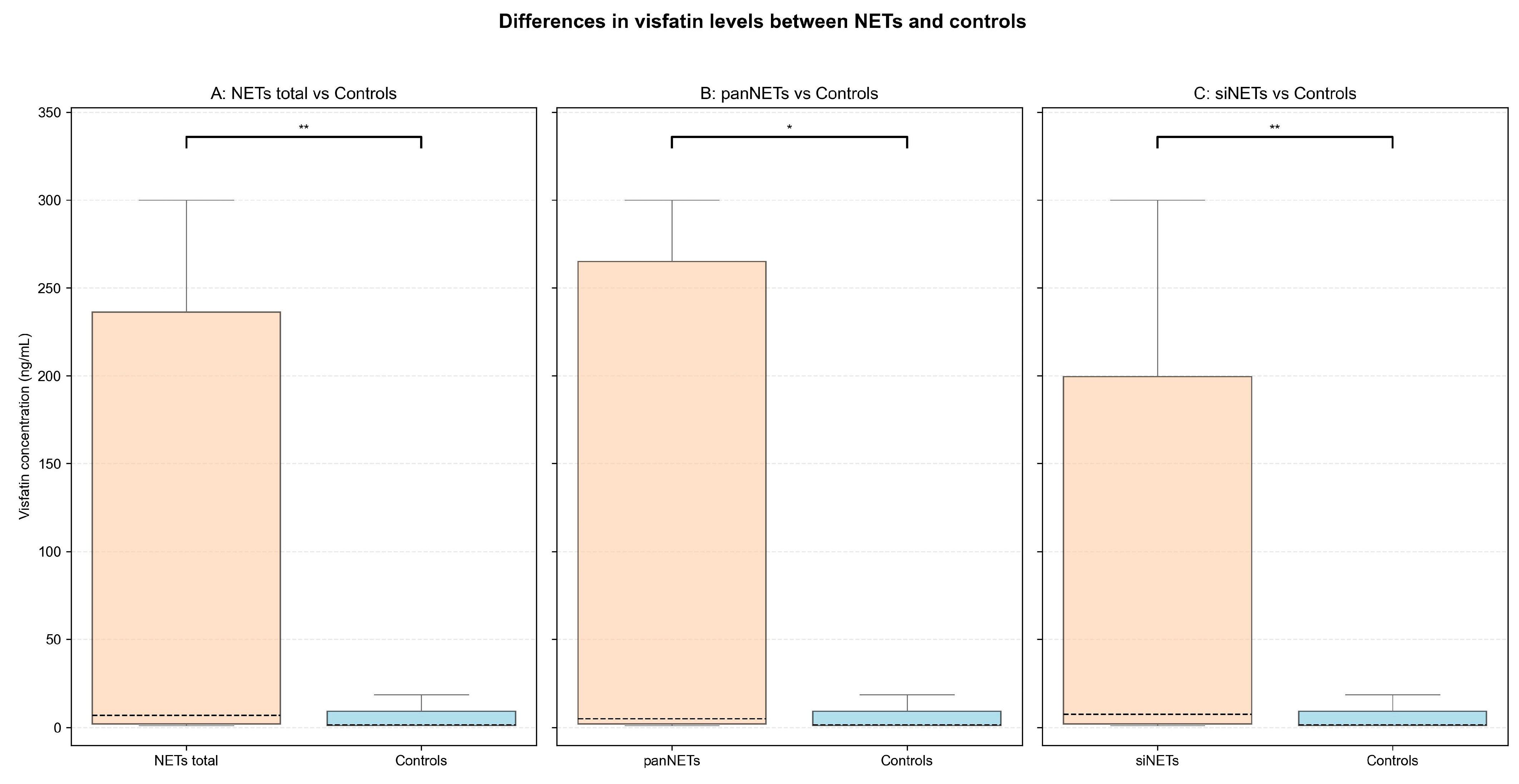
3.2. Serum Visfatin Concentrations in NETs vs. Controls

Median [IQR] serum visfatin levels were higher in patients with NETs regardless of tumor’s primary site (6.94 [2.11–236.17] ng/mL, p = 0.004) as well as when stratified by panNETs (4.98 [2.13–264.96] ng/mL, p = 0.019) and siNETs (7.46 [2.01–199.44] ng/mL, p = 0.007) vs. controls (1.59 [1.1–9.24] ng/mL). In NET patients, visfatin concentrations varied (IQR: 234.06 ng/mL), with 16 (20.8%) patients above and 14 (18.2%) below the assay’s reference range (1.1–300 ng/mL). In controls, only 1 (3.4%) subject exceeded the reference range, while 13 (44.8%) were below it. Detailed results of the analysis are presented in Table 2 and Figure 1 and the complete statistical analysis can be found in the Supplementary Table S1.

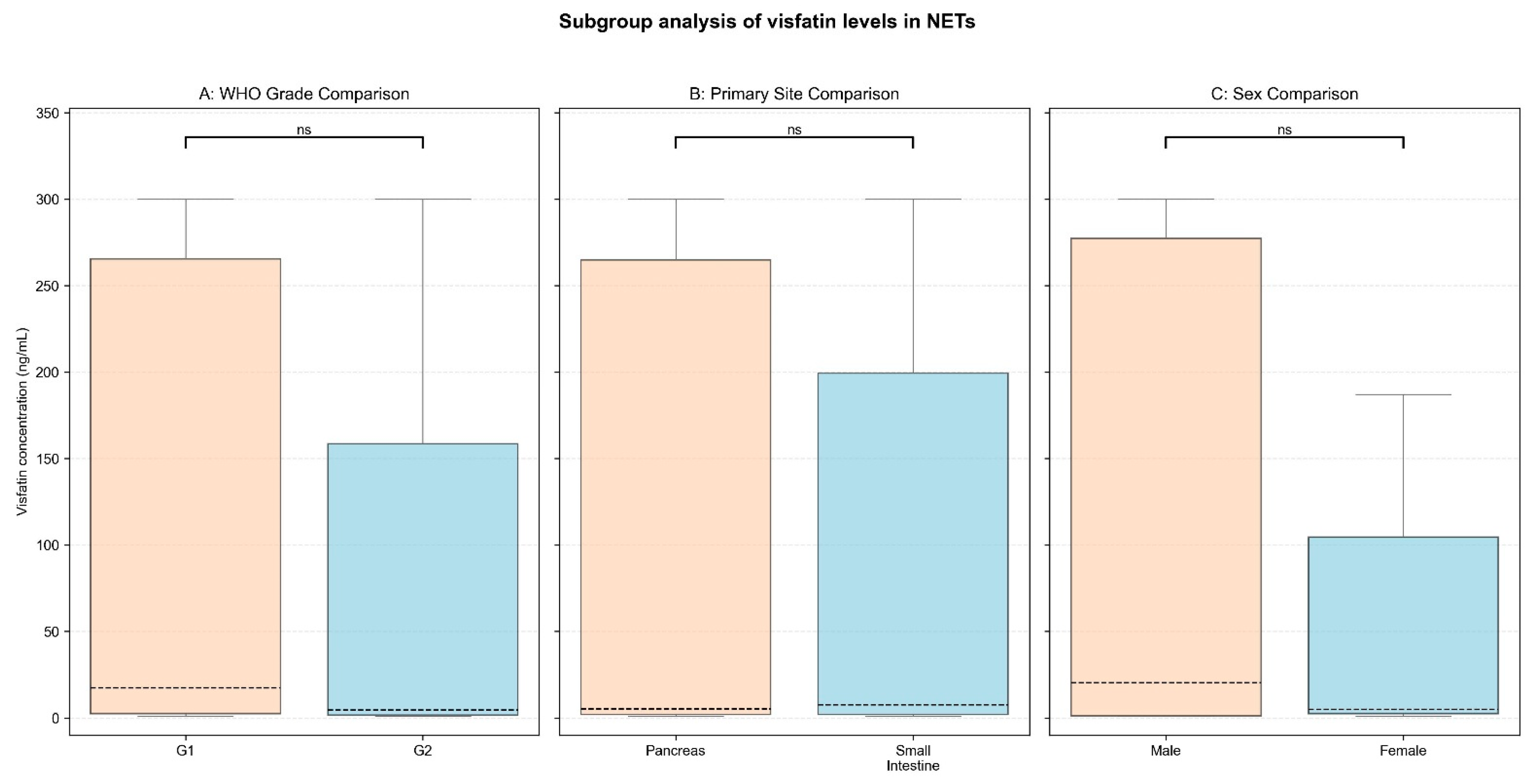
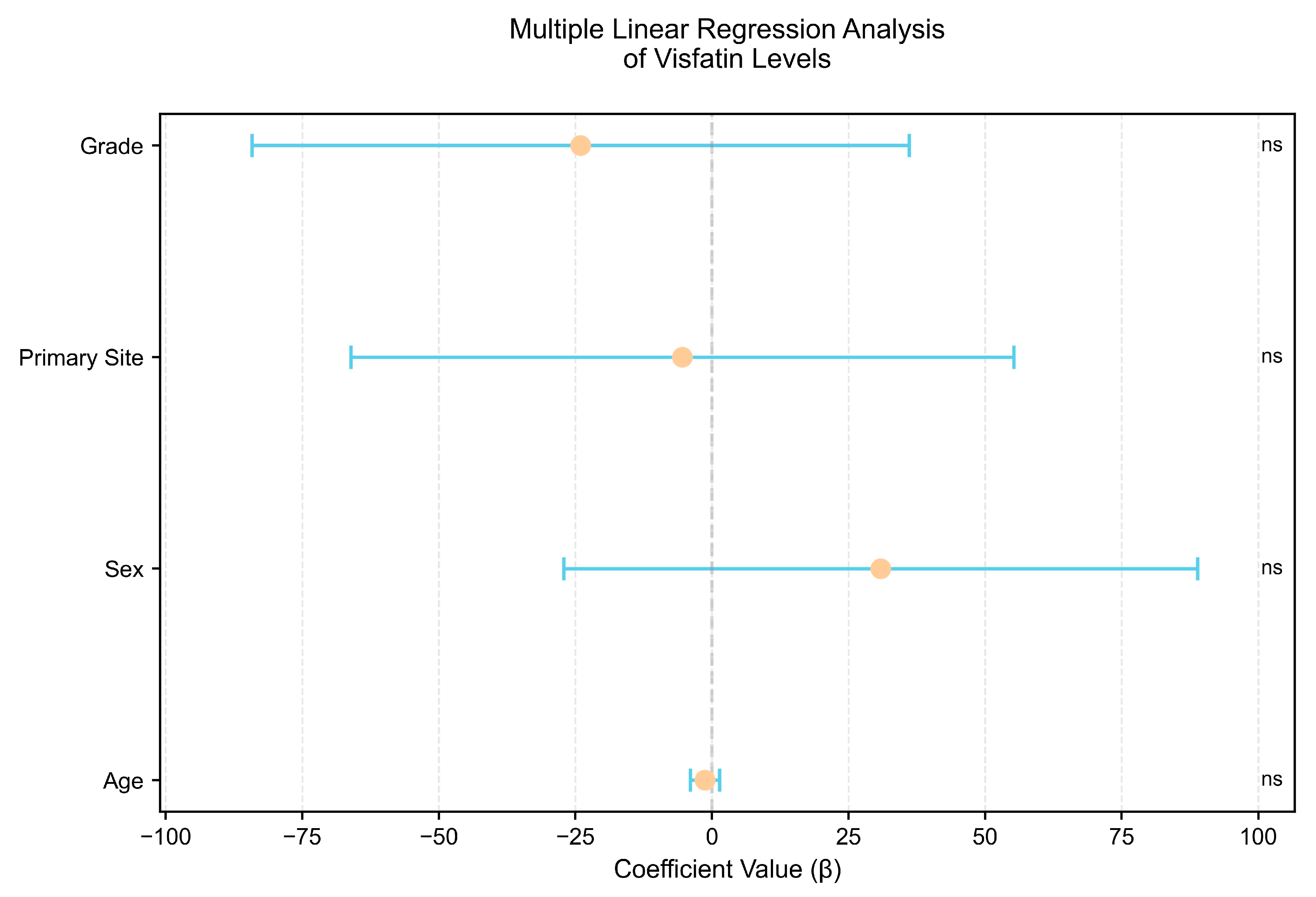
3.3. Subgroup Comparison and Correlation Analysis

Subgroup analyses were performed to determine if visfatin levels varied by NET primary site, WHO grade, or sex. We found no differences in visfatin concentrations depending on NET primary site (panNETs vs. siNETs, median [IQR]: 4.98 [2.13–264.96] vs. 7.46 [2.01–199.44] ng/mL, p = 0.95), tumor grade (G1 vs. G2, median [IQR]: 17.23 [2.44–265.55] vs. 4.45 [1.62–158.45] ng/mL, p = 0.31), and sex (male vs. female, median [IQR]: 20.27 [1.23–277.34] vs. 4.84 [2.42–104.56] ng/mL, p = 0.89). Furthermore, when stratified by both primary site and grade, the Kruskal–Wallis test revealed no significant variation in visfatin levels (p = 0.18). Serum visfatin did not correlate significantly with age in NET patients (R Spearman = −0.17, p = 0.13). Finally, multiple linear regression (adjusting for age, sex, primary site, and grade) confirmed no impact of these variables on serum visfatin (R2 = 0.036; all p > 0.2). The results are presented in Table 3 and Table 4 and Figure 2 and Figure 3.

3.4. Serum Visfatin’s Diagnostic Performance

The diagnostic utility of serum visfatin for distinguishing NET patients from controls was evaluated using ROC curve analysis. The analysis revealed a moderate area under the curve (AUC) value of 0.68 (0.59–0.77). A cutoff value of 2.11 ng/mL was established with Youden’s index, providing a sensitivity of 75.3% (65.7–85.0%) and a specificity of 58.6% (40.7–76.6%). At this cut-off, the positive predictive value (PPV) was 82.86% (74.0–91.7%) and the negative predictive value (NPV) was 47.22% (30.9–63.5%). The ROC curves are displayed in Figure 4.
The complete results of the statistical analysis are included in Supplementary Table S1.

4. Discussion

Our study reveals that serum visfatin levels are increased in patients with NETs compared to controls without an active malignancy. ROC curve analyses revealed moderate diagnostic performance for visfatin across all patients with NETs and when divided into panNETs and siNETs. Notably, visfatin levels were consistent in NET subgroups determined by patient and tumor characteristics. Given the absence of prior studies of serum visfatin in NETs, these results have to be discussed in the broader context of research in other malignancies and in tumor tissue.

4.1. Overview of Visfatin and Its Role in Tumorigenesis

eNAMPT/visfatin functions primarily as a proinflammatory cytokine/adipocytokine. It exerts its responses via membrane receptors, though a precise mechanism of action remains under investigation [7]. Visfatin also participates in glucose homeostasis, angiogenesis, and may function as an ectoenzyme. It is actively secreted via a non-classical pathway, stimulated by cellular stress (e.g., hypoxia and ischemia) as well as by inflammatory processes [7]. Various factors are suggested to increase circulating visfatin, including caloric intake, exercise, and circadian cycles. Table 5 displays examples of visfatin alterations in several pathological conditions [12,13]. Visfatin is involved in the adipose–malignancy interactions alongside other adipocytokines, such as resistin and leptin, and may contribute to increased neoplasm risk in obesity [14]. Emerging evidence points to a causative role of visfatin in breast cancer, liver cancer, and multiple myeloma [15,16,17]. Both eNAMPT and iNAMPT are generally considered oncogenic, although their role in cancer is complex. Elevated circulating visfatin may result from tumor cell secretion or overproduction within the tumor microenvironment [7,9]. NAMPT facilitates cancer cell proliferation via NAD+ supply, the activation of multiple signaling pathways, and apoptosis inhibition [7,8]. Additionally, visfatin promotes cancer cell migration and metastasis by upregulating SDF-1/CXCL12, TGF-β, and the gelatinases MMP-2/-9 [7].

4.2. Diagnostic Utility of Serum Visfatin in NETs

Our study revealed that patients with NETs exhibited significantly higher serum visfatin levels compared to the control group, regardless of the tumor’s primary site (Figure 1). ROC curve analysis showed moderate diagnostic performance of serum visfatin across all NET patients (AUC = 0.68; 95%CI: 0.59–0.77). A cut-off value of 2.11 ng/mL achieved a sensitivity of 75.3%, and a specificity of 58.6%. Subgroup analysis for panNET and siNET patients yielded similar ROC results, as shown in Figure 4.
Although circulating visfatin has not been previously researched in NETs, our results align with similar studies across various neoplasms. Increased circulating visfatin levels and NAMPT tissue overexpression was reported in several malignancies, although discrepancies exist between studies and between tissue and serum compartments (e.g., in thyroid cancer), as seen in Table 6 [10,40]. In NET animal models, visfatin is expressed in pituitary and gastrointestinal neuroendocrine cells and has a regulatory role in cell proliferation and hormone secretion [41,42,43]. NAMPT overexpression has also been observed in patient-derived panNET tissue, suggesting its potential role in tumor pathogenesis [44].

4.3. Association of Visfatin with Tumor and Patient Characteristics

Given the heterogeneity of NETs, we analyzed visfatin levels across NET primary sites, WHO Grades (panNETs vs. siNETs, G1 vs. G2), sex, and age. Although some trends were observed, there was no significant difference between variables, either individually or in composite groups (Table 3 and Table 4, Figure 2 and Figure 3). This consistency could be favorable in a biomarker setting. Contrasting with chromogranin A’s variability, serum visfatin may present a more consistent option, independent of tumor features [2].
In other malignancies, elevated visfatin levels were associated with advanced tumor size, stage, and disease progression, the presence of lymph node metastases, higher metastatic rates, and resistance to treatment [8]. However, most studies did not establish a significant correlation between visfatin concentrations and tumor histological grade [61]. A meta-analysis highlighted a consistent correlation between elevated serum visfatin and poor overall survival in endometrial, colorectal, breast, and bladder cancers [11,51,62]. Importantly, due to the cross-sectional design of this study, we were unable to assess the prognostic value of visfatin in NETs. Prospective studies are needed to explore these associations and clarify visfatin’s prognostic potential.
In previous studies, both positive and negative correlations between age/sex and serum visfatin can be found, with no significant sex differences [63,64]. Our study revealed no statistically significant differences between males and females and did not observe a correlation with age. These findings align with the perspective that underlying health conditions may have more impact on visfatin levels than sex or age.

4.4. Visfatin as a Therapeutic Target—NAMPT Inhibitors

Further evidence of NAMPT’s significance in neuroendocrine malignancies comes from studies on small cell lung cancer (SCLC), revealing a strong reliance of cancer cells on NAD+ synthesis through the NAMPT-dependent salvage pathway. SCLC and other cells of neuroendocrine differentiation could be particularly vulnerable to NAMPT inhibitors (NAMPT-i) [65]. This is supported by Audrito et al., suggesting that NAMPT overexpression correlates with tumorigenesis and may be utilized in NAMPT-i therapy [66]. Further preclinical studies have shown that NAMPT-i exhibit strong anti-tumor activity against neuroblastoma and SCLC cell lines. This effect is attributed to the high dependence of these tumors on oxidative phosphorylation for energy production and reduced expression of Yes-Associated Protein 1 (YAP1) [67]. One in vitro study suggests NAMPT-i as potential drug candidates for NET treatment [68].
Emerging data suggest that NAMPT-i could also improve existing NET therapies. PanNET cells frequently exhibit overactivity of the PI3K/AKT/mTOR pathway, and NAMPT has been linked with a resistance mechanism against everolimus, an mTOR inhibitor. Dual inhibition of NAMPT and mTOR may have a synergistic effect and enhance treatment efficacy [69]. Additionally, NAMPT inhibition sensitizes NET cells to radiation and has been shown to improve response to radioligand therapy with 177Lu-DOTATATE [70].
These findings highlight the potential of targeting NAMPT as a novel therapeutic option for NETs. Although circulating visfatin is not a direct substitute for NAMPT, its study can provide valuable insights into disease status and aid in personalizing treatment. While NAMPT-i are still in the early stages of development, some have already been researched in NENs and others have entered clinical trials, paving the way for their potential therapeutic implementation in the near future [70,71,72].

4.5. Limitations and Future Directions

This study has several limitations. As the first study examining serum visfatin levels in NETs, there is no previous data for direct comparison, and validation in an independent cohort is needed. NETs are heterogeneous and further analyses should include patients with additional primary tumor sites (e.g., pulmonary carcinoids or hindgut NETs). Moreover, our study was limited to G1 and G2 NETs. Including G3 NETs and comparing NETs with NECs would be valuable, as in vitro and pharmacological studies suggest that higher grade tumors may exhibit even higher visfatin levels [65,67].
Visfatin is affected by other comorbidities (Table 5), complicating the selection of an optimal control group. We minimized confounding factors by excluding subjects with conditions known to affect visfatin (e.g., diabetes, renal or liver failure, untreated hyperlipidemia, hypertension, thyroid dysfunction, recent surgery, or active malignancies other than NETs). Nevertheless, some selection bias cannot be entirely ruled out. Our control group consisted of patients with radically resected low-risk papillary thyroid cancer in a euthyroid state and healthy otherwise, without significant comorbidities. This selection appears justified, as previous research indicates that thyroid cancer is not associated with altered visfatin levels (indeed, a control group without any malignancy might exhibit even lower levels of visfatin) [10]. Moreover, the sex and age distributions of the controls differ from those of the test group. Although these factors do not appear to affect serum visfatin both within sex- and age-matched NET subgroups and in previous research, we acknowledge this as a limitation.
Finally, a cross-sectional study design prevents the evaluation of visfatin’s prognostic value in NETs. All NET patients were treated with SSA, the impact of which on serum visfatin remains unknown. In order to assess visfatin’s relationship with disease outcomes, treatment response, and tumor burden and to determine any treatment-related effects, future studies should include longitudinal measurements or compare treatment-naive patients with those undergoing therapy.

5. Conclusions

For the first time, we showed that serum visfatin levels have moderate diagnostic ability in distinguishing NETs from controls [73]. Visfatin is a monoanalyte and, as such, cannot describe disease status as comprehensively as next-generation biomarkers. However, it appears less variable across NET subgroups than other single biomarkers, such as chromogranin A. Visfatin/NAMPT plays an important role in cancer biology—in cell migration, tissue proliferation, and metabolic regulation—making it an attractive candidate both in biomarker research and as a therapeutic target. This study is limited by its single-center, cross-sectional design, which needs to be addressed in future studies through longitudinal measurements and larger patient cohorts. Nevertheless, visfatin’s promising results, combined with a low-cost approach, warrant further investigation.

Supplementary Materials

The following supporting information can be downloaded at: https://www.mdpi.com/article/10.3390/cancers17142343/s1, Supplementary Table S1: Complete statistical calculations.

Author Contributions

P.K.: conceptualization, methodology, validation, formal analysis, investigation, resources, data curation, writing—original draft, writing—review and editing, visualization, supervision, project administration, funding acquisition; A.M.: conceptualization, formal analysis, investigation, resources, data curation, writing—review and editing; J.M.: conceptualization, formal analysis, investigation, resources, data curation, writing—original draft, writing—review and editing, visualization; M.C.: conceptualization, formal analysis, investigation, resources, data curation, writing—original draft, writing—review and editing, visualization; G.M.: conceptualization, validation, formal analysis, investigation, writing—review and editing, visualization, supervision, project administration; M.R.: conceptualization, validation, formal analysis, investigation, writing—review and editing, visualization, supervision, project administration, funding acquisition. P.G.: conceptualization, methodology, validation, formal analysis, investigation, resources, data curation, writing—review and editing, supervision, project administration; All authors have read and agreed to the published version of the manuscript.

Funding

This research was funded by the Poznan University of Medical Sciences: “Grants from the statutory subsidy for Young Scientists—Ph.D. students of the Doctoral School for 2023—Large Research Grant”; grant number NMN0000067. P.K. is the administrator of the grant and the primary investigator, while P.G. is the project supervisor.

Institutional Review Board Statement

This study was conducted in accordance with the Declaration of Helsinki and was approved by the Regional Bioethical Committee at the Poznan University of Medical Sciences (protocol no. 849/21, approve date: 4 November 2021).

Informed Consent Statement

All study participants were of legal capability and provided written informed consent.

Data Availability Statement

Data is available as a supplementary file from the corresponding authors upon reasonable request.

Acknowledgments

Paweł Komarnicki is a participant of STER Internationalization of Doctoral Schools Program from NAWA Polish National Agency for Academic Exchange No. PPI/STE/2020/1/00014/DEC/02. We used a local generative AI tool in order to draft the outline of the article, identify potential errors, and proofread the manuscript: Mistral NeMo 12B (Mistral AI, Paris, France). Subsequently, we reviewed and edited generated content as required and take full responsibility for the publication.

Conflicts of Interest

P.K.: honoraria from Novartis, Ipsen, clinical trial remuneration from Neurocrine Biosciences, Crinetics Pharmaceuticals, Ascendis Pharma; A.M.: no conflict of interest; J.M.: honoraria from Novartis; M.C.: no conflict of interest; G.M.: no conflict of interest; M.R.: honoraria from Novartis, Pfizer, Ipsen, Berlin-Chemie Menarini, Genzyme, Merck, IBSA. P.G.: honoraria from Novartis, Ipsen. The funders had no role in the design of the study; in the collection, analyses, or interpretation of data; in the writing of the manuscript; or in the decision to publish the results.

Abbreviations

The following abbreviations are used in this manuscript:
NENsNeuroendocrine neoplasms
NETsNeuroendocrine tumors
NECsNeuroendocrine carcinomas
WHOWorld Health Organization
NAMPTNicotinamide phosphoribosyltransferase
PBEF1Pre-B-cell colony-enhancing factor 1
NAD+Nicotinamide adenine dinucleotide
eNAMPTExtracellular NAMPT
iNAMPTIntracellular NAMPT
siNETsSmall intestinal NETs
panNETsPancreatic NETs
TSHThyroid-stimulating hormone
NAMPT-iNAMPT inhibitors
SCLCSmall cell lung cancer

References

  1. Hofland, J.; Kaltsas, G.; De Herder, W.W. Advances in the Diagnosis and Management of Well-Differentiated Neuroendocrine Neoplasms. Endocr. Rev. 2020, 41, 371–403. [Google Scholar] [CrossRef]
  2. Komarnicki, P.; Musiałkiewicz, J.; Stańska, A.; Maciejewski, A.; Gut, P.; Mastorakos, G.; Ruchała, M. Circulating Neuroendocrine Tumor Biomarkers: Past, Present and Future. J. Clin. Med. 2022, 11, 5542. [Google Scholar] [CrossRef] [PubMed]
  3. Woliński, K.; Komarnicki, P.; Maciejewski, A.; Musiałkiewicz, J.; Gut, P.; Ruchała, M. Prevalence of Second Primary Malignancies in Patients With Well-Differentiated Neuroendocrine Tumors. Endocr. Pract. 2025, 31, 426–432. [Google Scholar] [CrossRef] [PubMed]
  4. De Herder, W.W.; Rehfeld, J.F.; Kidd, M.; Modlin, I.M. A Short History of Neuroendocrine Tumours and Their Peptide Hormones. Best Pract. Res. Clin. Endocrinol. Metab. 2016, 30, 3–17. [Google Scholar] [CrossRef]
  5. Oberg, K.; Modlin, I.M.; De Herder, W.; Pavel, M.; Klimstra, D.; Frilling, A.; Metz, D.C.; Heaney, A.; Kwekkeboom, D.; Strosberg, J.; et al. Consensus on Biomarkers for Neuroendocrine Tumour Disease. Lancet Oncol. 2015, 16, e435–e446. [Google Scholar] [CrossRef] [PubMed]
  6. Komarnicki, P.; Gut, P.; Cieślewicz, M.; Musiałkiewicz, J.; Maciejewski, A.; Czupińska, M.; Mastorakos, G.; Ruchała, M. Serum β-HCG as a Biomarker in Pancreatic Neuroendocrine Tumors: Rethinking Single-Analyte Approach. Cancers. 2024, 16, 2060. [Google Scholar] [CrossRef]
  7. Semerena, E.; Nencioni, A.; Masternak, K. Extracellular Nicotinamide Phosphoribosyltransferase: Role in Disease Pathophysiology and as a Biomarker. Front. Immunol. 2023, 14, 1268756. [Google Scholar] [CrossRef]
  8. Navas, L.E.; Carnero, A. NAD+ Metabolism, Stemness, the Immune Response, and Cancer. Signal Transduct. Target. Ther. 2021, 6, 2. [Google Scholar] [CrossRef]
  9. Grolla, A.A.; Torretta, S.; Gnemmi, I.; Amoruso, A.; Orsomando, G.; Gatti, M.; Caldarelli, A.; Lim, D.; Penengo, L.; Brunelleschi, S.; et al. Nicotinamide Phosphoribosyltransferase (NAMPT/PBEF/Visfatin) Is a Tumoural Cytokine Released from Melanoma. Pigment Cell Melanoma Res. 2015, 28, 718–729. [Google Scholar] [CrossRef]
  10. Sawicka-Gutaj, N.; Ziółkowska, P.; Derwich, A.; Gut, P.; Czarnywojtek, A.; Kloska, M.; Ruchała, M. Is ENAMPT/Visfatin a Potential Serum Marker of Papillary Thyroid Cancer? Ther. Adv. Endocrinol. Metab. 2022, 13, 20420188221090005. [Google Scholar] [CrossRef]
  11. Mohammadi, M.; Mianabadi, F.; Mehrad-Majd, H. Circulating Visfatin Levels and Cancers Risk: A Systematic Review and Meta-Analysis. J. Cell. Physiol. 2019, 234, 5011–5022. [Google Scholar] [CrossRef]
  12. Chang, Y.H.; Chang, D.M.; Lin, K.C.; Shin, S.J.; Lee, Y.J. Visfatin in Overweight/Obesity, Type 2 Diabetes Mellitus, Insulin Resistance, Metabolic Syndrome and Cardiovascular Diseases: A Meta-Analysis and Systemic Review. Diabetes. Metab. Res. Rev. 2011, 27, 515–527. [Google Scholar] [CrossRef]
  13. Yu, P.L.; Wang, C.; Li, W.; Zhang, F.X. Visfatin Level and The Risk of Hypertension and Cerebrovascular Accident: A Systematic Review and Meta-Analysis. Horm. Metab. Res. 2019, 51, 220–229. [Google Scholar] [CrossRef]
  14. Guiu, B.; Petit, J.M.; Bonnetain, F.; Ladoire, S.; Guiu, S.; Cercueil, J.-P.; Krause, D.; Hillon, P.; Borg, C.; Chauffert, B.; et al. Visceral Fat Area Is an Independent Predictive Biomarker of Outcome after First-Line Bevacizumab-Based Treatment in Metastatic Colorectal Cancer. Gut 2010, 59, 341–347. [Google Scholar] [CrossRef]
  15. Wang, Y.; Wang, F.; Wang, L.; Qiu, S.; Yao, Y.; Yan, C.; Xiong, X.; Chen, X.; Ji, Q.; Cao, J.; et al. NAD+ Supplement Potentiates Tumor-Killing Function by Rescuing Defective TUB-Mediated NAMPT Transcription in Tumor-Infiltrated T Cells. Cell Rep. 2021, 36, 109516. [Google Scholar] [CrossRef] [PubMed]
  16. Miethe, C.; Torres, L.; Beristain, J.; Zamora, M.; Price, R.S. The Role of Visfatin and Resistin in an in Vitro Model of Obesity-Induced Invasive Liver Cancer. Can. J. Physiol. Pharmacol. 2021, 99, 839–846. [Google Scholar] [CrossRef] [PubMed]
  17. Tentolouris, A.; Ntanasis-Stathopoulos, I.; Terpos, E. Obesity and Multiple Myeloma: Emerging Mechanisms and Perspectives. Semin. Cancer Biol. 2023, 92, 45–60. [Google Scholar] [CrossRef] [PubMed]
  18. Cheleschi, S.; Tenti, S.; Bedogni, G.; Fioravanti, A. Circulating Mir-140 and Leptin Improve the Accuracy of the Differential Diagnosis between Psoriatic Arthritis and Rheumatoid Arthritis: A Case-Control Study. Transl. Res. 2022, 239, 18–34. [Google Scholar] [CrossRef]
  19. Mohammed Ali, D.M.; Al-Fadhel, S.Z.; Al-Ghuraibawi, N.H.A.; Al-Hakeim, H.K. Serum Chemerin and Visfatin Levels and Their Ratio as Possible Diagnostic Parameters of Rheumatoid Arthritis. Reumatologia 2020, 58, 67–75. [Google Scholar] [CrossRef]
  20. Askari, A.; Arasteh, P.; Homayounfar, R.; Naghizadeh, M.M.; Ehrampoush, E.; Mousavi, S.M.; Alipoor, R. The Role of Adipose Tissue Secretion in the Creation and Pain Level in Osteoarthritis. Endocr. Regul. 2020, 54, 6–13. [Google Scholar] [CrossRef]
  21. Fioravanti, A.; Cheleschi, S.; De Palma, A.; Addimanda, O.; Mancarella, L.; Pignotti, E.; Pulsatelli, L.; Galeazzi, M.; Meliconi, R. Can Adipokines Serum Levels Be Used as Biomarkers of Hand Osteoarthritis? Biomarkers 2018, 23, 265–270. [Google Scholar] [CrossRef]
  22. Colombo, G.; Caviglia, G.P.; Ravera, A.; Tribocco, E.; Frara, S.; Rosso, C.; Travelli, C.; Genazzani, A.A.; Ribaldone, D.G. NAMPT and NAPRT Serum Levels Predict Response to Anti-TNF Therapy in Inflammatory Bowel Disease. Front. Med. 2023, 10, 1116862. [Google Scholar] [CrossRef]
  23. Neubauer, K.; Bednarz-Misa, I.; Walecka-Zacharska, E.; Wierzbicki, J.; Agrawal, A.; Gamian, A.; Krzystek-Korpacka, M. Oversecretion and Overexpression of Nicotinamide Phosphoribosyltransferase/Pre-B Colony-Enhancing Factor/Visfatin in Inflammatory Bowel Disease Reflects the Disease Activity, Severity of Inflammatory Response and Hypoxia. Int. J. Mol. Sci. 2019, 20, 166. [Google Scholar] [CrossRef] [PubMed]
  24. Bime, C.; Casanova, N.G.; Camp, S.M.; Oita, R.C.; Ndukum, J.; Hernon, V.R.; Oh, D.K.; Li, Y.; Greer, P.J.; Whitcomb, D.C.; et al. Circulating ENAMPT as a Biomarker in the Critically Ill: Acute Pancreatitis, Sepsis, Trauma, and Acute Respiratory Distress Syndrome. BMC Anesthesiol. 2022, 22, 182. [Google Scholar] [CrossRef]
  25. Lee, Y.C.; Lin, C.Y.; Chen, Y.H.; Chiu, W.C.; Wang, Y.Y.; Hsu, C.; Hu, S.C.S.; Su, Y.H.; Yuan, S.S.F. Essential Role of Visfatin in Lipopolysaccharide and Colon Ascendens Stent Peritonitis-Induced Acute Lung Injury. Int. J. Mol. Sci. 2019, 20, 1678. [Google Scholar] [CrossRef]
  26. Mir, M.M.; Mir, R.; Alghamdi, M.A.A.; Wani, J.I.; Sabah, Z.U.; Jeelani, M.; Marakala, V.; Sohail, S.K.; O’haj, M.; Alharthi, M.H.; et al. Differential Association of Selected Adipocytokines, Adiponectin, Leptin, Resistin, Visfatin and Chemerin, with the Pathogenesis and Progression of Type 2 Diabetes Mellitus (T2DM) in the Asir Region of Saudi Arabia: A Case Control Study. J. Pers. Med. 2022, 12, 735. [Google Scholar] [CrossRef] [PubMed]
  27. Mostafa, T.M.; El-Gharbawy, N.M.; Werida, R.H. Circulating IRAPe, Irisin, and IL-34 in Relation to Insulin Resistance in Patients With Type 2 Diabetes. Clin. Ther. 2021, 43, e230–e240. [Google Scholar] [CrossRef] [PubMed]
  28. Nourbakhsh, M.; Nourbakhsh, M.; Gholinejad, Z.; Razzaghy-Azar, M. Visfatin in Obese Children and Adolescents and Its Association with Insulin Resistance and Metabolic Syndrome. Scand. J. Clin. Lab. Investig. 2015, 75, 183–188. [Google Scholar] [CrossRef]
  29. Chen, C.C.; Li, T.C.; Li, C.I.; Liu, C.S.; Lin, W.Y.; Wu, M.T.; Lai, M.M.; Lin, C.C. The Relationship between Visfatin Levels and Anthropometric and Metabolic Parameters: Association with Cholesterol Levels in Women. Metabolism 2007, 56, 1216–1220. [Google Scholar] [CrossRef]
  30. Yin, C.; Hu, W.; Wang, M.; Xiao, Y. The Role of the Adipocytokines Vaspin and Visfatin in Vascular Endothelial Function and Insulin Resistance in Obese Children. BMC Endocr. Disord. 2019, 19, 127. [Google Scholar] [CrossRef]
  31. Zhong, M.; Tan, H.W.; Gong, H.P.; Wang, S.F.; Zhang, Y.; Zhang, W. Increased Serum Visfatin in Patients with Metabolic Syndrome and Carotid Atherosclerosis. Clin. Endocrinol. 2008, 69, 878–884. [Google Scholar] [CrossRef]
  32. Gunes, F.; Akbal, E.; Cakir, E.; Akyurek, O.; Altunbas, M.; Ozbek, M. Visfatin May Be a Novel Marker for Identifying Stages of Essential Hypertension in Advanced Age Patients. Intern. Med. 2012, 51, 553–557. [Google Scholar] [CrossRef] [PubMed]
  33. Liakos, C.I.; Sanidas, E.A.; Perrea, D.N.; Grassos, C.A.; Chantziara, V.; Viniou, N.A.; Barbetseas, J.D.; Papadopoulos, D.P. Apelin and Visfatin Plasma Levels in Healthy Individuals With High Normal Blood Pressure. Am. J. Hypertens. 2016, 29, 549–552. [Google Scholar] [CrossRef] [PubMed]
  34. Gu, S.J.; Xuan, H.F.; Lu, M.; Chen, X.Z.; Dong, W.F.; Yan, X.F.; Si, Y.; Gao, G.L.; Hu, D.X.; Miao, J.Q. Admission Plasma Visfatin Level Strongly Correlates with Hematoma Growth and Early Neurologic Deterioration in Patients with Acute Spontaneous Basal Ganglia Hemorrhage. Clin. Chim. Acta 2013, 425, 85–89. [Google Scholar] [CrossRef]
  35. Huang, Q.; Dai, W.M.; Jie, Y.Q.; Yu, G.F.; Fan, X.F.; Wu, A. High Concentrations of Visfatin in the Peripheral Blood of Patients with Acute Basal Ganglia Hemorrhage Are Associated with Poor Outcome. Peptides 2013, 39, 55–58. [Google Scholar] [CrossRef] [PubMed]
  36. Wang, J.X.; Hou, Y.; Ruan, S.P.; Wang, J.; Hu, X.M. Plasma Visfatin, a Possible Prognostic Marker in Aneurysmal Subarachnoid Hemorrhage. Peptides 2013, 50, 8–12. [Google Scholar] [CrossRef]
  37. Zhang, C.; Zhu, R.; Wang, H.; Tao, Q.; Lin, X.; Ge, S.; Zhai, Z. Nicotinamide Phosphate Transferase (NAMPT) Increases in Plasma in Patients with Acute Coronary Syndromes, and Promotes Macrophages to M2 Polarization. Int. Heart J. 2018, 59, 1116–1122. [Google Scholar] [CrossRef]
  38. Zheng, L.Y.; Xu, X.; Wan, R.H.; Xia, S.; Lu, J.; Huang, Q. Association between Serum Visfatin Levels and Atherosclerotic Plaque in Patients with Type 2 Diabetes. Diabetol. Metab. Syndr. 2019, 11, 60. [Google Scholar] [CrossRef]
  39. Kadoglou, N.P.E.; Sailer, N.; Moumtzouoglou, A.; Kapelouzou, A.; Gerasimidis, T.; Kostakis, A.; Liapis, C.D. Adipokines: A Novel Link between Adiposity and Carotid Plaque Vulnerability. Eur. J. Clin. Investig. 2012, 42, 1278–1286. [Google Scholar] [CrossRef]
  40. Sawicka-Gutaj, N.; Waligórska-Stachura, J.; Andrusiewicz, M.; Biczysko, M.; Sowiński, J.; Skrobisz, J.; Ruchała, M. Nicotinamide Phosphorybosiltransferase Overexpression in Thyroid Malignancies and Its Correlation with Tumor Stage and with Survivin/Survivin DEx3 Expression. Tumor Biol. 2015, 36, 7859–7863. [Google Scholar] [CrossRef]
  41. Szymanska, K.; Rytelewska, E.; Zaobidna, E.; Kiezun, M.; Gudelska, M.; Kopij, G.; Dobrzyn, K.; Mlyczynska, E.; Kurowska, P.; Kaminska, B.; et al. The Effect of Visfatin on the Functioning of the Porcine Pituitary Gland: An In Vitro Study. Cells 2023, 12, 2835. [Google Scholar] [CrossRef]
  42. Szymanska, K.; Zaobidna, E.; Rytelewska, E.; Mlyczynska, E.; Kurowska, P.; Dobrzyn, K.; Kiezun, M.; Kaminska, B.; Smolinska, N.; Rak, A.; et al. Visfatin in the Porcine Pituitary Gland: Expression and Regulation of Secretion during the Oestrous Cycle and Early Pregnancy. Sci. Rep. 2023, 13, 18253. [Google Scholar] [CrossRef]
  43. Marini, D.; Cappai, M.G.; Palmioli, E.; Battacone, G.; Maranesi, M.; Dobrzyń, K.; Mercati, F.; Dall’Aglio, C. Morphological Digital Assessment and Transcripts of Gastric and Duodenal Visfatin in Growing Piglets Fed with Increasing Amounts of Polyphenols from Olive Mill Waste Extract. Ann. Anat. Anat. Anzeiger 2025, 258, 152369. [Google Scholar] [CrossRef] [PubMed]
  44. Mpilla, G.; Aboukameel, A.; Muqbil, I.; Kim, S.; Beydoun, R.; Philip, P.A.; Mohammad, R.M.; Kamgar, M.; Shidham, V.; Senapedis, W.; et al. PAK4-NAMPT Dual Inhibition as a Novel Strategy for Therapy Resistant Pancreatic Neuroendocrine Tumors. Cancers 2019, 11, 1902. [Google Scholar] [CrossRef]
  45. Olesen, U.H.; Hastrup, N.; Sehested, M. Expression Patterns of Nicotinamide Phosphoribosyltransferase and Nicotinic Acid Phosphoribosyltransferase in Human Malignant Lymphomas. APMIS 2011, 119, 296–303. [Google Scholar] [CrossRef] [PubMed]
  46. Wang, Z.; Gao, S.; Sun, C.; Li, J.; Gao, W.; Yu, L. Clinical Significance of Serum Adiponectin and Visfatin Levels in Endometrial Cancer. Int. J. Gynaecol. Obstet. 2019, 145, 34–39. [Google Scholar] [CrossRef] [PubMed]
  47. Tian, W.; Zhu, Y.; Wang, Y.; Teng, F.; Zhang, H.; Liu, G.; Ma, X.; Sun, D.; Rohan, T.; Xue, F. Visfatin, a Potential Biomarker and Prognostic Factor for Endometrial Cancer. Gynecol. Oncol. 2013, 129, 505–512. [Google Scholar] [CrossRef]
  48. Dalamaga, M.; Archondakis, S.; Sotiropoulos, G.; Karmaniolas, K.; Pelekanos, N.; Papadavid, E.; Lekka, A. Could Serum Visfatin Be a Potential Biomarker for Postmenopausal Breast Cancer? Maturitas 2012, 71, 301–308. [Google Scholar] [CrossRef]
  49. Koike Folgueira, M.A.A.; Carraro, D.M.; Brentani, H.; Da Costa Patrão, D.F.; Mantovani Barbosa, E.; Mourão Netto, M.; Fígaro Caldeira, J.R.; Hirata Katayama, M.L.; Soares, F.A.; Tosello Oliveira, C.; et al. Gene Expression Profile Associated with Response to Doxorubicin-Based Therapy in Breast Cancer. Clin. Cancer Res. 2005, 11, 7434–7443. [Google Scholar] [CrossRef]
  50. Kim, J.G.; Kim, E.O.; Jeong, B.R.; Min, Y.J.; Park, J.W.; Kim, E.S.; Namgoong, I.S.; Kim, Y.I.; Lee, B.J. Visfatin Stimulates Proliferation of MCF-7 Human Breast Cancer Cells. Mol. Cells 2010, 30, 341–345. [Google Scholar] [CrossRef]
  51. Zhang, K.; Zhou, B.; Zhang, P.; Zhang, Z.; Chen, P.; Pu, Y.; Song, Y.; Zhang, L. Prognostic Value of Serum Nicotinamide Phosphoribosyltransferase in Patients with Bladder Cancer. Croat. Med. J. 2014, 55, 507–513. [Google Scholar] [CrossRef] [PubMed]
  52. El-Daly, U.M.; Saber, M.M.; Abdellateif, M.S.; Nassar, H.R.; Namour, A.E.; Ismail, Y.M.; Zekri, A.R.N. The Possible Role of Adipokines in HCV Associated Hepatocellular Carcinoma. Asian Pac. J. Cancer Prev. 2020, 21, 599–609. [Google Scholar] [CrossRef] [PubMed]
  53. Fazeli, M.S.; Dashti, H.; Akbarzadeh, S.; Assadi, M.; Aminian, A.; Keramati, M.R.; Nabipour, I. Circulating Levels of Novel Adipocytokines in Patients with Colorectal Cancer. Cytokine 2013, 62, 81–85. [Google Scholar] [CrossRef] [PubMed]
  54. Kosova, F.; Coskun, T.; Kaya, Y.; Kara, E.; Ari, Z. Adipocytokine Levels of Colon Cancer Patients before and after Treatment. Bratisl. Lek. Listy 2013, 114, 394–397. [Google Scholar] [CrossRef]
  55. Hufton, S.E.; Moerkerk, P.T.; Brandwijk, R.; De Bruïne, A.P.; Arends, J.W.; Hoogenboom, H.R. A Profile of Differentially Expressed Genes in Primary Colorectal Cancer Using Suppression Subtractive Hybridization. FEBS Lett. 1999, 463, 77–82. [Google Scholar] [CrossRef]
  56. Van Beijnum, J.R.; Moerkerk, P.T.M.; Gerbers, A.J.; De Brune, A.P.; Arends, J.W.; Hoogenboom, H.R.; Hufton, S.E. Target Validation for Genomics Using Peptide-Specific Phage Antibodies: A Study of Five Gene Products Overexpressed in Colorectal Cancer. Int. J. Cancer 2002, 101, 118–127. [Google Scholar] [CrossRef]
  57. Wang, B.; Hasan, M.K.; Alvarado, E.; Yuan, H.; Wu, H.; Chen, W.Y. NAMPT Overexpression in Prostate Cancer and Its Contribution to Tumor Cell Survival and Stress Response. Oncogene 2011, 30, 907–921. [Google Scholar] [CrossRef]
  58. Patel, S.T.; Mistry, T.; Brown, J.E.P.; Digby, J.E.; Adya, R.; Desai, K.M.; Randeva, H.S. A Novel Role for the Adipokine Visfatin/Pre-B Cell Colony-Enhancing Factor 1 in Prostate Carcinogenesis. Peptides 2010, 31, 51–57. [Google Scholar] [CrossRef]
  59. Bi, T.Q.; Che, X.M.; Liao, X.H.; Zhang, D.J.; Long, H.L.; Li, H.J.; Zhao, W. Overexpression of Nampt in Gastric Cancer and Chemopotentiating Effects of the Nampt Inhibitor FK866 in Combination with Fluorouracil. Oncol. Rep. 2011, 26, 1251–1257. [Google Scholar] [CrossRef]
  60. Long, H.L.; Che, X.M.; Bi, T.Q.; Li, H.J.; Liu, J.S.; Li, D.W. The Expression of Nicotinamide Phosphoribosyl Transferase and Vascular Endothelial Growth Factor-A in Gastric Carcinoma and Their Clinical Significance. Zhonghua Wai Ke Za Zhi 2012, 50, 839–842. [Google Scholar]
  61. Wang, Y.Y.; Hung, A.C.; Lo, S.; Yuan, S.S.F. Adipocytokines Visfatin and Resistin in Breast Cancer: Clinical Relevance, Biological Mechanisms, and Therapeutic Potential. Cancer Lett. 2021, 498, 229–239. [Google Scholar] [CrossRef] [PubMed]
  62. Cymbaluk-Płoska, A.; Chudecka-Głaz, A.; Pius-Sadowska, E.; Sompolska-Rzechuła, A.; Machaliński, B.; Menkiszak, J. Circulating Serum Level of Visfatin in Patients with Endometrial Cancer. Biomed Res. Int. 2018, 2018, 8576179. [Google Scholar] [CrossRef] [PubMed]
  63. Chan, T.F.; Chen, Y.L.; Lee, C.H.; Chou, F.H.; Wu, L.C.; Jong, S.B.; Tsai, E.M. Decreased Plasma Visfatin Concentrations in Women with Gestational Diabetes Mellitus. J. Soc. Gynecol. Investig. 2006, 13, 364–367. [Google Scholar] [CrossRef] [PubMed]
  64. Filippatos, T.D.; Derdemezis, C.S.; Kiortsis, D.N.; Tselepis, A.D.; Elisaf, M.S. Increased Plasma Levels of Visfatin/Pre-B Cell Colony-Enhancing Factor in Obese and Overweight Patients with Metabolic Syndrome. J. Endocrinol. Investig. 2007, 30, 323–326. [Google Scholar] [CrossRef]
  65. Nomura, M.; Ohuchi, M.; Sakamoto, Y.; Kudo, K.; Yaku, K.; Soga, T.; Sugiura, Y.; Morita, M.; Hayashi, K.; Miyahara, S.; et al. Niacin Restriction with NAMPT-Inhibition Is Synthetic Lethal to Neuroendocrine Carcinoma. Nat. Commun. 2023, 14, 8095. [Google Scholar] [CrossRef]
  66. Audrito, V.; Moiso, E.; Ugolini, F.; Messana, V.G.; Brandimarte, L.; Manfredonia, I.; Bianchi, S.; De Logu, F.; Nassini, R.; Szumera-Ciećkiewicz, A.; et al. Tumors Carrying BRAF-Mutations over-Express NAMPT That Is Genetically Amplified and Possesses Oncogenic Properties. J. Transl. Med. 2022, 20, 118. [Google Scholar] [CrossRef]
  67. Safari, M.; Scotto, L.; Litman, T.; Petrukhin, L.A.; Zhu, H.; Shen, M.; Robey, R.W.; Hall, M.D.; Fojo, T.; Bates, S.E. Novel Therapeutic Strategies Exploiting the Unique Properties of Neuroendocrine Neoplasms. Cancers 2023, 15, 4960. [Google Scholar] [CrossRef]
  68. Winter, J.; Kunze, R.; Veit, N.; Kuerpig, S.; Meisenheimer, M.; Kraus, D.; Glassmann, A.; Probstmeier, R. Targeting of Glucose Transport and the NAD Pathway in Neuroendocrine Tumor (NET) Cells Reveals New Treatment Options. Cancers 2023, 15, 1415. [Google Scholar] [CrossRef]
  69. Mpilla, G.B.; Uddin, M.H.; Al-Hallak, M.N.; Aboukameel, A.; Li, Y.; Kim, S.H.; Beydoun, R.; Dyson, G.; Baloglu, E.; Senapedis, W.T.; et al. PAK4-NAMPT Dual Inhibition Sensitizes Pancreatic Neuroendocrine Tumors to Everolimus. Mol. Cancer Ther. 2021, 20, 1836–1845. [Google Scholar] [CrossRef]
  70. Elf, A.K.; Bernhardt, P.; Hofving, T.; Arvidsson, Y.; Forssell-Aronsson, E.; Wängberg, B.; Nilsson, O.; Johanson, V. NAMPT Inhibitor GMX1778 Enhances the Efficacy of 177Lu-DOTATATE Treatment of Neuroendocrine Tumors. J. Nucl. Med. 2017, 58, 288–292. [Google Scholar] [CrossRef]
  71. Wen, F.; Gui, G.; Wang, X.; Ye, L.; Qin, A.; Zhou, C.; Zha, X. Drug Discovery Targeting Nicotinamide Phosphoribosyltransferase (NAMPT): Updated Progress and Perspectives. Bioorg. Med. Chem. 2024, 99, 117595. [Google Scholar] [CrossRef] [PubMed]
  72. Dayton, T.L.; Alcala, N.; Moonen, L.; den Hartigh, L.; Geurts, V.; Mangiante, L.; Lap, L.; Dost, A.F.M.; Beumer, J.; Levy, S.; et al. Druggable Growth Dependencies and Tumor Evolution Analysis in Patient-Derived Organoids of Neuroendocrine Neoplasms from Multiple Body Sites. Cancer Cell 2023, 41, 2083–2099. [Google Scholar] [CrossRef] [PubMed]
  73. Komarnicki, P.; Gut, P.; Musiałkiewicz, J.; Czupińska, M.; Mastorakos, G.; Ruchala, M. Increased Serum Visfatin in Neuroendocrine Tumors Shows Promise as a Diagnostic Biomarker: A Single-Center, Cross-Sectional Study. Endocr. Abstr. 2025, 110, P465. [Google Scholar]
Figure 1. Differences in visfatin levels between NET patients and controls. The box and whiskers plots display the distribution of visfatin concentrations between (A) all patients with NETs (NETs total), (B) patients with pancreatic NETs (panNETs), (C) patients with small intestinal NETs (siNETs) and controls. The dashed line within the box indicates the median visfatin concentration in the group, with IQR highlighted by the edges of the box. Whiskers extend to 1.5 IQR. The significance level of each comparison is highlighted above the box plots: * p < 0.05, ** p < 0.01.
Cancers 17 02343 g001
Figure 2. Differences in visfatin levels between NET subgroups. The box and whiskers plots display the distribution of visfatin concentrations determined by (A) WHO grade, (B) NET primary site, (C) NET patients’ sex. The dashed line within the box marks the median visfatin concentration in the group, with IQR highlighted by the edges of the box. Whiskers extend to 1.5 IQR. The significance level of each comparison is highlighted above the box plots: ns, non-significant.
Cancers 17 02343 g002
Figure 3. Multiple linear regression analysis of visfatin concentration in NET patients. The forest plot visualizes β-coefficients with 95% CI for each variable. The markers represent the regression coefficient, with whiskers marking the CI. The significance level of each comparison is highlighted to the right of each comparison: ns, non-significant.
Cancers 17 02343 g003
Figure 4. Diagnostic performance of serum visfatin in NET patients. The receiver operating characteristic curves display the performance of serum visfatin concentrations as a diagnostic biomarker in (A) all study NET patients, (B) pancreatic NET patients (panNETs), and (C) small intestinal NET patients (siNETs) vs. controls. The dot indicates the optimal threshold for sensitivity and specificity.
Cancers 17 02343 g004
Table 1. Baseline demographics and clinical characteristics. The results are displayed as frequencies with percentages in parentheses, or as * the median [IQR].
VariableNETs Patients
Total NETs (n = 77)Pancreatic NETs (n = 33)Small Intestinal NETs (n = 44)Controls (n = 29)
Demographics    
 Age, years71.0 [63.0–77.0] *70.0 [59.0–78.0] *71.0 [65.8–76.0] *56.0 [43.0–64.0] *
 Sex    
  Male39 (50.7%)17 (51.5%)22 (50%)4 (13.8%)
  Female38 (49.3%)16 (48.5%)22 (50%)25 (86.2%)
Clinical Characteristics    
 NET primary site    
  Pancreas33 (42.9%)   
 Small Intestine44 (57.1%)   
 Functioning NETs    
  Total26 (33.8%)5 (15.2%)21 (47.7%) 
  Carcinoid Syndrome21 (27.3%)-21 (47.7%) 
  Insulinoma4 (5.2%)4 (12.1%)- 
  Glucagonoma1 (0.1%)1 (0.3%)- 
 WHO Grade    
  G135 (45.5%)10 (30.3%)25 (56.8%) 
 G242 (54.5%)23 (69.7%)19 (43.2%) 
Table 2. Serum visfatin levels in NET patients vs. controls.
SubgroupMedian [IQR] (ng/mL)Mann–Whitney U StatisticEffect Size (r)p-Value
NETsControls
Total NETs6.94 [2.11–236.17]1.59 [1.10–9.24]1518.50.360.004
panNETs4.98 [2.13–264.96]642.00.340.019
siNETs7.46 [2.01–199.44]876.50.370.007
Table 3. Subgroup comparisons and correlation analysis of serum visfatin in NET patients.
VariableSubgroupMedian [IQR] (ng/mL)Mann–Whitney U StatisticEffect Size (r)p-Value
Primary SitepanNETs4.98 [2.13–264.96]732.50.010.95
siNETs7.46 [2.01–199.44]
WHO GradeG117.23 [2.44–265.55]834.00.110.31
G24.45 [1.62–158.45]
Sex (NETs)Male20.27 [1.23–277.34]755.50.020.89
Female4.84 [2.42–104.56]
Kruskal–Wallis H statisticDegrees of freedom 
Primary Site * WHO Grade4.912430.18
R Spearman 
Age (NETs)−0.17310.13
Table 4. Multiple linear regression analysis of factors associated with serum visfatin levels in NET patients.
VariableRegression Coefficient (5–95% CI)Standard Errorp-Value
Age−1.28 (−3.96–1.41)1.350.35
Sex (Male)30.88 (−27.14–88.90)29.110.29
Primary Site (siNET)−5.41 (−66.10–55.28)30.440.86
Grade (G2)−24.04 (−84.18–36.10)30.170.43
R-squaredAdjusted R-squared
Model Fit0.036−0.018
Table 5. Selected pathological conditions associated with elevated serum visfatin.
Conditions with Elevated Serum VisfatinStudy
Inflammatory diseasesRheumatoid arthritisCheleschi [18]
Ali [19]
Osteoarthritis Askari [20]
Fioravanti [21]
Inflammatory bowel diseaseColombo [22]
Neubauer [23]
Lung injuryBime [24]
Lee [25]
Metabolic disordersType 2 diabetesMir [26]
Mostafa [27]
Insulin resistanceNourbakhsh [28]
Chen [29]
ObesityYin [30]
Nourbakhsh [28]
Metabolic syndromeZhong [31]
Cardiovascular
diseases
HypertensionGunes [32]
Liakos [33]
Cerebrovascular accidentsGu [34]
Huang [35]
Wang [36]
Acute coronary syndromeZhang [37]
AtherosclerosisZheng [38]
Kadoglou [39]
Table 6. Visfatin/NAMPT alterations in blood and tumor tissue across various malignancies.
StudyMalignancyAssessmentAlteration
Olesen [45]Hematopoietic malignanciesTumor
Wang [46]Endometrial cancerCirculating
Tian [47]Endometrial cancerTumor
Dalamaga [48]Breast cancerCirculating
Folgueira [49]
Kim [50]
Breast cancerTumor
Zhang [51]Bladder cancerCirculating
Sawicka-Gutaj [10]Thyroid cancerCirculating
Sawicka- Gutaj [40]Thyroid cancerTumor
El-Daly [52]Hepatocellular carcinomaCirculating
Fazeli [53]
Kosova [54]
Colorectal cancerCirculating↑/↔
Hufton [55]
Beijnum [56]
Colorectal cancerTumor
Wang [57]
Patel [58]
Prostate cancerTumor
Bi [59]
Long [60]
Gastric cancerTumor
Disclaimer/Publisher’s Note: The statements, opinions and data contained in all publications are solely those of the individual author(s) and contributor(s) and not of MDPI and/or the editor(s). MDPI and/or the editor(s) disclaim responsibility for any injury to people or property resulting from any ideas, methods, instructions or products referred to in the content.

