Article

Artery-First Approach During Minimally Invasive Pancreatoduodenectomy for Pancreatic Cancer: Outcomes from a Single Center and Comparison Between Laparoscopic and Robotic Approaches

ASST Grande Ospedale Metropolitano Niguarda, Division of Minimally-Invasive Surgical Oncology, Piazza Ospedale Maggiore, 3, 20162 Milan, Italy
*
Author to whom correspondence should be addressed.
Cancers 2025, 17(13), 2103; https://doi.org/10.3390/cancers17132103
Submission received: 1 May 2025 / Revised: 15 June 2025 / Accepted: 20 June 2025 / Published: 23 June 2025
(This article belongs to the Special Issue Clinical Surgery for Hepato-Pancreato-Biliary (HPB) Cancer)

Simple Summary

Pancreatic cancer remains one of the most challenging periampullary malignancies to treat due to its late-stage diagnosis and complex surgical resectability. The Artery-First Approach (AFA) is a promising strategy in pancreatic surgery; however, its feasibility and reproducibility during Minimally Invasive Pancreatoduodenectomy (MIPD) remain under investigation. The present study analyzed 71 patients undergoing AFA during both laparoscopic and robotic pancreatoduodenectomy (LPD and RPD). It showed that both right and posterior AFA are feasible during MIPD for pancreatic cancer and reported a comparison of LPD and RPD outcomes. Although no significant differences in surgical and oncological outcomes were observed, RPD had enormous advantages in terms of enhanced surgical ergonomics, precision, and ease of dissection in tight operative fields, contributing to its adoption as the preferred approach for MIPD. This study may provide valuable insights into the role of AFA when performed using minimally invasive surgery.

Abstract

Background: Despite benefits during open pancreatoduodenectomy (PD), the artery-first approach (AFA) during minimally invasive PD (MIPD) has been poorly investigated. Methods: Data of consecutive patients undergoing MIPD (both laparoscopic (LPD) and robotic (RPD)) from 2020 to 2024 for pancreatic cancer (PC) were prospectively collected and retrospectively analyzed, comparing the surgical and oncological outcomes of LPD with right AFA and RPD with posterior AFA. The rate of ineffective AFA (IAFA), defined in the case of excessive resection time, estimated blood loss, or conversion to laparotomy, was also investigated. Results: 71 patients undergoing MIPD were selected (32 LPD and 39 RPD). Baseline patients’ characteristics only differed for a higher rate of neoadjuvant treatment in LPD and RPD groups, respectively (23.1% vs. 0%, p = 0.0036). No patients underwent conversion. R0 resection was obtained in 74.6% of patients. No difference between the groups was found regarding intraoperative, postoperative, and oncological outcomes except for a greater number of lymph nodes harvested in RPD (24 vs. 17, p = 0.023). IAFA was observed in 12.7% of patients, without difference between the groups (9.4 vs. 15.4%, p = 0.499, in LPD and RPD, respectively). Conclusions: MIPD with AFA was feasible and safe in patients affected by PC. RPD using posterior AFA had a higher number of lymph nodes retrieved when compared to LPD using right AFA. RPD has provided subjective advantages for the surgeon in terms of handling, safety, and reproducibility, although these have not translated into better outcomes.

Graphical Abstract

1. Introduction

Pancreatoduodenectomy (PD) is the mainstay of treatment for periampullary tumors [1,2]. The superior mesenteric artery (SMA) represents a crucial vascular landmark during PD for several reasons. The neoplastic involvement of the SMA is one of the main causes of the local unresectability of periampullary neoplasms [3]. Moreover, the SMA is considered the medial boundary of the mesopancreas and its lateral aspect should be dissected in order to guarantee an adequate oncological radicality [4,5]. Furthermore, the SMA is deeply located, dorsally to the superior mesenteric vein (SMV), and surrounded by frail vessels, making its dissection challenging and at high risk of intraoperative bleeding [4,6,7,8]. The role of the SMA as a vascular landmark during PD has progressively gained attention, offering the rationale for a novel surgical strategy, namely the artery-first approach (AFA) [9]. It is based on both the early identification of the SMA and the early ligation of the peripancreatic vessels, especially the inferior pancreatico-duodenal artery (IPDA), allowing a correct resectability assessment prior to any irreversible procedure, and an in-flow control [1,10]. Six main types of AFA have been described for open PD, each of them showing particular advantages in specific conditions according to the tumor site, patient’s anatomical features, and surgeon’s preference [11]. Minimally invasive approaches for pancreatic resections, including minimally invasive PD (MIPD), have been progressively adopted in the treatment of periampullary tumors with encouraging results; however, few reports focused on the approach to the SMA during MIPD [12,13]. Pancreatic cancer (PC) is the most frequent type of periampullary tumor [14]. Due to its particular behavior, consisting of a more advanced stage at diagnosis and a tendency to infiltrate the nerve sheaths, PC usually has a worse prognosis, a lower resectability rate, and requires a more demanding surgery, making it the perfect scope for AFA [15]. The aim of this study was to investigate the outcomes of AFA in patients undergoing MIPD for PC, in a Western tertiary referral center. Preoperative factors associated with AFA efficacy were also investigated.

2. Materials and Methods

2.1. Study Overview

Since September 2020, laparoscopic PD (LPD) with AFA has been offered to all patients who are candidates for PD in our division, with the exemption of those presenting with major vascular involvement on preoperative imaging or anesthesiologic contraindications to pneumoperitoneum. The robotic approach was introduced in our practice in September 2022, after an adequate learning curve in other fields of surgery (colorectal, oesophagogastric, and left pancreatic resections), and has rapidly become the only minimally invasive approach for PD. Selection criteria for MIPD gradually evolve over time also including patients undergoing neoadjuvant treatment and with limited vascular involvement. Data of consecutive patients who underwent MIPD (both laparoscopic and robotic) from September 2020 to March 2024 for PC were prospectively collected and retrospectively analyzed. The study protocol followed the ethical guidelines of the 1975 Declaration of Helsinki (as revised in Brazil in 2013). Results are reported according to Strengthening the Reporting of Observational Studies in Epidemiology (STROBE) [16].

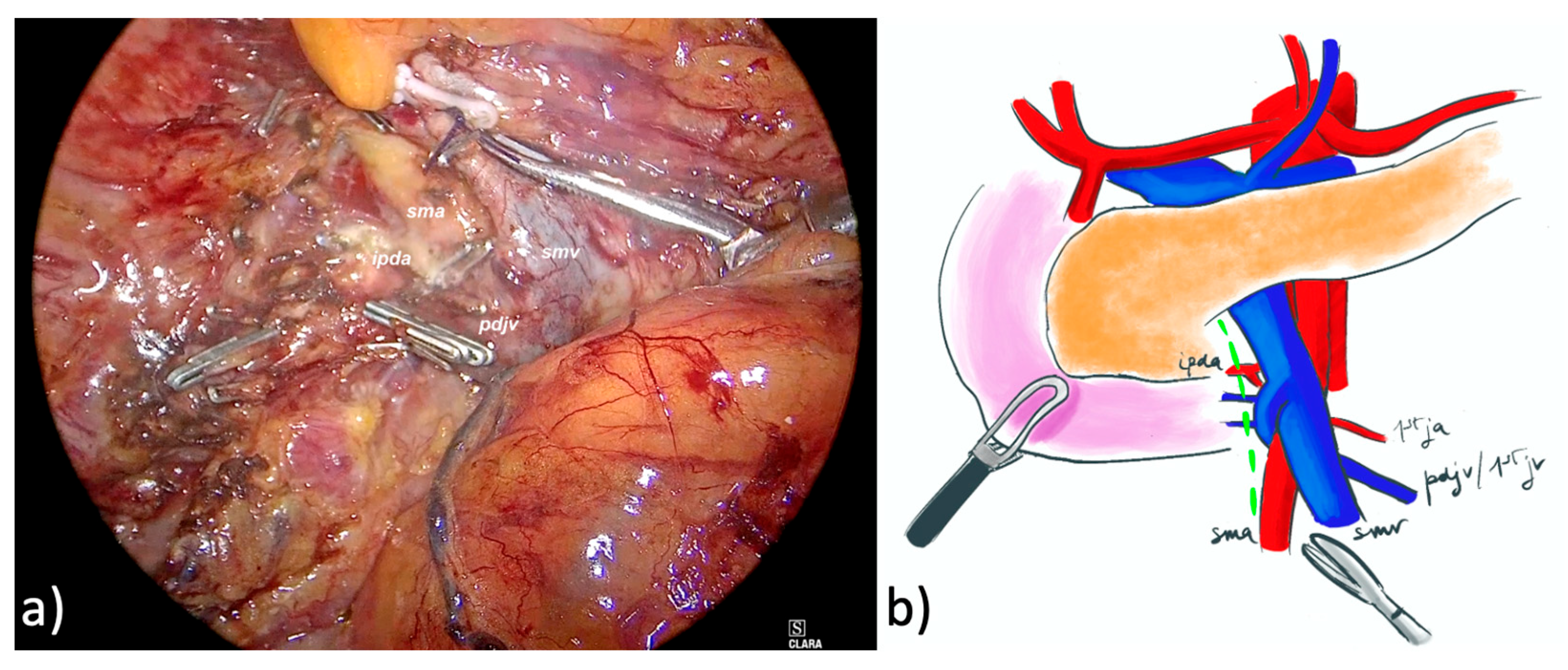
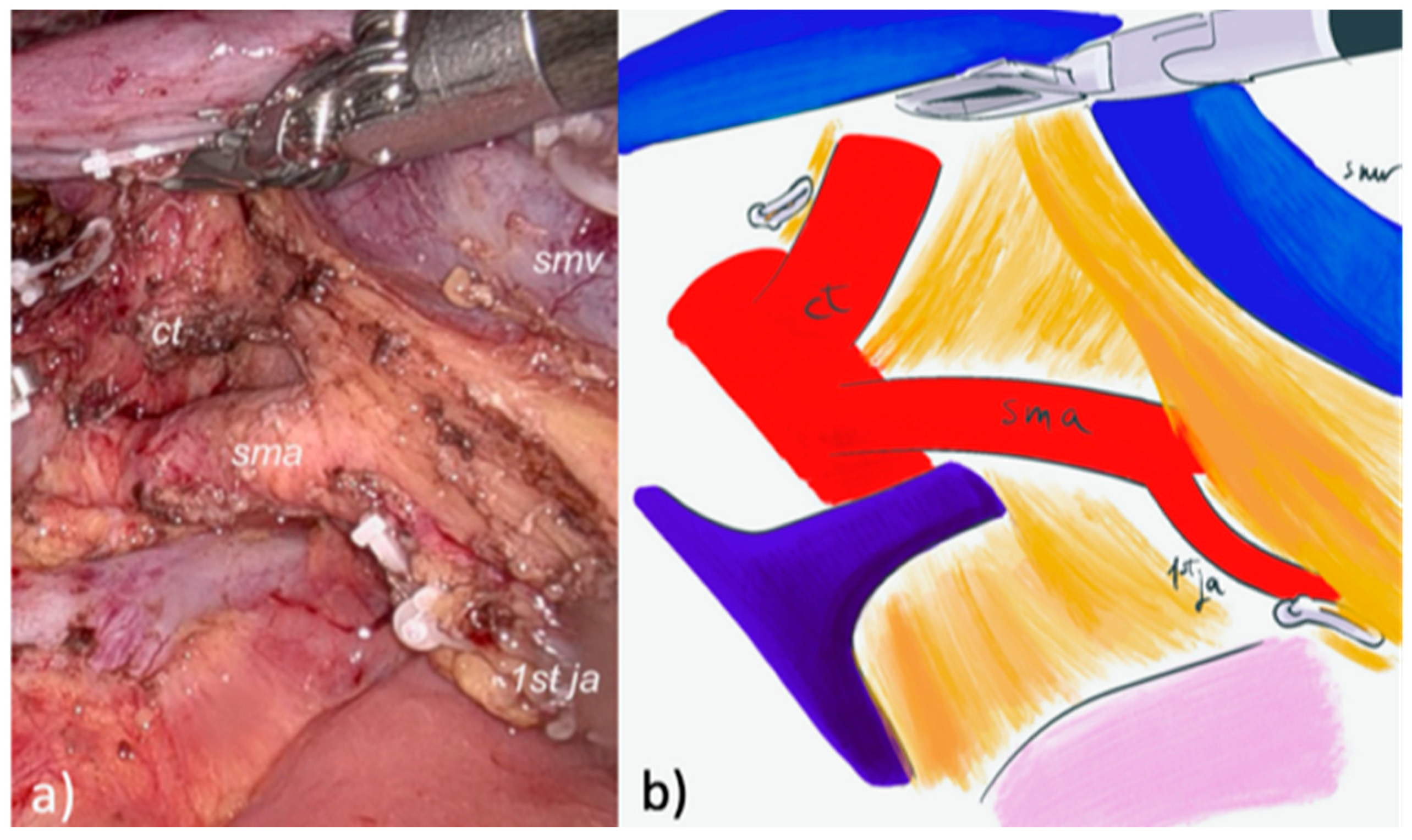
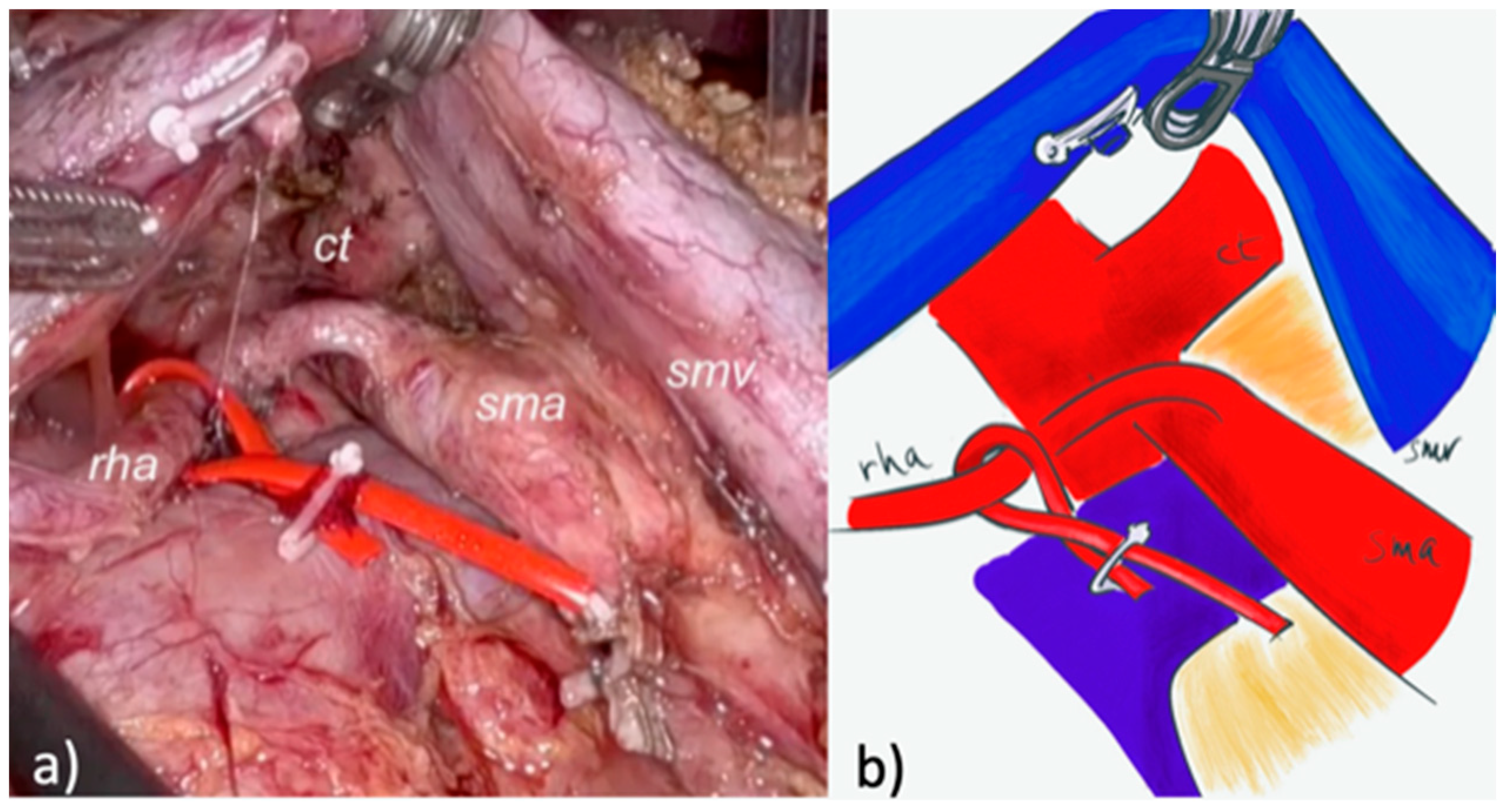
2.2. Surgical Technique and Perioperative Management

2.2.1. LPD

The whole description of the technique adopted for LPD was previously reported and is here briefly described [17,18]. A modified right AFA using the proximal dorsal jejunal vein preisolation method was used [19]; it was based on the early sealing of the right gastroepiploic vein and the wide dissection of the SMV on its right lateral aspect until the proximal dorsal jejunal vein was identified and its pancreatic tributaries divided (Figure 1).
The SMA was identified behind the proximal dorsal jejunal vein and dissected along its right aspect through its origin. The lymphovascular tissue between the uncinate process and the anterior-right lateral aspect of the SMA was removed; the IPDA was identified and sealed with clips. After an extensive Kocher maneuver, the Treitz ligament was divided and the first jejunal loop was de-rotated beneath the mesenteric axis and sectioned. The gastric antrum or duodenum and the gastroduodenal artery were sectioned using a stapler. The pancreatic neck was underpassed and divided using cold scissors, as well as the common hepatic duct. Frozen sections of both pancreatic and bile duct stumps were always performed to confirm negative resection margins. After ligation of the superior pancreatico-duodenal veins, the SMV was shifted to the patient’s left by the first assistant, and all the tissue between the celiac trunk and the SMA was removed completing the mesopancreas excision. The fistula risk score (FRS) was calculated [20]. Pancreaticojejunostomy was performed according to Cattell-Warren or Blumgart, depending on the surgeon’s preference and regardless of the surgical approach [21]. A single-layer end-to-side hepaticojejunostomy was performed. After antecolic transposition, manual double-layer hand-sewn duodenojejunostomy or mechanic gastrojejunostomy was performed. One or two spiral drains entering the right side of the patient were positioned near the pancreaticojejunostomy. At the end of the procedure, the nasogastric tube was removed. An intraoperative near-zero fluid balance was applied. According to institutional protocol, patients’ mobilization and liquid diet were allowed when tolerated. On postoperative day 2 patients received a solid diet. Drains were mobilized and removed according to an early-removal policy in case of negative drain fluid amylase and reassurance fluid characteristics [22].

2.2.2. Robotic PD (RPD)

A pure robotic approach (4 robotic trocars and 2 laparoscopic trocars) with the Da Vinci™ Xi system (Intuitive Surgical®, Inc., Sunnyvale, CA, USA) was used for RPD. A modified posterior AFA was used. After the gastrocolic ligament was opened and the right colonic flexure mobilized, an extensive Kocher maneuver was carried out exposing the inferior vena cava and the left renal vein. The right crura was exposed and the right aspect of the celiac trunk was identified and cleared. The SMA was identified at its origin and dissected in a cranial-to-caudal fashion, following a periadventitial plane, until the IPDA was identified at its origin and sealed with clips (Figure 2 and Figure 3).
The first jejunal loop was then de-rotated through the Treitz window and sectioned using a stapler. The tributary veins from the uncinate process to the proximal dorsal jejunal vein, or directly to the SMV, were transected, and the dissection of the posterior mesopancreas was completed exposing the dorsal aspect of the SMV (Figure 2). After the gastric antrum or duodenum and the gastroduodenal artery were sectioned using a stapler, a complete lymphadenectomy of stations 8–9–12 was performed. The pancreatic neck was underpassed and divided using cold scissors, as well as the common hepatic duct. After ligation of the right gastroepiploic vein, the remaining pancreatico-duodenal veins were sectioned with the removal of all the tissue between the celiac trunk and the SMA, completing the mesopancreas excision. The following steps were the same as those for LPD.

2.3. Definitions and Variables

Patients’ baseline characteristics also included surgical approach (laparoscopic or robotic) and the presence of anatomical vascular variations in the hepatic artery at the preoperatively contrast-enhanced computed tomography imaging. Types III, IV, VI, VII, VIII, and IX of anomalies according to Michels’ classification were considered as aberrant right hepatic arteries. Surgical outcomes were associated with procedures, vascular resection, conversion rate, duration of resection phase, and overall operative time. Conversion from MIPD to open surgery was defined as the need to complete the resection phase by any type of laparotomy. The FRS was calculated based on pancreatic texture measured by palpation of the extracted specimen, Wirsung diameter assessed intraoperatively at the time of transaction, estimated blood loss, and pathology [20]. The pathological radicality of resection, pTNM, and pathologic stage were classified according to the 8th edition of the American Joint Committee on Cancer staging system [23]. A minimum of 15 lymph nodes (LNs) harvested was chosen to consider the lymphadenectomy adequate [24]. Resection margins were defined according to the Royal College of Pathologists; margin status was considered R1 when the distance between the tumor and any resection margins was ≤1 mm. The Clavien-Dindo classification was used to grade postoperative complications; those graded > 2 were considered severe [25]. Pancreas-specific complications were recorded separately and defined in accordance with the International Study Group of Pancreatic Surgery [26,27,28]. Ineffective AFA (IAFA) was defined in the case of resection time or estimated blood loss > 90th percentile, or conversion to laparotomy.

2.4. Study Endpoints

The primary endpoint was IAFA during MIPD. The secondary endpoints were surgical outcomes (conversion rate, duration of resection phase and overall operative time, postoperative complications), pathological outcomes (radicality of resection, number of LNs harvested), and identification of factors associated with IAFA in patients undergoing MIPD. A subanalysis comparing patients undergoing LPD and RPD was also performed.

2.5. Statistical Analysis

Data are expressed as median and interquartile range and number and relative percentage. The normal distribution of continuous variables was assessed with the Shapiro–Wilk test. Univariate analysis was carried out to identify variables which were independently associated with IAFA; the variables with p values < 0.10 at univariate analysis were further analyzed with multivariable logistic regression. Categorical variables were analyzed using the Fisher exact test or Chi-Square test, as appropriate, while continuous ones were analyzed using the Student t test or Mann–Whitney test. p Values were considered significant when <0.05. Data analysis was performed using JMP®, Version 16.0 (SAS Institute Inc., Cary, NC, USA, 1989–2019).

3. Results

3.1. Patient Selection

In the study period, 209 patients underwent PD and 109 of these were treated for PC. After the exception of 38 patients receiving open PD, 71 patients undergoing MIPD were selected: 32 underwent LPD and 39 RPD. Baseline patients’ characteristics are shown in Table 1.
No difference was found between the subgroups (LPD and RPD) in terms of baseline characteristics (sex, age, BMI, ASA score, Age-adjusted Charlson Comorbidity Index, previous abdominal surgery, major venous involvement, vascular anomalies, and preoperative biliary drainage). More patients in the RPD group received neoadjuvant treatment (23.1% vs. 0%, p 0.0036).

3.2. Operative and Postoperative Outcomes

Intraoperative outcomes are shown in Table 2.
No patients underwent conversion. The median operative time and resection time of the entire cohort were 545 min and 310 min, respectively. The median estimated blood loss was 250 mL. The majority of patients had an intermediate or low risk of fistula based on FRS. No difference between the groups was found regarding intraoperative outcomes. Postoperative outcomes are reported in Table 3.
The overall and severe complication rate of the entire cohort was 45.1% and 21.1%, respectively. Four patients (5.6%) required reoperation. Pancreatic fistula and postpancreatectomy hemorrhage occurred in 8.4% and 9.9% of patients, respectively. The median length of stay was 11 days with an 8.4% readmission rate. The groups did not differ in terms of postoperative outcomes. IAFA was observed in 12.7% of patients, without difference between the groups (9.4 vs. 15.4%, p 0.499, in LPD and RPD, respectively). The main causes of IAFA were excessive blood loss (2 and 3 patients in LPD and RPD, p 1.000, respectively) and resection time (1 and 3 patients in LPD and RPD, p 0.622, respectively) without difference between the groups. At univariate analysis, male sex and ASA > 2 were detected as possible factors associated with IAFA (p < 0.10) and were included in the logistic regression. In multivariable analysis, only male sex was an independent predictor of IAFA. Univariate and multivariable analyses for IAFA are reported in Table 4.

3.3. Pathological and Oncological Outcomes

Pathological and oncological outcomes are reported in Table 5. R0 resection was obtained in 74.6% of patients. No R2 resection was reported. Among patients with R1 resection, posterior and medial resection margin was involved in 19.7% and 5.6%, respectively. The RPD group showed a greater number of LNs harvested (24 vs. 17, p = 0.023), with no difference between the groups regarding the median number of positive LNs (p = 0.630) and LNs ratio (p = 0.291). The rate of patients receiving adjuvant therapy was 59.7%, with no difference between patients undergoing LPD and RPD. No difference between the groups was found in terms of tumor features and stage.

4. Discussion

The present study showed that MIPD with AFA was feasible and safe for the treatment of patients affected by PC. Male sex was an independent predictor of IAFA. No difference was observed in patients undergoing MIPD regardless of the approach, except for a higher number of LNs harvested with RPD.
AFA has been proposed as a novel surgical strategy with potential advantages during PD, namely early determination of resectability and control of vascular in-flow for the pancreatic head, higher R0 rate, and avoiding vascular injury in patients with arterial anomalies [1,29]. Some doubts about the usefulness of AFA have been raised by a randomized study that showed no benefits in comparison to conventional PD [30]. However, a recent meta-analysis reported better outcomes for patients undergoing PD with AFA in comparison with standard PD regarding both overall and pancreas-specific complications, blood loss, length of stay, and margin radicality, suggesting potential clinical benefit for patients undergoing AFA in terms of postoperative recovery and oncological outcomes [1]. Considering that all the studies included in the meta-analysis selected exclusively patients undergoing open PD, the efficacy of AFA remains debated for MIPD [1]. Even though MIPD may ideally represent an optimal application for AFA, due to a careful dissection along embryological and periadventitial planes, the need to operate in very limited and tight spaces and the difficulty of performing effective tractions and counteractions could limit its feasibility, hindering the reproducibility of the results obtained during open PD. In the present series, AFA was always feasible during MIPD, with no conversion. The rate of IAFA was 12.7% and it was mainly related to excessively long resection time or blood loss; this data should be analyzed considering the characteristics of patients all affected by PC. In a previous study, patients affected by PC showed a trend through longer operative time and higher blood loss, even if not statistically relevant, as compared with those affected by other periampullary tumors [18]. However, these potential limits did not hinder patients’ safety as demonstrated by the absence of conversion, the adequacy of LN retrieval, and the acceptable postoperative outcomes in terms of complications and length of stay. The only factor associated with IAFA was male sex, while BMI and previous abdominal surgery were not. Many authors consider high BMI or previous abdominal surgery as exclusion criteria for MIPD. Interestingly, in the present study, neither of these variables was associated with IAFA; instead, male sex was a significant factor. This finding may suggest that, when performed by experienced surgeons, no absolute exclusion criteria should be applied to MIPD. However, caution should be applied when performing MIPD in males with high BMI, as these factors are associated with visceral fat and thickened mesentery [31]. Both these factors can make the procedure particularly challenging, potentially leading to longer operative time and increased blood loss. In particular, visceral obesity has proved to correlate with more complex procedures and increased postoperative complications after PD. Some authors even suggested considering the visceral fat area as a factor influencing surgical difficulty during robotic distal pancreatectomy [32]. Furthermore, the male sex has been recently proposed as a risk factor for postoperative pancreatic fistula in the setting of MIPD due to its association with visceral fat, fatty pancreas, and more challenging reconstructive phase [33].
A recent review identified a few studies focusing on AFA during MIPD, describing four main approaches to the SMA: anterior, posterior, right, and left [13]. Among these, the right AFA was the most frequently used due to the wider working space and the easy accessibility to the SMA; however, the right approach requires wide isolation of the anterolateral aspect of the SMV at an early phase of the dissection and the section of the tributary veins moving from the pancreatic head to the first jejunal vein or directly in the SMV, with potential risk of time-consuming and harmful bleeding, especially in obese patients with thickened mesenteric roots [19]. On the contrary, only a few studies reported experiences regarding the left approach, probably due to the difficulty in creating an adequate working space between the transverse mesocolon and the small intestine [34,35,36]. The anterior approach was described in a single case series, which underlined its usefulness in particular when facing major venous involvement needing resection [37].
The potential advantages of the posterior approach include the exploration and dissection of the SMA before venous evaluation, the possibility of performing an interaortocaval LNs biopsy, a safer probing of the SMV, and a more conducive lymphadenectomy of the hepatoduodenal ligament [38]. Accordingly, many authors advocated for the efficacy and potential benefits of the posterior approach during both open and MIPD, allowing for safer identification of the SMA at its origin, easy clearance of the lymphatic tissue on the right lateral aspect of the SMA and celiac trunk, and earlier assessment of disease extension and arterial control prior to sectioning the pancreatic neck or dissecting the SMV [11,39]. However, the dissection of the posterior plane along the anterior aspect of the aorta and above the left renal vein may not always be easy during open surgery, especially in the case of obese patients, due to the difficulty of moving the duodenopancreatic block on the left and the ventral-to-dorsal approach. During LPD, especially in patients with thickened retropancreatic tissues as in cases of pancreatitis, the posterior approach may even be unfeasible because of the rigid and straight instruments.
Only anecdotal reports focusing on AFA during RPD have been published in the literature. While previous studies have compared the surgical and oncological outcomes of patients undergoing open and minimally invasive resection for PC, no studies have specifically examined differences between the AFAs used for RPD and LPD [13,40,41]. Robotic surgery has shown clear advantages in comparison to laparoscopic one in terms of articulation of instruments, endowrist technology, mitigation of unintentional tremors, and immersive enhanced three-dimensional vision [42,43]. Some authors have argued that the advantages of robotic surgery could be more evident as the complexity of the intervention increases, suggesting an ideal field of application for pancreatic surgery [44]. Consistently, the implementation of robotic surgery was associated with improved outcomes during pancreatic and liver surgery; in particular, shifting from LPD to RPD resulted in a reduced operative time and blood loss and a higher number of retrieved LNs [45]. For adequate nodal staging, standard lymphadenectomy, with the removal of at least 15 LNs, is recommended because the LN status is an important prognostic factor, especially for patients affected by PC [45]. In the present study, even though an adequate number of LNs was harvested by using both approaches, RPD was associated with a greater number of LNs retrieved probably explained by a magnified visualization and a more precise dissection; however, no difference between the groups was found regarding LNs ratio. On the contrary, R0 was slightly in favor of LPD, even if not reach statistical significance; considering the number of LNs harvested, which confirmed a meticulous dissection, this data could be related to progressive modification of the inclusion criteria for MIPD, justified by the increasing acquisition of technical skills and the implementation of the robotic approach, as witnessed by the higher number of patients receiving neoadjuvant treatment and a trend in a greater number of vascular involvement and history of previous surgery in patients undergoing RPD. However, the R1 rate reported in the literature for patients affected by PC ranged between 30 and 35% in both open and MIPDs, which was consistent with our results (18.8% and 30.8% in LPD and RPD, respectively).
A report from a prospective national registry confirmed that the application of the robotic platform to pancreatic surgery was associated with reduced rates of conversion and blood loss and favored a wider diffusion of MIPD [46]. The present study well reflects the global trend in shifting from LPD to RPD; since its introduction in our clinical practice in 2022, RPD has rapidly become the only approach for MIPD. Although no significant difference was found in surgical outcomes between LPD and RPD, the benefit perceived by the surgeon in terms of safety, reproducibility, ease of instrument handling, and ergonomics has led to a complete abandonment of the laparoscopic approach in favor of the robotic one.
This study has several limitations that warrant caution in interpreting the results. The data were analyzed retrospectively and based on a single-center experience. The findings may have been influenced by changes in perioperative management and surgical expertise over time; in particular, the outcomes of RPD could have benefited from the learning curve previously established with LPD [21]. On the other hand, LPDs were performed by an experienced laparoscopic surgeon with more than 100 laparoscopic gastrectomies and 300 laparoscopic colectomies [21]. The same surgeon initiated the robotic program and began performing RPD after a structured training program, but with limited robotic experience. Therefore, the different learning curve for each approach, which was much higher for LPD, may have influenced the results in favor of LPD. A higher rate of neoadjuvant therapy was observed in the RPD group due to a gradual change in selection criteria over time. Although the influence of this specific factor on the study outcomes is impossible to analyze and beyond the aim of this study, no difference was observed between the groups in the resectability rate as demonstrated by the similar results in terms of blood loss, conversions, operative time, and R0. This could suggest, at most, a potential benefit of RPD in managing more complex conditions. Additionally, the data were derived from a single center with specialized expertise in advanced minimally invasive and pancreatic surgery, which may limit their generalizability to other settings. For each minimally invasive approach (laparoscopic and robotic), an AFA was used, restricting the ability to identify potential advantages of other approaches. The small sample size could introduce a risk of type II error.

5. Conclusions

This paper reported outcomes of MIPD with AFA showing its feasibility and safety in patients affected by PC. When comparing RPD with posterior AFA and LPD with right AFA no difference was found in terms of surgical and oncological outcomes, despite RPD having a higher number of LNs retrieved. RPD has provided subjective advantages for the surgeon in terms of handling, safety, and reproducibility, although these have not translated into better results with respect to the defined outcomes.

Author Contributions

Conceptualization, M.M. and G.F.; methodology, A.G., M.P. and A.Z.; software, A.G.; formal analysis, A.G. and A.Z.; data curation, P.C., C.F. and G.M.; writing—original draft preparation, M.M.; writing—review, and editing, A.G., P.C., M.M. and P.D.M.; supervision, G.F. All authors have read and agreed to the published version of the manuscript.

Funding

This research received no external funding.

Institutional Review Board Statement

The study was conducted in accordance with the Declaration of Helsinki. Ethical review and approval were waived for this study due to its retrospective comparative nature.

Informed Consent Statement

Informed consent was obtained from all subjects involved in the study.

Data Availability Statement

The data presented in this study are available on request from the corresponding author due to their containing information that could compromise the privacy of research participants (specify the reason for the restriction).

Conflicts of Interest

The authors declare no conflicts of interest.

Abbreviations

The following abbreviations are used in this manuscript:
PDPancreatoduodenectomy
SMASuperior Mesenteric Artery
SMVSuperior Mesenteric Vein
AFAArtery-first Approach
IPDAInferior Pancreatico-duodenal Artery
MIPDMinimally Invasive Pancreaticoduodenectomy
PCPancreatic Cancer
LPDLaparoscopic Pancreaticoduodenectomy
FRSFistula Risk Score
BMIBody Mass Index
LNLymph Node
IAFAIneffective Artery-first Approach

References

  1. Jiang, X.; Yu, Z.; Ma, Z.; Deng, H.; Ren, W.; Shi, W.; Jiao, Z. Superior mesenteric artery first approach can improve the clinical outcomes of pancreaticoduodenectomy: A meta-analysis. Int. J. Surg. 2020, 73, 14–24. [Google Scholar] [CrossRef] [PubMed]
  2. van Roessel, S.; Mackay, T.M.; Tol, J.A.M.G.; van Delden, O.M.; van Lienden, K.P.; Nio, C.Y.; Phoa, S.S.K.S.; Fockens, P.; van Hooft, J.E.; Verheij, J.; et al. Impact of expanding indications on surgical and oncological outcome in 1434 consecutive pancreatoduodenectomies. HPB 2019, 21, 865–875. [Google Scholar] [CrossRef] [PubMed]
  3. Isaji, S.; Mizuno, S.; Windsor, J.A.; Bassi, C.; Fernández-Del Castillo, C.; Hackert, T.; Hayasaki, A.; Katz, M.H.G.; Kim, S.W.; Kishiwada, M.; et al. International consensus on definition and criteria of borderline resectable pancreatic ductal adenocarcinoma 2017. Pancreatology 2018, 18, 2–11. [Google Scholar] [CrossRef]
  4. Fernandes Ede, S.M.; Strobel, O.; Girão, C.; Moraes-Junior, J.M.A.; Torres, O.J.M. What do surgeons need to know about the mesopancreas. Langenbecks Arch. Surg. 2021, 406, 2621–2632. [Google Scholar] [CrossRef] [PubMed]
  5. Butler, J.R.; Ahmad, S.A.; Katz, M.H.; Cioffi, J.L.; Zyromski, N.J. A systematic review of the role of periadventitial dissection of the superior mesenteric artery in affecting margin status after pancreatoduodenectomy for pancreatic adenocarcinoma. HPB 2016, 18, 305–311. [Google Scholar] [CrossRef]
  6. Croome, K.P.; Farnell, M.B.; Que, F.G.; Reid-Lombardo, K.M.; Truty, M.J.; Nagorney, D.M.; Kendrick, M.L. Pancreaticoduodenectomy with Major Vascular Resection: A Comparison of Laparoscopic Versus Open Approaches. J. Gastrointest. Surg. 2015, 19, 189–194. [Google Scholar] [CrossRef]
  7. Kendrick, M.L.; Sclabas, G.M. Major venous resection during total laparoscopic pancreaticoduodenectomy. HPB 2011, 13, 454–458. [Google Scholar] [CrossRef]
  8. Khatkov, I.E.; Izrailov, R.E.; Khisamov, A.A.; Tyutyunnik, P.S.; Fingerhut, A. Superior mesenteric-portal vein resection during laparoscopic pancreatoduodenectomy. Surg. Endosc. 2017, 31, 1488–1495. [Google Scholar] [CrossRef]
  9. Weitz, J.; Rahbari, N.; Koch, M.; Büchler, M.W. The “artery first” approach for resection of pancreatic head cancer. J. Am. Coll. Surg. 2010, 210, e1–e4. [Google Scholar] [CrossRef]
  10. Varty, P.P.; Yamamoto, H.; Farges, O.; Belghiti, J.; Sauvanet, A. Early retropancreatic dissection during pancreaticoduodenectomy. Am. J. Surg. 2005, 189, 488–491. [Google Scholar] [CrossRef]
  11. Sanjay, P.; Takaori, K.; Govil, S.; Shrikhande, S.V.; Windsor, J.A. “Artery-first” approaches to pancreatoduodenectomy. Br. J. Surg. 2012, 99, 1027–1035. [Google Scholar] [CrossRef]
  12. van Hilst, J.; de Graaf, N.; Abu Hilal, M.; Besselink, M.G. The Landmark Series: Minimally Invasive Pancreatic Resection. Ann. Surg. Oncol. 2021, 28, 1447–1456. [Google Scholar] [CrossRef] [PubMed]
  13. Nagakawa, Y.; Watanabe, Y.; Kozono, S.; Boggi, U.; Palanivelu, C.; Liu, R.; Wang, S.E.; He, J.; Nishino, H.; Ohtsuka, T.; et al. Study group of Precision Anatomy for Minimally Invasive Hepato-Biliary-Pancreatic surgery (PAM-HBP surgery). Surgical approaches to the superior mesenteric artery during minimally invasive pancreaticoduodenectomy: A systematic review. J. Hepatobiliary Pancreat. Sci. 2022, 29, 114–123. [Google Scholar] [CrossRef] [PubMed]
  14. Uijterwijk, B.A.; Lemmers, D.H.; Fusai, G.K.; Groot Koerkamp, B.; Koek, S.; Zerbi, A.; Sparrelid, E.; Boggi, U.; Luyer, M.; Ielpo, B.; et al. Different Periampullary Types and Subtypes Leading to Different Perioperative Outcomes of Pancreatoduodenectomy: Reality and Not a Myth; An International Multicenter Cohort Study. Cancers 2024, 16, 899. [Google Scholar] [CrossRef]
  15. He, C.; Mao, Y.; Wang, J.; Duan, F.; Lin, X.; Li, S. Nomograms predict long-term survival for patients with periampullary adenocarcinoma after pancreatoduodenectomy. BMC Cancer 2018, 18, 327. [Google Scholar] [CrossRef] [PubMed]
  16. von Elm, E.; Altman, D.G.; Egger, M.; Pocock, S.J.; Gøtzsche, P.C.; Vandenbroucke, J.P.; STROBE Initiative. The Strengthening the Reporting of Observational Studies in Epidemiology (STROBE) Statement: Guidelines for Reporting Observational Studies. Ann. Intern. Med. 2007, 147, 573. [Google Scholar] [CrossRef]
  17. Mazzola, M.; Giani, A.; Crippa, J.; Morini, L.; Zironda, A.; Bertoglio, C.L.; De Martini, P.; Magistro, C.; Ferrari, G. Totally laparoscopic versus open pancreaticoduodenectomy: A propensity score matching analysis of short-term outcomes. Eur. J. Surg. Oncol. 2021, 47, 674–680. [Google Scholar] [CrossRef]
  18. Mazzola, M.; Giani, A.; Bertoglio, C.L.; Carnevali, P.; De Martini, P.; Benedetti, A.; Giusti, I.; Magistro, C.; Ferrari, G. Standardized right artery first approach during laparoscopic pancreaticoduodenectomy for periampullary neoplasms: Technical aspects and perioperative outcomes. Surg. Endosc. 2023, 37, 759–765. [Google Scholar] [CrossRef]
  19. Nagakawa, Y.; Hosokawa, Y.; Sahara, Y.; Takishita, C.; Hijikata, Y.; Osakabe, H.; Nakajima, T.; Shirota, T.; Katsumata, K.; Nakamura, M.; et al. Approaching the superior mesenteric artery from the right side using the proximal-dorsal jejunal vein preisolation method during laparoscopic pancreaticoduodenectomy. Surg. Endosc. 2018, 32, 4044–4051. [Google Scholar] [CrossRef]
  20. Callery, M.P.; Pratt, W.B.; Kent, T.S.; Chaikof, E.L.; Vollmer, C.M., Jr. A prospectively validated clinical risk score accurately predicts pancreatic fistula after pancreatoduodenectomy. J. Am. Coll. Surg. 2013, 216, 1–14. [Google Scholar] [CrossRef]
  21. Mazzola, M.; Giani, A.; Veronesi, V.; Bernasconi, D.P.; Benedetti, A.; Magistro, C.; Bertoglio, C.L.; De Martini, P.; Ferrari, G. Multidimensional evaluation of the learning curve for totally laparoscopic pancreaticoduodenectomy: A risk-adjusted cumulative summation analysis. HPB 2023, 25, 507–517. [Google Scholar] [CrossRef]
  22. Salvia, R.; Marchegiani, G.; Andrianello, S.; Balduzzi, A.; Masini, G.; Casetti, L.; Esposito, A.; Landoni, L.; Malleo, G.; Paiella, S.; et al. Redefining the Role of Drain Amylase Value for a Risk-Based Drain Management after Pancreaticoduodenectomy: Early Drain Removal Still Is Beneficial. J. Gastrointest. Surg. 2021, 25, 1461–1470. [Google Scholar] [CrossRef] [PubMed]
  23. Amin, M.B.; Edge, S.B.; Greene, F.L.; Byrd, D.R.; Brookland, R.K.; Washington, M.K. AJCC Cancer Staging Manual; Springer: Cham, Switzerland, 2018. [Google Scholar]
  24. Pedrazzoli, S. Extent of lymphadenectomy to associate with pancreaticoduodenectomy in patients with pancreatic head cancer for better tumor staging. Cancer Treat. Rev. 2015, 41, 577–587. [Google Scholar] [CrossRef] [PubMed]
  25. Dindo, D.; Demartines, N.; Clavien, P.-A. Classification of Surgical Complications. Ann. Surg. 2004, 240, 205–213. [Google Scholar] [CrossRef] [PubMed]
  26. Bassi, C.; Marchegiani, G.; Dervenis, C.; Sarr, M.; Abu Hilal, M.; Adham, M.; Allen, P.; Andersson, R.; Asbun, H.J.; Besselink, M.G.; et al. International Study Group on Pancreatic Surgery (ISGPS). The 2016 update of the International Study Group (ISGPS) definition and grading of postoperative pancreatic fistula: 11 Years After. Surgery 2017, 161, 584–591. [Google Scholar] [CrossRef]
  27. Wente, M.N.; Bassi, C.; Dervenis, C.; Fingerhut, A.; Gouma, D.J.; Izbicki, J.R.; Neoptolemos, J.P.; Padbury, R.T.; Sarr, M.G.; Traverso, L.W.; et al. Delayed gastric emptying (DGE) after pancreatic surgery: A suggested definition by the International Study Group of Pancreatic Surgery (ISGPS). Surgery 2007, 142, 761–768. [Google Scholar] [CrossRef]
  28. Wente, M.N.; Veit, J.A.; Bassi, C.; Dervenis, C.; Fingerhut, A.; Gouma, D.J.; Izbicki, J.R.; Neoptolemos, J.P.; Padbury, R.T.; Sarr, M.G.; et al. Postpancreatectomy hemorrhage (PPH): An International Study Group of Pancreatic Surgery (ISGPS) definition. Surgery 2007, 142, 20–25. [Google Scholar] [CrossRef]
  29. Müller, P.C.; Müller, B.P.; Hackert, T. Contemporary artery-first approaches in pancreatoduodenectomy. Br. J. Surg. 2023, 110, 1570–1573. [Google Scholar] [CrossRef]
  30. Lemieux, S.; Prud’homme, D.; Bouchard, C.; Tremblay, A.; Deprés, J.P. Sex differences in the relation of visceral adipose tissue acculumation to total body fatness. Am. J. Clin. Nutr. 1993, 58, 463–467. [Google Scholar] [CrossRef]
  31. Takagi, K.; Yamada, M.; Fuji, T.; Yasui, K.; Nishiyama, T.; Nagai, Y. Impact of visceral fat area on surgical difficulty during robotic distal pancreatectomy (TAKUMI-2). Surg. Endosc. 2025, 39, 3137–3145. [Google Scholar] [CrossRef]
  32. Mungroop, T.H.; Klompmaker, S.; Wellner, U.F.; Steyeberg, E.W.; Coratti, A.; D’Hondt, M.; de Pastena, M.; Dokmak, S.; Khatkov, I.; Saint-Marc, O.; et al. Consortium on minimally invasive pancreatic surgery (E-MIPS). Updated alternative fistula risk score (ua-FRS) to include minimally invasive pancreaticoduodenectomy: Pan-European validation. Ann. Surg. 2021, 273, 334–340. [Google Scholar] [CrossRef]
  33. Sabater, L.; Cugat, E.; Serrablo, A.; Suarez-Artacho, G.; Diez-Valladares, L.; Santoyo-Santoyo, J.; Martín-Pérez, E.; Ausania, F.; Lopez-Ben, S.; Jover-Navalon, J.M.; et al. Does the artery-first approach improve the rate of R0 resection in pancreatoduodenectomy? Ann. Surg. 2019, 270, 738–746. [Google Scholar] [CrossRef]
  34. Morales, E.; Zimmitti, G.; Codignola, C.; Manzoni, A.; Garatti, M.; Sega, V.; Rosso, E. Follow “the superior mesenteric artery”: Laparoscopic approach for total mesopancreas excision during pancreaticoduodenectomy. Surg. Endosc. 2019, 33, 4186–4191. [Google Scholar] [CrossRef] [PubMed]
  35. Cho, A.; Yamamoto, H.; Kainuma, O. Tips of laparoscopic pancreaticoduodenectomy: Superior mesenteric artery first approach (with video). J. Hepatobiliary Pancreat. Sci. 2014, 21, e19–e21. [Google Scholar] [CrossRef] [PubMed]
  36. Liao, C.-H.; Liu, Y.-Y.; Wang, S.-Y.; Liu, K.-H.; Yeh, C.-N.; Yeh, T.-S. The feasibility of laparoscopic pancreaticoduodenectomy-a stepwise procedure and learning curve. Langenbecks Arch. Surg. 2017, 402, 853–861. [Google Scholar] [CrossRef] [PubMed]
  37. Cai, Y.; Gao, P.; Li, Y.; Wang, X.; Peng, B. Laparoscopic pancreaticoduodenectomy with major venous resection and reconstruction: Anterior superior mesenteric artery first approach. Surg. Endosc. 2018, 32, 4209–4215. [Google Scholar] [CrossRef]
  38. Wang, X.M.; Sun, W.D.; Hu, M.H.; Wang, G.N.; Jiang, Y.Q.; Fang, X.S.; Han, M. Inferoposterior duodenal approach for laparoscopic pancreaticoduodenectomy. World J. Gastroenterol. 2016, 22, 2142–2148. [Google Scholar] [CrossRef]
  39. Ielpo, B.; Anselmo, A.; Masuda, Y.; Xuan, M.Y.H.; Burdio, F.; De Blasi, V.; Sanchez-Velazquez, P.; Giuliani, A.; Azagra, J.S.; Viola, G.M.; et al. Superior Mesenteric Artery First Approach for Minimally Invasive Pancreaticoduodenectomy: A Step-By-Step Surgical Technique Video. Ann. Surg. Oncol. 2023, 30, 1500–1503. [Google Scholar] [CrossRef]
  40. Sun, R.; Yu, J.; Zhang, Y.; Liang, Z.; Han, X. Perioperative and oncological outcomes following minimally invasive versus open pancreaticoduodenectomy for pancreatic duct adenocarcinoma. Surg. Endosc. 2021, 35, 2273–2285. [Google Scholar] [CrossRef]
  41. Giani, A.; Mazzola, M.; Paterno, M.; Zironda, A.; Calcagno, P.; Zuppi, E.; De Martini, P.; Ferrari, G. Oncological Outcomes of Open Versus Minimally Invasive Surgery for Ductal Adenocarcinomas of Pancreatic Head: A Propensity Score Matching Analysis. Curr. Oncol. 2024, 31, 6096–6109. [Google Scholar] [CrossRef]
  42. Hoehn, R.S.; Nassour, I.; Adam, M.A.; Winters, S.; Paniccia, A.; Zureikat, A.H. National Trends in Robotic Pancreas Surgery. J. Gastrointest. Surg. 2021, 25, 983–990. [Google Scholar] [CrossRef] [PubMed]
  43. Nota, C.L.; Zwart, M.J.; Fong, Y.; Hagendoorn, J.; Hogg, M.E.; Koerkamp, B.G.; Besselink, M.G.; Molenaar, I.Q. Dutch Pancreatic Cancer Group. Developing a robotic pancreas program: The Dutch experience. J. Visc. Surg. 2017, 3, 106. [Google Scholar] [CrossRef] [PubMed]
  44. Emmen, A.M.L.H.; Görgec, B.; Zwart, M.J.W.; Daams, F.; Erdmann, J.; Festen, S.; Gouma, D.J.; van Gulik, T.M.; van Hilst, J.; Kazemier, G.; et al. Impact of shifting from laparoscopic to robotic surgery during 600 minimally invasive pancreatic and liver resections. Surg. Endosc. 2023, 37, 3291–3292. [Google Scholar] [CrossRef] [PubMed]
  45. Tol, J.A.; Gouma, D.J.; Bassi, C.; Dervenis, C.; Montorsi, M.; Adham, M.; Andrén-Sandberg, A.; Asbun, H.J.; Bockhorn, M.; Büchler, M.W.; et al. Definition of a standard lymphadenectomy in surgery for pancreatic ductal adenocarcinoma: A consensus statement by the International Study Group on Pancreatic Surgery (ISGPS). Surgery 2014, 156, 591–600. [Google Scholar] [CrossRef]
  46. Boggi, U.; Donisi, G.; Napoli, N.; Partelli, S.; Esposito, A.; Ferrari, G.; Butturini, G.; Morelli, L.; Abu Hilal, M.; Viola, M.; et al. Prospective minimally invasive pancreatic resections from the IGOMIPS registry: A snapshot of daily practice in Italy on 1191 between 2019 and 2022. Updates Surg. 2023, 75, 1439–1456. [Google Scholar] [CrossRef]
Figure 1. Dissection of the mesopancreas using laparoscopic lateral artery-first approach. (a) intraoperative vision of the surgical field with right lateral aspect of the superior mesenteric vein (smv) and the superior mesenteric artery (sma). (b) Schematic illustration showing smv, sma, inferior pancreatico-duodenal artery (ipda), first jejunal artery (1st ja), proximal dorsal jejunal vein (pdjv), first jejunal vein (1st jv).
Cancers 17 02103 g001
Figure 2. Dissection of the mesopancreas using robotic posterior artery-first approach. (a) intraoperative vision of the surgical field with the posterior aspect of the superior mesenteric vein and right lateral aspect of the superior mesenteric artery. (b) Schematic illustration showing superior mesenteric vein (smv), superior mesenteric artery (sma), celiac trunk (ct), first jejunal artery (1st ja).
Cancers 17 02103 g002
Figure 3. Completion of mesopancreas excision using robotic posterior artery-first approach, in patient with aberrant hepatic artery from superior mesenteric artery. (a) intraoperative vision of the surgical field with superior mesenteric vein/portal vein (blu vessel loops) and the aberrant hepatic artery (red vessel loop). (b) Schematic illustration showing superior mesenteric vein (smv), superior mesenteric artery (sma), replaced right hepatic artery (rha).
Cancers 17 02103 g003
Table 1. Baseline characteristics of patients undergoing MIPD.
CharacteristicOverall (71)LPD (32)RPD (39)p
Sex (F)39 (54.9)19 (59.4)20 (51.3)0.495
Age (years) *72 (65–80)73 (60.5–80.7)71 (65–79)0.556
BMI (kg/m2) *24.7 (22.5–27.6)25.6 (22.6–27.8)24.6 (22.0–26.7)0.291
ACCI *5 (4–6)5 (4–6)5 (4–6)0.610
ASA 0.741
ASA 13 (4.2)2 (6.2)1 (2.5)
ASA 252 (73.2)23 (71.9)29 (74.4)
ASA 316 (22.6)7 (21.9)9 (23.1)
Previous abdominal surgery32 (45.1)11 (34.4)21 (53.8)0.100
Neoadjuvant therapy9 (12.7)0 (0)9 (23.1)0.0036
Venous involvement12 (16.9)4 (12.5)8 (20.5)0.370
Vascular anomalies18 (25.3)9 (28.1)9 (23.1)0.626
Preoperative biliary drainage36 (50.7)14 (43.8)22 (56.4)0.288
Values in parenthesis are percentages unless indicated otherwise: * Numbers are expressed as median and interquartile range. MIPD: minimally invasive pancreaticoduodenectomy, LPD: laparoscopic pancreaticoduodenectomy, RPD: robotic pancreaticoduodenectomy, BMI: body mass index, ECOG PS: Eastern Cooperative Oncology Group Performance Status, ACCI: Age-adjusted Charlson Comorbidity Index, ASA: American Society of Anesthesiologists Physical Status Classification System.
Table 2. Intraoperative outcomes of patients undergoing MIPD.
CharacteristicOverall (71)LPD (32)RPD (39)p
Vascular resection 0.059
No66 (92.9)32 (100)34 (87.2)
PV1 (1.5)0 (0)1 (2.5)
SMV4 (5.6)0 (0)4 (10.3)
Operative time *545 (500–600)555 (510–597.5)540 (480–620)0.561
Resection time *310 (265–350)305 (271.2–328.7)318 (260–370)0.708
Conversion0 (0)0 (0)0 (0)/
Wirsung diameter (mm) *4 (2–5)4 (2–5)4 (2–5)0.276
Blood loss (mL) 0.231
<40049 (69.0)25 (78.1)24 (61.5)
401–70013 (18.4)4 (12.5)9 (23.1)
701–10006 (8.4)3 (9.4)3 (7.7)
>10003 (4.2)0 (0)3 (7.7)
Blood loss (mL) *250 (150–500)300 (200–487.5)200 (150–600)0.561
Pancreatic texture 0.283
Soft24 (33.8)9 (28.1)15 (38.5)
Intermediate7 (9.9)5 (15.6)2 (5.1)
Firm40 (56.3)18 (56.3)22 (56.4)
FRS *3 (1–5)2 (0.2–5)3 (1–4)0.706
FRS moderate-high35 (49.3)15 (46.9)20 (51.3)0.711
Class of FRS 0.146
Negligible (0 pt)15 (21.1)8 (25.0)7 (17.9)0.640
Low (1–2 pt)20 (28.2)9 (28.1)11 (28.2)
Moderate (3–6 pt)31 (43.7)14 (43.8)17 (43.6)
High (7–10 pt)5 (7.0)1 (3.1)4 (10.3)
Values in parenthesis are percentages unless indicated otherwise: * Numbers are expressed as median and interquartile range. MIPD: minimally invasive pancreaticoduodenectomy, LPD: laparoscopic pancreaticoduodenectomy, RPD: robotic pancreaticoduodenectomy, PV: portal vein, SMV: superior mesenteric vein, FRS: Fistula risk score.
Table 3. Postoperative outcomes of patients undergoing MIPD.
CharacteristicOverall (71)LPD (32)RPD (39)p
Length of stay (day) *11 (8–16)10.5 (8–19.2)11 (8–16)0.893
Overall complications32 (45.1)13 (40.6)19 (48.7)0.495
Reoperation4 (5.6)3 (9.4)1 (2.5)0.215
Complications: 0.551
140 (56.3)20 (62.6)20 (51.3)
214 (19.7)4 (12.5)10 (25.7)
3a10 (14.1)4 (12.5)6 (15.5)
3b3 (4.2)2 (6.2)1 (2.5)
41 (1.5)0 (0)1 (2.5)
90-day mortality3 (4.2)2 (6.2)1 (2.5)0.442
Comprehensive CI *0 (0–25.7)0 (0–24.9)0 (0–25.7)0.587
Severe complications15 (21.1)8 (25.0)7 (17.9)0.468
POPF6 (8.4)3 (9.4)3 (7.7)0.799
POPF grade 0.691
Grade B3 (4.2)1 (3.1)2 (5.0)
Grade C3 (4.2)2 (6.2)1 (2.5)
DGE11 (15.5)5 (15.6)6 (15.4)0.977
DGE grade 0.330
Grade A4 (5.6)3 (9.4)1 (2.5)
Grade B7 (9.9)2 (6.2)5 (12.8)
Grade C0 (0)0 (0)0 (0)
Biliary leak4 (5.6)3 (9.4)1 (2.5)0.215
Biliary leak grade 0.280
Grade A2 (2.8)1 (3.1)1 (2.5)
Grade B2 (2.8)2 (6.2)0 (0)
Grade C0 (0)0 (0)0 (0)
PPH7 (9.9)3 (9.4)4 (10.3)0.901
PPH grade 0.669
Grade A1 (1.5)0 (0)1 (2.5)
Grade B3 (4.2)1 (3.1)2 (5.0)
Grade C3 (4.2)2 (6.2)1 (2.5)
90 day-readmission6 (8.4)2 (6.2)4 (10.3)0.545
IAFA9 (12.7)3 (9.4)6 (15.4)0.499
Values in parenthesis are percentages unless indicated otherwise: * Numbers are expressed as median and interquartile range. MIPD: minimally invasive pancreaticoduodenectomy, LPD: laparoscopic pancreaticoduodenectomy, RPD: robotic pancreaticoduodenectomy, CI: Complication Index, POPF: Postoperative Pancreatic Fistula, DGE: Delayed Gastric Emptying, PPH: Post Pancreatectomy Hemorrhage, IAFA: Ineffective Artery-First Approach.
Table 4. Univariate and multivariate analyses for IAFA among patients undergoing MIPD.
Univariate AnalysisMultivariable Analysis
OR (95% CI)p ValueOR (95% CI)p Value
Sex (male)5.181 (0.994–27.027)0.035 5.181 (1.105–37.037)0.036
Age > 701.029 (0.252–4.203)0.968
BMI > 300.982 (0.106–9.067)0.987
ASA > 23.333 (0.776–14.325)0.0923.310 (0.687–15.605)0.131
ACCI > 51.620 (0.397–6.620)0.498
Previous abdominal surgery2.245 (0.387–13.013)0.319
Vascular involvement0.580 (0.066–5.120)0.620
RHA anomalies0.821 (0.154–4.369)0.817
Preoperative biliary drainage2.133 (0.489–9.303)0.305
Ca 19.9 > 500 U/mL0.680 (0.075–6.124)0.730
Neoadjuvant chemotherapy2.245 (0.387–13.013)0.319
Robotic approach1.758 (0.403–7.667)0.449
Tumor size > 25 mm1.620 (0.397–6.620)0.380
Positive lymph nodes (yes)0.844 (0.206–3.457)0.814
IAFA: Ineffective Artery-First Approach, MIPD: minimally invasive pancreaticoduodenectomy, OR: odds ratio, CI: confidence interval, BMI: body mass index, ASA: American Society of Anesthesiologists Physical Status Classification System, ACCI: Age-adjusted Charlson Comorbidity Index, RHA: right hepatic artery.
Table 5. Pathological and oncological outcomes of patients undergoing MIPD.
CharacteristicOverall (71)LPD (32)RPD (39)p
Tumor size (mm) *25 (20–30)25 (20–30)25 (21–30)0.847
Grading 0.281
not applicable3 (4.2)0 (0)3 (7.7)
Gx1 (1.5)0 (0)1 (2.5)
G10 (0)0 (0)0 (0)
G236 (50.7)16 (50.0)20 (51.3)
G331 (43.7)16 (50.0)15 (38.5)
Perineural invasion47 (66.2)25 (78.1)28 (71.8)0.593
Lymphovascular invasion37 (52.1)16 (50.0)21 (53.8)0.746
Lymph nodes harvested *22 (15–27)17 (15–24.7)24 (17–28)0.023
Positive lymph nodes *1 (0–3)1 (0–3)1 (0–3)0.630
pN+42 (59.1)21 (65.7)21 (53.8)0.315
Lymph node ratio *0.05 (0–0.12)0.06 (0–0.16)0.04 (0–0.11)0.291
R053 (74.6)26 (81.2)27 (69.2)0.246
R1 0.108
Posterior margin14 (19.7)6 (100)8 (66.7)
Medial margin4 (5.6)0 (0)4 (33.3)
pT 0.590
pT0 2 (2.8)0 (0)2 (5.1)
pTis1 (1.5)0 (0)1 (2.5)
pT1a3 (4.2)1 (3.1)2 (5.1)
pT1b3 (4.2)1 (3.1)2 (5.1)
pT1c15 (21.1)6 (18.7)9 (23.1)
pT243 (60.6)21 (65.7)22 (56.4)
pT34 (5.6)3 (9.4)1 (2.5)
pN 0.570
pN029 (40.9)11 (34.4)18 (46.2)
pN129 (40.9)15 (46.9)14 (35.9)
pN213 (18.4)6 (18.7)7 (17.9)
TNM Stage > IIa41 (57.8)21 (65.7)20 (51.3)0.223
TNM Stage 0.577
Ia16 (22.6)5 (15.6)11 (28.2)
Ib13 (18.4)6 (18.7)7 (17.9)
IIa1 (1.5)0 (0)1 (2.5)
IIb28 (39.1)15 (46.9)13 (33.5)
III13 (18.4)6 (18.7)7 (17.9)
Performed adjuvant therapy40/67 †† (59.7)20 (62.6)20/35 (57.1)0.655
Values in parenthesis are percentages unless indicated otherwise: * Numbers are expressed as median and interquartile range. MIPD: minimally invasive pancreaticoduodenectomy, LPD: laparoscopic pancreaticoduodenectomy, RPD: robotic pancreaticoduodenectomy. pT0 indicates patients undergoing neoadjuvant treatment with no evidence of residual tumor on the surgical specimen. †† Patients who underwent totally neoadjuvant chemotherapy were excluded. For pTNM stage the 8th edition of AJCC TNM staging system was used.
Disclaimer/Publisher’s Note: The statements, opinions and data contained in all publications are solely those of the individual author(s) and contributor(s) and not of MDPI and/or the editor(s). MDPI and/or the editor(s) disclaim responsibility for any injury to people or property resulting from any ideas, methods, instructions or products referred to in the content.

