Review

Mechanical Modulation, Physiological Roles, and Imaging Innovations of Intercellular Calcium Waves in Living Systems

1
Department of Mechanical and Aerospace Engineering, Herbert Wertheim College of Engineering, University of Florida, Gainesville, FL 32611, USA
2
Department of Chemistry, University of Florida, Gainesville, FL 32611, USA
3
J. Crayton Pruitt Family Department of Biomedical Engineering, University of Florida, Gainesville, FL 32611, USA
4
Independent Researcher, Watertown, MA 02472, USA
5
Division of Chemical Biology & Medicinal Chemistry, College of Pharmacy, University of Texas at Austin, Austin, TX 78712, USA
6
Department of Computer and Information Science and Engineering, University of Florida, Gainesville, FL 32603, USA
7
Department of Molecular Genetics and Microbiology, University of Florida, Gainesville, FL 32603, USA
8
Department of Biology, University of Florida, Gainesville, FL 32611, USA
9
University of Florida Health Cancer Center (UFHCC), Gainesville, FL 32610, USA
10
College of Medicine, University of Florida, Gainesville, FL 32610, USA
11
College of Dentistry, University of Florida, Gainesville, FL 32610, USA
*
Authors to whom correspondence should be addressed.
These authors contributed equally to this work.
Cancers 2025, 17(11), 1851; https://doi.org/10.3390/cancers17111851
Submission received: 25 April 2025 / Revised: 26 May 2025 / Accepted: 28 May 2025 / Published: 31 May 2025
(This article belongs to the Special Issue Cancer Mechanosensing)

Simple Summary

Intercellular calcium waves (ICWs) are a class of molecular signals for long-range intercellular communication, influencing many key biological activities. Despite increasing evidence that mechanical signals across molecular to tissue scales initiate and modulate ICWs, the mechanisms by which cells transduce these forces into ICW-associated biochemical and genetic activities remain incompletely understood. This review discusses how mechanical stimuli interact with upstream molecules and organelles and how these signals propagate through downstream networks in living cells and tissues. By highlighting the roles of ICWs in various physiological and pathological contexts, with particular focus on cancer, this review proposes potential actionable mechano-therapeutic targets. Furthermore, this review summarizes recent advances in imaging and artificial intelligence technologies that have deepened the understanding of ICW dynamics. This review aims to offer a comprehensive framework of ICW mechanobiology and to highlight actionable mechano-therapeutic targets against ICW-related immune evasion and mechano-drug resistance in cancer.

Abstract

Long-range intercellular communication is essential for multicellular biological systems to regulate multiscale cell–cell interactions and maintain life. Growing evidence suggests that intercellular calcium waves (ICWs) act as a class of long-range signals that influence a broad spectrum of cellular functions and behaviors. Importantly, mechanical signals, ranging from single-molecule-scale to tissue-scale in vivo, can initiate and modulate ICWs in addition to relatively well-appreciated biochemical and bioelectrical signals. Despite these recent conceptual and experimental advances, the full nature of underpinning mechanotransduction mechanisms by which cells convert mechanical signals into ICW dynamics remains poorly understood. This review provides a systematic analysis of quantitative ICW dynamics around three main stages: initiation, propagation, and regeneration/relay. We highlight the landscape of upstream molecules and organelles that sense and respond to mechanical stimuli, including mechanosensitive membrane proteins and cytoskeletal machinery. We clarify the roles of downstream molecular networks that mediate signal release, spread, and amplification, including adenosine triphosphate (ATP) release, purinergic receptor activation, and gap junction (GJ) communication. Furthermore, we discuss the broad pathophysiological implications of ICWs, covering pathophysiological processes such as cancer metastasis, tissue repair, and developmental patterning. Finally, we summarize recent advances in optical imaging and artificial intelligence (AI)/machine learning (ML) technologies that reveal the precise spatial-temporal-functional dynamics of ICWs and ATP waves. By synthesizing these insights, we offer a comprehensive framework of ICW mechanobiology and propose new directions for mechano-therapeutic strategies in disease diagnosis, cancer immunotherapies, and drug discovery.

1. Introduction

Mechanotransduction is the process by which cells sense and transduce mechanical stimuli into biochemical signaling and gene expression. It has a fundamental role in regulating cell functions and behaviors [1,2,3,4,5,6,7,8]. In particular, the mechanotransduction that occurs during multiscale cell–cell interactions influences diverse biological processes in animals [9,10,11,12,13,14], terrestrial plants [15,16,17,18], bacteria [19,20,21], and yeast [22,23,24]. Dysregulation of mechanotransduction is central to various pathologies characterized by aberrant cell–cell interactions, such as cancer metastasis and cardiac arrhythmia [12,14,25,26,27,28,29,30]. Therefore, a deeper understanding of how cells utilize mechanotransduction to maintain proper multiscale cell–cell interactions will provide key insights into the fundamentals of biology and bioengineering [30,31,32,33,34,35,36,37].
Calcium (Ca2+) is a crucial signaling messenger used by all eukaryotes [27,38,39,40,41]. Calcium signals impact nearly every aspect of cell biology across a wide range of spatial-temporal scales, from changes in protein conformation to functions of organelles, to the coordination of collective cell behaviors [42,43,44,45,46,47]. Disruptions in calcium signaling cause numerous human diseases, including but not limited to Alzheimer’s disease, heart failure, immunodeficiency, metabolic disorders, and cancer [48,49,50,51,52]. Intercellular calcium waves (ICWs) represent increases in cytoplasmic calcium ion concentration that occur in an initiating trigger cell among a cluster of cells and appear as propagating waves that radially transmit to the surrounding and distant cells [27,40,46,53]. Traditionally, electrically excitable cells such as neurons and myocytes are known to generate long-distance ICWs because they express diverse voltage-sensitive ion channels and proteins that enable transmitting fluctuations of membrane electrical voltage and triggering calcium influxes [54,55,56,57]. The excitable cells utilize ICWs as a fundamental mechanism for long-distance cell–cell interaction, with their electrophysiological mechanisms having been extensively investigated [58,59]. However, little is known about (1) whether and how electrically non-excitable cells such as epithelial cells produce spontaneous long-distance ICWs, and (2) whether and how non-electrical signals, especially mechanical forces, initiate and modulate ICWs. Because most cells in our bodies are electrically non-excitable, understanding ICW mechanobiology in non-excitable cells is warranted to discover new rules of life, promoting the creation of new therapeutic strategies which leverage mechanobiology principles.
In this review, we focus on reporting the quantitative interplay between mechanical signals and three main stages of ICW dynamics: initiation, propagation, and relay of long-distance ICWs. Several recent reviews have provided excellent summaries of the relationships between biochemical signals and ICWs [2,44,45,60,61]. However, a major knowledge gap is how cells sense and transduce the landscape of mechanical signals into ICWs and what the relationships are between the mechanical forces, cellular signaling, gene expression, and cell functions [62]. The goal of this review is to synergistically report (1) the recent advances in the understanding of mechanical influences in three main stages of ICW dynamics and the corresponding mechano-sensitive molecular effectors (Section 2); (2) the physiological roles of ICWs and ATP signals (Section 3); and (3) the recent technological advances in fluorescence-based calcium and ATP imaging and artificial intelligence (AI)/machine learning (ML)-enabled data analysis (Section 4). We conclude by establishing a new conceptual framework and proposing new paradigms on the future research directions of mechano-therapeutics to bridge the knowledge gap (Section 5).

2. Functional Interplay Between Mechanical Signals and Calcium Dynamics

To date, most long-distance ICWs observed in the electrically non-excitable cells were in cells cultured in stiff mechanical microenvironments (polystyrene (PS) petri dishes; elastic modulus = 3 GPa = 3 × 10⁹ Pa). Their initiation and propagation were triggered by the experimenter-applied mechanical, biochemical, or oncogenic stimuli [40,46,61,62,63,64,65,66]. As a result, these were not spontaneous. Interestingly, most reported spontaneous long-distance ICWs or increase of intracellular Ca2+ signals in non-excitable cells were observed in isolated tissues or during animal embryogenesis in vivo [67,68,69,70,71,72,73,74,75,76,77,78], where the mechanical microenvironment (10s kPa = 10 s × 103 Pa) is 3–6 orders of magnitude softer than that in petri dishes in vitro. This phenotypic correlation inspires the hypothesis that mechanical signals in tissues may play a key regulatory role in the initiation, propagation, and function of ICWs. In Section 2, we analyze three main stages of ICW dynamics under the influence of mechanical signals and summarize their mechano-sensitive molecular effectors.

2.1. Mechanically Induced Initiation of ICWs

2.1.1. Mechano-Regulated, Non-Spontaneous ICW Initiation

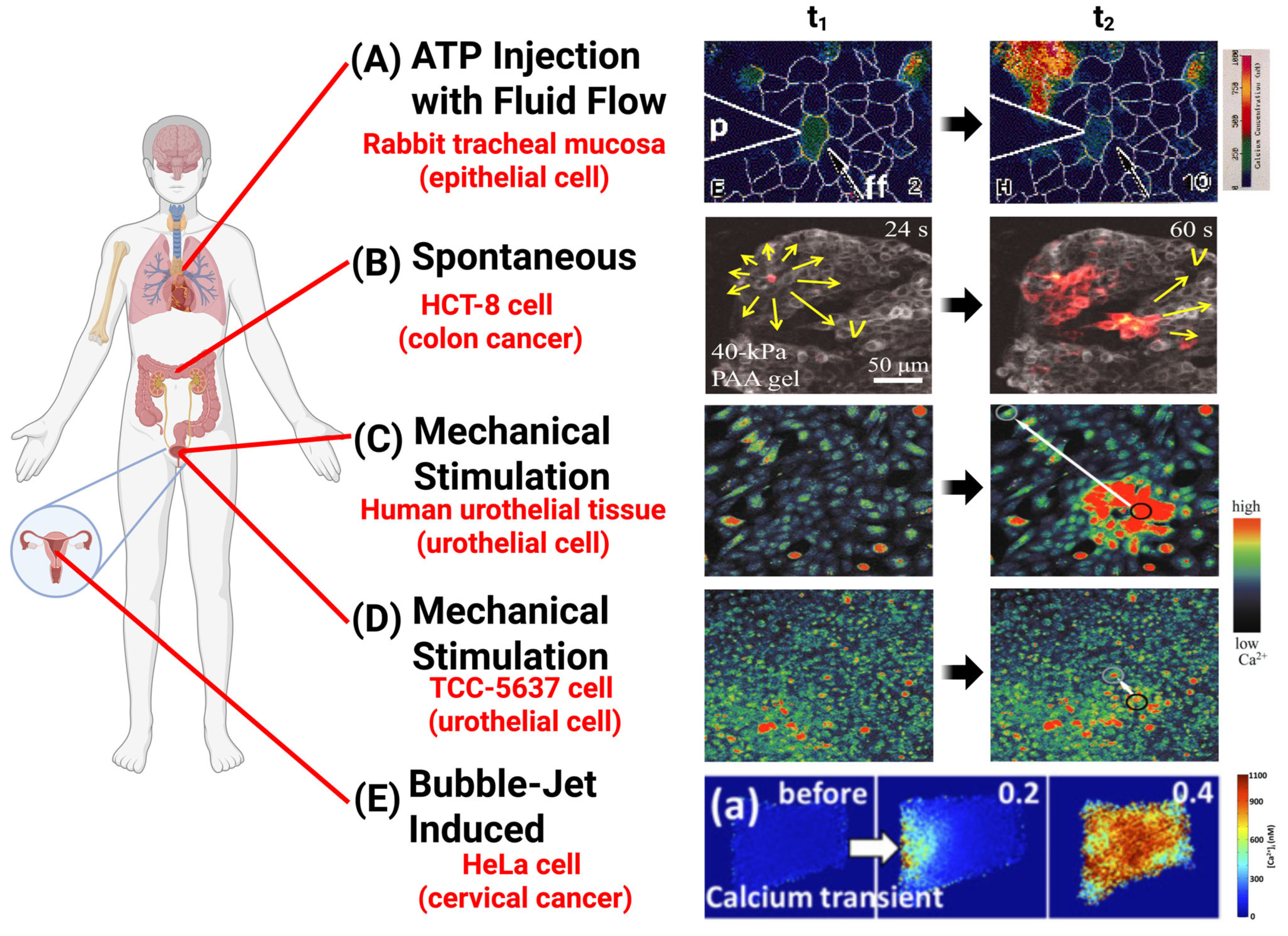
In research laboratories, external mechanical forces are widely applied in experiments to induce intracellular calcium dynamics and to reveal new signaling pathways (Figure 1). Utilizing different methods of mechanical application, these multiscale forces have been reported to activate calcium activities demonstrating a broad spectrum of spatial-temporal-functional dynamics (Table 1 and Table 2).
Table 1. Summary of available force application methods that activate calcium signals in distinct cell types and their physiological functions.
Category of Force Types Methods of Application Key Parameters Molecular Mechanisms Function
Molecular-Scale Mechanical StimulationOptical Laser Tweezers Fibronectin (Fn)-coated bead (diameter = 10 μm), force = 300 pN [79]Induce Ca2+ signals at the plasma membrane and ER via TRPM7 mechanosensitive channels. ICWs in MSCs depend on TRPM7-mediated calcium signaling, which regulates differentiation.
Light-Activated Molecular Machines Forces = 10−12 to 10−9 N, laser = 400–405 nm (3.2 × 10² to 9.0 × 10² W/cm2), duration = 250 ms (in vitro), 1–2 s (Hydra) [80]Induce ICWs via IP3-mediated signaling pathways. MM-induced calcium wave generation can control biological behaviors coordinated in the networks of cells, such as contraction.
Pipette Poking/Probing Blunt-end glass micropipette, tip diameter = 50 μm [76]ATP release via mechano-volume-sensitive Cl anion channels, activating receptors (P2X/P2Y) on wave-receiving cells. Induced ICWs propagated at velocities of ~15 μm/s and distances of 200–300 μm, transmitting signals to adjacent cells.
Scrape with Pipette Tip Bent 200 μL micropipette tip [74]ATP release stimulates calcium waves through purinergic receptor activation. Induced ICWs regulated intercellular communication.
Glass Micropipette with Micromanipulator Micropipette tip coupled with 200 ng/mL EGF for 5 min [81] EGF activates PLC-mediated calcium signaling. -
Tip diameter = 1 μm, movement = 2–5 μm, controlled by a piezoelectric device [82] IP3 moves between gap junctions in epithelial respiratory tract cells. -
Tip diameter  <  1 μm, touching less than 1 % of the cell membrane [83]Induces a rapid Ca2+ spike and ICWs through gap junctions. ICWs facilitate intercellular communication, regulate responses to mechanical and metabolic stress, and maintain metabolic homeostasis.
Tip diameter of 1 μm, moved downward by 10 μm over 0.5 s coupled with 3 M KCl, delivered at 150 hPa for 1 s [84]Ultrafast wave of calcium, traveling at approximately 15 mm/s. ICWs synchronize contraction, regulate blood flow, and coordinate rapid vasomotor responses in SMCs.
Microinjector Capillary Micrometer precision [85]ICWs are inhibited by GJ-blocking heptanol, indicating gap junction dependence. ICWs maintain intercellular communication and coordinated cellular responses in urothelial cells.
Force Probe or 30-Gauge Syringe Needle Force range = ~2–300 μN, stimulation duration = 20–2000 ms [86] Not specified. Induced ICWs regulate endothelial communication, which is critical for immune modulation and tissue healing.
Glass Microelectrode Tip diameter = 1 μm [87]Not specified. -
Subcellular/Cellular-Scales Mechanical StimulationFocused Ultrasound (FUS) Amplitude = 46 MHz (12 Vp–p), pulse repetition frequency = 1 kHz, duty cycle = 5% [88]PANX1 mechanosensitive channels mediate calcium wave (propagation distance > 1 mm) initiation. FUS-induced ICWs in PC-3 cells promote ATP release and cytokine/chemokine secretion via PANX1.
Bubble-Jetting Methods RGD-coated beads (6 μm), γ = Sd/Rmax = 1.2 to 2.4 (Sd = 30–60 μm) [89]Intracellular calcium waves elicited by tandem bubble-induced jetting flow. The bubble-induced rapid Ca2+ influx showed loss of F-actin stress fibers, cell shrinkage, and apoptosis.
Parallel-Plate Flow ChamberShear stress from 100 to 400 μN/cm2 for 3 s [90]Raising shear stress induced localized ATP release from caveolin-1-rich membrane domains, which activated purinergic receptors and initiated intracellular Ca2+ waves.The shear stress triggered Ca2+ wave in HPAECs; contributed to cell shear-sensing.
Tissue-Scale Mechanical StimulationMechanical StretchingStretching speed at 100 μm/s and distance at 200 μm (17.5% elongation) [91]Stimulate Piezo1-dependent calcium influx and ATP release.
Applied Mechanical Loading Diaphragm backpressure = 15 kPa, duration = 300 s [92]Induces ICWs through physical deformation and ATP release. ICWs regulate organ growth through calcium spikes, transients, and waves.
Figure 1. Intercellular calcium waves (ICWs) imaged in vitro in response to different forms of mechanical and biochemical stimulation. (A) [Ca2+] imaged in airway epithelial cell monolayer at t1 = 2 s and t2 = 10 s after local application of extracellular ATP in the presence of external fluid flow (ff, direction represented by white arrow) [93]. (B) Spontaneous ICW imaged in human HCT-8 colon cancer on 40-kPa polyacrylamide (PAA) gel at t1 = 24 s and t2 = 60 s. The yellow arrows indicate direction and velocity of wave propagation [53]. (C,D) [Ca2+] imaged in human urothelial cells and 5637 human transitional cell carcinoma (TCC) cells, respectively, at t1 = prior to single-cell micromanipulator stimulation and t2 = instance of maximal calcium wave distance propagation (white arrow) [85]. (E) Intracellular calcium transient progression imaged in singular HeLa cervical cancer cell in response to tandem bubble-induced jetting flow at t = 0, 0.2, and 0.4 s, respectively [89].
Cancers 17 01851 g001
Table 2. Summary of diverse mechanical stimuli and corresponding molecular transducers that regulate different propagation stages of ICWs.
Mechanical Force TypeMolecular TransducersMechanotransduction Pathway
Shear StressCaveolin-1, P2X/P2Y purinergic receptors; Integrins Shear stress induces ATP release from caveolin-1-enriched membrane domains, which activates P2X/P2Y purinergic receptors and initiates ICWs [90]. Similarly, laser-induced tandem bubble-jetting flow activates integrins, the mechanosensitive ion channel TRPM7, leading to calcium influx and subsequent ER-mediated calcium-induced calcium release [89].
Tension and StretchIntegrins, Piezo1, Gq-PLC-IP3R pathwayMechanical stretching or increased ECM stiffness is sensed by integrins and Gq-PLC-IP3R pathway-mediated calcium release [53,94,95,96]; Piezo1 mediates direct calcium influx under stretch-induced membrane tension [97,98,99,100].
Point Stress and CompressionTRPM7, Cl channels, IP3R, P2X/P2Y receptors, Connexin-based Gap JunctionsLocal mechanical indentation activates volume-sensitive Cl channels and TRPM7, resulting in ATP release and P2X/P2Y purinergic receptor activation [74,76,79]; IP3 is generated and triggers IP3R-mediated calcium release [81,82,86], which propagates through GJ [83,84,85,87].
Membrane TensionPiezo1 (Force-from-Lipids); PANX1 (ER), IP3R (Ultrasound-induced ER deformation)In cytoskeleton-deficient conditions, membrane tension activates Piezo1 through a force-from-lipids mechanism, leading to calcium influx [101]; FUS induces ER membrane deformation, activating PANX1 channels and IP3Rs to release calcium [88,102].
Stress RelaxationIP3R, Inx2 (Gap Junctions in Drosophila)Mechanical stress release leads to the generation of IP3, which activates IP3Rs and promotes calcium release from the ER and propagation through GJs (Inx2) [92,103].
Nanoscale Molecular ForceIP3RLight-activated molecular machines deliver nanoscale rotational forces, stimulating Gq-PLC-IP3 pathway and leading to calcium release and ICW generation [80].
At the molecular scale, mechanical stimuli that are applied by precise and minimally invasive tools primarily target subcellular structures (Figure 2A). For example, a light-activated fast-rotating molecular machine (MM) can be delivered into cytoplasm to exert unidirectional forces in the range of 10−12 N (=1 pN) to 10−9 N (=1 nN) to stimulate ICWs through the pathway of inositol trisphosphate (IP3)-mediated signaling. This MM technology uses laser stimulation at 3.2 × 10² W/cm2 (in vitro; 400 nm), 5.1 × 10² W/cm2 (cardiomyocytes; 400 nm), and 9.0 × 10² W/cm2 (Hydra; 405 nm) for 250 ms in vitro and 1–2 s for Hydra, depending on specific research goals [80]. Similarly, optical laser tweezers can apply a mechanical force of 300 pN to the fibronectin (Fn)-coated bead (10-μm diameter), which is attached to the plasma membrane. This force can induce Ca2+ signals at the plasma membrane and the endoplasmic reticulum (ER) in human mesenchymal stem cells (MSCs) [79].
Using a highly precise micropipette or force probe, contact-based mechanical stimulation can be achieved at a molecular scale, offering spatial precision in the micrometer and nanonewton range. Using a blunt-end glass micropipette with a tip diameter of 50 μm, mechanical poking or probing on local regions of cellular membrane in human DU-145 prostate cancer cells can induce intercellular propagation of ICWs, with wave velocities reaching approximately 15 μm/s and propagation distances achieving 200–300 μm. Some studies suggest that the propagation of Ca2+ waves occurs through the mechano-regulated secretion of ATP molecules from wave-initiating cells and the activation of purinergic receptors on wave-receiving distant cells. Mechanistically, the mechano-volume-sensitive Cl anion channels on cells are activated by cell swelling or local changes in cellular mechanical strain and trigger the release of ATP molecules [104]. Similarly, linear mechanically scraping epithelial cancer cells using a bent 200-μL micropipette tip stimulates extracellular ATP release and initial calcium waves through the activation of purinergic receptors on distant cells [74]. Additionally, using a micromanipulator-controlled glass micropipette to perform mechanical stimulation, coupled with chemical epidermal growth factors (EGF) via EGF receptors (EGFR) at a concentration of 200 ng/mL for 5 min, intracellular calcium transients and intercellular calcium waves are activated in MDA-MB-468 cells [81]. A piezoelectric device driving a fine glass microprobe with a tip diameter of 1 μm and precise movements of 2–5 μm induces intracellular Ca2+ levels in epithelial cells via the movement of IP3 through GJs between epithelial respiratory tract cells [82]. The combination of microinjector capillary and pharmacological GJ (gap junctions)-blocking heptanol reveals the GJ-dependent propagation mechanism of ICWs [85]. The force probe or 30-gauge syringe needle can apply forces ranging from ~2–300 μN with a stimulation duration of 20–2000 ms on cells and induce calcium waves [85]. Glass microelectrodes with a tip of 1 μm diameter have been employed to investigate calcium communication induced by mechanical force [87]. Briefly touching less than 1 % of the cell membrane with a glass micropipette (tip diameter  <  1 μm) induces a rapid intracellular Ca2+ spike, which spreads to neighboring cells. This technique, applied to bovine corneal endothelial cells (BCEC), enables the study of intercellular communication and GJ function [83]. Similarly, ICWs in vascular smooth muscle cells (SMCs) were triggered by either mechanical stimulation applied locally using a micropipette (tip diameter of 1 μm, moved downward by 10 μm over 0.5 s) or chemical stimulation using locally applied KCl (3 M KCl, delivered under controlled pressure of 15 kPa for 1 s). These stimuli depolarize the membrane, allow the propagation of electrical signals into neighboring cells, and consequentially activate voltage-gated calcium channels to facilitate calcium influx. The resulting ultrafast calcium waves, traveling at approximately 15 mm/s, were inhibited when GJs between cells or voltage-operated calcium channels were blocked [84].
Together, these methods provide precise, physiologically relevant, and minimally invasive ways to dissect the fundamental roles of cellular mechanical forces in initiating non-spontaneous intracellular calcium signaling and propagating ICWs, demonstrating how mechanical forces are translated into calcium signaling at the smallest biological scales.
At the subcellular and cellular scales, a variety of methods demonstrate how forces can induce ICWs from organelles and influence cellular behaviors (Figure 2B). Hydrodynamic forces generated by bubble-jetting methods, which use RGD-coated beads of 6-μm and combine with single-bubble treatment (γ = Sd/Rmax = 1.2 to 2.4 (Sd = 30 μm to 60 μm)), can elicit the propagation of a calcium wave in HeLa cells by activating integrins, which triggers the mechanosensitive ion channel TRPM7, leading to calcium influx and subsequent ER-mediated calcium-induced calcium release. [89]. Similarly, in human pulmonary artery endothelial cells (HPAECs) that are exposed to shear stress in a parallel-plate flow chamber (100 to 400 μN/cm2 for 3 s), highly localized ATP release occurs at caveolin-1-rich plasma membrane domains (caveolae), which activates nearby P2X/P2Y purinergic receptors and triggers intracellular Ca2+ increases that initiate Ca2+ waves propagating across the cell monolayer [90]. On the subcellular level, a focused ultrasound (FUS) that has an amplitude of 46 MHz (12 Vp–p), pulse repetition of 1 kHz frequency, and duty of 5% cycle, stimulates ICWs in PC-3 prostate cancer cells via mechanosensitive pannexin-1 (PANX1) channels in the ER without cytoskeletal dependence [88]. Importantly, the FUS-activated calcium elevation has been reported for invasive prostate (PC-3 and DU-145) and bladder (T24/83) cancer cell lines, but not for non-invasive cell lines (BPH-1, PNT1A, and RT112/84). The ICWs propagate from the cells at the focus of the transducer to other cells over spatial distances greater than 1 mm [102]. These subcellular- and cellular-scale mechanical stimulations elucidate the roles of mechanical signals in activating organelle-based calcium waves and intercellular communications through distinct mechanosensitive pathways.
At the tissue and organ scales, macroscopic mechanical forces, including compression, stretching, shear stress, and membrane tension, can be transduced into intracellular and intercellular calcium signals (Figure 2C). The mechanosensitive Piezo1 channel has a critical role during this transduction in multiple cancer models, including gastric, breast, prostate, glioma, and osteosarcoma cancers [97,105]. Mechanical stretching of bladder urothelium cells with stretching speed at 100 μm/s and stretching displacement at 200 μm (actual cell extension rate, 17.5%) stimulates calcium influx and ATP release, which is dependent on Piezo1 activation [98]. Similarly, mechanical loading applied on discs of Drosophila wings at a diaphragm backpressure of 15 kPa for 300 s, such as the release of loads from external sources, has been shown to stimulate ICWs [92]. Together, tissue-scale mechanical forces, including stretching, compression, and shear stress, provide insights into the critical roles of physical deformation in triggering ATP release and calcium influx across heterogeneous cell populations, offering a framework for understanding system-wide mechanotransduction in both physiological and pathological processes.
Depending on the cell types and experimental settings, mechanical stimuli have dual roles in initiating ICWs, either as the primary driver or through the coordination with biochemical pathways. On the one hand, light-activated molecular machines, optical tweezers, and FUS can dominantly trigger ICWs via direct mechanotransduction pathways involving IP3 signaling, mechanosensitive ion channels (e.g., TRPM7, PANX1), or intracellular calcium mobilization. On the other hand, pipette scraping, shear stress, and membrane stretch as mechanical cues that rely on ATP release or EGF receptor activation subsequently propagate ICWs through purinergic signaling, receptor-mediated pathways, or GJ communication.
Comparison of current experimental models of mechanically induced ICWs reveals that most studies have been conducted in 2D monolayer cell models. This is mainly due to the advantages of 2D models, including ease of applying mechanical interventions, compatibility with optical imaging, and reproducibility. However, 2D models lack complex 3D architecture and mechanical microenvironments in physiological tissues.
In contrast, studies of mechanically induced ICWs in 3D in vitro, ex vivo, or in vivo models have been reported, but are very limited to date. For example, in an ex vivo Drosophila wing disc model cultured with 15% fly extract, the release of 15 kPa compressive mechanical stress via the regulated environment for microorgans chip (REM-Chip) device triggered IP3- and GJ-mediated ICWs [92]. Most studies using 3D models and in vivo models have focused on spontaneous ICWs or calcium waves induced by biochemical stimuli (e.g., KCl injection, caged-ATP release). This is mainly due to the more complex structures of 3D models, making it difficult to achieve both 3D precise mechanical control and high-resolution imaging simultaneously. These limitations highlight a critical technological gap and underscore the current need for new 3D model-based mechanobiological tools to deepen our understanding of physiologically relevant ICWs.

2.1.2. Spontaneous Initiation Mechanisms of Multiscale ICWs

Spontaneous ICWs are a well-known phenomenon in electrically excitable cells including neurons [72,106], astrocytes [77,107,108,109], glial cells [46,87,110,111,112,113], and muscle cells [46,80,84,114,115,116]. In neurons, spontaneous ICWs have been linked to various developmental phenomena including regulation of neurite extension [117], guidance of neocortical growth [113], and neuronal differentiation [118]. Spontaneous ICWs have been linked to cochlear development in pre-hearing mammalian cochlea [119]. In extraocular muscles, spontaneous slow ICWs (max duration of 2–12 s, velocity of 25–50 μm/s) have been linked to localized myofiber contractions [115]. In smooth muscle cells, a mechanical signaling system that utilizes ECM stiffness to modulate calcium wave frequency and agonist sensitivity was identified, likely contributing to diseases like asthma and hypertension, independent of GJs or diffusing signals [114].
Spontaneous ICWs have infrequently been observed in cancer cell lines as well. HCT-8 colon cancer and DU145 prostate cancer were recently shown to generate spontaneous calcium transients and ICWs without active mechanical probing. In vitro cultures and ex vivo HCT-8 xenograft tumor slices revealed that both calcium transient duration and ICW propagation distance are influenced by substrate stiffness, with distinctly low and high substrate stiffnesses (250 Pa and 3 GPa) provoking decreased transient duration and propagation distance compared to intermediate stiffnesses (10, 20, and 40 kPa) [53]. These substrate-stiffness-dependent spontaneous ICWs were shown to modulate tumor growth in mouse models. Considering that mechanical stiffness of solid tumors is higher than that of healthy tissues, this finding potentially suggests a promoting role of tumor stiffness and ICWs on cancer progression.
Spontaneous ICWs also play a role in Drosophila organogenesis [92], tissue repair [120], smooth muscle differentiation [121], and actomyosin regulation [122]. In Drosophila imaginal wing disc development, spontaneous ICWs are regulated by biochemical signaling and release of mechanical stress [92]. Spontaneous oscillatory ICWs have been observed in Drosophila imaginal discs, where wave properties including initiation and oscillation frequency were found to be influenced by factors such as rearing temperature, cellular organization, and high regional Wingless (Wnt) signaling [122].
Finally, while intracellular Ca2+ oscillations lack the spreading characteristic of ICWs, these oscillations play a regulatory role in cell processes such as cell division, migration, fertilization, and apoptosis, which may shed light on the potential physiological impact of ICWs [42,45]. In human mesenchymal stem cells (HMSCs), spontaneous Ca2+ oscillations are shown to be regulated by extracellular substrate rigidity via the RhoA/ROCK signaling pathway, whereupon they influence cell differentiation outcomes [123]. In esophageal squamous cell carcinoma (ESCC), Orai1-mediated hyperactive intracellular Ca2+ oscillations have been shown to promote tumorigenic behavior such as cell proliferation, migration, and invasion in both in vitro cultures and in vivo xenografted mice [124].

2.2. Molecular Effectors Underpinning the Mechano-Regulated Initiation of ICWs

2.2.1. Roles of Interacting Cytoskeleton and Mechanosensitive Ion Channels

Upon mechanical stimulation of a cell, the mechanical signal propagates through various force-sensing molecules and organelles. These effectors can be broadly categorized into three functional classes: (1) Mechanosensitive ion channels, such as transient receptor potential melastatin 7 (TRPM7) [79], Piezo-1 [97,98,99,100], pannexin-1 (PANX1) [88], connexin channels [125], and cytoskeletal components, which activate cascades of signal transduction to induce Ca2+ release from the ER and subsequent cytoplasmic Ca2+ spikes in adjacent cells, thus initiating an ICW [46,94]. (2) Intracellular calcium release channels, primarily the inositol 1,4,5-trisphosphate receptor (IP3R) [53,102,117,122], which mediate calcium release from the endoplasmic reticulum (ER) in response to IP3 binding. In some cases, calcium signaling can be tuned by mechanical substrate stiffness [53,123]. Importantly, increasing studies suggest that the participation of the cytoskeleton into these mechanotransduction processes can be case dependent [79,88].
Beyond ion channels, mechanotransduction also involves integrins and actin cytoskeleton. Acting as transmembrane receptors, integrins anchor cells to the extracellular matrix and serve as key mechanosensors. When mechanical forces like tension are applied to the ECM or cells sense high environmental stiffness, integrins cluster and activate intracellular signaling pathways [95]. These signals are transmitted to the cytoskeleton, including actin filaments, intermediate filaments, and microtubules, which reorganize and enable the adaptation of cellular shape, stiffness, and motility [96,126]. This dynamic feedback loop enables cells to sense mechanical cues and convert them into biochemical signals to regulate processes such as migration, proliferation, differentiation, ion channel gating, gene expression, and stem cell fate [127]. Some studies report that the cytoskeleton plays a critical role in the mechano-transduction process contributing to ICW initiation. In human mesenchymal stem cells (MSCs), the cytoskeletal network of contractile actomyosin machinery and microtubules were demonstrated to transmit mechanical force to the ER, potentially triggering Ca2+ release from the ER through activation of inositol 1,4,5-trisphosphate receptors (IP3Rs) or other transient receptor potential (TRP) family ion channels. Additionally, the mechanosensitive channel TRPM7, which transports Ca2+, Mg2+, and Zn2+ ions, was found to play a critical role. TRPM7 facilitates force transmission to the ER, exerts downstream effects on IP3R, or can be directly coupled to IP3R via adaptor proteins, including ankyrins [79]. In Drosophila imaginal discs, the relaxation of mechanical stress, rather than the stress itself, was found to initiate ICWs in a process dependent on IP3-mediated Ca2+ release from ER and propagation through GJs (Inx2) [92,103]. The temporal and spatial characteristics of the ICW response to mechanical loading and release were primarily dictated by baseline spontaneous ICW activity, with higher baseline levels correlating with prolonged burst duration (~275 s vs. ~125 s) and greater area fraction (~0.75 vs. ~0.25) [92].
On the other hand, some ICWs’ initial path from mechanical stimulation may bypass the cytoskeleton entirely. Mechanosensitive PANX1 channels localized in the ER mediate calcium release from internal stores in response to FUS, potentially due to membrane deformation induced by FUS acoustic pressure waves leading to PANX1 activation. This signaling pathway is independent of the cytoskeletal integrity in invasive PC-3 prostate cancer and HEK 293T human embryonic kidney cells and critically relies on both PANX1 and IP3R for ICW initiation and propagation [88]. However, how mechanical forces interact with PANX1 and IP3R on the ER remains to be determined. Similarly, in DU-145 prostate cancer cells, volume-sensitive anion channels in the plasma membrane mediate ATP secretion in response to pipette-based poking stimulation. During this process, the release of Ca2+ signals from the ER and the propagation of ICWs occur independently of the cytoskeleton, because the disruption of microfilaments and microtubules by cytochalasin B and nocodazole has shown no effect on propagation of calcium wave [76]. Additionally, Piezo1, a mechanosensitive ion channel [128,129], was activated using Yoda1 as a chemical substitute for mechanical stimulation in colon cancer stem-like cells (CCSCs), HCT-116, and HCT-8 cells. Activation of Piezo1 induced Ca2+ influx and subsequently activated NFAT1, which is a critical regulator for maintaining the stemness of CCSCs [99]. In cytoskeleton-deficient HEK293 cells’ membrane blebs, mechanical forces can activate the Piezo1 pathway via a “Force-from-Lipids” mechanism, inducing calcium ion influx [101].
Overall, these studies suggest that diverse mechanotransduction mechanisms can regulate ICW initiation and propagation, with or without cytoskeletal involvement. While the exact mechanisms by which mechanical stimulation is transduced into downstream ICW propagation remain unclear in many cell lines, exploring the mechanistic diversity and cell-type specificity of these pathways will promote a deeper understanding of how mechanical signals regulate calcium dynamics. Additionally, targeting specific mechanosensitive ion channels, such as TRPM7, Piezo1, or PANX1, may provide new therapeutic strategies for diseases associated with calcium signaling, such as cancer, heart diseases, and inflammation [130,131,132,133,134,135,136].

2.2.2. Roles of IP3 and IP3R in Intracellular Calcium Release

Following the receiving of mechanical signals, IP3 activated by the Gq-PLC-IP3R pathway is frequently recognized as a direct effector for the initiation of intracellular calcium release. IP3 binds to the IP3 receptor (IP3R) on the ER, triggering the release of intracellular Ca2+ ions into the cytosol [53,92,94,122]. At low cytoplasmic concentrations, these initial Ca2+ ions can stimulate increased release of Ca2+ from the ER through biphasic modulation of IP3R channel gating, leading to Ca2+-induced Ca2+ release (CICR) [86]. Calcium diffusing from one open channel can trigger adjacent channels to open, amplifying the release in a self-reinforcing loop, which is eventually countered by inhibitory feedback at high Ca2+ concentrations [137]. Since both IP3 production and IP3R opening are subject to positive and negative feedback regulations by Ca2+ and IP3 in a concentration-dependent manner, with Ca2+ exerting both stimulatory and inhibitory effects, intracellular Ca2+ oscillations emerge from complex signal amplification and refractory dynamics [64,122]. Significantly, the remodeling of IP3R-mediated Ca2+ signaling is recognized as a central key mediator that controls the cellular processes in cancer progression [138].
Elevated substrate stiffness may increase plasma membrane tension, activating cytoskeletal proteins via integrins, actin filaments, and G-protein coupled receptors (GPCRs) including GPR68 and H1R. These activated GPCRs dissociate the Gq protein, whose alpha subunit activates Phospholipase-C (PLC) to cleave Phosphatidylinositol 4,5-bisphosphate (PIP2) into diacylglycerol (DAG) and IP3. This IP3 then activates IP3R to release ER Ca2+, raising [Ca2+]cyt. Alternatively, increased cytoskeletal tension on stiffer substrates could directly open IP3Rs through interaction with cytoskeletal proteins. Elevated [Ca2+]cyt subsequently activates cyclin D1/CDK4 and cyclin E/CDK2 signaling, facilitating the G1/S transition and promoting cancer cell proliferation and tumor growth in vivo [53,94]. However, how the mechanical signals mechanistically regulate the detailed spatial-temporal dynamics of these molecular networks to produce Ca2+ oscillation remains to be elucidated.
Upon release from intracellular stores such as the ER, calcium ions act as key initiators of intercellular communication by triggering a cascade of downstream signaling events. The initial release of calcium binds to calcium-binding proteins like calmodulin, which activates various kinases and phosphatases, including protein kinase C (PKC) and proto-oncogene tyrosine-protein kinase Src. Consequently, they modulate cellular processes such as gene expression, cytoskeletal remodeling, and cell proliferation [85,88]. Calcium influx is further facilitated by stretch-activated and store-operated channels, such as TRPM7, which are essential for mechanosensitive calcium entry into the cytoplasm [79]. This influx amplifies the cytosolic calcium concentration, initiating additional calcium release through CICR, thereby propagating the calcium signal across neighboring cells [85,86].
Ryanodine receptors (RyRs) are another channel protein responsible for releasing Ca2+ from the ER into the cytoplasm [139,140]. Unlike IP3Rs, however, RyRs are infrequently in ICWs of transient generations. RyR and TRP channels are essentially not expressed in Drosophila imaginal discs, suggesting that ICWs present are not propagated by any RyR-dependent mechanism [122]. Application of the RyR inhibitor dantrolene (Dan) had no effect on ICWs present in RasV12-transformed mammalian epithelial cells [27], nor did it affect Ca2+ transients in HCT-8 colon cancer cells [53]. However, while RyRs do not contribute to the IP3R-mediated Ca2+ release via CICR in single colonic smooth muscle cells, RyR blockers like ryanodine can still inhibit Ca2+ signaling if RyRs were already activated (e.g., by caffeine or depolarization), indirectly affecting IP3-mediated release by depleting the ER of Ca2+ [141].

2.3. Multiscale Release, Propagation, and Regeneration of ICWs

2.3.1. Overview of Primary Mechanisms

ICWs propagate through a combination of mechanisms that ensure efficient communications between cells and tissues [109]. After intracellular calcium is released into the cytoplasm, it can initiate ICWs via two primary methods: (1) ATP release into the extracellular environment (Figure 3A) or (2) direct transfer of signaling molecules through GJs (Figure 3B). In the ATP-mediated pathway, calcium stimulates the diffusive release of ATP, which binds to purinergic receptors (e.g., P2X, P2X2, P2Y, etc.) on neighboring cells, triggering calcium release in those cells. This method is typically faster (vavg = 7 μm/s) but is often limited to shorter distances (100 μm) [142]. Conversely, GJs allow for the direct transfer of IP3 molecules between adjacent cells, facilitating slower but longer-range signal propagation. As IP3 binds to IP3Rs in adjacent cells, it triggers ER Ca2+ release and CICR to propagate the wave [143,144]. The resulting cytoplasmic Ca2+ can further activate PLCs, leading to additional IP3 production and amplifying the intercellular Ca2+ signal [145]. Another unique form of GJ-mediated communication occurs through tunneling nanotubes (TNTs; Figure 3C)-thin, actin-rich membrane extensions connected by GJs which bridge distances between cells, enabling ICW propagation via direct transfer of IP3 and Ca2+ [146,147,148].
In addition to these classical transmission pathways, ICWs can also propagate further distances through regenerative wave spread that involves active signal amplification by downstream cells [109]. ICWs have also been shown to spread via less-investigated paracrine mechanisms involving molecules other than ATP, including nitric oxide [149,150] and prostaglandins [151]. In astrocytes, NO promotes the propagation of ICWs through distinct pathways depending on the types of stimulation. Micropipette poking can trigger NO production, which induces plasma membrane depolarization and activates L-type voltage-gated Ca2+ channels, enhancing Ca2+ influx and supporting ICWs spread [152]. Glutamate stimulation activates metabotropic glutamate receptors (mGluRs), leading to eNOS-derived NO that directly S-nitrosylates CALHM1 channels, resulting in ATP release and downstream purinergic receptor-dependent activation of Cx43 and Panx-1 channels, and amplifying and coordinating calcium transients with wave-like propagation [153].
The speed and propagation distance of ICWs depend on several interacting factors, including the initiation mechanism (mechanical or non-mechanical) [76,81], mode of spread (e.g., ATP release, gap junctions, tunneling nanotubes) [142,143,144], presence of regenerative wave amplification [109], cell type [53,89,90], and the spatial organization of the cell group (e.g., monolayer vs. 3D tissue) [53,154]. Because these properties arise from a wide variety of interacting biophysical factors, currently there is no precise way or formula known to accurately pre-determine ICW speed and distance. The exact pathway or formula remains to be investigated. The following Section 2.3.2, Section 2.3.3, Section 2.3.4, Section 2.3.5, Section 2.3.6 discuss each mechanism in detail.
Figure 3. Mechanisms of ICW propagation. (AC) Mechanical signals from force prodding and/or substrate tension activate GPCRs, triggering conformational changes in the α subunit which then activates PLC to cleave PIP2 into IP3 and DAG [46,60]. IP3 activates IP3Rs on the ER, stimulating the release of Ca2+ from the ER into the cytoplasm [45,137,138,147]. Cytoplasmic Ca2+ further sensitizes IP3Rs to IP3, inciting positive-feedback calcium-induced calcium release (CICR) [122,145]. Mechanical stimulation may also directly trigger ER Ca2+ release by transmitting force through cytoskeletal proteins like ankyrins, which link to IP3Rs or other mechanosensitive channels, inducing their activation [79]. (A) ICW propagation via ATP signaling. Increased cytoplasmic Ca2+ stimulates extracellular vesicles and ATP transporters to release ATP into extracellular space, where it acts as a long-distance signaling molecule by binding to purinergic receptors (P2X, P2Y) on receiving cells, causing subsequent Ca2+ release by stimulating the PLC-IP3-IP3R- Ca2+ pathway [53,74,109,144,155]. (B) ICW propagation via gap junctions. IP3 signaling molecules pass directly from an initiating cell to an adjacent receiving cell through gap junctions, stimulating intracellular Ca2+ release by binding to IP3Rs on the receiving cell’s ER. Rising levels of cytoplasmic Ca2+ activate the PLC-IP3-IP3R-Ca2 pathway, driving further intracellular Ca2+ release [83,85,143,144,156]. (C) ICW propagation via tunneling nanotubes. IP3 signaling molecules travel from initiating to receiving cells via tunneling nanotubes (TNTs) connected by gap junctions, initiating calcium release in the receiving cell via the PLC-IP3-IP3R-Ca2 pathway and CICR [146,147,148].
Cancers 17 01851 g003

2.3.2. ATP-Mediated ICW Spread

Various studies have shown that ICWs’ long-range spatial propagation is critically dependent on the spatially diffusive release of ATP, known as ATP waves (Figure 3A). These ATP molecules function as endocrine signaling molecules to initiate and sustain ICWs [81,85,93,109,147,157,158,159]. P2 purinergic receptors P2X and P2Y are commonly shown to play a role in propagating ICWs by accepting extracellular ATP and inducing downstream Ca2+ activities [142].
For instance, the addition of ATP-degrading apyrase abolishes ICWs in human mesenchymal stem cells (HMSCs) [79], while both apyrase or purinergic receptor blockers Suramin and PPADS abolish ICWs in FUS-stimulated PC-3 human prostate cancer cells [88]. Purinergic receptor antagonists, ATP depletion, and ATP scavenging each completely inhibit ICW propagation in DU-145 prostate cancer cells, suggesting a wave propagation mechanism reliant on ATP release, mechanosensitive anion channels, and purinergic ATP receptors [76]. Stanniocalcin-1 (STC1) regulates ICWs in human pulmonary (A549) and prostate (PC3) epithelial cells by stimulating ATP release from bystander cells, indicating a cooperative mechanism for promoting ICW spread [74].
Extracellular ATP injection is another common method of triggering ICW initiation independent of mechanical stimulation, highlighting the pivotal biochemical roles that ATP can play in initiating ICWs. This approach highlights the sufficiency of ATP in generating ICWs, even in the absence of other potential contributing factors. For instance, exogenous ATP application has been shown to reliably initiate ICWs in various cell types, including astrocytes, endothelial cells, and epithelial cells [81,85,142]. These results not only underscore ATP’s central role in ICW initiation but also provide a reproducible means of studying calcium signaling dynamics across diverse cellular environments.

2.3.3. GJ-Mediated ICW Spread

GJs are specialized intercellular channels composed primarily of connexin43 (Cx43; Figure 3B), which assembles into hexameric connexons that dock between adjacent cells to facilitate selective cytoplasmic exchange of calcium ions and small signaling molecules [85,86,156,160]. GJs facilitate the spread of the calcium wave, ensuring synchronous cellular responses throughout the tissue. In various studies, the inhibition or blocking of GJs with chemicals including octanol [142,157], carbenoxolone [122,142], heptanol [85], and halothane [82] result in decreased ICW spread or duration, indicating the dependence of ICW spread on GJs in these conditions.
Studies on HeLa M-sec cervical cancer cells [147], H-SY5Y neuroblastoma cells, and HEK cells [148] indicate that passive Ca2+ diffusion through GJs is insufficient for ICW propagation, with IP3 transfer serving instead as the primary mediator [122]. The IP3 signaling pathway amplifies intracellular waves by increasing IP3 levels in adjacent cells, triggering calcium release from the ER and sustaining signal propagation over longer distances [86,145,161]. Several recent studies demonstrate GJ-reliant ICW mechanisms. ICW propagation (v = 12.1 ms−1) in NIH-3T3 mouse fibroblast cells was abolished completely with octanol, a Cx43 inhibitor [157]. ROS 17/2.8 rat osteosarcoma cell lines, which express Cx43 and lack P2Y2 receptors, were shown to transmit slow (7.4 µm/s) GJ-dependent ICWs [144]. As calcium signals propagate through the tissue, ATP may be released from PANX1 hemichannels, acting as a secondary messenger [76,88]. ATP activates purinergic receptors (P2X, P2Y) on neighboring cells, further amplifying calcium release from their ER stores and perpetuating the calcium wave, thus ensuring the synchronization of collective cellular responses [158,162].

2.3.4. Combined ATP- and GJ-Mediated Mechanism

Notably, these mechanisms are not mutually exclusive but can be simultaneously implemented by cells (Figure 3). Their relative contribution is dynamically modulated based on cell types, connexin expression, ATP availability, and microenvironment, enabling efficient communication between cells and tissues through the propagation of ICWs. ICWs in HeLa cells propagate via both ATP-mediated extracellular signaling and GJ-mediated intracellular pathways, with the dominant mechanism depending on connexin expression and ATP availability [45]. Bone cells largely rely on an ATP-diffusion-dependent mechanism for propagating ICWS, with GJs playing a significantly lesser role [27], whereas astrocytes preferentially utilize the GJ-IP3 pathway to mediate calcium wave propagation [117]. In the adult mouse organ of Corti, two separate types of ICWs were identified with distinct propagation mechanisms: slow (1–3 μm/s), periodic, GJ-mediated ICWs propagating longitudinally along the cochlea were inhibited by GJ blockers 1-octanol and carbenoxolone, while fast (v = 7 μm/s) ICWs stimulated by extracellular ATP were modulated by P2 receptor antagonists PPADS and suramin, likely mediated by an ATP-dependent mechanism involving activation of P2X2 purinergic receptors identified in supporting cells and outer hair cell stereocilia [142].
While most cell lines rely on ATP- and GJ-dependent mechanisms for ICW propagation, few exhibit ICW spread without either pathway, underscoring their essential roles in cellular communication. However, some smooth muscle cells may propagate calcium waves through a distinct ECM-stiffness-dependent mechanical signaling mechanism independent of gap junctions and extracellular diffusion, posing a unique exception to the rule [114].

2.3.5. Tunneling Nanotubes

Tunneling nanotubes (TNTs) represent a specialized extension of GJ-mediated ICW propagation (Figure 3C). They maintain GJ connectivity while enabling signal transmission over semi-long distances (approx. 10–70 µm) [146,147,148,160]. These dynamic, actin-based membrane protrusions establish direct cytoplasmic continuity between distant cells, facilitating the transfer of ions and signaling molecules such as Ca2+ and IP3. TNTs also enable the transfer of larger cargoes, such as organelles, between cells, and can adapt to cellular stress, as seen in astrocytes [108,163].
Like in GJ-mediated ICW propagation, photo-released IP3 experiments show that IP3 diffusion through TNTs, rather than the direct transfer of Ca2+, is a key driver of intercellular Ca2+ signaling [147]. TNTs have been directly implicated in ICW propagation in multiple cell types, including HeLa M-Sec cells [147,164] and SH-SY5Y cultured neuroblastoma cells [148]. TNTs have been found in Wnt/Ca2+ communication between neurons [108], which is significant because a noncanonical, β-catenin-independent [165,166,167,168] Wnt-PLC-IP3-Connexin-Ca2+ signaling axis has been demonstrated to play an important role in influencing ICWs in zebrafish spinal cords [169] and cardiac myocytes [168]. Together, these findings highlight the multifaceted nature of TNT-mediated communication, bridging the gap between direct cell-to-cell connections like GJs and broader signaling mechanisms like ATP waves.

2.3.6. Regenerative Wave Spread

Regular propagation mechanisms of ICWs mainly include ATP-, GJ-, and TNT-mediated modalities, and may include other pathways such as prostaglandins or NO. The initiation mechanisms of these propagations typically rely on exogenous signals, such as mechanical stimuli, chemical stimuli, or regulatory factors from neighboring cells (e.g., STC1). Upon activation by an initial mechanical stimulus, such as mechanical poking or mechanical microenvironmental influences, paracrine biochemical signals (such as ATP or NO) are transmitted to adjacent cells via one or more of these pathways, facilitating intercellular communication. Unlike the unidirectional transmission of propagation, the regenerative waves are amplified by the downstream cells that autonomously boost and relay the signals. The speed and duration of the regenerative waves depend on the tissue structure and cell type, and it possesses greater robustness and adaptability over longer distances (up to thousands of cells). A recent report suggests that the influences of mechanical microenvironments, not mechanical poking, can regulate the magnitudes of wave speeds and wave distance, indicating previously understudied roles of mechanical inputs in mediating regenerative and trigger waves [53]. This phenomenon is supported by experimental and modeling studies that reveal its underlying mechanisms and implications for efficient and robust signal transmission across cellular networks [87,109,122].
MacDonald et al. (2008) developed a diffusion model demonstrating that regenerative ATP release from downstream astrocytes significantly enhances ICW propagation. The dual signaling model—combining initial point-source ATP release and downstream regenerative release—provides an efficient and energetically robust mechanism for intercellular communication, aligning well with observed spatiotemporal kinetics [109]. In Drosophila imaginal discs, wavefronts of ICW maintained their strength over several thousand cells, suggesting cell-autonomous regeneration of the ICW signal via a GJ-mediated mechanism [122]. Similarly, intermediate cells in micropatterned BV-2 microglial assemblies can act as regenerative amplifiers, enhancing ICW transmission to outermost cells [87]. Together, these findings highlight regenerative wave propagation as a fundamental mechanism for sustaining and enhancing long-distance ICW transmission, ensuring efficient and robust signal spread across diverse cellular systems.

3. Physiological Roles of Calcium Waves and ATP Signals

3.1. Cancer Cells

Ca2+ signaling plays a well-established role in physiological processes essential for tumor progression [2,170,171,172,173]. As a specialized form of Ca2+ signaling, ICWs enable coordinated cellular responses with tumorigenic roles [115], including metastasis [161], migration and invasion [174], and tumor growth [53].
In many cases, ICWs have been proven as drivers of tumorigenesis and related pathological processes. For instance, in human HCT-8 colon cancer cells, mice inoculated with IP3-sponge-transduced cells—which showed a 16-fold reduction in spontaneous calcium transients and ICWs compared to controls—developed tumors with 1.6 times lower weight, demonstrating a causal role of ICWs in driving tumorigenesis in vivo [53]. In oncogenic RasV12-transformed mammalian epithelial cells and zebrafish embryos, ICWs initiated by a transformed cell induced apical extrusion and polarized movement of surrounding cells through an IP3R-GJ-TRPC1-mediated mechanism [27].
In other cases, ICWs act as biomarkers for cancer, though an unexplored causal role may still be at play. Scratch-wound assays in wild-type C1 CRC cells induce extracellular Ca2+ influx through voltage-gated calcium channels (VGCCs), driving initial calcium transients and promoting BCL9 interaction with paraspeckle proteins. Subsequently, BCL9 translocation into paraspeckles promotes sustained ICW propagation, cytoplasmic projections, tumor progression, tissue remodeling, and stromal cell infiltration [175]. Studies using FUS have revealed that invasive cancer cell lines (e.g., PC-3, MDA-MB-231) exhibit robust calcium oscillations in response to stimulation, whereas non-invasive cells of the same type do not. This suggests that invasive cells may possess intrinsic properties that make them particularly receptive to calcium signaling, potentially enhancing their metastatic capabilities [88,176]. The underlying mechanisms remain to be investigated.
Despite growing evidence linking ICWs to the promotion of tumorigenesis, some studies suggest modulated calcium signaling—including calcium waves—may suppress tumors via induction of apoptosis [177,178,179]. Electromagnetic fields modeled after ICWs have been shown to induce apoptosis in B16-BL6 melanoma cells, possibly indicating a tumor-suppressive role of ICWs in some cell lines [146]. Given that ICWs often propagate through extracellular ATP signaling, studies implicating ATP waves in cancerous phenotypes may, in part, reflect underlying ICW activity that has yet to be fully characterized [155]. Further investigation into the mechanisms and context-dependent functional effects of ICWs could provide critical insights into their potential as new therapeutic targets or biomarkers in oncology.

3.2. ICWs in Non-Cancer Cells

In non-cancerous biological systems, ICWs mediate various critical functions [42,44] including neuronal plasticity [113,117,118], stem cell differentiation [123,180], organ developmental regulation [92,94], tissue repair [120,181,182], viral priming [183,184], and muscle contraction [80,84,115,116].
In neurons, ICWs have been shown to regulate neuronal division in the ventricular zone of the neocortex [113]. Substrates’ mechanical rigidity regulates Ca2+ oscillation via the RhoA pathway in HMCSC stem cells, potentially influencing cell differentiation [123]. In Drosophila wing imaginal discs, ICWs exhibit spatiotemporal patterns that regulate organ growth through calcium spikes, transients, and waves. Perturbations to these signals can result in abnormal organ sizes, with either excessive or insufficient calcium signaling reducing growth [92,94]. ICWs mediate wound healing, as seen in polarized hepatic cells where ICWs guide preferential cell growth toward the wound edge [181]. In epithelial tissues, regenerative calcium waves traverse thousands of cells, enabling synchronized responses such as cell extrusion [27] and tissue repair [182]. Similarly, mechanical injury in Drosophila wing discs triggers slow IP3R-mediated ICWs, promoting tissue repair [120]. Calcium is also shown to regulate patterning and actomyosin organization in the Drosophila disc epithelium [122]. Rotavirus infection induces ICWs through ADP signaling, effectively priming surrounding cells for rapid viral spread [183,184]. In smooth muscle cells, the influx of calcium ions activates myosin light chain kinase (MLCK) to trigger contraction, while modulation of ATP-sensitive potassium channels regulates membrane potential and excitability [84]. These coordinated propagations of calcium and ATP signals are essential in urothelial cells, helping regulate their responses to mechanical and metabolic stress [85].
The interactions between mechanosensitive molecules and calcium signals underpin a myriad of pathological processes and therapeutic treatments. For example, malocclusion is a highly prevalent dental disease and severely impacts on oral health, such as causing periodontitis. Periodontitis increases the risk of general systemic diseases, including respiratory tract infections, Alzheimer’s disease, diabetes, and cardiovascular diseases [185,186]. Mechanotransduction via calcium influx and mechanoreceptor-induced signaling are critical steps during orthodontic tooth movement (OTM) for treating patients with malocclusions [187]. Mechanoreceptors located on the cell membrane, such as ion channels Piezo1, can receive mechanical forces and convert the signals to activate intracellular signaling, such as yes-associated protein (YAP) [188]. Researchers show that Piezo1 can increase intracellular calcium levels, leading to the dephosphorylation and nuclear translocation of YAP, transforming it into a transcriptional co-activator in the nucleus under mechanical stimulus [189,190]. Furthermore, Piezo1 controls the YAP-dependent expression of type II and IX collagens in osteoblasts under mechanical loads and influences osteoclast development [191,192], while YAP could regulate Piezo1 expression in turn. Nuclear localization of YAP could activate Piezo1 and enhance osteogenesis, and YAP, in collaboration with the G-protein-coupled estrogen receptor pathway, suppresses Piezo1 activation [193]. Some researchers show that Piezo1 exerts a signaling transduction role in mechanical stress-induced osteoclastogenesis, in which the markers cyclooxygenase-2, receptor activator of NF-kB ligand, and prostaglandin E2 were significantly upregulated under 2 g/cm2 static compressive loading for 0.5, 3, 6, and 12 h [194]. Piezo1 can also regulate periodontal ligament stem cell (PDLSCs) differentiation during OTM. Moreover, heavy mechanical forces up-regulate Piezo1 in PDL cells, reducing mitochondrial calcium influx and leading to the reduced cytoplasmic release of mitochondrial DNA. The subsequent signals inhibit the activation of the cGAS-STING signaling cascade and monocyte-to-osteoclast differentiation. Suppression of Piezo1 or up-regulation of STING expression under heavy mechanical force significantly increases osteoclast activity and accelerates OTM [195]. Furthermore, Piezo1 deficiency in osteoblastic cells leads to loss of bone mass and spontaneous fractures with increased bone resorption [190]. Piezo1-deficient mice are resistant to bone loss and bone resorption induced by hind limb unloading, demonstrating that Piezo1 can affect osteoblast–osteoclast crosstalk in response to mechanical forces [191].

3.3. Physiological Conclusions and Therapeutic Targets

Ultimately, ICWs play a fundamental role in mediating diverse physiological and pathological processes, from organ development and tissue repair to viral priming and muscle contraction. Their mechanosensitive nature enables precise regulation of multiscale cellular responses, particularly through calcium signaling pathways.
The intricate interplay between mechanical forces, calcium influx, and intracellular signaling cascades highlights the potential for targeted therapeutic interventions leveraging the mechanosensitive properties of ICWs and their physiological effects. Relevant mechanobiological strategies for cancer treatment include targeting proteins necessary for ECM stiffness (e.g., TGF-β, collagen), ECM crosslinking (e.g., Pan LOX, LOX, LOXL2), ECM mechanosensors and mechanotransducers (e.g., integrins, Piezo channels), and nuclear mechanotransduction through interventions including drug treatment, mechanical stretch, and low-intensity pulsed ultrasound (LIPU) [196,197,198].
Targeted manipulation of key molecular components, such as Piezo1, a mechanosensitive ion channel that controls ICW dynamics, provides an avenue for controlling ICW dynamics and selectively inducing antitumor responses. In prostate cancer cells, compression of the cell membrane by a glass probe activates Piezo1, which enhances ICW and subsequently activates the Akt/mTOR signaling cascade, promoting cancer cell proliferation, migration, and cell cycle progression. Knockdown of Piezo1 by shRNA or the Piezo1-specific antagonist GsMTx4 inhibited tumor growth in vitro and in vivo in DU145 cells and xenograft nude mouse models [199]. Similarly, cyclic mechanical stretch induces ICWs in Tpm2.1 knockdown cells via Piezo1, leading to calpain–2-mediated apoptosis. Restoration of the actin-regulatory protein Tpm2.1 suppressed ICWs and reversed the apoptotic response [200].
In addition to molecular targets, mechanical stimuli, including bubble-jetting flow and FUS, have been shown to be effective in modulating ICWs, providing a potential platform for non-invasive mechanotherapy of tumors. These stimuli may modulate calcium-sensitive apoptotic pathways and alter gene expression, thereby altering cancer cell viability, invasiveness, and response to therapies. Using laser-induced tandem bubble-jetting flow to stimulate single HeLa cells induces integrin-mediated ICWs that modulate intracellular calcium signaling pathways linked to cytoskeletal remodeling and membrane repair, inspiring a targeted mechanotherapeutic strategy to regulate cells using ICW without compromising membrane integrity [89]. In murine B16F10 melanoma cells expressing the mechanosensitive channel of large conductance (MSCL), localized ultrasound mechanical stimuli trigger the activation of MSCL, leading to sustained calcium influx that induces apoptosis via ER stress and a downstream calpain–caspase–3 pathway. Those methano-regulated apoptotic tumor cells released antigenic debris that promoted dendritic cell (DC) maturation and CD8+ T cell activation, resulting in a robust and tumor-specific immune response [201].

4. Cutting-Edge Technologies for Ca2+ and ATP Imaging

4.1. Functional Imaging of Ca2+ Dynamics

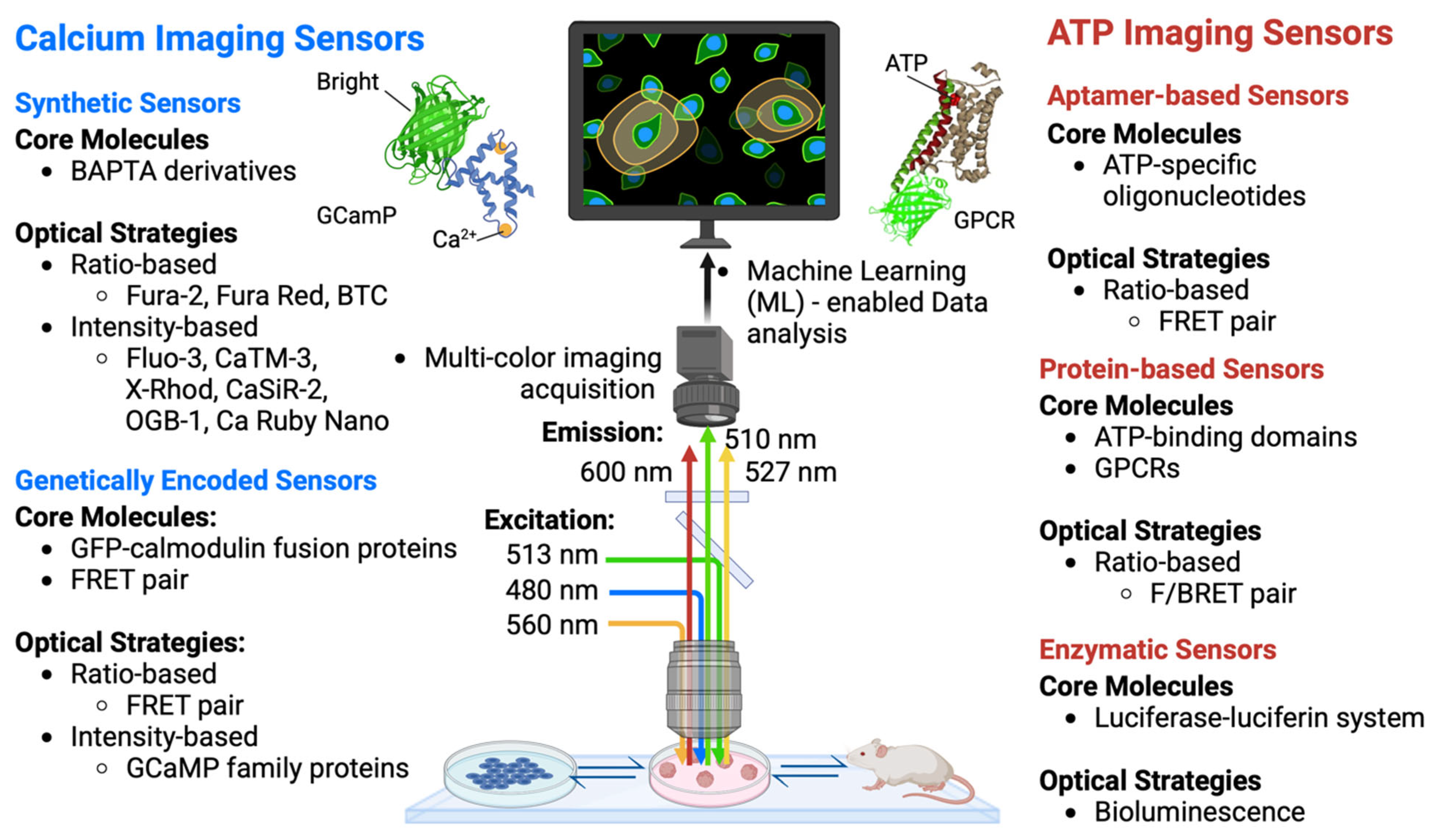
Given the importance of calcium ions in various physiological processes, there have been continuous research efforts in the last few decades to develop and improve fluorescence sensors for optical calcium imaging (Figure 4) [53,202,203,204,205]. Synthetic calcium sensors used today are mostly derivatives of 1,2-bis(o-aminophenoxy)ethane-N,N,N′,N′-tetraacetic acid, or BAPTA [206,207,208]. Compared to an ethylene glycol tetraacetic acid (EGTA) moiety, BAPTA has the advantage of an optical readout change upon calcium binding, fast binding kinetics, and independence from pH, while maintaining its high selectivity for calcium ions. Binding/chelating of calcium to BAPTA results in a shift from an “aniline-like” structure to a “benzene-like” structure that can be exploited for fluorescence property changes.
Broadly speaking, there are two parallel strategies in modulating optical readout changes for calcium imaging (Figure 4): (1) ratio-based indicators and (2) intensity-based indicators. Ratio-based indicators change the shape of their absorption or emission spectra. Upon binding between calcium ions and ratio-based indicators, the ratios of peak fluorescent intensity between two wavelengths change (e.g., fura-2, Fura Red, and BTC) [210,211,212]. In contrast, intensity-based indicators retain the overall shape of their absorption and emission spectra, but their fluorescence intensity typically increases upon binding between calcium ions and indicators (e.g., fluo-3, OGB-1, CaTM-3, Ca Ruby Nano, X-Rhod, CaSiR-2) [213,214,215]. While both strategies are widely implemented today, intensity-based indicators are relatively more appealing due to their ease of use, as they circumvent complex microscopic illumination, detection schemes, and ratiometric calibration.
Another class of calcium sensors gaining desired traction in cell biology and animal research is genetically encoded calcium indicators (GECIs) [216,217,218]. The GECIs are engineered proteins that exhibit a change in fluorescence upon physical binding between proteins and calcium ions. Because GECIs can be stably expressed in cell lines and transgenic animals, they have the advantages in biocompatibility and ease of use by eliminating the need to stain and wash, unlike their synthetic counterparts. GECIs are read out based on changes in fluorescent color ratio or intensity. To modulate changes in color ratio, a protein engineering strategy is to develop Förster resonant energy transfer (FRET) sensors where a FRET pair of fluorescence proteins is combined with a calcium binding module that changes conformation upon calcium binding to either facilitate or inhibit FRET [219,220]. Regarding the changes in intensity, a representative class of calcium sensor is the GCaMP family proteins. They are a fusion protein of circularly permuted green fluorescent protein (cpGFP) and Ca2+-binding calmodulin. When calcium ions bind to calmodulin, calmodulin changes the local microenvironment of the fluorophore in neighboring cpGFP and increases its fluorescence [221].
As the utility of calcium indicators is expanded through simultaneous imaging of multiple different proteins of interests, recent research shows rapid progress in expanding the color palette of calcium fluorescence indicators. While most of the early generations of synthetic and genetic calcium indicators emit green fluorescence, red/yellow-emitting indicators such as those in the RCaMP family have been developed to offer multiplexed imaging capabilities [222,223]. These advances hold the potential to image diverse ICWs in multiple distinct tissues and cell types simultaneously, leading to the decoding of multiscale intercellular calcium language in living systems.
Another important consideration in choosing the appropriate calcium sensors for imaging is the response time of the sensors compared to the ICW propagation speed. Recent advances in GECIs have seen accelerated response time on the order of 50–100 milliseconds, approaching to capturing a single electrical action potential [224,225]. Given that the fastest ICW speed is ~10–30 µm/s [53], which is equivalent to about one cell per second during propagation, those faster GECIs are well equipped to capture the ICW dynamics, such as fast GCaMP3 [226], GCaMP6fu [227], and GCaMP6f [228]. For high-resolution fluorescence imaging of ICW, the spatial resolution per the diffraction limit is on the order of ~300 nm [229]. The mammalian cell size is one or two orders of magnitude larger than the diffraction limit, making high-resolution fluorescence imaging a facile and sufficient platform for monitoring ICW propagation across cells.

4.2. Functional Imaging of ATP Dynamics

Extracellular ATP sensors are molecular tools designed for accurately measuring concentrations of ATP molecules across various biological contexts. These tools provide the capabilities to monitor real-time secretion and diffusion/reaction dynamics of ATP, leading to new insights into intracellular signaling and pathological processes. These biosensors rely on specific biorecognition elements and signal transduction mechanisms. The most notable two types are (1) binding-based and (2) enzymatic-reaction-based sensors, each employing distinct strategies for ATP detection.
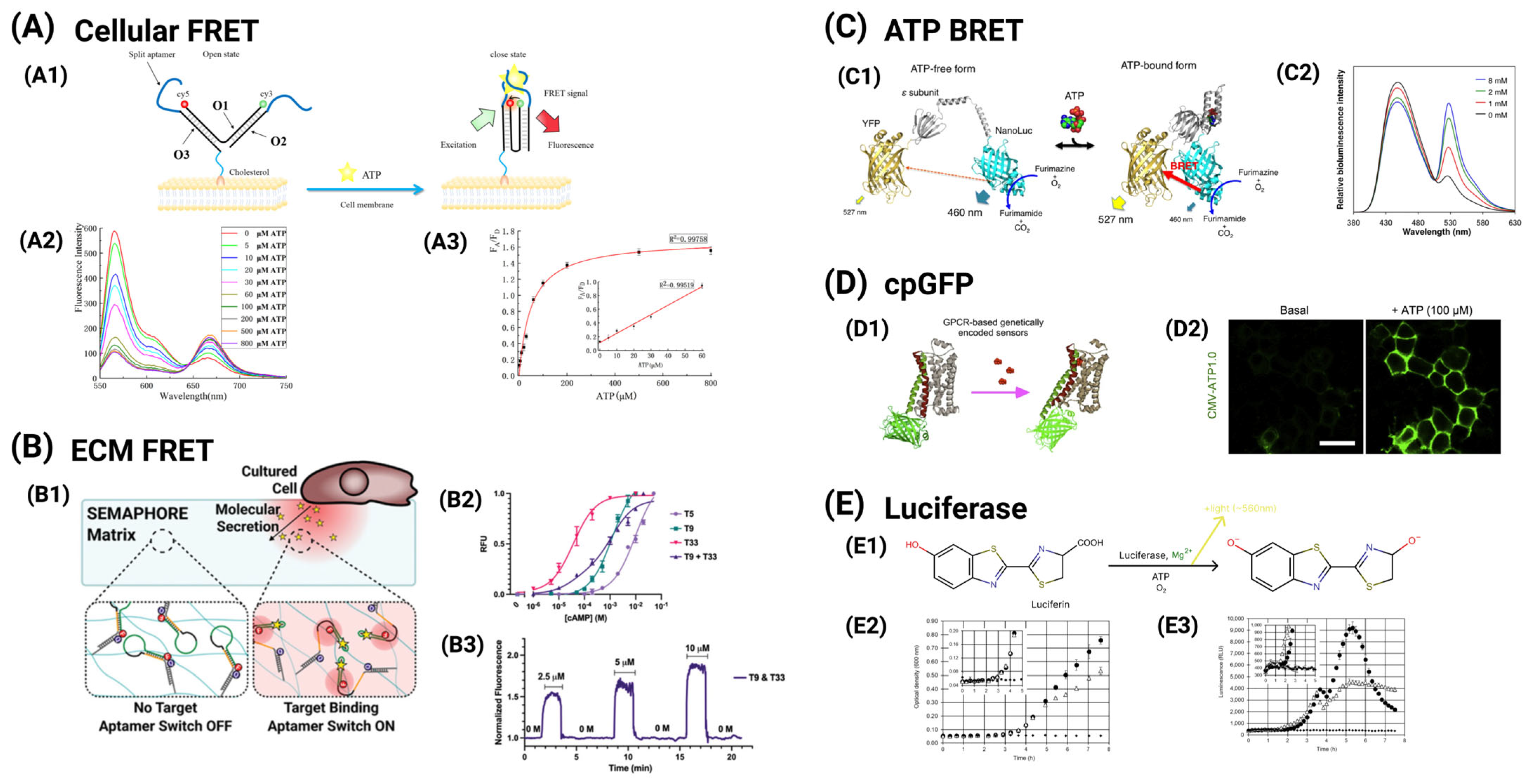
Aptamer-based biosensors utilize ATP-specific oligonucleotides as recognition elements [230,231,232,233]. They typically incorporate FRET motif to translate binding events into quantifiable signals (Figure 5A,B). When ATP binds to the aptamer, it induces oligonucleotide structure-switching that alters the proximity of a fluorescent dye to a quencher or enhances interactions between FRET pairs [234,235], resulting in a measurable fluorescence intensity shift that directly correlates with ATP concentration. Aptamer-based sensors are versatile, suitable for real-time monitoring on various substrates, including cell surfaces and hydrogels, and exhibit high specificity and speed (Figure 5C). Alternatively, recombinant-protein-scaffolds-containing-based sensors use engineered proteins, such as ATP-binding domains [236] (Figure 5D) or GPCRs [237] (Figure 5E), to detect ATP. These sensors employ either fluorescence/bioluminescence resonance energy transfer (F/BRET) [236] (Figure 5D) or circularly permuted FP (similar to the design strategy of GCaMP sensors) [238] as their signal transduction mechanisms. When ATP binds to the recognition site, it triggers a conformational change in the protein, which either affects the configuration of fluorescent proteins [239,240,241] tagged to the ATP binder or alters the distance between bioluminescent/fluorescent dyes and their FRET partners [242]. This change produces a detectable optical signal that can be correlated with ATP concentration.
Another prominent method for ATP detection is the enzymatic reaction-based approach [243,244,245], notably those that employ the luciferin/luciferase system [243]. These biosensors exploit biochemical reactions where the luciferase catalyzes the conversion of luciferin in the presence of ATP, resulting in light emission that serves as a quantifiable measure of ATP concentration.
The luciferase enzyme binds to both luciferin and ATP, facilitating a reaction that generates an oxidized form of luciferin that emits light [244]. This bioluminescent signal is highly sensitive and characterized by low background interference, making it an effective method for detecting ATP. However, despite its sensitivity, this method does have limitations, including their restricted dynamic range and dependence on the availability of luciferin, which can affect performance.

4.3. Artificial Intelligence (AI)/Machine Learning (ML)-Enabled Data Analysis

To effectively interpret the recorded spatial-temporal dynamics of cellular signals including both calcium/ATP and electrical spikes, computational techniques such as denoising, signal decomposition, and peak detection are often required to sequentially extract the information encoded in all firing dynamics (Figure 4). For many decades, researchers have been pursuing computational methods that can automatically detect spikes, including methods such as template matching [246,247]. The task of calcium imaging analysis is to identify calcium spikes from calcium imaging videos, which can be treated as a time series of brightness signals for each pixel in each frame. Unfortunately, this task is still challenging for many AI/ML models due to the complex inherent characteristics of the imaging data, such as non-linearity, heterogenous variations of cell size at pixel-level, decay of measured signals due to experimental limitations such as electrode drift or photo-bleaching, low signal-to-noise ratio (SNR), and the short duration of spiking events (which makes them resemble random noise). To the best of our knowledge, no existing method can universally and automatically identify all types of spikes. Therefore, we have been designing a case-by-case-based AI/ML pipeline to process and analyze neuronal spikes. However, modern AI/ML models typically require a large amount of human-labeled data to achieve good performance, demanding high levels of human labor and resources.
The typical design of an AI/ML pipeline in the field is as follows. First, imaging data are preprocessed by tailored signal processing techniques such as wavelet transform [248,249]. Second, machine learning methods are used to further process the data. For example, the principal component analysis (PCA) [250,251] enables identifying the projection direction of most variance. Third, all pixels that represent the objects of interest are clustered by methods such as K-means [252,253], the Gaussian mixture model (GMM) via expectation-maximization [254,255], or entirely manually labeled. With broader definition on spike signal detection, people use convolutional neural networks (CNN), recurrent neural networks (RNN), and long short-term memory (LSTM) [256] to find the spiking activities [257,258]. Although these newly developed deep neural networks are powerful, they have certain limitations. For example, their black-box nature makes it difficult to interpret the working mechanism, challenging to tune the model parameters, and computationally expensive to accomplish training and inference. Recent studies have applied this pipeline to ICW-related calcium imaging tasks. By combining calcium image preprocessing with spatial segmentation using fully convolutional networks, followed by temporal spike detection using long short-term memory (LSTM) models, calcium transients induced by mechanical stimulation can be accurately identified [259]. Additionally, 3D U-Net architectures can be directly applied to full-frame confocal imaging data to detect and classify local calcium release events automatically [260].
One of the fundamental challenges in applying AI/ML techniques of detecting ICW is the difficulty of obtaining large, high-quality labeled datasets. On the one hand, high-quality labeled spiking samples in calcium imaging are inherently expensive and difficult to obtain; detecting calcium spikes requires not only spatial segmentation but also precise temporal annotation of transient signal fluctuations embedded in noisy backgrounds. This process is highly labor-intensive and demands substantial domain expertise, and even trained experts may struggle to distinguish true spikes from noise. As a result, annotated datasets are typically small, costly to produce, and prone to inconsistencies due to subjective interpretations.
On the other hand, deep learning models are fundamentally data-hungry for the context of detecting ICW. To contextualize the data demands of AI/ML models, it is useful to revisit early deep learning systems. For instance, AlexNet, a seminal CNN architecture, contained approximately 60 million parameters and was trained on 1.2 million labeled images from the ImageNet dataset. In contrast, ICW calcium imaging datasets often contain only a few thousand manually labeled spiking events. Note that the parameter size of modern DL models is often much larger, resulting in a severe data bottleneck. Compounding the problem, annotation in biological imaging remains subjective and labor-intensive, limiting scalability and reproducibility. Moreover, precise detection of ICW spike is inherently more challenging than traditional image classification tasks like ImageNet. Models such as AlexNet were trained to classify well-defined, static images into different categories, with high inter-class variability and abundant clean labels. In contrast, ICW analysis must detect sparse, transient, low-SNR (signal-to-noise ratio) events at the pixel level across time, often with ambiguous ground truth and significant biological variability. Even with similar data volumes, these differences make the modeling task fundamentally harder, further amplifying the limitations of current deep learning methods.
Unlike the classical ML models, the limited interpretability of deep learning models also plays a critical role in this context. Consider a simple linear classifier of the form Wx+b, where each parameter has a clear and intuitive meaning: for example, the weight vector W defines the slope and orientation of the decision boundary, while the bias term b shifts the boundary across the input space. Of course, such linear models are far too simple to capture the nonlinear dynamics of calcium spikes in ICW data. However, they serve as a clear example of what true interpretability looks like: each model parameter directly corresponds to a specific feature’s influence on the decision, and the entire decision boundary can be understood in terms of slope and offset. In contrast, most parameters in a deep neural network, often numbering in the millions, lack such intuitive meaning. Their role in the final prediction is entangled, and largely opaque, making it difficult to trace how a particular input leads to a specific output. This opacity limits their scientific interpretability, particularly when the goal is to understand underlying biological mechanisms or design interpretable experiments.
Therefore, despite the promise of deep learning in modeling complex signal dynamics, its application to ICW research must be approached with caution and deeper consideration. Hybrid strategies, combining classical interpretable models (e.g., dictionary learning) with deep networks, or leveraging unsupervised/weak-supervised methods, may provide more pragmatic and sustainable paths forward under real-world data constraints.
Hence, we highlight a new method that combines the wisdom of both classical ML methods and deep learning methods. Classical ML techniques refer to conventional ML models like PCA or dictionary learning, which can uncover hidden patterns in data using interpretable mathematical principles with affordable computational cost. These techniques contrast with deep learning approaches, which, although powerful, often function as black boxes that are computationally intensive and difficult to interpret and train. Our method sequentially combines classical ML techniques and deep learning techniques. Following standard filtering and other signal processing methods, the novelty of our method is to use periodic dictionary learning [261] to find the periodic signal like a pre-selection, which is more computationally friendly thanks to the highly parallel nature of dictionary representation inference (one needs only matrix multiplication to inference the representation) [262]. We propose to train both a classical machine learning model and a deep neural network to recognize patterns of interest. One may use either one approach or combine both, depending on the final performance.

5. Summary and Outlook

As a fundamental mechanism that regulates cellular communication [55], neuronal survival [263], synaptic plasticity, and memory formation [264,265], ICWs have been extensively studied in electrically excitable neurons. This review synthesizes recent advances in understanding how electrically non-excitable cells transduce mechanical stimuli into ICW dynamics across molecular, cellular, and tissue scales. We focus our quantitative discussions on how multiple classes of mechanical forces, including those externally applied (e.g., ultrasound, pipette poking, or mechanical stretching) or intrinsically generated (e.g., matrix stiffness, cytoskeletal stress, and membrane tension), regulate three key stages of ICWs: initiation, propagation, and regeneration or relay. Mechanosensitive ion channels, such as Piezo1 and TRPM7, along with ER-localized PANX1 channels, transduce mechanical forces into calcium release, often via IP3-dependent ER store depletion. In the downstream process, ICWs propagate through ATP purinergic signaling or GJ-mediated IP3 diffusion. These signal pathways consequently drive metastasis, proliferation, and stemness in cancer cells, while regulating tissue repair, developmental patterning, and organ growth in non-cancer cells. To advance the study of ICW dynamics, we summarize the new technologies including genetically encoded calcium and ATP biosensors and AI/ML algorithms that enable real-time visualization and analysis of 2D/3D ICW propagation, linking it to mechanotransduction, gene expression (YAP/TAZ activation), and pathological processes.
In addition to summarizing the molecular mechanisms of ICW initiation and propagation, we review the major knowledge gaps that remain to be filled. First, the quantitative properties of ICWs, such as speed, duration, and spatial extent in response to different mechanical stimuli, require further investigations. Second, the role of IP3 signaling in mechanically triggered ICWs, particularly in how cytoskeletal tension affects IP3 production and calcium release, also needs to be further understood. Third, the feedback mechanisms between ICWs and gene expression remain unclear, including the factors that influence ICW properties and the downstream mechanisms by which ICWs further regulate the activity of transcription factors. Furthermore, the differences in ICWs’ behavior between cancerous and non-cancerous tissues need to be elucidated. This difference provides a key opportunity to develop a new framework to deepen understanding of the roles that ICWs play in disease progression and to identify novel therapies. Overall, addressing these questions will enhance our understanding of ICW mechanobiology and drive the development of novel clinical approaches for cancer and other mechanobiology-related diseases.

Author Contributions

Data curation, C.M., Y.F., O.P.N., J.D., Y.T., J.H.; Writing—original draft preparation, C.M., Y.F., O.P.N., J.D., Y.T., J.H.; Writing—review and editing, C.L., A.L., H.T., Y.M., Q.V., L.W., D.S., J.P., X.Y., K.H., T.G., J.G., X.T.; Supervision, J.G., X.T.; All authors have read and agreed to the published version of the manuscript.

Funding

This research was funded by the National Institute of General Medical Sciences of the National Institutes of Health under award numbers R35GM150812 (X.T.) and R35GM146877 (J.G.); the National Science Foundation under grant numbers 2308574 (X.T.) and CAREER award 2339756 (J.P.); the Air Force Office of Scientific Research under award number FA9550–23–1–0393 (X.T.); the UF Gatorade Award (X.T.); the National Science Foundation REU Site: Engineering for Healthcare under award number 1757128 (PIs: Douglas Spearot and Malisa Sarntinoranont); the National Institute of Biomedical Imaging and Bioengineering under award number R21EB031455 (J.P.); the UF Research Opportunity Seed Fund (X.T.); the Cancer Pilot Research Award from the NCI-designated UF Health Cancer Center (X.T. and Dietmar Siemann); and the University Research Scholars Program from the University of Florida Provost Office (X.T., Anne Donnelly, Olivia Tyler, and Ethan Lantzy). The NIH/NCI-designated UF Health Cancer Center (UFHCC) pilot program is also supported by the State of Florida through the Casey DeSantis Cancer Research Act (Fla. Stat. § 381.915). And The APC was waived by discount of the Special Issue.

Acknowledgments

We are heartily grateful for the insightful discussions with and generous support from all members of Tang (UF)’s, Wu (UF)’s, Siemann (UF)’s, Huang (UF)’s, Pan (UF)’s, George (UF)’s, Yang (UF)’s, and Guan (UTA)’s laboratories and all staff members of the MAE/BME/CISE/ECE/MSE/UFHCC at UF and Division of Chemical Biology and Medicinal Chemistry, College of Pharmacy at the University of Texas at Austin, especially Tongjun Gu, Alberto Riva, Steven Madore, Yanping Zhang, Christi L Swiers, HeYang Wang, Andrea Kannita Noy, Hayley Sussman, and Mitchell Litvinov.

Conflicts of Interest

The authors declare no conflict of interest.

References

  1. Xin, Y.; Li, K.; Huang, M.; Liang, C.; Siemann, D.; Wu, L.; Tan, Y.; Tang, X. Biophysics in Tumor Growth and Progression: From Single Mechano-Sensitive Molecules to Mechanomedicine. Oncogene 2023, 42, 3457–3490. [Google Scholar] [CrossRef] [PubMed]
  2. Liang, C.; Huang, M.; Li, T.; Li, L.; Sussman, H.; Dai, Y.; Siemann, D.W.; Xie, M.; Tang, X. Towards an integrative understanding of cancer mechanobiology: Calcium, YAP, and microRNA under biophysical forces. Soft Matter 2022, 18, 1112. [Google Scholar] [CrossRef] [PubMed]
  3. Huang, M.; Wang, H.; Mackey, C.; Chung, M.C.; Guan, J.; Zheng, G.; Roy, A.; Xie, M.; Vulpe, C.; Tang, X. YAP at the Crossroads of Biomechanics and Drug Resistance in Human Cancer. Int. J. Mol. Sci. 2023, 24, 12491. [Google Scholar] [CrossRef] [PubMed]
  4. Chaudhuri, O.; Cooper-White, J.; Janmey, P.A.; Mooney, D.J.; Shenoy, V.B. Effects of Extracellular Matrix Viscoelasticity on Cellular Behaviour. Nature 2020, 584, 535–546. [Google Scholar] [CrossRef]
  5. Nia, H.T.; Munn, L.L.; Jain, R.K. Physical traits of cancer. Science 2020, 370, eaaz0868. [Google Scholar] [CrossRef]
  6. Guimarães, C.F.; Gasperini, L.; Marques, A.P.; Reis, R.L. The Stiffness of Living Tissues and Its Implications for Tissue Engineering. Nat. Rev. Mater. 2020, 5, 351–370. [Google Scholar] [CrossRef]
  7. Liu, Z.; Li, Y.R.; Yang, Y.; Zhu, Y.; Yuan, W.; Hoffman, T.; Wu, Y.; Zhu, E.; Zarubova, J.; Shen, J.; et al. Viscoelastic synthetic antigen-presenting cells for augmenting the potency of cancer therapies. Nat. Biomed. Eng. 2024, 8, 1615. [Google Scholar] [CrossRef]
  8. Hasani-Sadrabadi, M.M.; Majedi, F.S.; Zarubova, J.; Thauland, T.J.; Arumugaswami, V.; Hsiai, T.K.; Bouchard, L.-S.; Butte, M.J.; Li, S. Harnessing Biomaterials to Amplify Immunity in Aged Mice through T Memory Stem Cells. ACS Nano 2024, 18, 6908. [Google Scholar] [CrossRef]
  9. Murthy, S.; Dubin, A.; Patapoutian, A. Piezos thrive under pressure: Mechanically activated ion channels in health and disease. Nat. Rev. Mol. Cell Biol. 2017, 18, 771. [Google Scholar] [CrossRef]
  10. Wolfenson, H.; Yang, B.; Sheetz, M.P. Steps in Mechanotransduction Pathways that Control Cell Morphology. Annu. Rev. Physiol. 2019, 81, 585. [Google Scholar] [CrossRef]
  11. Aydin, O.; Passaro, A.P.; Raman, R.; Spellicy, S.E.; Weinberg, R.P.; Kamm, R.D.; Sample, M.; Truskey, G.A.; Zartman, J.; Dar, R.D.; et al. Principles for the design of multicellular engineered living systems. APL Bioeng. 2022, 6, 010903. [Google Scholar] [CrossRef] [PubMed]
  12. Tang, X.; Kuhlenschmidt, T.B.; Li, Q.; Ali, S.; Lezmi, S.; Chen, H.; Pires-Alves, M.; Laegreid, W.W.; Saif, T.A.; Kuhlenschmidt, M.S. A mechanically-induced colon cancer cell population shows increased metastatic potential. Mol. Cancer 2014, 13, 1. [Google Scholar] [CrossRef] [PubMed]
  13. Tang, X.; Kuhlenschmidt, T.; Zhou, J.; Bell, P.; Wang, F.; Kuhlenschmidt, M.; Saif, T. Mechanical force affects expression of an in vitro metastasis-like phenotype in HCT-8 cells. Biophys. J. 2010, 99, 2460. [Google Scholar] [CrossRef] [PubMed]
  14. Tang, X.; Wen, Q.; Kuhlenschmidt, T.; Kuhlenschmidt, M.; Janmey, P.; Saif, T. Attenuation of cell mechanosensitivity in colon cancer cells during in vitro metastasis. PLoS ONE 2012, 7, e50443. [Google Scholar] [CrossRef]
  15. Naglich, A.; LeDuc, P. Plant Movement Response to Environmental Mechanical Stimulation Toward Understanding Predator Defense. Adv. Sci. 2024, 11, 2404578. [Google Scholar] [CrossRef]
  16. Radin, I.; Richardson, R.A.; Coomey, J.H.; Weiner, E.R.; Bascom, C.S.; Li, T.; Bezanilla, M.; Haswell, E.S. Plant PIEZO homologs modulate vacuole morphology during tip growth. Science 2021, 373, 586. [Google Scholar] [CrossRef]
  17. Hamant, O.; Haswell, E.S. Life behind the wall: Sensing mechanical cues in plants. BMC Biol. 2017, 15, 59. [Google Scholar] [CrossRef]
  18. Huynh, T.-P.; Haick, H. Learning from an Intelligent Mechanosensing System of Plants. Adv. Mater. Technol. 2019, 4, 1800464. [Google Scholar] [CrossRef]
  19. Gordon, V.D.; Wang, L. Bacterial mechanosensing: The force will be with you, always. J. Cell Sci. 2019, 132, jcs227694. [Google Scholar] [CrossRef]
  20. Bruni, G.N.; Weekley, R.A.; Dodd, B.J.T.; Kralj, J.M. Voltage-gated calcium flux mediates Escherichia coli mechanosensation. Proc. Natl. Acad. Sci. USA 2017, 114, 9445–9450. [Google Scholar] [CrossRef]
  21. Persat, A. Bacterial mechanotransduction. Curr. Opin. Microbiol. 2017, 36, 1–6. [Google Scholar] [CrossRef] [PubMed]
  22. Najrana, T.; Sanchez-Esteban, J. Mechanotransduction as an Adaptation to Gravity. Front. Pediatr. 2016, 4, 140. [Google Scholar] [CrossRef] [PubMed]
  23. Mishra, R.; Minc, N.; Peter, M. Cells under pressure: How yeast cells respond to mechanical forces. Trends Microbiol. 2022, 30, 495. [Google Scholar] [CrossRef]
  24. Matsuda, A.; Mofrad, M.R.K. On the nuclear pore complex and its emerging role in cellular mechanotransduction. APL Bioeng. 2022, 6, 011504. [Google Scholar] [CrossRef]
  25. Frey, N.; Sönmez, U.M.; Minden, J.; LeDuc, P. Microfluidics for understanding model organisms. Nat. Commun. 2022, 13, 3195. [Google Scholar] [CrossRef]
  26. Xu, F.; Guo, H.; Zustiak, S.P.; Genin, G.M. Targeting the physical microenvironment of tumors for drug and immunotherapy. Adv. Drug Deliv. Rev. 2023, 196, 114768. [Google Scholar] [CrossRef]
  27. Takeuchi, Y.; Narumi, R.; Akiyama, R.; Vitiello, E.; Shirai, T.; Tanimura, N.; Kuromiya, K.; Ishikawa, S.; Kajita, M.; Tada, M.; et al. Calcium Wave Promotes Cell Extrusion. Curr. Biol. 2020, 30, 670. [Google Scholar] [CrossRef]
  28. Tang, X.; Bajaj, P.; Bashir, R.; Saif, T. How far cardiac cells can see each other mechanically. Soft Matter 2011, 7, 6151. [Google Scholar] [CrossRef]
  29. Bajaj, P.; Tang, X.; Saif, T.A.; Bashir, R. Stiffness of the substrate influences the phenotype of embryonic chicken cardiac myocytes. J. Biomed. Mater. Res. A 2010, 95, 1261. [Google Scholar] [CrossRef]
  30. Dooling, L.J.; Saini, K.; Anlaş, A.A.; Discher, D.E. Tissue mechanics coevolves with fibrillar matrisomes in healthy and fibrotic tissues. Matrix Biol. 2022, 111, 153. [Google Scholar] [CrossRef]
  31. Coyle, S.; Doss, B.; Huo, Y.; Singh, H.R.; Quinn, D.; Hsia, K.J.; LeDuc, P.R. Cell alignment modulated by surface nano-topography—Roles of cell-matrix and cell-cell interactions. Acta Biomater. 2022, 142, 149. [Google Scholar] [CrossRef] [PubMed]
  32. Sundaram, S.; Chen, C.S. Next-generation engineered microsystems for cell biology: A systems-level roadmap. Trends Cell Biol. 2022, 32, 490. [Google Scholar] [CrossRef] [PubMed]
  33. Chi, S.; Cui, Y.; Wang, H.; Jiang, J.; Zhang, T.; Sun, S.; Zhou, Z.; Zhong, Y.; Xiao, B. Astrocytic Piezo1-mediated mechanotransduction determines adult neurogenesis and cognitive functions. Neuron 2022, 110, 2984–2999. [Google Scholar] [CrossRef]
  34. Bai, H.; Si, L.; Jiang, A.; Belgur, C.; Zhai, Y.; Plebani, R.; Oh, C.Y.; Rodas, M.; Patil, A.; Nurani, A.; et al. Mechanical control of innate immune responses against viral infection revealed in a human lung alveolus chip. Nat. Commun. 2022, 13, 1928. [Google Scholar] [CrossRef]
  35. Williams, L.N.; LaPlaca, M.C. Editorial overview—Neural engineering: Traumatic brain injury. Curr. Opin. Biomed. Eng. 2023, 27, 100468. [Google Scholar] [CrossRef]
  36. Narayan, O.P.; Dong, J.; Huang, M.; Chen, L.; Liu, L.; Nguyen, V.; Dozic, A.V.; Liu, X.; Wang, H.; Yin, Q.; et al. Reversible light-responsive protein hydrogel for on-demand cell encapsulation and release. Acta Biomater. 2025, 193, 202. [Google Scholar] [CrossRef]
  37. Wu, Y.; Huang, Z.; Liu, Y.; He, P.; Wang, Y.; Yan, L.; Wang, X.; Gao, S.; Zhou, X.; Yoon, C.W.; et al. Ultrasound Control of Genomic Regulatory Toolboxes for Cancer Immunotherapy. Nat. Commun. 2024, 15, 10444. [Google Scholar] [CrossRef]
  38. Luan, S.; Wang, C. Calcium Signaling Mechanisms Across Kingdoms. Annu. Rev. Cell Dev. Biol. 2021, 37, 311. [Google Scholar] [CrossRef]
  39. Clapham, D.E. Calcium Signaling. Cell 2007, 131, 1047. [Google Scholar] [CrossRef]
  40. Leybaert, L.; Sanderson, M.J. Intercellular Ca2+ Waves: Mechanisms of Initiation and Propagation. In Encyclopedia of Biological Chemistry; Elsevier: Amsterdam, The Netherlands, 2013; pp. 613–618. [Google Scholar]
  41. King, M.M.; Kayastha, B.B.; Franklin, M.J.; Patrauchan, M.A. Calcium Regulation of Bacterial Virulence. In Calcium Signaling; Advances in Experimental Medicine and Biology; Springer: Berlin/Heidelberg, Germany, 2020; pp. 827–855. [Google Scholar]
  42. Berridge, M.J.; Lipp, P.; Bootman, M.D. The Versatility and Universality of Calcium Signalling. Nat. Rev. Mol. Cell Biol. 2000, 1, 11–21. [Google Scholar] [CrossRef]
  43. Tian, W.; Wang, C.; Gao, Q.; Li, L.; Luan, S. Calcium spikes, waves and oscillations in plant development and biotic interactions, Nat. Plants 2020, 6, 750. [Google Scholar]
  44. Berridge, M.J.; Bootman, M.D.; Roderick, H.L. Calcium Signalling: Dynamics, Homeostasis and Remodelling. Nat. Rev. Mol. Cell Biol. 2003, 4, 517–529. [Google Scholar] [CrossRef] [PubMed]
  45. Berridge, M.J. The inositol trisphosphate/calcium signaling pathway in health and disease. Physiol. Rev. 2016, 96, 1261. [Google Scholar] [CrossRef]
  46. Leybaert, L.; Sanderson, M.J. Intercellular Ca2+ waves: Mechanisms and function. Physiol. Rev. 2012, 92, 1359. [Google Scholar] [CrossRef]
  47. Hashimura, H.; Morimoto, Y.V.; Hirayama, Y.; Ueda, M. Calcium responses to external mechanical stimuli in the multicellular stage of Dictyostelium discoideum. Sci. Rep. 2022, 12, 12428. [Google Scholar] [CrossRef]
  48. Feske, S. Immunodeficiency due to defects in store-operated calcium entry. Ann. N. Y. Acad. Sci. 2011, 1238, 74. [Google Scholar] [CrossRef]
  49. Chen, M.; Van Hook, M.J.; Thoreson, W.B. Ca2+ diffusion through endoplasmic reticulum supports elevated intraterminal Ca2+ levels needed to sustain synaptic release from rods in darkness. J. Neurosci. 2015, 35, 11364. [Google Scholar] [CrossRef]
  50. Monteith, G.R.; McAndrew, D.; Faddy, H.M.; Roberts-Thomson, S.J. Calcium and cancer: Targeting Ca2+ transport. Nat. Rev. Cancer 2007, 7, 519. [Google Scholar] [CrossRef]
  51. Izu, L.T.; Xie, Y.; Sato, D.; Bányász, T.; Chen-Izu, Y. Ca2+ Waves in the Heart. J. Mol. Cell. Cardiol. 2013, 58, 118–124. [Google Scholar] [CrossRef]
  52. Monteith, G.R.; Prevarskaya, N.; Roberts-Thomson, S.J. The calcium-cancer signalling nexus. Nat. Rev. Cancer 2017, 17, 367. [Google Scholar] [CrossRef]
  53. Liang, C.; Zhang, Q.; Chen, X.; Liu, J.; Tanaka, M.; Wang, S.; Lepler, S.E.; Jin, Z.; Siemann, D.W.; Zeng, B.; et al. Human cancer cells generate spontaneous calcium transients and intercellular waves that modulate tumor growth. Biomaterials 2022, 290, 121823. [Google Scholar] [CrossRef] [PubMed]
  54. Zeng, H.; Sanes, J.R. Neuronal cell-type classification: Challenges, opportunities and the path forward. Nat. Rev. Neurosci. 2017, 18, 530. [Google Scholar] [CrossRef] [PubMed]
  55. McMillen, P.; Oudin, M.J.; Levin, M.; Payne, S.L. Beyond Neurons: Long Distance Communication in Development and Cancer. Front. Cell Dev. Biol. 2021, 9, 739024. [Google Scholar] [CrossRef] [PubMed]
  56. He, L.; Wang, L.; Zeng, H.; Tan, P.; Ma, G.; Zheng, S.; Li, Y.; Sun, L.; Dou, F.; Siwko, S.; et al. Engineering of a bona fide light-operated calcium channel. Nat. Commun. 2021, 12, 164. [Google Scholar] [CrossRef]
  57. Ma, G.; Wei, M.; He, L.; Liu, C.; Wu, B.; Zhang, S.L.; Jing, J.; Liang, X.; Senes, A.; Tan, P.; et al. Inside-out Ca2+ signalling prompted by STIM1 conformational switch. Nat. Commun. 2015, 6, 7826. [Google Scholar] [CrossRef]
  58. Goldman, D. Potential, impedance, and rectification in membrane. J. Gen. Physiol. 1943, 27, 37. [Google Scholar] [CrossRef]
  59. Hille, B. Ion Channels of Excitable Membranes, 3rd ed.; Sinauer Associates, Inc.: Sunderland, MA, USA, 2001. [Google Scholar]
  60. Carafoli, E. Calcium signaling: A tale for all seasons. Proc. Natl. Acad. Sci. USA 2002, 99, 1115. [Google Scholar] [CrossRef]
  61. Sanderson, M.J.; Charles, A.C.; Boitano, S.; Dirksen, E.R. Mechanisms and function of intercellular calcium signaling. Mol. Cell. Endocrinol. 1994, 98, 173. [Google Scholar] [CrossRef]
  62. Zhang, Y.; Zhang, Q.; Li, Q.; Shi, Z.; Pan, C.; Yan, R.; Fei, D.; Xu, S.; Luo, Y. Mechanical stress facilitates calcium influx and growth of alveolar epithelial cells via activation of the BDKRB1/Ca2+/CaMKII/MEK1/ERK axis. Respir. Res. 2025, 26, 168. [Google Scholar] [CrossRef]
  63. Zimmermann, B.; Walz, B. Serotonin-induced intercellular calcium waves in salivary glands of the blowfly Calliphora erythrocephala. J. Physiol. 1997, 500, 17. [Google Scholar] [CrossRef]
  64. Paemeleire, K.; Martin, P.E.M.; Coleman, S.L.; Fogarty, K.E.; Carrington, W.A.; Leybaert, L.; Tuft, R.A.; Evans, W.H.; Sanderson, M.J. Intercellular calcium waves in HeLa cells expressing GFP-labeled connexin 43, 32, or 26. Mol. Biol. Cell 2000, 11, 1815. [Google Scholar] [CrossRef] [PubMed]
  65. Lia, A.; Henriques, V.J.; Zonta, M.; Chiavegato, A.; Carmignoto, G.; Gómez-Gonzalo, M.; Losi, G. Calcium Signals in Astrocyte Microdomains, a Decade of Great Advances. Front. Cell. Neurosci. 2021, 15, 673433. [Google Scholar] [CrossRef] [PubMed]
  66. Chen, Z.; Lu, X.; Watsky, M.A. Transient plasma membrane disruption induced calcium waves in mouse and human corneal epithelial cells. PLoS ONE 2024, 19, e0301495. [Google Scholar] [CrossRef]
  67. Hao, M.M.; Bergner, A.J.; Hirst, C.S.; Stamp, L.A.; Casagranda, F.; Bornstein, J.C.; Boesmans, W.; Berghe, P.V.; Young, H.M. Spontaneous calcium waves in the developing enteric nervous system. Dev. Biol. 2017, 428, 74. [Google Scholar] [CrossRef]
  68. Zhang, W.; Couldwell, W.T.; Song, H.; Takano, T.; Lin, J.H.C.; Nedergaard, M. Tamoxifen-induced enhancement of calcium signaling in glioma and MCF-7 breast cancer cells. Cancer Res. 2000, 60, 5395. [Google Scholar]
  69. Haas, B.; Schipke, C.G.; Peters, O.; Söhl, G.; Willecke, K.; Kettenmann, H. Activity-dependent ATP-waves in the Mouse Neocortex are Independent from Astrocytic Calcium Waves. Cereb. Cortex 2006, 16, 237. [Google Scholar] [CrossRef]
  70. Ohno, Y.; Otaki, J.M. Spontaneous long-range calcium waves in developing butterfly wings. BMC Dev. Biol. 2015, 15, 17. [Google Scholar] [CrossRef]
  71. Ready, D.F.; Chang, H.C. Calcium waves facilitate and coordinate the contraction of endfeet actin stress fibers in Drosophila interommatidial cells. Development 2021, 148, dev199700. [Google Scholar] [CrossRef]
  72. Rochefort, N.L.; Garaschuk, O.; Milos, R.-I.; Narushima, M.; Marandi, N.; Pichler, B.; Kovalchuk, Y.; Konnerth, A. Sparsification of neuronal activity in the visual cortex at eye-opening. Proc. Natl. Acad. Sci. USA 2009, 106, 15049. [Google Scholar] [CrossRef]
  73. Jaffe, L.F. Fast calcium waves. Cell Calcium 2010, 48, 102. [Google Scholar] [CrossRef]
  74. Block, G.J.; DiMattia, G.D.; Prockop, D.J. Stanniocalcin-1 Regulates Extracellular ATP-Induced Calcium Waves in Human Epithelial Cancer Cells by Stimulating ATP Release from Bystander Cells. PLoS ONE 2010, 5, e10237. [Google Scholar] [CrossRef] [PubMed]
  75. Zhu, D.; Feng, L.; Feliu, N.; Guse, A.H.; Parak, W.J. Stimulation of Local Cytosolic Calcium Release by Photothermal Heating for Studying Intra- and Intercellular Calcium Waves. Adv. Mater. 2021, 33, 2008261. [Google Scholar] [CrossRef] [PubMed]
  76. Sauer, H.; Hescheler, J.; Wartenberg, M. Mechanical strain-induced Ca2+ waves are propagated via ATP release and purinergic receptor activation. Am. J. Physiol. Cell Physiol. 2000, 279, C295–C307. [Google Scholar] [CrossRef]
  77. Stout, C.E.; Costantin, J.L.; Naus, C.C.G.; Charles, A.C. Intercellular calcium signaling in astrocytes via ATP release through connexin hemichannels. J. Biol. Chem. 2002, 277, 10482. [Google Scholar] [CrossRef]
  78. Hayashi, K.; Yamamoto, T.S.; Ueno, N. Intracellular calcium signal at the leading edge regulates mesodermal sheet migration during Xenopus gastrulation. Sci. Rep. 2018, 8, 2433. [Google Scholar] [CrossRef]
  79. Kim, T.J.; Joo, C.; Seong, J.; Vafabakhsh, R.; Botvinick, E.L.; Berns, M.W.; Palmer, A.E.; Wang, N.; Ha, T.; Jakobsson, E.; et al. Distinct Mechanisms Regulating Mechanical Force-Induced Ca2+ Signals at the Plasma Membrane and the ER in Human MSCs. eLife 2015, 4, e04876. [Google Scholar] [CrossRef]
  80. Beckham, J.L.; van Venrooy, A.R.; Kim, S.; Li, G.; Li, B.; Duret, G.; Arnold, D.; Zhao, X.; Li, J.T.; Santos, A.L.; et al. Molecular machines stimulate intercellular calcium waves and cause muscle contraction. Nat. Nanotechnol. 2023, 18, 1051. [Google Scholar] [CrossRef]
  81. Bryant, J.A.; Finn, R.S.; Slamon, D.J.; Cloughesy, T.F.; Charles, A.C. EGF activates intracellular and intercellular calcium signaling by distinct pathways in tumor cells. Cancer Biol. Ther. 2004, 3, 1243. [Google Scholar] [CrossRef]
  82. Sanderson, M.J.; Charles, A.C.; Dirksen, E.R. Mechanical stimulation and intercellular communication increases intracellular Ca2+ in epithelial cells. Cell Regul. 1990, 1, 585. [Google Scholar] [CrossRef]
  83. Iyyathurai, J.; Himpens, B.; Bultynck, G.; D’hondt, C. Calcium Wave Propagation Triggered by Local Mechanical Stimulation as a Method for Studying Gap Junctions and Hemichannels. In Gap Junction Protocols; Springer: Berlin/Heidelberg, Germany, 2016; pp. 203–211. [Google Scholar]
  84. Quijano, J.C.; Vianay, B.; Bény, J.-L.; Meister, J.-J. Ultrafast Ca2+ wave in cultured vascular smooth muscle cells aligned on a micropatterned surface. Cell Calcium 2013, 54, 436. [Google Scholar] [CrossRef]
  85. Leinonen, P.; Aaltonen, V.; Koskela, S.; Lehenkari, P.; Korkiamäki, T.; Peltonen, J. Impaired gap junction formation and intercellular calcium signaling in urinary bladder cancer cells can be improved by Gö6976. Cell Commun. Adhes. 2007, 14, 125. [Google Scholar] [CrossRef] [PubMed]
  86. Junkin, M.; Lu, Y.; Long, J.; Deymier, P.A.; Hoying, J.B.; Wong, P.K. Mechanically induced intercellular calcium communication in confined endothelial structures. Biomaterials 2013, 34, 2049. [Google Scholar] [CrossRef] [PubMed]
  87. Xing, F.; Zhang, P.; Jiang, P.; Chen, Z.; Yang, J.; Hu, F.; Drevenšek-Olenik, I.; Zhang, X.; Pan, L.; Xu, J. Spatiotemporal Characteristics of Intercellular Calcium Wave Communication in Micropatterned Assemblies of Single Cells. ACS Appl. Mater. Interfaces 2018, 10, 2937. [Google Scholar] [CrossRef] [PubMed]
  88. Lee, N.S.; Yoon, C.W.; Wang, Q.; Moon, S.; Koo, K.M.; Jung, H.; Chen, R.; Jiang, L.; Lu, G.; Fernandez, A.; et al. Focused Ultrasound Stimulates ER Localized Mechanosensitive PANNEXIN-1 to Mediate Intracellular Calcium Release in Invasive Cancer Cells. Front. Cell Dev. Biol. 2020, 8, 504. [Google Scholar] [CrossRef]
  89. Li, F.; Yang, C.; Yuan, F.; Liao, D.; Li, T.; Guilak, F.; Zhong, P. Dynamics and mechanisms of intracellular calcium waves elicited by tandem bubble-induced jetting flow. Proc. Natl. Acad. Sci. USA 2018, 115, E353–E362. [Google Scholar] [CrossRef]
  90. Yamamoto, K.; Furuya, K.; Nakamura, M.; Kobatake, E.; Sokabe, M.; Ando, J. Visualization of flow-induced ATP release and triggering of Ca2+ waves at caveolae in vascular endothelial cells. J. Cell Sci. 2011, 124, 3477. [Google Scholar] [CrossRef]
  91. Kim, T.; Lei, L.; Seong, J.; Suh, J.; Jang, Y.; Jung, S.H.; Sun, J.; Kim, D.; Wang, Y. Matrix Rigidity-Dependent Regulation of Ca2+ at Plasma Membrane Microdomains by FAK Visualized by Fluorescence Resonance Energy Transfer. Adv. Sci. 2019, 6, 1801290. [Google Scholar] [CrossRef]
  92. Narciso, C.E.; Contento, N.M.; Storey, T.J.; Hoelzle, D.J.; Zartman, J.J. Release of Applied Mechanical Loading Stimulates Intercellular Calcium Waves in Drosophila Wing Discs. Biophys. J. 2017, 113, 491. [Google Scholar] [CrossRef]
  93. Hansen, M.; Boitano, S.; Dirksen, E.R.; Sanderson, M.J. Intercellular calcium signaling induced by extracellular adenosine 5′-triphosphate and mechanical stimulation in airway epithelial cells. J. Cell Sci. 1993, 106, 995. [Google Scholar] [CrossRef]
  94. Parkash, J.; Asotra, K. Calcium wave signaling in cancer cells. Life Sci. 2010, 87, 587. [Google Scholar] [CrossRef]
  95. Uray, I.P.; Uray, K. Mechanotransduction at the Plasma Membrane-Cytoskeleton Interface. Int. J. Mol. Sci. 2021, 22, 11566. [Google Scholar] [CrossRef] [PubMed]
  96. Ndiaye, A.-B.; Koenderink, G.H.; Shemesh, M. Intermediate Filaments in Cellular Mechanoresponsiveness: Mediating Cytoskeletal Crosstalk from Membrane to Nucleus and Back. Front. Cell Dev. Biol. 2022, 10, 882037. [Google Scholar] [CrossRef] [PubMed]
  97. De Felice, D.; Alaimo, A. Mechanosensitive Piezo Channels in Cancer: Focus on Altered Calcium Signaling in Cancer Cells and in Tumor Progression. Cancers 2020, 12, 1780. [Google Scholar] [CrossRef]
  98. Miyamoto, T.; Mochizuki, T.; Nakagomi, H.; Kira, S.; Watanabe, M.; Takayama, Y.; Suzuki, Y.; Koizumi, S.; Takeda, M.; Tominaga, M. Functional Role for Piezo1 in Stretch-evoked Ca2+ Influx and ATP Release in Urothelial Cell Cultures. J. Biol. Chem. 2014, 289, 16565. [Google Scholar] [CrossRef]
  99. Li, R.; Wang, D.; Li, H.; Lei, X.; Liao, W.; Liu, X.-Y. Identification of Piezo1 as a potential target for therapy of colon cancer stem-like cells. Discov. Oncol. 2023, 14, 95. [Google Scholar] [CrossRef]
  100. Yao, M.; Tijore, A.; Cheng, D.; Li, J.V.; Hariharan, A.; Martinac, B.; Van Nhieu, G.T.; Cox, C.D.; Sheetz, M. Force- and cell state–dependent recruitment of Piezo1 drives focal adhesion dynamics and calcium entry. Sci. Adv. 2022, 8, eabo1461. [Google Scholar] [CrossRef]
  101. Cox, C.D.; Bae, C.; Ziegler, L.; Hartley, S.; Nikolova-Krstevski, V.; Rohde, P.R.; Ng, C.-A.; Sachs, F.; Gottlieb, P.A.; Martinac, B. Removal of the mechanoprotective influence of the cytoskeleton reveals PIEZO1 is gated by bilayer tension. Nat. Commun. 2016, 7, 10366. [Google Scholar] [CrossRef]
  102. Weitz, A.C.; Lee, N.S.; Yoon, C.W.; Bonyad, A.; Goo, K.S.; Kim, S.; Moon, S.; Jung, H.; Zhou, Q.; Chow, R.H.; et al. Functional Assay of Cancer Cell Invasion Potential Based on Mechanotransduction of Focused Ultrasound. Front. Oncol. 2017, 7, 161. [Google Scholar] [CrossRef]
  103. Soundarrajan, D.K.; Huizar, F.J.; Paravitorghabeh, R.; Robinett, T.; Zartman, J.J. From spikes to intercellular waves: Tuning intercellular calcium signaling dynamics modulates organ size control. PLoS Comput. Biol. 2021, 17, e1009543. [Google Scholar] [CrossRef]
  104. Sabirov, R.Z.; Okada, Y. ATP Release via Anion Channels. Purinergic Signal. 2005, 1, 311–328. [Google Scholar] [CrossRef]
  105. Pan, Y.; Shi, L.Z.; Yoon, C.W.; Preece, D.; Gomez-Godinez, V.; Lu, S.; Carmona, C.; Woo, S.H.; Chien, S.; Berns, M.W.; et al. Mechanosensor Piezo1 mediates bimodal patterns of intracellular calcium and FAK signaling. EMBO J. 2022, 41, e111799. [Google Scholar] [CrossRef] [PubMed]
  106. Charles, A.C.; Kodali, S.K.; Tyndale, R.F. Intercellular Calcium Waves in Neurons. Mol. Cell. Neurosci. 1996, 7, 337. [Google Scholar] [CrossRef] [PubMed]
  107. Riera, J.; Hatanaka, R.; Ozaki, T.; Kawashima, R. Modeling the spontaneous Ca2+ oscillations in astrocytes: Inconsistencies and usefulness. J. Integr. Neurosci. 2011, 10, 439. [Google Scholar] [CrossRef] [PubMed]
  108. Scemes, E.; Giaume, C. Astrocyte calcium waves: What they are and what they do. Glia 2006, 54, 716. [Google Scholar] [CrossRef]
  109. MacDonald, C.L. Diffusion modeling of ATP signaling suggests a partially regenerative mechanism underlies astrocyte intercellular calcium waves. Front. Neuroeng. 2008, 1, 260. [Google Scholar] [CrossRef]
  110. Jiang, P.; Xing, F.; Guo, B.; Yang, J.; Li, Z.; Wei, W.; Hu, F.; Lee, I.; Zhang, X.; Pan, L.; et al. Nucleotide transmitters ATP and ADP mediate intercellular calcium wave communication via P2Y12/13 receptors among BV-2 microglia. PLoS ONE 2017, 12, e0183114. [Google Scholar] [CrossRef]
  111. Seyedbarhagh, M.; Ahmadi, A.; Ahmadi, M. Deep Brain Stimulation on Ca2+ Signalling and Neuro-Glial Network: A Digital Implementation. IEEE Trans. Med. Robot. Bionics 2023, 5, 704. [Google Scholar] [CrossRef]
  112. Suadicani, S.O.; Cherkas, P.S.; Zuckerman, J.; Smith, D.N.; Spray, D.C.; Hanani, M. Bidirectional calcium signaling between satellite glial cells and neurons in cultured mouse trigeminal ganglia. Neuron Glia Biol. 2010, 6, 43. [Google Scholar] [CrossRef]
  113. Weissman, T.A.; Riquelme, P.A.; Ivic, L.; Flint, A.C.; Kriegstein, A.R. Calcium waves propagate through radial glial cells and modulate proliferation in the developing neocortex. Neuron 2004, 43, 647. [Google Scholar] [CrossRef]
  114. Stasiak, S.E.; Jamieson, R.R.; Bouffard, J.; Cram, E.J.; Parameswaran, H. Intercellular communication controls agonist-induced calcium oscillations independently of gap junctions in smooth muscle cells. Sci. Adv. 2020, 6, eaba1149. [Google Scholar] [CrossRef]
  115. Feng, C.-Y.; Hennig, G.W.; Corrigan, R.D.; Smith, T.K.; von Bartheld, C.S. Analysis of spontaneous and nerve-evoked calcium transients in intact extraocular muscles in vitro. Exp. Eye Res. 2012, 100, 73. [Google Scholar] [CrossRef] [PubMed]
  116. McHale, N.; Hollywood, M.; Sergeant, G.; Thornbury, K. Origin of spontaneous rhythmicity in smooth muscle. J. Physiol. 2006, 570, 23. [Google Scholar] [CrossRef] [PubMed]
  117. Spitzer, N.C.; Olson, E.; Gu, X. Spontaneous calcium transients regulate neuronal plasticity in developing neurons. J. Neurobiol. 1995, 26, 316. [Google Scholar] [CrossRef]
  118. Gu, X.; Spitzer, N.C. Breaking the Code: Regulation of Neuronal Differentiation by Spontaneous Calcium Transients. Dev. Neurosci. 1997, 19, 33. [Google Scholar] [CrossRef]
  119. De Faveri, F.; Ceriani, F.; Marcotti, W. In vivo spontaneous Ca2+ activity in the pre-hearing mammalian cochlea. Nat. Commun. 2025, 16, 29. [Google Scholar] [CrossRef]
  120. Restrepo, S.; Basler, K. Drosophila wing imaginal discs respond to mechanical injury via slow InsP3R-mediated intercellular calcium waves. Nat. Commun. 2016, 7, 12450. [Google Scholar] [CrossRef]
  121. Chevalier, N.R.; Zig, L.; Gomis, A.; Amedzrovi Agbesi, R.J.; El Merhie, A.; Pontoizeau, L.; Le Parco, I.; Rouach, N.; Arnoux, I.; de Santa Barbara, P.; et al. Calcium wave dynamics in the embryonic mouse gut mesenchyme: Impact on smooth muscle differentiation. Commun. Biol. 2024, 7, 1277. [Google Scholar] [CrossRef]
  122. Balaji, R.; Bielmeier, C.; Harz, H.; Bates, J.; Stadler, C.; Hildebrand, A.; Classen, A.-K. Calcium spikes, waves and oscillations in a large, patterned epithelial tissue. Sci. Rep. 2017, 7, 42786. [Google Scholar] [CrossRef]
  123. Kim, T.J.; Seong, J.; Ouyang, M.; Sun, J.; Lu, S.; Jun, P.H.; Wang, N.; Wang, Y. Substrate rigidity regulates Ca2+ oscillation via RhoA pathway in stem cells. J. Cell. Physiol. 2008, 218, 285. [Google Scholar] [CrossRef]
  124. Zhu, H.; Zhang, H.; Jin, F.; Fang, M.; Huang, M.; Yang, C.S.; Chen, T.; Fu, L.; Pan, Z. Elevated Orai1 expression mediates tumor-promoting intracellular Ca2+ oscillations in human esophageal squamous cell carcinoma. Oncotarget 2014, 5, 3455. [Google Scholar] [CrossRef]
  125. Decrock, E.; Hoorelbeke, D.; Ramadan, R.; Delvaeye, T.; De Bock, M.; Wang, N.; Krysko, D.V.; Baatout, S.; Bultynck, G.; Aerts, A.; et al. Calcium, oxidative stress and connexin channels, a harmonious orchestra directing the response to radiotherapy treatment? Biochim. Biophys. Acta (BBA)-Mol. Cell Res. 2017, 1864, 1099. [Google Scholar] [CrossRef] [PubMed]
  126. Chuang, Y.-C.; Chen, C.-C. Force from Filaments: The Role of the Cytoskeleton and Extracellular Matrix in the Gating of Mechanosensitive Channels. Front. Cell Dev. Biol. 2022, 10, 886048. [Google Scholar] [CrossRef] [PubMed]
  127. Putra, V.D.L.; Kilian, K.A.; Tate, M.L.K. Biomechanical, biophysical and biochemical modulators of cytoskeletal remodelling and emergent stem cell lineage commitment. Commun. Biol. 2023, 6, 75. [Google Scholar] [CrossRef] [PubMed]
  128. Wu, Y.; Liu, Y.; Huang, Z.; Wang, X.; Jin, Z.; Li, J.; Limsakul, P.; Zhu, L.; Allen, M.; Pan, Y.; et al. Control of the activity of CAR-T cells within tumours via focused ultrasound. Nat. Biomed. Eng. 2021, 5, 1336. [Google Scholar] [CrossRef]
  129. Mulhall, E.M.; Gharpure, A.; Lee, R.M.; Dubin, A.E.; Aaron, J.S.; Marshall, K.L.; Spencer, K.R.; Reiche, M.A.; Henderson, S.C.; Chew, T.L.; et al. Direct observation of the conformational states of PIEZO1. Nature 2023, 620, 1117. [Google Scholar] [CrossRef]
  130. Bracho-Sanchez, E.; Rocha, F.G.; Bedingfield, S.K.; Partain, B.D.; Macias, S.L.; Brusko, M.A.; Colazo, J.M.; Fettis, M.M.; Farhadi, S.A.; Helm, E.Y.; et al. Suppression of local inflammation via galectin-anchored indoleamine 2,3-dioxygenase. Nat. Biomed. Eng. 2023, 7, 1156. [Google Scholar] [CrossRef]
  131. Kwiatkowski, A.J.; Helm, E.Y.; Stewart, J.; Leon, J.; Drashansky, T.; Avram, D.; Keselowsky, B. Design principles of microparticle size and immunomodulatory factor formulation dictate antigen-specific amelioration of multiple sclerosis in a mouse model. Biomaterials 2023, 294, 122001. [Google Scholar] [CrossRef]
  132. Macias, S.L.; Keselowsky, B.G. Perspectives on immunometabolism at the biomaterials interface. Mol. Asp. Med. 2022, 83, 100992. [Google Scholar] [CrossRef]
  133. Porras, A.M.; Hutson, H.N.; Berger, A.J.; Masters, K.S. Engineering approaches to study fibrosis in 3-D in vitro systems. Curr. Opin. Biotechnol. 2016, 40, 24. [Google Scholar] [CrossRef]
  134. Porras, A.M.; van Engeland, N.C.A.; Marchbanks, E.; McCormack, A.; Bouten, C.V.C.; Yacoub, M.H.; Latif, N.; Masters, K.S. Robust Generation of Quiescent Porcine Valvular Interstitial Cell Cultures. J. Am. Heart Assoc. 2017, 6, e005041. [Google Scholar] [CrossRef]
  135. Del Pino Herrera, A.; Ferrall-Fairbanks, M.C. A war on many fronts: Cross disciplinary approaches for novel cancer treatment strategies. Front. Genet. 2024, 15, 1383676. [Google Scholar] [CrossRef] [PubMed]
  136. Casson, C.L.; John, S.A.; Ferrall-Fairbanks, M.C. Mathematical modeling of cardio-oncology: Modeling the systemic effects of cancer therapeutics on the cardiovascular system. Semin. Cancer Biol. 2023, 97, 30. [Google Scholar] [CrossRef] [PubMed]
  137. Lock, J.T.; Parker, I. Termination of Ca2+ puffs during IP3-evoked global Ca2+ signals. Cell Calcium 2021, 100, 102494. [Google Scholar] [CrossRef] [PubMed]
  138. Ismatullah, H.; Jabeen, I.; Saeed, M.T. Biological Regulatory Network (BRN) Analysis and Molecular Docking Simulations to Probe the Modulation of IP3R Mediated Ca2+ Signaling in Cancer. Genes 2020, 12, 34. [Google Scholar] [CrossRef]
  139. Woll, K.A.; Van Petegem, F. Calcium-release channels: Structure and function of IP 3 receptors and ryanodine receptors. Physiol. Rev. 2022, 102, 209. [Google Scholar] [CrossRef]
  140. Van Petegem, F. Ryanodine Receptors: Structure and Function. J. Biol. Chem. 2012, 287, 31624. [Google Scholar] [CrossRef]
  141. MacMillan, D.; Chalmers, S.; Muir, T.C.; McCarron, J.G. IP3-mediated Ca2+ increases do not involve the ryanodine receptor, but ryanodine receptor antagonists reduce IP3-mediated Ca2+ increases in guinea-pig colonic smooth muscle cells. J. Physiol. 2005, 569, 533. [Google Scholar] [CrossRef]
  142. Sirko, P.; Gale, J.E.; Ashmore, J.F. Intercellular Ca2+ signalling in the adult mouse cochlea. J. Physiol. 2019, 597, 303. [Google Scholar] [CrossRef]
  143. Suadicani, S.O.; Flores, C.E.; Urban-Maldonado, M.; Beelitz, M.; Scemes, E. Gap junction channels coordinate the propagation of intercellular Ca2+ signals generated by P2Y receptor activation. Glia 2004, 48, 217. [Google Scholar] [CrossRef]
  144. Jørgensen, N.R.; Geist, S.T.; Civitelli, R.; Steinberg, T.H. ATP- and Gap Junction–dependent Intercellular Calcium Signaling in Osteoblastic Cells. J. Cell Biol. 1997, 139, 497. [Google Scholar] [CrossRef]
  145. Politi, A.; Gaspers, L.D.; Thomas, A.P.; Höfer, T. Models of IP3 and Ca2+ Oscillations: Frequency Encoding and Identification of Underlying Feedbacks. Biophys. J. 2006, 90, 3120. [Google Scholar] [CrossRef] [PubMed]
  146. Sherer, N.M. Long-distance relationships: Do membrane nanotubes regulate cell–cell communication and disease progression? Mol. Biol. Cell 2013, 24, 1095. [Google Scholar] [CrossRef] [PubMed]
  147. Lock, J.T.; Parker, I.; Smith, I.F. Communication of Ca2+ signals via tunneling membrane nanotubes is mediated by transmission of inositol trisphosphate through gap junctions. Cell Calcium 2016, 60, 266. [Google Scholar] [CrossRef]
  148. Smith, I.F.; Shuai, J.; Parker, I. Active generation and propagation of Ca2+ signals within tunneling membrane nanotubes. Biophys. J. 2011, 100, L37. [Google Scholar] [CrossRef]
  149. Uhrenholt, T.R.; Schjerning, J.; Vanhoutte, P.M.; Jensen, B.L.; Skøtt, O. Intercellular calcium signaling and nitric oxide feedback during constriction of rabbit renal afferent arterioles. Am. J. Physiol.-Ren. Physiol. 2007, 292, F1124. [Google Scholar] [CrossRef]
  150. Li, N.; Sul, J.-Y.; Haydon, P.G. A Calcium-Induced Calcium Influx Factor, Nitric Oxide, Modulates the Refilling of Calcium Stores in Astrocytes. J. Neurosci. 2003, 23, 10302. [Google Scholar] [CrossRef]
  151. Young, R.C.; Schumann, R.; Zhang, P. The signaling mechanisms of long distance intercellular calcium waves (far waves) in cultured human uterine myocytes. J. Muscle Res. Cell Motil. 2002, 23, 279. [Google Scholar] [CrossRef]
  152. Willmott, N.J.; Wong, K.; Strong, A.J. Intercellular Ca2+ waves in rat hippocampal slice and dissociated glial–neuron cultures mediated by nitric oxide. FEBS Lett. 2000, 487, 239. [Google Scholar] [CrossRef]
  153. Puebla, M.; Muñoz, M.F.; Lillo, M.A.; Contreras, J.E.; Figueroa, X.F. Control of astrocytic Ca2+ signaling by nitric oxide-dependent S-nitrosylation of Ca2+ homeostasis modulator 1 channels. Biol. Res. 2024, 57, 19. [Google Scholar] [CrossRef]
  154. Hsiao, M.-Y.; Liao, D.; Xiang, G.; Zhong, P. Intercellular Calcium Waves and Permeability Change Induced by Vertically Deployed Surface Acoustic Waves in a Human Cerebral Microvascular Endothelial Cell Line (hCMEC/D3) Monolayer. Ultrasound Med. Biol. 2023, 49, 1153. [Google Scholar] [CrossRef]
  155. Vultaggio-Poma, V.; Sarti, A.C.; Di Virgilio, F. Extracellular ATP: A Feasible Target for Cancer Therapy. Cells 2020, 9, 2496. [Google Scholar] [CrossRef] [PubMed]
  156. Kirichenko, E.Y.; Skatchkov, S.N.; Ermakov, A.M. Structure and Functions of Gap Junctions and Their Constituent Connexins in the Mammalian CNS. Biochem. (Mosc. ), Suppl. Ser. A Membr. Cell Biol. 2021, 15, 107. [Google Scholar] [CrossRef] [PubMed]
  157. Chen, P.; Feng, X.; Chen, D.; Liu, C.; Du, W.; Liu, B.-F. Investigating intercellular calcium waves by microfluidic gated pinched-flow. Sens. Actuators B Chem. 2016, 234, 583. [Google Scholar] [CrossRef]
  158. Huo, B.; Lu, X.L.; Costa, K.D.; Xu, Q.; Guo, X.E. An ATP-dependent mechanism mediates intercellular calcium signaling in bone cell network under single cell nanoindentation. Cell Calcium 2010, 47, 234. [Google Scholar] [CrossRef]
  159. Stamatakis, M.; Mantzaris, N.V. Modeling of ATP-mediated signal transduction and wave propagation in astrocytic cellular networks. J. Theor. Biol. 2006, 241, 649. [Google Scholar] [CrossRef]
  160. Bao, L.; Sachs, F.; Dahl, G. Connexins are mechanosensitive. Am. J. Physiol. Cell Physiol. 2004, 287, C1389. [Google Scholar] [CrossRef]
  161. Rain, B.D.; Plourde-Kelly, A.D.; Lafrenie, R.M.; Dotta, B.T. Induction of apoptosis in B16-BL melanoma cells following exposure to electromagnetic fields modeled after intercellular calcium waves. FEBS Open Bio 2024, 14, 515. [Google Scholar] [CrossRef]
  162. Povstyan, O.V.; Harhun, M.I.; Gordienko, D.V. Ca2+ entry following P2X receptor activation induces IP3 receptor-mediated Ca2+ release in myocytes from small renal arteries. Br. J. Pharmacol. 2011, 162, 1618. [Google Scholar] [CrossRef]
  163. Zhou, C.; Huang, M.; Wang, S.; Chu, S.; Zhang, Z.; Chen, N. Tunneling nanotubes: The transport highway for astrocyte-neuron communication in the central nervous system. Brain Res. Bull. 2024, 209, 110921. [Google Scholar] [CrossRef]
  164. Watkins, S.C.; Salter, R.D. Functional Connectivity between Immune Cells Mediated by Tunneling Nanotubules. Immunity 2005, 23, 309. [Google Scholar] [CrossRef]
  165. Vargas, J.Y.; Loria, F.; Wu, Y.J.; Córdova, G.; Nonaka, T.; Bellow, S.; Syan, S.; Hasegawa, M.; van Woerden, G.M.; Trollet, C.; et al. The Wnt/Ca2+ pathway is involved in interneuronal communication mediated by tunneling nanotubes. EMBO J. 2019, 38, e101230. [Google Scholar] [CrossRef] [PubMed]
  166. Kohn, A.D.; Moon, R.T. Wnt and calcium signaling: β-Catenin-independent pathways. Cell Calcium 2005, 38, 439. [Google Scholar] [CrossRef] [PubMed]
  167. Thrasivoulou, C.; Millar, M.; Ahmed, A. Activation of Intracellular Calcium by Multiple Wnt Ligands and Translocation of β-Catenin into the Nucleus. J. Biol. Chem. 2013, 288, 35651. [Google Scholar] [CrossRef]
  168. Ai, Z.; Fischer, A.; Spray, D.C.; Brown, A.M.C.; Fishman, G.I. Wnt-1 regulation of connexin43 in cardiac myocytes. J. Clin. Investig. 2000, 105, 161. [Google Scholar] [CrossRef]
  169. Zhang, J.; Chandrasekaran, G.; Li, W.; Kim, D.Y.; Jeong, I.Y.; Lee, S.H.; Liang, T.; Bae, J.Y.; Choi, I.; Kang, H.; et al. Wnt-PLC-IP3-Connexin-Ca2+ axis maintains ependymal motile cilia in zebrafish spinal cord. Nat. Commun. 2020, 11, 1860. [Google Scholar] [CrossRef]
  170. Chen, Y.-F.; Chen, Y.-T.; Chiu, W.-T.; Shen, M.-R. Remodeling of calcium signaling in tumor progression. J. Biomed. Sci. 2013, 20, 23. [Google Scholar] [CrossRef]
  171. Cui, C.; Merritt, R.; Fu, L.; Pan, Z. Targeting calcium signaling in cancer therapy. Acta Pharm. Sin. B 2017, 7, 3. [Google Scholar] [CrossRef]
  172. Stewart, T.A.; Yapa, K.T.D.S.; Monteith, G.R. Altered calcium signaling in cancer cells. Biochim. Biophys. Acta Biomembr. 2015, 1848, 2502. [Google Scholar] [CrossRef]
  173. Liang, C.; Zeng, B.; Tanaka, M.; Noy, A.K.; Barrett, M.; Hengartner, E.; Cochrane, A.; Garzon, L.; Litvinov, M.; Siemann, D.; et al. Mechano-Regulated Intercellular Waves Among Cancer Cells. Int. Conf. Comput. Exp. Eng. Sci. 2024, 32, 1. [Google Scholar] [CrossRef]
  174. Sharma, S.; Kalra, H.; Akundi, R.S. Extracellular ATP Mediates Cancer Cell Migration and Invasion Through Increased Expression of Cyclooxygenase 2. Front. Pharmacol. 2021, 11, 617211. [Google Scholar] [CrossRef]
  175. Jiang, M.; Kang, Y.; Sewastianik, T.; Wang, J.; Tanton, H.; Alder, K.; Dennis, P.; Xin, Y.; Wang, Z.; Liu, R.; et al. BCL9 provides multi-cellular communication properties in colorectal cancer by interacting with paraspeckle proteins. Nat. Commun. 2020, 11, 19. [Google Scholar] [CrossRef] [PubMed]
  176. Ali, M.Y.; Chuang, C.-Y.; Saif, M.T.A. Reprogramming cellular phenotype by soft collagen gels. Soft Matter 2014, 10, 8829. [Google Scholar] [CrossRef] [PubMed]
  177. Harr, M.W.; Distelhorst, C.W. Apoptosis and Autophagy: Decoding Calcium Signals that Mediate Life or Death. Cold Spring Harb. Perspect. Biol. 2010, 2, a005579. [Google Scholar] [CrossRef]
  178. Pinton, P.; Giorgi, C.; Siviero, R.; Zecchini, E.; Rizzuto, R. Calcium and apoptosis: ER-mitochondria Ca2+ transfer in the control of apoptosis. Oncogene 2008, 27, 6407. [Google Scholar] [CrossRef]
  179. Danese, A.; Leo, S.; Rimessi, A.; Wieckowski, M.R.; Fiorica, F.; Giorgi, C.; Pinton, P. Cell death as a result of calcium signaling modulation: A cancer-centric prospective. Biochim. Biophys. Acta (BBA)-Mol. Cell Res. 2021, 1868, 119061. [Google Scholar] [CrossRef]
  180. Tonelli, F.M.P.; Santos, A.K.; Gomes, D.A.; da Silva, S.L.; Gomes, K.N.; Ladeira, L.O.; Resende, R.R. Stem Cells and Calcium Signaling. In Calcium Signaling. In Calcium Signaling; Springer: Berlin/Heidelberg, Germany, 2012; pp. 891–916. [Google Scholar]
  181. Sung, Y.-J.; Sung, Z.; Ho, C.-L.; Lin, M.-T.; Wang, J.-S.; Yang, S.-C.; Chen, Y.-J.; Lin, C.-H. Intercellular calcium waves mediate preferential cell growth toward the wound edge in polarized hepatic cells. Exp. Cell Res. 2003, 287, 209. [Google Scholar] [CrossRef]
  182. Ghilardi, S.J.; O’Reilly, B.M.; Sgro, A.E. Intracellular signaling dynamics and their role in coordinating tissue repair. WIREs Syst. Biol. Med. 2020, 12, e1479. [Google Scholar] [CrossRef]
  183. Chang-Graham, A.L.; Perry, J.L.; Engevik, M.A.; Engevik, K.A.; Scribano, F.J.; Gebert, J.T.; Danhof, H.A.; Nelson, J.C.; Kellen, J.S.; Strtak, A.C.; et al. Rotavirus induces intercellular calcium waves through ADP signaling. Science 2020, 370, eabc3621. [Google Scholar] [CrossRef]
  184. Perry, J.L.; Scribano, F.J.; Gebert, J.T.; Engevik, K.A.; Ellis, J.M.; Hyser, J.M. Host IP3 R channels are dispensable for rotavirus Ca2+ signaling but critical for intercellular Ca2+ waves that prime uninfected cells for rapid virus spread. mBio 2024, 15, e02145-23. [Google Scholar] [CrossRef]
  185. Fei, W. The Multidimensional Impact of Oral Diseases on Overall Health. Front. Med. Sci. Res. 2024, 6, 90–97. [Google Scholar]
  186. Macey, R.; Thiruvenkatachari, B.; O’Brien, K.; Batista, K.B.S.L. Do malocclusion and orthodontic treatment impact oral health? A systematic review and meta-analysis. Am. J. Orthod. Dentofac. Orthop. 2020, 157, 738. [Google Scholar] [CrossRef] [PubMed]
  187. Amin, M.N.; Permatasari, N. The Role of Stem Cell on Orthodontic Tooth Movement Induced-Alveolar Bone Remodeling. Res. J. Pharm. Technol. 2023, 16, 123–128. [Google Scholar] [CrossRef]
  188. Wang, L.; You, X.; Zhang, L.; Zhang, C.; Zou, W. Mechanical regulation of bone remodeling. Bone Res. 2022, 10, 16. [Google Scholar] [CrossRef]
  189. Dasgupta, I.; McCollum, D. Control of cellular responses to mechanical cues through YAP/TAZ regulation. , J. Biol. Chem. 2019, 294, 17693. [Google Scholar] [CrossRef]
  190. Sayedyahossein, S.; Thines, L.; Sacks, D.B. Ca2+ signaling and the Hippo pathway: Intersections in cellular regulation. Cell Signal. 2023, 110, 110846. [Google Scholar] [CrossRef]
  191. Wang, L.; You, X.; Lotinun, S.; Zhang, L.; Wu, N.; Zou, W. Mechanical sensing protein PIEZO1 regulates bone homeostasis via osteoblast-osteoclast crosstalk. Nat. Commun. 2020, 11, 282. [Google Scholar] [CrossRef]
  192. Kong, K.; Chang, Y.; Hu, Y.; Qiao, H.; Zhao, C.; Rong, K.; Zhang, P.; Zhang, J.; Zhai, Z.; Li, H. TiO2 Nanotubes Promote Osteogenic Differentiation Through Regulation of Yap and Piezo1. Front. Bioeng. Biotechnol. 2022, 10, 872088. [Google Scholar] [CrossRef]
  193. Sun, Y.; Leng, P.; Guo, P.; Gao, H.; Liu, Y.; Li, C.; Li, Z.; Zhang, H. G protein coupled estrogen receptor attenuates mechanical stress-mediated apoptosis of chondrocyte in osteoarthritis via suppression of Piezo1. Mol. Med. 2021, 27, 96. [Google Scholar] [CrossRef]
  194. Jin, Y.; Li, J.; Wang, Y.; Ye, R.; Feng, X.; Jing, Z.; Zhao, Z. Functional role of mechanosensitive ion channel Piezo1 in human periodontal ligament cells. Angle Orthod. 2015, 85, 87. [Google Scholar] [CrossRef]
  195. Zhu, Y.; Meng, X.; Zhai, Q.; Xin, L.; Tan, H.; He, X.; Li, X.; Yang, G.; Song, J.; Zheng, L. Heavy mechanical force decelerates orthodontic tooth movement via Piezo1-induced mitochondrial calcium down-regulation. Genes Dis. 2025, 12, 101434. [Google Scholar] [CrossRef]
  196. Kumari, A.; Veena, S.M.; Luha, R.; Tijore, A. Mechanobiological Strategies to Augment Cancer Treatment. ACS Omega 2023, 8, 42072. [Google Scholar] [CrossRef]
  197. Yoon, C.W.; Pan, Y.; Wang, Y. The application of mechanobiotechnology for immuno-engineering and cancer immunotherapy. Front. Cell Dev. Biol. 2022, 10, 1064484. [Google Scholar] [CrossRef] [PubMed]
  198. Ng, X.Y.; Peh, G.; Morales-Wong, F.; Gabriel, R.; Soong, P.L.; Lin, K.-H.; Mehta, J.S. Towards Clinical Application: Calcium Waves for In Vitro Qualitative Assessment of Propagated Primary Human Corneal Endothelial Cells. Cells 2024, 13, 2012. [Google Scholar] [CrossRef]
  199. Han, Y.; Liu, C.; Zhang, D.; Men, H.; Huo, L.; Geng, Q.; Wang, S.; Gao, Y.; Zhang, W.; Zhang, Y.; et al. Mechanosensitive ion channel Piezo1 promotes prostate cancer development through the activation of the Akt/mTOR pathway and acceleration of cell cycle. Int. J. Oncol. 2019, 55, 629. [Google Scholar] [CrossRef]
  200. Tijore, A.; Yao, M.; Wang, Y.H.; Hariharan, A.; Nematbakhsh, Y.; Doss, B.L.; Lim, C.T.; Sheetz, M. Selective killing of transformed cells by mechanical stretch. Biomaterials 2021, 275, 120866. [Google Scholar] [CrossRef]
  201. He, T.; Wang, H.; Wang, T.; Pang, G.; Zhang, Y.; Zhang, C.; Yu, P.; Chang, J. Sonogenetic nanosystem activated mechanosensitive ion channel to induce cell apoptosis for cancer immunotherapy. Chem. Eng. J. 2021, 407, 127173. [Google Scholar] [CrossRef]
  202. Liang, C.; Huang, M.; Tanaka, M.; Lightsey, S.; Temples, M.; Lepler, S.E.; Sheng, P.; Mann, W.P.; Widener, A.E.; Siemann, D.W.; et al. Functional Interrogation of Ca2+ Signals in Human Cancer Cells In Vitro and Ex Vivo by Fluorescent Microscopy and Molecular Tools. In Microfluidic Systems for Cancer Diagnosis; Springer: New York, NY, USA, 2023; pp. 95–125. [Google Scholar]
  203. Farhi, S.L.; Parot, V.J.; Grama, A.; Yamagata, M.; Abdelfattah, A.S.; Adam, Y.; Lou, S.; Kim, J.J.; Campbell, R.E.; Cox, D.D.; et al. Wide-Area All-Optical Neurophysiology in Acute Brain Slices. J. Neurosci. 2019, 39, 4889. [Google Scholar] [CrossRef]
  204. Werley, C.A.; Boccardo, S.; Rigamonti, A.; Hansson, E.M.; Cohen, A.E. Multiplexed Optical Sensors in Arrayed Islands of Cells for multimodal recordings of cellular physiology. Nat. Commun. 2020, 11, 3881. [Google Scholar] [CrossRef]
  205. Combs, D.J.; Moult, E.M.; England, S.K.; Cohen, A.E. Mapping Uterine Calcium Dynamics during the Ovulatory Cycle in Live Mice. PNAS Nexus 2024, 3, pgae446. [Google Scholar] [CrossRef]
  206. Zhou, X.; Belavek, K.J.; Miller, E.W. Origins of Ca2+ Imaging with Fluorescent Indicators. Biochemistry 2021, 60, 3547. [Google Scholar] [CrossRef]
  207. Collatz, M.B.; Rüdel, R.; Brinkmeier, H. Intracellular calcium chelator BAPTA protects cells against toxic calcium overload but also alters physiological calcium responses. Cell Calcium 1997, 21, 453. [Google Scholar] [CrossRef] [PubMed]
  208. Kang, K.; Kim, J.; Ryu, B.; Lee, S.; Oh, M.; Baek, J.; Ren, X.; Canavero, S.; Kim, C.; Chung, H.M. BAPTA, a calcium chelator, neuroprotects injured neurons in vitro and promotes motor recovery after spinal cord transection in vivo. CNS Neurosci. Ther. 2021, 27, 919. [Google Scholar] [CrossRef] [PubMed]
  209. Lin, L.; Gupta, S.; Zheng, W.S.; Si, K.; Zhu, J.J. Genetically encoded sensors enable micro- and nano-scopic decoding of transmission in healthy and diseased brains. Mol. Psychiatry 2021, 26, 443. [Google Scholar] [CrossRef] [PubMed]
  210. Martínez, M.; Martínez, N.; Silva, W. Measurement of the Intracellular Calcium Concentration with Fura-2 AM Using a Fluorescence Plate Reader. Bio Protoc. 2017, 7, e2411. [Google Scholar] [CrossRef]
  211. Wendt, E.R.; Ferry, H.; Greaves, D.R.; Keshav, S. Ratiometric Analysis of Fura Red by Flow Cytometry: A Technique for Monitoring Intracellular Calcium Flux in Primary Cell Subsets. PLoS ONE 2015, 10, e0119532. [Google Scholar] [CrossRef]
  212. Hyrc, K.L.; Bownik, J.M.; Goldberg, M.P. Ionic selectivity of low-affinity ratiometric calcium indicators: Mag-Fura-2, Fura-2FF and BTC. Cell Calcium 2000, 27, 75. [Google Scholar] [CrossRef]
  213. Meng, G.; Pan, L.; Li, C.; Hu, F.; Shi, X.; Lee, I.; Drevenšek-Olenik, I.; Zhang, X.; Xu, J. Temperature-induced labelling of Fluo-3 AM selectively yields brighter nucleus in adherent cells. Biochem. Biophys. Res. Commun. 2014, 443, 888. [Google Scholar] [CrossRef]
  214. Baudon, A.; Clauss-Creusot, E.; Darbon, P.; Patwell, R.; Grinevich, V.; Charlet, A. Calcium imaging and BAPTA loading of amygdala astrocytes in mouse brain slices. STAR Protoc. 2022, 3, 101159. [Google Scholar] [CrossRef]
  215. Hendel, T.; Mank, M.; Schnell, B.; Griesbeck, O.; Borst, A.; Reiff, D.F. Fluorescence Changes of Genetic Calcium Indicators and OGB-1 Correlated with Neural Activity and Calcium In Vivo and In Vitro. J. Neurosci. 2008, 28, 7399. [Google Scholar] [CrossRef]
  216. Zhang, Y.; Rózsa, M.; Liang, Y.; Bushey, D.; Wei, Z.; Zheng, J.; Reep, D.; Broussard, G.J.; Tsang, A.; Tsegaye, G.; et al. Fast and sensitive GCaMP calcium indicators for imaging neural populations. Nature 2023, 615, 884. [Google Scholar] [CrossRef]
  217. Inoue, M. Genetically encoded calcium indicators to probe complex brain circuit dynamics in vivo. Neurosci. Res. 2021, 169, 2. [Google Scholar] [CrossRef] [PubMed]
  218. Zhong, C.; Schleifenbaum, J. Genetically Encoded Calcium Indicators: A New Tool in Renal Hypertension Research. Front. Med. 2019, 6, 128. [Google Scholar] [CrossRef] [PubMed]
  219. Suzuki, J.; Kanemaru, K.; Iino, M. Genetically Encoded Fluorescent Indicators for Organellar Calcium Imaging. Biophys. J. 2016, 111, 1119. [Google Scholar] [CrossRef]
  220. Piljić, A.; de Diego, I.; Wilmanns, M.; Schultz, C. Rapid Development of Genetically Encoded FRET Reporters. ACS Chem. Biol. 2011, 6, 685. [Google Scholar] [CrossRef]
  221. Kostyuk, A.I.; Demidovich, A.D.; Kotova, D.A.; Belousov, V.V.; Bilan, D.S. Circularly Permuted Fluorescent Protein-Based Indicators: History, Principles, and Classification. Int. J. Mol. Sci. 2019, 20, 4200. [Google Scholar] [CrossRef]
  222. Oheim, M.; van ’t Hoff, M.; Feltz, A.; Zamaleeva, A.; Mallet, J.-M.; Collot, M. New red-fluorescent calcium indicators for optogenetics, photoactivation and multi-color imaging. Biochim. Biophys. Acta (BBA)-Mol. Cell Res. 2014, 1843, 2284. [Google Scholar] [CrossRef]
  223. Shemetov, A.A.; Monakhov, M.V.; Zhang, Q.; Canton-Josh, J.E.; Kumar, M.; Chen, M.; Matlashov, M.E.; Li, X.; Yang, W.; Nie, L.; et al. A near-infrared genetically encoded calcium indicator for in vivo imaging. Nat. Biotechnol. 2021, 39, 368. [Google Scholar] [CrossRef]
  224. Tang, S.; Reddish, F.; Zhuo, Y.; Yang, J.J. Fast kinetics of calcium signaling and sensor design. Curr. Opin. Chem. Biol. 2015, 27, 90. [Google Scholar] [CrossRef]
  225. Zhang, Y.; Looger, L.L. Fast and sensitive GCaMP calcium indicators for neuronal imaging. J. Physiol. 2024, 602, 1595. [Google Scholar] [CrossRef]
  226. Sun, X.R.; Badura, A.; Pacheco, D.A.; Lynch, L.A.; Schneider, E.R.; Taylor, M.P.; Hogue, I.B.; Enquist, L.W.; Murthy, M.; Wang, S.S.-H. Fast GCaMPs for improved tracking of neuronal activity. Nat. Commun. 2013, 4, 2170. [Google Scholar] [CrossRef]
  227. Kerruth, S.; Coates, C.; Dürst, C.D.; Oertner, T.G.; Török, K. The kinetic mechanisms of fast-decay red-fluorescent genetically encoded calcium indicators. J. Biol. Chem. 2019, 294, 3934. [Google Scholar] [CrossRef] [PubMed]
  228. Chen, T.W.; Wardill, T.J.; Sun, Y.; Pulver, S.R.; Renninger, S.L.; Baohan, A.; Schreiter, E.R.; Kerr, R.A.; Orger, M.B.; Jayaraman, V.; et al. Ultrasensitive fluorescent proteins for imaging neuronal activity. Nature 2013, 499, 295. [Google Scholar] [CrossRef] [PubMed]
  229. Huang, B.; Bates, M.; Zhuang, X. Super-Resolution Fluorescence Microscopy. Annu. Rev. Biochem. 2009, 78, 993. [Google Scholar] [CrossRef]
  230. Yuan, J.; Deng, Z.; Liu, H.; Li, X.; Li, J.; He, Y.; Qing, Z.; Yang, Y.; Zhong, S. Cell-Surface-Anchored Ratiometric DNA Nanoswitch for Extracellular ATP Imaging. ACS Sens. 2019, 4, 1648. [Google Scholar] [CrossRef]
  231. Xia, T.; Yuan, J.; Fang, X. Conformational Dynamics of an ATP-Binding DNA Aptamer: A Single-Molecule Study. J. Phys. Chem. B 2013, 117, 14994. [Google Scholar] [CrossRef]
  232. Yao, W.; Wang, L.; Wang, H.; Zhang, X.; Li, L. An aptamer-based electrochemiluminescent biosensor for ATP detection. Biosens. Bioelectron. 2009, 24, 3269. [Google Scholar] [CrossRef]
  233. Zhou, Z.-M.; Yu, Y.; Zhao, Y.-D. A new strategy for the detection of adenosine triphosphate by aptamer/quantum dot biosensor based on chemiluminescence resonance energy transfer. Analyst 2012, 137, 4262. [Google Scholar] [CrossRef]
  234. Park, C.H.; Thompson, I.A.P.; Newman, S.S.; Hein, L.A.; Lian, X.; Fu, K.X.; Pan, J.; Eisenstein, M.; Soh, H.T. Real-Time Spatiotemporal Measurement of Extracellular Signaling Molecules Using an Aptamer Switch-Conjugated Hydrogel Matrix. Adv. Mater. 2024, 36, 2306704. [Google Scholar] [CrossRef]
  235. Zheng, G.; Zhao, L.; Yuan, D.; Li, J.; Yang, G.; Song, D.; Miao, H.; Shu, L.; Mo, X.; Xu, X.; et al. A genetically encoded fluorescent biosensor for monitoring ATP in living cells with heterobifunctional aptamers. Biosens. Bioelectron. 2022, 198, 113827. [Google Scholar] [CrossRef]
  236. Yoshida, T.; Kakizuka, A.; Imamura, H. BTeam, a Novel BRET-based Biosensor for the Accurate Quantification of ATP Concentration within Living Cells. Sci. Rep. 2016, 6, 39618. [Google Scholar] [CrossRef]
  237. Wu, Z.; He, K.; Chen, Y.; Li, H.; Pan, S.; Li, B.; Liu, T.; Xi, F.; Deng, F.; Wang, H.; et al. A sensitive GRAB sensor for detecting extracellular ATP in vitro and in vivo. Neuron 2022, 110, 770–782. [Google Scholar] [CrossRef] [PubMed]
  238. Feng, J.; Dong, H.; Lischinsky, J.E.; Zhou, J.; Deng, F.; Zhuang, C.; Miao, X.; Wang, H.; Li, G.; Cai, R.; et al. Monitoring norepinephrine release in vivo using next-generation GRABNE sensors. Neuron 2024, 112, 1930. [Google Scholar] [CrossRef]
  239. Lobas, M.A.; Tao, R.; Nagai, J.; Kronschläger, M.T.; Borden, P.M.; Marvin, J.S.; Looger, L.L.; Khakh, B.S. A genetically encoded single-wavelength sensor for imaging cytosolic and cell surface ATP. Nat. Commun. 2019, 10, 711. [Google Scholar] [CrossRef]
  240. Marvin, J.S.; Kokotos, A.C.; Kumar, M.; Pulido, C.; Tkachuk, A.N.; Yao, J.S.; Brown, T.A.; Ryan, T.A. iATPSnFR2: A high-dynamic-range fluorescent sensor for monitoring intracellular ATP. Proc. Natl. Acad. Sci. USA 2024, 121, e2314604121. [Google Scholar] [CrossRef]
  241. Mu, X.; Evans, T.D.; Zhang, F. ATP biosensor reveals microbial energetic dynamics and facilitates bioproduction. Nat. Commun. 2024, 15, 5299. [Google Scholar] [CrossRef]
  242. Tsuyama, T.; Kishikawa, J.; Han, Y.-W.; Harada, Y.; Tsubouchi, A.; Noji, H.; Kakizuka, A.; Yokoyama, K.; Uemura, T.; Imamura, H. In Vivo Fluorescent Adenosine 5′-Triphosphate (ATP) Imaging of Drosophila melanogaster and Caenorhabditis elegans by Using a Genetically Encoded Fluorescent ATP Biosensor Optimized for Low Temperatures. Anal. Chem. 2013, 85, 7889. [Google Scholar] [CrossRef]
  243. Ihssen, J.; Jovanovic, N.; Sirec, T.; Spitz, U. Real-time monitoring of extracellular ATP in bacterial cultures using thermostable luciferase. PLoS ONE 2021, 16, e0244200. [Google Scholar] [CrossRef]
  244. Morciano, G.; Sarti, A.C.; Marchi, S.; Missiroli, S.; Falzoni, S.; Raffaghello, L.; Pistoia, V.; Giorgi, C.; Di Virgilio, F.; Pinton, P. Use of luciferase probes to measure ATP in living cells and animals. Nat. Protoc. 2017, 12, 1542. [Google Scholar] [CrossRef]
  245. Wang, C.; Huang, C.-Y.C.; Lin, W.-C. Optical ATP biosensor for extracellular ATP measurement. Biosens. Bioelectron. 2013, 43, 355. [Google Scholar] [CrossRef]
  246. Abeles, M.; Goldstein, M.H. Multispike train analysis. Proc. IEEE 1977, 65, 762. [Google Scholar] [CrossRef]
  247. Gerstein, G.L.; Clark, W.A. Simultaneous Studies of Firing Patterns in Several Neurons. Science 1964, 143, 1325. [Google Scholar] [CrossRef] [PubMed]
  248. Nenadic, Z.; Burdick, J.W. Spike Detection Using the Continuous Wavelet Transform. IEEE Trans. Biomed. Eng. 2005, 52, 74. [Google Scholar] [CrossRef]
  249. Quiroga, R.Q.; Nadasdy, Z.; Ben-Shaul, Y. Unsupervised Spike Detection and Sorting with Wavelets and Superparamagnetic Clustering. Neural Comput. 2004, 16, 1661. [Google Scholar] [CrossRef]
  250. Adamos, D.A.; Kosmidis, E.K.; Theophilidis, G. Performance evaluation of PCA-based spike sorting algorithms. Comput. Methods Programs Biomed. 2008, 91, 232. [Google Scholar] [CrossRef]
  251. Gibson, S.; Judy, J.W.; Markovic, D. Technology-Aware Algorithm Design for Neural Spike Detection, Feature Extraction, and Dimensionality Reduction. IEEE Trans. Neural Syst. Rehabil. Eng. 2010, 18, 469. [Google Scholar] [CrossRef]
  252. Chah, E.; Hok, V.; Della-Chiesa, A.; Miller, J.J.H.; O’Mara, S.M.; Reilly, R.B. Automated spike sorting algorithmbased on Laplacian eigenmaps and k -means clustering. J. Neural Eng. 2011, 8, 016006. [Google Scholar] [CrossRef]
  253. İnan, Z.H.; Kuntalp, M. A study on fuzzy C-means clustering-based systems in automatic spike detection. Comput. Biol. Med. 2007, 37, 1160. [Google Scholar] [CrossRef]
  254. Souza, B.C.; Lopes-dos-Santos, V.; Bacelo, J.; Tort, A.B.L. Spike sorting with Gaussian mixture models. Sci. Rep. 2019, 9, 3627. [Google Scholar] [CrossRef]
  255. Wood, E.; Fellows, M.; Donoghue, J.R.; Black, M.J. Automatic Spike Sorting for Neural Decoding. In Proceedings of the 26th Annual International Conference of the IEEE Engineering in Medicine and Biology Society, San Francisco, CA, USA, 1–5 September 2004; pp. 4009–4012. [Google Scholar]
  256. Xu, Z.; Wang, T.; Cao, J.; Bao, Z.; Jiang, T.; Gao, F. BECT Spike Detection Based on Novel EEG Sequence Features and LSTM Algorithms. IEEE Trans. Neural Syst. Rehabil. Eng. 2021, 29, 1734. [Google Scholar] [CrossRef]
  257. Saif-ur-Rehman, M.; Lienkämper, R.; Parpaley, Y.; Wellmer, J.; Liu, C.; Lee, B.; Kellis, S.; Andersen, R.; Iossifidis, I.; Glasmachers, T.; et al. SpikeDeeptector: A deep-learning based method for detection of neural spiking activity. J. Neural Eng. 2019, 16, 056003. [Google Scholar] [CrossRef]
  258. Rácz, M.; Liber, C.; Németh, E.; Fiáth, R.; Rokai, J.; Harmati, I.; Ulbert, I.; Márton, G. Spike detection and sorting with deep learning. J. Neural Eng. 2020, 17, 016038. [Google Scholar] [CrossRef] [PubMed]
  259. Zhou, J.; Wu, H.; Zhang, X.; Xia, G.; Gong, X.; Yue, D.; Fan, Y.; Wang, B.; Wang, G.; Li, Y.; et al. Deep learning models for image and data processes of intracellular calcium ions. Cell Signal 2022, 91, 110225. [Google Scholar] [CrossRef] [PubMed]
  260. Dotti, P.; Fernandez-Tenorio, M.; Janicek, R.; Márquez-Neila, P.; Wullschleger, M.; Sznitman, R.; Egger, M. A deep learning-based approach for efficient detection and classification of local Ca2+ release events in Full-Frame confocal imaging. Cell Calcium 2024, 121, 102893. [Google Scholar] [CrossRef]
  261. Tenneti, S.V.; Vaidyanathan, P.P. Nested Periodic Matrices and Dictionaries: New Signal Representations for Period Estimation. IEEE Trans. Signal Process. 2015, 63, 3736. [Google Scholar] [CrossRef]
  262. Tosic, I.; Frossard, P. Dictionary Learning. IEEE Signal Process. Mag. 2011, 28, 27. [Google Scholar] [CrossRef]
  263. English, K.; Shepherd, A.; Uzor, N.-E.; Trinh, R.; Kavelaars, A.; Heijnen, C.J. Astrocytes rescue neuronal health after cisplatin treatment through mitochondrial transfer. Acta Neuropathol. Commun. 2020, 8, 36. [Google Scholar] [CrossRef]
  264. Zhang, H.; Sun, S.; Wu, L.; Pchitskaya, E.; Zakharova, O.; Tacer, K.F.; Bezprozvanny, I. Store-Operated Calcium Channel Complex in Postsynaptic Spines: A New Therapeutic Target for Alzheimer’s Disease Treatment. J. Neurosci. 2016, 36, 11837. [Google Scholar] [CrossRef]
  265. Wegierski, T.; Kuznicki, J. Neuronal calcium signaling via store-operated channels in health and disease. Cell Calcium 2018, 74, 102. [Google Scholar] [CrossRef]
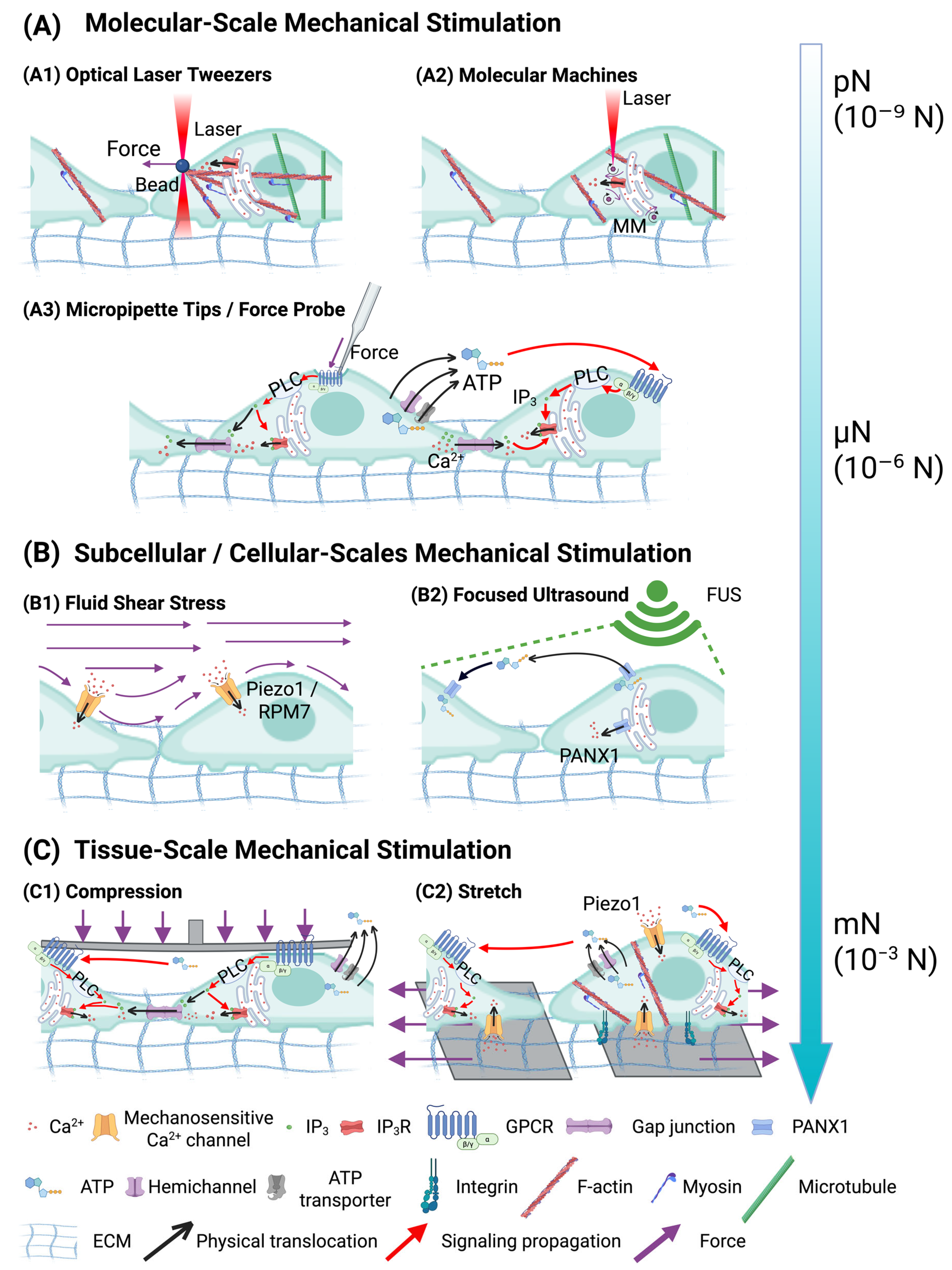
Figure 2. Mechanical stimulation methods across multiple scales regulate calcium and ATP signaling. The force range spans from piconewtons (pN) at the molecular level to millinewtons (mN) at the tissue level. (A) Molecular-scale mechanical stimulation. (A1) Optical laser tweezers apply force via beads [79]. (A2) Light-activated molecular machines (MM) stimulate intracellular calcium signaling [80]. (A3) Micropipette tips or force probes mechanically trigger mechano-volume-sensitive Cl- anion channels and PLC-IP3R-mediated calcium release and ATP propagation [74,76,81,85]. (B) Subcellular/cellular-scale mechanical stimulation. (B1) Fluid shear stress activating mechanosensitive channels like Piezo1 and TRPM7 [89]. (B2) Focused ultrasound (FUS) directly stimulates PANX1 hemichannels, facilitating intercellular calcium release [88]. (C) Tissue-scale mechanical stimulation includes (C1) compression and (C2) stretch, both enhancing calcium signaling via mechanosensitive channels and PLC-IP3R activation [91,92].
Cancers 17 01851 g002
Figure 4. The sensors for optical imaging of calcium and ATP dynamics. A schematic representation of the core components required for imaging calcium and ATP signals is shown. Calcium imaging utilizes both synthetic and genetically encoded sensors, with optical strategies including ratio-based and intensity-based approaches. ATP imaging involves aptamer-, protein-, and enzymatic-based sensors with ratio-based (F/BRET) and bioluminescence optical strategies. Different sensors use specific excitation and emission wavelength lasers, enabling multi-color imaging acquisition. The upper left diagram illustrates a representative class of calcium sensors, the GCaMP family proteins. The upper right diagram represents an ATP sensor, GPCRs, adapted from Lin, L, licensed under CC BY 4.0 [209]. Machine learning-enabled data analysis further refines the detection and interpretation of signals, providing precise dynamic imaging.
Cancers 17 01851 g004
Figure 5. (A) Depiction of the mechanism behind the ratiometric DNA nanoswitch anchored to the cell surface used for imaging extracellular ATP (A1) [230]. The fluorescence spectra illustrate the response of the DNA nanoswitch to various ATP concentrations in vitro at 37 °C, showcasing the FRET ratio of FA/FD in relation to ATP concentrations (A2). The inset presents the calibration curve for concentrations ranging from 5 to 60 μM (A3). (B) Cells are cultured directly on the SEMAPHORE matrix [234]. Secreted signaling molecules diffuse into the hydrogel, where they engage with target-specific aptamer switches, resulting in localized fluorescent signaling. The fluorescence response of the SEMAPHORE system, which contains cAMP-responsive aptamer switches of varying poly-T linker lengths, is measured over time (B1). The plot illustrate the fluorescence response of the SEMAPHORE system (B2) and the time-resolved measurement of the SEMAPHORE fluorescent response (B3). The system exhibits rapid and reversible cAMP detection in response to varying concentrations of cAMP. (C) Schematic representation of BTeam [236]. In the absence of ATP, the ε subunit remains extended and flexible, which separates YFP and NLuc, leading to low BRET efficiency (C1). Conversely, the presence of ATP induces a conformational change in the ε subunit, bringing YFP and NLuc closer together, thereby enhancing BRET efficiency (C2). It is important to note that the ε subunit can reversibly bind and release ATP without hydrolysis. The ATP-dependent luminescence spectral changes of purified BTeam are also illustrated. (D) Illustration of the principles behind GRAB-based ATP sensors, which utilize the human P2Y1 receptor as a scaffold linked to circularly permuted enhanced green fluorescence protein (cpGFP). ATP binding initiates a conformational change that enhances the fluorescence signal. (D1) The illustration of sensor composed of a conformationally sensitive circularly permutated GFP (cpGFP) and a ligand-binding protein [209]. (D2) Representative fluorescence images of HEK293T cells expressing the ATP1.0 sensor are presented, demonstrating both basal conditions and the response to the presence of 100 μM ATP [237]. (E) Overview of ATP measurement using luciferase assays [243]. Bacterial growth in liquid media is monitored through optical density assessments (E2) and luminescence measurements (eATP, utilizing thermostable luciferase and D-luciferin) (E3). Filled circles represent S. aureus, open triangles denote E. coli, and filled diamonds indicate the sterile control. The initial cell density was 100,000 CFU/mL.
Cancers 17 01851 g005
Disclaimer/Publisher’s Note: The statements, opinions and data contained in all publications are solely those of the individual author(s) and contributor(s) and not of MDPI and/or the editor(s). MDPI and/or the editor(s) disclaim responsibility for any injury to people or property resulting from any ideas, methods, instructions or products referred to in the content.