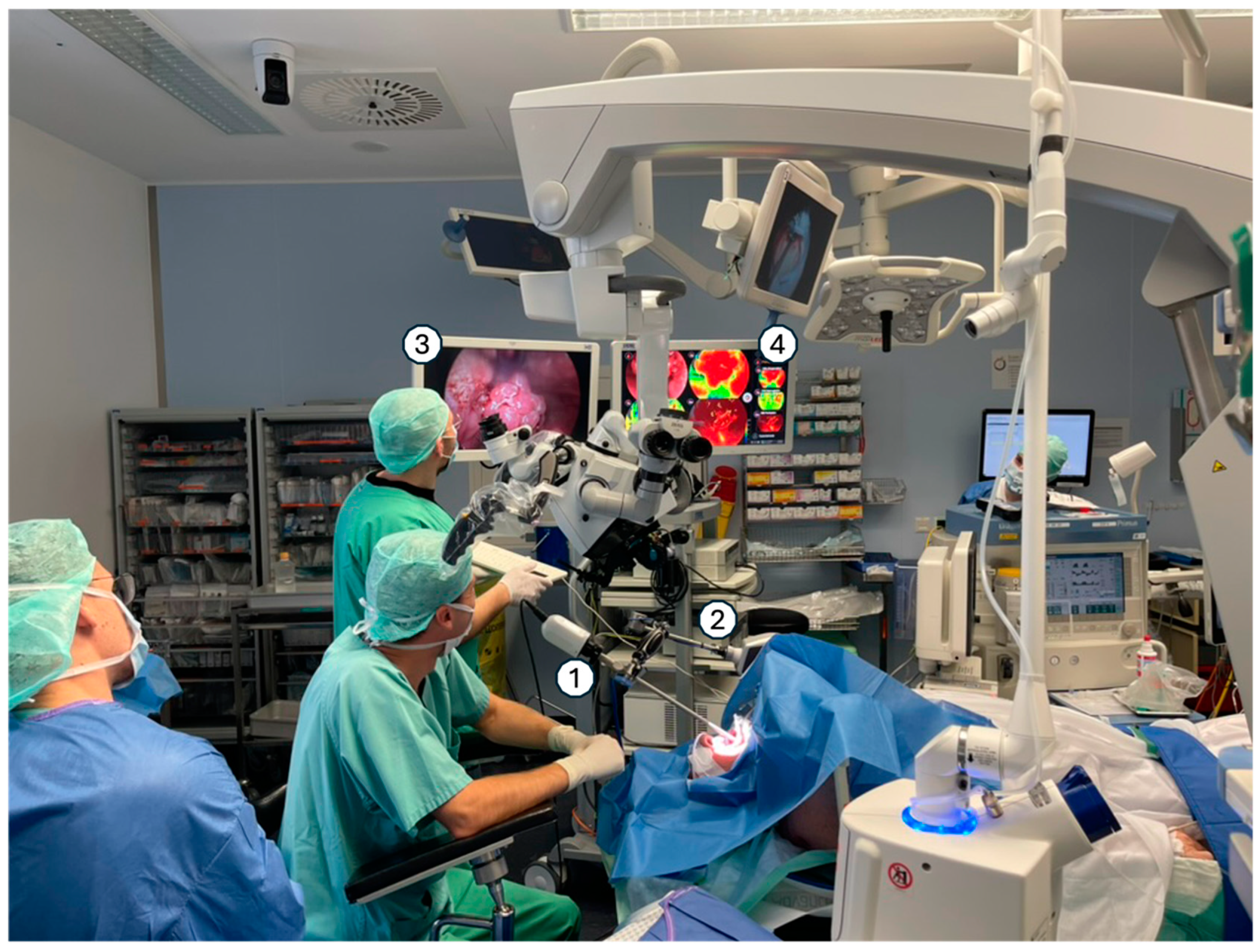
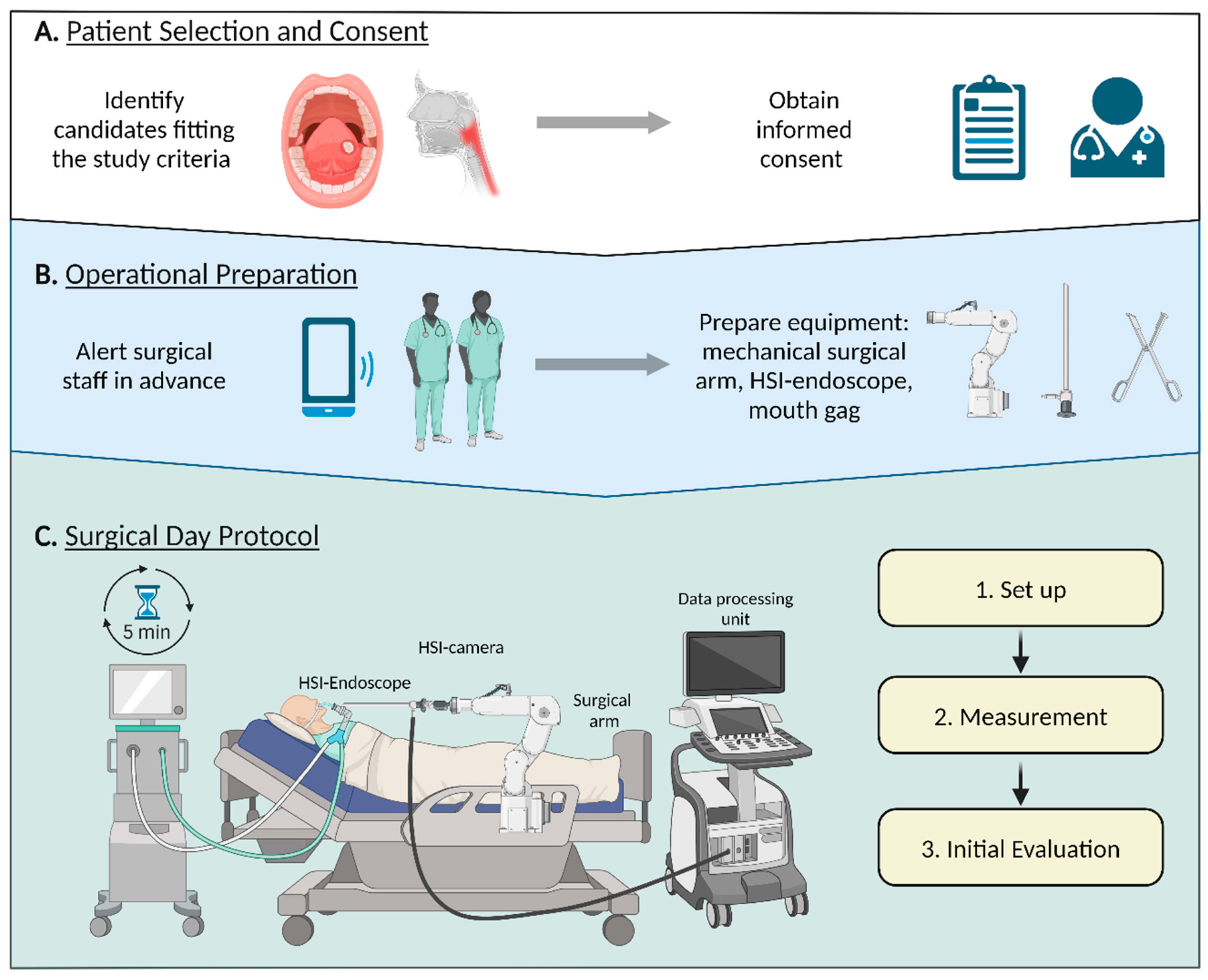
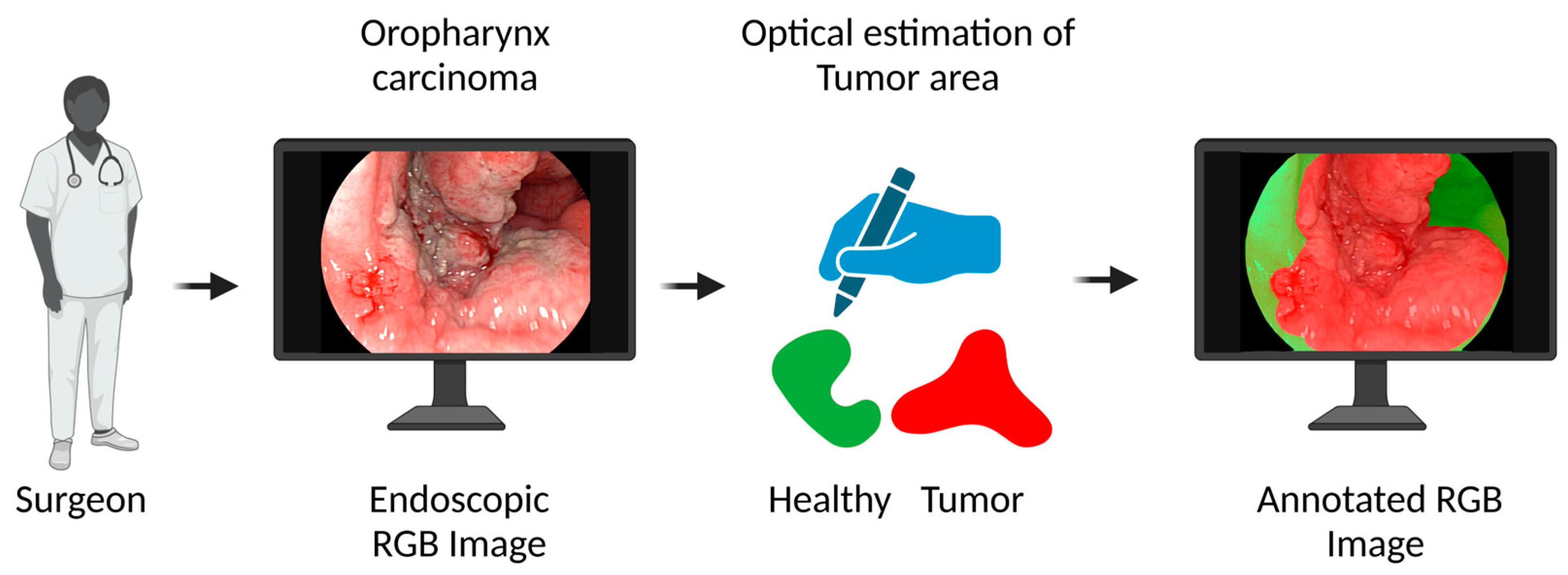
Endoscopic In Vivo Hyperspectral Imaging for Head and Neck Tumor Surgeries Using a Medically Approved CE-Certified Camera with Rapid Visualization During Surgery
by
 , , , , , , , , and
Ayman Bali
1,
Thomas Bitter
2,
Marcela Mafra
1,
Jonas Ballmaier
2,
Mussab Kouka
2 ,
,
Gerlind Schneider
2,
Anna Mühlig
1,3,
Nadja Ziller
1 ,
,
Theresa Werner
1,
Ferdinand von Eggeling
1,
Orlando Guntinas-Lichius
1,2 and
and
David Pertzborn
1,* 1
Clinical Biophotonics & MALDI Imaging, Department of Otorhinolaryngology, Jena University Hospital, 07747 Jena, Germany
2
Department of Otorhinolaryngology, Jena University Hospital, 07747 Jena, Germany
3
Comprehensive Cancer Center Central Germany, 07747 Jena, Germany
*
Author to whom correspondence should be addressed.
Cancers 2024, 16(22), 3785; https://doi.org/10.3390/cancers16223785
Submission received: 17 October 2024
/
Revised: 6 November 2024
/
Accepted: 8 November 2024
/
Published: 10 November 2024
(This article belongs to the Section Methods and Technologies Development)
Simple Summary
During head and neck cancer surgery, accurately identifying the borders of malignant tumors is crucial but remains challenging. This study investigates the use of hyperspectral imaging (HSI) to support surgeons in real-time tumor visualization during surgery. The research focuses on integrating HSI into the surgical workflow, emphasizing the optimization of this process to ensure efficiency and ease of use for medical staff. By facilitating faster and more accurate evaluation of tumor margins, this approach has the potential to enhance surgical precision, minimize incomplete tumor resections, and ultimately improve patient outcomes.
Abstract
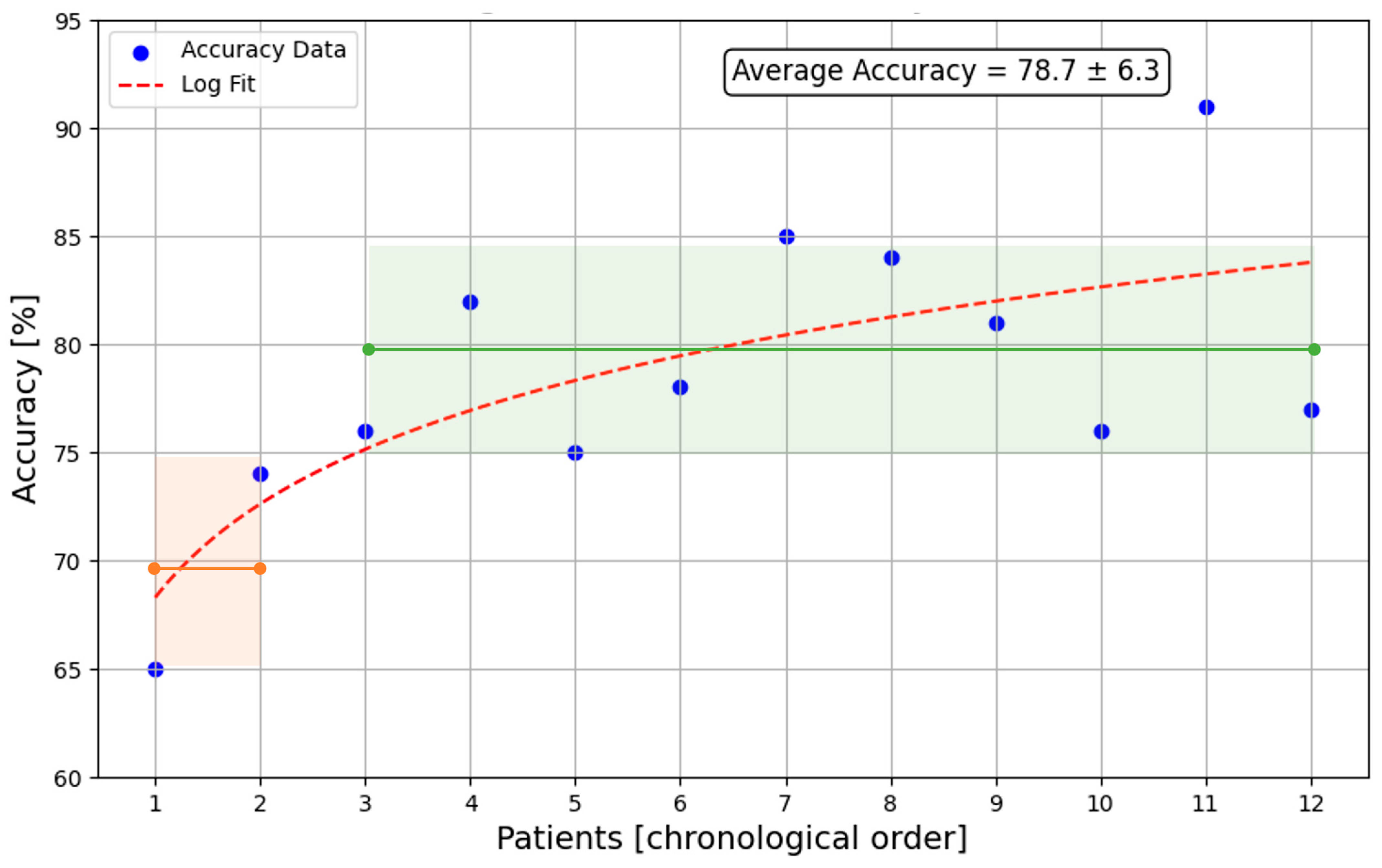
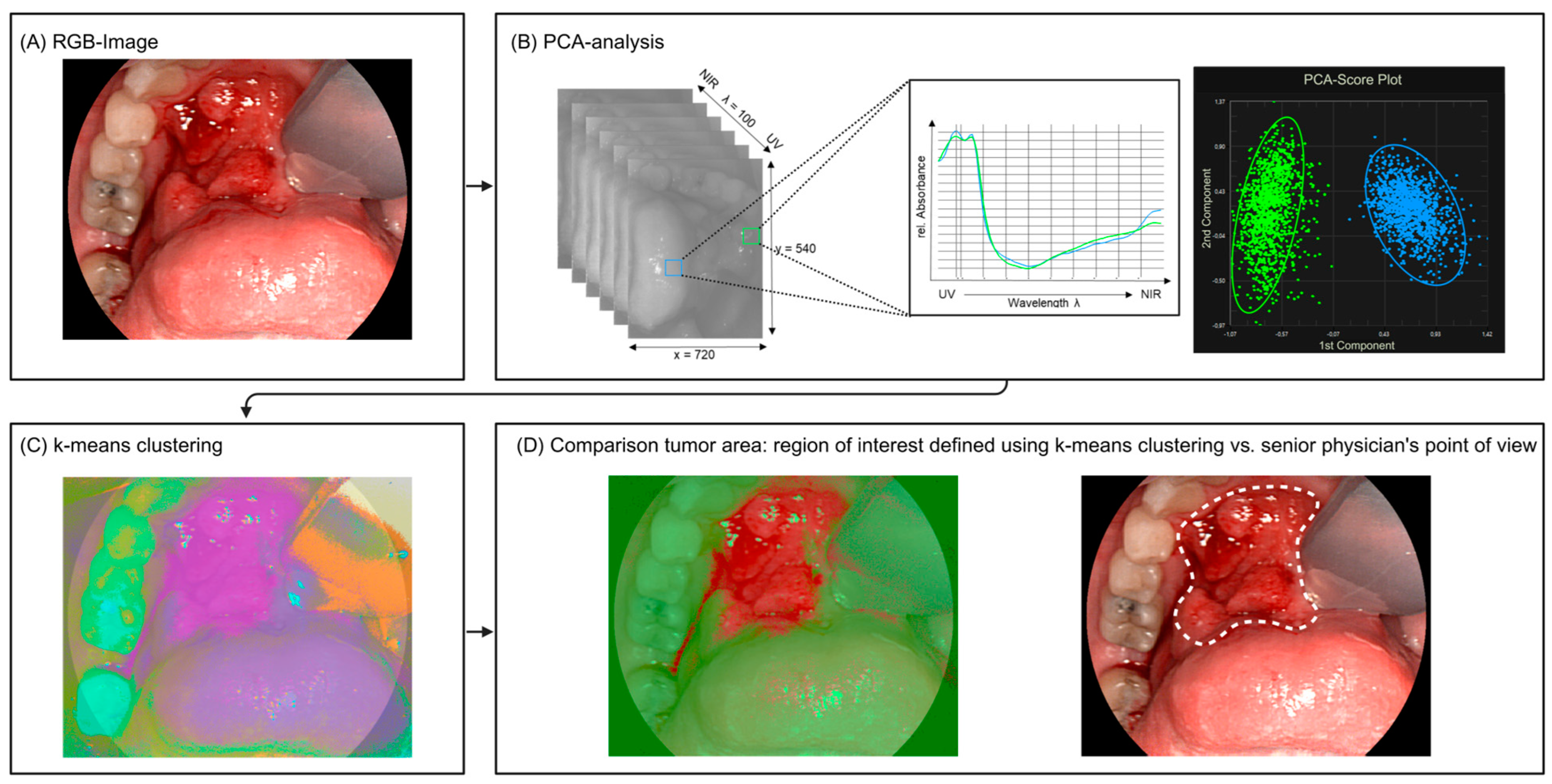
Background: In vivo visualization of malignant tumors remains the main challenge during head and neck cancer surgery. This can result in inadequate tumor margin assessment and incomplete tumor resection, adversely affecting patient outcomes. Hyperspectral imaging (HSI) is a promising approach to address this issue. However, its application in surgery has been limited by the lack of medically approved HSI devices compliant with MDR regulations, as well as challenges regarding the integration into the surgical workflow. Methods: In this feasibility study, we employed endoscopic HSI during surgery to visualize the tumor sites of 12 head and neck cancer patients. We optimized the HSI workflow to minimize time required during surgery and to reduce the adaptation period needed for surgeons to adjust to the new workflow. Additionally, we implemented data processing to enable real-time classification and visualization of HSI within the intraoperative setting. HSI evaluation was conducted using principal component analysis and k-means clustering, with this clustering validated through comparison with expert annotations. Results: Our complete HSI workflow requires two to three minutes, with each HSI measurement—including evaluation and visualization—taking less than 10 s, achieving an accuracy of 79%, sensitivity of 72%, and specificity of 84%. Medical personnel became proficient with the HSI system after two surgeries. Conclusions: This study presents an HSI workflow for in vivo tissue differentiation during head and neck cancer surgery, providing accurate and visually accessible results within minimal time. This approach enhances the in vivo evaluation of tumor margins, leading to more clear margins and, consequently, improved patient outcomes.