Review

Addressing Genetic Tumor Heterogeneity, Post-Therapy Metastatic Spread, Cancer Repopulation, and Development of Acquired Tumor Cell Resistance

by
Dennis Christoph Harrer
1,
Florian Lüke
1,2,
Tobias Pukrop
1,3,
Lina Ghibelli
4,
Albrecht Reichle
1,* and
Daniel Heudobler
1,3
1
Department of Internal Medicine III, Hematology and Oncology, University Hospital Regensburg, Franz-Josef-Strauß-Allee 11, 93053 Regensburg, Germany
2
Division of Personalized Tumor Therapy, Fraunhofer Institute for Toxicology and Experimental Medicine, 30625 Regensburg, Germany
3
Bavarian Cancer Research Center (BZKF), University Hospital Regensburg, 93053 Regensburg, Germany
4
Department of Biology, University of Rome “Tor Vergata”, 00133 Rome, Italy
*
Author to whom correspondence should be addressed.
Cancers 2024, 16(1), 180; https://doi.org/10.3390/cancers16010180
Submission received: 16 November 2023 / Revised: 8 December 2023 / Accepted: 12 December 2023 / Published: 29 December 2023
(This article belongs to the Special Issue Advances in Cancer Therapeutics)

Abstract

:

Simple Summary

The present review summarizes and interprets uniform therapy schemes for rescuing relapsed or refractory (r/r) neoplasias of quite different histologic origins. Exploiting tumor tissues’ plasticity by reprogramming hallmarks of cancer into biologic hallmarks controlling tumor regrowth with metronomic chemotherapy and simultaneous targeting of nuclear and/or cytokine receptors plus/minus targeted therapies, termed tumor tissue editing, may induce cCR or long-term tumor control, as indicated by data from clinical trials designed for the treatment of r/r neoplasias of quite different histologic origins. Tumor tissue editing may overcome unique post-therapy disease traits that arise following treatment of r/r tumor disease with standard therapy regimens using maximum tolerable doses, i.e., metastatic spread, cancer repopulation, and acquired tumor cell resistance (M-CRAC), by attenuating, or resolving M-CRAC. The introduction of M-CRAC control in future therapeutic considerations may help to overcome the multifold translational challenges of precision medicine in the large group of r/r neoplasias without driver mutations.

Abstract

The concept of post-therapy metastatic spread, cancer repopulation and acquired tumor cell resistance (M-CRAC) rationalizes tumor progression because of tumor cell heterogeneity arising from post-therapy genetic damage and subsequent tissue repair mechanisms. Therapeutic strategies designed to specifically address M-CRAC involve tissue editing approaches, such as low-dose metronomic chemotherapy and the use of transcriptional modulators with or without targeted therapies. Notably, tumor tissue editing holds the potential to treat patients, who are refractory to or relapsing (r/r) after conventional chemotherapy, which is usually based on administering a maximum tolerable dose of a cytostatic drugs. Clinical trials enrolling patients with r/r malignancies, e.g., non-small cell lung cancer, Hodgkin’s lymphoma, Langerhans cell histiocytosis and acute myelocytic leukemia, indicate that tissue editing approaches could yield tangible clinical benefit. In contrast to conventional chemotherapy or state-of-the-art precision medicine, tissue editing employs a multi-pronged approach targeting important drivers of M-CRAC across various tumor entities, thereby, simultaneously engaging tumor cell differentiation, immunomodulation, and inflammation control. In this review, we highlight the M-CRAC concept as a major factor in resistance to conventional cancer therapies and discusses tissue editing as a potential treatment.

1. Introduction

The treatment of metastatic tumors poses a crucial challenge in anti-cancer therapy. Although the therapeutic armamentarium has steadily grown and overall survival (OS) rates have significantly improved, continuous complete remissions (cCRs) are still scarce in patients with refractory or relapsing (r/r) metastatic tumor disease [1,2,3,4,5,6,7].
Conventional tumor therapy based on the application of a maximum tolerable dose to achieve maximum apoptosis induction in cancer cells is still a vital pillar of anti-cancer therapy [1,6,8]. However, disease relapse after initially successful chemotherapy is frequently observed, and relapsing cancer cells often display elevated resistance to traditional chemotherapy schedules [1].
Seeking to explain tumor progression after conventional tumor therapy, the concept of post-therapy metastatic spread, cancer repopulation, and acquired tumor cell resistance (M-CRAC) is primarily predicated on tumor cell heterogeneity generated as a result of post-chemotherapy genetic damage and subsequent tissue repair mechanisms [9,10]. Due to M-CRAC processes, tumor cells and the associated tumor microenvironment give rise to significant resistance and pave the way for r/r metastatic disease [8,11].
Contrary to conventional chemotherapy, immunotherapy or targeted therapies with small molecules, tumor tissue editing approaches therapeutically design the tumor phenotype by simultaneously engaging tumor cell differentiation, immunosurveillance, inflammation, and tumor metabolism via redirection of cancer hallmarks. These factors are important drivers of M-CRAC in a multitude of histologically different relapsed or refractory tumor entities treated with tumor tissue editing approaches [12,13,14,15,16,17,18,19,20,21,22,23,24,25,26,27,28,29,30,31].
Tissue editing approaches encompass low-dose metronomic chemotherapy, the use of transcriptional modulators, and targeted therapies. Clinically, treatment regimens based on tissue editing can achieve continuous complete remissions in patients with a variety of different r/r malignancies, such as Hodgkin’s lymphoma, Langerhans cell histiocytosis, renal clear cell carcinoma, cholangiocarcinoma, and angiosarcoma. This definitively shows that M-CRAC induced by preceding systemic therapies may be overcome, even by induction of cCR in single patients with histologically different r/r tumor disease [13,16,24,25,26].
In the present review, we introduce the M-CRAC concept as a key factor contributing to clinical resistance to standard systemic tumor therapies. In addition, we highlight tumor tissue editing strategies as specific interventions to counteract M-CRAC and maintain long-lasting disease control.

2. M-CRAC as a Target for Tumor Tissue Editing in Refractory or Relapsed Neoplasias

The novel treatment paradigm ‘tumor tissue editing’ adopts the use of tissue editing technologies for correcting genetic or epigenetic abnormalities in tumor tissues [32,33,34,35,36]. Tumor tissue editing methodologies are now focused on achieving phenotypic, therapeutically relevant editing of tumors [37].
Tumor tissue editing is defined as the therapy-guided targeted evolution of tumor tissues to establish biologic hallmarks that facilitate tumor control or initiate complete remission in relapsed or refractory tumor disease [38]. Appropriate editing schedules combine bioactive therapeutic principles to readjust aberrant tissue homeostasis at primary and metastatic tumor sites by using differential regulatory active therapeutic techniques [39,40]. Bioactive drugs must not demonstrate any monoactivity in the respective tumor disease [40]. The combined regulatory activity profile concertedly redirecting hallmarks of cancer is of pivotal therapeutic interest (Table 1).
The operational procedure facilitating tumor tissue editing, i.e., anakoinosis, promotes therapy-initiated reprogramming and cell recruitment in tumor tissues, which finally contributes to the tumors’ plasticity in therapeutic intention [39,40]. Tumor-associated supervising communication lines that cumulatively constitute tumor-type specific communication protocols among different tumor cell compartments are supposed to be respective therapeutic targets of editing techniques [40,48].
The editing procedure relies on tumor-specific communication lines to establish phenotypic plasticity. The extent and quality of tumor systems’ plasticity reflect evolutionary system states and the developmental and medical histories of tumor diseases [12,44,49,50]. Tumor tissue plasticity is constituted by timely and spatially developing tumor cell autonomous and non-autonomous processes and cannot be described by genetic/molecular genetic aberrations alone, particularly if driver mutations are absent, as in most tumors [51,52,53].
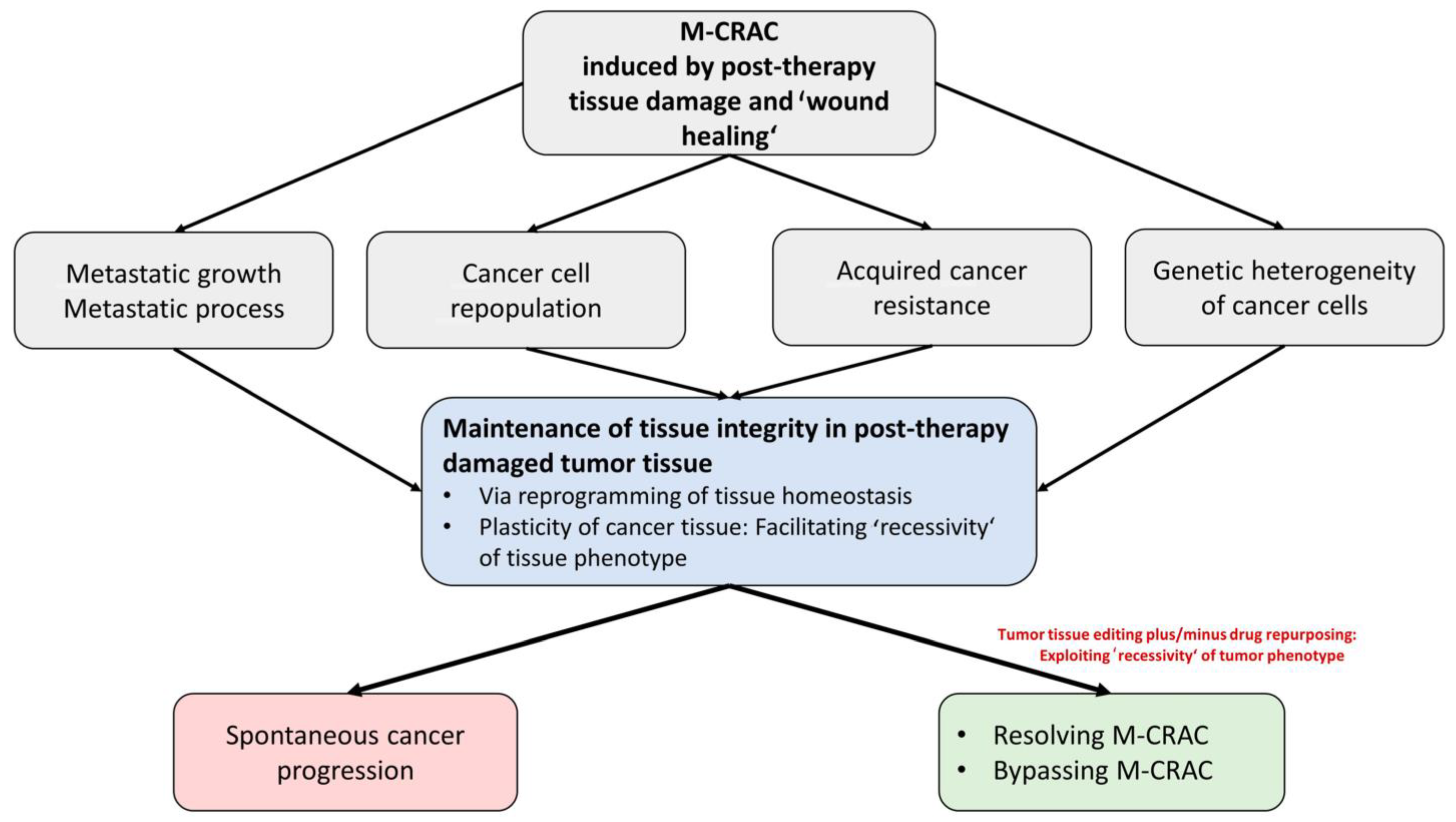
Tumor tissue editing approaches turned out to efficaciously control or resolve M-CRAC by redirecting cancer associated hallmarks into biologic hallmarks, attenuating tumor growth, and inducing alternative patterns of tumor cell death in r/r tumor disease. As shown, clinical trials using editing approaches may induce long-term tumor control, objective response or even cCR [38] (Figure 1, Table 1).
The novel treatment strategy is highly integrative and provides, beyond its biomodulatory activity profile, opportunities for drug repurposing, e.g., by providing edited non-oncogene addiction. Moreover, editing techniques may establish transdifferentiation and differentiation of tumor cells for M-CRAC control and tumor cell death induction [12,37,49].
Important aspects of tissue editing approaches include the possibility of exploiting the synergistic potency and efficacy of biomodulatorily active drug combinations [54]. The combinatorial, metronomic administration of drugs targeting the network infrastructure of tumor tissues proved to be efficacious in various, histologically quite different tumors. Interestingly, edited tumor tissues provide novel activity profiles for otherwise less efficacious approved targeted therapies. As clinically shown, the use of repurposed targeted therapies, e.g., mTOR inhibitors, may contribute to long-term tumor control or cCR in r/r tumor disease, e.g., Hodgkin’s lymphoma or uveal melanoma [22,37].
Tumor tissue editing approaches for rescuing r/r neoplasias via M-CRAC control and tumor cell death induction may include different metronomically scheduled cytotoxic drugs (trofosfamide, treosulfan, capecitabine). Low-dose metronomic chemotherapy serves as a prerequisite for the activity of otherwise inefficacious transcriptional modulators [40]. Tissue editing approaches use transcriptional modulators, such as nuclear receptor agonists or cytokines, as indicated in Table 1. Pioglitazone itself is a dual-receptor agonist for the peroxisome-proliferator-activated receptor PPARα and γ (PPARα/γ). Therefore, all the mentioned tumor tissue editing trials used dual or triple transcriptional regulation in addition to low-dose metronomic chemotherapy (Table 1) [55].
With tissue editing techniques, quite different neoplasias in the r/r stage could be either stabilized for the long term or controlled by objective response, even by cCR [13,16,19,22,25,26]. Further, in castration-resistant prostate cancer (CRPC) or r/r multiple myeloma (MM), disease control was stable even after discontinuation of the tumor editing approach. In CRPC, hormone sensitivity was reestablished in single cases [19]. Stable M-CRAC control beyond discontinuation of tumor tissue editing in CRPC and r/r multiple myeloma reveals reprogramming of the tumor tissue, i.e., simultaneously involving the stroma and tumor cells [14,19].
Thus, tumor tissue editing techniques facilitate the achievement of quite different endpoints that finally contribute to M-CRAC control. Objective response is not a prerequisite for M-CRAC control [14].

3. Tissue Editing Methods Redirect Cancer-Related Hallmarks into Novel Biologic Hallmarks Facilitating Tumor Response

How tissue editing approaches work at the tumor tissue communication level cannot yet be pinned down to single pathways [40].
Studies on non-PML-AML provide the insight that only the triple combination of azacitidine, ATRA, and pioglitazone may induce cytologically proven differentiation as a triple combination. These results, however, underline that the synergism of each drug with one another is a prerequisite for therapeutically redirecting leukemia plasticity [45,50].
Also, clinical data on r/r RCCC, showing cCR induction with the combination pioglitazone plus interferon-α on the background of ‘very’-low-dose capecitabine, are not well experimentally supported [25]. However, reprogramming the oncogenic stress response is the prerequisite for dual control of tumor-associated inflammation with pioglitazone and interferon-α. C-reactive protein (CRP) is a strong marker indicating tumor response [17,43]. In RCCC, tumor cells may directly produce CRP aside from liver-mediated secretion [56]. Different tissue editing approaches, as indicated in Table 1, result in diversified reprogramming results of tumor tissues.
Also, the recently published survival benefit of patients with relapsed head and neck cancer receiving metronomic methotrexate, celecoxib, and erlotinib combined with low-dose nivolumab in comparison to those receiving the same metronomic schedule without nivolumab cannot be based on pathway analyses providing distinct mechanisms of action promoting OS benefit with quadruple therapy [57]. Enhancement of the immune response might be the main emphasis of the nivolumab arm.
In general, the studies on tissue editing demonstrate the efficacy of tumor editing techniques in selectively redirecting cancer hallmarks for tumor control.
Within tissue editing approaches, pleiotropic tissue activity profiles of single biomodulatory drugs lead in concerted tissue communication guiding activity profiles that are focused and successfully reprogram the oncogenic stress response as a prerequisite for redirecting single cancer hallmarks into biologic hallmarks attenuating tumor growth [40]. This way, tumor tissue editing approaches therapeutically exploit tumor plasticity (Figure 1). The reprogramming process is termed anakoinosis in anticipation of future descriptions of communicative lines describing the molecular background of the respective pharmacologic interactions on the tissue level in more detail [40]. The activity profiles of metronomic chemotherapy, pioglitazone, and other transcriptional modulators in reviews did not predict the outcome provided by tissue editing protocols that have been successfully applied in many histologically highly different neoplasias [55,58,59].

4. Tissue Editing Approaches: Impact on the Genomic Evolution of Tumors

A remarkable clinical result is that tumor tissue editing approaches may induce CR or cCR in r/r neoplasias, even within small study populations (Table 1).
Recent studies found molecular genetic markers or histologies associated with resistance to metronomic chemotherapy in head and neck cancer or triple-negative breast cancer [60,61]. These data might not be directly transferable to tissue editing approaches. Metronomic low-dose chemotherapy facilitates the use of the clinically decisive step, concerted transcriptional modulation. As shown in non-PML AML, even complex aberrant karyotypes may respond with CR to editing methods [45].
Hodgkin’s lymphoma in the relapsed or refractory stage demonstrates that an adequate tissue editing approach overcomes resistance mechanisms of quite different origin, namely those induced by radiotherapy, immunotherapy, and chemotherapy, as indicated by consecutive CR and cCR induction [13]. Thus, editing-specific genomic evolution of resistance plays no key role when using an adequate editing methodology.
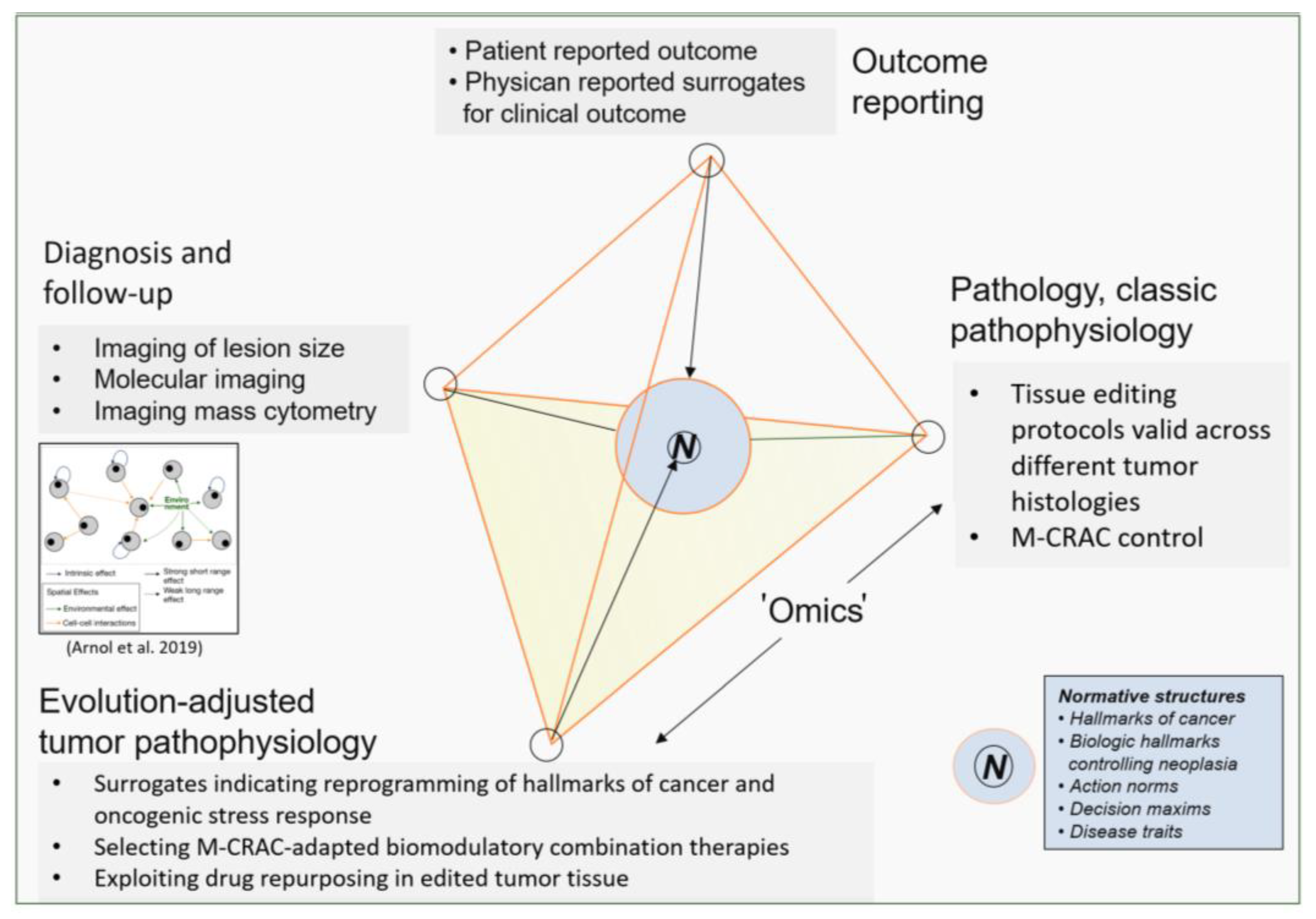
Nevertheless, developing resistance may limit response to tissue editing approaches in r/r tumor disease. Up to now, there was no urgent need to perform genetic analyses at the time of tumor progression, because there are fewer concerns about the rapid evolution of resistance to tissue editing approaches. Currently, the main therapeutic focus is the implementation of the most suitable editing approach, at best in a personalized manner. Further studies must establish missing biomarkers for the personalized selection of editing techniques and corresponding follow-up parameters. The tissue editing methodology is at its beginning (Figure 2).

5. M-CRAC as Driver of Disease Relapse and Chemoresistance: The Therapeutic View

Definition of M-CRAC: M-CRAC-associated disease traits, i.e., metastatic spread, cancer repopulation, and acquired tumor cell resistance as well as genetic and/or molecular-genetic tumor cell heterogeneity, may be clinically separated as a unique post-therapeutic response pattern to systemic tumor therapies based on the clinical finding that M-CRAC may be successfully resolved or attenuated by the introduction of tumor tissue editing techniques designed for the treatment of r/r neoplasias of different histologic origin (Table 1) [12,13,14,15,16,17,18,19,20,21,22,23,24,25,26,27,28]. In particular, the differentially developing resistance patterns in tumors, originating either from resistant clones during initial tumor growth or from the multifold resistance patterns induced by preceding systemic tumor therapies, describe the therapeutic challenges for establishing M-CRAC control and indicate the necessity for novel therapeutic solutions. The multifaceted M-CRAC disease traits have been intensively studied, e.g., in r/rMM [62,63,64,65].
The M-CRAC concept summarizes disease traits promoting tumor progression or relapse following any kind of prior therapy, such as chemotherapy, immunotherapy, and targeted therapies with small molecules. M-CARC is often associated with mixed or organ-specific response patterns to systemic tumor therapy [9,10,66,67].
The availability of specific tumor-type-adapted tissue editing techniques for rescuing relapsed or refractory tumor disease of quite different histologic origins support the consistency of the M-CRAC concept (Table 1).
Biologic processes promoting M-CRAC: Intrinsic, i.e., systemic, and tumor-specific factors guide the biology of metastatic tumor disease. Tumor-cell-specific parameters are commonly considered targets for systemic tumor therapy to attain maximum apoptosis induction [68]. Additionally, extrinsic, primarily non-genetic factors, termed the ‘exposome’ of the host, impact tumor biology and long-term outcome following systemic tumor therapy and play a pivotal role in the development of M-CRAC [8,69,70]. All preceding chemotherapies, immunotherapies, targeted therapies with small molecules, and radiation therapies contribute to the ‘exposome’ and are directly involved in the development of M-CRAC. In aggregate, M-CRAC describes the oncogenic potential of prior tumor therapies promoting pathophysiological disease traits associated with tumor progression or relapse [8,9].
Potential mechanisms underlying M-CRAC: Damage response to systemic tumor therapy reshapes cancer tissue by a multitude of coordinated, timely, spatially, and dynamically developing cellular and tissue events, summarized in M-CRAC [9,71]. The enormous range of post-therapy tumor system states, arising on the basis of the tumor tissue’s plasticity, facilitates the maintenance of the tumor tissue’s integrity via wound healing mechanisms [72]. Multifold concurrent events linked to M-CRAC-related disease traits are closely interwoven and underline the oncogenic role of tumor cell death during post-therapy tissue repair [72,73,74,75,76,77].
Apoptotic tumor cells, hypoxia, and inflammation may promote compensatory tumor cell proliferation, for instance through the caspase-3-cytosolic phospholipase A(2) alpha (cPLA-2)-COX-2-PGE-2-STAT3 Phoenix rising pathway. [10,68,78,79]. Maximum-tolerated doses (MTD) of chemotherapy may promote M-CRAC via the epithelial–mesenchymal transition (EMT) orchestrated by hypoxia-inducible factors (HIF)-1α and (HIF)-2α [79,80,81,82,83,84,85].
For future research, experimental studies are vital to investigate how the post-therapy oncogenic potential of pulsed therapies, summarized in M-CRAC-related disease traits, might be accessible with differential tissue editing approaches [10,11,86,87,88,89,90].
The therapeutic accessibility of M-CRAC via tumor tissue editing techniques is surprising, as tumor progression following systemic tumor therapy is spatially and temporally heterogeneous. Tumor cells are epigenetically remodeled and may successively become genetically heterogeneous. Moreover, stromal cells may gain genetic aberrations, cancer stem cells become functionally altered, non-mutated tumor suppressors are down-regulated, epithelial–mesenchymal transition is promoted, and autophagy becomes dysregulated. Finally, context-dependent roles of TGFβ occur, tumor and stroma cell metabolism are disturbed, immunosurveillance is decreased, pro-inflammatory processes in hypoxia are highly activated, and angiogenesis is altered [91,92,93,94,95,96,97,98,99,100,101,102,103,104,105,106,107,108].
As a result of the multi-leveled repair processes, the phenotypic outcome of M-CRAC seems to be poorly predictable. The cellular tumor tissue compartments exploit the whole repertoire of cellular plasticity to guarantee tissue integrity at the expense of M-CRAC development [109,110,111,112,113,114]. The evolution within cancer tissues to establish novel tissue homeostasis under hypoxic, pro-inflammatory, and immunosuppressive conditions and the presence of damaged cells involves all cellular compartments [3,109,115,116,117]. In the end, even secondary malignancies are possible as a final ‘reconstitution’ process.
The induction of cCR in r/r neoplasias indicates resolution of M-CRAC. Differentiation induction may completely bypass M-CRAC, as shown by hematologic or molecular-genetic complete remission in relapsed or refractory acute myelocytic leukemia (AML), even in those with adverse risk according to European LeukemiaNet (ELN) [12,45,46,47].
This means the clinically circumscriptive phenotype defined as M-CRAC is differentially accessible with tumor tissue editing techniques using biomodulatory techniques, i.e., anakoinosis, for successfully targeting the evolutionary preserved plasticity of tumor tissues in therapeutic intention (Figure 1). Different tumor histologies share targets available for M-CRAC control, even if in quite different contexts, as exemplified for pioglitazone (Table 1).
An important mechanism of action for meeting the multifold therapeutic challenges provoked by M-CRAC might be the therapeutic alteration and redirection of the tumor-specific, survival-related inevitable stress response patterns via metronomic low-dose chemotherapy and, additionally, via targeted transcriptional modulation. The feasibility of establishing edited non-oncogene addiction to mTOR in r/r HL, followed by the achievement of cCR, supports the explanation that Hodgkin tissue cannot manage any more stress responses, irrespective of the kind of pre-treatment [13].
The available data on tissue editing trials cannot yet specify whether differential editing approaches are necessary for M-CRAC control to meet specific tissue alterations given by preceding systemic tumor therapies. However, different prior systemic targeted therapies, chemotherapies, and immunotherapies within one histologic tumor type were rescued with unique tissue editing techniques, as exemplarily shown in r/r acute myelocytic leukemia (AML) and r/r Hodgkin lymphoma (HL) [12,13,45,46,47].
Explaining M-CRAC by tumor tissue editing techniques: Tissue editing techniques therapeutically target the network infrastructure of tumor tissues and the tumor’s phenotypic plasticity, which contrasts with frequently used approaches separately targeting tumor and/or stroma cells (Figure 1). Therapeutical reprogramming of the tumor infrastructure promotes new ways of tumor cell death. Moreover, differentiation induction and post-therapy wound healing processes may be modulated to resolve or bypass M-CRAC [25,50,118,119,120]. The induction of cCR in r/r metastatic neoplasias shows that tumor cell heterogeneity in metastatic r/r disease may be overcome by functionally editing the whole repertoire of heterogeneous tumor phenotypes and genetically heterogeneous tumor cells [13,16,24,25,26]. Tissue editing techniques demonstrate, even in small study populations and in histologically quite heterogeneous neoplasias successful control of r/r metastatic disease (Table 1) [12,13,14,15,16,17,18,19,20,21,22,23,24,25,26,27,28].
Single pathways are not attributable as cause of complex post-therapy disease traits mediated by M-CRAC. Tissue editing techniques may address M-CRAC, which emerged as regulatory and differentially accessible phenomenon. M-CRAC control aims at reprogramming communication networks that are supported by tumor and stroma cells.

6. Specific Therapeutic Access to M-CRAC with Tumor Tissue Editing Approaches

Therapeutic challenges in treating M-CRAC with chemotherapy, immunotherapy, or targeted therapies with small molecules: Enhancing apoptosis induction by the application of maximum tolerable doses is the main aim of most systemic tumor therapies. Irrespective of the drug combination, the drug-related ‘exposome’ leaves major therapeutic challenges, as reflected by a high frequency of relapsed or refractory diseases. Systemic tumor therapy rarely achieves long-term tumor control or cCR in most r/r metastatic neoplasias despite the steadily increasing and diversifying therapeutic repertoire. Reasons are tissue repair processes following apoptosis, hypoxia, cell damage, immunosuppression, metabolic alterations and inflammation [1,2,4,5,6,7,8,121].
M-CRAC is a major obstacle counteracting the initially induced tumor response and therefore affects the overall outcome. The approved pulsed chemotherapy regimens, relying on the application of maximum tolerable doses, primarily aim at tumor cell destruction, resulting in the stimulation of tissue repair and potential M-CRAC development [1].
Therefore, induction therapies designed to induce maximum rates of tumor cell death should be complemented with therapies targeting and guiding the tumor tissues’ post-therapy functional phenotypes to simultaneously address M-CRAC [83].
Differential techniques of tumor tissue editing uncover M-CRAC as a unique but differentially accessible pathophysiological phenomenon. Three therapeutic elements systematically addressed M-CRA: Low-dose metronomic chemotherapy, epigenetic modifiers (azacitidine), and transcriptional modulation via nuclear receptors (PPARα/γ glucocorticoid receptor, RXR, or cytokine receptors). Moreover, the combination with classic targeted therapies could be beneficial, as exemplarily shown for mTOR inhibitors by the edited non-oncogene addiction in r/r HL for mTOR (Figure 1, Table 1) [13,45].
Resolution of M-CRAC has been achieved as indicated by cCR in r/r cholangiocellular carcinoma, renal clear cell carcinoma, angiosarcoma, multisystem Langerhans cell histiocytosis, Hodgkin’s lymphoma, or by CR in non-promyelocytic acute myelocytic leukemia (non-PML-AML) [12,13,16,24,25,45,122]. Tissue editing induced long-term tumor control in all studied r/r neoplasias, in high grade gliomas stable disease as best response, and in all the other neoplasias at least partial remissions (Table 1).
Tumor plasticity is the starting point and final target for tumor tissue editing methods [9,37]. Differential therapeutic emphasis on distinctively developed tumor-associated hallmarks may help to select specific tumor tissue editing techniques (Figure 1).
All stroma components, i.e., mesenchymal cells, cancer-associated fibroblasts (CAFs), adipocytes, endothelial cells and hematologic cells, such as myeloid-derived suppressive cells, tumor-associated macrophages (TAMs) and T-cells, are involved in M-CRAC control, but to different extents depending on the focus of the chosen biomodulatory drug combination [55,59]. Enhancement of immunosurveillance seems to frequently play a crucial role in successful editing techniques, besides strong inflammation control, differentiation induction and metabolic reprogramming (Table 1, Figure 1) [23,42,57].
Metronomic low-dose chemotherapy as a prerequisite for transcriptional editing During long-term administration of metronomic low-dose chemotherapy in schedules for tumor tissue editing, scheduled dose reductions of cytotoxic drugs were allowed, even up to a quarter of the initially recommended starting dose [17]. Thus, the initially intended monoactivity of metronomic ‘very’ low-dose chemotherapy to induce at least PR or even cCR is out of range.
The study on r/r renal clear cell carcinoma (RCCC) impressively shows the effect of ‘very’ low doses of capecitabine. The addition of low-dose interferon-α to pioglitazone and cyclooxygenase-2 (COX-2) inhibition may induce cCR on the background of very low-doses of capecitabine, namely, 1 g absolute BID × 2 weeks, followed by a 1-week rest period, and scheduled dose reductions of capecitabine to 0.5 g or 0.25 g absolute BID × 2 weeks, followed by a 1-week rest period. In contrast to the approved standard dose of capecitabine, 1250 mg/m2 BID within the same schedule, the starting dose of capecitabine in the phase II editing trials was 40% in comparison to the standard dose capecitabine, and 20% or 10% if scheduled dose reduction was performed in phase II editing trials for RCCC [17,43].
Metronomic, very low-dose chemotherapy promotes a continuous pattern of stress responses [55,123,124,125]. The present clinical data reveal that metronomic chemotherapy at ‘very’ low-doses limits tumor tissue plasticity by stress response, probably decreasing phenotypic heterogeneity of tumor cell niches as tissue stress generally induces a tighter phenotype [123,126,127,128,129,130,131]. Thus, metronomic chemotherapy might induce phenotypic integration of inflammation control or differentiation by editing techniques and, consecutively, may serve as an enhancer of pro-anakoinotic effects mediated by added transcriptional modulators [55,123]. Exposure of tumor tissues to stress in addition to the tumor intrinsic management of oncogenic stress for preserving tumor integrity and promotion, or inhibition of salvage pathways managing the stress response, may induce tumor cell death pathways. Whereas non-tumor cells compensate for therapeutically induced perturbations, as indicated by the modest toxicity profile of editing trials (Table 1) [132,133]. Tumor tissue editing techniques finally inhibit the relief of stress in tumor tissue, which cancer cells are relying on for survival. This way, tumor tissue editing approaches seem to resolve or attenuate M-CRAC.
Only combined transcriptional regulation with nuclear receptor agonists or cytokines (interferon-α) led either to decisive inflammation control and cCR, e.g., in r/r renal clear cell carcinoma, r/r LCH and r/r HL, or to differentiation induction in AML, or to the establishment of non-oncogene addiction in r/rHL [12,17,25,45].
The clinical results demonstrate that combined transcriptional modulation on-topic unlocks the phenotypic plasticity of tumor tissues for M-CRAC control (Figure 1 and Figure 2). Unlocking is profoundly associated with the reprogramming of cancer hallmarks in edited tumor tissues, on the background of the reprogrammed tumor system’s stress response, which facilitates a tighter phenotype at the phenotypically and genetically heterogeneous tumor sites. The tight phenotype might be an explanation for why tumor tissue editing may overcome genetic heterogeneity at different metastatic tumor sites and associated resistance.
The combination of both treatment components, metronomic low-dose chemotherapy plus transcriptional modulation, is profoundly associated with the reprogramming of cancer hallmarks to biologic hallmarks controlling M-CRAC in respective edited tumor tissues (Figure 2).
The prerequisite of exceptionally low doses of metronomic chemotherapy for clinically efficacious transcriptional modulation reveals a novel aspect of the mechanisms of action of metronomic chemotherapy. Both treatment components, low-dose metronomic chemotherapy and transcriptional modulation, provide poor or no monoactivity in r/r tumors [40,55].
Concerted reprogramming of cancer hallmarks with metronomic (very) low-dose chemotherapy and dual/triple transcriptional modulation induces a series of clinically important phenomena beyond M-CRAC control. These are tumor cell death, edition of non-oncogene addiction, maintenance of disease control beyond discontinuation of editing therapy, or re-establishment of hormone sensitivity in CRPC [14,19,37].

7. Repurposing Chemotherapy: Metronomic Low-Dose Chemotherapy

Metronomic chemotherapy is clinically efficacious despite frequently applied scheduled dose reductions (Table 1). Clinical efficacy of ‘very’ low-doses within tissue editing protocols demonstrates the meaning of ‘low-dose’ metronomic chemotherapy. Thus, the clinical biomodulatory effect of metronomic low-dose chemotherapy is not necessarily based on the cumulative achievement of maximum tolerable doses with equally split daily doses applied during a 3 to 4-week treatment cycle.
A main result of the presented series of tissue editing trials is that metronomic ‘very’ low-dose chemotherapy is sufficient and essential for reprogramming of the oncogenic stress response and for transcriptional tumor editing. Metronomic low-dose chemotherapy may concretely contribute to stimulating the immune response, particularly in concert with additional immunomodulators (immune checkpoint inhibitors, pioglitazone). Metronomic low-dose chemotherapy may inhibit tumor-associated inflammation, e.g., in combination with pioglitazone, interferon-α, and angiogenesis, and/or may promote tumor differentiation, e.g., in combination with all-trans retinoic acid. Finally, metronomic low-dose chemotherapy facilitates edited non-oncogene addiction, namely the successful use of repurposed targeted therapies, e.g., mTOR inhibitors, associated with resolution of M-CRAC in r/r Hodgkin’s lymphoma and consecutive cCR [13,17,45,57].
Low-dose metronomic chemotherapy, as used in tissue editing approaches, represents a completely repurposed way of applying chemotherapy. Alkylating agents or methotrexate have been frequently applied with respect to their immunomodulatory activity profile (Table 1) [57].
Reprogramming the tumor-associated stress response, the novel activity profile of metronomic chemotherapy, determines the therapeutic difference in comparison to the well-known mechanisms of action observed following pulsed chemotherapy. Prolonged therapy-free intervals for recovering toxicity, the risk of infections, and M-CRAC development are well-known problems associated with conventional chemotherapy [134]. Focusing on tumor cell death with maximum tolerable doses always bears the risk of post-therapy M-CRAC induction due to evolving molecular-genetic and genetic tumor heterogeneity and drug resistance.
Tissue editing approaches integrating low-dose metronomic chemotherapy induce, as shown, not only replication arrest or dormancy, associated with stable disease or stable objective response, even beyond discontinuation of tissue editing, but also CR and cCR in r/r neoplasias (Table 1) [19,25].
Thus, the overall impact of low-dose metronomic chemotherapy is not simply to save treatment costs in countries that cannot afford expensive targeted therapies or to integrate a mostly non-approved therapy into ‘Common Sense Oncology’ [135,136]. Rather, metronomic chemotherapy may address the weaknesses of pulsed chemotherapies, targeted therapies, oncogene-directed, or non-oncogene ‘addicted’ therapies by targeting the tumor’s plasticity via phenotypic editing of undruggable genetic defects in tumor cells, for example, in AML patients with complex chromosomal aberrations, including defects in gatekeeper genes such as TP53 mutations and PTEN downregulation, which may be counteracted with pioglitazone [13,45,100]. Gatekeeper defective genes, e.g., aberrant expression of PTEN could probably be normalized, as clinically suggested in r/r Hodgkin’s lymphoma, by edited non-oncogene addiction to mTOR [13,137].

8. Examples of M-CRAC Control and Tissue Editing in the Clinical Setting

In a large series of studies, tissue editing approaches were applied for the treatment of relapsed or refractory tumor diseases of quite different histologic origins, e.g., carcinoma, sarcoma, and hematologic neoplasias (Table 1) [12,13,14,15,16,17,18,19,20,21,22,23,24,25,26,27,28]. A recent randomized trial compared in r/r metastatic non-small cell lung cancer (NSCLC) (second-to-fifth-line treatments) such as two biomodulatory therapy approaches, nivolumab at the approved dose level versus metronomic low-dose chemotherapy, pioglitazone, COX-2 inhibitors, and clarithromycin. The experimental arm failed to show superiority in progression-free survival (PFS). Nevertheless, the fact that overall survival (OS) was similar between the treatment arms gives rise to the hypothesis that the well-tolerable biomodulatory therapy may edit and prime tumor tissues for efficacious, consecutive immune checkpoint inhibitor (ICPi) therapy in the experimental arm [23,138,139].
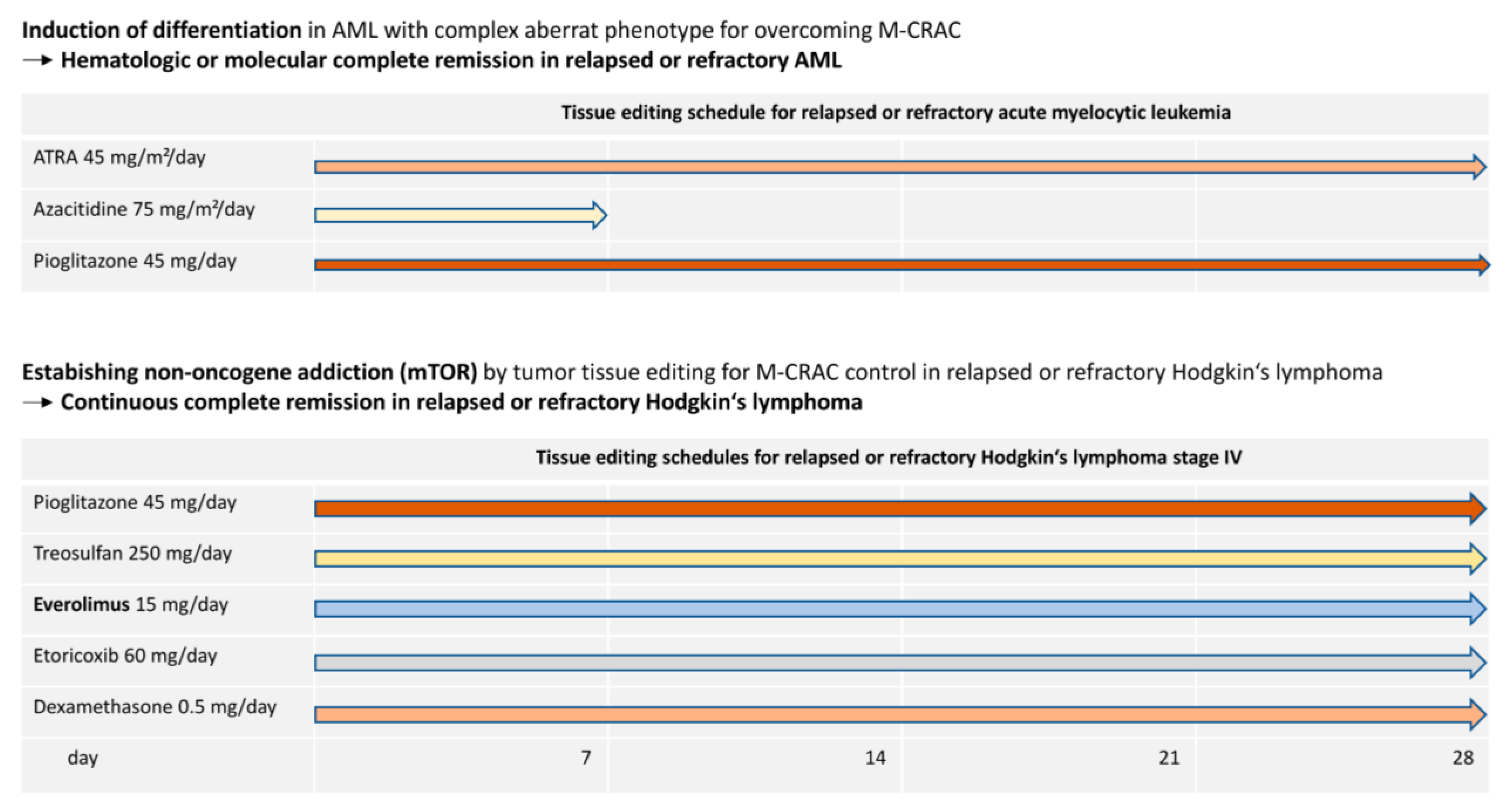
In r/r Hodgkin’s lymphoma, two cohorts of patients are retrospectively summarized (Figure 3) [13,27]. Patients had either all received, at that time, approved systemic treatments or were ineligible for standard treatment, including immune checkpoint inhibitors. One patient received a previous allogenic hematopoietic stem-cell transplant (alloHSCT). The rescue therapy consisted of an all-oral tissue editing approach including metronomic, daily, low-dose treosulfan, pioglitazone, etoricoxib, and dexamethasone, plus, in repurposed edited tissue, everolimus 15 mg p.o. daily to achieve serum trough levels of 15 ng/mL (MEPED schedule for r/r HL). Everolimus has some but poor monoactivity in r/r Hodgkin’s disease [140]. All patients achieved continuous, complete remission with MEPED. After entering CR, three transplant-eligible patients underwent consolidating alloHSCT and achieved consecutive cCR.
As shown, in Hodgkin’s lymphoma, both lymphoma cell death and M-CRAC control may be achieved with an on-topic guided therapeutic procedure using inflammation control, enhancement of immunosurveillance, and metabolic reprogramming [55,141]. An earlier small series of patients showed that response to treatment positively correlated with systemic inflammation control [27].
The addition of everolimus to pulsed standard chemotherapy in r/r HL failed to show any beneficial clinical effect [142]. Therefore, the clinical data reveal that tissue editing effects in r/rHL are associated with an edited non-oncogene addiction to mTOR [142] (Figure 1).
Multisystem Langerhans cell histiocytosis (mLCH) is an inflammation-driven malignant hematologic disease with strong PPARγ expression on malignant histiocytes [27,143]. A concerted anti-inflammatory, immunomodulatory, and metabolic reprogramming approach in r/r multi-system LCH with low-dose trofosfamide, etoricoxib, pioglitazone, and low-dose dexamethasone led to long-term control; cCR was observed in cases with involvement of the pituitary gland or meningeal involvement [16,27].
A tissue editing approach tailored to the treatment of AML comprising low-dose azacitidine (AZA), pioglitazone, and all-trans retinoic acid (ATRA) could induce hematologic or molecular complete remission in non-promyelocytic leukemia (non-PML) AML by inducing differentiation of blasts to granulocytes, which has also been corroborated in vitro (Figure 3) [12,44,50]. The differentiated leukemic blasts may gain functional importance, as suggested by the resolution of fungal pneumonia during therapy. Differentiation induction in AML blasts might provide a broad pattern of cell death pathways, like in neutrophils [144]. As in r/r Hodgkin’s lymphoma, relapse following alloHSCT may be rescued in AML by differentiation induction [45,47].
Response duration at different organ sites (skin versus bone marrow) was organ-dependent in AML [46]. Recent negative results of ATRA in addition to low-dose chemotherapy underline the important therapeutic impact of pioglitazone and azacitidine in addition to ATRA for M-CRAC control [145].
Differential tumor tissue editing approaches for overcoming M-CRAC: The four chosen examples of r/r tumor neoplasias treated via tumor tissue editing techniques demonstrate the successful use of quite different editing approaches. All approaches facilitate M-CRAC control in r/r tumor disease via concerted inflammation control in r/r LCH and r/r HL, differentiation induction in r/r AML with adverse risk features according to European LeukemiaNet (ELN), and the efficacious targeting of edited non-oncogene addiction in r/r HL and melanoma with mTOR inhibitors [12,13,16,23,41,45] (Figure 3). In r/r NSCLC, an immunomodulatory effect of the editing schedule may be suggested due to the observed efficacy of rescue therapy with immune checkpoint inhibitors [23].
Thus, from a therapeutic point of view, the resolution or attenuation of the clinically defined M-CRAC phenomenon is highly differentially accessible despite a uniquely arising therapeutic problem, M-CRAC.
Clinical data on successful tumor tissue editing in r/r neoplasias descriptively separate M-CRAC and, therefore, provide an example for knowledge-generating patient care. Experimental studies on tumor tissue editing must still describe in more detail the concrete mechanisms of action responsible for M-CRAC control (Figure 2 and Figure 4). However, it is clinically obvious that tumor tissue editing techniques may overcome M-CRAC.

9. Addressing M-CRAC Control with a Novel Therapy Model for r/r Neoplasias

Tumor tissue editing, as clinically demonstrated, promotes therapeutic access to system-relevant normative structures and functions (Figure 4) and reverses tumor-promoting normative functions, such as hallmarks of cancer, into biologic hallmarks controlling tumor growth or inducing cCR in r/r tumor disease. The clinically accessible surrogates, such as inflammation control, differentiation induction, establishment of non-oncogene addiction, enhancement of immunosurveillance, or the reprogramming of tumor metabolism (pioglitazone) reveal the possibility of selecting differential editing approaches even within a histologic tumor type. The clinically monitored surrogates that indicate the reprogramming of cancer hallmarks do not sufficiently cover the suggested activity profiles of the used transcriptional modulators [55]. For example, pioglitazone participates in modulating metabolic processes and immunosurveillance. Alternative apoptosis pathways, like ferroptosis, may be initiated with pioglitazone [146].
The activity profiles of the chosen biomodulatory drugs in differential tissue editing protocols may be context-dependently specified, as impressively shown for pioglitazone [55]. Pioglitazone and metronomic low-dose chemotherapy are the backbone in all histologically quite different r/r neoplasias (Table 1). Therefore, tumors share intra- and extracellular communication protocols. Tissue editing protocols provide unique accessibility in r/r hematologic and oncologic neoplasias and are the basis of context-dependently targeting M-CRAC (Table 1).
The novel tumor model is currently primarily based on patient-reported outcomes and clinically evaluable outcome parameters, such as the correlation of inflammation and M-CRAC control, differentiation induction, establishment of non-oncogene addiction, or enhancement of immune surveillance [23]. Alternative ways of tumor cell death induction may be suggested from the known mechanisms of action of single drugs used in editing schedules [55].
In the future, the huge repertoire of biomodulatory active drugs could expand, dependent on novel editing-relevant surrogates, the reprogramming of cancer hallmarks and ways of oncogenic stress response. Accordingly, novel biomodulatory combinations may be established and drug repurposing exploited, particularly with respect to edited non-oncogene addiction (Table 1, Figure 1) [37,147]. Molecular imaging, imaging-based 3D invasion culture, imaging mass cytometry and ‘omic’ technologies could be pivotal techniques for studying mechanisms involved in providing therapeutically relevant tumor plasticity and for guiding the appropriate selection of biomodulatory active drugs addressing the M-CRAC problem (Figure 4) [148,149,150,151,152].
The new therapy model represents a substantial advancement as complete remission or continuous complete remission may be achieved in histologically quite different r/r neoplsias, i.e., r/r non-PML AML, RCCC, angiosarcoma, cholangiocellular carcinoma, HL, and mLCH [13,16,24,25,26,45]. If long-term disease control is the best response to tissue editing protocols, competitive classic maintenance or rescue therapies are available, particularly in tumors with druggable driver mutations or high tumor mutational burden [23,153,154,155]. However, a general advantage of tissue editing approaches is modest toxicity, as exemplified in a randomized trial for r/r hepatocellular carcinoma in comparison to nivolumab [23]. Classic maintenance therapies may also show interesting biologic effects, such as an impact on consecutive progression-free survival (PFS2), e.g., in multiple myeloma, or an improvement in overall survival, as exemplarily shown for non-small cell lung cancer [153,154].

10. Patient Selection Criteria for Treatment with Tumor Tissue Editing Approaches

Regulatory authorities cannot approve tumor tissue editing approaches due to missing phase III trials. The first randomized phase III trial by Patil et al. might lead to the approval of metronomic chemotherapy plus a reduced dose of nivolumab combined with erlotinib in relapsed head and neck cancer. The presented trials are phase I and II trials for relapsed and refractory neoplasias. Thus, only individual treatment concepts can be an indication for tumor tissue editing approaches. The formal establishment of differential tumor tissue editing techniques urgently requires larger phase II or III trials.

11. Conclusions and Future Directions

The oncogenic potential of systemic tumor therapies, as reflected by the promotion of metastases, tumor repopulation, acquired cancer cell resistance, and genetic heterogeneity (M-CRAC), poses a crucial barrier to long-term survival in metastatic cancer. Successful patient care in refractory or relapsed neoplasias with a unique therapeutic principle, tumor tissue editing, facilitated clinically separating the oncogenic potential of M-CRAC as a phenomenon that arises as a post-therapeutic disease. Tumor tissue editing successfully resolves M-CRAC or bypasses M-CRAC with differentiation induction (Table 1). Moreover, clinical data on M-CRAC control additionally reveal that heterogeneous types of macro-metastasis and organ parenchymal interfaces promoting tumor invasion may be controlled or resolved by tissue editing techniques [156].
Tumor tissue editing may achieve both tumor cell death and M-CRAC control by using on-topic guided editing techniques. These techniques encompass differentiation induction, inflammation control, enhancement of immunosurveillance, and metabolic reprogramming with pioglitazone [55,141,157]. Resistance mechanisms, developing in response to qualitatively quite different preceding systemic or local radiotherapies, may be controlled by editing techniques, as exemplified in r/r AML and r/r Hodgkin’s lymphoma [12,13,44,45,46,47].
Weiss et al. identified shared nodules promoting metastasis and therapy resistance in tumors and suggested new opportunities to improve tumor therapy with novel therapeutic strategies [11]. Clinical trials on tissue editing in relapsed or refractory tumor diseases with different histologies now indicate that the novel biomodulatory strategies may overcome significant obstacles to long-term disease control induced by pulsed standard therapies.
Successfully introduced tissue editing schedules reveal that editing must be specifically adapted according to tumor histology. Nevertheless, histologically different neoplasias share common patterns of targets for the unique therapeutic reprogramming of cancer hallmarks with respective differential recombination of transcriptional modulators (Table 1). Stress response to low-dose metronomic chemotherapy is suggested to be a prerequisite for the differential combined activity of transcriptional modulators or for the concertedly edited stress response, referred to as non-oncogene addiction [133,158].
Tissue editing identifies M-CRAC as a unique disease trait by reversing tumor promoting hallmarks of cancer into biologic hallmarks, controlling, resolving, or bypassing M-CRAC. The clinical results give hints on mechanisms of action as indicated by clinical surrogates, inflammation control, differentiation induction and establishment of non-oncogene addiction. The shared ‘tissue nodes’ promoting M-CRAC are objects for further research.
The novel therapy model addressing M-CRAC control may initialize multileveled, clinically linked studies on tumor tissue editing for deciphering communicative networks that provide tumor-inherent therapeutic plasticity as a prerequisite for successful editing (Figure 4). Biomodulatory drug combinations may therapeutically disturb the tumor’s novel, evolved homeostatic mechanisms for managing the stress response [132]. Targeting tumor cell survival with tumor tissue editing techniques shows promise for principally controlling M-CRAC and for inducing tumor cell death with biomodulatory therapeutic techniques.
Designing tissue editing therapies is just the beginning, and novel concepts may include quite different drugs, including immunotherapeutics. Immunotherapy could be applied in novel settings to promote synergistic potency and efficacy with novel biomodulatory tumor tissue reprogramming, i.e., ‘pro-anakoinotic’ therapy schedules [40,57].
In addition, the control of the M-CRAC disease traits following classic induction therapies warrants the evaluation of differential tissue editing approaches. The introduction of M-CRAC control in future therapeutic considerations may help overcome the multifold translational challenges of precision medicine by repurposing targeted therapies. Such therapy approaches could be beneficial for the very large group of r/r tumor diseases without driver mutations [159,160,161,162].

Author Contributions

The authors D.H., D.C.H., F.L., L.G. and A.R. made substantial contributions to the conception of the review. D.C.H., D.H., F.L. and A.R., contributed to writing—review and editing and to the interpretation of the reviewed data and drafted the work. T.P. and L.G. revised the work critically. All authors approved the submitted paper and agreed to all aspects of the work, ensuring that questions related to the accuracy or integrity of any part of the work were appropriately investigated and resolved and appropriately documented in the literature. All authors have read and agreed to the published version of the manuscript.

Funding

This research received no external funding.

Conflicts of Interest

The authors declare no conflict of interest.

References

  1. Christensen, D.S.; Ahrenfeldt, J.; Sokač, M.; Kisistók, J.; Thomsen, M.K.; Maretty, L.; McGranahan, N.; Birkbak, N.J. Treatment Represents a Key Driver of Metastatic Cancer Evolution. Cancer Res. 2022, 82, 2918–2927. [Google Scholar] [CrossRef] [PubMed]
  2. Shepherd, J.H.; Ballman, K.; Polley, M.-Y.C.; Campbell, J.D.; Fan, C.; Selitsky, S.; Fernandez-Martinez, A.; Parker, J.S.; Hoadley, K.A.; Hu, Z.; et al. CALGB 40603 (Alliance): Long-Term Outcomes and Genomic Correlates of Response and Survival After Neoadjuvant Chemotherapy with or Without Carboplatin and Bevacizumab in Triple-Negative Breast Cancer. JCO 2022, 40, 1323–1334. [Google Scholar] [CrossRef] [PubMed]
  3. Shaked, Y. The pro-tumorigenic host response to cancer therapies. Nat. Rev. Cancer 2019, 19, 667–685. [Google Scholar] [CrossRef] [PubMed]
  4. Bethge, W.A.; Martus, P.; Schmitt, M.; Holtick, U.; Subklewe, M.; von Tresckow, B.; Ayuk, F.; Wagner-Drouet, E.M.; Wulf, G.G.; Marks, R.; et al. GLA/DRST real-world outcome analysis of CAR T-cell therapies for large B-cell lymphoma in Germany. Blood 2022, 140, 349–358. [Google Scholar] [CrossRef] [PubMed]
  5. Gökbuget, N.; Zugmaier, G.; Dombret, H.; Stein, A.; Bonifacio, M.; Graux, C.; Faul, C.; Brüggemann, M.; Taylor, K.; Mergen, N.; et al. Curative outcomes following blinatumomab in adults with minimal residual disease B-cell precursor acute lymphoblastic leukemia. Leuk. Lymphoma 2020, 61, 2665–2673. [Google Scholar] [CrossRef] [PubMed]
  6. Overmoyer, B. Combination chemotherapy for metastatic breast cancer: Reaching for the cure. JCO 2003, 21, 580–582. [Google Scholar] [CrossRef]
  7. Westphal, T.; Gampenrieder, S.P.; Rinnerthaler, G.; Greil, R. Cure in metastatic breast cancer. Memo 2018, 11, 172–179. [Google Scholar] [CrossRef]
  8. Morad, G.; Helmink, B.A.; Sharma, P.; Wargo, J.A. Hallmarks of response, resistance, and toxicity to immune checkpoint blockade. Cell 2021, 184, 5309–5337. [Google Scholar] [CrossRef]
  9. Heudobler, D.; Ghibelli, L.; Reichle, A. Editorial: Anakoinosis for promoting tumor tissue editing: Novel therapeutic opportunities for establishing clinically relevant tumor control by targeting tumor plasticity. Front. Oncol. 2022, 12, 1005381. [Google Scholar] [CrossRef]
  10. Corsi, F.; Capradossi, F.; Pelliccia, A.; Briganti, S.; Bruni, E.; Traversa, E.; Torino, F.; Reichle, A.; Ghibelli, L. Apoptosis as Driver of Therapy-Induced Cancer Repopulation and Acquired Cell-Resistance (CRAC): A Simple In Vitro Model of Phoenix Rising in Prostate Cancer. Int. J. Mol. Sci. 2022, 23, 1152. [Google Scholar] [CrossRef]
  11. Weiss, F.; Lauffenburger, D.; Friedl, P. Towards targeting of shared mechanisms of cancer metastasis and therapy resistance. Nat. Rev. Cancer 2022, 22, 157–173. [Google Scholar] [CrossRef] [PubMed]
  12. Heudobler, D.; Klobuch, S.; Lüke, F.; Hahn, J.; Grube, M.; Kremers, S.; Südhoff, T.; Westermann, J.; Huetter-Kroenke, M.-L.; Paschka, P.; et al. Low-Dose Azacitidine, Pioglitazone and All-Trans Retinoic Acid Versus Standard-Dose Azacitidine in Patients ≥ 60 Years with Acute Myeloid Leukemia Refractory to Standard Induction Chemotherapy (AMLSG 26-16/AML-ViVA): Results of the Safety Run-in Phase I. Blood 2019, 134, 1382. [Google Scholar] [CrossRef]
  13. Lüke, F.; Harrer, D.C.; Menhart, K.; Wolff, D.; Holler, E.; Hellwig, D.; Herr, W.; Grube, M.; Vogelhuber, M.; Reichle, A.; et al. Biomodulatory Treatment Regimen, MEPED, Rescues Relapsed and Refractory Classic Hodgkin’s Disease. Front. Pharmacol. 2021, 12, 599561. [Google Scholar] [CrossRef] [PubMed]
  14. Heudobler, D.; Elger, T.; Mayer, S.; Hart, C.; Vogelhuber, M.; Grube, M.; Hahn, J.; Lüke, F.; Ditz, D.; Günther, A.; et al. Biomodulatory therapy approach with lenalidomide in combination with pioglitazone, dexamethasone, and metronomic low-dose chemotherapy with treosulfan in patients with relapsed/refractory multiple myeloma > second-line. JCO 2019, 37, 8037. [Google Scholar] [CrossRef]
  15. Fante, M.A.; Felsenstein, M.; Mayer, S.; Gerken, M.; Klinkhammer-Schalke, M.; Herr, W.; Vogelhuber, M.; Reichle, A.; Heudobler, D. All-Oral Low-Dose Chemotherapy TEPIP is Effective and Well-Tolerated in Relapsed/Refractory Patients with Aggressive B-Cell Lymphoma. Front. Oncol. 2022, 12, 852987. [Google Scholar] [CrossRef] [PubMed]
  16. Harrer, D.C.; Jakob, M.; Vogelhuber, M.; Lüke, F.; Utpatel, K.; Corbacioglu, S.; Herr, W.; Reichle, A.; Heudobler, D. Biomodulatory therapy induces durable remissions in multi-system Langerhans cell histiocytosis. Leuk. Lymphoma 2022, 63, 2858–2868. [Google Scholar] [CrossRef]
  17. Walter, B.; Schrettenbrunner, I.; Vogelhuber, M.; Grassinger, J.; Bross, K.; Wilke, J.; Suedhoff, T.; Berand, A.; Wieland, W.F.; Rogenhofer, S.; et al. Pioglitazone, etoricoxib, interferon-α, and metronomic capecitabine for metastatic renal cell carcinoma: Final results of a prospective phase II trial. Med. Oncol. 2012, 29, 799–805. [Google Scholar] [CrossRef]
  18. Vogelhuber, M.; Feyerabend, S.; Stenzl, A.; Suedhoff, T.; Schulze, M.; Huebner, J.; Oberneder, R.; Wieland, W.; Mueller, S.; Eichhorn, F.; et al. Biomodulatory Treatment of Patients with Castration-Resistant Prostate Cancer: A Phase II Study of Imatinib with Pioglitazone, Etoricoxib, Dexamethasone and Low-Dose Treosulfan. Cancer Microenviron. 2015, 8, 33–41. [Google Scholar] [CrossRef]
  19. Hart, C.; Vogelhuber, M.; Wolff, D.; Klobuch, S.; Ghibelli, L.; Foell, J.; Corbacioglu, S.; Rehe, K.; Haegeman, G.; Thomas, S.; et al. Anakoinosis: Communicative Reprogramming of Tumor Systems—for Rescuing from Chemorefractory Neoplasia. Cancer Microenviron. 2015, 8, 75–92. [Google Scholar] [CrossRef]
  20. Walter, I.; Schulz, U.; Vogelhuber, M.; Wiedmann, K.; Endlicher, E.; Klebl, F.; Andreesen, R.; Herr, W.; Ghibelli, L.; Hackl, C.; et al. Communicative reprogramming non-curative hepatocellular carcinoma with low-dose metronomic chemotherapy, COX-2 inhibitor and PPAR-gamma agonist: A phase II trial. Med. Oncol. 2017, 34, 192. [Google Scholar] [CrossRef]
  21. Reichle, A.; Lugner, A.; Ott, C.; Klebl, F.; Vogelhuber, M.; Berand, A.; Andreesen, R. Control of cancer-associated inflammation and survival: Results from a prospective randomized phase II trial in gastric cancer. JCO 2009, 27, e15584. [Google Scholar] [CrossRef]
  22. Hart, C.; Vogelhuber, M.; Hafner, C.; Landthaler, M.; Berneburg, M.; Haferkamp, S.; Herr, W.; Reichle, A. Biomodulatory metronomic therapy in stage IV melanoma is well-tolerated and may induce prolonged progression-free survival, a phase I trial. J. Eur. Acad. Dermatol. Venereol. 2016, 30, e119–e121. [Google Scholar] [CrossRef] [PubMed]
  23. Heudobler, D.; Schulz, C.; Fischer, J.R.; Staib, P.; Wehler, T.; Südhoff, T.; Schichtl, T.; Wilke, J.; Hahn, J.; Lüke, F.; et al. A Randomized Phase II Trial Comparing the Efficacy and Safety of Pioglitazone, Clarithromycin and Metronomic Low-Dose Chemotherapy with Single-Agent Nivolumab Therapy in Patients with Advanced Non-small Cell Lung Cancer Treated in Second or Further Line (ModuLung). Front. Pharmacol. 2021, 12, 599598. [Google Scholar] [CrossRef] [PubMed]
  24. Vogt, T.; Hafner, C.; Bross, K.; Bataille, F.; Jauch, K.-W.; Berand, A.; Landthaler, M.; Andreesen, R.; Reichle, A. Antiangiogenetic therapy with pioglitazone, rofecoxib, and metronomic trofosfamide in patients with advanced malignant vascular tumors. Cancer 2003, 98, 2251–2256. [Google Scholar] [CrossRef]
  25. Reichle, A.; Vogt, T. Systems biology: A therapeutic target for tumor therapy. Cancer Microenviron. 2008, 1, 159–170. [Google Scholar] [CrossRef] [PubMed]
  26. Reichle, A.; Klebl, F.; Bross, K.; Kullmann, F.; Wild, P.; Berand, A.; Krause, S.W.; Schölmerich, J.; Andreesen, R. Pioglitazone and Rofecoxib Combined with Angiostatically Scheduled Capecitabine in Far-Advanced Hepatobiliary Carcinoma. In From Molecular to Modular Tumor Therapy; Reichle, A., Ed.; Springer Netherlands: Dordrecht, The Netherlands, 2010; pp. 341–352. ISBN 978-90-481-9530-5. [Google Scholar]
  27. Ugocsai, P.; Wolff, D.; Menhart, K.; Hellwig, D.; Holler, E.; Herr, W.; Reichle, A. Biomodulatory metronomic therapy induces PET-negative remission in chemo- and brentuximab-refractory Hodgkin lymphoma. Br. J. Haematol. 2016, 172, 290–293. [Google Scholar] [CrossRef] [PubMed]
  28. Reichle, A.; Vogt, T.; Kunz-Schughart, L.; Bretschneider, T.; Bachthaler, M.; Bross, K.; Freund, S.; Andreesen, R. Anti-inflammatory and angiostatic therapy in chemorefractory multisystem Langerhans’ cell histiocytosis of adults. Br. J. Haematol. 2005, 128, 730–732. [Google Scholar] [CrossRef] [PubMed]
  29. Petrella, G.; Corsi, F.; Ciufolini, G.; Germini, S.; Capradossi, F.; Pelliccia, A.; Torino, F.; Ghibelli, L.; Cicero, D.O. Metabolic Reprogramming of Castration-Resistant Prostate Cancer Cells as a Response to Chemotherapy. Metabolites 2022, 13, 65. [Google Scholar] [CrossRef]
  30. Walter, B.; Rogenhofer, S.; Vogelhuber, M.; Berand, A.; Wieland, W.F.; Andreesen, R.; Reichle, A. Modular therapy approach in metastatic castration-refractory prostate cancer. World J. Urol. 2010, 28, 745–750. [Google Scholar] [CrossRef]
  31. Hau, P.; Kunz-Schughart, L.; Bogdahn, U.; Baumgart, U.; Hirschmann, B.; Weimann, E.; Muhleisen, H.; Ruemmele, P.; Steinbrecher, A.; Reichle, A. Low-dose chemotherapy in combination with COX-2 inhibitors and PPAR-gamma agonists in recurrent high-grade gliomas—a phase II study. Oncology 2007, 73, 21–25. [Google Scholar] [CrossRef]
  32. Nakade, S.; Yamamoto, T.; Sakuma, T. Cancer induction and suppression with transcriptional control and epigenome editing technologies. J. Hum. Genet. 2018, 63, 187–194. [Google Scholar] [CrossRef] [PubMed]
  33. Frezza, V.; Chellini, L.; Del Verme, A.; Paronetto, M.P. RNA Editing in Cancer Progression. Cancers 2023, 15, 5277. [Google Scholar] [CrossRef] [PubMed]
  34. Dominguez, A.A.; Lim, W.A.; Qi, L.S. Beyond editing: Repurposing CRISPR-Cas9 for precision genome regulation and interrogation. Nat. Rev. Mol. Cell Biol. 2016, 17, 5–15. [Google Scholar] [CrossRef] [PubMed]
  35. Voigt, P.; Reinberg, D. Epigenome editing. Nat. Biotechnol. 2013, 31, 1097–1099. [Google Scholar] [CrossRef] [PubMed]
  36. Reichle, A.; Hildebrandt, G.C. Principles of modular tumor therapy. Cancer Microenviron. 2009, 2, 227–237. [Google Scholar] [CrossRef] [PubMed]
  37. Lüke, F.; Harrer, D.C.; Pantziarka, P.; Pukrop, T.; Ghibelli, L.; Gerner, C.; Reichle, A.; Heudobler, D. Drug Repurposing by Tumor Tissue Editing. Front. Oncol. 2022, 12, 900985. [Google Scholar] [CrossRef] [PubMed]
  38. Heudobler, D.; Reichle, A.; Ghibelli, L. Editorial: Anakoinosis: An Innovative Anticancer Therapy Targeting the Aberrant Cancer Tissue Homeostasis. Front. Pharmacol. 2021, 12, 779021. [Google Scholar] [CrossRef]
  39. Heudobler, D.; Rechenmacher, M.; Lüke, F.; Vogelhuber, M.; Klobuch, S.; Thomas, S.; Pukrop, T.; Hackl, C.; Herr, W.; Ghibelli, L.; et al. Clinical Efficacy of a Novel Therapeutic Principle, Anakoinosis. Front. Pharmacol. 2018, 9, 1357. [Google Scholar] [CrossRef]
  40. Heudobler, D.; Lüke, F.; Vogelhuber, M.; Klobuch, S.; Pukrop, T.; Herr, W.; Gerner, C.; Pantziarka, P.; Ghibelli, L.; Reichle, A. Anakoinosis: Correcting Aberrant Homeostasis of Cancer Tissue-Going Beyond Apoptosis Induction. Front. Oncol. 2019, 9, 1408. [Google Scholar] [CrossRef]
  41. Reichle, A.; Vogt, T.; Coras, B.; Terheyden, P.; Neuber, K.; Trefzer, U.; Schultz, E.; Berand, A.; Bröcker, E.B.; Landthaler, M.; et al. Targeted combined anti-inflammatory and angiostatic therapy in advanced melanoma: A randomized phase II trial. Melanoma Res. 2007, 17, 360–364. [Google Scholar] [CrossRef]
  42. Vogt, T.; Coras, B.; Hafner, C.; Landthaler, M.; Reichle, A. Antiangiogenic therapy in metastatic prostate carcinoma complicated by cutaneous lupus erythematodes. Lancet Oncol. 2006, 7, 695–697. [Google Scholar] [CrossRef] [PubMed]
  43. Reichle, A.; Grassinger, J.; Bross, K.; Wilke, J.; Suedhoff, T.; Walter, B.; Wieland, W.-F.; Berand, A.; Andreesen, R. C-reactive Protein in Patients with Metastatic Clear Cell Renal Carcinoma: An Important Biomarker for Tumor-associated Inflammation. Biomark. Insights 2007, 1, 87–98. [Google Scholar] [CrossRef] [PubMed]
  44. Thomas, S.; Schelker, R.; Klobuch, S.; Zaiss, S.; Troppmann, M.; Rehli, M.; Haferlach, T.; Herr, W.; Reichle, A. Biomodulatory therapy induces complete molecular remission in chemorefractory acute myeloid leukemia. Haematologica 2015, 100, e4–e6. [Google Scholar] [CrossRef] [PubMed]
  45. Heudobler, D.; Luke, F.; Hahn, J.; Grube, M.; Schlosser, P.; Kremers, S.; Sudhoff, T.; Westermann, J.; Hutter-Kronke, M.L.; Schlenk, R.F.; et al. Low-dose azacitidine, pioglitazone and all-trans retinoic acid is safe in patients aged ≥60 years with acute myeloid leukemia refractory to standard induction chemotherapy (AMLSG 26-16/AML-ViVA): Results of the safety run-in phase. Haematologica 2023. [CrossRef] [PubMed]
  46. Heudobler, D.; Klobuch, S.; Thomas, S.; Hahn, J.; Herr, W.; Reichle, A. Cutaneous Leukemic Infiltrates Successfully Treated with Biomodulatory Therapy in a Rare Case of Therapy-Related High Risk MDS/AML. Front. Pharmacol. 2018, 9, 1279. [Google Scholar] [CrossRef] [PubMed]
  47. Kattner, A.-S.; Holler, E.; Herr, W.; Reichle, A.; Wolff, D.; Heudobler, D. Successful Treatment of Early Relapsed High-Risk AML After Allogeneic Hematopoietic Stem Cell Transplantation with Biomodulatory Therapy. Front. Oncol. 2020, 10, 443. [Google Scholar] [CrossRef] [PubMed]
  48. Arnol, D.; Schapiro, D.; Bodenmiller, B.; Saez-Rodriguez, J.; Stegle, O. Modeling Cell-Cell Interactions from Spatial Molecular Data with Spatial Variance Component Analysis. Cell Rep. 2019, 29, 202–211.e6. [Google Scholar] [CrossRef]
  49. Ishay-Ronen, D.; Diepenbruck, M.; Kalathur, R.K.R.; Sugiyama, N.; Tiede, S.; Ivanek, R.; Bantug, G.; Morini, M.F.; Wang, J.; Hess, C.; et al. Gain Fat-Lose Metastasis: Converting Invasive Breast Cancer Cells into Adipocytes Inhibits Cancer Metastasis. Cancer Cell 2019, 35, 17–32.e6. [Google Scholar] [CrossRef]
  50. Klobuch, S.; Steinberg, T.; Bruni, E.; Mirbeth, C.; Heilmeier, B.; Ghibelli, L.; Herr, W.; Reichle, A.; Thomas, S. Biomodulatory Treatment with Azacitidine, All-trans Retinoic Acid and Pioglitazone Induces Differentiation of Primary AML Blasts into Neutrophil Like Cells Capable of ROS Production and Phagocytosis. Front. Pharmacol. 2018, 9, 1380. [Google Scholar] [CrossRef]
  51. Weber, R.J.; Desai, T.A.; Gartner, Z.J. Non-autonomous cell proliferation in the mammary gland and cancer. Curr. Opin. Cell Biol. 2017, 45, 55–61. [Google Scholar] [CrossRef]
  52. Thies, K.A.; Lefler, J.E.; Leone, G.; Ostrowski, M.C. PTEN in the Stroma. Cold Spring Harb. Perspect. Med. 2019, 9, a036111. [Google Scholar] [CrossRef] [PubMed]
  53. Berger, A.H.; Knudson, A.G.; Pandolfi, P.P. A continuum model for tumour suppression. Nature 2011, 476, 163–169. [Google Scholar] [CrossRef] [PubMed]
  54. Jaaks, P.; Coker, E.A.; Vis, D.J.; Edwards, O.; Carpenter, E.F.; Leto, S.M.; Dwane, L.; Sassi, F.; Lightfoot, H.; Barthorpe, S.; et al. Effective drug combinations in breast, colon and pancreatic cancer cells. Nature 2022, 603, 166–173. [Google Scholar] [CrossRef] [PubMed]
  55. Heudobler, D.; Rechenmacher, M.; Lüke, F.; Vogelhuber, M.; Pukrop, T.; Herr, W.; Ghibelli, L.; Gerner, C.; Reichle, A. Peroxisome Proliferator-Activated Receptors (PPAR)γ Agonists as Master Modulators of Tumor Tissue. Int. J. Mol. Sci. 2018, 19, 3540. [Google Scholar] [CrossRef] [PubMed]
  56. Jabs, W.J.; Busse, M.; Krüger, S.; Jocham, D.; Steinhoff, J.; Doehn, C. Expression of C-reactive protein by renal cell carcinomas and unaffected surrounding renal tissue. Kidney Int. 2005, 68, 2103–2110. [Google Scholar] [CrossRef] [PubMed]
  57. Patil, V.M.; Noronha, V.; Menon, N.; Rai, R.; Bhattacharjee, A.; Singh, A.; Nawale, K.; Jogdhankar, S.; Tambe, R.; Dhumal, S.; et al. Low-Dose Immunotherapy in Head and Neck Cancer: A Randomized Study. JCO 2023, 41, 222–232. [Google Scholar] [CrossRef]
  58. Chen, Y.-L.; Chang, M.-C.; Cheng, W.-F. Metronomic chemotherapy and immunotherapy in cancer treatment. Cancer Lett. 2017, 400, 282–292. [Google Scholar] [CrossRef]
  59. Cazzaniga, M.E.; Cordani, N.; Capici, S.; Cogliati, V.; Riva, F.; Cerrito, M.G. Metronomic Chemotherapy. Cancers 2021, 13, 2236. [Google Scholar] [CrossRef]
  60. Joaquin Garcia, A.; Rediti, M.; Venet, D.; Majjaj, S.; Kammler, R.; Munzone, E.; Gianni, L.; Thürlimann, B.; Laáng, I.; Colleoni, M.; et al. Differential Benefit of Metronomic Chemotherapy Among Triple-Negative Breast Cancer Subtypes Treated in the IBCSG Trial 22-00. Clin. Cancer Res. 2023, 29, 4908–4919. [Google Scholar] [CrossRef]
  61. Sambath, J.; Noronha, V.; Manda, S.S.; Mishra, R.; Chandrani, P.; Patil, V.; Menon, N.; Chougule, A.; Ramachandran, V.; Limaye, S.; et al. Whole exome sequencing uncovers HRAS mutations as potential mediators of resistance to metronomic chemotherapy. Gene 2024, 893, 147952. [Google Scholar] [CrossRef]
  62. Vo, J.N.; Wu, Y.-M.; Mishler, J.; Hall, S.; Mannan, R.; Wang, L.; Ning, Y.; Zhou, J.; Hopkins, A.C.; Estill, J.C.; et al. The genetic heterogeneity and drug resistance mechanisms of relapsed refractory multiple myeloma. Nat. Commun. 2022, 13, 3750. [Google Scholar] [CrossRef] [PubMed]
  63. Haertle, L.; Barrio, S.; Munawar, U.; Han, S.; Zhou, X.; Vogt, C.; Fernández, R.A.; Bittrich, M.; Ruiz-Heredia, Y.; Da Viá, M.; et al. Cereblon enhancer methylation and IMiD resistance in multiple myeloma. Blood 2021, 138, 1721–1726. [Google Scholar] [CrossRef] [PubMed]
  64. Schürch, C.M.; Rasche, L.; Frauenfeld, L.; Weinhold, N.; Fend, F. A review on tumor heterogeneity and evolution in multiple myeloma: Pathological, radiological, molecular genetics, and clinical integration. Virchows Arch. 2020, 476, 337–351. [Google Scholar] [CrossRef] [PubMed]
  65. Larrayoz, M.; Garcia-Barchino, M.J.; Celay, J.; Etxebeste, A.; Jimenez, M.; Perez, C.; Ordoñez, R.; Cobaleda, C.; Botta, C.; Fresquet, V.; et al. Preclinical models for prediction of immunotherapy outcomes and immune evasion mechanisms in genetically heterogeneous multiple myeloma. Nat. Med. 2023, 29, 632–645. [Google Scholar] [CrossRef] [PubMed]
  66. Klein, C.A. Cancer progression and the invisible phase of metastatic colonization. Nat. Rev. Cancer 2020, 20, 681–694. [Google Scholar] [CrossRef] [PubMed]
  67. Dong, Z.-Y.; Zhai, H.-R.; Hou, Q.-Y.; Su, J.; Liu, S.-Y.; Yan, H.-H.; Li, Y.-S.; Chen, Z.-Y.; Zhong, W.-Z.; Wu, Y.-L. Mixed Responses to Systemic Therapy Revealed Potential Genetic Heterogeneity and Poor Survival in Patients with Non-Small Cell Lung Cancer. Oncologist 2017, 22, 61–69. [Google Scholar] [CrossRef]
  68. Morana, O.; Wood, W.; Gregory, C.D. The Apoptosis Paradox in Cancer. Int. J. Mol. Sci. 2022, 23, 1328. [Google Scholar] [CrossRef]
  69. Wild, C.P. The exposome: From concept to utility. Int. J. Epidemiol. 2012, 41, 24–32. [Google Scholar] [CrossRef]
  70. Miller, G.W.; Jones, D.P. The nature of nurture: Refining the definition of the exposome. Toxicol. Sci. 2014, 137, 1–2. [Google Scholar] [CrossRef]
  71. Danenberg, E.; Bardwell, H.; Zanotelli, V.R.T.; Provenzano, E.; Chin, S.-F.; Rueda, O.M.; Green, A.; Rakha, E.; Aparicio, S.; Ellis, I.O.; et al. Breast tumor microenvironment structures are associated with genomic features and clinical outcome. Nat. Genet. 2022, 54, 660–669. [Google Scholar] [CrossRef]
  72. Dvorak, H.F. Tumors: Wounds that do not heal. Similarities between tumor stroma generation and wound healing. N. Engl. J. Med. 1986, 315, 1650–1659. [Google Scholar] [CrossRef] [PubMed]
  73. Zimmerlin, L.; Park, T.S.; Zambidis, E.T.; Donnenberg, V.S.; Donnenberg, A.D. Mesenchymal stem cell secretome and regenerative therapy after cancer. Biochimie 2013, 95, 2235–2245. [Google Scholar] [CrossRef] [PubMed]
  74. Stein, J.E.; Lipson, E.J.; Cottrell, T.R.; Forde, P.M.; Anders, R.A.; Cimino-Mathews, A.; Thompson, E.D.; Allaf, M.E.; Yarchoan, M.; Feliciano, J.; et al. Pan-Tumor Pathologic Scoring of Response to PD-(L)1 Blockade. Clin. Cancer Res. 2020, 26, 545–551. [Google Scholar] [CrossRef] [PubMed]
  75. Cottrell, T.R.; Thompson, E.D.; Forde, P.M.; Stein, J.E.; Duffield, A.S.; Anagnostou, V.; Rekhtman, N.; Anders, R.A.; Cuda, J.D.; Illei, P.B.; et al. Pathologic features of response to neoadjuvant anti-PD-1 in resected non-small-cell lung carcinoma: A proposal for quantitative immune-related pathologic response criteria (irPRC). Ann. Oncol. 2018, 29, 1853–1860. [Google Scholar] [CrossRef] [PubMed]
  76. Goff, P.H.; Riolobos, L.; LaFleur, B.J.; Spraker, M.B.; Seo, Y.D.; Smythe, K.S.; Campbell, J.S.; Pierce, R.H.; Zhang, Y.; He, Q.; et al. Neoadjuvant Therapy Induces a Potent Immune Response to Sarcoma, Dominated by Myeloid and B Cells. Clin. Cancer Res. 2022, 28, 1701–1711. [Google Scholar] [CrossRef]
  77. Rawson, R.V.; Adhikari, C.; Bierman, C.; Lo, S.N.; Shklovskaya, E.; Rozeman, E.A.; Menzies, A.M.; van Akkooi, A.C.J.; Shannon, K.F.; Gonzalez, M.; et al. Pathological response and tumour bed histopathological features correlate with survival following neoadjuvant immunotherapy in stage III melanoma. Ann. Oncol. 2021, 32, 766–777. [Google Scholar] [CrossRef]
  78. Labrie, M.; Brugge, J.S.; Mills, G.B.; Zervantonakis, I.K. Therapy resistance: Opportunities created by adaptive responses to targeted therapies in cancer. Nat. Rev. Cancer 2022, 22, 323–339. [Google Scholar] [CrossRef]
  79. Khalaf, K.; Hana, D.; Chou, J.T.-T.; Singh, C.; Mackiewicz, A.; Kaczmarek, M. Aspects of the Tumor Microenvironment Involved in Immune Resistance and Drug Resistance. Front. Immunol. 2021, 12, 656364. [Google Scholar] [CrossRef]
  80. Jing, X.; Yang, F.; Shao, C.; Wei, K.; Xie, M.; Shen, H.; Shu, Y. Role of hypoxia in cancer therapy by regulating the tumor microenvironment. Mol. Cancer 2019, 18, 157. [Google Scholar] [CrossRef]
  81. McAleese, C.E.; Choudhury, C.; Butcher, N.J.; Minchin, R.F. Hypoxia-mediated drug resistance in breast cancers. Cancer Lett. 2021, 502, 189–199. [Google Scholar] [CrossRef]
  82. Buyuk, B.; Jin, S.; Ye, K. Epithelial-to-Mesenchymal Transition Signaling Pathways Responsible for Breast Cancer Metastasis. Cell. Mol. Bioeng. 2022, 15, 1–13. [Google Scholar] [CrossRef] [PubMed]
  83. Savage, P.; Stebbing, J.; Bower, M.; Crook, T. Why does cytotoxic chemotherapy cure only some cancers? Nat. Clin. Pract. Oncol. 2009, 6, 43–52. [Google Scholar] [CrossRef] [PubMed]
  84. D’Alterio, C.; Scala, S.; Sozzi, G.; Roz, L.; Bertolini, G. Paradoxical effects of chemotherapy on tumor relapse and metastasis promotion. Semin. Cancer Biol. 2020, 60, 351–361. [Google Scholar] [CrossRef] [PubMed]
  85. Norton, L. Evolving concepts in the systemic drug therapy of breast cancer. Semin. Oncol. 1997, 24, S10-3–S10-10. [Google Scholar]
  86. Welch, D.R.; Hurst, D.R. Defining the Hallmarks of Metastasis. Cancer Res. 2019, 79, 3011–3027. [Google Scholar] [CrossRef]
  87. Friedl, P.; Alexander, S. Cancer invasion and the microenvironment: Plasticity and reciprocity. Cell 2011, 147, 992–1009. [Google Scholar] [CrossRef]
  88. Quail, D.F.; Joyce, J.A. Microenvironmental regulation of tumor progression and metastasis. Nat. Med. 2013, 19, 1423–1437. [Google Scholar] [CrossRef]
  89. Hamidi, H.; Ivaska, J. Every step of the way: Integrins in cancer progression and metastasis. Nat. Rev. Cancer 2018, 18, 533–548. [Google Scholar] [CrossRef]
  90. Obenauf, A.C.; Zou, Y.; Ji, A.L.; Vanharanta, S.; Shu, W.; Shi, H.; Kong, X.; Bosenberg, M.C.; Wiesner, T.; Rosen, N.; et al. Therapy-induced tumour secretomes promote resistance and tumour progression. Nature 2015, 520, 368–372. [Google Scholar] [CrossRef]
  91. Wang, Y.; Li, J.-Q.; Yang, Z.-L.; Wang, L.; Zhang, J.-C.; Sun, Y.-F.; Li, Z.-Y.; Qin, L. NCAPH regulates gastric cancer progression through DNA damage response. Neoplasma 2022, 69, 283–291. [Google Scholar] [CrossRef]
  92. Konen, J.M.; Rodriguez, B.L.; Padhye, A.; Ochieng, J.K.; Gibson, L.; Diao, L.; Fowlkes, N.W.; Fradette, J.J.; Peng, D.H.; Cardnell, R.J.; et al. Dual Inhibition of MEK and AXL Targets Tumor Cell Heterogeneity and Prevents Resistant Outgrowth Mediated by the Epithelial-to-Mesenchymal Transition in NSCLC. Cancer Res. 2021, 81, 1398–1412. [Google Scholar] [CrossRef] [PubMed]
  93. Thomalla, D.; Beckmann, L.; Grimm, C.; Oliverio, M.; Meder, L.; Herling, C.D.; Nieper, P.; Feldmann, T.; Merkel, O.; Lorsy, E.; et al. Deregulation and epigenetic modification of BCL2-family genes cause resistance to venetoclax in hematologic malignancies. Blood 2022, 140, 2113–2126. [Google Scholar] [CrossRef] [PubMed]
  94. Lim, S.; Metzger, E.; Schüle, R.; Kirfel, J.; Buettner, R. Epigenetic regulation of cancer growth by histone demethylases. Int. J. Cancer 2010, 127, 1991–1998. [Google Scholar] [CrossRef] [PubMed]
  95. Pan, G.; Liu, Y.; Shang, L.; Zhou, F.; Yang, S. EMT-associated microRNAs and their roles in cancer stemness and drug resistance. Cancer Commun. 2021, 41, 199–217. [Google Scholar] [CrossRef] [PubMed]
  96. Bakir, B.; Chiarella, A.M.; Pitarresi, J.R.; Rustgi, A.K. EMT, MET, Plasticity, and Tumor Metastasis. Trends Cell Biol. 2020, 30, 764–776. [Google Scholar] [CrossRef] [PubMed]
  97. Blau, O.; Baldus, C.D.; Hofmann, W.-K.; Thiel, G.; Nolte, F.; Burmeister, T.; Türkmen, S.; Benlasfer, O.; Schümann, E.; Sindram, A.; et al. Mesenchymal stromal cells of myelodysplastic syndrome and acute myeloid leukemia patients have distinct genetic abnormalities compared with leukemic blasts. Blood 2011, 118, 5583–5592. [Google Scholar] [CrossRef] [PubMed]
  98. Rasche, L.; Chavan, S.S.; Stephens, O.W.; Patel, P.H.; Tytarenko, R.; Ashby, C.; Bauer, M.; Stein, C.; Deshpande, S.; Wardell, C.; et al. Spatial genomic heterogeneity in multiple myeloma revealed by multi-region sequencing. Nat. Commun. 2017, 8, 268. [Google Scholar] [CrossRef]
  99. Afify, S.M.; Hassan, G.; Seno, A.; Seno, M. Cancer-inducing niche: The force of chronic inflammation. Br. J. Cancer 2022, 127, 193–201. [Google Scholar] [CrossRef]
  100. Teresi, R.E.; Waite, K.A. PPARγ, PTEN, and the Fight against Cancer. PPAR Res. 2008, 2008, 932632. [Google Scholar] [CrossRef]
  101. Álvarez-Garcia, V.; Tawil, Y.; Wise, H.M.; Leslie, N.R. Mechanisms of PTEN loss in cancer: It’s all about diversity. Semin. Cancer Biol. 2019, 59, 66–79. [Google Scholar] [CrossRef]
  102. Aramini, B.; Masciale, V.; Grisendi, G.; Bertolini, F.; Maur, M.; Guaitoli, G.; Chrystel, I.; Morandi, U.; Stella, F.; Dominici, M.; et al. Dissecting Tumor Growth: The Role of Cancer Stem Cells in Drug Resistance and Recurrence. Cancers 2022, 14, 976. [Google Scholar] [CrossRef] [PubMed]
  103. Arumugam, T.; Ramachandran, V.; Fournier, K.F.; Wang, H.; Marquis, L.; Abbruzzese, J.L.; Gallick, G.E.; Logsdon, C.D.; McConkey, D.J.; Choi, W. Epithelial to mesenchymal transition contributes to drug resistance in pancreatic cancer. Cancer Res. 2009, 69, 5820–5828. [Google Scholar] [CrossRef] [PubMed]
  104. Berger, A.H.; Pandolfi, P.P. Haplo-insufficiency: A driving force in cancer. J. Pathol. 2011, 223, 137–146. [Google Scholar] [CrossRef] [PubMed]
  105. Cheng, H.S.; Yip, Y.S.; Lim, E.K.Y.; Wahli, W.; Tan, N.S. PPARs and Tumor Microenvironment: The Emerging Roles of the Metabolic Master Regulators in Tumor Stromal-Epithelial Crosstalk and Carcinogenesis. Cancers 2021, 13, 2153. [Google Scholar] [CrossRef] [PubMed]
  106. Chowdhury, P.S.; Chamoto, K.; Kumar, A.; Honjo, T. PPAR-Induced Fatty Acid Oxidation in T Cells Increases the Number of Tumor-Reactive CD8+ T Cells and Facilitates Anti-PD-1 Therapy. Cancer Immunol. Res. 2018, 6, 1375–1387. [Google Scholar] [CrossRef]
  107. Denison, T.A.; Bae, Y.H. Tumor heterogeneity and its implication for drug delivery. J. Control. Release 2012, 164, 187–191. [Google Scholar] [CrossRef]
  108. Du, L.; Cheng, Q.; Zheng, H.; Liu, J.; Liu, L.; Chen, Q. Targeting stemness of cancer stem cells to fight colorectal cancers. Semin. Cancer Biol. 2022, 82, 150–161. [Google Scholar] [CrossRef]
  109. Greten, F.R.; Grivennikov, S.I. Inflammation and Cancer: Triggers, Mechanisms, and Consequences. Immunity 2019, 51, 27–41. [Google Scholar] [CrossRef]
  110. Peng, D.; Fu, M.; Wang, M.; Wei, Y.; Wei, X. Targeting TGF-β signal transduction for fibrosis and cancer therapy. Mol. Cancer 2022, 21, 104. [Google Scholar] [CrossRef]
  111. Sanford-Crane, H.; Abrego, J.; Sherman, M.H. Fibroblasts as Modulators of Local and Systemic Cancer Metabolism. Cancers 2019, 11, 619. [Google Scholar] [CrossRef]
  112. Alieva, M.; van Rheenen, J.; Broekman, M.L.D. Potential impact of invasive surgical procedures on primary tumor growth and metastasis. Clin. Exp. Metastasis 2018, 35, 319–331. [Google Scholar] [CrossRef] [PubMed]
  113. Chen, X.; Zeh, H.J.; Kang, R.; Kroemer, G.; Tang, D. Cell death in pancreatic cancer: From pathogenesis to therapy. Nat. Rev. Gastroenterol. Hepatol. 2021, 18, 804–823. [Google Scholar] [CrossRef] [PubMed]
  114. Kasprzak, A. The Role of Tumor Microenvironment Cells in Colorectal Cancer (CRC) Cachexia. Int. J. Mol. Sci. 2021, 22, 1565. [Google Scholar] [CrossRef] [PubMed]
  115. Labi, V.; Erlacher, M. How cell death shapes cancer. Cell Death Dis. 2015, 6, e1675. [Google Scholar] [CrossRef] [PubMed]
  116. Haj-Shomaly, J.; Vorontsova, A.; Barenholz-Cohen, T.; Levi-Galibov, O.; Devarasetty, M.; Timaner, M.; Raviv, Z.; Cooper, T.J.; Soker, S.; Hasson, P.; et al. T Cells Promote Metastasis by Regulating Extracellular Matrix Remodeling following Chemotherapy. Cancer Res. 2022, 82, 278–291. [Google Scholar] [CrossRef] [PubMed]
  117. Ferri-Borgogno, S.; Zhu, Y.; Sheng, J.; Burks, J.K.; Gomez, J.A.; Wong, K.K.; Wong, S.T.C.; Mok, S.C. Spatial Transcriptomics Depict Ligand-Receptor Cross-talk Heterogeneity at the Tumor-Stroma Interface in Long-Term Ovarian Cancer Survivors. Cancer Res. 2023, 83, 1503–1516. [Google Scholar] [CrossRef] [PubMed]
  118. Ishay-Ronen, D.; Christofori, G. Targeting Cancer Cell Metastasis by Converting Cancer Cells into Fat. Cancer Res. 2019, 79, 5471–5475. [Google Scholar] [CrossRef]
  119. Bar-Hai, N.; Ishay-Ronen, D. Engaging plasticity: Differentiation therapy in solid tumors. Front. Pharmacol. 2022, 13, 944773. [Google Scholar] [CrossRef]
  120. Deyell, M.; Garris, C.S.; Laughney, A.M. Cancer metastasis as a non-healing wound. Br. J. Cancer 2021, 124, 1491–1502. [Google Scholar] [CrossRef]
  121. Meng, F.; Henson, R.; Patel, T. Chemotherapeutic stress selectively activates NF-kappa B-dependent AKT and VEGF expression in liver cancer-derived endothelial cells. Am. J. Physiol. Cell Physiol. 2007, 293, C749-60. [Google Scholar] [CrossRef]
  122. From Molecular to Modular Tumor Therapy; Reichle, A. (Ed.) Springer: Dordrecht, The Netherlands, 2010; ISBN 978-90-481-9530-5. [Google Scholar]
  123. Matesanz, S.; Blanco-Sánchez, M.; Ramos-Muñoz, M.; de La Cruz, M.; Benavides, R.; Escudero, A. Phenotypic integration does not constrain phenotypic plasticity: Differential plasticity of traits is associated to their integration across environments. New Phytol. 2021, 231, 2359–2370. [Google Scholar] [CrossRef] [PubMed]
  124. Zhong, H.; Lai, Y.; Zhang, R.; Daoud, A.; Feng, Q.; Zhou, J.; Shang, J. Low Dose Cyclophosphamide Modulates Tumor Microenvironment by TGF-β Signaling Pathway. Int. J. Mol. Sci. 2020, 21, 957. [Google Scholar] [CrossRef] [PubMed]
  125. Doloff, J.C.; Waxman, D.J. Transcriptional profiling provides insights into metronomic cyclophosphamide-activated, innate immune-dependent regression of brain tumor xenografts. BMC Cancer 2015, 15, 375. [Google Scholar] [CrossRef] [PubMed]
  126. Muñoz, R.; Girotti, A.; Hileeto, D.; Arias, F.J. Metronomic Anti-Cancer Therapy: A Multimodal Therapy Governed by the Tumor Microenvironment. Cancers 2021, 13, 5414. [Google Scholar] [CrossRef] [PubMed]
  127. Chhabra, Y.; Weeraratna, A.T. Fibroblasts in cancer: Unity in heterogeneity. Cell 2023, 186, 1580–1609. [Google Scholar] [CrossRef] [PubMed]
  128. Bondarenko, M.; Le Grand, M.; Shaked, Y.; Raviv, Z.; Chapuisat, G.; Carrère, C.; Montero, M.-P.; Rossi, M.; Pasquier, E.; Carré, M.; et al. Metronomic Chemotherapy Modulates Clonal Interactions to Prevent Drug Resistance in Non-Small Cell Lung Cancer. Cancers 2021, 13, 2239. [Google Scholar] [CrossRef]
  129. Ho-Xuan, H.; Lehmann, G.; Glazar, P.; Gypas, F.; Eichner, N.; Heizler, K.; Schlitt, H.J.; Zavolan, M.; Rajewsky, N.; Meister, G.; et al. Gene Expression Signatures of a Preclinical Mouse Model during Colorectal Cancer Progression under Low-Dose Metronomic Chemotherapy. Cancers 2020, 13, 49. [Google Scholar] [CrossRef]
  130. Tran, A.P.; Ali Al-Radhawi, M.; Kareva, I.; Wu, J.; Waxman, D.J.; Sontag, E.D. Delicate Balances in Cancer Chemotherapy: Modeling Immune Recruitment and Emergence of Systemic Drug Resistance. Front. Immunol. 2020, 11, 1376. [Google Scholar] [CrossRef]
  131. Mitra Ghosh, T.; White, J.; Davis, J.; Mazumder, S.; Kansom, T.; Skarupa, E.; Barnett, G.S.; Piazza, G.A.; Bird, R.C.; Mitra, A.K.; et al. Identification and Characterization of Key Differentially Expressed Genes Associated with Metronomic Dosing of Topotecan in Human Prostate Cancer. Front. Pharmacol. 2021, 12, 736951. [Google Scholar] [CrossRef]
  132. Nagel, R.; Semenova, E.A.; Berns, A. Drugging the addict: Non-oncogene addiction as a target for cancer therapy. EMBO Rep. 2016, 17, 1516–1531. [Google Scholar] [CrossRef]
  133. Lines, C.L.; McGrath, M.J.; Dorwart, T.; Conn, C.S. The integrated stress response in cancer progression: A force for plasticity and resistance. Front. Oncol. 2023, 13, 1206561. [Google Scholar] [CrossRef] [PubMed]
  134. Benzekry, S.; Pasquier, E.; Barbolosi, D.; Lacarelle, B.; Barlési, F.; André, N.; Ciccolini, J. Metronomic reloaded: Theoretical models bringing chemotherapy into the era of precision medicine. Semin. Cancer Biol. 2015, 35, 53–61. [Google Scholar] [CrossRef] [PubMed]
  135. André, N.; Banavali, S.; Snihur, Y.; Pasquier, E. Has the time come for metronomics in low-income and middle-income countries? Lancet Oncol. 2013, 14, e239–e248. [Google Scholar] [CrossRef] [PubMed]
  136. Ergun, Y. Common Sense Oncology: Including everyone. Lancet Oncol. 2023, 24, e403. [Google Scholar] [CrossRef] [PubMed]
  137. Georgakis, G.V.; Li, Y.; Rassidakis, G.Z.; Medeiros, L.J.; Mills, G.B.; Younes, A. Inhibition of the phosphatidylinositol-3 kinase/Akt promotes G1 cell cycle arrest and apoptosis in Hodgkin lymphoma. Br. J. Haematol. 2006, 132, 503–511. [Google Scholar] [CrossRef] [PubMed]
  138. Martin, C.; Lupinacci, L.; Perazzo, F.; Bas, C.; Carranza, O.; Puparelli, C.; Kowalyszyn, R.; Magri, I.; Varela, M.; Richardet, E.; et al. Efficacy and Safety of Nivolumab in Previously Treated Patients with Non-Small-cell Lung Cancer: Real World Experience in Argentina. Clin. Lung Cancer 2020, 21, e380–e387. [Google Scholar] [CrossRef] [PubMed]
  139. Youn, B.; Trikalinos, N.A.; Mor, V.; Wilson, I.B.; Dahabreh, I.J. Real-world use and survival outcomes of immune checkpoint inhibitors in older adults with non-small cell lung cancer. Cancer 2020, 126, 978–985. [Google Scholar] [CrossRef]
  140. Johnston, P.B.; Inwards, D.J.; Colgan, J.P.; Laplant, B.R.; Kabat, B.F.; Habermann, T.M.; Micallef, I.N.; Porrata, L.F.; Ansell, S.M.; Reeder, C.B.; et al. A Phase II trial of the oral mTOR inhibitor everolimus in relapsed Hodgkin lymphoma. Am. J. Hematol. 2010, 85, 320–324. [Google Scholar] [CrossRef]
  141. Ballav, S.; Biswas, B.; Sahu, V.K.; Ranjan, A.; Basu, S. PPAR-γ Partial Agonists in Disease-Fate Decision with Special Reference to Cancer. Cells 2022, 11, 3215. [Google Scholar] [CrossRef]
  142. Gillessen, S.; Hüttmann, A.; Vucinic, V.; Müller, H.; Plütschow, A.; Viardot, A.; Topp, M.S.; Kobe, C.; Böll, B.; Eichenauer, D.A.; et al. Reinduction therapy with everolimus in combination with dexamethasone, high-dose cytarabin and cisplatinum in patients with relapsed or refractory classical Hodgkin lymphoma: An experimental phase I/II multicentre trial of the German Hodgkin Study Group (GHSG HD-R3i). Br. J. Haematol. 2022, 196, 606–616. [Google Scholar] [CrossRef]
  143. Brown, R.E. The NF-kappaB pathway and the successful application of anti-inflammatory and angiostatic therapy in Langerhans’ cell histiocytosis. Br. J. Haematol. 2005, 130, 147–148. [Google Scholar] [CrossRef] [PubMed]
  144. Singhal, A.; Kumar, S. Neutrophil and remnant clearance in immunity and inflammation. Immunology 2022, 165, 22–43. [Google Scholar] [CrossRef] [PubMed]
  145. Schlenk, R.F.; Weber, D.; Krzykalla, J.; Kindler, T.; Wulf, G.; Hertenstein, B.; Salih, H.R.; Südhoff, T.; Krauter, J.; Martens, U.; et al. Randomized phase-III study of low-dose cytarabine and etoposide + /- all-trans retinoic acid in older unfit patients with NPM1-mutated acute myeloid leukemia. Sci. Rep. 2023, 13, 14809. [Google Scholar] [CrossRef]
  146. Ye, L.; Jin, F.; Kumar, S.K.; Dai, Y. The mechanisms and therapeutic targets of ferroptosis in cancer. Expert Opin. Ther. Targets 2021, 25, 965–986. [Google Scholar] [CrossRef] [PubMed]
  147. La Rojo de Vega, M. A holistic view of cancer. Cancer Cell 2023, 41, 373. [Google Scholar] [CrossRef] [PubMed]
  148. Meier-Menches, S.M.; Neuditschko, B.; Janker, L.; Gerner, M.C.; Schmetterer, K.G.; Reichle, A.; Gerner, C. A Proteomic Platform Enables to Test for AML Normalization In Vitro. Front. Chem. 2022, 10, 826346. [Google Scholar] [CrossRef] [PubMed]
  149. Muqaku, B.; Eisinger, M.; Meier, S.M.; Tahir, A.; Pukrop, T.; Haferkamp, S.; Slany, A.; Reichle, A.; Gerner, C. Multi-omics Analysis of Serum Samples Demonstrates Reprogramming of Organ Functions Via Systemic Calcium Mobilization and Platelet Activation in Metastatic Melanoma. Mol. Cell. Proteom. 2017, 16, 86–99. [Google Scholar] [CrossRef]
  150. Giesen, C.; Wang, H.A.O.; Schapiro, D.; Zivanovic, N.; Jacobs, A.; Hattendorf, B.; Schüffler, P.J.; Grolimund, D.; Buhmann, J.M.; Brandt, S.; et al. Highly multiplexed imaging of tumor tissues with subcellular resolution by mass cytometry. Nat. Methods 2014, 11, 417–422. [Google Scholar] [CrossRef]
  151. Tietscher, S.; Wagner, J.; Anzeneder, T.; Langwieder, C.; Rees, M.; Sobottka, B.; de Souza, N.; Bodenmiller, B. A comprehensive single-cell map of T cell exhaustion-associated immune environments in human breast cancer. Nat. Commun. 2023, 14, 98. [Google Scholar] [CrossRef]
  152. Weiss, F.; Atlasy, N.; van Reijmersdal, V.; Stunnenberg, H.; Hulsbergen-Veelken, C.; Friedl, P. 3D spheroid culture to examine adaptive therapy response in invading tumor cells. In Vitro Model. 2022, 1, 463–471. [Google Scholar] [CrossRef]
  153. Jackson, G.H.; Davies, F.E.; Pawlyn, C.; Cairns, D.A.; Striha, A.; Collett, C.; Hockaday, A.; Jones, J.R.; Kishore, B.; Garg, M.; et al. Lenalidomide maintenance versus observation for patients with newly diagnosed multiple myeloma (Myeloma XI): A multicentre, open-label, randomised, phase 3 trial. Lancet Oncol. 2019, 20, 57–73. [Google Scholar] [CrossRef] [PubMed]
  154. De Castro, G.; Kudaba, I.; Wu, Y.-L.; Lopes, G.; Kowalski, D.M.; Turna, H.Z.; Caglevic, C.; Zhang, L.; Karaszewska, B.; Laktionov, K.K.; et al. Five-Year Outcomes with Pembrolizumab Versus Chemotherapy as First-Line Therapy in Patients with Non-Small-Cell Lung Cancer and Programmed Death Ligand-1 Tumor Proportion Score ≥1% in the KEYNOTE-042 Study. JCO 2023, 41, 1986–1991. [Google Scholar] [CrossRef] [PubMed]
  155. Duvivier, H.L.; Rothe, M.; Mangat, P.K.; Garrett-Mayer, E.; Ahn, E.R.; Al Baghdadi, T.; Alva, A.S.; Dublis, S.A.; Cannon, T.L.; Calfa, C.J.; et al. Pembrolizumab in Patients with Tumors with High Tumor Mutational Burden: Results from the Targeted Agent and Profiling Utilization Registry Study. JCO 2023, 41, JCO2300702. [Google Scholar] [CrossRef] [PubMed]
  156. Blazquez, R.; Sparrer, D.; Wendl, C.; Evert, M.; Riemenschneider, M.J.; Krahn, M.P.; Erez, N.; Proescholdt, M.; Pukrop, T. The macro-metastasis/organ parenchyma interface (MMPI)—A hitherto unnoticed area. Semin. Cancer Biol. 2020, 60, 324–333. [Google Scholar] [CrossRef] [PubMed]
  157. Szatmari, I.; Töröcsik, D.; Agostini, M.; Nagy, T.; Gurnell, M.; Barta, E.; Chatterjee, K.; Nagy, L. PPARγ regulates the function of human dendritic cells primarily by altering lipid metabolism. Blood 2007, 110, 3271–3280. [Google Scholar] [CrossRef]
  158. Luo, J.; Solimini, N.L.; Elledge, S.J. Principles of Cancer Therapy: Oncogene and Non-oncogene Addiction. Cell 2009, 136, 823–837. [Google Scholar] [CrossRef] [PubMed]
  159. Yang, S.-R.; Schultheis, A.M.; Yu, H.; Mandelker, D.; Ladanyi, M.; Büttner, R. Precision medicine in non-small cell lung cancer: Current applications and future directions. Semin. Cancer Biol. 2022, 84, 184–198. [Google Scholar] [CrossRef]
  160. Szeitz, B.; Megyesfalvi, Z.; Woldmar, N.; Valkó, Z.; Schwendenwein, A.; Bárány, N.; Paku, S.; László, V.; Kiss, H.; Bugyik, E.; et al. In-depth proteomic analysis reveals unique subtype-specific signatures in human small-cell lung cancer. Clin. Transl. Med. 2022, 12, e1060. [Google Scholar] [CrossRef]
  161. Pich, O.; Bailey, C.; Watkins, T.B.K.; Zaccaria, S.; Jamal-Hanjani, M.; Swanton, C. The translational challenges of precision oncology. Cancer Cell 2022, 40, 458–478. [Google Scholar] [CrossRef]
  162. Liu, R.; Rizzo, S.; Waliany, S.; Garmhausen, M.R.; Pal, N.; Huang, Z.; Chaudhary, N.; Wang, L.; Harbron, C.; Neal, J.; et al. Systematic pan-cancer analysis of mutation-treatment interactions using large real-world clinicogenomics data. Nat. Med. 2022, 28, 1656–1661. [Google Scholar] [CrossRef]
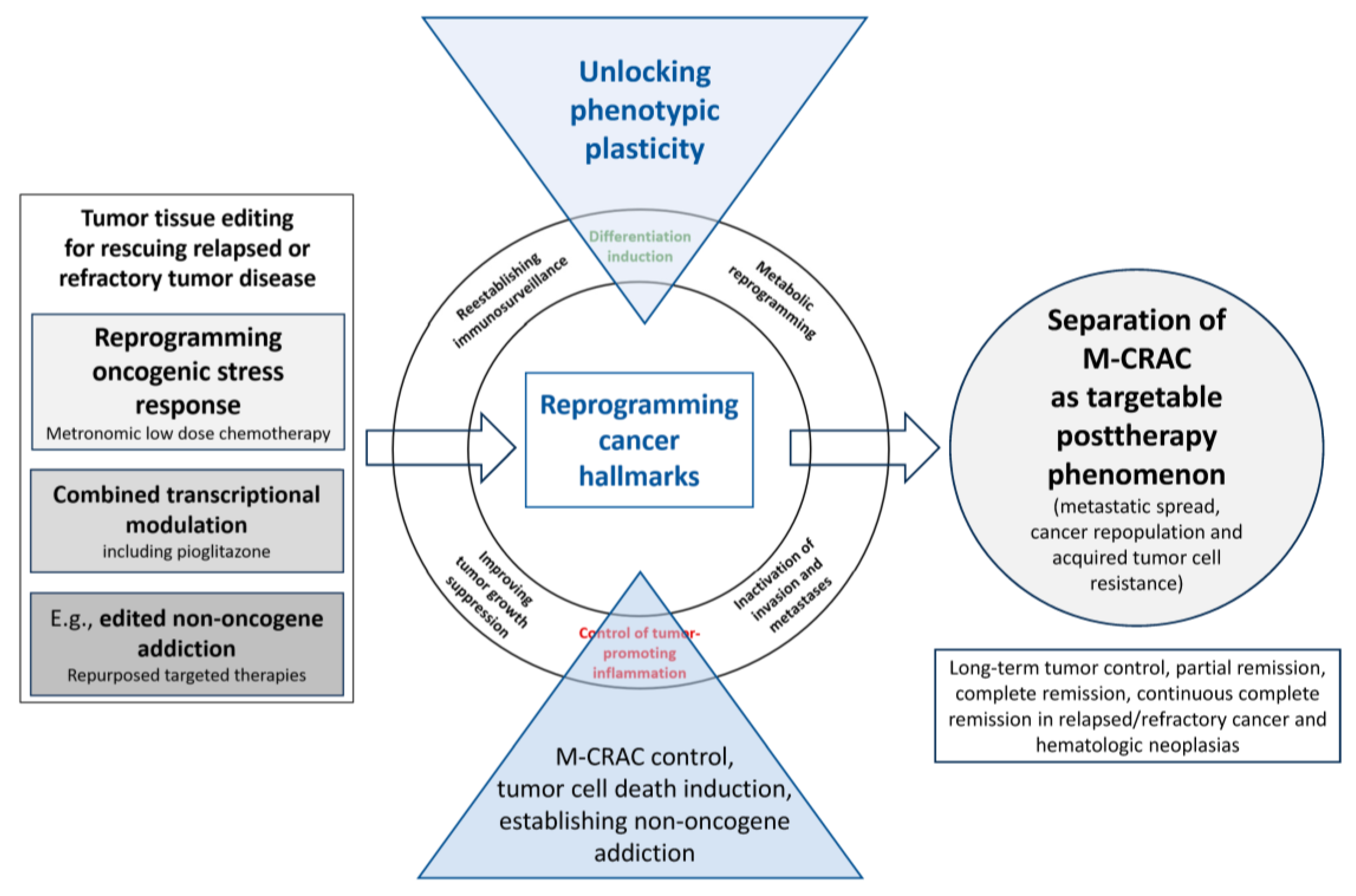
Figure 1. Tumor tissue editing for rescuing relapsed or refractory tumor disease separates M-CRAC as targetable phenomenon: suggested mechanisms of action.
Cancers 16 00180 g001
Figure 2. Disease traits of M-CRAC trigger spontaneous cancer progression. The therapeutic tumor tissue editing tool may control, resolve, or bypass M-CRAC via access to shared triggers for tumor promotion present in histologically different tumor types.
Cancers 16 00180 g002
Figure 3. Differential tumor tissue editing approaches for overcoming M-CRAC.
Cancers 16 00180 g003
Figure 4. A novel therapy model: tumor tissue editing for M-CRAC control derived from knowledge-generating patient care with tumor tissue editing techniques. Arnol et al. drafted a communication-derived tumor model [40,48].
Cancers 16 00180 g004
Table 1. Tumor tissue editing protocols for relapsed or refractory tumors: transcriptional modulation plus low-dose metronomic chemotherapy (MCT) for M-CRAC control and resolution (CR: complete remission, cCR: continuous complete remission). PR = partial remission. SD= stable disease. ®: randomization. n.d = not done.
Transcriptional RegulationMCT, Targeted Therapyr/r Neoplasia [12,13,14,15,16,17,18,19,20,21,22,23,24,25,26,27,28,29,30]Reprogramming Cancer HallmarksBest ResponseReference
Citation
Pioglitazone, rofecoxibMCT Hepatocellular carcinomaInflammation controlPR[20]
Pioglitazone, rofecoxibMCTCholangiocellular carcinoman.d.cCR[26]
Pioglitazone, rofecoxibMCTHigh-grade gliomasn.d.SD[31]
Pioglitazone, rofecoxibMCTAngiosarcoma n.d.cCR[24]
Rofecoxib plus/minus pioglitazoneMCTMetastatic gastric cancer® n.d.PR[21]
Rofecoxib plus/minus pioglitazone
Pioglitazone, etoricoxib
MCT
MCT + temsirolimus
r/r metastatic melanoma
uveal melanoma
® Inflammation controlPR
SD
[22,41]
Pioglitazone, etoricoxibMCT, clarithromycin
vs. nivolumab
r/r Non-small cell lung cancer ® Enhancing immune surveillancePR[23]
Pioglitazone, dexamethasone, etoricoxibMCT
MCT + imatinib
Castration-refractory prostate cancern.d.PR[18,30,42]
Pioglitazone, dexamethasone, etoricoxibMCT, everolimusr/r Hodgkin’s lymphomaInflammation controlcCR[13,27]
Pioglitazone, dexamethasone, etoricoxibMCTr/r Multisystem Langerhans cell histiocytosisInflammation controlcCR[16,28]
Pioglitazone, rofecoxib
Pioglitazone, rofecoxib, + interferon-α
MCT
MCT
r/r Renal clear cell carcinoma
No sufficient inflammation control
Inflammation control
SD
cCR
[17,25,43]
Pioglitazone, dexamethasoneMCT, lenalidomider/r Multiple myeloman.d.PR[14]
Pioglitazone, all-trans retinoic acidAzacytidiner/r Non-promyelocytic acute myelocytic leukemiaDifferentiation inductionCR[44,45,46,47]
Disclaimer/Publisher’s Note: The statements, opinions and data contained in all publications are solely those of the individual author(s) and contributor(s) and not of MDPI and/or the editor(s). MDPI and/or the editor(s) disclaim responsibility for any injury to people or property resulting from any ideas, methods, instructions or products referred to in the content.