Efficacy of a Novel Augmented Reality Navigation System Using 3D Computer Graphic Modeling in Endoscopic Transsphenoidal Surgery for Sellar and Parasellar Tumors
Abstract
Simple Summary
Abstract
1. Introduction
2. Materials and Methods
2.1. Patients
2.2. DCG Model Creation from Clinical Radiographic Images
2.2.1. Radiographic Image Acquisition
2.2.2. Image Processing
2.2.3. Creation of the Fused Image
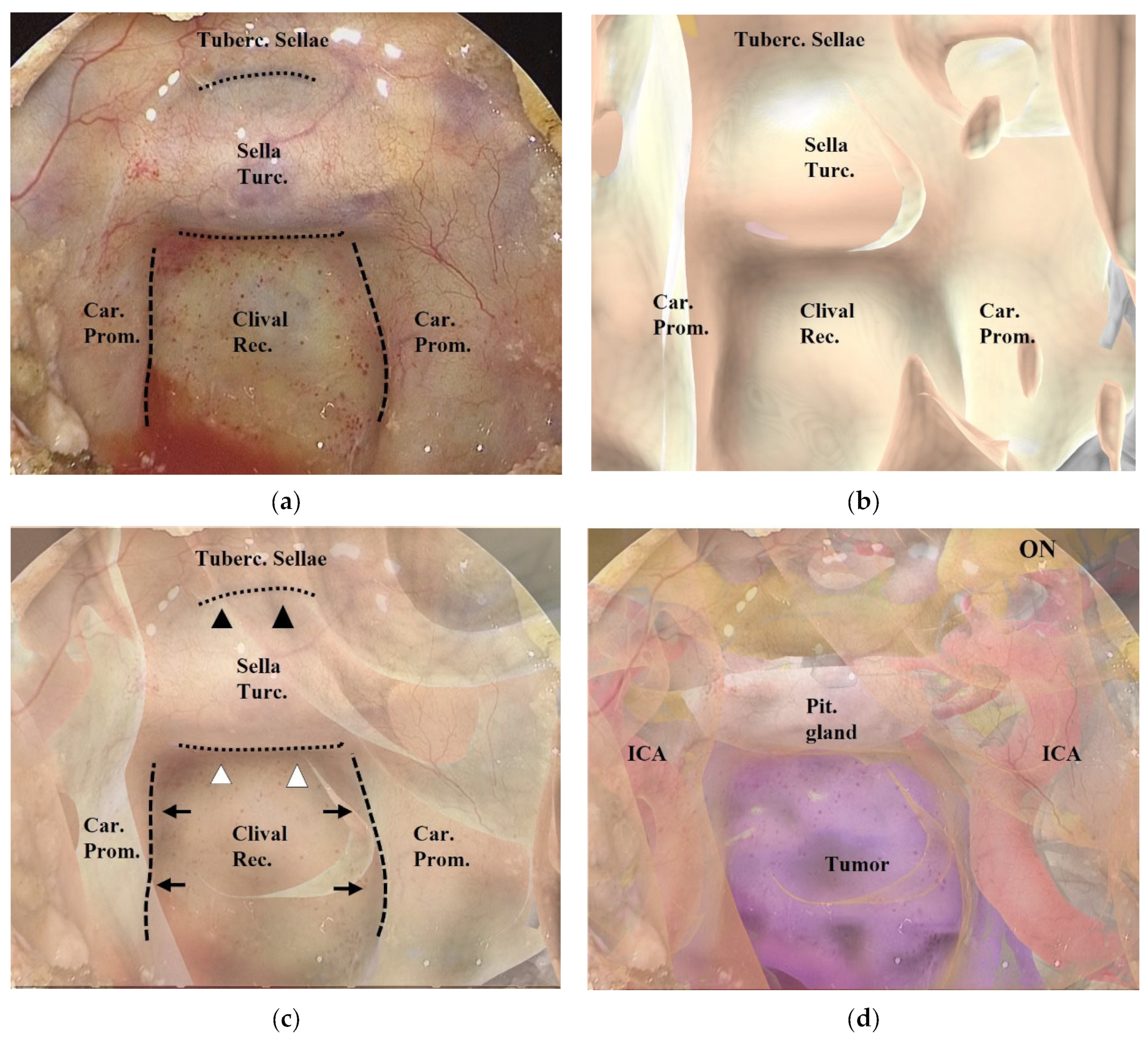
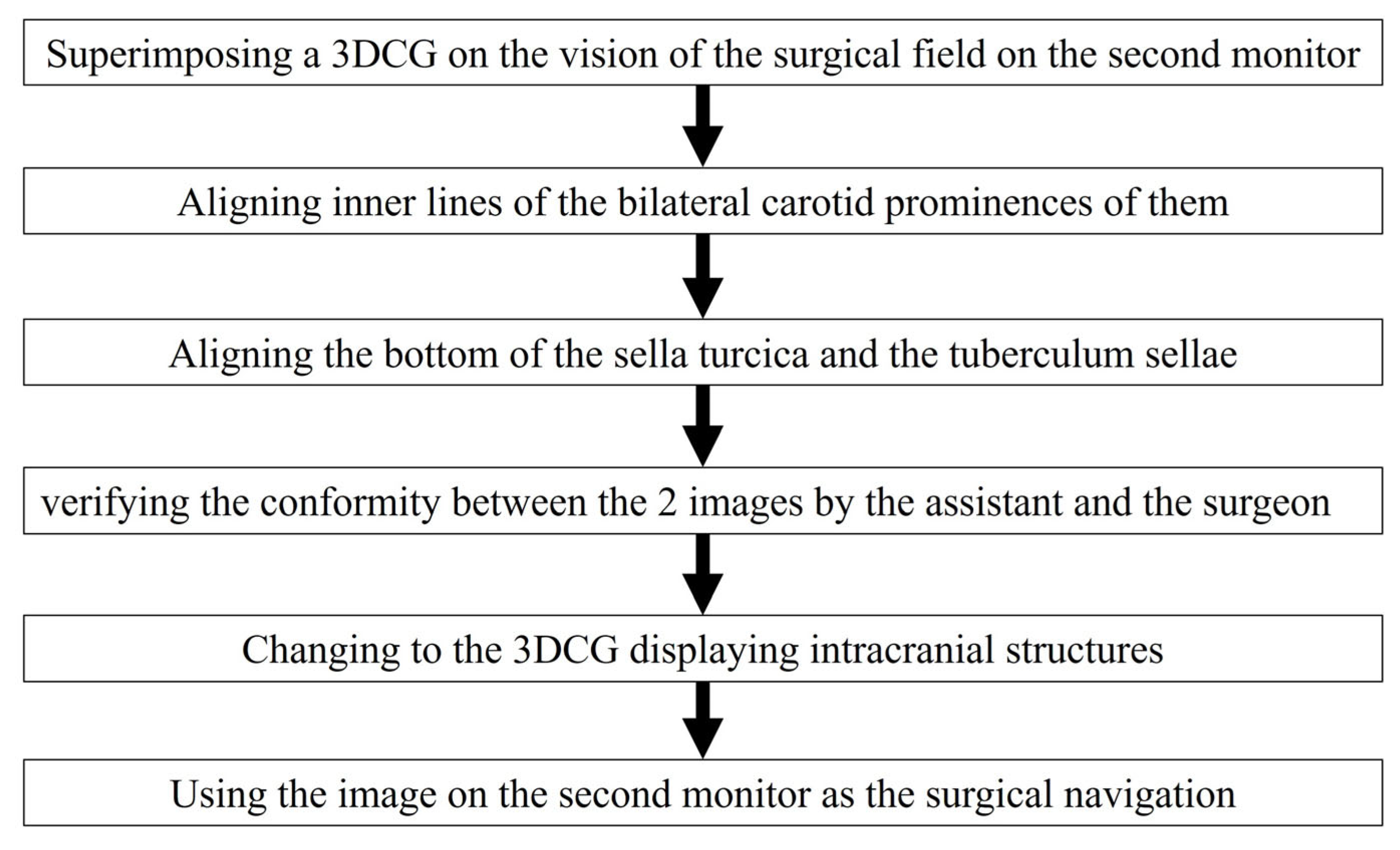
2.2.4. Creation of the AR Navigation Image on the Second Monitor during Endoscopic Surgery
2.3. Evaluation of the AR Navigation System in ETS
3. Results
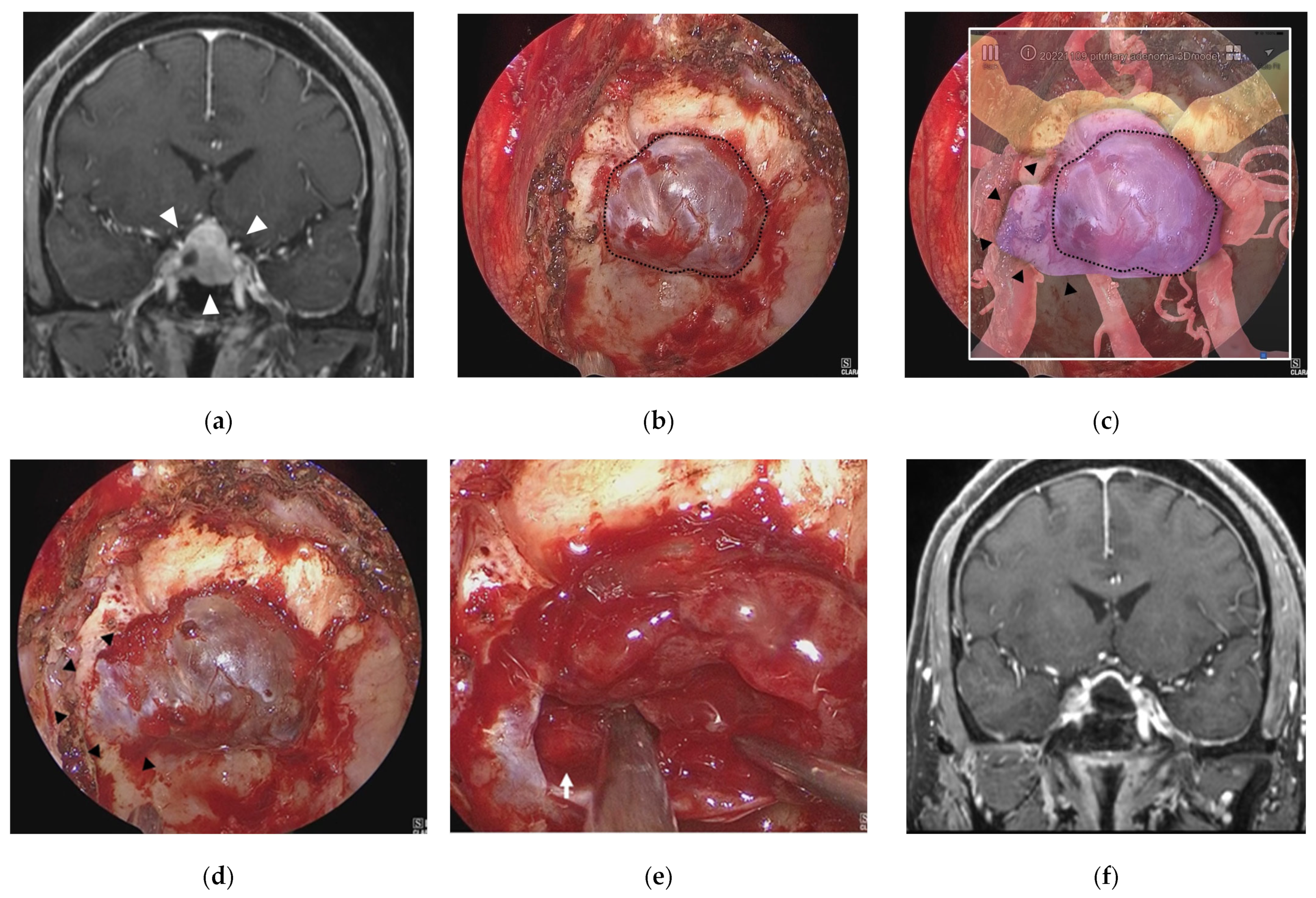
3.1. Illustrative Case 1 (Case 4)
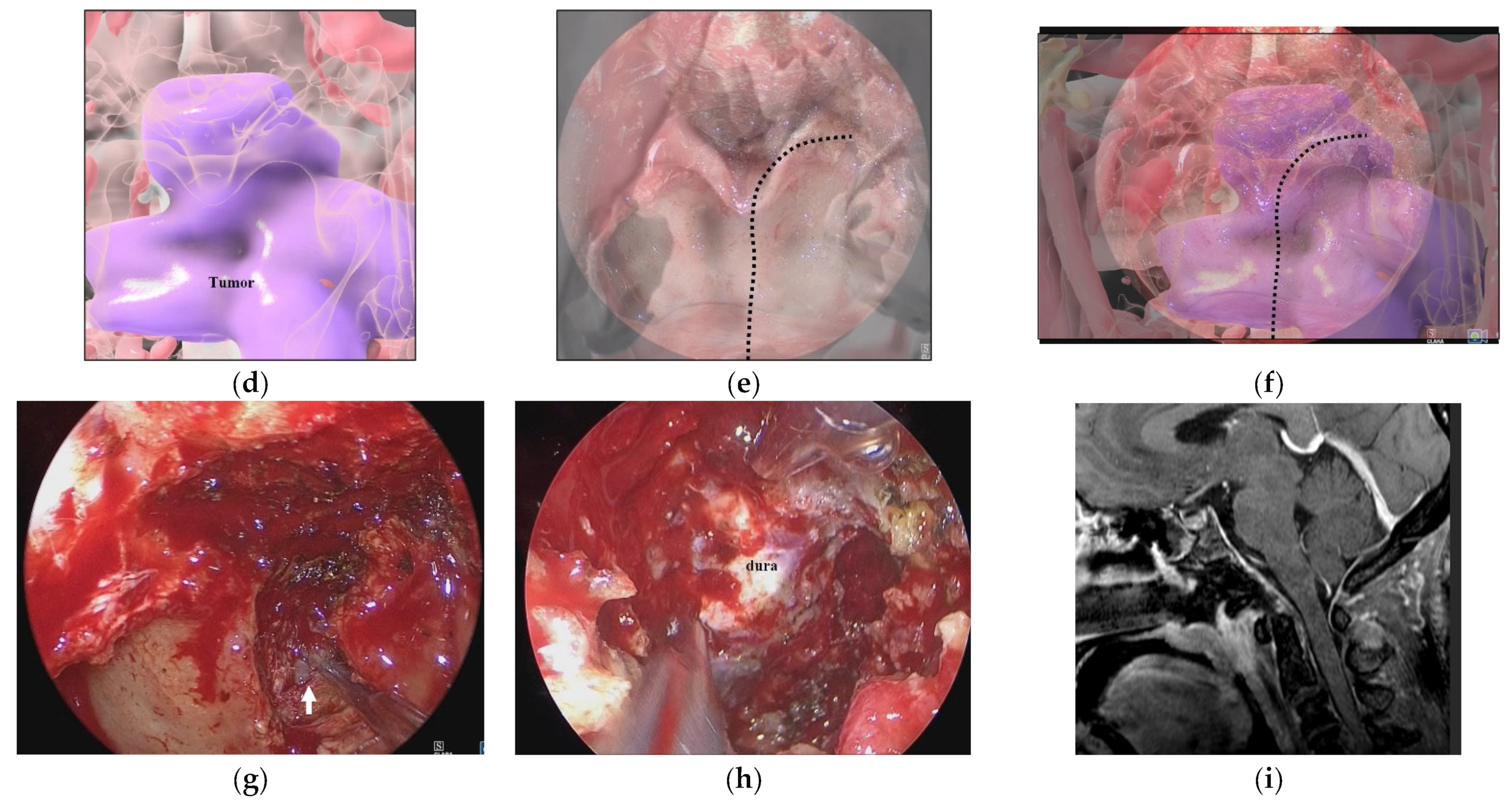
3.2. Illustrative Case 2 (Case 9)
3.3. Illustrative Case 3 (Case 10)
4. Discussion
5. Conclusions
Supplementary Materials
Author Contributions
Funding
Institutional Review Board Statement
Informed Consent Statement
Data Availability Statement
Acknowledgments
Conflicts of Interest
References
- Cavallo, L.M.; Frank, G.; Cappabianca, P.; Solari, D.; Mazzatenta, D.; Villa, A.; Zoli, M.; D’Enza, A.I.; Esposito, F.; Pasquini, E. The endoscopic endonasal approach for the management of craniopharyngiomas: A series of 103 patients. J. Neurosurg. 2014, 121, 100–113. [Google Scholar] [CrossRef] [PubMed]
- Cavallo, L.M.; Somma, T.; Solari, D.; Iannuzzo, G.; Frio, F.; Baiano, C.; Cappabianca, P. Endoscopic endonasal transsphenoidal surgery: History and evolution. World Neurosurg. 2019, 127, 686–694. [Google Scholar] [CrossRef] [PubMed]
- Chibbaro, S.; Cornelius, J.F.; Froelich, S.; Tigan, L.; Kehrli, P.; Debry, C.; Romano, A.; Herman, P.; George, B.; Bresson, D. Endoscopic endonasal approach in the management of skull base chordomas—Clinical experience on a large series, technique, outcome, and pitfalls. Neurosurg. Rev. 2014, 37, 217–224, discussion 224–215. [Google Scholar] [CrossRef] [PubMed]
- De Divitiis, E.; Cappabianca, P.; Cavallo, L.M.; Esposito, F.; de Divitiis, O.; Messina, A. Extended endoscopic transsphenoidal approach for extrasellar craniopharyngiomas. Neurosurgery 2007, 61 (Suppl. S2), 219–227, discussion 228. [Google Scholar] [CrossRef] [PubMed]
- Jho, H.-D.; Carrau, R.L. Endoscopic endonasal transsphenoidal surgery: Experience with 50 patients. J. Neurosurg. 1997, 87, 44–51. [Google Scholar] [CrossRef]
- Almeida, J.P.; De Andrade, E.J.; Vescan, A.; Zadeh, G.; Recinos, P.F.; Kshettry, V.R.; Gentili, F. Surgical anatomy and technical nuances of the endoscopic endonasal approach to the anterior cranial fossa. J. Neurosurg. Sci. 2020, 65, 103–117. [Google Scholar] [CrossRef]
- Castelnuovo, P.G.M.; Dallan, I.; Battaglia, P.; Bignami, M. Endoscopic endonasal skull base surgery: Past, present and future. Eur. Arch. Oto-Rhino-Laryngol. 2010, 267, 649–663. [Google Scholar] [CrossRef]
- Fernandez-Miranda, J.C.; Zwagerman, N.T.; Abhinav, K.; Lieber, S.; Wang, E.W.; Snyderman, C.H.; Gardner, P.A. Cavernous sinus compartments from the endoscopic endonasal approach: Anatomical considerations and surgical relevance to adenoma surgery. J. Neurosurg. 2018, 129, 430–441. [Google Scholar] [CrossRef]
- Osawa, H.; Sakurada, T.; Sasaki, J.; Araki, E. Successful surgical repair of a bilateral coronary-to-pulmonary artery fistula. Ann. Thorac. Cardiovasc. Surg. 2009, 15, 50–52. [Google Scholar]
- Quirk, B.; Connor, S. Skull base imaging, anatomy, pathology and protocols. Pract. Neurol. 2019, 20, 39–49. [Google Scholar] [CrossRef]
- Xu, Y.; Mohyeldin, A.; Asmaro, K.P.; Nunez, M.A.; Doniz-Gonzalez, A.; Vigo, V.; Cohen-Gadol, A.A.; Fernandez-Miranda, J.C. Intracranial breakthrough through cavernous sinus compartments: Anatomic study and implications for pituitary adenoma surgery. Oper. Neurosurg. 2022, 23, 115–124. [Google Scholar] [CrossRef]
- Inoue, A.; Suehiro, S.; Ohnishi, T.; Nishida, N.; Takagi, T.; Nakaguchi, H.; Miyake, T.; Shigekawa, S.; Watanabe, H.; Matsuura, B.; et al. Simultaneous combined endoscopic endonasal and transcranial surgery for giant pituitary adenomas: Tips and traps in operative indication and procedure. Clin. Neurol. Neurosurg. 2022, 218, 107281. [Google Scholar] [CrossRef]
- Koutourousiou, M.; Fernandez-Miranda, J.C.; Stefko, S.T.; Wang, E.W.; Snyderman, C.H.; Gardner, P.A. Endoscopic endonasal surgery for suprasellar meningiomas: Experience with 75 patients. J. Neurosurg. 2014, 120, 1326–1339. [Google Scholar] [CrossRef]
- Dolati, P.; Gokoglu, A.; Eichberg, D.; Zamani, A.; Golby, A.; Al-Mefty, O. Multimodal navigated skull base tumor resection using image-based vascular and cranial nerve segmentation: A prospective pilot study. Surg. Neurol. Int. 2015, 6, 172. [Google Scholar] [CrossRef]
- Jean, W.C.; Huang, M.C.; Felbaum, D.R. Optimization of skull base exposure using navigation-integrated, virtual reality templates. J. Clin. Neurosci. 2020, 80, 125–130. [Google Scholar] [CrossRef]
- Louis, R.G.; Steinberg, G.K.; Duma, C.; Britz, G.; Mehta, V.; Pace, J.; Selman, W.; Jean, W.C. Early Experience with virtual and synchronized augmented reality platform for preoperative planning and intraoperative navigation: A case series. Oper. Neurosurg. 2021, 21, 189–196. [Google Scholar] [CrossRef]
- Singh, H.; Essayed, W.I.; Schwartz, T.H. Endoscopic technology and repair techniques. Handb. Clin. Neurol. 2020, 170, 217–225. [Google Scholar] [CrossRef]
- Zhu, J.-H.; Yang, R.; Guo, Y.-X.; Wang, J.; Liu, X.-J.; Guo, C.-B. Navigation-guided core needle biopsy for skull base and parapharyngeal lesions: A five-year experience. Int. J. Oral Maxillofac. Surg. 2021, 50, 7–13. [Google Scholar] [CrossRef]
- Bopp, M.H.A.; Saß, B.; Pojskić, M.; Corr, F.; Grimm, D.; Kemmling, A.; Nimsky, C. Use of neuronavigation and augmented reality in transsphenoidal pituitary adenoma surgery. J. Clin. Med. 2022, 11, 5590. [Google Scholar] [CrossRef]
- Fick, T.; van Doormaal, J.; Hoving, E.; Regli, L.; van Doormaal, T. Holographic patient tracking after bed movement for augmented reality neuronavigation using a head-mounted display. Acta Neurochir. 2021, 163, 879–884. [Google Scholar] [CrossRef]
- Hooten, K.G.; Lister, J.R.; Lombard, G.; Lizdas, D.E.; Lampotang, S.; Rajon, D.A.; Bova, F.; Murad, G.J. Mixed reality ventriculostomy simulation: Experience in neurosurgical residency. Neurosurgery 2014, 10, 576–581, discussion 581. [Google Scholar] [CrossRef] [PubMed]
- Ivan, M.E.; Eichberg, D.G.; Di, L.; Shah, A.H.; Luther, E.M.; Lu, V.M.; Komotar, R.J.; Urakov, T.M. Augmented reality head-mounted display–based incision planning in cranial neurosurgery: A prospective pilot study. Neurosurg. Focus 2021, 51, E3. [Google Scholar] [CrossRef] [PubMed]
- Li, L.; Yang, J.; Chu, Y.; Wu, W.; Xue, J.; Liang, P.; Chen, L. A novel augmented reality navigation system for endoscopic sinus and skull base surgery: A feasibility study. PLoS ONE 2016, 11, e0146996. [Google Scholar] [CrossRef] [PubMed]
- Mishra, R.; Narayanan, M.K.; Umana, G.E.; Montemurro, N.; Chaurasia, B.; Deora, H. virtual reality in neurosurgery: Beyond neurosurgical planning. Int. J. Environ. Res. Public Health 2022, 19, 1719. [Google Scholar] [CrossRef] [PubMed]
- Weigl, M.; Stefan, P.; Abhari, K.; Wucherer, P.; Fallavollita, P.; Lazarovici, M.; Weidert, S.; Euler, E.; Catchpole, K. Intra-operative disruptions, surgeon’s mental workload, and technical performance in a full-scale simulated procedure. Surg. Endosc. 2015, 30, 559–566. [Google Scholar] [CrossRef]
- Zeiger, J.; Costa, A.; Bederson, J.; Shrivastava, R.K.; Iloreta, A.M.C. Use of mixed reality visualization in endoscopic endonasal skull base surgery. Oper. Neurosurg. 2019, 19, 43–52. [Google Scholar] [CrossRef]
- Dixon, B.J.; Daly, M.J.; Chan, H.; Vescan, A.; Witterick, I.J.; Irish, J.C. Augmented real-time navigation with critical structure proximity alerts for endoscopic skull base surgery. Laryngoscope 2014, 124, 853–859. [Google Scholar] [CrossRef]
- Caversaccio, M.; Langlotz, F.; Nolte, L.P.; Hausler, R. Impact of a self-developed planning and self-constructed navigation system on skull base surgery: 10 years experience. Acta Otolaryngol. 2007, 127, 403–407. [Google Scholar] [CrossRef]
- Kawamata, T.; Iseki, H.; Shibasaki, T.; Hori, T. Endoscopic augmented reality navigation system for endonasal transsphenoidal surgery to treat pituitary tumors: Technical note. Neurosurgery 2002, 50, 1393–1397. [Google Scholar]
- Pennacchietti, V.; Stoelzel, K.; Tietze, A.; Lankes, E.; Schaumann, A.; Uecker, F.C.; Thomale, U.W. First experience with augmented reality neuronavigation in endoscopic assisted midline skull base pathologies in children. Child’s Nerv. Syst. 2021, 37, 1525–1534. [Google Scholar] [CrossRef]
- Orringer, D.; Golby, A.; Jolesz, F. Neuronavigation in the surgical management of brain tumors: Current and future trends. Expert Rev. Med. Devices 2012, 9, 491–500. [Google Scholar] [CrossRef]
- Kin, T.; Oyama, H.; Kamada, K.; Aoki, S.; Ohtomo, K.; Saito, N. Prediction of surgical view of neurovascular decompression using interactive computer graphics. Neurosurgery 2009, 65, 121–128, discussion 128–129. [Google Scholar] [CrossRef]
- Kin, T.; Shin, M.; Oyama, H.; Kamada, K.; Kunimatsu, A.; Momose, T.; Saito, N. Impact of multiorgan fusion imaging and interactive 3-dimensional visualization for intraventricular neuroendoscopic surgery. Oper. Neurosurg. 2011, 69, ons40–ons48, discussion ons48. [Google Scholar] [CrossRef]
- Koike, T.; Kin, T.; Tanaka, S.; Takeda, Y.; Uchikawa, H.; Shiode, T.; Saito, T.; Takami, H.; Takayanagi, S.; Mukasa, A.; et al. Development of innovative neurosurgical operation support method using mixed-reality computer graphics. World Neurosurg. X 2021, 11, 100102. [Google Scholar] [CrossRef]
- Kin, T.; Nakatomi, H.; Shojima, M.; Tanaka, M.; Ino, K.; Mori, H.; Kunimatsu, A.; Oyama, H.; Saito, N. A new strategic neurosurgical planning tool for brainstem cavernous malformations using interactive computer graphics with multimodal fusion images. J. Neurosurg. 2012, 117, 78–88. [Google Scholar] [CrossRef]


| Case | Age (y) | Sex | Diagnosis | Approach |
|---|---|---|---|---|
| 1 | 47 | male | Petroclival meningioma | Transsphenoidal-transclival |
| 2 | 58 | male | NF-PitNET | Transsphenoidal |
| 3 | 82 | male | Clival chordoma | Transsphenoidal-transclival |
| 4 | 58 | male | NF-PitNET | Transsphenoidal |
| 5 | 39 | male | Skull base chondrosarcoma | Transsphenoidal-transmaxillary |
| 6 | 52 | female | Intraorbital cavernous malformation | Transsphenoidal-transethmoidal |
| 7 | 16 | male | Craniopharyngioma | Transsphenoidal |
| 8 | 57 | female | PitNET | Transsphenoidal |
| 9 | 50 | female | Craniovertebral junction chordoma | Transsphenoidal-transclival |
| 10 | 75 | female | Tuberculum sellae meningioma | Transsphenoidal |
| 11 | 58 | male | Somatotroph-PitNET | Transsphenoidal |
| 12 | 60 | female | NF-PitNET | Transsphenoidal |
| 13 | 55 | male | NF-PitNET | Transsphenoidal |
| 14 | 74 | female | NF-PitNET | Transsphenoidal |
| 15 | 56 | female | Petroclival meningioma | Transsphenoidal-transclival |
| Result of Assessment | Score |
|---|---|
| Misleading, confusing | 1 |
| Not misleading but not useful | 2 |
| Useful but less so than conventional neuronavigation | 3 |
| As useful as conventional neuronavigation | 4 |
| More useful than conventional neuronavigation | 5 |
| Case | Diagnosis | Surgeon 1 | Surgeon 2 | Resident 1 | Resident 2 | Resident 3 | 95% CI |
|---|---|---|---|---|---|---|---|
| 1 | petroclival meningioma | 5 | 5 | 5 | 4 | 5 | 4.4–5.2 |
| 2 | PitNET | 4 | 4 | 5 | 4 | 5 | 3.91–4.89 |
| 3 | clival chordoma | 4 | 4 | 5 | 4 | 5 | 3.91–4.89 |
| 4 | PitNET | 5 | 5 | 5 | 5 | 4 | 4.4–5.2 |
| 5 | skull base chondrosarcoma | 4 | 5 | 3 | 5 | 5 | 3.8–5.4 |
| 6 | intraorbital cavernous malformation | 5 | 5 | 5 | 5 | 5 | 5 |
| 7 | craniopharyngioma | 5 | 4 | 5 | 3 | 5 | 3.8–5.4 |
| 8 | PitNET | 4 | 4 | 5 | 5 | 4 | 4.11–5.09 |
| 9 | craniovertebral junction chordoma | 5 | 5 | 5 | 5 | 5 | 5 |
| 10 | tuberculum sellae meningioma | 5 | 5 | 5 | 5 | 5 | 5 |
| 11 | PitNET | 5 | 5 | 5 | 5 | 5 | 5 |
| 12 | PitNET | 4 | 4 | 4 | 5 | 5 | 4.11–5.09 |
| 13 | PitNET | 4 | 4 | 4 | 4 | 4 | 4 |
| 14 | PitNET | 4 | 5 | 5 | 5 | 5 | 4.4–5.2 |
| 15 | petroclival meningioma | 5 | 5 | 5 | 5 | 5 | 5 |
| Case | Age | Sex | Diagnosis | Symptoms at ETS | Tumor Volumes (cm3) | Level of Resection | Symptoms after ETS | Postoperative Complications |
|---|---|---|---|---|---|---|---|---|
| 1 | 47 | M | Petroclival Meningioma | hearing deterioration | 26.25 | NTR | improved | none |
| 2 | 58 | M | PitNET | oculomotor nerve and abducens nerve palsy | 1.76 | GTR | improved | none |
| 3 | 82 | M | clival chordoma | abducens nerve palsy | 12.90 | GTR | improved | none |
| 4 | 58 | M | PitNET | None | 4.35 | GTR | none | none |
| 5 | 39 | M | skull base chondrosarcoma | unilateral temporal hemianopsia | 1.35 | GTR | no change | none |
| 6 | 52 | F | intraorbital cavernous malformation | impaired vision, oculomotor disorder | 4.37 | GTR | improved | none |
| 7 | 16 | M | Craniopharyngioma | headache, vertigo (hydrocephalus) | 6.78 | NTR | improved | none |
| 8 | 57 | F | PitNET | bitemporal hemianopsia | 3.70 | GTR | improved | none |
| 9 | 50 | F | craniovertebral junction chordoma | hypoglossal nerve palsy | 23.23 | GTR | no change | none |
| 10 | 75 | F | tuberculum sellae meningioma | impaired vision, unilateral temoral hemianopsia | 1.94 | STR | improved | none |
| 11 | 58 | M | PitNET | none | 2.31 | GTR | none | none |
| 12 | 60 | F | PitNET | homonymous hemianopsia | 6.35 | NTR | improved | none |
| 13 | 55 | M | PitNET | bitemporal hemianopsia | 5.25 | GTR | improved | none |
| 14 | 74 | F | PitNET | Bitemporal hemianopsia | 5.31 | GTR | improved | none |
| 15 | 56 | F | Petroclival Meningioma | vertigo, tinnitus | 17.76 | STR | improved | Transient abducens nerve palsy |
Disclaimer/Publisher’s Note: The statements, opinions and data contained in all publications are solely those of the individual author(s) and contributor(s) and not of MDPI and/or the editor(s). MDPI and/or the editor(s) disclaim responsibility for any injury to people or property resulting from any ideas, methods, instructions or products referred to in the content. |
© 2023 by the authors. Licensee MDPI, Basel, Switzerland. This article is an open access article distributed under the terms and conditions of the Creative Commons Attribution (CC BY) license (https://creativecommons.org/licenses/by/4.0/).
Share and Cite
Goto, Y.; Kawaguchi, A.; Inoue, Y.; Nakamura, Y.; Oyama, Y.; Tomioka, A.; Higuchi, F.; Uno, T.; Shojima, M.; Kin, T.; et al. Efficacy of a Novel Augmented Reality Navigation System Using 3D Computer Graphic Modeling in Endoscopic Transsphenoidal Surgery for Sellar and Parasellar Tumors. Cancers 2023, 15, 2148. https://doi.org/10.3390/cancers15072148
Goto Y, Kawaguchi A, Inoue Y, Nakamura Y, Oyama Y, Tomioka A, Higuchi F, Uno T, Shojima M, Kin T, et al. Efficacy of a Novel Augmented Reality Navigation System Using 3D Computer Graphic Modeling in Endoscopic Transsphenoidal Surgery for Sellar and Parasellar Tumors. Cancers. 2023; 15(7):2148. https://doi.org/10.3390/cancers15072148
Chicago/Turabian StyleGoto, Yoshiaki, Ai Kawaguchi, Yuki Inoue, Yuki Nakamura, Yuta Oyama, Arisa Tomioka, Fumi Higuchi, Takeshi Uno, Masaaki Shojima, Taichi Kin, and et al. 2023. "Efficacy of a Novel Augmented Reality Navigation System Using 3D Computer Graphic Modeling in Endoscopic Transsphenoidal Surgery for Sellar and Parasellar Tumors" Cancers 15, no. 7: 2148. https://doi.org/10.3390/cancers15072148
APA StyleGoto, Y., Kawaguchi, A., Inoue, Y., Nakamura, Y., Oyama, Y., Tomioka, A., Higuchi, F., Uno, T., Shojima, M., Kin, T., & Shin, M. (2023). Efficacy of a Novel Augmented Reality Navigation System Using 3D Computer Graphic Modeling in Endoscopic Transsphenoidal Surgery for Sellar and Parasellar Tumors. Cancers, 15(7), 2148. https://doi.org/10.3390/cancers15072148


