Article

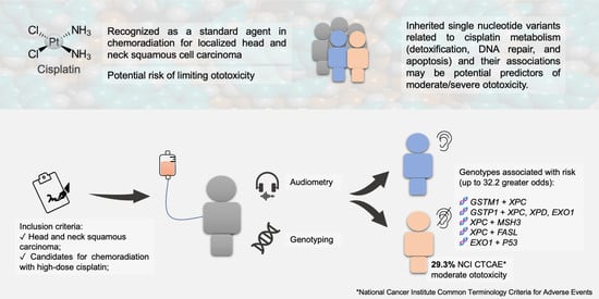
Association of Clinical Aspects and Genetic Variants with the Severity of Cisplatin-Induced Ototoxicity in Head and Neck Squamous Cell Carcinoma: A Prospective Cohort Study

by
Ligia Traldi Macedo
1,2,†,
Ericka Francislaine Dias Costa
1,†,
Bruna Fernandes Carvalho
1,
Gustavo Jacob Lourenço
1,
Luciane Calonga
3,
Arthur Menino Castilho
3,
Carlos Takahiro Chone
3 and
Carmen Silvia Passos Lima
1,3,*
1
Laboratory of Cancer Genetics, Faculty of Medical Sciences, University of Campinas, Campinas 13083-970, SP, Brazil
2
Department of Anesthesiology, Oncology and Radiology, Faculty of Medical Sciences, University of Campinas, Campinas 13083-888, SP, Brazil
3
Department of Ophthalmology and Otorhinolaryngology, Faculty of Medical Sciences, University of Campinas, Campinas 13083-888, SP, Brazil
*
Author to whom correspondence should be addressed.
These authors contributed equally to this work.
Cancers 2023, 15(6), 1759; https://doi.org/10.3390/cancers15061759
Submission received: 3 February 2023 / Revised: 1 March 2023 / Accepted: 7 March 2023 / Published: 14 March 2023

Abstract

:

Simple Summary

Cisplatin is recognized as the standard agent for head and neck squamous cell carcinoma therapy, despite the relevant risk of permanent hearing damage. The aim of this study was to evaluate the possible associations of the clinicopathological features and inherited genotypes encoding cisplatin metabolism in eighty-nine patients undergoing chemoradiation with the risk of hearing loss. We were able to confirm race, body mass index, and cumulative cisplatin dose as independent clinical risk factors. Patients with specific isolated and combined genotypes encoding cisplatin efflux (GSTM1, GSTP1 c.313A>G), DNA repair (XPC c.2815A>C, XPD c.934G>A, EXO1 c.1762G>A, MSH3 c.3133A>G), and apoptosis-related proteins (FASL c.-844A>T, P53 c.215G>C) presented up to 32.22 higher odds of moderate or severe ototoxicity. These findings reinforce the importance of inherited nucleotide variants involved in cisplatin metabolism as candidate variables for predictive models of adverse events.

Abstract

Background: Cisplatin (CDDP) is a major ototoxic chemotherapy agent for head and neck squamous cell carcinoma (HNSCC) treatment. Clinicopathological features and genotypes encode different stages of CDDP metabolism, as their coexistence may influence the prevalence and severity of hearing loss. Methods: HNSCC patients under CDDP chemoradiation were prospectively provided with baseline and post-treatment audiometry. Clinicopathological features and genetic variants encoding glutathione S-transferases (GSTT1, GSTM1, GSTP1), nucleotide excision repair (XPC, XPD, XPF, ERCC1), mismatch repair (MLH1, MSH2, MSH3, EXO1), and apoptosis (P53, CASP8, CASP9, CASP3, FAS, FASL)-related proteins were analyzed regarding ototoxicity. Results: Eighty-nine patients were included, with a cumulative CDDP dose of 260 mg/m2. Moderate/severe ototoxicity occurred in 26 (29%) patients, particularly related to hearing loss at frequencies over 3000 Hertz. Race, body-mass index, and cumulative CDDP were independent risk factors. Patients with specific isolated and combined genotypes of GSTM1, GSTP1 c.313A>G, XPC c.2815A>C, XPD c.934G>A, EXO1 c.1762G>A, MSH3 c.3133A>G, FASL c.-844A>T, and P53 c.215G>C SNVs had up to 32.22 higher odds of presenting moderate/severe ototoxicity. Conclusions: Our data present, for the first time, the association of combined inherited nucleotide variants involved in CDDP efflux, DNA repair, and apoptosis with ototoxicity, which could be potential predictors in future clinical and genomic models.

Graphical Abstract

1. Introduction

Head and neck squamous cell carcinoma (HNSCC) is the sixth most common cancer worldwide, with 878,348 new cases and 444,347 deaths estimated in 2020 [1,2]. Approximately 75% of patients with HNSCC present locally advanced disease at diagnosis, and the standard therapy for most cases involves chemoradiation or the induction of multi-agent chemotherapy, in which cisplatin (CDDP) is usually included [3,4]. Alternative treatments, such as carboplatin or cetuximab, were studied in the context of chemoradiation, though their equivalence regarding efficacy has yet to be validated by randomized trials [5]. Additionally, in patients with treatment-naïve metastatic disease or platinum-sensitive relapse, CDDP-based regimens are commonly used in clinical practice, with benefits in progression-free survival (PFS) and overall survival (OS) [6,7]. Nonetheless, CDDP is related to significant adverse events, such as nausea, vomiting, nephrotoxicity, hypersensitivity reactions, and ototoxicity [3,8,9]. Among these, hearing impairment is a current concern since there are, to date, no effective otoprotective measures, resulting in potentially permanent and quality-of-life-limiting damage [10,11].
Every year, one in five patients submitted to CDDP-based chemotherapy will suffer severe to profound hearing loss [10,12,13]. Regarding chemoradiation for HNSCC, major losses are described in higher frequencies, with reported pure-tone median threshold increases ranging from 9.52 to 25 decibels (dB) at 4 kilohertz (kHz) and 18.57 to 27.14 dB at 8 kHz [14,15]. This event has a major negative impact on the quality of life [16] and requires essential care regarding dosage management and the duration of therapy [17]. Despite the association of cumulative CDDP dose, history of noise exposure, and smoking as independent risk factors, the prevalence and intensity of hearing impairment are remarkably heterogeneous among patients with similar characteristics and regimens [18]. This finding indicates the involvement of unknown risk factors, with single-nucleotide variants (SNVs), on genes encoding proteins related to CDDP metabolism, being potential candidates for this risk [19,20].
Numerous proteins act in the mechanisms of CDDP cellular detoxification, as well as in the pathways of damage repair and apoptosis [21,22] (Figure 1).
The detoxification of CDDP occurs mainly through its conjugation with glutathione, encoded by the Mu1 (GSTM1), Theta1 (GSTT1), and Pi1 (GSTP1) genes [23], in which the lack of functional proteins involved in this cascade may contribute to intracellular CDDP accumulation and cytotoxic effects [24]. The cytotoxic activity of CDDP is also attributed to its DNA binding, leading to the activation of repair mechanisms. The DNA lesion induced by CDDP can be removed through the nucleotide excision repair (NER) pathway [25], mediated by the xeroderma pigmentosum (XPC, XPD, and XPF) [26,27] and excision repair cross-complementation group 1 (ERCC1) genes [28], as well as by the mismatch repair (MMR) pathway, mediated through proteins encoded by MutL homolog 1 (MLH1) [29], MutS homolog 2 (MSH2) [30], MutS homolog 3 (MSH3), and exonuclease 1 (EXO1) genes [29]. If the repair is ineffective, apoptosis is mediated by proteins encoded by P53, Caspase 8 (CASP8), CASP9, CASP3, Fas cell surface death receptor (FAS), and Fas ligand (FASL) tumor necrosis factors [21,31]. Defects in these pathways may promote increased DNA damage and/or apoptosis, with greater potential for toxicity [32].
Genome-wide studies have described SNVs in acylphosphatase 2 (ACYP2), involved in calcium homeostasis [33,34,35] and Mendelian deafness WFS1 genes [20,33,36,37], as predictors of CDDP-induced ototoxicity. Genes encoding thiopurine S- (TPMT) and cathecol-O methyltransferases (COMT) have also been described as potential risk factors [35]. In CDDP-treated patients, GSTM1, GSTT1 [18,38,39,40,41,42], and GSTP1 c.313A>G [38,39,41,43] were seen in pediatric solid or adult testicular tumors with controversial results in ototoxicity, while XPC c.2815A>C SNV influenced ototoxicity in osteosarcoma patients [44].
To our knowledge, the only cohort that evaluated SNVs in genes of distinct pathways of CDDP metabolism, damage repair, and apoptosis (GSTM1, GSTT1, GSTP1 c.313A>G, XPC c.2815A>C, XPD c.934G>A, XPD c.2251A>C, XPF c.2505T>C, ERCC1 c.354C>T, MLH1 c.-93G>A, MSH2 c.211 +9G>C, MSH3 c.3133A>G, EXO1 c.1762G>A, P53 c.215G>C, FAS c.-671A>G, FAS c.-1378G>A, FASL c.-844C>T, CASP3 c.-1191A>G, and CASP3 c.-182-247G>T) in the ototoxicity of HNSCC treated with CDDP chemoradiation was previously conducted by our group, and the functional roles of each SNV described in the literature are presented in Table A1. We found that GSTT1 [45], EXO1 [19], XPC [46], and FASL [47] SNVs altered the occurrence of all-grade ototoxicity.
Since there is scarce information regarding pure tone and audiometric speech changes in patients under CDDP chemoradiation, considering that moderate/severe ototoxicity influences quality of life and the fact that patients may inherit defects in more than one pathway, we conducted a descriptive and pharmacogenetic study focusing isolated factors related to CDDP metabolism, aiming to contribute to the prompt recognition of patients at high risk of ototoxicity before treatment initiation and thus enabling treatment modifications.

2. Materials and Methods

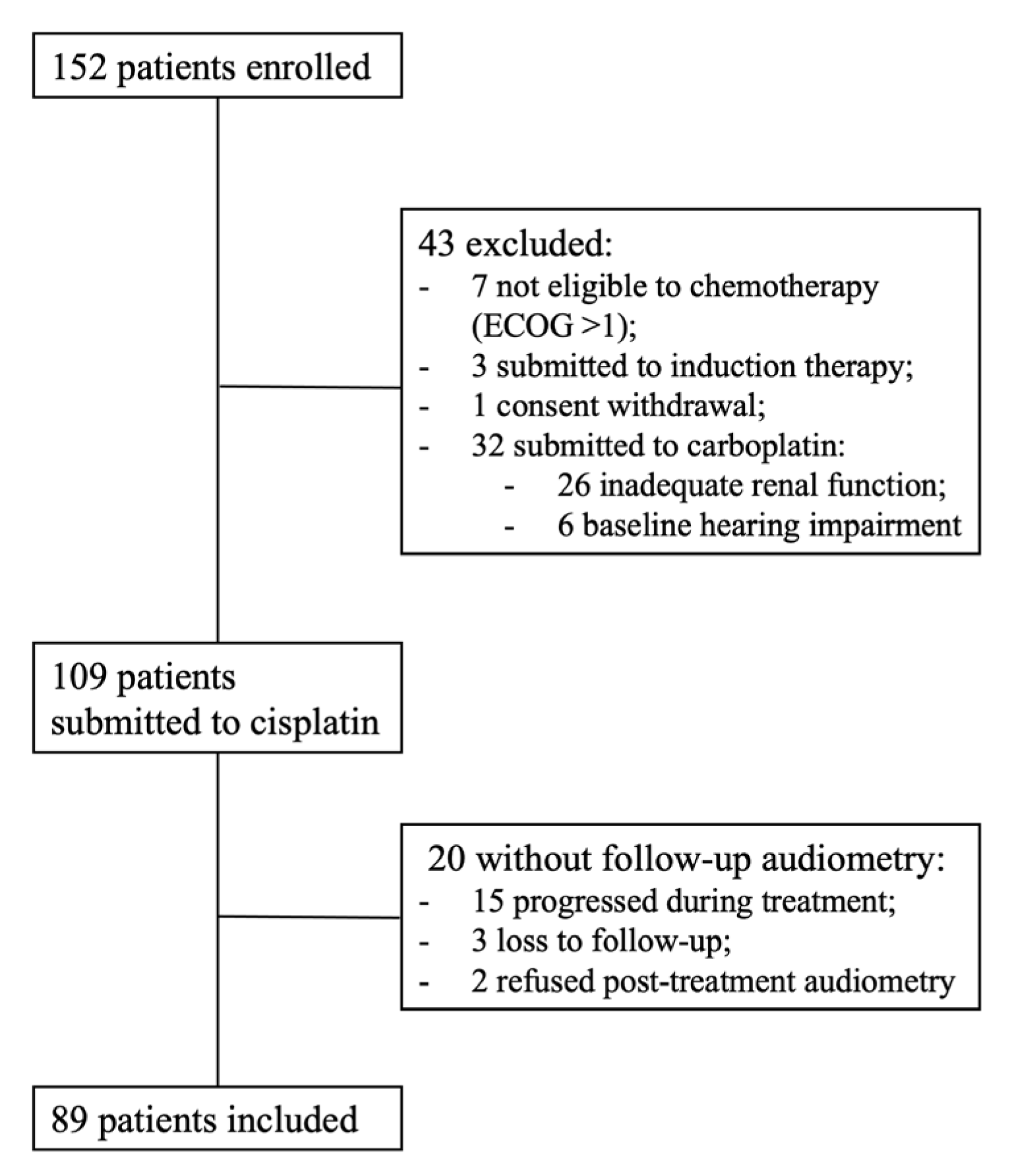
2.1. Study Population

This cohort prospectively enrolled HNSCC patients who were eligible for treatment with definitive chemoradiation at the Clinical Oncology Service of the University of Campinas, Brazil, between June 2011 and February 2014. Eastern Cooperative Oncology Group (ECOG) performance status of equal to or less than 1 [48], creatinine clearance greater than 45 mL/min, and the absence of baseline moderate or severe hearing impairment were required. Patients who were not candidates for treatment with CDDP or who were under induction, adjuvant, or palliative therapy were excluded.
Patients received high-dose CDDP (starting dose of 80–100 mg/m2 on days 1, 22, and 43) [49] associated with RT (35 sessions; planned total radiation dose of 70 Gray—Gy). All patients received anti-emetic prophylaxis with intravenous ondansetron and dexamethasone pre-infusion, in addition to oral dexamethasone and metoclopramide, for the following three days. Mannitol and hydration with saline solution, potassium chloride, and magnesium sulfate were administered, as reported [46]. Dose delays and reductions were applied in toxicity events with grades equal to or greater than 3, according to the National Cancer Institute criteria for adverse events (NCI CTCAE) [50]. Patients were followed from recruitment to 30 days after treatment completion.
The study was approved by the local institutional review board (Protocol 274/2011 and 62870722.1.0000.5404), and all patients enrolled in the study agreed to participate and declared consent in accordance with the Declaration of Helsinki. The results of this study were reported following the Strengthening the Reporting of Observational Studies in Epidemiology (STROBE) guidelines [51].

2.2. Clinical Data

Data related to age, gender, race, history of tobacco and alcohol use [52], ECOG status [48], body mass index (BMI) [53], and presence of diabetes [54] or systemic hypertension [55] as comorbidities of interest were collected. Regarding disease characteristics, primary tumor location, tumor side, and histological grade were also computed. Diagnosis and tumor staging followed the American Joint Committee on Cancer criteria [56]. Data related to cumulative CDDP dose, radiotherapy (RT) technique (2D or 3D), and final total dose in Gy, including total doses from supraclavicular fossa, cervico-facial, cervico-posterior, and boost, were also registered for analysis.

2.3. Hearing Assessment

Patients were submitted to otoscopic examination before any audiometric measurements. If there were identifiable diseases of the external acoustic meatus, tympanic membranes, middle ears, or other conditions that could interfere with the audiological evaluation, patients received treatment and were followed up until resolution. Audiometric evaluations were performed on two occasions, before treatment initiation and up to 30 days after therapy completion in an acoustic booth previously calibrated to meet the specifications of internal noise levels allowed according to the International Organization for Standardization ((ISO) 8253-1:2010 criteria, using the Interacoustics audiometer model AC 30 (Interacoustics A/S, Middlefart, Denmark).

2.3.1. Pure Tone Audiometry

Pure tone audiometry was conducted in air and bone conduction for both the left and right sides. For the air conduction assessment, the tonal auditory thresholds were measured at sound frequencies 0.25, 0.50, 1, 2, 3, 4, 6, and 8 kHz, with earphones model TDH 39, applying the descending–ascending technique. In each test, the smallest sound stimulus perceived by the patient in at least 50% of the presentations was considered. Bone conduction evaluation was performed in a similar descending–ascending manner, registering minimum dB thresholds at frequencies 0.25, 0.50, 1, 2, 3, and 4 kHz through a conduction receiver bow fixed on the mastoid (Interacoustics A/S, Middlefart, Denmark). The corresponding hearing thresholds for each frequency in both ears were collected, considering that the normal expected range was lower than 15 dB [57].

2.3.2. Speech Audiometry

Speech audiometry was performed when applicable, assessing the speech recognition threshold (SRT) in dB for the repetition of 50% disyllabic words for left and right ears. The speech discrimination score (SDS), calculating the percentage of syllables repeated correctly, was also registered for each side when possible [58]. SRT is normally within 10 dB of pure tone average thresholds, while the normal range of SDS is 92% to 100% [59]. Patients unable to speak owing to disease-related limitations or other causes had their exam halted and reasons noted.

2.4. Hearing Loss Classification

2.4.1. Global Burden of Disease (GBD) Hearing Loss Classification

The GBD Hearing Loss Classification was performed, assessing the ISO threshold average for 0.5, 1, 2, and 4 kHz in dB, as recommended by the World Health Organization [60], pre- and post-treatment in air and bone conduction assessments [61]. Patients were categorized according to the criteria of unilateral (<20 dB in the better and >35 dB in the worst ears, respectively), mild (20 to 34 dB in the better ear), moderate (35–49 dB in the better ear), moderately severe (50–64 dB in the better ear), severe (65–79 dB in the better ear), and profound (80–94 dB in the better ear) hearing loss.

2.4.2. National Cancer Institute Common Terminology Criteria for Adverse Events (NCI CTCAE)

Hearing loss in the right and left ears were also classified based on grades (G) 1 to 4, according to the NCI CTCAE v4.0 criteria [50] following the monitoring of at least 1, 2, 3, 4, 6, and 8 kHz audiogram, as follows: G 1, “threshold shift of 15 to 25 dB averaged at two contiguous test frequencies in at least one ear”; G 2, “threshold shift of >25 dB averaged at two contiguous test frequencies in at least one ear”; G 3, “threshold shift of >25 dB averaged at three contiguous test frequencies in at least one ear”; G 4, “decrease in hearing to profound bilateral loss (absolute threshold >80 dB hearing loss at 2 kHz and above)”.

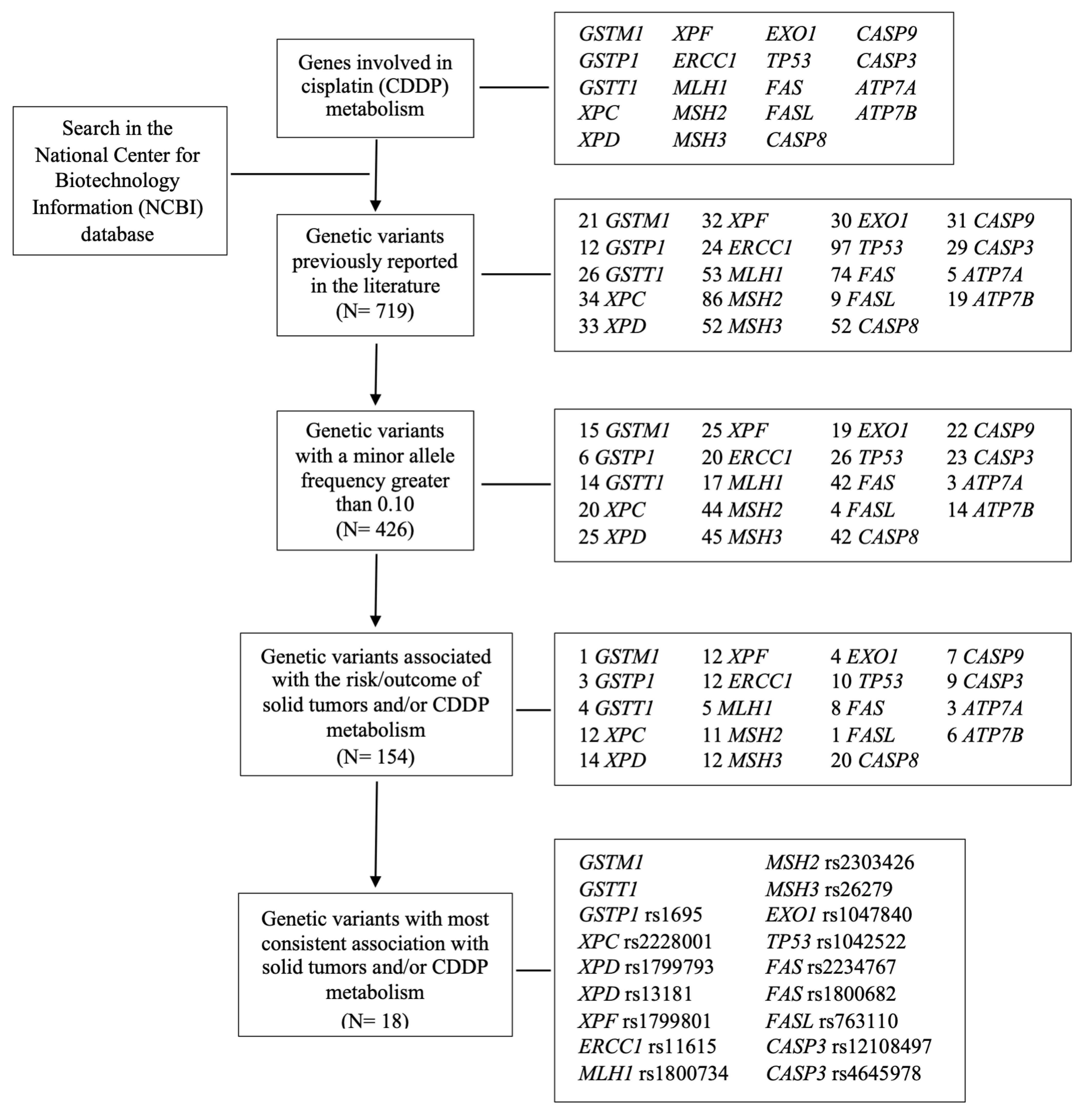
2.5. Genetic Variants Analysis

Genetic variants were selected for study based on the National Center for Biotechnology Information database, minor allele frequency greater than 10%, previous association with risk/outcome of solid tumors and/or CDDP metabolism, and the availability of financial resources (Figure A1). For genotyping, DNA samples from peripheral blood were collected, where the genotypes of the GSTM1 and GSTT1 variants [62] were obtained through the multiplex polymerase chain reaction (PCR) followed by digestion assays with enzymes of restriction. The additional genetic variants were evaluated by real-time PCR using TaqMan® SNP Genotyping Assays (Applied Biosystems®, Thermo Fisher Scientific Inc., Waltham, MA, USA), as follows: GSTP1 c.313A>G (rs1695) [63], XPC c.2815A>C (rs2228001) [64], XPD c.934G>A (rs1799793) [65], XPD c.2251A>C (rs13181) [65], XPF c.2505T>C (rs1799801) [66], ERCC1 c.354C>T (rs11615) [67], MLH1 c.-93G>A (rs1800734) [68], MSH2 c.211 +9G>C (rs2303426) [69], MSH3 c.3133A>G (rs26279) [70], EXO1 c.1762G>A (rs1047840) [71], P53 c.215G>C (rs1042522) [72], FAS c.-1378G>A (rs2234767) [73], FAS c.-671A>G (rs1800682) [74], FASL c.-844C>T (rs763110) [74], CASP3 c.-1191A>G (rs12108497) [75], and CASP3 c.-182-247G>T (rs4647601) [76]. Positive and negative controls were used in all genotyping reactions, and replications of 10% randomly selected samples were also performed in independent experiments, with 100% agreement.

2.6. Statistical Analysis

Descriptive statistics were performed according to the variables under study, with mean values and standard deviation (SD) in normal distribution or median and interquartile ranges (IQR) when applicable.
Pre- and post-treatment pure tone thresholds were described individually, as well as the averages of high-frequency minimum thresholds (considering 3, 4, 6, and 8 kHz) and ISO averages (0.5, 1, 2, and 4 kHz) in each ear. Wilcoxon’s signed-rank test for paired data was applied in the comparison of speech audiometry, pure tone averages, and frequency thresholds before and after chemoradiation, with the latter controlling for false discovery rates with the Benjamini–Hochberg correction in multiple testing [77]. Cochran’s Q test was used for the GBD classification of hearing loss before and after therapy. To assess the influence of clinicopathological aspects and genotypes related to high-frequency minimum threshold average changes from baseline, we performed multiple linear regression. Data were transformed into ranks. The significance level adopted for the study was 5%.
The main endpoint of this study was the proportion of patients with NCI CTCAE hearing loss G equal to or greater than 3 during follow-up based on audiometry monitoring. Multiple logistic regression was used to obtain the odds ratio (ORs) adjusted for any specific discrepancies for each independent variable, considering a 95% confidence interval (CI). Variables were selected using a conditional stepwise approach, permitting a p-value of under 0.10 in univariate regression.
Post hoc power analyses (PA) were also conducted for associations, taking into consideration p-value and CI as the measures of statistical significance [78,79]. After multivariate analysis, results with p ≤ 0.05 were validated using bootstrap [80] to verify the stability of risk estimates and account for missing data (1000 replications). Isolated SNVs associated with the increase in the hearing thresholds or grade 3 ototoxicity and combined SNVs associated with an increase in hearing thresholds or grade 3 ototoxicity with PA >70% were selected for this study.
All tests were performed using the Statistical Analysis System (SAS) for Windows, version 9.4 (SAS Institute Inc., 2002–2008, Cary, NC, USA) and Stata Statistical Software: Release 15 (Stata Corp LP, College Station, TX, USA).

3. Results

3.1. Study Population

In a median follow-up of 142 days, 152 patients were enrolled, of whom 89 were included in the analysis with the completion of baseline and post-treatment audiometry (Figure 2).
The median age was 56 years, and most patients were male and white, with a high rate of tobacco and alcohol consumption. Median BMI was within the normally acceptable range, most presented an ECOG status of 0, and the proportion of diabetes and hypertension was 10 and 26.9%, respectively. Most primary tumors were in the oral cavity or oropharynx, evenly distributed between the right and left sides of the head and neck, well or moderately differentiated, and at advanced stages (III or IV). The median cumulative CDDP dose among patients was 260 mg/m2. Eighty-eight patients received 2D RT, with a total dose of 70 Gy. The clinicopathological aspects of patients enrolled in the study are further detailed in Table 1.

3.2. Hearing Impairment in Monitoring Audiometry

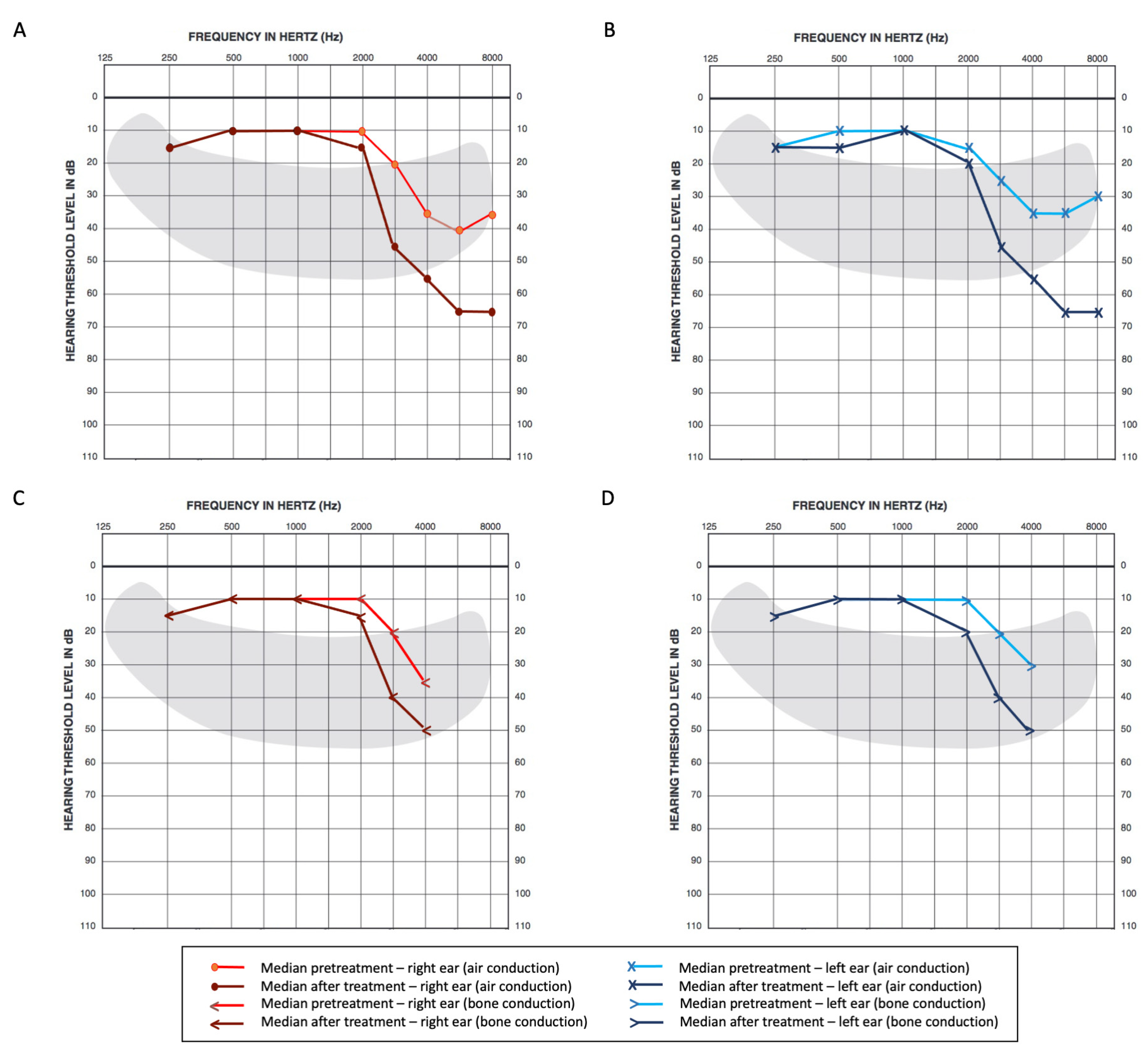
3.2.1. Pure Tone Audiometry

Analyzing the median thresholds for each frequency upon baseline, we were able to observe a normal range below 2 kHz and a trend toward higher thresholds, starting from 3 kHz in both conduction modalities (Figure 3).
After treatment, there was significant damage regarding higher frequencies over 2 kHz, which was more evident in air conduction analysis. Hearing thresholds for each frequency in pure tone audiometry are further detailed in Table 2.
Following the ISO average criteria, a median increase of 5 dB (p < 0.001) on the right side and 6.25 dB (p < 0.001) on the left side was observed when comparing baseline to post-treatment assessments. For bone conduction, there was a median increase of 6.25 (p < 0.001) on the right side and 6.25 dB (p < 0.001) on the left side, respectively (Table 3).
Regarding high-frequency average thresholds for pure tone air conduction audiometry, there was a median increase of 18 dB (p < 0.001) on the right and 19 dB (p < 0.001) on the left sides observed after exposure to CDDP and RT.

3.2.2. Speech Audiometry

Data from speech audiometry were retrievable in 62 patients since 27 had limited speech capability (nine were submitted to tracheostomy and eighteen presented tumors in the oral cavity). The median baseline SRT was 10 and 15 dB in the right and left ears, respectively. Pre- and post-treatment median differences were null on both sides (Table 3). Regarding SDS, median baseline and post-treatment scores were 96% and 92%, respectively, for both ears, with a decrease of 4% on the right side.

3.2.3. GBD Classification for Hearing Loss

Before chemoradiation, mild hearing loss was seen in about one-third and one-quarter of patients analyzed by air and bone conduction, respectively, and only three patients presented moderate hearing impairment in both assessments. After treatment, there was a significant increase in the proportion of mild and moderate hearing loss identified in air and bone conduction (chi2 20.16, p < 0.001 for air; chi2 18.24, p < 0.001 for bone conduction), although a severe degree of hearing loss was not observed throughout the study (Table 3). The unilateral loss was more evident in air conduction after treatment, although the same proportion was not observed in bone conduction analyses. All patients with unilateral damage after treatment had pharyngeal carcinoma located on the side of hearing loss and with changes at baseline.

3.2.4. Hearing Impairment in Monitoring Audiometry According to NCI CTCAE Criteria

The proportion of any-grade hearing impairment by air conduction after chemoradiation was 76.4% (68 out of 89 patients). The ototoxicity of G1 and G2 was observed in 23 (25.8%) and 19 (21.3%) patients, respectively. G3 or moderate/severe ototoxicity occurred in 26 (29.3%) participants, and G4 was not identified in this study.

3.3. Clinicopathological Aspects and Genotypes in Hearing Impairment

3.3.1. Average of Minimum Threshold for Pure Tone Air Conduction Audiometry at High Frequencies (3, 4, 6, and 8 kHz)

In univariate analysis, gender and cumulative CDDP dose were associated with hearing loss in the right ear, while BMI was associated with hearing loss in both ears. Only cumulative CDDP dose was associated with hearing loss in the right ear in multivariate analysis (regression coefficient = 0.08, p = 0.02), where the higher the dose of CDDP, the greater the hearing impairment (Supplemental Table S1).
When SNVs were analyzed individually, it was observed that patients with XPC c.2815AA genotype presented higher average threshold increases after CDDP chemoradiation than those with XPC c.2815AC or CC genotypes (23.8 versus 17.5 dB in the right ear; 27.5 versus 16.3 dB in left ear), as represented in Table 4. Higher average threshold increases were also seen after treatment in patients with combined genotypes GSTM1 null plus EXO1 c.1762GA or AA (21.3 versus 5.0 dB in the right ear; 22.5 versus 8.8 dB in the left ear) and with GSTP1 c.313AG or GG plus XPC c.2815AA (30.0 versus 17.5 dB in the right ear; 38.8 versus 16.3 dB in the left ear) in comparison to other related variants. The analyses of isolated and combined SNVs with biological significance with hearing loss at high frequencies are presented in Supplemental Table S2 and Table S3, respectively.

3.3.2. Hearing Impairment in Monitoring Audiometry According to NCI CTCAE Criteria

Race and BMI were significantly associated with the risk of G3 ototoxicity in univariate and multivariate analyses, but potential clinical risk factors such as age, gender, diabetes, hypertension, smoking, alcohol consumption, tumor stage, tumor side, and CDDP cumulative dose did not alter the risk of ototoxicity in univariate analysis (Supplemental Table S4). The occurrence of moderate/severe ototoxicity was more common in non-white than in white patients (66.7% versus 25.0%, respectively), with OR = 5.43 (95% CI: 1.21–24.27, p = 0.02) in multivariate analysis. BMI was also a potential predictor, as participants with grade 3 hearing impairment presented lower median BMI (17.8 versus 19.7), with the OR = 0.82 higher for every decrease in BMI (95% CI: 0.72–0.98, p = 0.03) in multivariate analysis.
When analyzed individually (Table 5), two SNVs were identified as independent factors for this outcome. Patients with GSTP1 c.313AG or GG genotypes had about 4.20 higher odds of having grade 3 or greater ototoxicity. Moreover, XPC c.2815AA genotype was associated with greater odds of severe hearing impairment, with a reported OR of 3.13 (p = 0.01) in the multivariate regression model. In associations of SNVs, it was observed that patients with GSTM1 null plus the XPC c.2815AA genotype had 8.19 greater odds of having moderate/severe hearing impairment (p = 0.02, PA = 99%). GSTP1 c.313AG or GG genotypes plus XPC c.2815AA, XPD c.934AA and EXO1 c.1762AA had ORs of 32.22 (p = 0.004, PA = 97%), 19.44 (p = 0.02, PA = 92%), and 12.08 (p = 0.01, PA = 81%), respectively. In addition, we observed relevant associations amongst DNA repair and apoptosis-related SNVs in patients with XPC c.2815AA genotype plus MSH3 c.3133A>G and FASL c.-844CC, where individuals with the respective profile had OR 17.09 (p = 0.009, PA = 88%) and 22.29 (p = 0.01, PA = 82%). Finally, patients with the combined genotypes EXO1 c.1792GA or AA and P53 c.215 CC had OR 20.97 (p = 0.02, PA = 85%). Further details of other SNVs and their combinations are summarized in Supplemental Table S5 and Table S6, respectively.

4. Discussion

In this clinical and pharmacogenetic cohort, it was possible to reaffirm the clinical relevance of hearing loss induced by CDDP. CDDP induces ototoxicity through the promotion of oxidative stress and inflammation in the cochlea, with the increased generation of reactive oxygen species (ROS) [81]. The long-term accumulation of CDDP in the cochlear endolymph was also described through plasma mass spectrometry in preclinical models, justifying the potential for permanent damage [82].
Firstly, substantial hearing loss before treatment was observed in our cohort; high-frequency minimum thresholds were higher at baseline, ranging from 35 to 40 dB over 4 kHz. This may be attributable to the high proportion of smokers in our sample since smoking is a reported risk factor for loss at high frequencies [18,83,84]. A history of noise exposure, not assessed in this cohort, could also explain this finding as well as uneven losses in left and right ears not associated with the tumor side [85,86,87]. We were able to observe a meaningful change after CDDP exposure in regard to minimum hearing thresholds, particularly in higher frequencies in univariate analysis, as suggested by previous studies [15], with limitations involving higher pitch sounds. Caballero and colleagues described similar findings in a cohort of 103 patients, with significantly meaningful changes after CDDP exposure (median change of 9.5 dB in the right and 18.75 dB in the left ears for 4 kHz; 18.6 dB in the right and 28.7 dB in the left, for 8 kHz). The limitations to quality of life related to hearing loss from CDDP have already been reported in a recent systematic review from Pearson and colleagues [16]. Regarding additional clinical factors, cumulative CDDP dose was observed as a risk factor for greater change in high-frequency averages (3, 4, 6, and 8 kHz) for the right ear, which prompted the inclusion of this variable in multivariate analysis for both sides.
When accounting for the 0.25 to 4 kHz interval, there was also a significant relative increase in mild and moderate hearing loss after CDDP in our analysis, following GBD classification. The percentage of 64.1% with a threshold ≥ 20 dB after treatment is markedly superior to the overall prevalence reported in the literature for the general population (19.3%) [88], pointing to the cytotoxic effects of CDDP on hearing impairment. To our knowledge, this is the first study to report this classification before and after CDDP in patients diagnosed with HNSCC [89]. Unilateral hearing damage was observed in four patients (4.5%) after therapy under pure tone audiometry air conduction, from which two (2.2%) had reported losses in both conduction modalities (air and bone). It is worth commenting that all patients with unilateral damage had pharyngeal carcinoma located on the side of hearing loss, and most had changes at baseline. Even though the RT technique and CDDP dose did not differ amongst patients, the location of the tumor in relation to the inner ear could have influenced this finding.
On the other hand, median outcomes from speech audiometry (SRT and SDS) were practically unchanged after platinum exposure. One possible explanation for this may be related to the fact that human speech usually ranges from 0.25 to 4 kHz [90], while CDDP-related hearing loss involves more relevant changes beyond 3 kHz. In an isolated acoustic environment, frequencies related to speech may be unaltered, though it is possible to expect a greater extent of limitation in terms of communication with background noise, which was not assessed in this cohort. The largest study analyzing speech audiometry after CDDP exposure was performed by Shahbazi and colleagues [91], evaluating the prevalence of speech recognition disability, defined as SRT greater than 15 dB, in testicular cancer survivors. In 1347 patients, speech recognition disability was identified in 10.4%, and the association of the cumulative CDDP dose could also not be confirmed. Those findings are distinct from our analysis, where 51.6% could be classified as speech-disabled before therapy and 60.7% after therapy. The study populations are markedly different since the Platinum Study [91] included younger patients not bearing primary tumors in the head and neck and without risk factors such as tobacco and alcohol consumption. There are, to date, only scarce amounts of the literature data on speech audiometry for HNSCC, thus limiting further comparisons.
When considering the NCI CTCAE criteria for the classification of hearing loss, the proportion of 29.5% moderate/severe ototoxicity was marginally higher than previously reported literature data, ranging from 20 to 25% in adults [10,37]. Except for race and BMI, other clinical variables such as age, sex, tumor location, and staging could not be identified as prognostic factors in this analysis, and although cumulative CDDP is recognized as a risk factor for hearing damage [37], this association could not be observed in the present data for this outcome specifically. Some recent studies have suggested the presence of emotional stress as a possible risk factor for enhanced tumorigenesis and neurotoxicity induced by chemotherapy in general [92]. A cross-sectional analysis of 623 cancer survivors described a higher association of tinnitus (p = 0.029) and hearing loss (p = 0.007) amongst patients with higher distress scores [93]. Due to the characteristics of the study design, it is not possible to differentiate stress as an independent risk factor, as opposed to a consequence of long-term toxicity. Even though a longitudinal study of the current analysis could potentially assess this variable, distress scores were not previously planned and included in this cohort.
In this study, GSTP1 c313AG or GG and XPC c.2815 AA genotypes increased the odds of moderate/severe ototoxicity 4.20- and 3.13-fold, respectively. Preclinical studies have demonstrated that GSTP1 c313 A>G encodes a change from isoleucine to valine in codon 105, leading to reduced protein activity and detoxification [63], while the XPC c.2815 C allele promotes the change from lysine to glutamine in codon 939, also diminishing protein activity and, consequently, DNA repair (Table A1) [64]. There is, however, marked heterogeneity of clinical effects in terms of the currently available literature. For instance, GSTP1 c313AG or GG was associated with an increased risk of moderate/severe hearing impairment in 106 [41] and 64 children [43], respectively, treated with platinum agents, using the Brock hearing loss classification of equal or greater than 2 [20]. The association between cumulative CDDP and ototoxicity was found in the study conducted by Lui and colleagues [41] but not in the study by Sherief and colleagues [43]. Even though our findings in a previous analysis of the data [45] are similar and in agreement with the functional roles of GSTP1 c313A>G [94], this SNV was not related to CDDP-induced ototoxicity in an additional cohort that recruited 71 children and young patients with various solid tumors [38], while in 173 patients with testicular carcinoma, post-treatment audiometric evaluations prompted divergent results, even though baseline assessments were not retrievable [39]. Reported results were also conflicting for isolated XPC c.2815A>G [35]. The XPC c.2815AA genotype was associated with an increased risk of any grade of toxicity [46] in a previous analysis of the data conducted by our group, and the same effect was observed in a smaller subset of patients with osteosarcoma [44]. Nonetheless, Lui and colleagues [41] did not present a significant association among 106 pediatric patients treated with platin analogs. Functional analyses performed for this variant [64] suggest the presence of the C allele reduces DNA repair, which would theoretically increase the risk of toxicity in contrast to what is currently reported, though an additional assay from Khan and colleagues [26] did not demonstrate a clear difference for the rate of nucleotide excision repair. Differences in the results obtained from the studies are not easily explained and may have originated from limitations related to sample size, patient baseline characteristics, tumor types, and treatment administered. There are also markedly distinct hearing loss classifications applied in previous cohorts, hampering proper direct comparisons with NCI CTCAE v4.03. Larger cohorts, in addition to further functional assays, would be ideal to better confront these findings.
The metabolism of CDDP is known to involve cellular efflux, NER, and MMR damage repair, as well as apoptosis [37]. We were able to observe meaningful interactions between variants encoding those distinct pathways, suggesting that toxicity may be enhanced by the coexistence of more than one mutation in the different stages of CDDP metabolism and cytotoxic effect. The combination of GSTM1 null plus XPC c.2815AA, GSTP1 c.313AG or GG plus XPC c.2815AA, XPD c.934AA or EXO1 c.1762AA, or XPC c.2815AA plus MSH3 c.3133AA or FASL c.-844CC (Table A1) intensified the odds of moderate/severe ototoxicity up to 32.22-fold. The variant alleles from SNVs XPD c.934 (A) [65], EXO1 c.1762 (A) [71], and MSH3 c.3133 (A) [70] have been shown to reduce DNA repair activity by encoding amino acid replacements with the consequent loss of protein function or expression (Table A1). Additionally, the SNV FASL c.-844 is located within the enhancer-biding region of FASL, and luciferase assays have described the variant genotype TT to promote protein affinity twice lower than wild CC donors, leading to less protein expression and, as such, reduced apoptosis [74]. Hence, the combination of genotypes enhancing CDDP accumulation and reducing repair or activating apoptosis could potentiate the risk of ototoxicity, as observed in this analysis. To our knowledge, no studies focusing on the effects of the combinations of SNVs on the genes of distinct pathways of CDDP metabolism have been conducted to date.
An association with MMR and apoptosis mechanisms was also noted in this study, as the combination of EXO1 c.1762GA or AA and P53 c.215CC genotypes increased the risk for events with OR 20.97. The P53 c.215 wild allele encoding arginine (G) was described as more efficient in inducing apoptosis than the proline (C) variant [72]. The P53 protein signaling pathway promotes cell death triggered by the generation of ROS [35]. In addition to apoptosis, P53 is related to cell cycle arrest, cell senescence, and DNA repair [95]. Cellular senescence is a state of permanent cell cycle arrest that is able to promote local inflammation and tissue damage [96]. In vitro studies have suggested that early senescence in response to genotoxic stress was P53-dependent and EXO1-depleted [97,98]. Moreover, Benkafadar and colleagues [99] observed that the response to ROS-induced DNA damage leads to cochlear cell senescence by activating the P53 pathway and hence contributing to age-related hearing loss. To date, there is a lack of evidence for a direct association between ototoxicity by CDDP and cell senescence. However, the accumulation of senescent neuronal cells is associated with CDDP-induced peripheral neuropathy in mice [100]. Thus, we may infer that patients with EXO1 c.1792GA or AA and P53 c.215CC combined genotypes were more efficient in promoting cell cycle arrest and senescence of sensory cells after injury by CDDP and, consequently, were at greater risk of hearing loss when compared to patients carrying other genotypes.
We are aware that this study is limited for its sample size; thus, similarly to previous studies, lacking power for further SNVs combinations or polygenic evaluations and correction for other possible confounders. Though statistical tools were used to stabilize risk, such as bootstrap and power post hoc calculations, there may still be unknown influential factors not identifiable in this sample. It must also be considered that not all SNVs in the genes related to CDDP detoxification, DNA repair, and the apoptosis of damaged cells were evaluated in this study; only those recognized with a greater potential to induce ototoxicity were evaluated here. Thus, it is possible that other SNVs with equal or even greater importance in CDDP ototoxicity will be identified in future studies. Furthermore, other known SNVs for CDDP-induced ototoxicity unrelated to stages regarding drug absorption, distribution, metabolism, and excretion were not assessed and could be additional confounders. There is evidence supporting additional SNVs as risk factors for ototoxicity induced by CDDP related to ACYP2 [33,34], TPMT [35], COMT [35], and WFS1 [36] genes. ACYP2 is known to influence ATP-dependent calcium signaling, which may play a role in sensorineural hearing loss [33]. TPMT and COMT are methyltransferases that may inactivate CDDP and purine compounds. The Mendelian deafness genes, amongst which WSF1 is included, encode proteins reported to control endothelial reticulum stress response, thus influencing inner ear cellular damage [36]. Apart from known and unknown genetic risk factors for toxicity and hearing loss, clinical variables, such as a history of noise exposure [18] and distress scores [93], were also not collected from this cohort. We believe, however, that the exclusion of patients with reported hearing loss and hearing impairment in audiometry before treatment could, to some extent, attenuate these limitations.
It is also important to consider distinct patient characteristics when assessing the generalizability of this study for other tumors since the population was predominantly male, with a high frequency of smokers and alcohol users, as well as locally advanced stages of HNSCC. Treatment approaches in the field of RT may also be distinct and could influence the prevalence and severity of adverse events, mainly in institutions with more frequent use of intensity-modulated RT. Though prespecified treatment protocols were strictly followed, therefore preventing confounding to some extent, heterogeneity in therapy protocols could affect the generalizability of these results.

5. Conclusions

The results of this cohort suggest, for the first time, the interactions of inherited genetic abnormalities involved in CDDP metabolism as potential candidate targets for future risk models in ototoxicity. The development of genetic and clinical risk prediction tools is essential, not only for optimizing treatment selection based on efficacy but also to assist in supportive care during therapy. We believe these results may be included in future polygenic and clinical predictive models.

Supplementary Materials

The following supporting information can be downloaded at: https://www.mdpi.com/article/10.3390/cancers15061759/s1; Table S1. Clinicopathological factors and minimum threshold averages for pure tone air conduction audiometry in high frequencies (3, 4, 6, and 8 kHz): linear regression coefficients; Table S2. Single nucleotide variants encoding cisplatin metabolism and pure tone high-frequency threshold averages in air conduction (3, 4, 6, and 8 kHz); Table S3. Combinations of single nucleotide variants encoding cisplatin metabolism and their association with pure tone high-frequency threshold averages (3, 4, 6, and 8 kHz); Table S4. Clinical characteristics of patients stratified by intensity of ototoxicity classified by NCI CTCAE v4.0 criteria; Table S5. Single nucleotide variants and hearing impairment (according to NCI criteria—NCI CTCAE v4.0) related to cisplatin-based chemoradiation; Table S6. Associations for single nucleotide variants and hearing impairment (according to NCI criteria—NCI CTCAE v4.0) related to cisplatin-based chemoradiation.

Author Contributions

Conceptualization, L.T.M., E.F.D.C. and C.S.P.L.; methodology, L.T.M., E.F.D.C., B.F.C., G.J.L. and L.C.; software, L.T.M. and G.J.L.; validation, C.T.C., A.M.C. and C.S.P.L.; formal analysis, L.T.M. and E.F.D.C.; investigation, E.F.D.C. and L.C.; writing—original draft preparation, L.T.M., E.F.D.C. and B.F.C.; writing—review and editing, L.C., C.T.C., A.M.C. and C.S.P.L.; supervision, C.T.C., A.M.C. and C.S.P.L.; project administration, C.S.P.L.; funding acquisition, G.J.L. and C.S.P.L. All authors have read and agreed to the published version of the manuscript.

Funding

This research was funded by Fundação de Amparo a Pesquisa do Estado de São Paulo (FAPESP), grant number 12/01807-2.

Institutional Review Board Statement

The study was conducted in accordance with the Declaration of Helsinki and approved by the Institutional Ethics Committee from the State University of Campinas—UNICAMP (Protocol numbers 274/2011 and 62870722.1.0000.5404).

Informed Consent Statement

Informed consent was obtained from all subjects involved in the study.

Data Availability Statement

The data are not publicly accessible due to privacy and ethical restrictions but can be made available upon request from the corresponding author.

Acknowledgments

We would like to thank the Department of Statistics staff from the Faculty of Medical Sciences, University of Campinas, for their kind support and guidance. We would also like to thank the patients who agreed to participate in this study and the Fundação de Amparo a Pesquisa do Estado de São Paulo (FAPESP) for the financial support.

Conflicts of Interest

The authors declare no conflict of interest. The funders had no role in the design of the study; in the collection, analyses, or interpretation of data; in the writing of the manuscript; or in the decision to publish the results.

Appendix A

Table A1. Summary of evidence on single nucleotide variants encoding cisplatin metabolism and their expected functions.
SNVSNV ID (rs)Ch RegionGene LocationCoding ChangeAlleleFunctionAlleleFunctionAssayRef.
GSTM1NA1p13.3NADeletionPresentNormal detoxificationNullNo detoxificationLuciferase[62]
GSTT1NA22q11.23NADeletionPresentNormal detoxificationNullNo detoxificationLuciferase[62]
GSTP1 c.313A>G169511q13.2Exon 5Ile105ValANormal detoxificationGReduced detoxificationIH[63]
XPC c.2815A>C22280013p25.1Exon 15Lys939GlnANormal repairCReduced repairComet assay[64]
XPD c.934G>A179979319q13.32Exon 10Asp312AsnGNormal repairAReduced repairHCR[65]
XPD c.2251A>C1318119q13.32Exon 23Lys751GlnANormal repairCReduced repairHCR[65]
XPF c.2505T>C179980116p13.12Exon 11Ser835SerTNormal repairCReduced repairIH[66]
ERCC1 c.354C>T1161519q13.32Exon 4Asn118AsnCNormal repairTReduced repairAAS[67]
MLH1 c.-93G>A18007343p22.2PromoterNAGNormal repairAReduced repairLuciferase[68]
MSH2 c.211+9G>C23034262p21IntronNAGReduced repairCNormal repairWestern-Blot[69]
MSH3 c.3133G>A262795q14.1Exon 23Ala1045ThrGNormal repairAReduced repairExpression by qPCR[70]
EXO1 c.1762G>A10478401q43Exon 12Glu267LysGNormal repairAReduced repairWestern-Blot[71]
TP53 c.215G>C104252217p13.1Exon 4Arg72ProGNormal apoptosisCReduced apoptosisWestern-Blot[72]
FAS c.-1378G>A223476710q23.31PromoterNAGNormal apoptosisAReduced apoptosisEMSA[73]
FAS c.-671A>G180068210q23.31IntronNAANormal apoptosisGReduced apoptosisLuciferase [74]
FASL c.-844C>T7631101q24.3PromoterNACNormal apoptosisTReduced apoptosisLuciferase [74]
CASP3 c.-1191A>G121084974q35.1PromoterNAANormal apoptosisGReduced apoptosisExpression by qPCR[75]
CASP3 c.-182-247G>T46476014q35.1IntronNAGNormal apoptosisTReduced apoptosisExpression by qPCR[76]
AAS: Atomic absorbance spectrometry; Ala: alanine; Arg: arginine; Asn: asparagine; Asp: aspartic acid; Ch: chromosome; EMSA: Electrophoresis mobility shift assay; Glu: glutamic acid; Gln: glutamine; HCR: Host cell reactivation; ID: identification; IH: immunohistochemistry; Ile: isoleucine; Lys: lysine; NA: not applicable; Pro: proline; qPCR: quantitative PCR; rs: reference number; Ser: serine; SNV: single nucleotide variant; Thr: threonine; Val: valine.
Figure A1. Flowchart illustrating the process used to select the genetic variants included in the study. Initially, the National Center for Biotechnology Information database was searched to identify variations in genes associated with the metabolism of cisplatin (CDDP). A total of 719 genetic variations were identified. Next, 426 genetic variations were selected based on a minor allele frequency greater than 0.10. Subsequently, 154 genetic variations previously associated with the risk or outcome of solid tumors and/or the metabolism of CDDP were selected, and 18 of them with most consistent association were included in the study.
Cancers 15 01759 g0a1

References

  1. Siegel, R.L.; Miller, K.D.; Fuchs, H.E.; Jemal, A. Cancer Statistics, 2021. CA Cancer J. Clin. 2021, 71, 7–33. [Google Scholar] [CrossRef]
  2. Sung, H.; Ferlay, J.; Siegel, R.L.; Laversanne, M.; Soerjomataram, I.; Jemal, A.; Bray, F. Global Cancer Statistics 2020: GLOBOCAN Estimates of Incidence and Mortality Worldwide for 36 Cancers in 185 Countries. CA Cancer J. Clin. 2021, 71, 209–249. [Google Scholar] [CrossRef] [PubMed]
  3. Vermorken, J.B.; Remenar, E.; van Herpen, C.; Gorlia, T.; Mesia, R.; Degardin, M.; Stewart, J.S.; Jelic, S.; Betka, J.; Preiss, J.H.; et al. Cisplatin, Fluorouracil, and Docetaxel in Unresectable Head and Neck Cancer. N. Engl. J. Med. 2007, 357, 1695–1704. [Google Scholar] [CrossRef] [Green Version]
  4. Argiris, A.; Karamouzis, M.V.; Raben, D.; Ferris, R.L. Head and Neck Cancer. Lancet Lond. Engl. 2008, 371, 1695–1709. [Google Scholar] [CrossRef]
  5. Bonner, J.A.; Harari, P.M.; Giralt, J.; Azarnia, N.; Shin, D.M.; Cohen, R.B.; Jones, C.U.; Sur, R.; Raben, D.; Jassem, J.; et al. Radiotherapy plus Cetuximab for Squamous-Cell Carcinoma of the Head and Neck. N. Engl. J. Med. 2006, 354, 567–578. [Google Scholar] [CrossRef] [Green Version]
  6. Vermorken, J.B.; Mesia, R.; Rivera, F.; Remenar, E.; Kawecki, A.; Rottey, S.; Erfan, J.; Zabolotnyy, D.; Kienzer, H.-R.; Cupissol, D.; et al. Platinum-Based Chemotherapy plus Cetuximab in Head and Neck Cancer. N. Engl. J. Med. 2008, 359, 1116–1127. [Google Scholar] [CrossRef] [PubMed] [Green Version]
  7. Burtness, B.; Harrington, K.J.; Greil, R.; Soulières, D.; Tahara, M.; de Castro, G.; Psyrri, A.; Basté, N.; Neupane, P.; Bratland, Å.; et al. Pembrolizumab Alone or with Chemotherapy versus Cetuximab with Chemotherapy for Recurrent or Metastatic Squamous Cell Carcinoma of the Head and Neck (KEYNOTE-048): A Randomised, Open-Label, Phase 3 Study. Lancet 2019, 394, 1915–1928. [Google Scholar] [CrossRef] [PubMed]
  8. Albers, J.W.; Chaudhry, V.; Cavaletti, G.; Donehower, R.C. Interventions for Preventing Neuropathy Caused by Cisplatin and Related Compounds. Cochrane Database Syst. Rev. 2014, CD005228. [Google Scholar] [CrossRef]
  9. Pincinato, E.C.; Visacri, M.B.; de Souza, C.M.; Tuan, B.T.; Ferrari, G.B.; de Oliveira, D.N.; Barbosa, C.R.; Rodrigues, R.F.; Granja, S.; Ambrósio, R.F.L.; et al. Impact of Drug Formulation and Free Platinum/Cisplatin Ratio on Hypersensitivity Reactions to Cisplatin: Formulation Matters. J. Clin. Pharm. Ther. 2015, 40, 41–47. [Google Scholar] [CrossRef]
  10. Ganesan, P.; Schmiedge, J.; Manchaiah, V.; Swapna, S.; Dhandayutham, S.; Kothandaraman, P.P. Ototoxicity: A Challenge in Diagnosis and Treatment. J. Audiol. Otol. 2018, 22, 59–68. [Google Scholar] [CrossRef] [Green Version]
  11. Laurell, G. Pharmacological Intervention in the Field of Ototoxicity. HNO 2019, 67, 434–439. [Google Scholar] [CrossRef] [PubMed] [Green Version]
  12. Bokemeyer, C.; Berger, C.; Hartmann, J.; Kollmannsberger, C.; Schmoll, H.; Kuczyk, M.; Kanz, L. Analysis of Risk Factors for Cisplatin-Induced Ototoxicity in Patients with Testicular Cancer. Br. J. Cancer 1998, 77, 1355–1362. [Google Scholar] [CrossRef] [PubMed]
  13. Frisina, R.D.; Wheeler, H.E.; Fossa, S.D.; Kerns, S.L.; Fung, C.; Sesso, H.D.; Monahan, P.O.; Feldman, D.R.; Hamilton, R.; Vaughn, D.J.; et al. Comprehensive Audiometric Analysis of Hearing Impairment and Tinnitus After Cisplatin-Based Chemotherapy in Survivors of Adult-Onset Cancer. J. Clin. Oncol. 2016, 34, 2712–2720. [Google Scholar] [CrossRef] [PubMed]
  14. Chen, W.C.; Jackson, A.; Budnick, A.S.; Pfister, D.G.; Kraus, D.H.; Hunt, M.A.; Stambuk, H.; Levegrun, S.; Wolden, S.L. Sensorineural Hearing Loss in Combined Modality Treatment of Nasopharyngeal Carcinoma. Cancer 2006, 106, 820–829. [Google Scholar] [CrossRef] [PubMed]
  15. Caballero, M.; Mackers, P.; Reig, O.; Buxo, E.; Navarrete, P.; Blanch, J.L.; Grau, J.J. The Role of Audiometry Prior to High-Dose Cisplatin in Patients with Head and Neck Cancer. Oncology 2017, 93, 75–82. [Google Scholar] [CrossRef] [PubMed]
  16. Pearson, S.E.; Taylor, J.; Patel, P.; Baguley, D.M. Cancer Survivors Treated with Platinum-Based Chemotherapy Affected by Ototoxicity and the Impact on Quality of Life: A Narrative Synthesis Systematic Review. Int. J. Audiol. 2019, 58, 685–695. [Google Scholar] [CrossRef]
  17. Rademaker-Lakhai, J.M.; Crul, M.; Zuur, L.; Baas, P.; Beijnen, J.H.; Simis, Y.J.W.; van Zandwijk, N.; Schellens, J.H.M. Relationship Between Cisplatin Administration and the Development of Ototoxicity. J. Clin. Oncol. 2006, 24, 918–924. [Google Scholar] [CrossRef]
  18. Talach, T.; Rottenberg, J.; Gal, B.; Kostrica, R.; Jurajda, M.; Kocak, I.; Lakomy, R.; Vogazianos, E. Genetic Risk Factors of Cisplatin Induced Ototoxicity in Adult Patients. Neoplasma 2016, 63, 263–268. [Google Scholar] [CrossRef]
  19. Silva Nogueira, G.A.; Dias Costa, E.F.; Lopes-Aguiar, L.; Penna Lima, T.R.; Visacri, M.B.; Pincinato, E.C.; Lourenço, G.J.; Calonga, L.; Mariano, F.V.; Messias de Almeida Milani Altemani, A.; et al. Polymorphisms in DNA Mismatch Repair Pathway Genes Predict Toxicity and Response to Cisplatin Chemoradiation in Head and Neck Squamous Cell Carcinoma Patients. Oncotarget 2018, 9, 29538–29547. [Google Scholar] [CrossRef] [Green Version]
  20. Clemens, E.; Broer, L.; Langer, T.; Uitterlinden, A.G.; de Vries, A.C.H.; van Grotel, M.; Pluijm, S.F.M.; Binder, H.; Byrne, J.; Broeder, E. van D.; et al. Genetic Variation of Cisplatin-Induced Ototoxicity in Non-Cranial-Irradiated Pediatric Patients Using a Candidate Gene Approach: The International PanCareLIFE Study. Pharm. J. 2020, 20, 294–305. [Google Scholar] [CrossRef]
  21. Siddik, Z.H. Cisplatin: Mode of Cytotoxic Action and Molecular Basis of Resistance. Oncogene 2003, 22, 7265–7279. [Google Scholar] [CrossRef] [PubMed] [Green Version]
  22. Kuo, M.T.; Chen, H.H.W.; Song, I.-S.; Savaraj, N.; Ishikawa, T. The Roles of Copper Transporters in Cisplatin Resistance. Cancer Metastasis Rev. 2007, 26, 71–83. [Google Scholar] [CrossRef]
  23. Hayes, J.D.; Flanagan, J.U.; Jowsey, I.R. Glutathione Transferases. Annu. Rev. Pharmacol. Toxicol. 2005, 45, 51–88. [Google Scholar] [CrossRef] [PubMed]
  24. Ishikawa, T.; Wright, C.; Ishizuka, H. GS-X Pump Is Functionally Overexpressed in Cis-Diamminedichloroplatinum (II)-Resistant Human Leukemia HL-60 Cells and down-Regulated by Cell Differentiation. J. Biol. Chem. 1994, 269, 29085–29093. [Google Scholar] [CrossRef] [PubMed]
  25. Shuck, S.C.; Short, E.A.; Turchi, J.J. Eukaryotic Nucleotide Excision Repair: From Understanding Mechanisms to Influencing Biology. Cell Res. 2008, 18, 64–72. [Google Scholar] [CrossRef] [Green Version]
  26. Khan, S.G. A New Xeroderma Pigmentosum Group C Poly(AT) Insertion/Deletion Polymorphism. Carcinogenesis 2000, 21, 1821–1825. [Google Scholar] [CrossRef] [Green Version]
  27. Lainé, J.P.; Mocquet, V.; Bonfanti, M.; Braun, C.; Egly, J.M.; Brousset, P. Common XPD (ERCC2) Polymorphisms Have No Measurable Effect on Nucleotide Excision Repair and Basal Transcription. DNA Repair 2007, 6, 1264–1270. [Google Scholar] [CrossRef]
  28. Goode, E.L.; Ulrich, C.M.; Potter, J.D. Polymorphisms in DNA Repair Genes and Associations with Cancer Risk. Cancer Epidemiol. Biomark. Prev. 2002, 11, 1513. [Google Scholar]
  29. Martin, L.P.; Hamilton, T.C.; Schilder, R.J. Platinum Resistance: The Role of DNA Repair Pathways. Clin. Cancer Res. 2008, 14, 1291–1295. [Google Scholar] [CrossRef] [Green Version]
  30. Mello, J.A.; Acharya, S.; Fishe, R.; Essigmann’, J.M. The Mismatch-Repair Protein HMSH2 Binds Selectively to DNA Adducts of the Anticancer Drug Cisplatin. Chem. Biol. 1996, 3, 579–589. [Google Scholar] [CrossRef] [Green Version]
  31. Singh, S. Cytoprotective and Regulatory Functions of Glutathione S-Transferases in Cancer Cell Proliferation and Cell Death. Cancer Chemother. Pharmacol. 2015, 75, 1–15. [Google Scholar] [CrossRef]
  32. De Zio, D.; Cianfanelli, V.; Cecconi, F. New Insights into the Link Between DNA Damage and Apoptosis. Antioxid. Redox Signal. 2013, 19, 559–571. [Google Scholar] [CrossRef] [PubMed] [Green Version]
  33. Drögemöller, B.I.; Brooks, B.; Critchley, C.; Monzon, J.G.; Wright, G.E.B.; Liu, G.; Renouf, D.J.; Kollmannsberger, C.K.; Bedard, P.L.; Hayden, M.R.; et al. Further Investigation of the Role of ACYP2 and WFS1 Pharmacogenomic Variants in the Development of Cisplatin-Induced Ototoxicity in Testicular Cancer Patients. Clin. Cancer Res. 2018, 24, 1866. [Google Scholar] [CrossRef] [PubMed] [Green Version]
  34. Driessen, C.M.; Ham, J.C.; te Loo, M.; van Meerten, E.; van Lamoen, M.; Hakobjan, M.H.; Takes, R.P.; van der Graaf, W.T.; Kaanders, J.H.; Coenen, M.J.H.; et al. Genetic Variants as Predictive Markers for Ototoxicity and Nephrotoxicity in Patients with Locally Advanced Head and Neck Cancer Treated with Cisplatin-Containing Chemoradiotherapy (The PRONE Study). Cancers 2019, 11, 551. [Google Scholar] [CrossRef] [PubMed] [Green Version]
  35. Tserga, E.; Nandwani, T.; Edvall, N.K.; Bulla, J.; Patel, P.; Canlon, B.; Cederroth, C.R.; Baguley, D.M. The Genetic Vulnerability to Cisplatin Ototoxicity: A Systematic Review. Sci. Rep. 2019, 9, 3455. [Google Scholar] [CrossRef] [Green Version]
  36. Wheeler, H.E.; Gamazon, E.R.; Frisina, R.D.; Perez-Cervantes, C.; El Charif, O.; Mapes, B.; Fossa, S.D.; Feldman, D.R.; Hamilton, R.J.; Vaughn, D.J.; et al. Variants in WFS1 and Other Mendelian Deafness Genes Are Associated with Cisplatin-Associated Ototoxicity. Clin. Cancer Res. 2017, 23, 3325. [Google Scholar] [CrossRef] [Green Version]
  37. Trendowski, M.R.; El Charif, O.; Dinh, P.C.; Travis, L.B.; Dolan, M.E. Genetic and Modifiable Risk Factors Contributing to Cisplatin-Induced Toxicities. Clin. Cancer Res. 2019, 25, 1147–1155. [Google Scholar] [CrossRef] [Green Version]
  38. Peters, U.; Preisler-Adams, S.; Hebeisen, A.; Hahn, M.; Seifert, E.; Lanvers, C.; Heinecke, A.; Horst, J.; Jürgens, H.; Lamprecht-Dinnesen, A. Glutathione S-Transferase Genetic Polymorphisms and Individual Sensitivity to the Ototoxic Effect of Cisplatin. Anticancer Drugs 2000, 11, 639–643. [Google Scholar] [CrossRef]
  39. Oldenburg, J.; Kraggerud, S.M.; Cvancarova, M.; Lothe, R.A.; Fossa, S.D. Cisplatin-Induced Long-Term Hearing Impairment Is Associated With Specific Glutathione S-Transferase Genotypes in Testicular Cancer Survivors. J. Clin. Oncol. 2007, 25, 708–714. [Google Scholar] [CrossRef]
  40. Choeyprasert, W.; Sawangpanich, R.; Lertsukprasert, K.; Udomsubpayakul, U.; Songdej, D.; Unurathapan, U.; Pakakasama, S.; Hongeng, S. Cisplatin-Induced Ototoxicity in Pediatric Solid Tumors: The Role of Glutathione S-Transferases and Megalin Genetic Polymorphisms. J. Pediatr. Hematol. Oncol. 2013, 35, e138–e143. [Google Scholar] [CrossRef] [Green Version]
  41. Lui, G.; Bouazza, N.; Denoyelle, F.; Moine, M.; Chastagner, P.; Corradini, N.; Entz-Werle, N.; Vérité, C.; Landmanparker, J.; Sudour-Bonnange, H.; et al. Association between Genetic Polymorphisms and Platinum-Induced Ototoxicity in Children. Oncotarget 2018, 9, 30883–30893. [Google Scholar] [CrossRef]
  42. Budai, B.; Prekopp, P.; Noszek, L.; Kovács, E.; Szőnyi, M.; Erdélyi, D.; Biró, K.; Geczi, L. GSTM1 Null and GSTT1 Null: Predictors of Cisplatin-Caused Acute Ototoxicity Measured by DPOAEs. J. Mol. Med. 2020, 98, 963–971. [Google Scholar] [CrossRef]
  43. Sherief, L.M.; Rifky, E.; Attia, M.; Ahmed, R.; Kamal, N.M.; Oshi, M.A.M.; Hanna, D. Platinum-Induced Ototoxicity in Pediatric Cancer Survivors: GSTP1 c.313A>G Variant Association. Medicine 2022, 101, e31627. [Google Scholar] [CrossRef] [PubMed]
  44. Caronia, D.; Patiño-García, A.; Milne, R.L.; Zalacain-Díez, M.; Pita, G.; Alonso, M.R.; Moreno, L.T.; Sierrasesumaga-Ariznabarreta, L.; Benítez, J.; González-Neira, A. Common Variations in ERCC2 Are Associated with Response to Cisplatin Chemotherapy and Clinical Outcome in Osteosarcoma Patients. Pharm. J. 2009, 9, 347–353. [Google Scholar] [CrossRef] [PubMed]
  45. Pincinato, E.C.; Costa, E.F.D.; Lopes-Aguiar, L.; Nogueira, G.A.S.; Lima, T.R.P.; Visacri, M.B.; Costa, A.P.L.; Lourenço, G.J.; Calonga, L.; Mariano, F.V.; et al. GSTM1, GSTT1 and GSTP1 Ile105Val Polymorphisms in Outcomes of Head and Neck Squamous Cell Carcinoma Patients Treated with Cisplatin Chemoradiation. Sci. Rep. 2019, 9, 9312. [Google Scholar] [CrossRef] [PubMed] [Green Version]
  46. Lopes-Aguiar, L.; Costa, E.F.D.; Nogueira, G.A.S.; Lima, T.R.P.; Visacri, M.B.; Pincinato, E.C.; Calonga, L.; Mariano, F.V.; de Almeida Milani Altemani, A.M.; Altemani, J.M.C.; et al. XPD c.934G>A Polymorphism of Nucleotide Excision Repair Pathway in Outcome of Head and Neck Squamous Cell Carcinoma Patients Treated with Cisplatin Chemoradiation. Oncotarget 2017, 8, 16190–16201. [Google Scholar] [CrossRef] [Green Version]
  47. Costa, E.F.D.; Lima, T.R.P.; Lopes-Aguiar, L.; Nogueira, G.A.S.; Visacri, M.B.; Quintanilha, J.C.F.; Pincinato, E.C.; Calonga, L.; Mariano, F.V.; de Almeida Milani Altemani, A.M.; et al. FAS and FASL Variations in Outcomes of Tobacco- and Alcohol-Related Head and Neck Squamous Cell Carcinoma Patients. Tumor Biol. 2020, 42, 1010428320938494. [Google Scholar] [CrossRef]
  48. Oken, M.; Creech, R.; Tormey, D.; Horton, J.; Davis, T.; McFadden, E.; Carbone, P. Toxicity and Response Criteria of the Eastern Cooperative Oncology Group. Am. J. Clin. Oncol. 1982, 6, 649–655. [Google Scholar] [CrossRef]
  49. Forastiere, A.A.; Zhang, Q.; Weber, R.S.; Maor, M.H.; Goepfert, H.; Pajak, T.F.; Morrison, W.; Glisson, B.; Trotti, A.; Ridge, J.A.; et al. Long-Term Results of RTOG 91-11: A Comparison of Three Nonsurgical Treatment Strategies to Preserve the Larynx in Patients with Locally Advanced Larynx Cancer. J. Clin. Oncol. Off. J. Am. Soc. Clin. Oncol. 2013, 31, 845–852. [Google Scholar] [CrossRef]
  50. Common Terminology Criteria for Adverse Events (CTCAE) v5.0. Published November, 27, 2017. Available online: https://ctep.cancer.gov/protocoldevelopment/electronic_applications/ctc.htm#ctc_50 (accessed on 15 July 2022).
  51. von Elm, E.; Altman, D.G.; Egger, M.; Pocock, S.J.; Gøtzsche, P.C.; Vandenbroucke, J.P. The Strengthening the Reporting of Observational Studies in Epidemiology (STROBE) Statement: Guidelines for Reporting Observational Studies. PLoS Med. 2007, 4, e296. [Google Scholar] [CrossRef] [Green Version]
  52. Hayes, R.B.; Bravo-Otero, E.; Kleinman, D.V.; Brown, L.M.; Fraumeni, J.F.; Harty, L.C.; Winn, D.M. Tobacco and Alcohol Use and Oral Cancer in Puerto Rico. Cancer Causes Control 1999, 10, 27–33. [Google Scholar] [CrossRef] [PubMed]
  53. Bray, G.A.; Heisel, W.E.; Afshin, A.; Jensen, M.D.; Dietz, W.H.; Long, M.; Kushner, R.F.; Daniels, S.R.; Wadden, T.A.; Tsai, A.G.; et al. The Science of Obesity Management: An Endocrine Society Scientific Statement. Endocr. Rev. 2018, 39, 79–132. [Google Scholar] [CrossRef] [PubMed] [Green Version]
  54. American Diabetes Association Professional Practice Committee 2. Classification and Diagnosis of Diabetes: Standards of Medical Care in Diabetes—2022. Diabetes Care 2021, 45, S17–S38. [Google Scholar] [CrossRef]
  55. Toto, R.D. Defining Hypertension: Role of New Trials and Guidelines. Clin. J. Am. Soc. Nephrol. CJASN 2018, 13, 1578–1580. [Google Scholar] [CrossRef] [PubMed] [Green Version]
  56. Lydiatt, W.M.; Patel, S.G.; O’Sullivan, B.; Brandwein, M.S.; Ridge, J.A.; Migliacci, J.C.; Loomis, A.M.; Shah, J.P. Head and Neck Cancers—Major Changes in the American Joint Committee on Cancer Eighth Edition Cancer Staging Manual. CA Cancer J. Clin. 2017, 67, 122–137. [Google Scholar] [CrossRef]
  57. Clark, J. Uses and Abuses of Hearing Loss Classification. ASHA 1981, 23, 493–500. [Google Scholar]
  58. American Speech-Language-Hearing Association Determining Threshold Level for Speech [Guidelines]. 1988. Available online: https://www.asha.org/policy/GL1988-00008/ (accessed on 15 July 2022).
  59. Jerger, J.; Speaks, C.; Trammell, J.L. A New Approach to Speech Audiometry. J. Speech Hear. Disord. 1968, 33, 318–328. [Google Scholar] [CrossRef]
  60. Informal Working Group on Prevention of Deafness and Hearing Impairment Programme Planning (1991: Geneva, Switzerland); World Health Organization. Programme for the Prevention of Deafness and Hearing Impairment. In Report of the Informal Working Group on Prevention of Deafness and Hearing Impairment Programme Planning, Geneva, 18–21 June 1991; World Health Organization: Geneva, Switzerland, 1991. [Google Scholar]
  61. Stevens, G.; Flaxman, S.; Brunskill, E.; Mascarenhas, M.; Mathers, C.D.; Finucane, M. On behalf of the Global Burden of Disease Hearing Loss Expert Group Global and Regional Hearing Impairment Prevalence: An Analysis of 42 Studies in 29 Countries. Eur. J. Public Health 2013, 23, 146–152. [Google Scholar] [CrossRef] [Green Version]
  62. Moyer, A.M.; Salavaggione, O.E.; Hebbring, S.J.; Moon, I.; Hildebrandt, M.A.T.; Eckloff, B.W.; Schaid, D.J.; Wieben, E.D.; Weinshilboum, R.M. Glutathione S-Transferase T1 and M1: Gene Sequence Variation and Functional Genomics. Clin. Cancer Res. 2007, 13, 7207–7216. [Google Scholar] [CrossRef] [Green Version]
  63. Terrier, P.; Townsend, A.J.; Coindre, J.M.; Triche, T.J.; Cowan, K.H. An Immunohistochemical Study of Pi Class Glutathione S-Transferase Expression in Normal Human Tissue. Am. J. Pathol. 1990, 137, 845–853. [Google Scholar]
  64. Zhu, Y.; Yang, H.; Chen, Q.; Lin, J.; Grossman, H.B.; Dinney, C.P.; Wu, X.; Gu, J. Modulation of DNA Damage/DNA Repair Capacity by XPC Polymorphisms. DNA Repair 2008, 7, 141–148. [Google Scholar] [CrossRef] [Green Version]
  65. Spitz, M.R.; Wu, X.; Wang, Y.; Wang, L.-E.; Shete, S.; Amos, C.I.; Guo, Z.; Lei, L.; Mohrenweiser, H.; Wei, Q. Modulation of Nucleotide Excision Repair Capacity by XPD Polymorphisms in Lung Cancer Patients. Cancer Res. 2001, 61, 1354. [Google Scholar] [PubMed]
  66. Vaezi, A.; Wang, X.; Buch, S.; Gooding, W.; Wang, L.; Seethala, R.R.; Weaver, D.T.; D’Andrea, A.D.; Argiris, A.; Romkes, M.; et al. XPF Expression Correlates with Clinical Outcome in Squamous Cell Carcinoma of the Head and Neck. Clin. Cancer Res. 2011, 17, 5513–5522. [Google Scholar] [CrossRef] [PubMed] [Green Version]
  67. Yu, J.J.; Lee, K.B.; Mu, C.; Li, Q.; Abernathy, T.V.; Bostick-Bruton, F.; Reed, E. Comparison of Two Human Ovarian Carcinoma Cell Lines (A2780/CP70 and MCAS) That Are Equally Resistant to Platinum, but Differ at Codon 118 of the ERCC1 Gene. Int. J. Oncol. 2000, 16, 555–615. [Google Scholar] [CrossRef] [PubMed]
  68. Perera, S.; Mrkonjic, M.; Rawson, J.B.; Bapat, B. Functional Effects of the MLH1-93G>A Polymorphism on MLH1/EPM2AIP1 Promoter Activity. Oncol. Rep. 2011, 25, 809–815. [Google Scholar] [CrossRef] [Green Version]
  69. Marra, G.; D’Atri, S.; Corti, C.; Bonmassar, L.; Cattaruzza, M.S.; Schweizer, P.; Heinimann, K.; Bartosova, Z.; Nyström-Lahti, M.; Jiricny, J. Tolerance of Human MSH2+/− Lymphoblastoid Cells to the Methylating Agent Temozolomide. Proc. Natl. Acad. Sci. USA 2001, 98, 7164–7169. [Google Scholar] [CrossRef] [PubMed] [Green Version]
  70. Nogueira, G.A.S.; Lourenço, G.J.; Oliveira, C.B.M.; Marson, F.A.L.; Lopes-Aguiar, L.; Costa, E.F.D.; Lima, T.R.P.; Liutti, V.T.; Leal, F.; Santos, V.C.A.; et al. Association between Genetic Polymorphisms in DNA Mismatch Repair-Related Genes with Risk and Prognosis of Head and Neck Squamous Cell Carcinoma. Int. J. Cancer 2015, 137, 810–818. [Google Scholar] [CrossRef]
  71. Wei, K.; Clark, A.B.; Wong, E.; Kane, M.F.; Mazur, D.J.; Parris, T.; Kolas, N.K.; Russell, R.; Hou, H.; Kneitz, B.; et al. Inactivation of Exonuclease 1 in Mice Results in DNA Mismatch Repair Defects, Increased Cancer Susceptibility, and Male and Female Sterility. Genes Dev. 2003, 17, 603–614. [Google Scholar] [CrossRef] [Green Version]
  72. Dumont, P.; Leu, J.I.-J.; Della Pietra, A.C.; George, D.L.; Murphy, M. The Codon 72 Polymorphic Variants of P53 Have Markedly Different Apoptotic Potential. Nat. Genet. 2003, 33, 357–365. [Google Scholar] [CrossRef]
  73. Sibley, K.; Rollinson, S.; Allan, J.M.; Smith, A.G.; Law, G.R.; Roddam, P.L.; Skibola, C.F.; Smith, M.T.; Morgan, G.J. Functional FAS Promoter Polymorphisms Are Associated with Increased Risk of Acute Myeloid Leukemia1. Cancer Res. 2003, 63, 4327–4330. [Google Scholar]
  74. Wu, J.; Metz, C.; Xu, X.; Abe, R.; Gibson, A.W.; Edberg, J.C.; Cooke, J.; Xie, F.; Cooper, G.S.; Kimberly, R.P. A Novel Polymorphic CAAT/Enhancer-Binding Protein Beta Element in the FasL Gene Promoter Alters Fas Ligand Expression: A Candidate Background Gene in African American Systemic Lupus Erythematosus Patients. J. Immunol. 2003, 170, 132–138. [Google Scholar] [CrossRef] [Green Version]
  75. Jang, J.S.; Kim, K.M.; Choi, J.E.; Cha, S.I.; Kim, C.H.; Lee, W.K.; Kam, S.; Jung, T.H.; Park, J.Y. Identification of Polymorphisms in the Caspase-3 Gene and Their Association with Lung Cancer Risk. Mol. Carcinog. 2008, 47, 383–390. [Google Scholar] [CrossRef] [PubMed]
  76. Chen, K.; Zhao, H.; Hu, Z.; Wang, L.-E.; Zhang, W.; Sturgis, E.M.; Wei, Q. CASP3 Polymorphisms and Risk of Squamous Cell Carcinoma of the Head and Neck. Clin. Cancer Res. 2008, 14, 6343. [Google Scholar] [CrossRef] [PubMed] [Green Version]
  77. Benjamini, Y.; Hochberg, Y. Controlling the False Discovery Rate: A Practical and Powerful Approach to Multiple Testing. J. R. Stat. Soc. Ser. B Methodol. 1995, 57, 289–300. [Google Scholar] [CrossRef]
  78. Levine, M.; Ensom, M.H.H. Post Hoc Power Analysis: An Idea Whose Time Has Passed? Pharm. J. Hum. Pharmacol. Drug Ther. 2001, 21, 405–409. [Google Scholar] [CrossRef]
  79. Wang, M.; Xu, S. Statistical Power in Genome-Wide Association Studies and Quantitative Trait Locus Mapping. Heredity 2019, 123, 287–306. [Google Scholar] [CrossRef]
  80. Efron, B. Bootstrap Methods: Another Look at the Jackknife. Ann. Stat. 1979, 7, 1–26. [Google Scholar] [CrossRef]
  81. Kopke, R.; Liu, W.; Gabaizadeh, R.; Jacono, A.; Feghali, J.; Spray, D.; Garcia, P.; Steinman, H.; Malgrange, B.; Ruben, R.; et al. Use of Organotypic Cultures of Corti’s Organ to Study the Protective Effects of Antioxidant Molecules on Cisplatin-Induced Damage of Auditory Hair Cells. Am. J. Otol. 1997, 18, 559–571. [Google Scholar]
  82. Breglio, A.M.; Rusheen, A.E.; Shide, E.D.; Fernandez, K.A.; Spielbauer, K.K.; McLachlin, K.M.; Hall, M.D.; Amable, L.; Cunningham, L.L. Cisplatin Is Retained in the Cochlea Indefinitely Following Chemotherapy. Nat. Commun. 2017, 8, 1654. [Google Scholar] [CrossRef] [Green Version]
  83. Nomura, K.; Nakao, M.; Morimoto, T. Effect of Smoking on Hearing Loss: Quality Assessment and Meta-Analysis. Prev. Med. 2005, 40, 138–144. [Google Scholar] [CrossRef]
  84. Capra Marques de Oliveira, D.C.; de Melo Tavares de Lima, M.A. Low and High Frequency Tonal Threshold Audiometry: Comparing Hearing Thresholds between Smokers and Non-Smokers. Braz. J. Otorhinolaryngol. 2009, 75, 738–744. [Google Scholar] [CrossRef] [Green Version]
  85. Le, T.N.; Straatman, L.V.; Lea, J.; Westerberg, B. Current Insights in Noise-Induced Hearing Loss: A Literature Review of the Underlying Mechanism, Pathophysiology, Asymmetry, and Management Options. J. Otolaryngol.—Head Neck Surg. 2017, 46, 41. [Google Scholar] [CrossRef] [PubMed] [Green Version]
  86. Berg, R.; Pickett, W.; Linneman, J.; Wood, D.; Marlenga, B. Asymmetry in Noise-Induced Hearing Loss: Evaluation of Two Competing Theories. Noise Health 2014, 16, 102–107. [Google Scholar] [PubMed]
  87. Schmidt, C.-M.; Knief, A.; Lagosch, A.K.; Deuster, D.; am Zehnhoff-Dinnesen, A. Left-Right Asymmetry in Hearing Loss Following Cisplatin Therapy in Children—The Left Ear Is Slightly but Significantly More Affected. Ear Hear. 2008, 29, 830–837. [Google Scholar] [CrossRef] [PubMed]
  88. GBD 2019 Hearing Loss Collaborators. Hearing Loss Prevalence and Years Lived with Disability, 1990–2019: Findings from the Global Burden of Disease Study 2019. Lancet 2021, 397, 996–1009. [Google Scholar] [CrossRef]
  89. Dillard, L.K.; Lopez-Perez, L.; Martinez, R.X.; Fullerton, A.M.; Chadha, S.; McMahon, C.M. Global Burden of Ototoxic Hearing Loss Associated with Platinum-Based Cancer Treatment: A Systematic Review and Meta-Analysis. Cancer Epidemiol. 2022, 79, 102203. [Google Scholar] [CrossRef]
  90. Hornsby, B.W.Y.; Ricketts, T.A. The Effects of Hearing Loss on the Contribution of High- and Low-Frequency Speech Information to Speech Understanding. II. Sloping Hearing Loss. J. Acoust. Soc. Am. 2006, 119, 1752–1763. [Google Scholar] [CrossRef]
  91. Shahbazi, M.; Zhang, X.; Dinh, P.C.; Sanchez, V.A.; Trendowski, M.R.; Shuey, M.M.; Nguyen, T.; Regeneron Genetics Center; Feldman, D.R.; Vaughn, D.J.; et al. Comprehensive Association Analysis of Speech Recognition Thresholds after Cisplatin-Based Chemotherapy in Survivors of Adult-Onset Cancer. Cancer Med. 2022, 12, 2999–3012. [Google Scholar] [CrossRef]
  92. Eckerling, A.; Ricon-Becker, I.; Sorski, L.; Sandbank, E.; Ben-Eliyahu, S. Stress and Cancer: Mechanisms, Significance and Future Directions. Nat. Rev. Cancer 2021, 21, 767–785. [Google Scholar] [CrossRef]
  93. Miaskowski, C.; Paul, S.M.; Mastick, J.; Abrams, G.; Topp, K.; Smoot, B.; Kober, K.M.; Chesney, M.; Mazor, M.; Mausisa, G.; et al. Associations Between Perceived Stress and Chemotherapy-Induced Peripheral Neuropathy and Otoxicity in Adult Cancer Survivors. J. Pain Symptom Manag. 2018, 56, 88–97. [Google Scholar] [CrossRef] [Green Version]
  94. Watson, M.A.; Stewart, R.K.; Smith, G.B.; Massey, T.E.; Bell, D.A. Human Glutathione S-Transferase P1 Polymorphisms: Relationship to Lung Tissue Enzyme Activity and Population Frequency Distribution. Carcinogenesis 1998, 19, 275–280. [Google Scholar] [CrossRef] [PubMed]
  95. Aubrey, B.J.; Kelly, G.L.; Janic, A.; Herold, M.J.; Strasser, A. How Does P53 Induce Apoptosis and How Does This Relate to P53-Mediated Tumour Suppression? Cell Death Differ. 2018, 25, 104–113. [Google Scholar] [CrossRef] [PubMed] [Green Version]
  96. Hernandez-Segura, A.; Nehme, J.; Demaria, M. Hallmarks of Cellular Senescence. Trends Cell Biol. 2018, 28, 436–453. [Google Scholar] [CrossRef]
  97. Aasland, D.; Götzinger, L.; Hauck, L.; Berte, N.; Meyer, J.; Effenberger, M.; Schneider, S.; Reuber, E.E.; Roos, W.P.; Tomicic, M.T.; et al. Temozolomide Induces Senescence and Repression of DNA Repair Pathways in Glioblastoma Cells via Activation of ATR–CHK1, P21, and NF-ΚB. Cancer Res. 2019, 79, 99–113. [Google Scholar] [CrossRef] [PubMed] [Green Version]
  98. Allmann, S.; Mayer, L.; Olma, J.; Kaina, B.; Hofmann, T.G.; Tomicic, M.T.; Christmann, M. Benzo[a]Pyrene Represses DNA Repair through Altered E2F1/E2F4 Function Marking an Early Event in DNA Damage-Induced Cellular Senescence. Nucleic Acids Res. 2020, 48, 12085–12101. [Google Scholar] [CrossRef] [PubMed]
  99. Benkafadar, N.; François, F.; Affortit, C.; Casas, F.; Ceccato, J.-C.; Menardo, J.; Venail, F.; Malfroy-Camine, B.; Puel, J.-L.; Wang, J. ROS-Induced Activation of DNA Damage Responses Drives Senescence-Like State in Postmitotic Cochlear Cells: Implication for Hearing Preservation. Mol. Neurobiol. 2019, 56, 5950–5969. [Google Scholar] [CrossRef] [Green Version]
  100. Acklin, S.; Zhang, M.; Du, W.; Zhao, X.; Plotkin, M.; Chang, J.; Campisi, J.; Zhou, D.; Xia, F. Depletion of Senescent-like Neuronal Cells Alleviates Cisplatin-Induced Peripheral Neuropathy in Mice. Sci. Rep. 2020, 10, 14170. [Google Scholar] [CrossRef]
Figure 1. Main pathways related to cisplatin (CDDP) influx (A), detoxification (B), DNA repair (C), apoptosis (D), and efflux (E). CDDP influx occurs through copper transport receptors 1 (CTR1) and 2 (CTR2). The glutathione S-transferases (GSTs), mu1 (GSTM1), theta1 (GSTT1), and Pi1 (GSTP1) conjugate CDDP with glutathione (GS) and enable its elimination. The DNA lesion induced by CDDP can be removed through the nucleotide excision repair (NER) pathway, mediated by the xeroderma pigmentosum (XPC, XPD, and XPF) and excision repair cross-complementation group 1 (ERCC1) genes, as well as by the mismatch repair (MMR) pathway mediated through proteins encoded by MutL homolog 1, 2, and 3 (MLH1, MSH2, and MSH3, respectively) and exonuclease 1 (EXO1) genes. If the repair is not effective, the apoptosis of cells is mediated by proteins encoded by the TP53, CASP8, CASP9, CASP3, Fas cell surface death receptor (FAS), and Fas ligand (FASL) tumor necrosis factor genes. CDDP efflux is mediated via ATPase copper transporters alpha (ATP7A) and beta (ATP7B) (Adapted from Kuo et al. 2007 [22]).
Cancers 15 01759 g001
Figure 2. CONSORT diagram for patient selection. ECOG: Eastern Cooperative Oncology Group performance status.
Cancers 15 01759 g002
Figure 3. Pure tone thresholds (medians) pre- and post-cisplatin exposure in air and bone conduction audiometry. (A) Pure tone thresholds (medians) for 0.25, 1, 2, 3, 4, 6, and 8 kHz in air conduction for the right ear, (B) Pure tone thresholds (medians) for 0.25, 1, 2, 3, 4, 6, and 8 kHz in air conduction for the left ear, (C) Pure tone thresholds (medians) for 0.25, 1, 2, 3 and 4 kHz in bone conduction for the right ear, (D) Pure tone thresholds (medians) for 0.25, 1, 2, 3 and 4 kHz in bone conduction for the left ear.
Cancers 15 01759 g003
Table 1. Clinical and pathological characteristics of patients included in the study.
VariableMedian (IQR) or N (%)
Age (years)56 (37–69)
Sex
 Male82 (92.1)
 Female7 (7.9)
Race (non-white)9 (10.1)
Tobacco consumption
 Smokers87 (97.7)
 Non-smokers2 (2.3)
Alcohol consumption
 Active81 (91)
 Abstainers8 (9)
ECOG performance status
 058 (63.5)
 131 (36.5)
Comorbidities
BMI19.4 (13.7–27.5)
Diabetes9 (10.1)
Hypertension24 (26.9)
Tumor location
 Oral cavity or oropharynx55 (61.8)
 Hypopharynx or larynx34 (38.2)
Tumor side
 Right37 (42.0)
 Left42 (47.7)
Bilateral/medial9 (10.2)
Histological grade
 Well or moderately differentiated73 (82.0)
 Poorly or undifferentiated16 (18.0)
Tumor stage
 I or II6 (6.7)
 III or IV83 (93.3)
Cumulative cisplatin dose (mg/m2)260 (160–300)
Radiotherapy technique (2D)88 (98.8)
Radiotherapy dose (Gy)
 Supraclavicular fossa50 (44–50)
 Facial (right and left)44 (44–44)
 Boost (right and left)20 (20–20)
IQR: interquartile range; ECOG: Eastern Cooperative Oncology Group status performance; BMI: body mass index; Gy: Gray.
Table 2. Hearing thresholds for each frequency in pure tone audiometry.
Frequency (kHz) Right Ear Left Ear
Pre-TreatmentPost-TreatmentDifferencep-ValueBH p-ValuePre-TreatmentPost-TreatmentDifferencep-ValueBH p-Value
Median (IQR)Median (IQR)Median (IQR)Median (IQR)
Air conduction
0.2515 (10–20)15 (10–20)0 (−5–+5)0.970.9715 (5–20)15 (10–20)0 (−5–+5)0.100.11
0.510 (5–15)10 (5–15)0 (−5–+5)0.400.4510 (5–15)15 (10–20)0 (−5–+5)0.060.09
110 (5–15)10 (5–15)0 (−5–+5)0.050.0710 (5–20)10 (5–20)0 (−5–+5)0.110.11
210 (5–20)15 (10–35)5 (0–15)<0.001<0.00115 (5–20)20 (10–35)5 (0–20)<0.001<0.001
320 (10–35)45 (20–60)10 (0–25)<0.001<0.00125 (10–35)45 (20–60)10 (0–30)<0.001<0.001
435 (20–50)55 (35–65)15 (0–25)<0.001<0.00135 (20–45)55 (45–65)10 (5–30)<0.001<0.001
640 (20–55)65 (45–75)15 (5–35)<0.001<0.00135 (20–55)65 (50–75)20 (5–35)<0.001<0.001
835 (15–55)65 (50–75)20 (10–35)<0.001<0.00130 (15–55)65 (55–75)30 (10–40)<0.001<0.001
Bone conduction
0.2515 (10–20)15 (10–20)0 (−5–+5)0.900.9915 (5–15)15 (10–20)0 (−5–+5)0.030.05
0.510 (5–15)10 (5–15)0 (−5–+5)0.990.9910 (5–15)10 (5–15)0 (−5–+5)0.130.15
110 (5–15)10 (5–15)0 (−5–+5)0.150.2310 (5–15)10 (5–20)0 (−5–+5)0.160.15
210 (5–20)15 (10–35)5 (0–10)<0.001<0.00110 (5–20)20 (10–35)5 (0–20)<0.001<0.001
320 (10–30)40 (20–55)10 (0–25)<0.001<0.00120 (10–35)40 (20–55)10 (0–25)<0.001<0.001
430 (15–45)50 (30–60)10 (0–30)<0.001<0.00130 (20–45)50 (40–60)10 (0–25)<0.001<0.001
BH: Benjamini–Hochberg correction by false discovery rate (according to ear side and conduction modality); IQR: interquartile range; kHz: kilo Hertz.
Table 3. Pure tone audiometry, speech audiometry, and hearing classifications before and after cisplatin chemoradiation treatment.
VariablePre-TreatmentPost-TreatmentDifferencep-Value
Median (IQR) or N (%)Median (IQR) or N (%)Median (IQR) or N (%)
Pure tone averages
ISO average (0.5, 1, 2, and 4 kHz)
Air conduction (dB)
 Right ear17.5 (8–83)22.5 (8.7–48.7)5 (−6.25–22.5)<0.001
 Left ear18.75 (5–43.7)25 (6.2–75)6.25 (−5–30)<0.001
Bone conduction (dB)
 Right ear16.25 (6.2–37.5)22.5 (10–45)6.25 (−7.5–20)<0.001
 Left ear16.25 (5–37.5)22.5 (6.2–52.5)6.25 (−5–20)<0.001
High-frequency averages (3, 4, 6, and 8 kHz)
Air conduction (dB)
 Right ear35 (0.25–8)54 (0.25–8)18 (−51–8)<0.001
 Left ear34 (0.25–8)55 (0.25–8)19 (−54–4)<0.001
Speech audiometry
SRT (dB)
 Right ear10 (5–30)15 (5–35)0 (−10–15)0.12
 Left ear15 (5–35)15 (5–45)0 (−10–25)0.30
SDS (%)
 Right ear96 (88–100)92 (80–100)−4 (−12–4)0.001
 Left ear96 (72–100)92 (72–100)0 (−16–12)0.06
GBD hearing loss classification
Air conduction
 No loss57 (64)32 (35.9) <0.001
 Unilateral1 (1.1)4 (4.5)
 Mild28 (31.4)37 (41.5)
 Moderate3 (3.4)16 (17.9)
Bone conduction
 No loss62 (69.6)39 (43.8) <0.001
 Unilateral1 (1.1)2 (2.2)
 Mild23 (25.8)38 (42.7)
 Moderate3 (3.4)10 (11.2)
IQR: interquartile range; ISO: International Organization for Standardization; dB: decibel; N: number; STR: speech recognition threshold; SDS: speech discrimination score; GBD: Global Burden of Disease.
Table 4. Significant associations of single nucleotide variants with high-frequency average thresholds (3, 4, 6, and 8 kHz) related to cisplatin-based chemoradiation.
VariableN Right Ear Left Ear
Pre-TreatmentPost-TreatmentDifferencePre-TreatmentPost-TreatmentDifference
Median (IQR)Median (IQR) Median (IQR)Median (IQR)
XPC c.2815A>C
 AA3426.3 (16.9–42.2)60.0 (43.4–67.8)23.8 (10.0–39.4)25.6 (18.8–46.3)57.5 (40.0–68.8)27.5 (8.8–40.3)
 AC or CC5535.0 (20.0–51.3)58.8 (32.5–68.8)17.5 (5.0–22.5)35.0 (20.0–48.8)55.0 (41.3–65.0)16.3 (6.3–26.3)
p-value 0.008 0.04
 PA 60.5 49.6
GSTP1 c.313A>G + XPC c.2815A>C
 AA + AC or CC2528.8 (18.8–46.9)50.0 (30.0–65.6)17.5 (7.5–23.8)33.8 (17.5–42.5)52.5 (38.1–62.5)16.3 (8.8–23.8)
 AG or GG + AA1920.0 (15.0–35.0)61.3 (50.0–67.5)30.0 (16.3–48.8)23.8 (13.8–41.3)62.5 (42.5–68.8)38.8 (16.3–52.5)
p-value 0.005 0.01
 PA 88.0 76.0
GSTM1 + EXO1 c.1762G>A
 Present + GG1321.3 (18.8–48.1)31.3 (25.6–63.8)5.0 (2.5–18.8)23.8 (17.5–48.1)38.8 (28.8–60.0)8.8 (5.0–15.0)
 Null + GA or AA 2737.5 (21.3–50.0)62.5 (57.5–68.8)21.3 (16.3–30.0)40.0 (25.0–48.8)61.3 (52.5–68.8)22.5 (11.3–28.8)
p-value 0.008 0.005
 PA 75.0 85.0
N: number of patients; IQR: interquartile range; PA: power analysis. Linear regression with audiometric patterns was adjusted for cumulative cisplatin dose.
Table 5. Significant associations for single nucleotide variants and hearing impairment (according to NCI CTCAE v4.03 criteria) related to cisplatin-based chemoradiation.
VariableN Ototoxicity
G0–G2G3–G4OR (95% CI)p-ValuePA (%)
GSTP1 c.313A>G
 AA4033 (52.4)7 (26.9)Reference0.01 165
 AG or GG4930 (47.6)19 (73.1)4.20 (1.34–13.16)
XPC c.2815A>C
 AC or CC5545 (71.4)10 (38.5)Reference0.01 265
 AA3418 (28.6)16 (61.5)3.13 (1.27–7.70)
GSTM1 + XPC c.2815A>C
 Present + AC or CC2219 (70.4)3 (27.3)Reference0.02 399
 Null + AA168 (29.6)8 (72.7)8.19 (1.28–52.20)
GSTP1 c.313A>G + XPC 2815A>C
 AA + AC or CC2524 (72.7)1 (9.1)Reference0.004 497
 AG or GG + AA199 (27.3)10 (90.9)32.22 (3.09–335.52)
GSTP1 c.313A>G + XPD c.934G>A
 AA + GG or GA3730 (96.8)7 (63.6)Reference0.02 592
 AG or GG + AA51 (3.2)4 (36.4)19.44 (1.59–237.72)
GSTP1 c.313A>G + EXO1 c.1762G>A
 AA + GG or GA3731 (93.9)6 (54.5)Reference0.01 681
 AG or GG + AA72 (6.1)5 (45.5)12.08 (1.60–91.01)
XPC c.2815A>C + MSH3 c.3133A>G
 AC or CC + AG or GG2321 (65.6)2 (22.2)Reference0.009 788
 AA + AA1811 (34.4)7 (77.8)17.09 (2.02–144.32)
XPC c.2815A>C + FASL c.-844C>T
 AC or CC + TT1211 (50.0)1 (7.7)Reference0.01 882
 AA + CC or CT2311 (50.0)12 (92.3)22.29 (1.79–276.99)
EXO1 c.1762G>A + P53 c.215G>C
 GG + GG or GC3124 (96.0)7 (58.3)Reference0.01 985
 GA or AA + CC61 (4.0)5 (41.7)20.97 (1.66–264.08)
NCI CTCAE: National Cancer Institute Criteria for Adverse Events; N: number of patients; PA: power analysis; G: grade, OR: odds ratio; CI: confidence interval; IQR: interquartile range. Logistic multivariate regression in hearing impairment (ototoxicity) was adjusted by race and body mass index. 1 p bootstrap = 0.01; 2 p bootstrap = 0.007; 3 p bootstrap = 0.01; 4 p bootstrap = 0.009; 5 p bootstrap = 0.002; 6 p bootstrap = 0.005; 7 p bootstrap = 0.001; 8 p bootstrap = 0.005; 9 p bootstrap = 0.005.
Disclaimer/Publisher’s Note: The statements, opinions and data contained in all publications are solely those of the individual author(s) and contributor(s) and not of MDPI and/or the editor(s). MDPI and/or the editor(s) disclaim responsibility for any injury to people or property resulting from any ideas, methods, instructions or products referred to in the content.

