Article

LL-37 Might Promote Local Invasion of Melanoma by Activating Melanoma Cells and Tumor-Associated Macrophages

Department of Dermatology, Tohoku University Graduate School of Medicine, Sendai 980-8574, Japan
*
Author to whom correspondence should be addressed.
Cancers 2023, 15(6), 1678; https://doi.org/10.3390/cancers15061678
Submission received: 24 November 2022 / Revised: 17 February 2023 / Accepted: 6 March 2023 / Published: 9 March 2023
(This article belongs to the Section Tumor Microenvironment)

Abstract

:

Simple Summary

LL-37 contributes the vertical invasion of tumor cells in melanoma. Indeed, the ratio of LL-37-expressing cells correlated positively to T stage severity. Moreover, LL-37 induced pro-angiogenic factors in both human and mouse systems. Our results suggested that LL-37 stimulates both tumor cells and macrophages to promote melanoma invasion by the induction of pro-angiogenic factors.

Abstract

LL-37 can stimulate various skin-resident cells to contribute to tumor development. Since tumor (T) stage is determined by the vertical invasion of tumor cells in melanoma, we hypothesized that the LL-37 expression level is correlated with the T stage in melanoma patients. Immunohistochemical staining of LL-37 was performed in each stage of melanoma (Tis-T4), suggesting the ratio of LL-37-expressing cells correlate positively to T stage severity. Next, to examine pro-angiogenetic factors induced by LL-37 stimulation, the B16F10 melanoma model was used. Intra-tumorally administered CRAMP, the mouse ortologe of LL-37, significantly increased the mRNA expression of CXCL5, IL23A, MMP1a, and MMP9 in B16F10 melanoma. To confirm the induction of pro-angiogenic factors, A375 human melanoma cells were stimulated by LL-37 in vitro. The mRNA expression of CXCL5, IL23A, and MMP9, but not MMP1, were significantly increased by LL-37 stimulation. Moreover, LL-37-stimulated A375 culture supernatant promoted tube networks, suggesting that these tumor-derived factors promote the pro-angiogenic effect on tumor development. In contrast to melanoma cell lines, M2 macrophages stimulated by LL-37 in vitro significantly increased their expression and secretion of MMP-1, but not MMP-9 expression. Collectively, these results suggest that LL-37 stimulates both tumor cells and macrophages to promote melanoma invasion by the induction of pro-angiogenic factors.

1. Introduction

LL-37 is an autoantigen derived from keratinocytes that can bind to surface scavenger receptors expressed on myeloid dendritic cells (DCs), plasmacytoid DCs, and macrophages to recognize extracellular self-DNA [1,2,3]. Among such scavenger receptors, CD163 is a specific surface marker of macrophages [4,5]. The expression of LL-37 on myeloid cells in dermis is correlated with levels of proinflammatory cytokines such as IL-23p19, IL-17A, and TNF-α [6,7], which could promote tumor progression via angiogenesis in melanoma and non-melanoma skin cancers [8,9,10,11]. Moreover, LL-37 is expressed in melanoma cells, potentially promoting their own proliferation, migration and invasion by autocrine and/or paracrine fashions [11,12,13]. Notably, dermal expression of LL-37 is increased in parallel with dermal invasion of tumor cells in skin cancers [14,15].
LL-37 is classically known as an antimicrobial peptide that possesses immunomodulatory and stimulatory effects on various skin-resident cells such as neutrophils, T cells and macrophages [16,17,18]. LL-37 stimulates these skin-resident cells to produce various chemokines, leading to recruit further immune cells to develop inflammatory microenvironment in the lesional skin of cutaneous disorders [18,19]. Notably, genetically lack of CRAMP (the mouse ortologe of LL-37) significantly reduces the neutrophils recruitment in muscle in the mouse Duchenne muscular dystrophy model [20], though its effects on skin inflammatory disorder is still controversial [21]. In addition to immunomodulatory effects, LL-37 promotes angiogenesis in the skin by the induction of vascular endothelial growth factor (VEGF) and matrix metalloproteinases (MMPs) [19]. Importantly, these angiogenetic factors from tumor cells as well as tumor associated macrophages (TAMs) play significant roles in development of skin cancers. Indeed, dermal expression of LL-37 as well as MMPs are increased in parallel with the dermal invasion of tumor cells in extramammary Paget’s cells in the skin and cutaneous squamous cell carcinoma [14,15]. Collectively, since LL-37 might be correlated with tumor progression in skin cancers, in this report, the expression of LL-37 in melanoma was investigated, along with possible local invasion mechanisms of melanoma through pro-angiogenesis via LL-37 pathways.

2. Materials and Methods

2.1. Reagents

Synthetic LL-37 peptides were synthesized and purchased from Genemed Synthesis Inc. (San Antonio, TX, USA). For immunohistochemical staining, monoclonal Abs against human LL37 (Santa Cruz, Dallas, TX, USA) and mouse anti-human CD163 phycoerythrin-conjugated monoclonal antibody (R&D Systems, Minneapolis, MN, USA) were used.
Tissue sample collection, immunohistochemical staining, and immunofluorescence staining were also carried out.
Archived formalin-fixed, paraffin-embedded skin specimens from melanoma patients treated in the Department of Dermatology at Tohoku University Graduate School of Medicine, Sendai, Japan, were collected. All patients gave their informed consent. The study was approved by the Ethics Committee of Tohoku University Graduate School of Medicine (permit number: 2021-1-1213) and conducted according to the guidelines of the Declaration of Helsinki.
The 10 non-invasive melanomas in each stage were processed for single staining of LL-37 (Table 1). Briefly, formalin-fixed, paraffin-embedded tissue samples were sectioned at 4 µm and deparaffinized. Antibody binding was demonstrated via alkaline phosphatase-conjugated anti-mouse Ig (Histofine SAB-AP(R) kit; Nichirei, Tokyo, Japan) for anti-LL37 Abs or their isotype controls. To quantify the immunohistochemical staining of each sample, positive cells were counted using a BZ-X800 microscope (KEYENCE, Tokyo, Japan). The percentage of IHC-positive cells per all tumour-infiltrating cells was counted automatically [14].
For cryosections, each sample was frozen in optimal cutting temperature embedding medium, and 6-µm-thick sections were fixed with cold acetone for 30 min and then blocked with IF buffer (PBS, 5% bovine serum albumin). Thereafter, each section was incubated with the relevant antibodies. The slides were mounted in DAPI Fluoromount g (Southern Biotech, Birmingham, AL, USA) and examined using a BZ-X800 microscope.

2.2. Tumor Inoculation and Treatment

B16F10 melanoma cells (100 μL of 2 × 10⁶ cells/mL) were subcutaneously injected into female C57BL/6 mice, as described previously [22]. For qRT-PCR analysis, CRAMP (mouse LL-37) (20 μg/mouse) was peritumorally injected on day 7, and the tumor was harvested on day 9. For qRT-PCR, whole tumor was frozen with liquid nitrogen and then crushed with Cryo-Press (MICROTEC, Chiba, Japan). Total RNA was extracted using ISOGEN (NIPPON GENE, Tokyo, Japan) according to the manufacturer’s instructions. The protocol for the animal study was approved by the ethics committee at Tohoku University Graduate School of Medicine for Animal Experimentation, Sendai, Japan (permit number: 2019MdLMO-134-03). The research complied with the Tohoku University Graduate School of Medicine’s Animal Experimentation Ethics guidelines and policies.

2.3. Cell Lines and Cell Culture

Human melanoma, A375, cell lines were purchased from the American Type Culture Collection (Manassas, VA, USA). The culture medium contained DMEM (Sigma-Aldrich, St. Louis, MO, USA) supplemented with 10% fetal bovine serum (Biological Industries, Kibbutz Beit Haemek, Israel), penicillin (100 units/mL), streptomycin (0.1 mg/mL), and amphotericin B (0.25 μg/mL). A375 cells were stimulated without or with 0.2 mM or 1 mM LL37. Cells were harvested 6 h after stimulation for RNA extraction. The supernatants were collected 48 h after stimulation for Western blot, ELISA assay, and tube formation assay.

2.4. RNA Extraction and Quantitative Real-Time PCR Experiments

Total RNA was extracted using an RNeasy Micro kit (Qiagen, Courtaboeuf, France) in accordance with the manufacturer’s instructions. RNA was eluted with 14 μL of RNase-free water. DNase I treatment (RNase-Free DNase Set; Qiagen) was performed to remove contaminating genomic DNA. Reverse transcription was performed with the SuperScript VILO cDNA Synthesis kit (Invitrogen, Carlsbad, CA, USA). Amplification reactions were performed using an Mx 3000P Real-Time Quantitative PCR System (Stratagene, San Diego, CA, USA). Relative mRNA expression levels were calculated for each gene and each time point after normalization against GAPDH using the ΔCt method or ΔΔCt method.

2.5. Western Blotting

A375 cells were seeded onto 6-well plates and cultured as described above. Cells were collected and disrupted in lysis buffer (Cell Signaling Technology, Boston, MA, USA). After adding SDS sample buffer (Cell Signaling Technology), lysates were electrophoretically separated on a 12% polyacrylamide gel (ATTO Corp., Tokyo, Japan). Proteins were electrophoretically transferred onto a polyvinylidene difluoride membrane (Bio-Rad, Hercules, CA, USA). The membrane was blocked in 5% nonfat dry milk in Tris-buffered saline (TBS) with 0.1% Tween-20 (TBST) for 1 h at room temperature. After several washes with TBST, the membrane was incubated overnight at 4 °C with primary mouse anti-human IL-23p19 antibody (Lifespan Bioscience, 1:1000) or mouse anti-human beta-actin antibody (Cell Signaling Technology, Tokyo, Japan; 1:1000). The membrane was washed several times in TBST followed by 1 h incubation with horseradish peroxidase–conjugated goat anti-mouse IgG secondary antibody (Santa Cruz, CA, USA).

2.6. In Vitro Angiogenesis Assay

Human dermal microvascular endothelial cells (HDMECs) (Lonza, Zurich, Switzerland) were co-cultured with culture supernatant of A375 treated with or without LL-37 (1 mM) alone for 48 h. Then, cells were treated with 10 μg/mL of mitomycin C (Sigma Aldrich, Tokyo, Japan) for 2 h. A 24-well plate was coated with 250 μL of growth factor-reduced Matrigel (BD Biosciences, San Jose, CA, USA). After the gel was solidified, cells were trypsinized and seeded onto the Matrigel at 7 Å, approximately 104 cells per well, and incubated for 24 h. Cells were treated with calcein AM before observation [23]. Six photographs were taken randomly from each well. The area of meshes and tube formation was calculated using a BZ-X800 microscope (KEYENCE) [14].

2.7. Culture of M2 Macrophages from Human Peripheral Blood Monocytes

CD14+ monocytes were isolated from peripheral blood mononuclear cells from healthy donors using MACS beads (CD14 microbeads, Miltenyi Biotec Inc., Sunnyvale, CA, USA) according to the manufacturer’s protocol. CD14+ monocytes (2 × 10⁵/mL) were cultured in complete medium containing 100 ng/mL of recombinant human M-CSF for 5 days, as previously reported [24,25]. On the fifth day, monocyte-derived macrophages were treated with or without graded LL-37 for 48 h, and culture supernatant was harvested.

2.8. Cytokine Enzyme-Linked Immunosorbent Assays (ELISAs)

The culture supernatants were collected as described in materials and methods, and the levels of secreted CXCL5, MMP-1, MMP-9, and IL-23p19 proteins were determined by ELISA, according to the manufacturer’s protocol (R&D Systems).

2.9. Statistical Analysis

For a single comparison between two groups, the Mann–Whitney U-test was used. For multi-group comparisons, the Kruskal–Wallis test was used. The level of significance was set at p < 0.05.

3. Results

LL-37 expression was increased in parallel with T stage in melanomas.
As we previously reported, since the expression of LL-37 was increased in parallel with the dermal invasion of various skin cancers such as cutaneous squamous cell carcinoma [14], we hypothesized that levels of LL-37 correlate with the dermal invasion of melanoma cells. To test this, immunohistochemical staining of 10 invasive melanomas in each T stage (T1-T4) (Figure 1a) and 10 melanoma-in situ patients was performed (Table 1). Numbers of LL-37+ cells were counted by a BZ-X800 digital microscope as we previously reported [14]. The ratio of LL-37+ cells in tumor-infiltrating leukocytes (TILs) was significantly increased in parallel with tumor stage (Figure 1b). Immunohistochemical staining showed that LL-37 was expressed on CD163+ macrophages (Figure 1c), in addition to melanoma cells, as previous reports suggested [11].
Intra-tumor injection of LL-37 significantly increased CXCL5, IL23A, MMP1a, and MMP9 mRNA expression in mouse B16F10 melanoma.
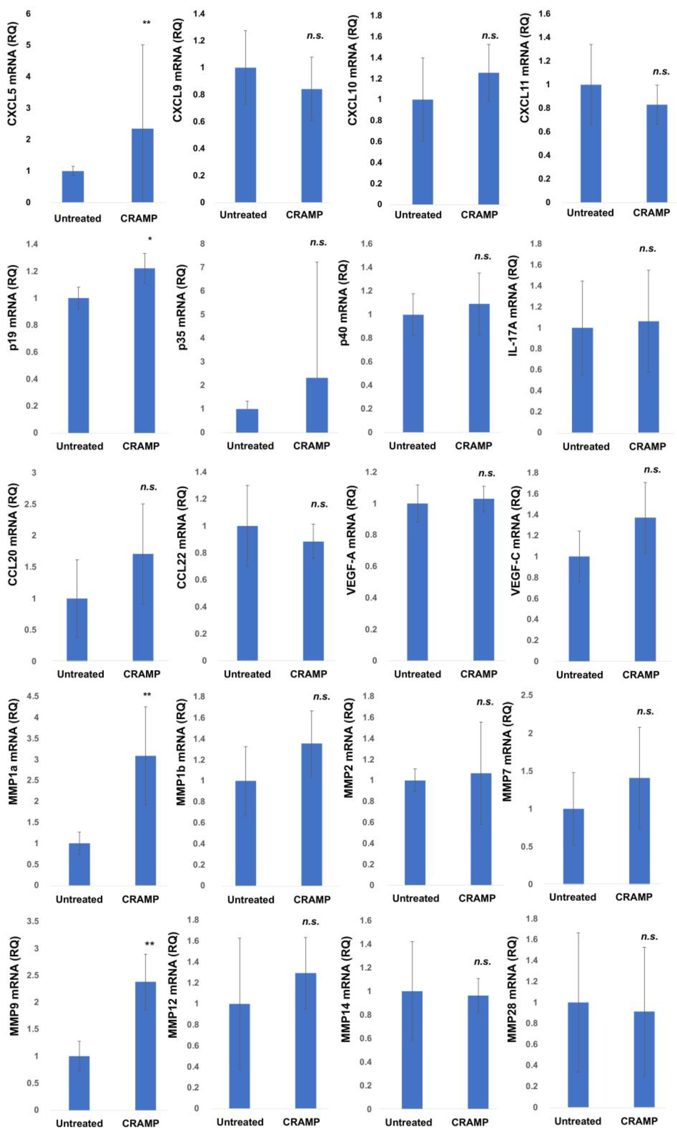
To investigate the immunomodulatory effects of LL-37 on the tumor microenvironment in vivo, the mouse B16F10 melanoma model was used. The peritumoral administration of LL-37 significantly increased the expression of CXCL5, IL23A, MMP1a, and MMP9 mRNA in the tumor microenvironment (Figure 2). There were no significant differences in the expression of CCL20, CCL22, CXCL9, CXCL10, CXCL11, VEGF-A, VEGF-C, IL-17A, IL-12p35, IL-12p40, MMP1b, MMP2, MMP7, MMP12, MMP14, and MMP28. The averages of the data from 15 mice are shown in Figure 2.
LL-37 increased the mRNA expression and protein production of CXCL5, IL-23p19, and MMP-9 in A375 human melanoma cell lines. n.s.: not significant
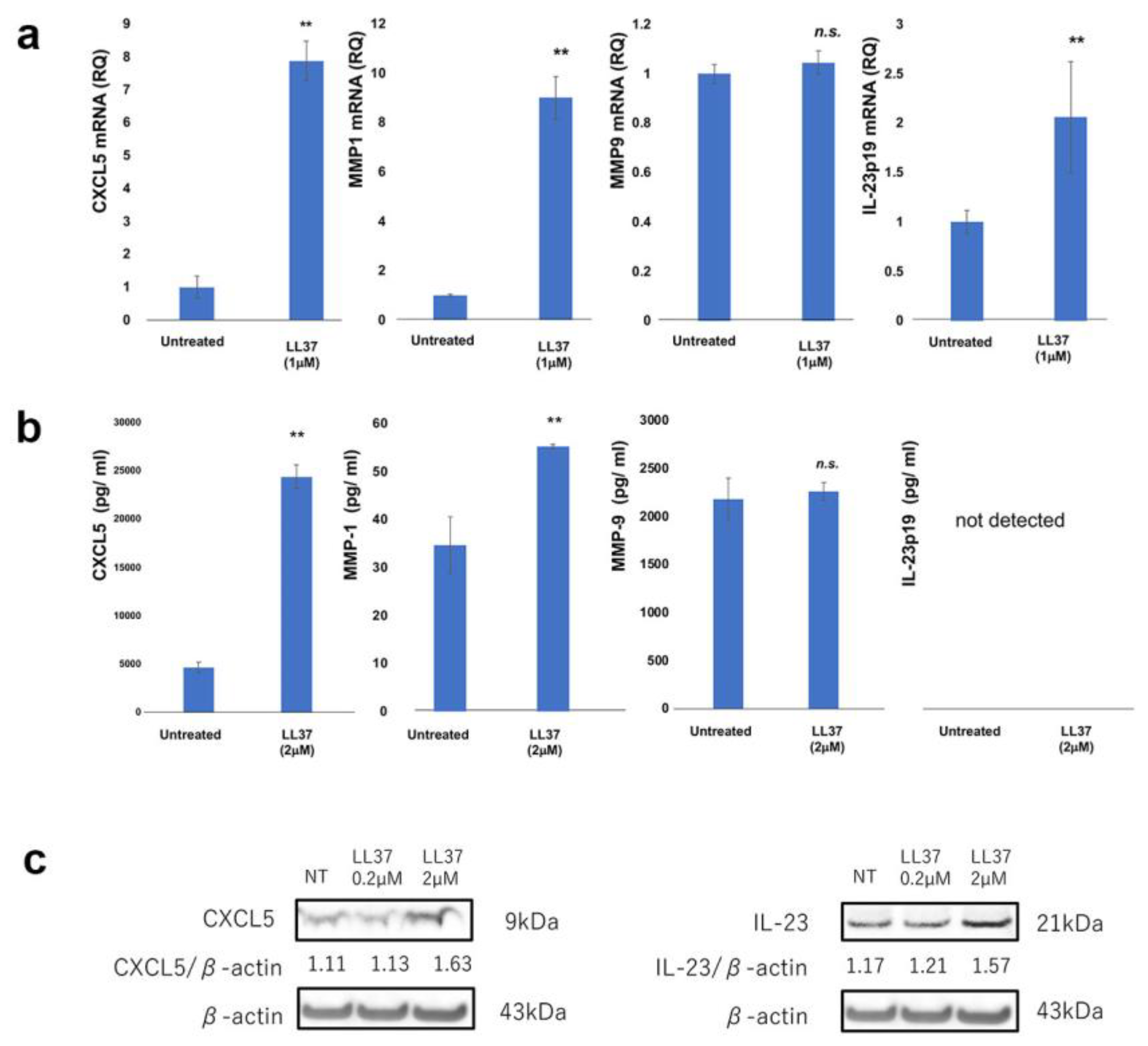
Next, to evaluate the LL-37-reactive cells in the melanoma microenvironment, the direct immunomodulatory effects of LL-37 on melanoma cells were evaluated. A375 melanoma cells were stimulated with LL-37, and the mRNA expression of CXCL5, IL23A, MMP1, and MMP9 were evaluated in vitro. The mRNA expression of CXCL5, IL23A, and MMP9 on A375 melanoma cells were significantly increased by LL-37, whereas there was no significant difference of MMP-1 with LL-37 stimulation (Figure 3a). To validate the increased mRNA expression of CXCL5, IL23A, and MMP9 on A375, the protein production of CXCL5, IL23A, and MMP-9 by A375 cells was investigated using an ELISA assay and Western blot. The production of CXCL5 and IL-23p19 by A375 cells was significantly increased by LL-37 stimulation (Figure 3b). The production of MMP-9 by A375 was significantly increased by LL-37 (Figure 3c). There was no significant increase in MMP-1 production by A375 (Figure 3b,c).
LL-37 increased the mRNA expression and protein production of CXCL5, IL-23p19, and MMP-1 in monocyte-derived M2 macrophages in vitro.
Since LL-37 was located at CD163+ M2 macrophages (Figure 1c), we further hypothesized that LL-37 increases the mRNA expression and production of angiogenetic factors (CXCL5, IL-23p19, MMP1, MMP9) from human CD163+ M2 macrophages. To test this hypothesis, monocyte-derived M2 macrophages were stimulated by LL-37 in vitro. LL-37 increased CXCL5, MMP1 and IL23A mRNA expression, but it did not increase MMP9 mRNA in vitro (Figure 4a). To validate the increased mRNA expression of CXCL5, MMP1, and IL23A on M2 macrophages, the protein production of CXCL5, MMP-1, and IL-23p19, as well as MMP-9, from M2 macrophages was investigated using an ELISA assay. Similar to mRNA expression, the production of CXCL5, MMP-1 (Figure 4b), and IL-23p19 (Figure 4c) was significantly increased by LL-37 stimulation, whereas there was no significant difference in MMP-9 production by M2 macrophages (Figure 4b).
LL-37 increased the angiogenetic activity of A375.
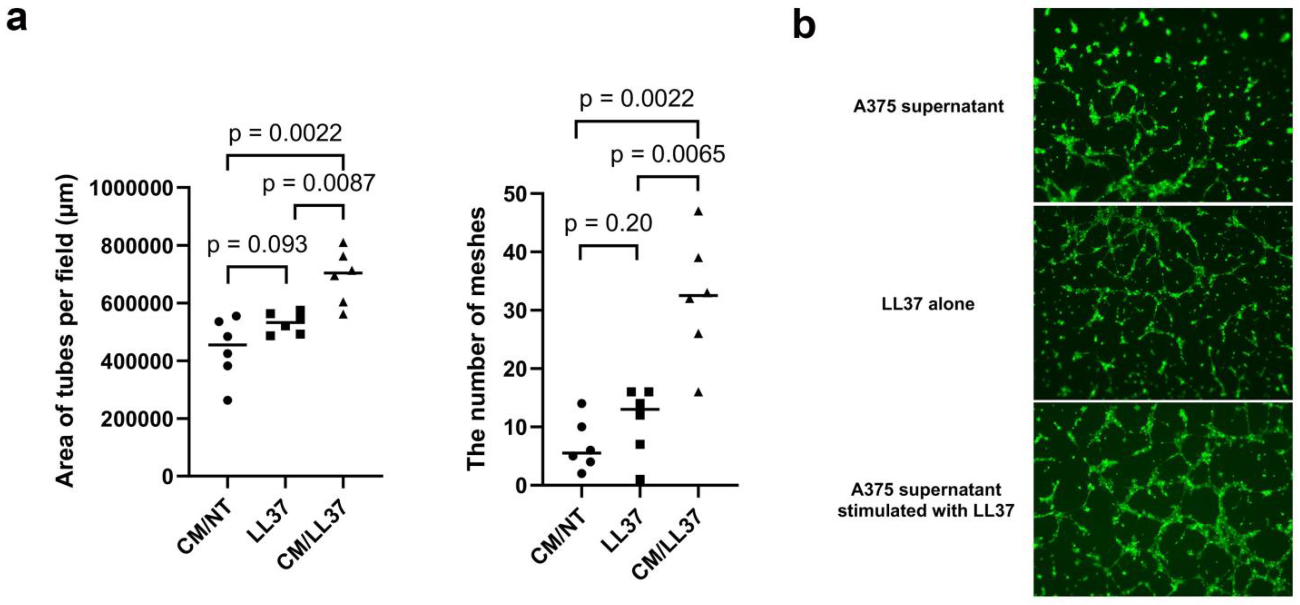
Since LL-37 increased CXCL5 and IL23A mRNA expression, as well as CXCL5 and IL-23p19 production, and since LL-37 increased only MMP-1 production from M2 macrophages, the pro-angiogenic activities of A375-related factors were further examined. To address this issue, an in vitro angiogenesis assay with Matrigel was used. HDMECs treated with LL-37-stimulated A375 culture supernatant formed tube networks, whereas HDMECs treated with LL-37 showed no tube networks under the same culture condition (Figure 5a). To objectively evaluate the activity of angiogenesis, the mesh area was calculated, and tube formation was counted using a BZ-X800 microscope. Tube formation area per field was significantly higher in HDMECs treated with A375+LL-37 culture supernatant than in HDMECs treated with or without LL-37 (Figure 5b).

4. Discussion

LL-37 is expressed in various skin cancers [14,15], and it even promotes the immunosuppressive microenvironment through the production of immunosuppressive chemokines by tumor-associated macrophages (TAMs) [15]. Since T stage determines the overall survival (OS) of melanoma patients [26], and since the expression of LL-37 was increased in parallel with T stage of melanoma as seen in the present study, it is important to understand the tumor-promoting effects of LL-37 on melanoma and its stromal cells. Indeed, peritumoral injection of CRAMP (the mouse ortologe of LL-37) increased the mRNA expression of CXCL5, IL23A, MMP1, and MMP9 in B16F10 melanoma in vivo, suggesting that LL-37 could enhance pro-angiogenesis in melanoma. Moreover, mRNA expression and protein production of these pro-angiogenetic factors was increased in A375 melanoma cells as well as CD163+ M2 macrophages by the stimulation of LL-37 in vitro. Notably, tube formation assay revealed that LL-37 increased angiogenetic activity of A375. Correctively, LL-37-induced CXCL5, IL-23p19, MMP-1 and MMP-9 could promote angiogenetic activity in melanoma, leading to the local invasion of melanoma which contribute to T stage in melanoma patients.
Among the pro-angiogenetic factors described above, IL-23p19 plays one of the central roles in promoting tumor growth in various cancers through its pro-angiogenesis activity [9]. Indeed, IL-23p19 increased the angiogenetic activity of cutaneous angiosarcoma [8]. In addition, IL-23p19 directly or indirectly promotes the infiltration of myeloid cells such as M2 macrophages and neutrophils, leading to the increased secretion of TGF-β, IL-10, and VEGF, and thus promoting angiogenesis in breast cancer [27]. Moreover, IL-23p19 increases IL-23p19 receptor expression on macrophages and enhances macrophage-mediated angiogenesis in hepatocellular carcinoma [28]. Notably, in our present study, IL-23p19 was increased in both melanoma cells and CD163+ M2 macrophages. Since recombinant IL-23p19 forms favorable tube networks in vitro [8], IL-23p19 could, at least in part, promote angiogenetic activity in tumor microenvironment in melanoma.
CXCL5 is a chemokine that promotes tumor formation by triggering the migration of CXCR2+ immune cells to tumors [29,30]. Not only recruiting immunosuppressive cells, such as precursors of TAMs, neutrophils, and myeloid-derived suppressor cells in the tumor microenvironment [30], CXCL5 can recruit endothelial cells (ECs) via its highly conserved glutamic acid-leucine-arginine ‘ELR’ motif [29,31], promoting cancer progression in various cancer types [20]. For example, the interaction between ECs and cancer cells enhances EC recruitment and promotes cancer progression through the EGFR-NF-kB-CXCL5-CXCR2 pathway in bladder cancer [32]. In addition, CXCL5 is positively correlated with the micro-vessel marker CD31, and it activates the AKT/NF-kB/FOXD1/VEGF-A pathway to enhance its tube formation ability in a CXCR2-dependent manner in colorectal cancer [33]. Moreover, CXCL5-overexpressing melanoma cells recruited high amounts of neutrophils and exhibited significantly increased lymphangiogenesis in a mouse melanoma xenograft model [34]. Notably, IL-17A promotes CXCR2-dependent angiogenesis in liver cancer [35]. Since IL-23p19 plays important roles in inducing Th17 cell proliferation even in the cancer microenvironment [36,37], increased levels of CXCL5 in parallel with IL-23p19 by LL-37 could trigger angiogenesis, leading to local invasion of melanoma in the primary lesion.
MMP-9, as well as MMP-1, is a pro-angiogenesis factor, and they could both be prognostic biomarkers for melanoma [38]. Indeed, high expression of MMP-9 protein, as well as infiltration of TAMs, was detected in proximity to intravascular pillars [33]. Notably, MMP-9 inhibition blocked formation of pillars in vessels, and the inhibition of MMP-9 promotes abrogated pillar formation in melanoma [39], leading to suppression of angiogenesis in melanomas. TAMs also produce various MMPs, which play critical roles in the tissue remodeling associated with protein cleavage to modify the immune microenvironment, angiogenesis, tissue repair, local invasion, and metastasis [40]. Notably, MMPs are among the central angiogenetic factors associated with M2-polarized TAMs in skin cancers [40]. For example, osteopontin signaling increased the secretion of MMP-9 from TAMs to promote angiogenesis and tumor progression in a melanoma model [41]. Interestingly, in our present study, LL-37 increased the protein production of MMP-9 from A375 melanoma cells, whereas LL-37 increased MMP-1 from M2 macrophages in vitro, suggesting that LL-37 might promote tumor invasion not only by the stimulation of melanoma cells but also by the stimulation of TAMs in melanoma microenvironment. In aggregate, our present study also suggested the pro-angiogenetic roles of TAMs through IL-23p19, CXCL5 and MMP-1 in melanoma.

5. Conclusions

Correctively, LL-37-induced CXCL5, IL-23p19, MMP-1 and MMP-9 could promote angiogenetic activity in melanoma tumor microenvironment, leading to the local invasion of melanoma which contributes to T stage in melanoma patients. Therefore, targeting LL-37, MMP-9, and TAMs could be a potential anti-angiogenic drug target to suppress the local invasion of melanoma cells, which might improve the OS of melanoma patients in the future (Figure 6).

Supplementary Materials

The following supporting information can be downloaded at: https://www.mdpi.com/article/10.3390/cancers15061678/s1, Figure S1: Raw data for Figure 3; Figure S2: Raw data for Figure 4.

Author Contributions

Conception and design: T.F. Development of methodology: T.I., T.T. and T.F. Acquisition of data: K.O., R.A., T.I., Y.R., J.E. and T.F. Analysis and interpretation of data: T.F. and T.I. Writing: review, and/or revision of the manuscript: T.F. Treating patients: K.O., T.F. and Y.K. Study supervision: Y.A. and T.F. All authors have read and agreed to the published version of the manuscript.

Funding

This study was supported in part by the Japan Agency for Medical Research and Development (21ym0126041h0001, 22ym0126041h0002), and in part by a grant-in-aid for scientific research from the Japan Society for the Promotion of Science (21K08318).

Institutional Review Board Statement

All patients gave their informed consent. The study was approved by the Ethics Committee of Tohoku University Graduate School of Medicine (permit number: 2021-1-1213) and conducted according to the guidelines of the Declaration of Helsinki. The protocol for the animal study was approved by the ethics committee at Tohoku University Graduate School of Medicine for Animal Experimentation, Sendai, Japan (permit number: 2019MdLMO-134-03). The research complied with the Tohoku University Graduate School of Medicine’s Animal Experimentation Ethics guidelines and policies.

Informed Consent Statement

All patients gave their informed consent. Written informed consent has been obtained from the patient(s) to publish this paper.

Data Availability Statement

All data generated or analyzed during this study are included in this article. Further enquiries can be directed to the corresponding author.

Conflicts of Interest

The authors have no conflict of interest to declare.

References

  1. Fabriek, B.O.; Dijkstra, C.D.; Berg, T.K.V.D. The macrophage scavenger receptor CD163. Immunobiology 2005, 210, 153–160. [Google Scholar] [CrossRef]
  2. Chamilos, G.; Gregorio, J.; Meller, S.; Lande, R.; Kontoyiannis, D.P.; Modlin, R.L.; Gilliet, M. Cytosolic sensing of extracellular self-DNA transported into monocytes by the antimicrobial peptide LL37. Blood 2012, 120, 3699–3707. [Google Scholar] [CrossRef] [Green Version]
  3. Takahashi, T.; Kulkarni, N.N.; Lee, E.Y.; Zhang, L.-J.; Wong, G.C.L.; Gallo, R.L. Cathelicidin promotes inflammation by enabling binding of self-RNA to cell surface scavenger receptors. Sci. Rep. 2018, 8, 4032. [Google Scholar] [CrossRef] [PubMed]
  4. Van Gorp, H.; Delputte, P.L.; Nauwynck, H.J. Scavenger receptor CD163, a Jack-of-all-trades and potential target for cell-directed therapy. Mol. Immunol. 2010, 47, 1650–1660. [Google Scholar] [CrossRef]
  5. Lau, S.K.; Chu, P.G.; Weiss, L.M. CD163: A Specific Marker of Macrophages in Paraffin-Embedded Tissue Samples. Am. J. Clin. Pathol. 2004, 122, 794–801. [Google Scholar] [CrossRef]
  6. Fuentes-Duculan, J.; Bonifacio, K.M.; Hawkes, J.E.; Kunjravia, N.; Cueto, I.; Li, X.; Gonzalez, J.; Garcet, S.; Krueger, J.G. Autoantigens ADAMTSL5 and LL37 are significantly upregulated in active Psoriasis and localized with keratinocytes, dendritic cells and other leukocytes. Exp. Dermatol. 2017, 26, 1075–1082. [Google Scholar] [CrossRef]
  7. Fang, H.; Hou, Y.; Zhuang, H.; Wang, C. The effects of Malassezia in the activation of Interleukin (IL)-23/IL-17 axis in Psoriasis. New Microbiol. 2022, 45, 130–137. [Google Scholar] [PubMed]
  8. Ohuchi, K.; Amagai, R.; Ikawa, T.; Muto, Y.; Roh, Y.; Endo, J.; Maekawa, T.; Kambayashi, Y.; Asano, Y.; Fujimura, T. Plasminogen activating inhibitor-1 promotes angiogenesis in cutaneous angiosarcomas. Exp. Dermatol. 2023, 32, 50–59. [Google Scholar] [CrossRef] [PubMed]
  9. Subhadarshani, S.; Yusuf, N.; Elmets, C.A. IL-23 and the Tumor Microenvironment. Adv. Exp. Med. Biol. 2021, 1290, 89–98. [Google Scholar]
  10. Wakita, D.; Sumida, K.; Iwakura, Y.; Nishikawa, H.; Ohkuri, T.; Chamoto, K.; Kitamura, H.; Nishimura, T. Tumor-infiltrating IL-17-producing gammadelta T cells support the progression of tumor by promoting angiogenesis. Eur. J. Immunol. 2010, 40, 1927–1937. [Google Scholar] [CrossRef]
  11. Kim, J.E.; Kim, H.J.; Choi, J.M.; Lee, K.H.; Kim, T.Y.; Cho, B.K.; Jung, J.Y.; Chung, K.Y.; Cho, D.; Park, H.J. The antimicrobial peptide human cationic antimicrobial protein-18/cathelicidin LL-37 as a putative growth factor for malignant melanoma. Br. J. Dermatol. 2010, 163, 959–967. [Google Scholar] [CrossRef]
  12. Jia, J.; Zheng, Y.; Wang, W.; Shao, Y.; Li, Z.; Wang, Q.; Wang, Y.; Yan, H. Antimicrobial peptide LL-37 promotes YB-1 expression, and the viability, migration and invasion of malignant elanoma cells. Mol. Med. Rep. 2016, 15, 240–248. [Google Scholar] [CrossRef] [PubMed] [Green Version]
  13. Manarang, J.C.; Otteson, D.C.; McDermott, A.M. Expression of Antimicrobial Peptides by Uveal and Cutaneous Melanoma Cells and Investigation of Their Role in Tumor Cell Migration and Vasculogenic Mimicry. Curr. Eye Res. 2017, 42, 1474–1481. [Google Scholar] [CrossRef] [PubMed]
  14. Fujimura, T.; Okabe, T.; Tanita, K.; Sato, Y.; Lyu, C.; Kambayashi, Y.; Maruyama, S.; Aiba, S.; Chunbing, L. A novel technique to diagnose non-melanoma skin cancer by thermal conductivity measurements: Correlations with cancer stromal factors. Exp. Dermatol. 2019, 28, 1029–1035. [Google Scholar] [CrossRef]
  15. Lyu, C.; Fujimura, T.; Amagai, R.; Ohuchi, K.; Sato, Y.; Tanita, K.; Matsushita, S.; Fujisawa, Y.; Otsuka, A.; Yamamoto, Y.; et al. Increased expression of dermal LL37 may trigger migration of CCR7+ regulatory T cells in extramammary Paget’s disease. J. Dermatol. Sci. 2020, 99, 65–68. [Google Scholar] [CrossRef]
  16. Lai, Y.P.; Gallo, R.L. AMPed up immunity: How antimicrobial peptides have multiple roles in immune defense. Trends Immunol. 2009, 30, 131–141. [Google Scholar] [CrossRef] [Green Version]
  17. Méndez-Samperio, P. The human cathelicidin hCAP18/LL-37: A multifunctional peptide involved in mycobacterial infections. Peptides 2010, 31, 1791–1798. [Google Scholar] [CrossRef] [PubMed]
  18. Amagai, R.; Takahashi, T.; Terui, H.; Fujimura, T.; Yamasaki, K.; Aiba, S.; Asano, Y. The Antimicrobial Peptide Cathelicidin Exerts Immunomodulatory Effects via Scavenger Receptors. Int. J. Mol. Sci. 2023, 24, 875. [Google Scholar] [CrossRef]
  19. Zeth, K.; Sancho-Vaello, E. The Human Antimicrobial Peptides Dermcidin and LL-37 Show Novel Distinct Pathways in Membrane Interactions. Front. Chem. 2017, 5, 86. [Google Scholar] [CrossRef] [Green Version]
  20. Choi, M.; Jo, J.; Lee, M.; Park, J.; Yao, T.; Park, Y. Cathelicidin-related antimicrobial peptide mediates skeletal muscle degeneration caused by injury and Duchenne muscular dystrophy in mice. J. Cachex Sarcopenia Muscle 2022, 13, 3091–3105. [Google Scholar] [CrossRef]
  21. Mihailovic, P.M.; Lio, W.M.; Yano, J.; Zhao, X.; Zhou, J.; Chyu, K.; Shah, P.K.; Cercek, B.; Dimayuga, P.C. The cathelicidin pro-tein CRAMP is a potential atherosclerosis self-antigen in ApoE(-/-) mice. PLoS ONE 2017, 12, e0187432. [Google Scholar] [CrossRef] [PubMed]
  22. Kakizaki, A.; Fujimura, T.; Furudate, S.; Kambayashi, Y.; Yamauchi, T.; Yagita, H.; Aiba, S. Immunomodulatory effect of peritumorally administered interferon-beta on melanoma through tumor-associated macrophages. Oncoimmunology 2015, 4, e1047584. [Google Scholar] [CrossRef] [PubMed]
  23. Ikawa, T.; Miyagawa, T.; Fukui, Y.; Toyama, S.; Omatsu, J.; Awaji, K.; Norimatsu, Y.; Watanabe, Y.; Yoshizaki, A.; Sato, S.; et al. Endothelial CCR6 expression due to FLI1 deficiency contributes to vasculopathy associated with systemic sclerosis. Arthritis Res. Ther. 2021, 23, 283. [Google Scholar] [CrossRef]
  24. Ohuchi, K.; Kambayashi, Y.; Hidaka, T.; Fujimura, T. Plasminogen Activating Inhibitor-1 Might Predict the Efficacy of Anti-PD1 Antibody in Advanced Melanoma Patients. Front. Oncol. 2021, 11, 798385. [Google Scholar] [CrossRef]
  25. Fujimura, T.; Kakizaki, A.; Furudate, S.; Aiba, S. A possible interaction between periostin and CD163+ skin-resident macrophages in pemphigus vulgaris and bullous pemphigoid. Exp. Dermatol. 2017, 26, 1193–1198. [Google Scholar] [CrossRef]
  26. Fujisawa, Y.; Yoshikawa, S.; Minagawa, A.; Takenouchi, T.; Yokota, K.; Uchi, H.; Noma, N.; Nakamura, Y.; Asai, J.; Kato, J.; et al. Classification of 3097 patients from the Japanese melanoma study database using the American joint committee on cancer eighth edition cancer staging system. J. Dermatol. Sci. 2019, 94, 284–289. [Google Scholar] [CrossRef] [PubMed] [Green Version]
  27. Nie, W.; Yu, T.; Sang, Y.; Gao, X. Tumor-promoting effect of IL-23 in mammary cancer mediated by infiltration of M2 macro-phages and neutrophils in tumor microenvironment. Biochem. Biophys. Res. Commun. 2017, 482, 1400–1406. [Google Scholar] [CrossRef] [PubMed]
  28. Zang, M.; Li, Y.; He, H.; Ding, H.; Chen, K.; Du, J.; Chen, T.; Wu, Z.; Liu, H.; Wang, D.; et al. IL-23 production of liver inflammatory macrophages to damaged hepatocytes promotes hepatocellular carcinoma development after chronic hepatitis B virus infection. Biochim. Biophys. Acta (BBA)-Mol. Basis Dis. 2018, 1864, 3759–3770. [Google Scholar] [CrossRef]
  29. Deng, J.; Jiang, R.; Meng, E.; Wu, H. CXCL5: A coachman to drive cancer progression. Front. Oncol. 2022, 12, 944494. [Google Scholar] [CrossRef]
  30. Fujimura, T.; Aiba, S. Significance of Immunosuppressive Cells as a Target for Immunotherapies in Melanoma and Non-Melanoma Skin Cancers. Biomolecules 2020, 10, 1087. [Google Scholar] [CrossRef]
  31. Strieter, R.M.; Polverini, P.J.; Kunkel, S.L.; Arenberg, D.A.; Burdick, M.D.; Kasper, J.; Dzuiba, J.; Damme, J.V.; Walz, A.; Marriott, D.; et al. The functional role of the ELR motif in CXC chemokine-mediated angiogenesis. J. Biol. Chem. 1995, 270, 27348–27357. [Google Scholar] [CrossRef] [PubMed] [Green Version]
  32. Huang, Z.; Zhang, M.; Chen, G.; Wang, W.; Zhang, P.; Yue, Y.; Guan, Z.; Wang, X.; Fan, J. Bladder cancer cells interact with vascular endothelial cells triggering EGFR signals to promote tumor progression. Int. J. Oncol. 2019, 54, 1555–1566. [Google Scholar] [CrossRef] [PubMed] [Green Version]
  33. Chen, C.; Xu, Z.Q.; Zong, Y.P.; Ou, B.C.; Shen, X.H.; Feng, H.; Zheng, M.H.; Zhao, J.K.; Lu, A.G. CXCL5 induces tumor angiogenesis via enhancing the expression of FOXD1 mediated by the AKT/NFkB pathway in colorectal cancer. Cell Death Dis 2019, 10, 178. [Google Scholar] [CrossRef] [Green Version]
  34. Soler-Cardona, A.; Forsthuber, A.; Lipp, K.; Ebersberger, S.; Heinz, M.; Schossleitner, K.; Buchberger, E.; Gröger, M.; Petzelbauer, P.; Hoeller, C.; et al. CXCL5 Facilitates Melanoma Cell–Neutrophil Interaction and Lymph Node Metastasis. J. Investig. Dermatol. 2018, 138, 1627–1635. [Google Scholar] [CrossRef] [Green Version]
  35. Liu, L.; Sun, H.; Wu, S.; Tan, H.; Sun, Y.; Liu, X.; Si, S.; Xu, L.; Huang, J.; Zhou, W.; et al. IL-17A promotes CXCR2-dependent angiogenesis in a mouse model of liver cancer. Mol. Med. Rep. 2019, 20, 1065–1074. [Google Scholar] [CrossRef] [Green Version]
  36. Sutton, C.E.; Lalor, S.J.; Sweeney, C.M.; Brereton, C.F.; Lavelle, E.C.; Mills, K.H. Interleukin-1 and IL-23 induce innate IL-17 production from gammadelta T cells, amplifying Th17 responses and autoimmunity. Immunity 2009, 31, 331–341. [Google Scholar] [CrossRef] [Green Version]
  37. Qian, X.; Gu, L.; Ning, H.; Zhang, Y.; Hsueh, E.C.; Fu, M.; Hu, X.; Wei, L.; Hoft, D.F.; Liu, J. Increased Th17 Cells in the Tumor Microenvironment Is Mediated by IL-23 via Tumor-Secreted Prostaglandin E2. J. Immunol. 2013, 190, 5894–5902. [Google Scholar] [CrossRef] [Green Version]
  38. Wang, T.; Zhang, Y.; Bai, J.; Xue, Y.; Peng, Q. MMP1 and MMP9 are potential prognostic biomarkers and targets for uveal melanoma. BMC Cancer 2021, 21, 1068. [Google Scholar] [CrossRef]
  39. Pandita, A.; Ekstrand, M.; Bjursten, S.; Zhao, Z.; Fogelstrand, P.; Le Gal, K.; Ny, L.; Bergo, M.O.; Karlsson, J.; Nilsson, J.A.; et al. Intussusceptive Angiogenesis in Human Metastatic Malignant Melanoma. Am. J. Pathol. 2021, 191, 2023–2038. [Google Scholar] [CrossRef]
  40. Fujimura, T. Stromal Factors as a Target for Immunotherapy in Melanoma and Non-Melanoma Skin Cancers. Int. J. Mol. Sci. 2022, 23, 4044. [Google Scholar] [CrossRef] [PubMed]
  41. Kale, S.P.; Raja, R.; Thorat, D.; Soundararajan, G.; Patil, T.V.; Kundu, G.C. Osteopontin signaling upregulates cyclooxygenase-2 expression in tumor-associated macrophages leading to enhanced angiogenesis and melanoma growth via α9β1 integrin. Oncogene 2013, 33, 2295–2306. [Google Scholar] [CrossRef] [PubMed]
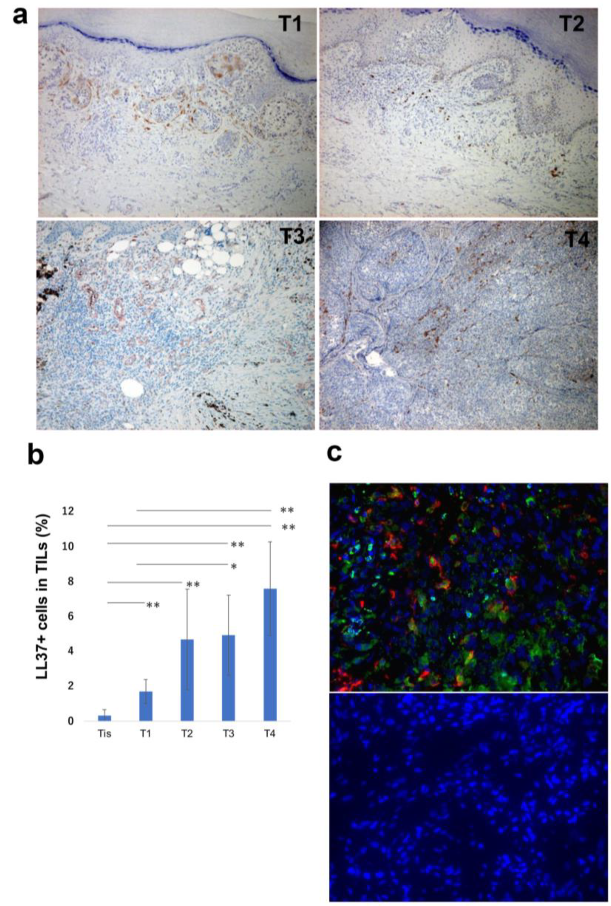
Figure 1. Representative paraffin-embedded tissue samples from lesional skin of patients with melanoma in each tumor stage (a). Quantitative analysis of LL-37+ cells: the IHC-positive cells within the lymphocyte fraction and the percentage of IHC-positive cells per all tumour-infiltrating cells were automatically counted using a BZ-X800 microscope (b). IF staining of melanoma for LL-37 (green), CD163 (red), and DAPI (blue, nuclei). A merged image is also shown, with green and red combining into yellow. The isotype control IgG1 stains as red or green (c). * marks a significant (p < 0.05) difference. ** marks a significant (p < 0.01) difference.
Cancers 15 01678 g001
Figure 2. Expression of chemokines, cytokines, and MMPs mRNA in B16F10 melanoma was analyzed by quantitative RT-PCR using the ΔΔCt method (n = 15). The data from each donor were obtained by triplicate assays, and then the mean ± SD was calculated (c). * marks a significant (p < 0.05) difference. ** marks a significant (p < 0.01) difference. n.s.: not significant.
Cancers 15 01678 g002
Figure 3. Expression of IL23A, CXCL5, MMP1, and MMP9 mRNA in A375 melanoma cells stimulated with or without LL-37 were analyzed by quantitative RT-PCR using the ΔΔCt method (n = 3). The data from each donor were obtained by triplicate assays, and then the mean ± SD was calculated. (a) Culture supernatant from A375 was harvested as described in Materials and Methods and measured by ELISA (n = 3) (b). MMP-1 and MMP-9 production was analyzed by Western blotting (c). Data from each donor were obtained from triplicate assays, and mean ± SD values were calculated. Representative data from at least three independent experiments are shown. * marks a significant (p < 0.05) difference. ** marks a significant (p < 0.01) difference. The uncropped blots are shown in Figure S1. n.s.: not significant.
Cancers 15 01678 g003
Figure 4. Expression of IL23A, CXCL5, MMP1, and MMP9 mRNA in M2 macrophages stimulated with or without LL-37 were analyzed by quantitative RT-PCR using the ΔΔCt method (n = 3). The data from each donor were obtained by triplicate assays, and then the mean ± SD was calculated. (a) Culture supernatant from M2 macrophages was harvested as described in Materials and Methods and measured by ELISA (n = 3). Data from each donor were obtained from triplicate assays, and mean ± SD values were calculated. Representative data from at least three independent experiments are shown (b). ** marks a significant (p < 0.01) difference. CXCL5 and IL-23p19 production was analyzed by Western blotting (c). The uncropped blots are shown in Figure S2. n.s.: not significant.
Cancers 15 01678 g004
Figure 5. The tube formation assay was performed by applying HDMECs treated with culture supernatant of recombinant LL-37-treated A375 or culture medium with or without recombinant LL-37 onto the Matrigel and incubating for 24 h. To eliminate the effect of proliferation, cells were treated with mitomycin C before the assay (a). Representative images are shown (b) (n = 5 for each group).
Cancers 15 01678 g005
Figure 6. Summary of the present study.
Cancers 15 01678 g006
Table 1. Characteristics of patients with melanoma.
AgeSexSubtypeTumor Thickness (mm)TNMStage
case 169Macral0TisN0M00
case 278Facral0TisN0M00
case 372Facral0TisN0M00
case 467Macral0TisN0M00
case 575Facral0TisN0M00
Tiscase 649Facral0TisN0M00
case 786Facral0TisN0M00
case 869Facral0TisN0M00
case 985Macral0TisN0M00
case 1021Fnon-CSD0TisN0M00
case 1178FCSD0.5T1aN0M0IA
case 1279FCSD0.5T1aN0M0IA
case 1362Macral0.35T1aN0M0IA
case 1465Macral0.90T1aN0M0IA
case 1548Facral0.90T1bN0M0IB
T1case 1636Facral0.60T1aN0M0IA
case 1743Facral0.80T1aN0M0IA
case 1872Macral0.42T1aN0M0IA
case 1975Macral0.70T1bN0M0IB
case 2065MCSD0.05T1bN0M0IA
case 2164Facral1.02T2bN0M0IIA
case 2246Facral1.05T2aN0M0IB
case 2381FCSD1.20T2aN0M0IB
case 2488FCSD1.50T2bN0M0IIA
case 2572Macral1.80T2bN0M0IIA
T2case 2665Mnon-CSD1.50T2aN0M0IB
case 2742Facral1.80T2aN0M0IB
case 2869Fnon-CSD1.03T2aN0M0IB
case 2921Facral1.30T2aN0M0IB
case 3021Fnon-CSD1.3T2aN0M0IB
case 3147Macral3.5T3aN3cM0IIIC
case 3263Facral3T3aN0M0IIA
case 3383FCSD2T3aN0M0IIA
case 3462FCSD3T3aN1aM0IIIB
case 3579Macral3.5T3aN1bM0IIIB
T3case 3679Facral3.2T3aN0M0IIA
case 3764Facral2T3bN1aM0IIIC
case 3884Facral2T3aN0M0IIA
case 3981Facral3T3aN0M0IIA
case 4071Mnon-CSD3T3aN0M0IIA
case 4174Facral6T4bN0M0IIC
case 4282Facral6T4aN0M0IIB
case 4377Macral6T4aN0M0IIB
case 4490FCSD4.4T4aN0M0IIB
case 4569Facral4.5T4bN1aM0IIIC
T4case 4639MCSD9T4bN2aM0IIIC
case 4770Macral14T4bN0M0IIC
case 4835Mnon-CSD20T4bN1aM1bIV
case 4984Macral6T4bN0M0IIC
case 5047MCSD8T4bN2bM0IIIC
(CSD: chronic sun damage).
Disclaimer/Publisher’s Note: The statements, opinions and data contained in all publications are solely those of the individual author(s) and contributor(s) and not of MDPI and/or the editor(s). MDPI and/or the editor(s) disclaim responsibility for any injury to people or property resulting from any ideas, methods, instructions or products referred to in the content.