Article

Consolidative Radiotherapy for Metastatic Urothelial Bladder Cancer Patients with No Progression and with No More than Five Residual Metastatic Lesions Following First-Line Systemic Therapy: A Retrospective Analysis

by
Amélie Aboudaram
1,
Léonor Chaltiel
2,
Damien Pouessel
3,
Pierre Graff-Cailleaud
4,
Nicolas Benziane-Ouaritini
5,
Paul Sargos
5,
Ulrike Schick
6,
Gilles Créhange
4,
Elizabeth Cohen-Jonathan Moyal
1,
Christine Chevreau
3 and
Jonathan Khalifa
1,*
1
Department of Radiation Oncology, Institut Claudius Regaud/Institut Universitaire du Cancer de Toulouse—Oncopole, 31000 Toulouse, France
2
Department of Biostatistics, Institut Claudius Regaud/Institut Universitaire du Cancer de Toulouse—Oncopole, 31059 Toulouse, France
3
Department of Medical Oncology, Institut Claudius Regaud/Institut Universitaire du Cancer de Toulouse—Oncopole, 31059 Toulouse, France
4
Department of Radiation Oncology, Institut Curie, 75248 Paris, France
5
Department of Radiation Oncology, Institut Bergonié, 33000 Bordeaux, France
6
Department of Radiation Oncology, CHU Brest, 29200 Brest, France
*
Author to whom correspondence should be addressed.
Cancers 2023, 15(4), 1161; https://doi.org/10.3390/cancers15041161
Submission received: 20 December 2022 / Revised: 3 February 2023 / Accepted: 10 February 2023 / Published: 11 February 2023
(This article belongs to the Special Issue Advance and New Insights in Bladder Cancer)

Abstract

:

Simple Summary

Radiotherapy with curative intent in the treatment of metastatic malignancies has shown promising results in several types of primary tumors but experience in bladder cancer is limited and scarce. The objective of our retrospective study was to assess the benefit of consolidative radiotherapy directed to the bladder and to residual metastases among patients with metastatic urothelial bladder cancer and with no progression following systemic therapy. In our analysis, radiotherapy was associated with a valuable and promising survival benefit compared with no local treatment following systemic therapy. The benefit of consolidative radiotherapy needs to be confirmed in prospective clinical trials in the future.

Abstract

Local consolidative radiotherapy in the treatment of metastatic malignancies has shown promising results in several types of tumors. The objective of this study was to assess consolidative radiotherapy to the bladder and to residual metastases in metastatic urothelial bladder cancer with no progression following first-line systemic therapy. Materials/methods: Patients who received first-line therapy for the treatment of metastatic urothelial bladder cancer (mUBC) and who were progression-free following treatment with no more than five residual metastases were retrospectively identified through the database of four Comprehensive Cancer Centers, between January 2005 and December 2018. Among them, patients who received subsequent definitive radiotherapy (of EQD2Gy > 45Gy) to the bladder and residual metastases were included in the consolidative group (irradiated (IR) group), and the other patients were included in the observation group (NIR group). Progression-free survival (PFS) and overall survival (OS) were determined from the start of the first-line chemotherapy using the Kaplan–Meier method. To prevent immortal time bias, a Cox model with time-dependent covariates and 6-month landmark analyses were performed to examine OS and PFS. Results: A total of 91 patients with at least stable disease following first-line therapy and with no more than five residual metastases were analyzed: 51 in the IR group and 40 in the NIR group. Metachronous metastatic disease was more frequent in the NIR group (19% vs. 5%, p = 0.02); the median number of metastases in the IR group vs. in the NIR group was 2 (1–9) vs. 3 (1–5) (p = 0.04) at metastatic presentation, and 1 (0–5) vs. 2 (0–5) (p = 0.18) after completion of chemotherapy (residual lesions), respectively. Two grade 3 toxicities (3.9%) and no grade 4 toxicity were reported in the IR group related to radiotherapy. With a median follow up of 85.9 months (95% IC (36.7; 101.6)), median OS and PFS were 21.7 months (95% IC (17.1; 29.7)) and 11.1 months (95% IC (9.9; 14.1)) for the whole cohort, respectively. In multivariable analysis, consolidative radiotherapy conferred a benefit in both PFS (HR = 0.49, p = 0.007) and OS (HR = 0.47, p = 0.015) in the whole population; in the landmark analysis at 6 months, radiotherapy was associated with improved OS (HR = 0.48, p = 0.026), with a trend for PFS (HR = 0.57, p = 0.082). Conclusion: Consolidative radiotherapy for mUBC patients who have not progressed after first-line therapy and with limited residual disease seems to confer both OS and PFS benefits. The role of consolidative radiotherapy in the context of avelumab maintenance should be addressed prospectively.

1. Introduction

Bladder cancer is the most common malignancy involving the urinary system, and urothelial (transitional cell) carcinoma is the predominant histological type in the United States and Europe, where it accounts for 90% of all bladder cancers [1]. Among patients with localized muscle-invasive disease, approximately 50% will relapse after radical cystectomy [2,3,4]. Moreover, 10–15% of patients present with unresectable or metastatic disease at diagnosis [5]. A cisplatin-based combination chemotherapy regimen (dose-dense MVAC: methotrexate, vinblastine, doxorubicin, and cisplatin; or GC: gemcitabine and cisplatin) is the preferred initial therapy for patients with metastatic urothelial bladder cancer (mUBC) who are eligible for cisplatin, providing an overall response rate (ORR) of 50–70%, a median overall survival (OS) of 13–15 months and a 5-year OS of approximately 15% [2,6,7,8,9]. Recently, the JAVELIN 100 trial has shown the benefit in OS of maintenance with avelumab in patients whose disease has not progressed following platinum-based chemotherapy (median OS, 21.4 months versus 14.3 months, p = 0.001), which now constitutes the standard of care [10].
Oligometastatic disease has emerged as a new paradigm to designate a metastatic state with a limited tumor burden, potentially amenable to cure when local treatments are combined with systemic therapies [11]. While this concept has gained evidence in several malignancies such as non-small cell cancer, colorectal cancer, breast cancer, or prostate cancer [12], the question as to whether the oligometastatic disease concept can be applied to patients with bladder cancer remains uncertain. Recent data suggest that, among patients with metastatic recurrence following radical cystectomy for UBC, a limited number of lesions (≤3) and a limited number of involved organs are independently correlated with cancer-specific survival [13], which argues in favor of local ablative treatment for well-selected patients with oligometastatic UBC.
Local ablative treatment for mUBC can be directed to both the bladder and the distant metastases. In the former case, beyond potential benefit to prevent hematuria or sepsis from primary tumor progression, recent evidence has suggested an oncological benefit of aggressive local treatment of the bladder [14,15]. In the latter case, several series have suggested the benefit of post-chemotherapy retroperitoneal lymph-node dissection among UBC patients with subdiaphragmatic nodal disease, and at least stable disease following chemotherapy resulted in durable long-term survival [16,17,18], with a 5-year OS of 49% in an Italian series [17]. Among patients with various anatomic sites of metastases, the 5-year median OS from metastasectomy ranged from 28% to 72% [19,20,21,22,23,24,25,26,27,28,29], and evidence of a benefit from metastasectomy among mUBC patients with peri-operative chemotherapy has been suggested in a recent meta-analysis [30]. However, in these reports, there was variability in the number of patients who received chemotherapy either before or after metastasectomy, and in the response to chemotherapy prior to metastasectomy, with inclusion of patients who were refractory to chemotherapy in one study [23]. Experience in radiotherapy for the metastases-directed therapy of mUBC is limited and scarce as it relies on heterogeneous populations of oligometastatic UBC and on both palliative and ablative radiation doses [31,32,33,34,35,36,37]. This strategy warrants further investigation, given that it is non-invasive compared with surgery. One way to select among oligometastatic mUBC patients those who would most benefit from local ablative treatment is to propose such a strategy to patients with oligopersistent cancer induced by systemic treatment [38].
The objective of this retrospective study was to assess the benefit of local consolidative radiotherapy (LCR) directed to the bladder and to residual metastases among patients with metastatic urothelial bladder cancer and with no progression following first-line systemic therapy.

2. Materials and Methods
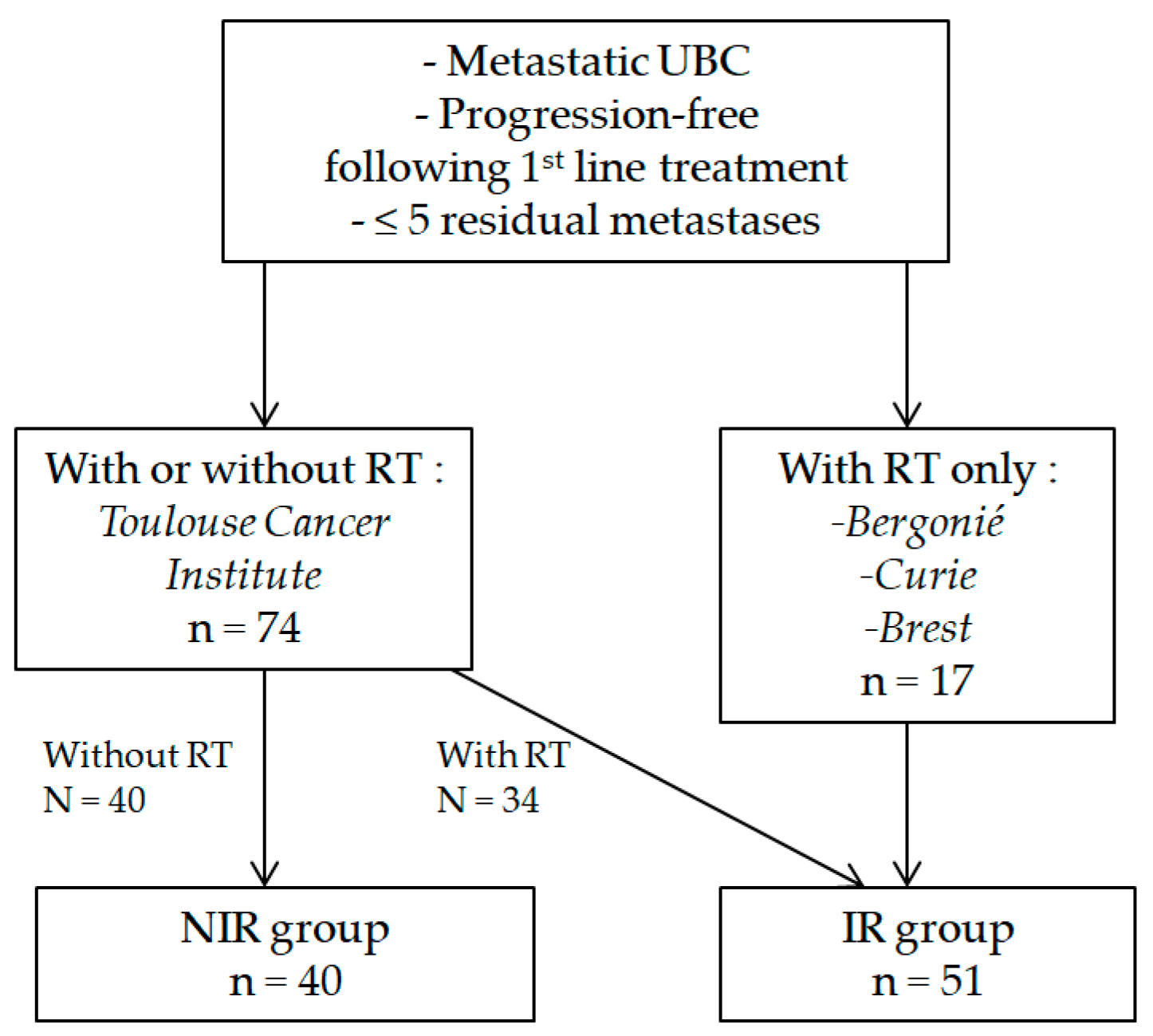
2.1. Patients

This was a multicenter retrospective study involving four cancer centers affiliated to the Groupe d’Etude des Tumeurs Uro-Génitales (GETUG). Patients were identified through the electronic database of each center between January 2011 and December 2018. Inclusion criteria were as follows: histologically proven metastatic urothelial bladder cancer (mUBC), either synchronous or metachronous, with chemotherapy and/or immunotherapy as the first line of treatment, with stable disease or objective response following treatment according to RECIST 1.1 criteria, with no more than five residual metastatic lesions based on a CT-scan or a 18FDG PET-CT, with (in the whole centers) or without (only in Toulouse Cancer Institute) local consolidative radiotherapy (LCR) to the primary tumor for synchronous setting and/or to the residual metastatic lesions.
The patients were then divided into two groups: those who received a LCR (irradiated (IR) group), and those who did not, which constituted the control group (NIR group).
We collected data on patients’ characteristics at diagnosis; disease characteristics including histological variants, timing of metastatic presentation (synchronous vs. metachronous), and number/location of metastases at presentation; previous treatment including first-line systemic treatment and local treatment for patients in a metachronous setting; a radiotherapy schedule in the IR group; and clinical outcome following first-line systemic therapy +/− irradiation.
Our study was approved by our local ethics committee and authorized by the CNIL (Commission nationale informatique et liberté), authorization no. 919375; CRC IUCT-O 19 URO 15.

2.2. Counting Metastatic Lesions

The metastasis count was based on the CT scan performed after first-line systemic treatment.
In case of distant lymph node involvement (above the aortic bifurcation), para-aortic lymph node involvement was counted as a single metastatic site regardless of the number of nodes involved, whereas other extra-pelvic lymph nodes were counted individually. At least one of the three following radiological criteria defined the suspicious character of an extra-pelvic lymph node: a small axis greater than 1 cm, and/or the presence of central necrosis, and/or contrast after injection of iodinated product.

2.3. Local Consolidative Radiotherapy

In the IR group, LCR was defined as an EQD2Gy dose of at least 45 Gy. Radiotherapy data were collected for each irradiated lesion regarding irradiated volume, radiotherapy technique, and dose/fractionation with an equivalent dose in the 2 Gy-fraction using an α/β ratio of 13 Gy for bladder cancer [39].

2.4. Follow Up

Standard follow up included physical examination and a body CT scan every 3 months. Disease status was assessed starting from the beginning of first-line systemic treatment, and progression was defined according to the RECIST 1.1 criteria. In the IR group, the post-irradiation response was reported for each irradiated site according to RECIST 1.1 criteria as well.

2.5. Toxicity Following Radiotherapy

In the IR group, toxicity according to CTCAE 7.0 criteria was reported for any irradiated lesion, i.e., bladder, pelvic nodes, and metastatic lesions; due to discrepancies in low-grade toxicity reporting in retrospective studies, only grade ≥ 3 toxicities were reported.

2.6. Statistical Analysis

Population characteristics were described by the usual statistics: frequencies, percentages, and number of missing data for each modality for qualitative variables; and median, minimum, maximum, and number of missing data for quantitative variables. Comparisons between groups were made using the χ2 test or Fisher’s exact test for qualitative variables and by the Kruskal–Wallis test for quantitative variables.
Overall survival was defined as the time from the date of initiation of first-line metastatic therapy to the date of all-cause death or the date of last news (censored data). In the case of death, the cause of death, whether or not related to the cancer pathology, was recorded.
PFS was defined as the time from the start of first-line metastatic therapy to the date of the first event or the date of last news (censored data). Events considered were progression and death from any cause.
Survival rates were estimated by the Kaplan–Meier method and presented with their 95% confidence intervals. Univariable analyses were performed using the logrank test for qualitative data. Associated variables with a p-value of less than 15% and clinically relevant in univariable analyses were considered in multivariable analyses, which were performed using the Cox proportional hazards model and presented with the hazard ratio (HR) and 95% confidence interval for each covariate. A Cox model with a time-dependent variable and a landmark analysis at 6 months (on the event-free living population at 6 months) were used to reduce the bias related to the delay between the initiation of the first line of treatment and the RT (immortal time bias), and to assess the effect of RT on OS and PFS.
All analyses were performed with STATA 16 software, and all tests used were 2-sided with a threshold α at 5%.

3. Results

3.1. Patients

In total, 91 patients were included: 51 in the IR group and 40 in the NIR group. The flow diagram for the selection of patients is presented in Figure 1.
The characteristics of the whole population and of the IR/NIR groups are presented in Table 1. The median age at metastatic presentation was 64 (37–83); ECOG performance status was 0 for 65.9% and 1 for 28.6% of patients, respectively. The distribution of T classification was slightly different between both groups (p = 0.019), with more T4 lesions in the IR group (19.1% vs. 5%); in situ carcinoma (CIS) was more frequent in the NIR group (37.9% vs. 12.1%, p = 0.018). Metachronous metastatic disease (following definitive treatment of localized UBC) was more frequent in the NIR group (70% vs. 35.3%, p = 0.001); the median number of metastases in the IR group vs. in the NIR group was 2 (1–9) vs. 3 (1–5) (p = 0.04) at metastatic presentation, respectively. Only one patient (in the IR group) had more than five metastases at presentation (nine lesions). First-line systemic therapy consisted of chemotherapy for all patients except two patients who received durvalumab (in the NIR group) and chemotherapy plus panitumumab (in the IR group), respectively; the median duration of first-line systemic therapy was 3.8 months (0.7–7.4) in the IR group and 3.9 months (1.6–4.9) in the NIR group (p = 0.248). Following first-line systemic treatment, a complete response (CR) was found in 27.3% vs. 21.1% in the IR vs. NIR group, and partial response (PR) in 45.5% vs. 42.1% (p = 0.289), respectively; the median number of residual lesions after completion of systemic treatment in the IR vs. NIR group was 1 (0–5) vs. 2 (0–5), respectively (p = 0.179). Other features were well balanced between both groups.
The landmark analysis was performed on 89 patients with no event (progression or death) at 6 months: 32 patients had radiotherapy within 6 months from the start of systemic therapy (IR group for landmark analysis) and 57 patients had no radiotherapy within 6 months (NIR group for landmark analysis).

3.2. Local Consolidative Radiotherapy

In the IR group (n = 51), the median time from the start of systemic treatment to radiotherapy was 5.7 months (1.6–9.6 months), and the median time from the completion of systemic treatment to radiotherapy was 1.9 months (−0.9–5.8 months),
Bladder irradiation was performed in 36 patients (70.6%), of whom 31 also received pelvic lymph node (PLN) irradiation; the main reason for bladder irradiation omission was a history of cystectomy (12/15). The median total dose to the bladder was 64 Gy (45–66 Gy), and the median number of fractions was 33 (15–36).
Metastasis irradiation was performed in 38 patients and was directed to a total of 56 metastases; a median of 1 lesion per patient was irradiated (range: 1–4).
The main metastatic irradiated site was extra-pelvic nodes (29/56; 51.8%). The median EQD2Gy of the 56 irradiated lesions was 53 Gy (45–132 Gy), with a median fraction size of 1.9 Gy (1.7–18 Gy).
The main characteristics of radiotherapy are presented in Table 2.

3.3. Treatment Response Analysis
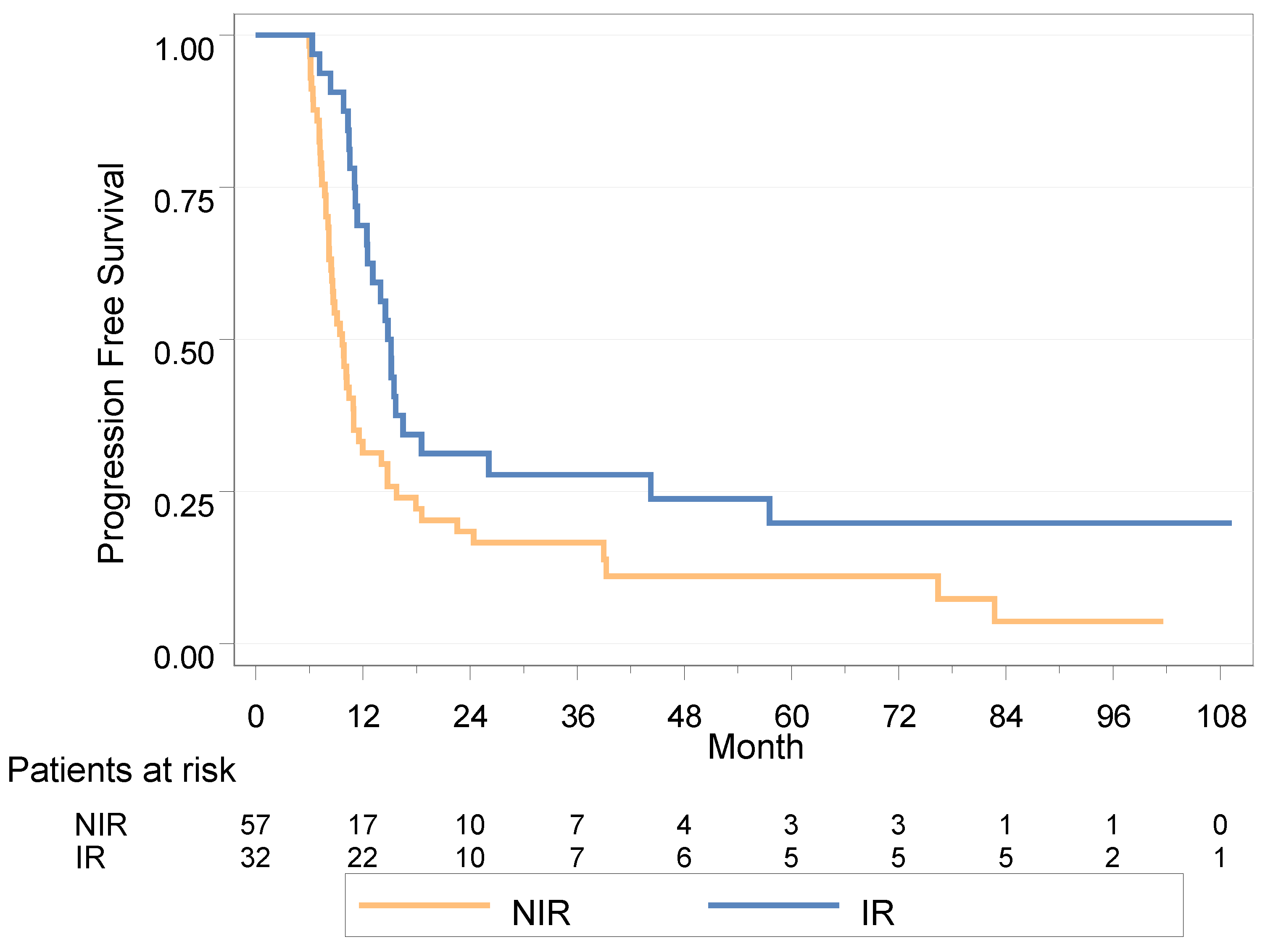
In the IR group, complete response (CR), partial response (PR), stable disease (SD) and progressive disease (PD) on irradiated lesions were observed in 46.2% (24/52), 19.2%, 25% and 9.6%, respectively.
Overall, the best RECIST response following first-line systemic treatment +/− local consolidative radiotherapy in the IR vs. NIR group was CR in 47.1% vs. 30%, PR in 23.5% vs. 37.5%, SD in 21.6% vs. 32.5%, and PD in 7.8% vs. 0%, respectively (p = 0.067).
Finally, progression occurred in 40 patients in the IR group (78.4%) vs. 37 patients in the NIR group (92.5%) (p = 0.099).

3.4. PFS

3.4.1. Whole Population

After a median follow up of 85.9 months (95% IC (36.7; 101.6)), the median PFS was 11.1 months (95% IC (9.9; 14.1)); using the Cox model with a time-dependent variable, the HR for PFS was 0.45 in the IR group (95% IC (0.28; 0.73), p = 0.001) (Figure 2).
In univariable analysis, a delay of 24 months or more between diagnosis and the appearance of metastases appeared deleterious in terms of PFS (HR = 1.80 (1.02–3.20), p = 0.040), as did the presence of a liver metastasis (HR = 2.60 (1.23–5.47), p = 0.009) (Table S1).
In multivariable analysis, PFS remained improved in the IR group (HR = 0.49 (0.29–0.82), p = 0.007), and liver metastasis remained associated with a decrease in PFS (HR = 2.31 (1.07–5.03), p = 0.034) (Table S1).

3.4.2. Landmark Population Analysis
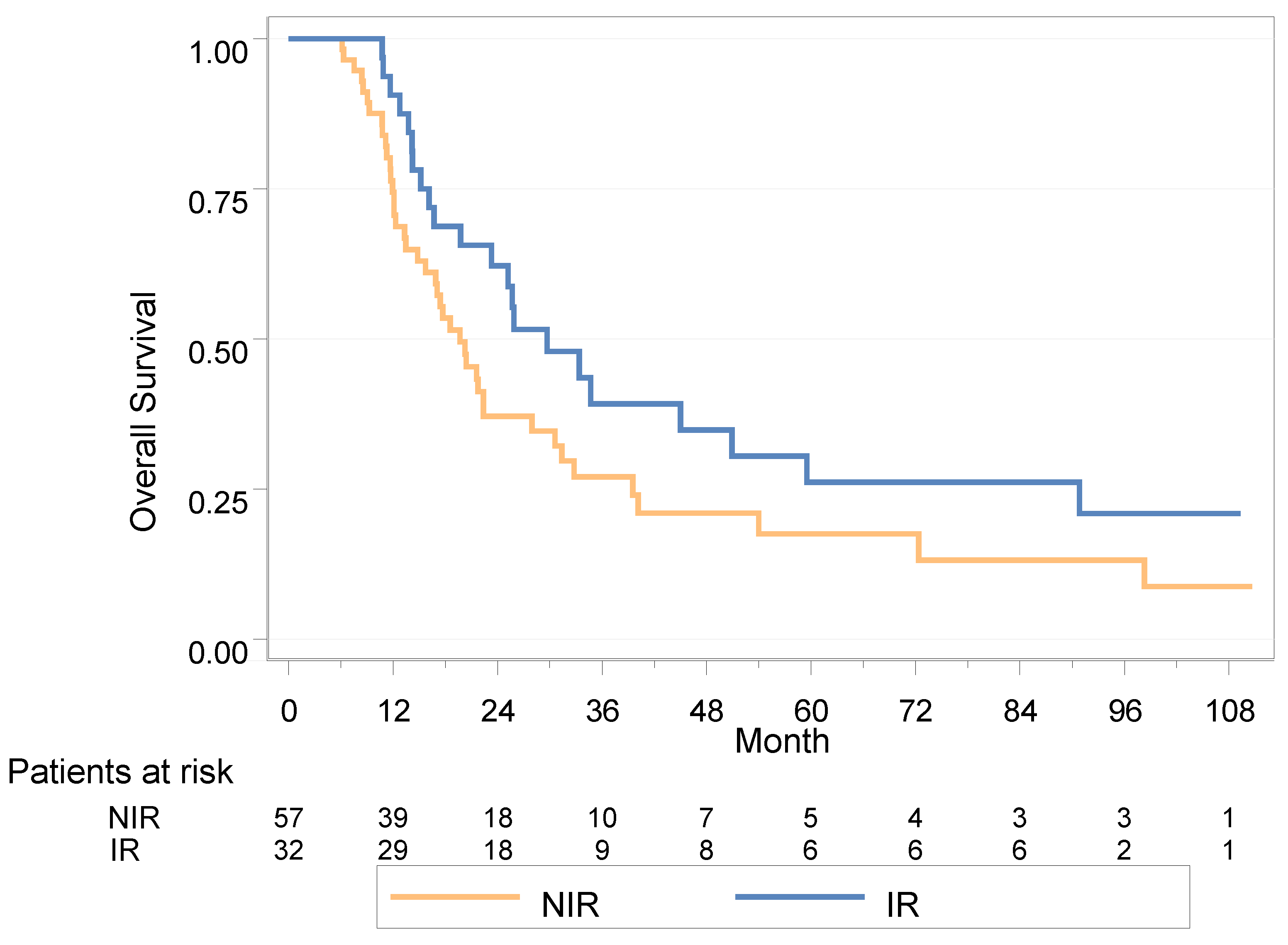
In the landmark population analysis at 6 months, the median PFS was 14.8 months in the IR group (11.4; 18.6) versus 9.7 months (8.2; 11) in the NIR group (HR = 0.52 (0.32–0.84), p = 0.006); the 2-year PFS was 31.3% vs. 18.5%, respectively.
In univariable analysis, a delay of 24 months or more between diagnosis and the appearance of metastases appeared deleterious in terms of PFS (HR = 1.89 (1.07–3.36), p = 0.026), as did the presence of a liver metastasis (HR = 2.76 (1.31–5.81), p = 0.005) (Table 3).
In multivariable analysis, none of the variables were statistically associated with PFS; in the IR group there was a trend to improved PFS (HR = 0.57 (0.31–1.07), p = 0.082) (Table 3).

3.5. OS

3.5.1. Whole Population

After a median follow up of 85.9 months (95% IC [36.7; 101.6]), the median OS was 21.7 months (95% IC (17.1–29.7)); using the Cox model with a time-dependent, variable the HR for OS was 0.53 in the IR group (95% IC (0.32–0.87), p = 0.011) (Figure 3).
In univariable analysis, none of the variables were statistically associated with OS; there was a trend of decreased OS among patients with in situ carcinoma (CIS+) (HR = 1.73 (0.90–3.32), p = 0.098) (Table S2).
In multivariable analysis, OS remained improved in the IR group (HR = 0.47, [0.25–0.86], p = 0.015) (Table S2).

3.5.2. Landmark Analysis

In the landmark population analysis at 6 months, the median OS was 29.7 months in the IR group (16.7–50.9) versus 19.7 months in the NIR group (14.8–28.0), HR = 0.63 (0.37–1.05), p = 0.074; 2-year OS was 62.2% vs. 37.1%, respectively.
In univariable analysis, none of the variables were statistically associated with OS, with a trend for CIS+ patients (HR = 1.80 (0.93–3.47), p = 0.076) and number of residual metastases (p = 0.12) (Table 4).
In multivariable analysis, OS remained improved in the IR group (HR = 0.48 (0.25–0.92), p = 0.026) (Table 4).

3.6. Toxicity in the IR Group

Among patients having received pelvic irradiation (bladder and/or PLN irradiation), the rate of grade ≥ 3 toxicity was 2.3% (1/43); it was a grade 3 gastro-intestinal toxicity (proctitis).
Among patients having received metastases-directed irradiation, the rate of grade ≥ 3 toxicity was 2.6% (1/38); it was a grade 3 gastro-intestinal toxicity due to pseudo-occlusive syndrome.

4. Discussion

In this retrospective multicentric study, we found that, among patients with at least stable disease and no more than five residual metastases following first-line systemic therapy for metastatic urothelial carcinoma, local consolidative radiotherapy to the bladder (when applicable) and on residual metastases provided significant survival benefit. In multivariable analysis, consolidative radiotherapy conferred a benefit in both PFS (HR = 0.49, p = 0.007) and OS (HR = 0.47, p = 0.015) in the whole population, and in OS in the landmark analysis at 6 months (HR = 0.48, p = 0.026), with only a trend for PFS (HR = 0.57, p = 0.082).
Given that radiotherapy in the IR group was performed during the follow up, the landmark analysis at 6 months (from the initiation of systemic therapy) has been realized to preventthe immortal time bias in the IR group [40].The landmark was set at 6 months because most patients in the IR group had received radiotherapy within 6 months of the start of analysis (32/51, 63% of the irradiated patients) and because a landmark beyond 6 months would have substantially decreased the event-free population.
Overall, the main biological rationale to propose local ablative treatment for metastatic malignancies, and especially oligometastatic disease, relies on the seed-and-soil theory. This refers to the secretion of tumor growth factors by the primary tumor, which may promote cancer-cell engraftment in distant organs as they constitute the source of the seed (the circulating-tumor cells), but also may prepare the metastatic soil via the secretion of cytokines that prime the microenvironment in which metastases can potentially develop [41]. In mUBC, data regarding ablative radiotherapy are scare with small series. Overall, in these studies, the 2-year PFS ranged from 19% to 40%, and the 2-year OS ranged from 26% to 75% [31,32,33,34,35,36,37]. However, these data are very heterogeneous in terms of disease characteristics (as high as 35% of oligoprogressive disease in the study by Franzese et al. [36]), radiotherapy schemes (irradiation or not of bladder along with metastases), and timing of radiation delivery compared with systemic therapy (no systemic therapy, upfront radiotherapy with systemic therapy, and systemic therapy followed by consolidative radiotherapy).
In our study, we decided to focus on patients with the oligometastatic state following systemic therapy as a way to select the potentially good candidates for treatment intensification (consolidation approach). We then compared both strategies: local consolidative treatment versus observation. Interestingly, all patients had genuine de novo oligometastatic disease (either synchronous (49.5%) or metachronous (50.5%)), except one patient (in the IR group) who had induced oligopersistent disease (with nine metastatic lesions at diagnosis), according to the ESTRO EORTC oligometastatic classification [38]. This constitutes a major strength of the study as a recent study including any solid malignancy treated with stereotactic radiotherapy to all metastases suggested that, compared with genuine oligometastatic disease, induced oligometastatic disease had a poorer outcome, and that this should be addressed separately in future trials [42,43]. This consolidative approach following systemic therapy has been shown to be promising, especially among patients with non-small-cell lung cancers compared with no local treatment [44,45]. In the largest phase II trial to date, after a median follow up of 38.8 months, there was a benefit from local consolidative therapy in both PFS and OS (median PFS 14.2 months vs. 4.4 months (p = 0.022) and median OS 41.2 months vs. 18.9 months (p = 0.017)) [44]. Our study, which used a similar design, suggests a similar benefit among mUBC. To the best of our knowledge, this is also the largest cohort of local ablative treatment for oligometastatic bladder cancer.
Moreover, while other series focused either on metastases-directed therapy [34] or on bladder-directed therapy [14,15], our series was innovative as it systematically included patients with local treatment of both primary tumor (except for previous history of cystectomy) and metastases.
On the other hand, our study had some limitations, mainly due to its retrospective nature.
Standards of care for mUBC have recently integrated avelumab maintenance following chemotherapy in non-progressive patients, due to a strong benefit in both PFS and OS in the JAVELIN trial [46]. The patients of our study would have typically been proposed such maintenance according to current guidelines. It is worth mentioning that a 2-year PFS (31%) and a 2-year OS (49%) in the JAVELIN trial compare favorably with our 2-year PFS and 2-year OS in the IR group of the landmark analysis, of respectively 31% and 62%. Whether or not local consolidative radiotherapy can provide similar additional benefit in the context of avelumab maintenance is unknown. In this regard, the ongoing GETUG-AFU V07/BLAD-RAD01 trial (NCT04428554) is a multicentric randomized phase II trial that assesses the benefit of local consolidative radiotherapy to both the bladder and residual metastases among patients without progression following chemotherapy and with no more than three residual lesions (stratified on the type of staging: positron emission tomography (PET) vs. CT only). In this trial, the standard of care with avelumab maintenance following chemotherapy is provided in both arms of treatment, with or without additional radiotherapy.
Finally, radiotherapy towards the tumor has the potency to modify the immune anti-tumor response, as it can induce both immune-stimulatory and immune-suppressive effects [47], and the hypothetical rationale for combining radiotherapy and immunotherapy, especially immune check point inhibitors, is manifold (stimulation of radiation immune-stimulatory effects through in situ vaccination, counteraction of radiation immune-suppressive effects, and immune-resistance tackling) [47,48,49,50]. Therefore, in addition to the mere additive effect of combining both maintenance avelumab and consolidative local radiotherapy, one cannot exclude a synergistic effect of such an association. In this perspective, the optimal fractionation for the irradiation of residual metastases should be determined properly, as the best schedule for maximizing in situ vaccination in combination with immunotherapy may be the delivery of fractions of 8–10 Gy [51,52,53].

5. Conclusions

Overall, we found in this retrospective multicentric study that consolidative radiotherapy to the primary tumor and to the residual metastases for mUBC patients who have not progressed after chemotherapy and with limited residual disease seems to confer both OS and PFS benefits. Prospective data in that field with the addition of avelumab are needed.

Supplementary Materials

The following supporting information can be downloaded at: https://www.mdpi.com/article/10.3390/cancers15041161/s1, Table S1: Univariable and multivariable analysis for progression free survival (whole population, Table S2: Univariable and multivariable analysis for overall survival (whole population).

Author Contributions

Conceptualization, J.K.; methodology, J.K. and A.A.; formal analysis, A.A. and L.C.; investigation, A.A., P.G.-C., N.B.-O., P.S., U.S., G.C. and E.C.-J.M.; data curation, A.A.; writing—original draft, J.K. and A.A.; writing—review & editing, A.A., D.P., P.G.-C., N.B.-O., P.S., U.S., G.C., E.C.-J.M. and C.C.; supervision, J.K. All authors have read and agreed to the published version of the manuscript.

Funding

This research received no external funding.

Institutional Review Board Statement

The study was conducted in accordance with the Declaration of Helsinki, and approved by the Ethics Committee of the Toulouse University Cancer Institute (CRC IUCT-O 19 URO 15; 5/11/2019), and authorized by the CNIL (Commission nationale informatique et liberté), authorization no. 919375.

Informed Consent Statement

This retrospective study relies on the re-utilization of clinical data from patients’ medical files. As such, French law classifies this study as a “non-interventional study that does not involve human subjects”. French regulatory authorities do not require patient written informed consent for a “non-interventional study that does not involve human subjects” so patient consent was waived for this reason. All patients in the study have however received a personalized information form as required by French authorities.

Data Availability Statement

De-identified data from this study are stored by JK.

Conflicts of Interest

The authors declare no conflict of interest.

References

  1. Saginala, K.; Barsouk, A.; Aluru, J.S.; Rawla, P.; Padala, S.A.; Barsouk, A. Epidemiology of Bladder Cancer. Med. Sci. 2020, 8, 15. [Google Scholar] [CrossRef]
  2. Alfred Witjes, J.; Lebret, T.; Compérat, E.M.; Cowan, N.C.; De Santis, M.; Bruins, H.M.; Hernández, V.; Espinós, E.L.; Dunn, J.; Rouanne, M.; et al. Updated 2016 EAU Guidelines on Muscle-Invasive and Metastatic Bladder Cancer. Eur. Urol. 2017, 71, 462–475. [Google Scholar] [CrossRef]
  3. Stein, J.P.; Lieskovsky, G.; Cote, R.; Groshen, S.; Feng, A.C.; Boyd, S.; Skinner, E.; Bochner, B.; Thangathurai, D.; Mikhail, M.; et al. Radical Cystectomy in the Treatment of Invasive Bladder Cancer: Long-Term Results in 1,054 Patients. J. Clin. Oncol. 2001, 19, 666–675. [Google Scholar] [CrossRef]
  4. Sargos, P.; Baumann, B.C.; Eapen, L.; Christodouleas, J.; Bahl, A.; Murthy, V.; Efstathiou, J.; Fonteyne, V.; Ballas, L.; Zaghloul, M.; et al. Risk Factors for Loco-Regional Recurrence after Radical Cystectomy of Muscle-Invasive Bladder Cancer: A Systematic-Review and Framework for Adjuvant Radiotherapy. Cancer Treat. Rev. 2018, 70, 88–97. [Google Scholar] [CrossRef] [PubMed]
  5. Kamat, A.M.; Hegarty, P.K.; Gee, J.R.; Clark, P.E.; Svatek, R.S.; Hegarty, N.; Shariat, S.F.; Xylinas, E.; Schmitz-Dräger, B.J.; Lotan, Y.; et al. ICUD-EAU International Consultation on Bladder Cancer 2012: Screening, Diagnosis, and Molecular Markers. Eur. Urol. 2013, 63, 4–15. [Google Scholar] [CrossRef]
  6. von der Maase, H.; Hansen, S.W.; Roberts, J.T.; Dogliotti, L.; Oliver, T.; Moore, M.J.; Bodrogi, I.; Albers, P.; Knuth, A.; Lippert, C.M.; et al. Gemcitabine and Cisplatin Versus Methotrexate, Vinblastine, Doxorubicin, and Cisplatin in Advanced or Metastatic Bladder Cancer: Results of a Large, Randomized, Multinational, Multicenter, Phase III Study. J. Clin. Oncol. 2000, 18, 3068–3077. [Google Scholar] [CrossRef] [PubMed]
  7. von der Maase, H.; Sengelov, L.; Roberts, J.T.; Ricci, S.; Dogliotti, L.; Oliver, T.; Moore, M.J.; Zimmermann, A.; Arning, M. Long-Term Survival Results of a Randomized Trial Comparing Gemcitabine Plus Cisplatin, With Methotrexate, Vinblastine, Doxorubicin, Plus Cisplatin in Patients With Bladder Cancer. J. Clin. Oncol. 2005, 23, 4602–4608. [Google Scholar] [CrossRef]
  8. Sternberg, C.N.; de Mulder, P.; Schornagel, J.H.; Theodore, C.; Fossa, S.D.; van Oosterom, A.T.; Witjes, J.A.; Spina, M.; van Groeningen, C.J.; Duclos, B.; et al. Seven Year Update of an EORTC Phase III Trial of High-Dose Intensity M-VAC Chemotherapy and G-CSF versus Classic M-VAC in Advanced Urothelial Tract Tumours. Eur. J. Cancer 2006, 42, 50–54. [Google Scholar] [CrossRef] [PubMed]
  9. Sternberg, C.N.; de Mulder, P.H.M.; Schornagel, J.H.; Théodore, C.; Fossa, S.D.; van Oosterom, A.T.; Witjes, F.; Spina, M.; van Groeningen, C.J.; de Balincourt, C.; et al. Randomized Phase III Trial of High–Dose-Intensity Methotrexate, Vinblastine, Doxorubicin, and Cisplatin (MVAC) Chemotherapy and Recombinant Human Granulocyte Colony-Stimulating Factor Versus Classic MVAC in Advanced Urothelial Tract Tumors: European Organization for Research and Treatment of Cancer Protocol No. 30924. J. Clin. Oncol. 2001, 19, 2638–2646. [Google Scholar] [CrossRef]
  10. Powles, T.; Park, S.H.; Voog, E.; Caserta, C.; Valderrama, B.P.; Gurney, H.; Kalofonos, H.; Radulovic, S.; Demey, W.; Ullén, A.; et al. Maintenance Avelumab + Best Supportive Care (BSC) versus BSC Alone after Platinum-Based First-Line (1L) Chemotherapy in Advanced Urothelial Carcinoma (UC): JAVELIN Bladder 100 Phase III Interim Analysis. J. Clin. Oncol. 2020, 38, LBA1. [Google Scholar] [CrossRef]
  11. Hellman, S.; Weichselbaum, R.R. Oligometastases. J. Clin. Oncol. 1995, 13, 8–10. [Google Scholar] [CrossRef]
  12. Palma, D.A.; Olson, R.; Harrow, S.; Gaede, S.; Louie, A.V.; Haasbeek, C.; Mulroy, L.; Lock, M.; Rodrigues, G.B.; Yaremko, B.P.; et al. Stereotactic Ablative Radiotherapy for the Comprehensive Treatment of Oligometastatic Cancers: Long-Term Results of the SABR-COMET Phase II Randomized Trial. J. Clin. Oncol. 2020, 38, 2830–2838. [Google Scholar] [CrossRef] [PubMed]
  13. Ogihara, K.; Kikuchi, E.; Watanabe, K.; Kufukihara, R.; Yanai, Y.; Takamatsu, K.; Matsumoto, K.; Hara, S.; Oyama, M.; Monma, T.; et al. Can Urologists Introduce the Concept of “Oligometastasis” for Metastatic Bladder Cancer after Total Cystectomy? Oncotarget 2017, 8, 111819–111835. [Google Scholar] [CrossRef] [PubMed]
  14. Seisen, T.; Sun, M.; Leow, J.J.; Preston, M.A.; Cole, A.P.; Gelpi-Hammerschmidt, F.; Hanna, N.; Meyer, C.P.; Kibel, A.S.; Lipsitz, S.R.; et al. Efficacy of High-Intensity Local Treatment for Metastatic Urothelial Carcinoma of the Bladder: A Propensity Score-Weighted Analysis From the National Cancer Data Base. J. Clin. Oncol. 2016, 34, 3529–3536. [Google Scholar] [CrossRef]
  15. Fischer-Valuck, B.W.; Patel, S.A.; Brenneman, R.J.; Christodouleas, J.; Sargos, P.; Kim, E.; Weiss, A.; Hershatter, B.; Rao, Y.J.; Picus, J.; et al. Association Between Local Radiation Therapy to the Primary Bladder Tumor and Overall Survival for Patients with Metastatic Urothelial Cancer Receiving Systemic Chemotherapy. Eur. Urol. Oncol. 2022, 5, 246–250. [Google Scholar] [CrossRef] [PubMed]
  16. de Vries, R.R.; Nieuwenhuijzen, J.A.; Meinhardt, W.; Bais, E.M.; Horenblas, S. Long-Term Survival after Combined Modality Treatment in Metastatic Bladder Cancer Patients Presenting with Supra-Regional Tumor Positive Lymph Nodes Only. Eur. J. Surg. Oncol. 2009, 35, 352–355. [Google Scholar] [CrossRef]
  17. Necchi, A.; Giannatempo, P.; Lo Vullo, S.; Farè, E.; Raggi, D.; Nicolai, N.; Piva, L.; Biasoni, D.; Torelli, T.; Catanzaro, M.; et al. Postchemotherapy Lymphadenectomy in Patients with Metastatic Urothelial Carcinoma: Long-Term Efficacy and Implications for Trial Design. Clin. Genitourin. Cancer 2015, 13, 80–86.e1. [Google Scholar] [CrossRef]
  18. Sweeney, P.; Millikan, R.; Donat, M.; Wood, C.G.; Radtke, A.S.; Pettaway, C.A.; Grossman, H.B.; Dinney, C.P.N.; Swanson, D.A.; Pisters, L.L. Is There a Therapeutic Role for Post-Chemotherapy Retroperitoneal Lymph Node Dissection in Metastatic Transitional Cell Carcinoma of the Bladder? J. Urol. 2003, 169, 2113–2117. [Google Scholar] [CrossRef] [PubMed]
  19. Han, W.S.; Kim, K.; Park, J.S. Result of Surgical Resection for Pulmonary Metastasis from Urothelial Carcinoma. Korean J. Thorac. Cardiovasc. Surg. 2012, 45, 242–245. [Google Scholar] [CrossRef]
  20. Kanzaki, R.; Higashiyama, M.; Fujiwara, A.; Tokunaga, T.; Maeda, J.; Okami, J.; Nishimura, K.; Kodama, K. Outcome of Surgical Resection of Pulmonary Metastasis from Urinary Tract Transitional Cell Carcinoma. Interact. Cardiovasc. Thorac. Surg. 2010, 11, 60–64. [Google Scholar] [CrossRef] [Green Version]
  21. Matsuguma, H.; Yoshino, I.; Ito, H.; Goya, T.; Matsui, Y.; Nakajima, J.; Ikeda, N.; Okumura, S.; Shiono, S.; Nomori, H.; et al. Is There a Role for Pulmonary Metastasectomy with a Curative Intent in Patients with Metastatic Urinary Transitional Cell Carcinoma? Ann. Thorac. Surg. 2011, 92, 449–453. [Google Scholar] [CrossRef] [PubMed]
  22. Luzzi, L.; Marulli, G.; Solli, P.; Cardillo, G.; Ghisalberti, M.; Mammana, M.; Carleo, F.; Spaggiari, L.; Rea, F. Long-Term Results and Prognostic Factors of Pulmonary Metastasectomy in Patients with Metastatic Transitional Cell Carcinoma. Thorac. Cardiovasc. Surg. 2017, 65, 567–571. [Google Scholar] [CrossRef] [PubMed]
  23. Otto, T.; Krege, S.; Suhr, J.; Rübben, H. Impact of Surgical Resection of Bladder Cancer Metastases Refractory to Systemic Therapy on Performance Score: A Phase II Trial. Urology 2001, 57, 55–59. [Google Scholar] [CrossRef] [PubMed]
  24. Abe, T.; Kitamura, H.; Obara, W.; Matsumura, N.; Tsukamoto, T.; Fujioka, T.; Hara, I.; Murai, S.; Shinohara, N.; Nonomura, K. Outcome of Metastasectomy for Urothelial Carcinoma: A Multi-Institutional Retrospective Study in Japan. J. Urol. 2014, 191, 932–936. [Google Scholar] [CrossRef]
  25. Bekku, K.; Saika, T.; Kobayashi, Y.; Kioshimoto, R.; Kanbara, T.; Nasu, Y.; Kumon, H. Could Salvage Surgery after Chemotherapy Have Clinical Impact on Cancer Survival of Patients with Metastatic Urothelial Carcinoma? Int. J. Clin. Oncol. 2013, 18, 110–115. [Google Scholar] [CrossRef]
  26. Dodd, P.M.; McCaffrey, J.A.; Herr, H.; Mazumdar, M.; Bacik, J.; Higgins, G.; Boyle, M.G.; Scher, H.I.; Bajorin, D.F. Outcome of Postchemotherapy Surgery after Treatment with Methotrexate, Vinblastine, Doxorubicin, and Cisplatin in Patients with Unresectable or Metastatic Transitional Cell Carcinoma. J. Clin. Oncol. 1999, 17, 2546–2552. [Google Scholar] [CrossRef]
  27. Siefker-Radtke, A.O.; Walsh, G.L.; Pisters, L.L.; Shen, Y.; Swanson, D.A.; Logothetis, C.J.; Millikan, R.E. Is There a Role for Surgery in the Management of Metastatic Urothelial Cancer? The M. D. Anderson Experience. J. Urol. 2004, 171, 145–148. [Google Scholar] [CrossRef] [PubMed]
  28. Lehmann, J.; Suttmann, H.; Albers, P.; Volkmer, B.; Gschwend, J.E.; Fechner, G.; Spahn, M.; Heidenreich, A.; Odenthal, A.; Seif, C.; et al. Surgery for Metastatic Urothelial Carcinoma with Curative Intent: The German Experience (AUO AB 30/05). Eur. Urol. 2009, 55, 1293–1299. [Google Scholar] [CrossRef]
  29. Kim, T.; Ahn, J.-H.; You, D.; Jeong, I.-G.; Hong, B.; Hong, J.H.; Ahn, H.; Lee, J.L. Pulmonary Metastasectomy Could Prolong Overall Survival in Select Cases of Metastatic Urinary Tract Cancer. Clin. Genitourin. Cancer 2015, 13, e297–e304. [Google Scholar] [CrossRef]
  30. Patel, V.; Collazo Lorduy, A.; Stern, A.; Fahmy, O.; Pinotti, R.; Galsky, M.D.; Gakis, G. Survival after Metastasectomy for Metastatic Urothelial Carcinoma: A Systematic Review and Meta-Analysis. Bladder Cancer 2017, 3, 121–132. [Google Scholar] [CrossRef] [Green Version]
  31. Shah, S.; Zhang, C.A.; Hancock, S.; Fan, A.; Skinner, E.; Srinivas, S. Consolidative Radiotherapy in Metastatic Urothelial Cancer. Clin. Genitourin. Cancer 2017, 15, 685–688. [Google Scholar] [CrossRef]
  32. Abe, T.; Minami, K.; Harabayashi, T.; Sazawa, A.; Chiba, H.; Kikuchi, H.; Miyata, H.; Frumido, J.; Matsumoto, R.; Osawa, T.; et al. Prognostic Impact of Local Radiotherapy on Metastatic Urothelial Carcinoma Patients Receiving Systemic Chemotherapy. Jpn. J. Clin. Oncol. 2020, 50, 206–213. [Google Scholar] [CrossRef]
  33. Leonetti, A.; D’Abbiero, N.; Baldari, G.; Andreani, S.; Ruffini, L.; Viansone, A.A.; Buti, S. Radiotherapy for the Treatment of Distant Nodes Metastases from Oligometastatic Urothelial Cancer: A Retrospective Case Series. Int. J. Urol. 2018, 25, 879–886. [Google Scholar] [CrossRef]
  34. Longo, N.; Celentano, G.; Napolitano, L.; La Rocca, R.; Capece, M.; Califano, G.; Ruvolo, C.C.; Mangiapia, F.; Fusco, F.; Morra, S.; et al. Metastasis-Directed Radiation Therapy with Consolidative Intent for Oligometastatic Urothelial Carcinoma: A Systematic Review and Meta-Analysis. Cancers 2022, 14, 2373. [Google Scholar] [CrossRef] [PubMed]
  35. Miranda, A.F.; Howard, J.M.; McLaughlin, M.; Meng, X.; Clinton, T.; Şanli, Ö.; Garant, A.; Bagrodia, A.; Margulis, V.; Lotan, Y.; et al. Metastasis-Directed Radiation Therapy after Radical Cystectomy for Bladder Cancer. Urol. Oncol. 2021, 39, 790.e1–790.e7. [Google Scholar] [CrossRef] [PubMed]
  36. Franzese, C.; Francolini, G.; Nicosia, L.; Alongi, F.; Livi, L.; Scorsetti, M. Stereotactic Body Radiation Therapy in the Management of Oligometastatic and Oligoprogressive Bladder Cancer and Other Urothelial Malignancies. Clin. Oncol. (R. Coll. Radiol.) 2021, 33, 50–56. [Google Scholar] [CrossRef]
  37. Augugliaro, M.; Marvaso, G.; Ciardo, D.; Zerini, D.; Riva, G.; Rondi, E.; Vigorito, S.; Comi, S.; de Cobelli, O.; Orecchia, R.; et al. Recurrent Oligometastatic Transitional Cell Bladder Carcinoma: Is There Room for Radiotherapy? Neoplasma 2019, 66, 160–165. [Google Scholar] [CrossRef] [PubMed]
  38. Guckenberger, M.; Lievens, Y.; Bouma, A.B.; Collette, L.; Dekker, A.; deSouza, N.M.; Dingemans, A.-M.C.; Fournier, B.; Hurkmans, C.; Lecouvet, F.E.; et al. Characterisation and Classification of Oligometastatic Disease: A European Society for Radiotherapy and Oncology and European Organisation for Research and Treatment of Cancer Consensus Recommendation. Lancet Oncol. 2020, 21, e18–e28. [Google Scholar] [CrossRef] [PubMed]
  39. Pos, F.J.; Hart, G.; Schneider, C.; Sminia, P. Radical Radiotherapy for Invasive Bladder Cancer: What Dose and Fractionation Schedule to Choose? Int. J. Radiat. Oncol. Biol. Phys. 2006, 64, 1168–1173. [Google Scholar] [CrossRef]
  40. Park, H.S.; Gross, C.P.; Makarov, D.V.; Yu, J.B. Immortal Time Bias: A Frequently Unrecognized Threat to Validity in the Evaluation of Postoperative Radiotherapy. Int. J. Radiat. Oncol. Biol. Phys. 2012, 83, 1365–1373. [Google Scholar] [CrossRef]
  41. Liu, Q.; Zhang, H.; Jiang, X.; Qian, C.; Liu, Z.; Luo, D. Factors Involved in Cancer Metastasis: A Better Understanding to “Seed and Soil” Hypothesis. Mol. Cancer 2017, 16, 176. [Google Scholar] [CrossRef] [PubMed]
  42. Willmann, J.; Vlaskou Badra, E.; Adilovic, S.; Ahmadsei, M.; Christ, S.M.; van Timmeren, J.E.; Kroeze, S.G.C.; Mayinger, M.; Guckenberger, M.; Andratschke, N. Evaluation of the Prognostic Value of the ESTRO EORTC Classification of Oligometastatic Disease in Patients Treated with Stereotactic Body Radiotherapy: A Retrospective Single Center Study. Radiother. Oncol. 2022, 168, 256–264. [Google Scholar] [CrossRef] [PubMed]
  43. Nevens, D.; Jongen, A.; Kindts, I.; Billiet, C.; Deseyne, P.; Joye, I.; Lievens, Y.; Guckenberger, M. Completeness of Reporting Oligometastatic Disease Characteristics in the Literature and Influence on Oligometastatic Disease Classification Using the ESTRO/EORTC Nomenclature. Int. J. Radiat. Oncol. Biol. Phys. 2022, 114, 587–595. [Google Scholar] [CrossRef]
  44. Gomez, D.R.; Tang, C.; Zhang, J.; Blumenschein, G.R.; Hernandez, M.; Lee, J.J.; Ye, R.; Palma, D.A.; Louie, A.V.; Camidge, D.R.; et al. Local Consolidative Therapy vs. Maintenance Therapy or Observation for Patients With Oligometastatic Non–Small-Cell Lung Cancer: Long-Term Results of a Multi-Institutional, Phase II, Randomized Study. J. Clin. Oncol. 2019, 37, 1558–1565. [Google Scholar] [CrossRef]
  45. Iyengar, P.; Wardak, Z.; Gerber, D.E.; Tumati, V.; Ahn, C.; Hughes, R.S.; Dowell, J.E.; Cheedella, N.; Nedzi, L.; Westover, K.D.; et al. Consolidative Radiotherapy for Limited Metastatic Non–Small-Cell Lung Cancer. JAMA Oncol. 2018, 4, e173501. [Google Scholar] [CrossRef] [PubMed]
  46. Powles, T.; Park, S.H.; Voog, E.; Caserta, C.; Valderrama, B.P.; Gurney, H.; Kalofonos, H.; Radulović, S.; Demey, W.; Ullén, A.; et al. Avelumab Maintenance Therapy for Advanced or Metastatic Urothelial Carcinoma. N. Engl. J. Med. 2020, 383, 1218–1230. [Google Scholar] [CrossRef]
  47. Walle, T.; Martinez Monge, R.; Cerwenka, A.; Ajona, D.; Melero, I.; Lecanda, F. Radiation Effects on Antitumor Immune Responses: Current Perspectives and Challenges. Ther. Adv. Med. Oncol. 2018, 10, 1758834017742575. [Google Scholar] [CrossRef]
  48. Deng, L.; Liang, H.; Burnette, B.; Beckett, M.; Darga, T.; Weichselbaum, R.R.; Fu, Y.-X. Irradiation and Anti–PD-L1 Treatment Synergistically Promote Antitumor Immunity in Mice. J. Clin. Investig. 2014, 124, 687–695. [Google Scholar] [CrossRef]
  49. Dovedi, S.J.; Adlard, A.L.; Lipowska-Bhalla, G.; McKenna, C.; Jones, S.; Cheadle, E.J.; Stratford, I.J.; Poon, E.; Morrow, M.; Stewart, R.; et al. Acquired Resistance to Fractionated Radiotherapy Can Be Overcome by Concurrent PD-L1 Blockade. Cancer Res. 2014, 74, 5458–5468. [Google Scholar] [CrossRef] [PubMed]
  50. Herrera, F.G.; Ronet, C.; de Olza, M.O.; Barras, D.; Crespo, I.; Andreatta, M.; Corria-Osorio, J.; Spill, A.; Benedetti, F.; Genolet, R.; et al. Low-Dose Radiotherapy Reverses Tumor Immune Desertification and Resistance to Immunotherapy. Cancer Discov. 2022, 12, 108–133. [Google Scholar] [CrossRef]
  51. Reits, E.A.; Hodge, J.W.; Herberts, C.A.; Groothuis, T.A.; Chakraborty, M.; Wansley, E.K.; Camphausen, K.; Luiten, R.M.; de Ru, A.H.; Neijssen, J.; et al. Radiation Modulates the Peptide Repertoire, Enhances MHC Class I Expression, and Induces Successful Antitumor Immunotherapy. J. Exp. Med. 2006, 203, 1259–1271. [Google Scholar] [CrossRef] [PubMed]
  52. Vanpouille-Box, C.; Alard, A.; Aryankalayil, M.J.; Sarfraz, Y.; Diamond, J.M.; Schneider, R.J.; Inghirami, G.; Coleman, C.N.; Formenti, S.C.; Demaria, S. DNA Exonuclease Trex1 Regulates Radiotherapy-Induced Tumour Immunogenicity. Nat. Commun. 2017, 8, 15618. [Google Scholar] [CrossRef] [PubMed]
  53. Khalifa, J.; Mazieres, J.; Gomez-Roca, C.; Ayyoub, M.; Moyal, E.C.J. Radiotherapy in the Era of Immunotherapy With a Focus on Non-Small-Cell Lung Cancer: Time to Revisit Ancient Dogmas? Front. Oncol. 2021, 11, 662236. [Google Scholar] [CrossRef] [PubMed]
Figure 1. Flow diagram for the selection of patients. Abbreviations: UBC: urothelial bladder cancer; RT: radiotherapy; NIR group: no irradiation group; IR group: irradiation group.
Cancers 15 01161 g001
Figure 2. Progression-free survival in landmark analysis at 6 months for NIR (no irradiation) vs. IR group (radiotherapy within 6 months).
Cancers 15 01161 g002
Figure 3. Overall survival in landmark analysis at 6 months for NIR (no irradiation) vs. IR group (radiotherapy within 6 months).
Cancers 15 01161 g003
Table 1. Patient characteristics.
TOTAL
N = 91
NIR
N = 40
IR
N = 51
p Value
Center
Toulouse Cancer Institute74 (81.3%)40 (100.0%)34 (66.7%)
Institut Bergonié10 (11.0%)010 (19.6%)
CHU Brest3 (3.3%)03 (5.9%)
Institut Curie 4 (4.4%)04 (7.8%)
Sex 0.176
Male81 (89.0%)38 (95.0%)43 (84.3%)
Female10 (11.0%)2 (5.0%)8 (15.7%)
Age 0.665
Median626263
(Range)(37.0–83.0)(45.0–83.0)(37.0–80.0)
ECOG performance status 0.715
060 (65.9%)27 (67.5%)33 (64.7%)
126 (28.6%)10 (25.0%)16 (31.4%)
24 (4.4%)2 (5.0%)2 (3.9%)
31 (1.1%)1 (2.5%)0 (0.0%)
Histological variant 0.526
No69 (83.1%)31 (86.1%)38 (80.9%)
Yes14 (16.9%)5 (13.9%)9 (19.1%)
Missing844
Type of variant (n = 14) 0.550
Epidermoid1 (7.1%)1 (20.0%)0 (0.0%)
Glandular1 (7.1%)0 (0.0%)1 (11.1%)
Nested2 (14.3%)1 (20.0%)1 (11.1%)
Micropapillary5 (35.7%)1 (20.0%)4 (44.4%)
Plasmocytoid3 (21.4%)2 (40.0%)1 (11.1%)
Sarcomatoid2 (14.3%)0 (0.0%)2 (22.2%)
Presence of CIS 0.018
No47 (75.8%)18 (62.1%)29 (87.9%)
Yes15 (24.2%)11 (37.9%)4 (12.1%)
Missing291118
T clinical status at diagnosis 0.019
T116 (18.4%)9 (22.5%)7 (14.9%)
T252 (59.8%)22 (55.0%)30 (63.8%)
T38 (9.2%)7 (17.5%)1 (2.1%)
T411 (12.6%)2 (5.0%)9 (19.1%)
Missing404
N clinical status at diagnosis 0.396
N046 (51.1%)23 (57.5%)23 (46.0%)
N110 (11.1%)3 (7.5%)7 (14.0%)
N215 (16.7%)8 (20.0%)7 (14.0%)
N319 (21.1%)6 (15.0%)13 (26.0%)
Missing101
Metastatic presentation 0.001
Metachronous46 (50.5%)28 (70.0%)18 (35.3%)
Synchronous45 (49.5%)12 (30.0%)33 (64.7%)
Time from diagnosis to metastasis (months) 0.001
Median0.513.10.0
(Range)(0.0–50.7)(0.0–250.7)(0.0–92.6)
Number of metastatic lesions at metastatic presentation 0.040
Median23 2
(Range)(1–9)(1–5)(1–9)
Topography of metastatic lesions at metastatic presentation
Bone 0.428
No62 (68.1%)29 (72.5%)33 (64.7%)
Yes29 (31.9%)11 (27.5%)18 (35.3%)
Lung 0.062
No73 (81.1%)29 (72.5%)44 (88.0%)
Yes17 (18.9%)11 (27.5%)6 (12.0%)
Missing101
Liver 0.293
No83 (91.2%)35 (87.5%)48 (94.1%)
Yes8 (8.8%)5 (12.5%)3 (5.9%)
Central Nervous System 0.440
No90 (98.9%)39 (97.5%)51 (100.0%)
Yes61 (67.0%)29 (72.5%)32 (62.7%)
Extra Pelvic Node 0.326
No30 (33.0%)11 (27.5%)19 (37.3%)
Yes61 (67.0%)29 (72.5%)32 (62.7%)
Duration of first-line systemic therapy 0.248
Median3.83.93.8
Range0.7–7.41.6–4.90.7–7.4
Number of residual metastatic lesions following first-line therapy 0.179
Median121
(range)(0–5)(0–5)(0–5)
Abbreviations: NIR (no irradiation); IR group (radiotherapy).
Table 2. Details of local consolidative radiotherapy in the IR group (n = 51).
Irradiated VolumeN (%)Total Dose
Median (Range)
Dose per Fraction (Gy)
Median (Range)
Number of Fractions
Median (Range)
Total Dose EQD2 Gy
Bladder36
(70.6%)
64.0
(45.0–66.0)
2.0
(1.7–3.0)
33
(15–36)
64.0
(48.0–66.0)
Pelvic Nodes39 *
(76.5%)
50.0
(24.0–63.0)
1.85
(1.6–3.0)
25
(10–34)
48.5
(24.0–63.5)
Metastasis38
(74.5%)
(56 lesions)
54.0
(30.0–67.5)
1.9
(1.7–18.0)
30
(3–34)
53.0
(45.0–132)
* Of whom eight patients had pelvic lymph node irradiation only without bladder irradiation.
Table 3. Univariable and multivariable analysis for progression-free survival (landmark population analysis at 6 months).
VariableUnivariableMultivariable
HR (95% CI)p-ValueHR (95% CI)p-Value
SexMale1
Female0.52 (0.22–1.21)0.123
Presence of histological variantNo1
Yes1.05 (0.55–2.00)0.881
Presence of CISNo1 1
Yes1.59 (0.86–2.96)0.1371.48 (0.79–2.80)0.224
T clinical status at diagnosisT1–T21
T3–T41.08 (0.62–1.88)0.787
N clinical status at diagnosisN01
N+0.97 (0.61–1.53)0.891
Metastatic presentationSynchronous1
Metachronous1.29 (0.82–2.02)0.271
Time from diagnosis to metastasis<24 months1 1
≥24 months1.89 (1.07–3.36)0.0262.01 (0.90–4.50)0.089
Number of metastatic lesions at metastatic presentation≤31
>31.11 (0.69–1.79)0.674
Lung metastatic lesionNo1
Yes0.98 (0.55–1.72)0.937
Liver metastatic lesionNo1 1
Yes2.76 (1.31–5.81)0.0051.60 (0.63–4.07)0.322
Extra-pelvic node only *No1 1
Yes0.65 (0.40–1.07)0.1130.56 (0.29–1.11)0.097
Number of residual metastatic lesions following first-line therapy01
11.06 (0.56–1.98)
≥21.38 (0.80–2.36)0.438
Local consolidative radiotherapyNo (NIR)1 1
Yes (IR)0.52 (0.34–0.84)0.0060.57 (0.31–1.07)0.082
* Extra-pelvic nodes are the only sites of metastases. Abbreviations: NIR (no irradiation); IR group (radiotherapy within 6 months).
Table 4. Univariable and multivariable analysis for overall survival (landmark analysis at 6 months).
VariableUnivariableMultivariable
HR (95% CI)p-ValueHR (95% CI)p-Value
SexMale1
Female0.55 (0.22–1.36)0.188
Presence of histological variantNo1
Yes1.06 (0.52–2.16)0.881
Presence of CISNo1 1
Yes1.80 (0.93–3.47)0.0761.49 (0.76–2.91)0.244
T clinical status at diagnosisT1–T21
T3–T41.12 (0.59–2.01)0.709
N clinical status at diagnosisN01
N+1.27 (0.77–2.08)0.348
Metastatic presentationSynchronous1
Metachronous1.06 (0.65–1.74)0.805
Time from diagnosis to metastasis<24 months1
≥24 months1.37 (0.76–2.49)0.293
Number of metastatic lesions at metastatic presentation≤31
>31.13 (0.68–1.88)0.635
Lung metastatic lesionNo1
Yes0.94 (0.51–1.74)0.844
Liver metastatic lesionNo1
Yes1.69 (0.76–3.74)0.192
Extra-pelvic node only *No1
Yes0.73 (0.42–1.26)0.254
Number of residual metastatic lesions following first-line therapy01 1
10.61 (0.29–1.29) 0.68 (0.26–1.80)
≥21.24 (0.70–2.18)0.1201.36 (0.69–2.68)0.367
Local consolidative radiotherapyNo (NIR)1 1
Yes (IR)0.63 (0.37–1.05)0.0740.48 (0.25–0.92)0.026
* Extra-pelvic nodes are the only sites of metastases. Abbreviations: NIR (no irradiation); IR group (radiotherapy within 6 months).
Disclaimer/Publisher’s Note: The statements, opinions and data contained in all publications are solely those of the individual author(s) and contributor(s) and not of MDPI and/or the editor(s). MDPI and/or the editor(s) disclaim responsibility for any injury to people or property resulting from any ideas, methods, instructions or products referred to in the content.

