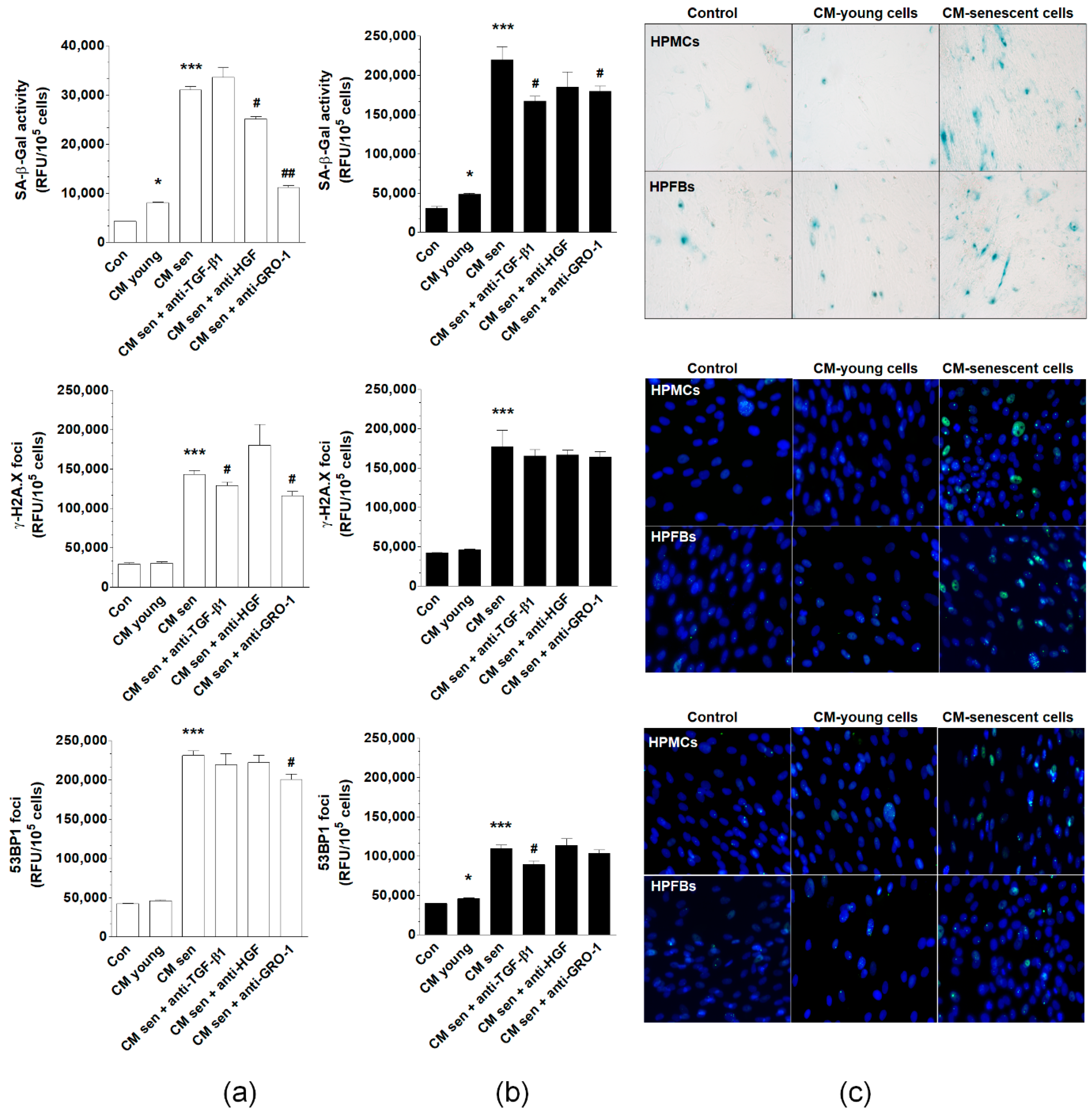
Correction: Pakuła et al. Deciphering the Molecular Mechanism of Spontaneous Senescence in Primary Epithelial Ovarian Cancer Cells. Cancers 2020, 12, 296


 , and
, and 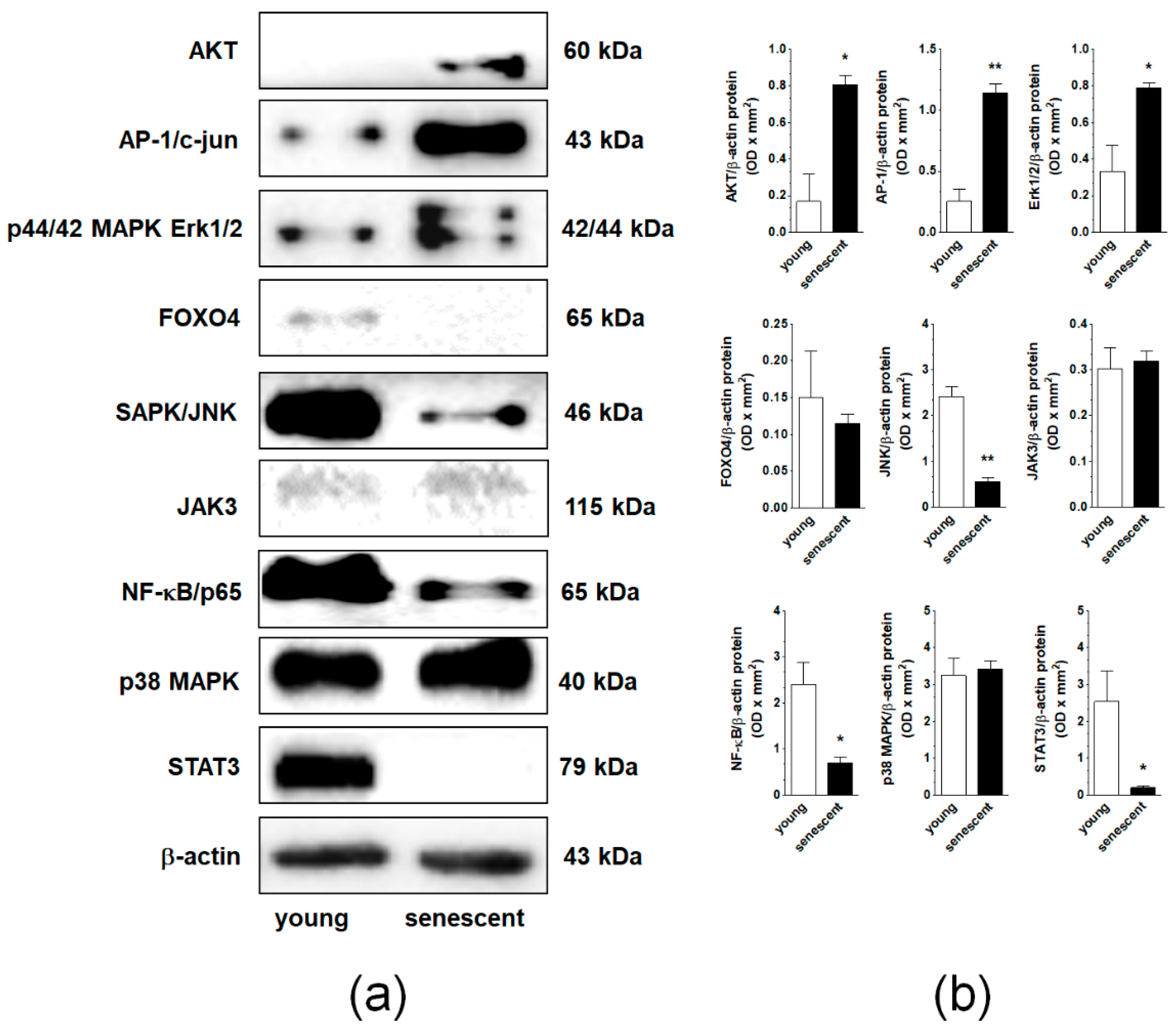
Error in Figures
Reference
- Pakuła, M.; Mały, E.; Uruski, P.; Witucka, A.; Bogucka, M.; Jaroszewska, N.; Makowska, N.; Niklas, A.; Moszyński, R.; Sajdak, S.; et al. Deciphering the molecular mechanism of spontaneous senescence of primary epithelial ovarian cancer cells. Cancers 2020, 12, 296. [Google Scholar] [CrossRef] [PubMed]


Disclaimer/Publisher’s Note: The statements, opinions and data contained in all publications are solely those of the individual author(s) and contributor(s) and not of MDPI and/or the editor(s). MDPI and/or the editor(s) disclaim responsibility for any injury to people or property resulting from any ideas, methods, instructions or products referred to in the content. |
© 2023 by the authors. Licensee MDPI, Basel, Switzerland. This article is an open access article distributed under the terms and conditions of the Creative Commons Attribution (CC BY) license (https://creativecommons.org/licenses/by/4.0/).
Share and Cite
Pakuła, M.; Mały, E.; Uruski, P.; Witucka, A.; Bogucka, M.; Jaroszewska, N.; Makowska, N.; Niklas, A.; Moszyński, R.; Sajdak, S.; et al. Correction: Pakuła et al. Deciphering the Molecular Mechanism of Spontaneous Senescence in Primary Epithelial Ovarian Cancer Cells. Cancers 2020, 12, 296. Cancers 2023, 15, 937. https://doi.org/10.3390/cancers15030937
Pakuła M, Mały E, Uruski P, Witucka A, Bogucka M, Jaroszewska N, Makowska N, Niklas A, Moszyński R, Sajdak S, et al. Correction: Pakuła et al. Deciphering the Molecular Mechanism of Spontaneous Senescence in Primary Epithelial Ovarian Cancer Cells. Cancers 2020, 12, 296. Cancers. 2023; 15(3):937. https://doi.org/10.3390/cancers15030937
Chicago/Turabian StylePakuła, Martyna, Ewa Mały, Paweł Uruski, Anna Witucka, Małgorzata Bogucka, Natalia Jaroszewska, Nicoletta Makowska, Arkadiusz Niklas, Rafał Moszyński, Stefan Sajdak, and et al. 2023. "Correction: Pakuła et al. Deciphering the Molecular Mechanism of Spontaneous Senescence in Primary Epithelial Ovarian Cancer Cells. Cancers 2020, 12, 296" Cancers 15, no. 3: 937. https://doi.org/10.3390/cancers15030937
APA StylePakuła, M., Mały, E., Uruski, P., Witucka, A., Bogucka, M., Jaroszewska, N., Makowska, N., Niklas, A., Moszyński, R., Sajdak, S., Tykarski, A., Mikuła-Pietrasik, J., & Książek, K. (2023). Correction: Pakuła et al. Deciphering the Molecular Mechanism of Spontaneous Senescence in Primary Epithelial Ovarian Cancer Cells. Cancers 2020, 12, 296. Cancers, 15(3), 937. https://doi.org/10.3390/cancers15030937