Review

The Role of Gene Fusions in Thymic Epithelial Tumors

by
Anja C. Roden
Department of Laboratory Medicine & Pathology, Mayo Clinic Rochester, Hilton 11, 200 First St SW, Rochester, MN 55905, USA
Cancers 2023, 15(23), 5596; https://doi.org/10.3390/cancers15235596
Submission received: 23 October 2023 / Revised: 14 November 2023 / Accepted: 23 November 2023 / Published: 27 November 2023

Abstract

:

Simple Summary

Thymic epithelial tumors (TET) are rare. Therefore, large studies, specifically concerning genetic alterations, are difficult in these tumors and require multi-institutional effort. However, such studies are important because alterations in certain genes of these tumors could be useful in the diagnosis of difficult cases, provide a better understanding of the nature of these tumors, or may even be therapeutically targetable. A few genetic alterations have already been identified in TET. This review will focus on gene fusions that have been reported in a subset of TET. The histopathologic features and clinical characteristics of these subtypes of TET and the potential clinical implications of the gene fusions for outcome of the patient and therapeutic options will be discussed.

Abstract

Thymic epithelial tumors (TET) are rare and large molecular studies are therefore difficult to perform. However, institutional case series and rare multi-institutional studies have identified a number of interesting molecular aberrations in TET, including gene fusions in a subset of these tumors. These gene fusions can aid in the diagnosis, shed light on the pathogenesis of a subset of tumors, and potentially may provide patients with the opportunity to undergo targeted therapy or participation in clinical trials. Gene fusions that have been identified in TET include MAML2 rearrangements in 50% to 56% of mucoepidermoid carcinomas (MAML2::CRTC1), 77% to 100% of metaplastic thymomas (YAP1::MAML2), and 6% of B2 and B3 thymomas (MAML2::KMT2A); NUTM1 rearrangements in NUT carcinomas (most commonly BRD4::NUTM1); EWSR1 rearrangement in hyalinizing clear cell carcinoma (EWSR1::ATF1); and NTRK rearrangement in a thymoma (EIF4B::NTRK3). This review focuses on TET in which these fusion genes have been identified, their morphologic, immunophenotypic, and clinical characteristics and potential clinical implications of the fusion genes. Larger, multi-institutional, global studies are needed to further elucidate the molecular characteristics of these rare but sometimes very aggressive tumors in order to optimize patient management, provide patients with the opportunity to undergo targeted therapy and participate in clinical trials, and to elucidate the pathogenesis of these tumors.
Keywords:
MAML2; NUTM1; EWSR1; YAP1; NTRK

1. Introduction

With the advantage of increasingly accessible and affordable molecular testing, gene alterations, including gene fusions, are being identified increasingly more often in thymomas and thymic carcinomas. The presence of gene fusions can aid in the diagnosis of neoplasms, provide more insight into the pathogenesis of these tumors, and potentially be useful for individualized management and targeted therapies. Given the paucity of thymic epithelial tumors (TET), large studies are challenging to perform even though multi-institutional studies of molecular alterations in these tumors have been accomplished [1].
In TET, recurring gene fusions have been identified involving mastermind transcriptional coactivator 2 (MAML2), Ewing sarcoma breakpoint region 1/EWS RNA binding protein 1 (EWSR1), NUT midline carcinoma family member 1 (NUTM1), and neurotrophic tyrosine kinase receptor (NTRK). MAML2 gene rearrangements have been detected in mucoepidermoid carcinomas [2,3,4], metaplastic thymomas [5,6,7,8], and occasional B2 and B3 thymomas [9]. Interestingly, fusion partners of MAML2 vary depending on the subtype of the TET. While CRCT1 was found to be fused with MAML2 in mucoepidermoid carcinomas, as in mucoepidermoid carcinomas elsewhere in the body, YAP1 was identified as the fusion partner of MAML2 in metaplastic thymomas and KMT2A in type B2 and B3 thymomas. EWSR1 gene rearrangements are present in hyalinizing clear cell carcinomas [10], similar to hyalinizing clear cell carcinomas in the head and neck [11,12] and lung [13,14,15]. Gene fusions involving the NUTM1 gene are also found in thymic tumors, specifically in NUT carcinomas, which are considered a subtype of thymic carcinomas [16,17]. In addition to these recurring and well-established gene fusions there is a rare report of NTRK fusion in a thymoma [18].
Recurring fusion genes have not been reported in thymic neuroendocrine neoplasms [19].
Herein, gene fusions in TET are reviewed and their importance for diagnosis and pathogenesis are outlined. Although currently they are not used as targets for therapy, potential treatment options and investigative studies are discussed. Table 1 summarizes all TET in which a gene fusion has been identified, including the frequency of the gene fusion, morphologic features of the tumor and other diagnostic features.

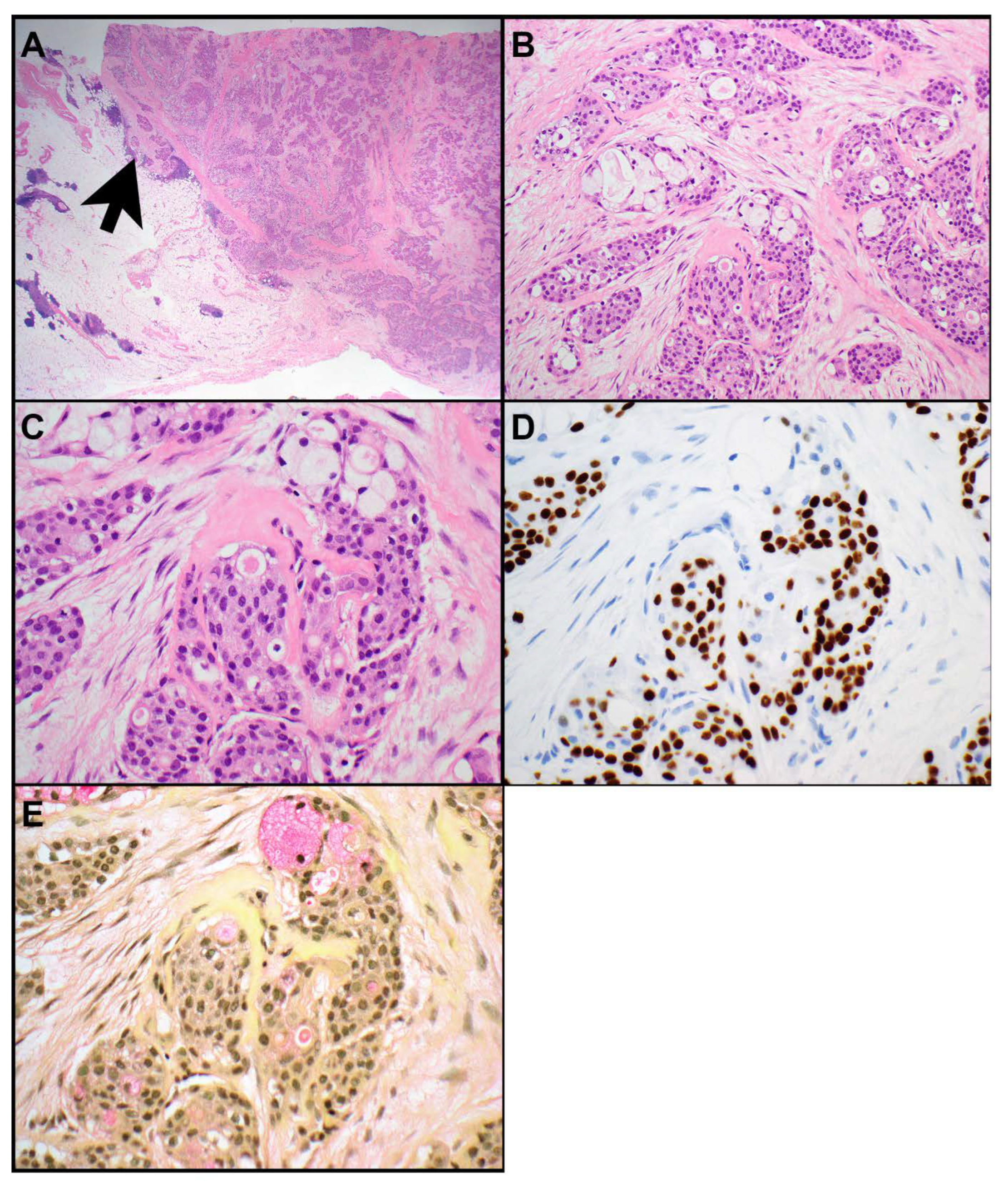
2. MAML2 Rearrangements

In the thymus, MAML2 rearrangements were first identified in mucoepidermoid carcinomas [2,3,4]. Subsequently they have also been found in metaplastic thymomas [5,6,7] and in rare B2 and B3 thymomas [9]. Despite the common MAML2 fusion partner, the corresponding fusion genes vary based on the tumor subtype in which they occur. For instance, in mucoepidermoid carcinomas, the reported fusion gene is cysteine-rich C-terminal 1 (CRTC1) leading to MAML2::CRTC1 fusions [2]. In metaplastic thymomas the fusion partner is Yes associated protein 1 (YAP1), leading to YAP1::MAML2 fusion genes [5,6,8]. In a subset of B2 and B3 thymomas the fusion partner is lysine methyltransferase 2A (KMT2A), resulting in KMT2A::MAML2 fusions [9]. Although these tumors have a common fusion gene, MAML2, they are morphologically and pathogenetically different.
Mucoepidermoid carcinomas are characterized by a triad of squamoid, intermediate, and mucus cells (Figure 1A–E) [3]. Their morphologic, immunophenotypic, and molecular features are essentially identical to mucoepidermoid carcinomas elsewhere, such as those of the head and neck, lung, or breast. In the thymus, mucoepidermoid carcinomas are considered a subtype of thymic carcinomas [20]. Here they can also arise in a cyst. MAML2 rearrangements are identified in 50 to 56% of thymic mucoepidermoid carcinomas [2,3,21]. The presence of MAML2 rearrangements aids in the diagnosis, specifically in high grade mucoepidermoid carcinomas, and can be utilized to distinguish mucoepidermoid carcinomas from thymic adenosquamous and squamous cell carcinomas as those do not harbor MAML2 rearrangements [3]. In a study of 20 thymic mucoepidermoid carcinomas, 56% harbored a t(11;19) (q21;p13) CRTC1::MAML2 gene fusion [2]. CRTC3::MAML2 [22] or EWSR1::POU5F1 [23] gene fusions, which have been identified in some head and neck mucoepidermoid carcinoma, have not been reported in thymic mucoepidermoid carcinomas. Furthermore, YAP1 and KMT2A were also not identified as fusion partners in thymic mucoepidermoid carcinomas [2]. Interestingly, the presence of CRTC1::MAML2 gene fusion in thymic mucoepidermoid carcinomas was associated with classic tumor histology, lower pT and TNM stage, and better overall survival [2].
A meta-analysis of thymic mucoepidermoid carcinoma [21], including 41 such patients from 24 studies, revealed a mean patient age of 49.8 years (range, 8 to 87 years) without any obvious sex predilection. The mean tumor size was 7.6 cm (range, 1.5 to 20 cm). Patients most commonly presented with dyspnea and/or chest pain; about a third of the patients were asymptomatic and the tumor was found incidentally. Various grading systems have been reported for mucoepidermoid carcinoma. In the thymus, the WHO does not recommend any particular grading system; however, in the lung, these tumors are divided into low- and high-grade tumors according to the WHO [20]. In the abovementioned meta-analysis most patients (67%) had low grade morphology with only 27% showing high grade features [21]. The 5 year and 10 year survivals have been reported as 69% and 43%, respectively [2]. Twenty-five and forty-five percent of patients died of disease with a median survival of 40 and 12 months, respectively [2,21]. Worse overall survival was associated with higher pT stage, higher TNM stage, residual tumors, greater tumor size, high grade tumor histology, and absence of CRTC1::MAML2 fusion [2]. Lymph node sampling was recommended and histologic grade and tumor stage/resectability were suggested as the main prognostic parameters [21]. Indeed, lymph node sampling is recommended in all resection specimens of TET; moreover, in thymic carcinomas which include mucoepidermoid carcinomas, deep thoracic lymph nodes (N2) should be sampled by surgeons [24].
The differential diagnosis of mucoepidermoid carcinomas in the thymic gland includes multiloculated thymic cyst, adenocarcinoma, and adenosquamous carcinoma. These entities do not harbor MAML2 rearrangements. However, as MAML2 rearrangements are only identified in about half of mucoepidermoid carcinomas, caution must be practiced in its absence. An invasive growth pattern of glands and epithelioid cells argues against a multiloculated thymic cyst. The presence of p63- or p40-positive tumor cells argues against an adenocarcinoma.
Metaplastic thymomas are very rare neoplasms that only occur in the thymus [25]. They are characterized by a biphasic morphology including a darker component of ovoid, bland appearing epithelial cells with inconspicuous nucleoli and a paler component of fibroblast-like spindle cells with ample pale cytoplasm (Figure 2A,B). The two components are either clearly separated or merged. Mitotic activity is not increased, and necrosis is not identified. The diagnosis in general is based on morphology. Keratin AE1/AE3 highlights the epithelial component and may or may not stain the spindle cell component. P40 marks only the epithelial component and is negative in the spindle cell component. MAML2 rearrangements have been identified in 77 to 100% of these tumors (Figure 2C,D) [5,6,7,8]. In metaplastic thymomas the fusion partner is YAP1, resulting in YAP1::MAML2 fusion genes fusing exon 1 or exons 1–5 on YAP1 with exons 2–5 on MAML2 [5,6]. However, fluorescence in situ hybridization (FISH) studies for MAML2 rearrangements are usually not necessary for the diagnosis. Moreover, the split signal that results from an intrachromosomal inversion on chromosome 11 in the YAP1::MAML2 fusion using a break apart FISH may be subtle and can be missed [7]. Recently, expression of YAP1-C terminus by immunohistochemistry has been shown to be highly sensitive and specific for metaplastic thymoma when evaluated in the context of other TET, including type A, AB, B1, B2, and B3 thymomas, and thymic carcinomas [7].
Both the epithelial and spindle cell component of metaplastic thymomas were considered to be of neoplastic nature by at least some authors, although molecular evidence was lacking [26]. More recently, MAML2 rearrangements have been observed in both the epithelial and spindle cell component (Figure 2C,D) suggesting that both components are at least clonal and likely neoplastic [6]. Mutations in POLE, HRAS, ALK, CDK4, PTEN, and BRAF were also identified in a number of metaplastic thymomas [6,8]. No other genetic alterations, including GTF2I mutation, have been found in these neoplasms [5,7,25].
Metaplastic thymomas are indolent tumors that occur at a median age of 50 years (range, 28 to 71 years) with a slight female predominance [7,20]. Reports of sarcomatoid carcinomas arising in metaplastic thymoma suggest that a small subset of these tumors may develop into a high-grade tumor [27,28]. The majority of patients do not develop metastases, recurrence, or mortality from the disease [5,6,7,8,25]. Only one patient has been reported to develop a recurrence and pleural metastases 14 months after resection of the primary tumor [26].
The differential diagnosis of metaplastic thymomas includes type A thymoma. Type A thymomas lack the biphasic appearance and are composed only of bland ovoid or slightly spindle cells without the spindle cell component. Additional tests are usually not necessary for the differential diagnosis. However, the majority (82% to 100%) of type A thymomas harbor a GTF2I mutation [8,29,30] which is absent in metaplastic thymomas. In contrast, YAP1::MAML2 fusions were not identified in type A thymoma or micronodular thymoma with lymphoid stroma, another entity that may be included in differential diagnoses of metaplastic thymomas [6,8]. In addition, at least a subset of type A thymomas has scattered thymocytes which are also absent in metaplastic thymomas. Sarcomatoid carcinoma can also mimic metaplastic thymoma. However, sarcomatoid carcinoma or carcinosarcoma have high-grade morphologies that typically show easily recognizable mitotic activity and necrosis.
Recently, two additional patients with mediastinal tumors with YAP1::MAML2 rearrangements were reported that followed a more aggressive clinical course [31]. One patient had a 9.8 cm mass invading the pericardium, right lung, and pulmonary artery and vein. A biopsy was interpreted as spindle sarcoma and the patient underwent neoadjuvant chemotherapy followed by resection of the thymic tumor and bilateral lung metastases. Microscopically the tumor was described as biphasic, composed of both epithelioid and spindle cell component; the epithelioid component expressed keratin and p63, the spindle cells were only focally positive for keratin. Targeted DNA/RNA sequencing revealed not only a YAP1::MAML2 fusion but also a TERT promoter mutation. The second patient was incidentally found to have a mediastinal mass which, on biopsy, was diagnosed as rhabdomyosarcoma. A resection specimen revealed poorly differentiated carcinomatous areas which expressed p63 and keratin and a rhabdomyosarcomatous area with expression of desmin. The tumor was interpreted as carcinosarcoma. The tumor had metastasized to a cervical lymph node and the lung. Sequencing revealed YAP1::MAML2 fusion in both components, a TP53 mutation in the carcinomatous and deep deletions of CDKN2A and CDKN2B in the sarcomatous component [31]. These two cases suggest that YAP1::MAML2 rearrangements may not only be found in pure metaplastic thymomas but also in more aggressive biphasic mediastinal tumors.
Thymomas are malignant neoplasms that occur in the prevascular mediastinum and in general have a more favorable outcome than thymic carcinomas. GTF2I mutations are the most prevalent genetic alterations in thymomas which most commonly occur in type A and AB thymomas [1,29,32]. MAML2 rearrangements have been identified in 6% of B2 and B3 thymoma but not in other thymomas [9]. In these tumors KMT2A on exons 8–11 fused with MAML2 on exon 2 [9]. These tumors had the typical morphology of B2 or B3 thymomas except for one case that also showed foci of thymic carcinoma. Interestingly, a KMT2A mutation has also been identified in a thymic papillary adenocarcinoma that occurred synchronously with a type A thymoma [33]. This 53 year-old woman presented with three nodules in the thorax including a 3 cm papillary adenocarcinoma in the anterior superior mediastinum, a 4 cm type A thymoma in the anterior mediastinum near the pericardium and a minimally invasive adenocarcinoma in the right upper lobe lung. Among other molecular alterations the thymic adenocarcinoma harbored a mutation in KMT2A c.2155A > C(p.S719R), while the type A thymoma harbored a different KMT2A mutation c.5343del(p.V178Yfs*38).
In thymic tumors, MAML2 rearrangements can aid in the diagnosis, specifically in mucoepidermoid carcinoma. These rearrangements may play a role in the OKpathogenesis, at least in a subset of TET. Studies in mice have confirmed CRTC1::MAML2 as an oncogenic driver for the development and maintenance of mucoepidermoid carcinomas [34]. While MAML2 rearrangements are currently not therapeutically targetable, these studies have also revealed EGFR and CDK4/6 inhibitors as potential therapeutic options in these tumors [34].

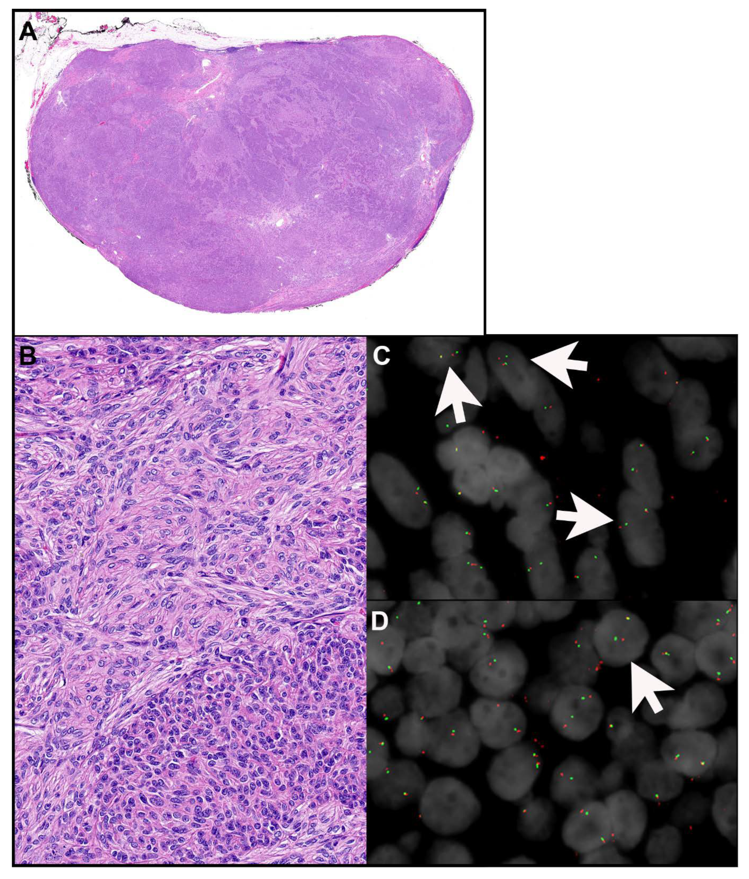
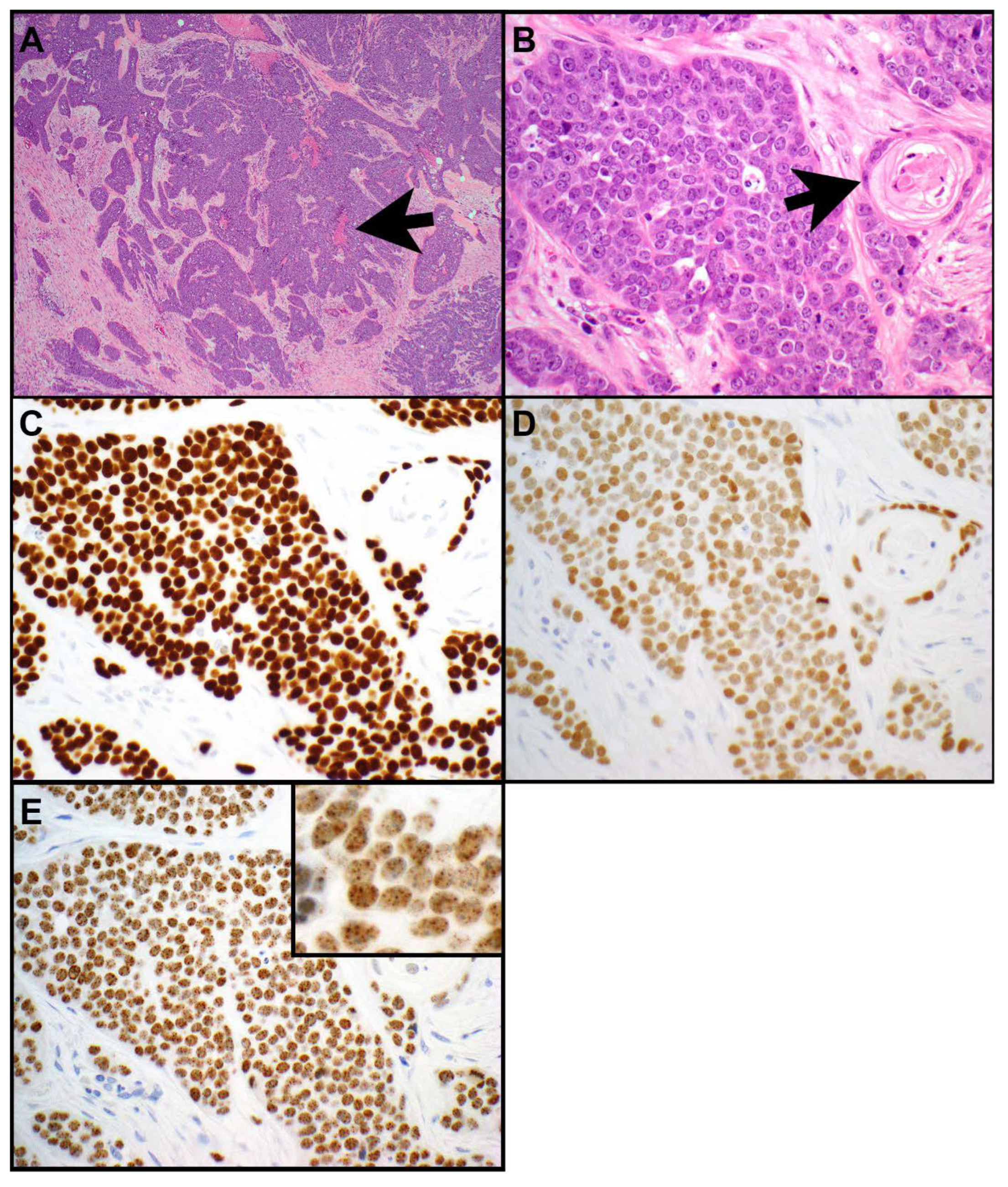
3. NUTM1 Rearrangements

In the mediastinum NUTM1 rearrangements are primarily seen in NUT carcinomas. More recently, these rearrangements have also been described in other small round blue cell tumors, although these tumors appear to be even rarer, specifically in the chest.
Nuclear protein in testis (NUT) carcinomas of the thorax are rare. These tumors are considered an aggressive subset of squamous cell carcinomas [16,35]. They are composed of small-to-medium-sized epithelioid cells with a high nuclear-to-cytoplasmic ratio, monotonous, round-to-oval nuclei, and prominent nucleoli (Figure 3A–E and Figure 4A–E) [36]. NUT carcinomas show high mitotic activity and necrosis. Numerous neutrophils can be seen intermixed with tumor cells in a subset of NUT carcinomas (Figure 4A) [37]. Abrupt squamous differentiation is classic for NUT carcinoma (Figure 3B) but is not always seen. Immunohistochemistry for NUT protein shows a characteristic speckled nuclear staining in the tumor cells (Figure 3E and Figure 4D) [36,38]. NUT carcinomas also frequently express markers of squamous differentiation, including p40 (Figure 3C), p63, and keratin 5 or 5/6 [39,40]; a subset of tumors expresses keratins such as AE1/AE3 and/or CAM5.2 [41,42]; and TTF-1 (Figure 3D and Figure 4B), CD34, or synaptophysin (Figure 4C) can also be seen in some of these tumors [40,43]. Rarely CD30, PLAP, and/or SALL4 expression has been reported in NUT carcinomas [44].
NUT protein is encoded by NUTM1. The most common molecular alteration, found in 70% to 78% of all NUT carcinomas, is a t(15;19) (q14;p13.1) translocation resulting in the BRD4::NUTM1 fusion oncogene [43,45,46]. Bromodomain containing protein 4 (BRD4) is a double bromodomain-containing gene which is involved in the regulation of cell cycle progression [17]. Less common translocations include BRD3::NUTM1 and NSD3::NUTM1 [43,45]. Other fusion partners of NTM1 are CHRM5, ZNF532, and ZNF592 [35,43,45]. NUTM1 rearrangements result in the expression of NUT protein which is normally only expressed in spermatids [47]. Immunohistochemistry for NUT reveals the abovementioned nuclear speckled staining pattern which is 100% specific and highly (87%) sensitive for NUT carcinoma in non-germ cell tumors [36,38]. Confirmatory ancillary tests are not necessary for the diagnosis. However, NUT immunohistochemistry may occasionally be negative. If in doubt, additional molecular or cytogenetic tests, such as next-generation sequencing (NGS), reverse transcription polymerase chain reaction (RT-PCR) for NUTM1::BRD4, fluorescence in situ hybridization (FISH) for NUTM1 rearrangement (Figure 4E), or karyotyping, should be performed. The karyotype of NUT carcinomas is usually simple, with the only aberration being the rearrangement involving the NUTM1 gene [46]. Besides NUTM1 rearrangements, no other molecular alterations have been reported in NUT carcinomas [45].
NUT carcinomas are highly aggressive neoplasms [48]. They are most commonly identified in the thorax (51% to 55%), specifically the mediastinum, followed by head and neck (40% to 41%) and other sites [45,49]. In the WHO, NUT carcinoma that occurs in the mediastinum is considered a subtype of thymic carcinoma [20]. The median age of patients is 16 to 50 years old (range, 0.1 to 78 years) [45,49]. There is no apparent sex predilection [45]. Most patients (51% to 77%) have metastatic disease at time of presentation with regional lymph node metastases identified in 68% of patients [45,49].
The prognosis of NUT carcinomas is poor [50,51]. The median overall survival is only 6.5 to 7 months, and the disease is usually fatal [45,49]. Evidence suggests that the prognosis may be associated with the primary tumor site and the molecular aberration. Chau NG et al. [45] showed that primary nonthoracic NUT carcinomas with BRD3::NUTM1 or NSD3::NUTM1 fusions have the best overall survival with 36.5 months, in contrast with primary nonthoracic NUT carcinoma with BRD4::NUTM1 fusion, which have a median overall survival of 10 months, and primary thoracic NUT carcinoma with a median overall survival of only 4.4 months [45]. Long-term survivors (3 years) were only identified in the first two groups [45]. Virarkar M et al. [49] also found that patients with primary head and neck NUT carcinomas have a better overall survival (16 months) than patients with pulmonary or other primary NUT carcinomas (6 months).
Chemotherapy is recommended for unresectable or metastatic tumors followed by local therapy [52]. If the tumor is localized, complete resection followed by radiation, possibly with concomitant chemotherapy, appears to be associated with better prognosis. However, many patients with NUT carcinoma present with metastatic disease and therefore are not surgical candidates. Clinical trials using bromodomain and extraterminal (BET) inhibitors, in combination with conventional chemotherapy (NCT05019716; NCT05372640), are currently listed [53].
The differential diagnosis of NUT carcinoma includes poorly differentiated squamous cell carcinoma, small cell carcinoma, large cell neuroendocrine carcinoma, lymphoma, and SMARCA4-deficient undifferentiated tumor (DUT) among other small round blue cell tumors. A NUT immunohistochemical test should be performed as the expression of other immunohistochemical tests, such as keratins, squamous markers, TTF-1, or synaptophysin, can be either lacking or pose pitfalls as described earlier. Moreover, especially in small biopsies and if crush artifact is present, many of the immunohistochemical tests may be negative and therefore one must be careful not to misdiagnose the tumor as small cell carcinoma. Again, a NUT immunostain should be performed in these situations. Given that NUT is normally expressed in spermatids it is not surprising that NUT may be expressed in a subset of germ cell tumors, including in 100% of spermatocytic seminomas, 74% of seminomas, and 100% of hepatoid and 7% of non-hepatoid yolk sac tumors [54]. However, strong and diffuse expression of NUT has been reported only in 71% of spermatocytic seminomas. Embryonal carcinomas were negative for NUT. The authors did not comment on whether NUT staining in germ cell tumors may be more homogeneous in the nucleus rather than speckled as seen in NUT carcinomas. However, morphological and immunophenotypical features should help to distinguish NUT carcinoma from germ cell tumors, particularly seminomas, which can occur in the prevascular mediastinum and may enter the differential diagnosis of NUT carcinomas.
Small round blue cell tumors with NUTM1 rearrangement (i.e., NUTM1-rearranged sarcomas) have also been described. These tumors have fusion partners, such as CIC, ATXN1, BCORL1, MXD1, and MX11 [35,43,45,55], that are different from those of NUT carcinomas. NUT sarcomas with features of extraskeletal myxoid chondrosarcoma and a solid pattern harboring an X::NUTM1 rearrangement [43], an NUT sarcoma with features of myxoid chondrosarcoma with a MGA::NUTM1 rearrangement [43], and a high-grade small round/epithelioid cell neoplasm with focal dense fibrous matrix and an MXD4::NUTM1 rearrangement [43] have also been described. These tumors have been reported in the foot (with lung metastasis 4 years later), lung (with liver metastasis at time of presentation), or chest/pleura, respectively [43]. Awareness of these neoplasms together with NUT immunostain and NGS is helpful in their diagnosis.

4. EWSR1 Rearrangements

Hyalinizing clear cell carcinomas (HCCC) with EWSR1 rearrangement can occur in the thymus and have been included in the 2021 WHO classification [10,20]. These tumors were first reported in the head and neck [11,56,57,58] and later also in the lung [13,15,59,60,61]. For primary thymic HCCC the 2021 WHO classification indicates that clear cell carcinomas without EWSR1 rearrangement may or may not represent true thymic clear cell carcinoma [20]. HCCC are characterized by cords, trabeculae, and nests of small-to-medium-sized epithelioid cells. The neoplastic cells have clear or eosinophilic cytoplasm and no significant mitotic activity. The cells are in a background of hyalinized fibrosis or myxohyaline stroma. Pools of mucin or glandular structures with mucin may be present. Lymphocytic infiltrates with germinal centers can be focally seen at the rim or within the lesion. HCCC express p40 and p63 [10,15], with a single case reported to be negative for p63 [62]. They are negative for other myoepithelial markers, TTF-1, napsin A, neuroendocrine markers, and CD117 [10,15,62]. While both reported cases of thymic HCCC showed EWSR1 rearrangement, in one of the two cases NGS revealed a fusion between exon 13 of EWSR1 and exon 6 of ATF1 [10]. Indeed, ATF1 has been reported as the most common fusion partner of EWSR1 in HCCC in other organs [13,15,59,62], only rarely have CREM [61,63,64] with or without IRF2::NTRK3 [65] been reported.
In the lung, HCCC has a mostly indolent behavior. In thymic HCCC no deaths have been reported, however, the number of reported cases is small [15]. One of the two reported patients with thymic HCCC, presented at Masaoka Koga stage IIB, received adjuvant radiation because of a positive resection margin, developed pulmonary metastases 12 months after resection, pleural and pericardial metastases 22 months after diagnosis and is alive with partial remission 40 months after diagnosis [10]. The other reported patient had no clinical information available.

5. NTRK Rearrangements

Chromosomal rearrangement of neutrotrophic tyrosine receptor kinase (NTRK) have only been identified in 0.3% of solid tumors. Importantly, TRK inhibitors have been shown to lead to tumor responses [18,66]. A rare case of thymoma harboring an NTRK fusion has been reported [18]. Specifically, a 50 year-old male was found to have multiple right pleural-based masses and a 5.8 cm mass in the lateral right hemidiaphragm together with multiple masses in the anterior mediastinum. A biopsy of a lung mass was interpreted as thymoma, suggestive of type B3. The patient was clinically stage IV. After two cycles of chemotherapy, side effects were intolerable and NGS was performed. This revealed an EIF4B::NTRK3 rearrangement in addition to high tumor mutational burden (31 mutation/Mb), TP53 variant, and FGFR1 copy number gain. Subsequently the patient was treated with entrectinib, a tyrosine kinase inhibitor selective for TRKA, B, C, ALK, and ROS1 which has been approved by the Food and Drug Administration to target both NTRK and ROS1 [67]. However, 10 months after treatment was initiated the patient experienced myasthenic crisis during which treatment was held. At that time imaging studies showed that the patient had a partial response with 35% reduction in the sum of the longest diameter by RECIST 1.1 criteria.

6. Conclusions

Recurring gene rearrangements have been identified in various TET. None of these gene rearrangements are disease-defining as they can occur in various neoplasms of the thymus. Furthermore, these fusions are not specific to TET and have also been identified in counterparts of the disease elsewhere. Nonetheless, some of these rearrangements, in the correct morphologic and immunophenotypic context, can aid in diagnosis. Others, although in only very rare occasions, may provide the opportunity for targeted therapy. For some tumors, these gene rearrangements may provide opportunities for targeted therapy in the future. The diagnosis of these tumors is important, as some of these neoplasms are very aggressive and the correct diagnosis provides the patient with the opportunity to participate in clinical trials. Moreover, these findings could play a role in pathogenesis in a subset of the tumors in the thymus. The fact that some genes that are involved in these rearrangements are recurring in various tumor subtypes may suggest similar pathogenetic mechanisms.
Molecular studies of these rare tumors need to be expanded in multi-institutional, global studies to provide patients with these sometimes quite aggressive neoplasms with access to individualized targeted treatment and management.

Funding

This research received no external funding.

Conflicts of Interest

Dr. AC Roden has received honoraria from the Pathology Learning Center and has served on the advisory board of Sanofi Oncology and as a consultant to Bristol Myers Squibb.

References

  1. Radovich, M.; Pickering, C.R.; Felau, I.; Ha, G.; Zhang, H.; Jo, H.; Hoadley, K.A.; Anur, P.; Zhang, J.; McLellan, M.; et al. The Integrated Genomic Landscape of Thymic Epithelial Tumors. Cancer Cell 2018, 33, 244–258.e210. [Google Scholar] [CrossRef]
  2. Murase, T.; Nakano, S.; Sakane, T.; Domen, H.; Chiyo, M.; Nagasaka, S.; Tanaka, M.; Kawahara, Y.; Toishi, M.; Tanaka, T.; et al. Thymic Mucoepidermoid Carcinoma: A Clinicopathologic and Molecular Study. Am. J. Surg. Pathol. 2022, 46, 1160–1169. [Google Scholar] [CrossRef] [PubMed]
  3. Roden, A.C.; Erickson-Johnson, M.R.; Yi, E.S.; Garcia, J.J. Analysis of MAML2 rearrangement in mucoepidermoid carcinoma of the thymus. Human Pathol. 2013, 44, 2799–2805. [Google Scholar] [CrossRef] [PubMed]
  4. Prieto-Granada, C.N.; Inagaki, H.; Mueller, J. Thymic Mucoepidermoid Carcinoma: Report of a Case With CTRC1/3-MALM2 Molecular Studies. Int. J. Surg. Pathol. 2015, 23, 277–283. [Google Scholar] [CrossRef] [PubMed]
  5. Vivero, M.; Davineni, P.; Nardi, V.; Chan, J.K.C.; Sholl, L.M. Metaplastic thymoma: A distinctive thymic neoplasm characterized by YAP1-MAML2 gene fusions. Mod. Pathol. 2020, 33, 560–565. [Google Scholar] [CrossRef]
  6. Zhao, J.; Zhao, R.; Xiang, C.; Shao, J.; Guo, L.; Han, Y. YAP1-MAML2 Fusion as a Diagnostic Biomarker for Metaplastic Thymoma. Front. Oncol. 2021, 11, 692283. [Google Scholar] [CrossRef]
  7. Wang, X.; Liu, L.L.; Li, Q.; Xia, Q.Y.; Li, R.; Ye, S.B.; Zhang, R.S.; Fang, R.; Chen, H.; Wu, N.; et al. Loss of YAP1 C-terminus expression as an ancillary marker for metaplastic thymoma: A potential pitfall in detecting YAP1::MAML2 gene rearrangement. Histopathology 2023, 83, 798–809. [Google Scholar] [CrossRef]
  8. Wang, M.; Xu, H.; Han, Q.; Wang, L. Significance of YAP1-MAML2 rearrangement and GTF2I mutation in the diagnosis and differential diagnosis of metaplastic thymoma. Ann. Med. 2023, 55, 2237040. [Google Scholar] [CrossRef]
  9. Massoth, L.R.; Hung, Y.P.; Dias-Santagata, D.; Onozato, M.; Shah, N.; Severson, E.; Duncan, D.; Gillespie, B.J.; Williams, N.F.; Ross, J.S.; et al. Pan-Cancer Landscape Analysis Reveals Recurrent KMT2A-MAML2 Gene Fusion in Aggressive Histologic Subtypes of Thymoma. JCO Precis. Oncol. 2020, 4, 109–115. [Google Scholar] [CrossRef]
  10. Porubsky, S.; Rudolph, B.; Ruckert, J.C.; Kuffer, S.; Strobel, P.; Roden, A.C.; Jain, D.; Tousseyn, T.; Van Veer, H.; Huang, J.; et al. EWSR1 translocation in primary hyalinising clear cell carcinoma of the thymus. Histopathology 2019, 75, 431–436. [Google Scholar] [CrossRef]
  11. Zhai, C.; Yuan, C.; Sun, J.; Song, W.; Wang, S.; Lin, L. Clinical and Histopathologic Analyses of Nasopharyngeal Hyalinizing Clear Cell Carcinoma: A Series of 26 Cases With Molecular Confirmation. Am. J. Surg. Pathol. 2023, 47, 1168–1175. [Google Scholar] [CrossRef] [PubMed]
  12. Zhao, Y.N.; Wang, X.; Liang, F.H.; Zhang, W.J.; Song, X.T. Hyalinizing clear cell carcinoma of salivary glands: A retrospective study focused on uncommon morphology, immunohistochemistry, and detection of gene fusion using fluorescence in situ hybridization. Pathol. Res. Pract. 2018, 214, 380–384. [Google Scholar] [CrossRef] [PubMed]
  13. Thakur, S.; Nambirajan, A.; Larsen, B.T.; Butt, Y.M.; Roden, A.C.; Kumar, S.; Jain, D. Primary Pulmonary Hyalinizing Clear Cell Carcinoma: Case Series With Review of Literature. Int. J. Surg. Pathol. 2022, 31, 1187–1194. [Google Scholar] [CrossRef] [PubMed]
  14. Takamatsu, M.; Sato, Y.; Muto, M.; Nagano, H.; Ninomiya, H.; Sakakibara, R.; Baba, S.; Sakata, S.; Takeuchi, K.; Okumura, S.; et al. Hyalinizing clear cell carcinoma of the bronchial glands: Presentation of three cases and pathological comparisons with salivary gland counterparts and bronchial mucoepidermoid carcinomas. Mod. Pathol. 2018, 31, 923–933. [Google Scholar] [CrossRef] [PubMed]
  15. Garcia, J.J.; Jin, L.; Jackson, S.B.; Larsen, B.T.; Lewis, J.E.; Sukov, W.R.; Roden, A.C. Primary pulmonary hyalinizing clear cell carcinoma of bronchial submucosal gland origin. Human Pathol. 2015, 46, 471–475. [Google Scholar] [CrossRef] [PubMed]
  16. French, C.A.; Kutok, J.L.; Faquin, W.C.; Toretsky, J.A.; Antonescu, C.R.; Griffin, C.A.; Nose, V.; Vargas, S.O.; Moschovi, M.; Tzortzatou-Stathopoulou, F.; et al. Midline carcinoma of children and young adults with NUT rearrangement. J. Clin. Oncol. 2004, 22, 4135–4139. [Google Scholar] [CrossRef]
  17. French, C.A.; Miyoshi, I.; Kubonishi, I.; Grier, H.E.; Perez-Atayde, A.R.; Fletcher, J.A. BRD4-NUT fusion oncogene: A novel mechanism in aggressive carcinoma. Cancer Res. 2003, 63, 304–307. [Google Scholar]
  18. Salame, H.; McKey, R.; Ballout, M.; Saad, W. The First Reported Case of Neurotrophic Tyrosine Receptor Kinase Fusion-Positive Thymoma Treated Successfully With Entrectinib. Cureus 2021, 13, e20588. [Google Scholar] [CrossRef]
  19. Li, X.; Li, M.; Shi, T.; Liu, R.; Ren, D.; Yang, F.; Wei, S.; Chen, G.; Chen, J.; Xu, S. Clinical implication of MEN1 mutation in surgically resected thymic carcinoid patients. J. Thorac. Dis. 2018, 10, E125–E129. [Google Scholar] [CrossRef]
  20. WHO Classification of Tumours Editorial Board. Thoracic tumours [Internet]. Lyon (France): International Agency for Research on Cancer; 2021 (WHO classification of tumours series, 5th ed.; vol. 5). Available online: https://tumourclassification.iarc.who.int/chapters/35 (accessed on 22 October 2023).
  21. Hamza, A.; Younes, A.I.; Kalhor, N. Thymic Mucoepidermoid Carcinoma: A Systematic Review and Meta-analysis. Adv. Anat. Pathol. 2019, 26, 341–345. [Google Scholar] [CrossRef]
  22. Nakayama, T.; Miyabe, S.; Okabe, M.; Sakuma, H.; Ijichi, K.; Hasegawa, Y.; Nagatsuka, H.; Shimozato, K.; Inagaki, H. Clinicopathological significance of the CRTC3-MAML2 fusion transcript in mucoepidermoid carcinoma. Mod. Pathol. 2009, 22, 1575–1581. [Google Scholar] [CrossRef] [PubMed]
  23. Moller, E.; Stenman, G.; Mandahl, N.; Hamberg, H.; Molne, L.; van den Oord, J.J.; Brosjo, O.; Mertens, F.; Panagopoulos, I. POU5F1, encoding a key regulator of stem cell pluripotency, is fused to EWSR1 in hidradenoma of the skin and mucoepidermoid carcinoma of the salivary glands. J. Pathol. 2008, 215, 78–86. [Google Scholar] [CrossRef] [PubMed]
  24. Bhora, F.Y.; Chen, D.J.; Detterbeck, F.C.; Asamura, H.; Falkson, C.; Filosso, P.L.; Giaccone, G.; Huang, J.; Kim, J.; Kondo, K.; et al. The ITMIG/IASLC Thymic Epithelial Tumors Staging Project: A Proposed Lymph Node Map for Thymic Epithelial Tumors in the Forthcoming 8th Edition of the TNM Classification of Malignant Tumors. J. Thorac. Oncol. 2014, 9, S88–S96. [Google Scholar] [CrossRef] [PubMed]
  25. Liu, B.; Rao, Q.; Zhu, Y.; Yu, B.; Zhu, H.Y.; Zhou, X.J. Metaplastic thymoma of the mediastinum. A clinicopathologic, immunohistochemical, and genetic analysis. Am. J. Clin. Pathol. 2012, 137, 261–269. [Google Scholar] [CrossRef]
  26. Yoneda, S.; Marx, A.; Muller-Hermelink, H.K. Low-grade metaplastic carcinomas of the thymus: Biphasic thymic epithelial tumors with mesenchymal metaplasia--an update. Pathol. Res. Pract. 1999, 195, 555–563. [Google Scholar] [CrossRef]
  27. Piao, Z.H.; Chen, J.P.; Chen, H.R.; Zhou, X.C. Malignant Transformation of Metaplastic Thymoma into High-Grade Sarcomatoid Carcinoma: A Case Report. Int. J. Surg. Pathol. 2022, 30, 564–568. [Google Scholar] [CrossRef]
  28. Moritani, S.; Ichihara, S.; Mukai, K.; Seki, Y.; Inoue, S.; Yasuda, A.; Hakiri, S.; Yatabe, Y.; Eimoto, T. Sarcomatoid carcinoma of the thymus arising in metaplastic thymoma. Histopathology 2008, 52, 409–411. [Google Scholar] [CrossRef]
  29. Petrini, I.; Meltzer, P.S.; Kim, I.K.; Lucchi, M.; Park, K.S.; Fontanini, G.; Gao, J.; Zucali, P.A.; Calabrese, F.; Favaretto, A.; et al. A specific missense mutation in GTF2I occurs at high frequency in thymic epithelial tumors. Nat. Genet 2014, 46, 844–849. [Google Scholar] [CrossRef]
  30. Feng, Y.; Lei, Y.; Wu, X.; Huang, Y.; Rao, H.; Zhang, Y.; Wang, F. GTF2I mutation frequently occurs in more indolent thymic epithelial tumors and predicts better prognosis. Lung Cancer 2017, 110, 48–52. [Google Scholar] [CrossRef]
  31. Kim, E.E.; Suh, Y.Y.; Lee, S.W.; Bae, J.M.; Lee, K.; Lee, S.; Yun, H.; Jung, K.C.; Koh, J. Expanding the Clinicopathologic Spectrum of YAP1::MAML2-Rearranged Thymic Neoplasm. Mod. Pathol. 2023, 36, 100048. [Google Scholar] [CrossRef]
  32. Lee, H.S.; Jang, H.J.; Shah, R.; Yoon, D.; Hamaji, M.; Wald, O.; Lee, J.S.; Sugarbaker, D.J.; Burt, B.M. Genomic Analysis of Thymic Epithelial Tumors Identifies Novel Subtypes Associated with Distinct Clinical Features. Clin. Cancer Res. 2017, 23, 4855–4864. [Google Scholar] [CrossRef] [PubMed]
  33. Zheng, Y.W.; Bai, L.L.; Jiang, G.Y.; Lin, X.Y.; Liu, Y.; Xu, H.T. Thymic adenocarcinoma accompanied by type A thymoma and pulmonary minimally invasive adenocarcinoma and harboring distinct gene alterations: A case report. Medicine 2021, 100, e25254. [Google Scholar] [CrossRef] [PubMed]
  34. Chen, Z.; Ni, W.; Li, J.L.; Lin, S.; Zhou, X.; Sun, Y.; Li, J.W.; Leon, M.E.; Hurtado, M.D.; Zolotukhin, S.; et al. The CRTC1-MAML2 fusion is the major oncogenic driver in mucoepidermoid carcinoma. JCI Insight 2021, 6, 139497. [Google Scholar] [CrossRef] [PubMed]
  35. Charlab, R.; Racz, R. The expanding universe of NUTM1 fusions in pediatric cancer. Clin. Transl. Sci. 2023, 16, 1331–1339. [Google Scholar] [CrossRef] [PubMed]
  36. French, C.A. Pathogenesis of NUT midline carcinoma. Annu. Rev. Pathol. 2012, 7, 247–265. [Google Scholar] [CrossRef]
  37. Kakkar, A.; Antony, V.M.; Irugu, D.V.K.; Adhikari, N.; Jain, D. NUT Midline Carcinoma: A Series of Five Cases, Including One with Unusual Clinical Course. Head Neck Pathol. 2018, 12, 230–236. [Google Scholar] [CrossRef]
  38. Haack, H.; Johnson, L.A.; Fry, C.J.; Crosby, K.; Polakiewicz, R.D.; Stelow, E.B.; Hong, S.M.; Schwartz, B.E.; Cameron, M.J.; Rubin, M.A.; et al. Diagnosis of NUT midline carcinoma using a NUT-specific monoclonal antibody. Am. J. Surg. Pathol. 2009, 33, 984–991. [Google Scholar] [CrossRef]
  39. Matsuda, K.; Kashima, J.; Yatabe, Y. The Isoform Matters in NUT Carcinoma: A Diagnostic Pitfall of p40 Immunohistochemistry. J. Thorac. Oncol. 2020, 15, e176–e178. [Google Scholar] [CrossRef]
  40. Pezzuto, F.; Fortarezza, F.; Mammana, M.; Pasello, G.; Pelosi, G.; Rea, F.; Calabrese, F. Immunohistochemical neuroendocrine marker expression in primary pulmonary NUT carcinoma: A diagnostic pitfall. Histopathology 2020, 77, 508–510. [Google Scholar] [CrossRef]
  41. Evans, A.G.; French, C.A.; Cameron, M.J.; Fletcher, C.D.; Jackman, D.M.; Lathan, C.S.; Sholl, L.M. Pathologic characteristics of NUT midline carcinoma arising in the mediastinum. Am. J. Surg. Pathol. 2012, 36, 1222–1227. [Google Scholar] [CrossRef]
  42. Bishop, J.A.; Westra, W.H. NUT midline carcinomas of the sinonasal tract. Am. J. Surg. Pathol. 2012, 36, 1216–1221. [Google Scholar] [CrossRef] [PubMed]
  43. Stevens, T.M.; Morlote, D.; Xiu, J.; Swensen, J.; Brandwein-Weber, M.; Miettinen, M.M.; Gatalica, Z.; Bridge, J.A. NUTM1-rearranged neoplasia: A multi-institution experience yields novel fusion partners and expands the histologic spectrum. Mod. Pathol. 2019, 32, 764–773. [Google Scholar] [CrossRef] [PubMed]
  44. Agaimy, A.; Haller, F.; Renner, A.; Niedermeyer, J.; Hartmann, A.; French, C.A. Misleading Germ Cell Phenotype in Pulmonary NUT Carcinoma Harboring the ZNF532-NUTM1 Fusion. Am. J. Surg. Pathol. 2022, 46, 281–288. [Google Scholar] [CrossRef] [PubMed]
  45. Chau, N.G.; Ma, C.; Danga, K.; Al-Sayegh, H.; Nardi, V.; Barrette, R.; Lathan, C.S.; DuBois, S.G.; Haddad, R.I.; Shapiro, G.I.; et al. An Anatomical Site and Genetic-Based Prognostic Model for Patients With Nuclear Protein in Testis (NUT) Midline Carcinoma: Analysis of 124 Patients. JNCI Cancer Spectr. 2020, 4, pkz094. [Google Scholar] [CrossRef] [PubMed]
  46. French, C.A.; Ramirez, C.L.; Kolmakova, J.; Hickman, T.T.; Cameron, M.J.; Thyne, M.E.; Kutok, J.L.; Toretsky, J.A.; Tadavarthy, A.K.; Kees, U.R.; et al. BRD-NUT oncoproteins: A family of closely related nuclear proteins that block epithelial differentiation and maintain the growth of carcinoma cells. Oncogene 2008, 27, 2237–2242. [Google Scholar] [CrossRef] [PubMed]
  47. Rousseaux, S.; Reynoird, N.; Khochbin, S. NUT Is a Driver of p300-Mediated Histone Hyperacetylation: From Spermatogenesis to Cancer. Cancers 2022, 14, 2234. [Google Scholar] [CrossRef] [PubMed]
  48. Kubonishi, I.; Takehara, N.; Iwata, J.; Sonobe, H.; Ohtsuki, Y.; Abe, T.; Miyoshi, I. Novel t(15;19)(q15;p13) chromosome abnormality in a thymic carcinoma. Cancer Res. 1991, 51, 3327–3328. [Google Scholar]
  49. Virarkar, M.; Mallery, M.; Saleh, M.; Ramani, N.S.; Morani, A.C.; Bhosale, P. Clinical, Radiographic, Pathologic Characterization and Survival Outcomes of Nuclear Protein of the Testis Carcinoma. J. Comput. Assist. Tomogr. 2021, 45, 431–441. [Google Scholar] [CrossRef]
  50. Bauer, D.E.; Mitchell, C.M.; Strait, K.M.; Lathan, C.S.; Stelow, E.B.; Luer, S.C.; Muhammed, S.; Evans, A.G.; Sholl, L.M.; Rosai, J.; et al. Clinicopathologic features and long-term outcomes of NUT midline carcinoma. Clin. Canc. Res. 2012, 18, 5773–5779. [Google Scholar] [CrossRef]
  51. Chau, N.G.; Mitchell, C.M.; Aserlind, A.; Grunfeld, N.; Kaplan, L.; Bauer, D.E.; Lathan, C.S.; Rodriguez-Galindo, C.; Hurwitz, S.; Tishler, R.B.; et al. Aggressive treatment and survival outcomes in NUT midline carcinoma (NMC) of the head and neck (HN). J. Clin. Oncol. 2014, 32, 5s. [Google Scholar] [CrossRef]
  52. Lemelle, L.; Flaadt, T.; Fresneau, B.; Moya-Plana, A.; Timmermann, B.; Roganovic, J.; Ferrari, A.; Fichera, G.; Lauer, U.M.; Ben-Ami, T.; et al. NUT Carcinoma in Children and Adolescents: The Expert European Standard Clinical Practice Harmonized Recommendations. J. Pediatr. Hematol. Oncol. 2023, 45, 165–173. [Google Scholar] [CrossRef]
  53. Available online: https://clinicaltrials.gov/ (accessed on 16 September 2023).
  54. Kao, C.S.; Badve, S.S.; Ulbright, T.M. The utility of immunostaining for NUT, GAGE7 and NY-ESO-1 in the diagnosis of spermatocytic seminoma. Histopathology 2014, 65, 35–44. [Google Scholar] [CrossRef]
  55. Le Loarer, F.; Pissaloux, D.; Watson, S.; Godfraind, C.; Galmiche-Rolland, L.; Silva, K.; Mayeur, L.; Italiano, A.; Michot, A.; Pierron, G.; et al. Clinicopathologic Features of CIC-NUTM1 Sarcomas, a New Molecular Variant of the Family of CIC-Fused Sarcomas. Am. J. Surg. Pathol. 2019, 43, 268–276. [Google Scholar] [CrossRef]
  56. Yang, X.H.; Liu, L.; Shi, Y.Y.; Hu, Y.J.; Hu, Q.G.; Zhang, P. Hyalinizing clear cell carcinoma of salivary gland origin in the head and neck: Clinical and histopathological analysis. Int. J. Oral. Maxillofac. Surg. 2018, 47, 692–698. [Google Scholar] [CrossRef]
  57. Hernandez-Prera, J.C.; Kwan, R.; Tripodi, J.; Chiosea, S.; Cordon-Cardo, C.; Najfeld, V.; Demicco, E.G. Reappraising hyalinizing clear cell carcinoma: A population-based study with molecular confirmation. Head Neck 2017, 39, 503–511. [Google Scholar] [CrossRef]
  58. Shah, A.A.; LeGallo, R.D.; van Zante, A.; Frierson, H.F., Jr.; Mills, S.E.; Berean, K.W.; Mentrikoski, M.J.; Stelow, E.B. EWSR1 genetic rearrangements in salivary gland tumors: A specific and very common feature of hyalinizing clear cell carcinoma. Am. J. Surg. Pathol. 2013, 37, 571–578. [Google Scholar] [CrossRef]
  59. Zhang, Y.; Han, W.; Zhou, J.; Yong, X. Primary lung hyalinizing clear cell carcinoma: A diagnostic challenge in biopsy. Diagn Pathol. 2022, 17, 35. [Google Scholar] [CrossRef]
  60. Li, Z.; Li, W.; Xue, L. Primary pulmonary hyalinizing clear cell carcinoma with vocal-cord squamous cell carcinoma: A case report with systematic review. Diagn Pathol. 2023, 18, 90. [Google Scholar] [CrossRef]
  61. Grosjean, V.; Fournel, P.; Picot, T.; Tiffet, O.; Forest, F. Hyalinizing clear cell carcinoma of the lung with EWSR1::CREM fusion. Histopathology 2023, 83, 333–335. [Google Scholar] [CrossRef]
  62. Wang, H.; Li, W.Y.; Kuo, Y.J.; Yeh, Y.C.; Hsieh, M.S. Primary pulmonary hyalinising clear cell carcinoma with mucin production and delayed metastases after 16 years. Pathology 2016, 48, 518–521. [Google Scholar] [CrossRef]
  63. Komatsu, M.; Sakai, Y.; Nishikubo, M.; Tane, S.; Nishio, W.; Kajimoto, K.; Hirose, T. EWSR1-CREM fusion in pulmonary mesenchymal neoplasm showing distinctive clear cell morphology. Pathol. Int. 2020, 70, 1020–1026. [Google Scholar] [CrossRef]
  64. Chapman, E.; Skalova, A.; Ptakova, N.; Martinek, P.; Goytain, A.; Tucker, T.; Xiong, W.; Leader, M.; Kudlow, B.A.; Haimes, J.D.; et al. Molecular Profiling of Hyalinizing Clear Cell Carcinomas Revealed a Subset of Tumors Harboring a Novel EWSR1-CREM Fusion: Report of 3 Cases. Am. J. Surg. Pathol. 2018, 42, 1182–1189. [Google Scholar] [CrossRef]
  65. Wu, Y.L.; Wu, F.; Cao, M.F.; Lan, Y.; Du, M.S.; Yu, S.T.; Wang, Y.; Yan, X.C.; Bian, X.W.; Duan, G.J. Primary pulmonary hyalinizing clear cell carcinoma with fusions of both EWSR1::CREM and IRF2::NTRK3: Report of a case with an aggressive behavior. Front. Oncol. 2023, 13, 1175279. [Google Scholar] [CrossRef]
  66. Doebele, R.C.; Drilon, A.; Paz-Ares, L.; Siena, S.; Shaw, A.T.; Farago, A.F.; Blakely, C.M.; Seto, T.; Cho, B.C.; Tosi, D.; et al. Entrectinib in patients with advanced or metastatic NTRK fusion-positive solid tumours: Integrated analysis of three phase 1-2 trials. Lancet Oncol. 2020, 21, 271–282. [Google Scholar] [CrossRef]
  67. Osman, H.M.; Tuncbilek, M. Entrectinib: A New Selective Tyrosine Kinase Inhibitor Approved for the Treatment of Pediatric and Adult Patients with NTRK Fusionpositive, Recurrent or Advanced Solid Tumors. Curr. Med. Chem. 2022, 29, 2602–2616. [Google Scholar] [CrossRef]
Figure 1. Mucoepidermoid carcinoma. (A). This circumscribed neoplasm (right side) is focally invading (arrow) into the surrounding thymic gland tissue (left side). (B,C). The tumor is composed of squamous and intermediate cells which express p40 (D) and of mucus-containing cells. (E). Mucicarmine highlights cytoplasmic mucin. Magnification, H&E × 12.5 (A), ×200 (B), ×400 (C), p40 × 400 (D), mucicarmine × 400 (E).
Cancers 15 05596 g001
Figure 2. Metaplastic thymoma. (A) This well-circumscribed neoplasm shows a biphasic morphology composed of darker and paler areas. (B) The paler component (upper part) contains spindle cells with ample pale cytoplasm. The darker component (lower part) is composed of bland, slightly elongated epithelial cells that harbor oval nuclei. (C,D) Break-apart fluorescence in situ hybridization studies reveal MAML2 rearrangement using MAML2 3′ (green)/5′ (red) (11q21) probe (arrows point towards cells that show disruption of one red and green signal) in the spindle (C) and in the epithelioid (D) component. Magnification, H&E x scanned (A), ×400 (B), MAML2 FISH × 1000 (oil immersion) (C,D). Figure 2C,D contributed by Dr. Rhett P. Ketterling, MD and Jamie V. Berg, Mayo Clinic Rochester.
Cancers 15 05596 g002
Figure 3. NUT carcinoma. (A) Irregular nests of tumor cells are surrounded by a desmoplastic stroma. Areas of necrosis are present (arrow). (B) The tumor cells are monotonous, round, with a high nuclear-to-cytoplasmic ratio and prominent nucleoli. High mitotic activity is present. The arrow points towards abrupt squamous differentiation. The neoplastic cells express p40 (C), TTF-1 (clone SPT24; weaker expression; note TTF-1 is diffusely expressed by the same population of neoplastic cells that also expresses p40) (D), and NUT protein (E). The NUT immunohistochemical test shows the characteristic stippled nuclear expression in the tumor cells ((E) insert). Magnification, H&E × 40 (A), ×400 (B), p40 × 400 (C), TTF-1 × 400 (D), NUT × 400 (E), × 600 ((E) insert).
Cancers 15 05596 g003
Figure 4. NUT carcinoma. (A) Medium-sized neoplastic cells with prominent nucleoli are associated with numerous neutrophils. The neoplastic cells are focally positive for TTF-1 (clone SPT24, staining of scattered tumor cells) (B), synaptophysin (C), and NUT (D). Although NUT shows the characteristic nuclear speckled staining pattern, the overall expression is weak. (E). Therefore, break-apart fluorescence in situ hybridization studies using NUTM1 (15q14) 3′ (red)/5′ (green) probe were performed which confirmed NUTM1 rearrangement (arrows point towards cells that show 1 red, 1 green, and 1 green/red dot, indicating the separation of 5′NUTM1 and 3′NUTM1 signals). Magnification, H&E × 400 (A), TTF-1 × 400 (B), synaptophysin × 400 (C), NUT × 400 (D), ×600 ((D) insert), FISH NUTM1 × 1000 (oil immersion). Figure 4E was contributed by Daniel Sill, Mayo Clinic Rochester.
Cancers 15 05596 g004
Table 1. Morphologic and diagnostic features of thymic epithelial tumors with gene fusions.
GeneGene FusionThymic Epithelial Tumor (Frequency of Gene Fusion in %)Morphologic FeaturesOther Diagnostic Features
MAML2MAML2::CRTC1Mucoepidermoid carcinoma (50–56)Triad of squamoid, intermediate, mucus cellsIHC: P63, p40 highlight squamoid and intermediate cells
Histochemistry: Mucicarmine highlights cytoplasmic mucin
YAP1::MAML2Metaplastic thymoma (77–100)Biphasic with component of bland ovoid epithelial cells and paler spindle cells with ample cytoplasmIHC: Keratin AE1/AE3 highlights epithelial component and possibly spindle cell component. P40 highlights epithelial component only.
MAML2::KMT2AType B2 and B3 thymoma (6)Type B2 thymoma: Mixture of polygonal neoplastic cells and thymocytes
Type B3 thymoma: Predominance of polygonal neoplastic cells with or without a few scattered thymocytes
IHC: Keratin AE1/AE3, p40 highlight neoplastic cells, TdT marks thymocytes in type B2 thymoma and, if present, in type B3 thymoma
NUTM1BRD4::NUTM1
Other fusion partners: BRD3, NSD3, CHRM5, ZNF532, ZNF592
NUT carcinoma (70–78, BRD4::NUTM1)Monotonous small-to-medium-sized epithelioid cells with high nuclear-to-cytoplasmic ratio and prominent nucleoli. Abrupt squamous differentiation in subset of cases. Necrosis and high mitotic activity.IHC: NUT shows speckled nuclear expression; p40, p63, keratin 5 or 5/6 commonly expressed
EWSR1EWSR1::ATF1Hyalinizing clear cell carcinoma
(1 case reported)
IHC: p40, p63
NTRK3EIF4B::NTRK3Thymoma (1 case reported) See above under thymoma
IHC, immunohistochemistry.
Disclaimer/Publisher’s Note: The statements, opinions and data contained in all publications are solely those of the individual author(s) and contributor(s) and not of MDPI and/or the editor(s). MDPI and/or the editor(s) disclaim responsibility for any injury to people or property resulting from any ideas, methods, instructions or products referred to in the content.

