Article

Association between Immune Checkpoint Inhibitor Treatment Outcomes and Body Composition Factors in Metastatic Renal Cell Carcinoma Patients

Department of Urology, Dokkyo Medical University, 880 Kitakobayashi, Mibu, Shimotsuga 321-0293, Tochigi, Japan
*
Author to whom correspondence should be addressed.
Cancers 2023, 15(23), 5591; https://doi.org/10.3390/cancers15235591
Submission received: 15 October 2023 / Revised: 15 November 2023 / Accepted: 22 November 2023 / Published: 26 November 2023

Abstract

:

Simple Summary

Although body composition-related biomarkers are associated with the prognosis of patients with cancer, whether they are associated with the therapeutic effects of immune checkpoint inhibitors remains unclear. We found that among body composition-related biomarkers, sarcopenia based on skeletal muscle mass was strongly associated with the treatment efficacy of immune checkpoint inhibitors in patients with metastatic renal cell carcinoma. However, body composition-related biomarkers based on subcutaneous or visceral fat were not associated with treatment efficacy. The skeletal muscle releases myokines to activate the immune system. Therapeutic interventions for sarcopenia may not only improve patients’ quality of life but also improve the therapeutic effects of immune checkpoint inhibitors and prolong the prognosis of patients.

Abstract

Introduction: Immune checkpoint inhibitors (ICIs) have revolutionized the treatment of metastatic renal cell carcinoma (mRCC); however, validating body composition-related biomarkers for their efficacy remains incomplete. We evaluated the association between body composition-related markers and the prognosis of patients with mRCC who received ICI-based first-line therapies. Patients and Methods: We retrospectively investigated 60 patients with mRCC who underwent ICI-based therapy as their first-line treatment between 2019 and 2023. Body composition variables, including skeletal muscle, subcutaneous fat, and visceral fat indices, were calculated using baseline computed tomography scans. Sarcopenia was defined according to sex-specific cut-off values of the skeletal mass index. The associations between body composition indices and objective response rate (ORR), disease control rate (DCR), progression-free survival (PFS), and overall survival (OS) were evaluated. Results: Patients with sarcopenia had lower ORR and DCR than those without sarcopenia (33.3% vs. 61.1%, p = 0.0436 and 52.4% vs. 94.4%, p = 0.0024, respectively). Patients with sarcopenia had a significantly shorter median PFS (14 months vs. not reached, p = 0.0020) and OS (21 months vs. not reached, p = 0.0023) than patients without sarcopenia did. Sarcopenia was a significant predictor of PFS (hazard ratio [HR], 4.31; 95% confidence interval [CI], 1.65–14.8; p = 0.0018) and OS (HR, 5.44; 95% CI, 1.83–23.4; p = 0.0013) along with poor IMDC risk. No association was found between the subcutaneous, visceral, and total fat indices and the therapeutic effect of ICI-based therapy. Conclusions: Sarcopenia was associated with a lower response and shorter survival rates in patients with mRCC who received first-line ICI-based therapy.

1. Introduction

Immune checkpoint inhibitors (ICIs), either alone or in combination with tyrosine kinase inhibitors (TKIs), provide improved outcomes for patients with metastatic renal cell carcinoma (mRCC) and have become the standard first-line treatment in recent years [1]. Nivolumab, a programmed cell death protein-1 (PD-1) inhibitor, was the first approved ICI for mRCC [2]. Additionally, the combination of nivolumab and ipilimumab, a cytotoxic T-lymphocyte-associated protein 4 inhibitor, was approved as a first-line treatment option because of its potential to provide a high complete response (CR) rate and long-term survival benefit [3]. The approval of various combinations of ICIs and TKIs has led to a growing utilization of ICI-based therapies in patients with mRCC. However, not all patients respond to these treatments. Therefore, clinical biomarkers are needed to predict which patients will benefit from ICI-based therapies. Despite extensive research into potential biomarkers via blood tests [4], pathological analyses [5], and genetic testing of tumor tissue [6], robust biomarkers have not yet been established. There is a need for clinically available biomarkers based on information derived before ICI administration in routine clinical practice.
The prognostic role of body mass index (BMI), a body composition-based index, has been studied extensively in multiple types of cancers, including RCC. Although patients with a high BMI have a higher risk of developing RCC, these patients tend to have a better prognosis, a phenomenon often referred to as the “obesity paradox” [7]. Although the relationship between BMI and prognosis in patients with mRCC receiving ICIs is inconclusive, a recent study reported a better prognosis in patients with high BMI compared with normal-weight patients [8]. BMI is simple and easy to calculate; however, it is an imperfect index that treats all body composition components equally, which can lead to significant heterogeneity in skeletal muscle and fat mass even among patients with identical BMI. Therefore, detailed body composition measurements, such as muscle and fat masses, are gaining attention. The standard method for measuring body composition involves measuring the areas of skeletal muscle and subcutaneous and visceral fat components based on computed tomography (CT) scans of the height of the third lumbar vertebra. These body composition indices have been reported to be associated with survival, perioperative complications, and adverse events of chemotherapy in some types of cancer [9,10].
Several studies have examined the association between these body composition markers and treatment efficacy in patients with mRCC undergoing ICI-based therapy. Martini et al. evaluated fat and skeletal muscle mass as well as intramuscular fat (myosteatosis) in patients with mRCC receiving ICI-based therapy and reported that patients with high subcutaneous and visceral fat and less myosteatosis had a better prognosis than those counterpart [11]. Ged et al. evaluated skeletal muscle, subcutaneous fat, and visceral fat mass, showing that patients with high muscle mass had a better prognosis than those with low muscle mass [12]. Recently, McManus and colleagues reported that the prognosis was better in patients with low skeletal muscle mass and subcutaneous fat mass, contrary to the results from previous reports [13]. Thus, further study is warranted to evaluate these inconclusive findings regarding the association between body composition markers and the treatment efficacy of ICI-based therapy in patients with mRCC. Therefore, this study aimed to investigate the association between body composition markers and other clinical factors and clinical outcomes in patients with mRCC who received ICI-based therapy as first-line therapy.

2. Patients and Methods

2.1. Patients

We retrospectively investigated 60 patients with mRCC who received ICI-based therapy between 2019 and 2023 at Dokkyo Medical University Hospital. All patients were clinically diagnosed with mRCC and received ICI-based first-line therapy. All patients underwent an analyzable abdominal CT scan within 30 days of the start of ICI-based therapy. The following clinical information was obtained from medical records at the beginning of ICI therapy: age, sex, weight, height, prior nephrectomy, Eastern Cooperative Oncology Group performance status (ECOG PS), tumor histology, International Metastatic RCC Database Consortium (IMDC) risk classification, site of metastasis, neutrophil-lymphocyte ratio (NLR), platelet-lymphocyte ratio (PLR), and C-reactive protein. The occurrence of immune-related Adverse Events (irAEs) was assessed using medical records and graded according to the Common Terminology Criteria for Adverse Events version 5.0.

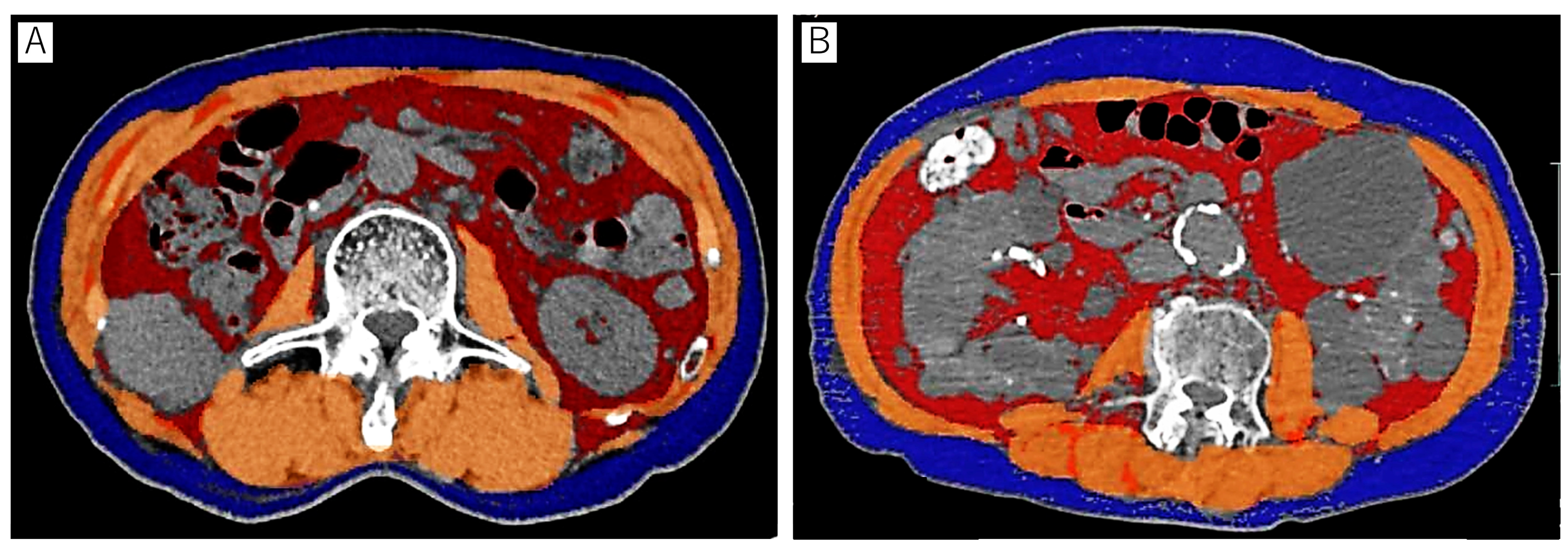
2.2. Body Composition Analysis

The BMI at the initiation of ICI-based therapy was calculated using the standard BMI formula (kg/m2) = [weight in kg]/[height in m]2. The patients were classified into normal weight (BMI: 18.5 to <25 kg/m2), overweight (BMI: 25 to < 30 kg/m2), and obese (BMI ≥30 kg/m2) according to the World Health Organization (WHO) categories for BMI.
Body composition parameters, including skeletal muscle area, subcutaneous fat area, and visceral fat area, were meticulously assessed by a single urologist, Dr. K. Takei, employing a state-of-the-art 3-dimensional image analysis system (SYNAPSE VINCENT; FUJIFILM Healthcare Corporation, Tokyo, Japan) (Figure 1). The third lumbar vertebra (L3) was used as the landmark to measure these parameters. In assessing the skeletal muscle area, we precisely identified the cross-sectional area of major skeletal muscle groups, including the rectus abdominis, internal and external obliques, lateral obliques, psoas, quadratus lumborum, and erector spinae. Hounsfield unit (HU) thresholds were used to differentiate tissues: −190 to −30 HU for subcutaneous fat tissue, −150 to −50 HU for visceral fat tissue, and −29 to 150 HU for skeletal muscle. The skeletal muscle index (SMI) was calculated as the skeletal muscle area at L3 divided by the square of height. The subcutaneous fat index (SFI) and visceral fat index (VFI) were similarly calculated as the subcutaneous fat area and the visceral fat area divided by the square of the height. Total fat index (TFI) was calculated as the sum of SFI and VFI.
Sarcopenia was precisely defined using sex-specific cut-off values for SMI, as outlined in the study by Martin et al. These cut-offs were determined as follows: for men with a BMI < 25 kg/m2, an SMI < 43 cm2/m2 was indicative of sarcopenia; for men with a BMI ≥ 25 kg/m2, an SMI < 53 cm2/m2 was considered indicative; and for women, an SMI < 41 cm2/m2 was used to define sarcopenia [14]. As no validated cut-off exists for SFI, VFI, and TFI, they were classified into high and low using sex-specific median values as cut-off, similar to previous studies [12,13].

2.3. Statistical Analyses

Overall survival (OS) and progression-free survival (PFS) were calculated from the start of the ICI-based therapy for each event. The objective response rate (ORR) was the proportion of patients who achieved CR or partial response (PR). The disease control rate (DCR) was defined as the proportion of patients who achieved CR, PR, or stable disease (SD) for >3 months according to the RECIST guidelines.
Continuous variables were reported as median/interquartile range, and categorical variables were reported as number (percentage). Categorical variables were analyzed using Fisher’s exact test, and continuous variables were analyzed using the Mann–Whitney U test. PFS and OS were estimated using the Kaplan–Meier survival curve method and were compared using the log-rank test. Univariate and multivariate analyses with Cox proportional hazards regression models were used to identify the factors related to survival. All statistical analyses were performed using JMP version 13 (SAS Institute, Cary, NC, USA), and statistical significance was defined as p < 0.05.

3. Results

3.1. Patient Characteristics

The clinical characteristics of the 60 patients are presented in Table 1. The median age was 71 years, and 46 patients (77%) were men. Thirty-four (57%) patients received ICI combination therapy (ipilimumab–nivolumab), and the remaining twenty-six (43%) received ICI-TKI combination therapy (avelumab–axitinib in 12, pembrolizumab–axitinib in 5, pembrolizumab–envatinib in 5, and nivolumab–cabozantinib in 4). Prior nephrectomy was performed in 27 (45%) patients, and 38 (63%) had good PS (≤1). Forty patients (67%) had clinical T stage 3 or greater, and forty-four (73%) had clear cell histology. The IMDC risk was intermediate in 38 (63%) patients, whereas the remaining 22 (37%) had poor risk. The median BMI and SMI values were 22.9 kg/m2 and 41.1 cm2/m2, respectively. Sex-specific median values of SFI, VFI, and TFI were as follows: (SFI: 28.7 cm2/m2 in men, 60.5 cm2/m2 in women; VFI: 44.1 cm2/m2 in men, 27.8 cm2/m2 in women: TFI: 77.3 cm2/m2 in men, 89.5 cm2/m2 in women).
According to the WHO criteria, 16 patients were categorized as overweight, and the remaining 44 were classified as having average weights. According to the criteria described by Martin et al. [14], 42 patients were identified as having sarcopenia. Patients with sarcopenia had more non-clear cell histology, higher NLR, higher PLR, lower BMI, lower SMI, lower VFI, and lower TFI (p = 0.02) than those without sarcopenia (Table 2).

3.2. Association between Body Composition Markers and Objective Responses

The ORR and DCR of the 60 patients were 41.6% and 65%, respectively. Both patients with and without sarcopenia achieved CR, PR, and SD; however, patients who developed progressive disease (PD) were mainly found to have sarcopenia. Patients with sarcopenia had a lower ORR and DCR than those without sarcopenia (33.3% vs. 61.1%, p = 0.0436 and 52.4% vs. 94.4%, p = 0.0024, respectively). BMI, SFI, VFI, and TFI were not correlated with ORR and DCR.

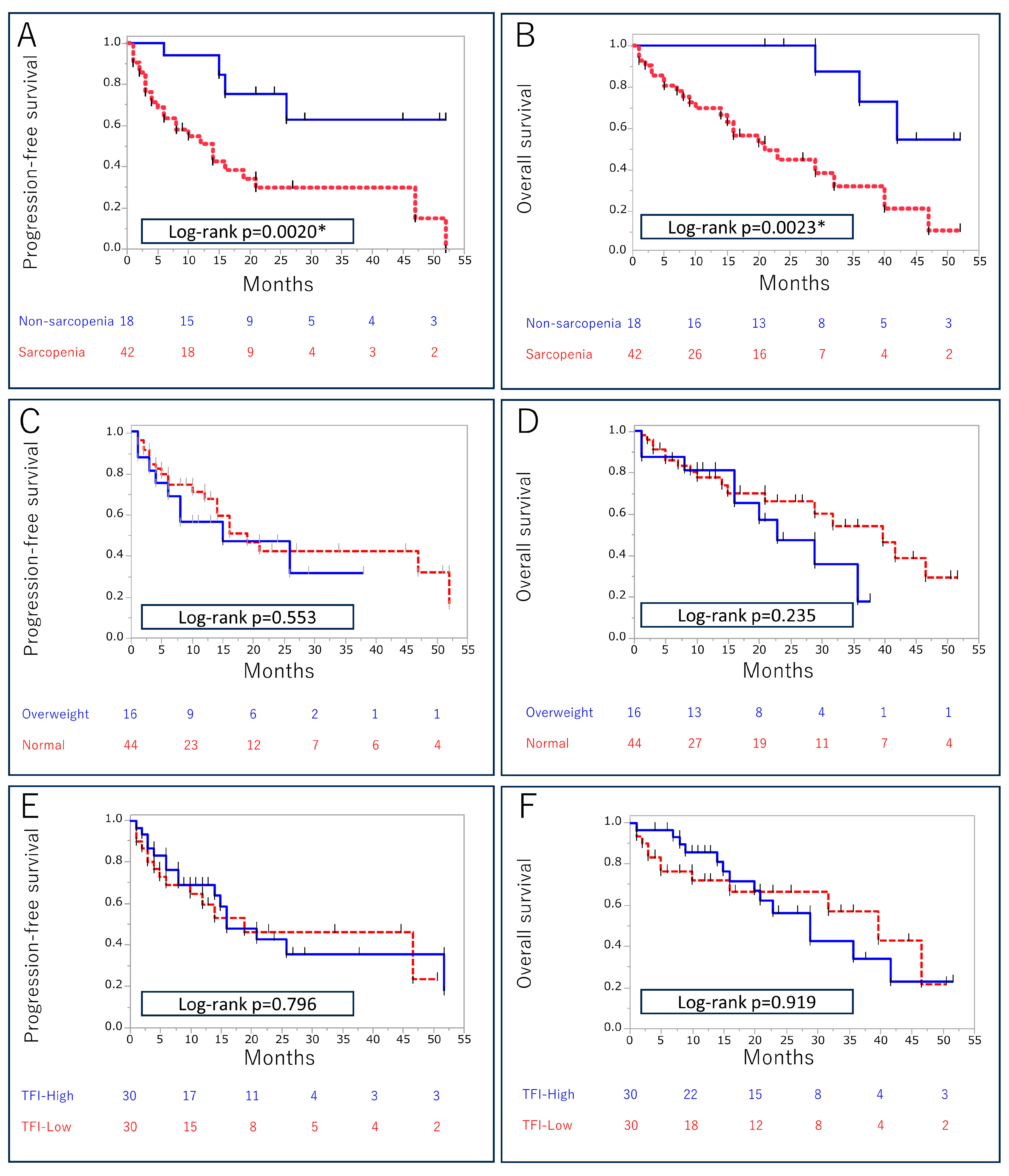
3.3. Association between Body Composition Markers and Survival Outcomes

During follow-up (median, 15 months; range, 1–52 months), 30 (50%) patients developed PD, and 26 (43%) died of the disease. Patients with sarcopenia had significantly shorter median PFS (14 months vs. not reached, p = 0.0020) and OS (21 months vs. not reached, p = 0.0023) compared with patients without sarcopenia (Figure 2). The negative impact of sarcopenia on PFS and OS was confirmed in a subgroup analysis of patients receiving ICIs and ICI-TKI therapy. BMI, SFI, VFI, and TFI were not correlated with PFS or OS.
Table 3 shows the Cox regression analysis for PFS. In univariate analysis, age, IMDC risk, NLR, PLR, and sarcopenia were associated with PFS. In multivariate analysis, sarcopenia was a significant predictor of PFS (hazard ratio [HR], 4.31; 95% confidence interval [CI], 1.65–14.8; p = 0.0018) along with poor IMDC risk (HR, 2.61; 95% CI, 1.22–5.67; p = 0.0136). In univariate Cox regression analysis for OS (Table 4), IMDC risk, NLR, and sarcopenia were associated with OS. In multivariate analysis, sarcopenia was a significant predictor of OS (HR, 5.44; 95% CI, 1.83–23.4; p = 0.0013) along with poor IMDC risk (HR, 3.30; 95% CI, 1.48–7.76; p = 0.0037).

3.4. Association between Body Composition Markers and irAEs

During follow-up, 34 patients experienced irAEs of all grades, and 21 had irAEs ≥ grade 3 (Table 5). The frequently observed irAEs were as follows: hepatic dysfunction (n = 5), adrenal insufficiency (n = 4), interstitial pneumonia (n = 3), rheumatoid arthritis (n = 3), thyroiditis (n = 2), hypophysitis (n = 2). irAEs of all grades were more common in patients without sarcopenia than in those with sarcopenia (78% vs. 48%; p = 0.0463). However, the frequency of severe (≥grade 3) irAEs was not associated with the presence of sarcopenia. The BMI, SFI, VFI, and TFI were not associated with the frequency of all-grade irAEs. Treatment discontinuation due to irAEs occurred in 13 patients. The frequency of treatment discontinuation due to irAEs was eight (19%) in patients with sarcopenia and five (27%) in patients without sarcopenia, with no significant difference (p = 0.503).

4. Discussion

Numerous studies have reported that body composition markers affect treatment outcomes, prognosis, and adverse events in various cancers, including mRCC. Among patients with mRCC receiving ICI-based therapy, several studies have reported a better prognosis in individuals with higher BMI and muscle and fat mass; however, these results are still inconclusive [11,12,13]. In this study, we investigated whether body composition markers correlate with the treatment efficacy and survival outcomes of patients administered ICI-based therapy as first-line therapy. Our results showed that the ORR and DCR were significantly lower in patients with sarcopenia than in those without it, and patients who developed PD were primarily those in the sarcopenia group. Additionally, patients with sarcopenia had a significantly shorter OS and PFS than those without sarcopenia. Furthermore, multivariate analysis revealed that sarcopenia was an independent prognostic factor along with poor IMDC risk. None of the indices related to fat components demonstrated a significant correlation with the treatment efficacy of ICI-based therapy. These results suggest that sarcopenia negatively affects treatment efficacy and prognosis in patients with mRCC treated with ICI-based therapy.
These findings suggested a possible association between sarcopenia and an immunosuppressive microenvironment. One of the mechanisms by which sarcopenia causes immunosuppression is a decrease in myokines [15]. Skeletal muscles have recently been recognized as endocrine organs capable of secreting more than 600 molecules, termed myokines [16]. Skeletal muscle can modulate the tumor microenvironment by secreting these myokines. One of the myokines that affect the tumor microenvironment is interleukin (IL)-15, which is abundantly expressed in skeletal muscle and activates natural killer (NK) and T cell immunity [17]. IL-15 has been found to co-localize within the tumor microenvironment with cluster of differentiation (CD)8+ T and NK cells [18], and the injection of IL-15 suppressed tumor burden by increasing intratumoral CD8+ T and NK cells in a mouse model [19]. To enhance the therapeutic efficacy of ICIs, recombinant human IL-15 or its agonists have been investigated in clinical trials for leukemia [20], non-small cell lung, [21], and ovarian cancers [22]. In addition to the administration of IL-15, exercise-induced increases in IL-15 were reported to enhance CD8+ T cell immunity and suppress tumor growth in mouse models. These findings suggest that IL-15 secreted from skeletal muscle promotes anti-tumor immunity and that patients with sarcopenia may have impaired immunity due to decreased IL-15.
Another myokine that may have anti-cancer potential is decorin, ubiquitously expressed in connective tissues [23]. Decorin acts as a soluble inhibitor of pan-receptor tyrosine kinases, such as the Met receptor and vascular endothelial growth factor receptor [24]. As these receptor tyrosine kinases have essential roles in the development and progression of RCC, and TKIs for these receptors are used in the treatment of mRCC, skeletal muscle-derived decorin might have anti-tumor effects in mRCC.
Chronic inflammation is another factor that may explain the link between sarcopenia and the immunosuppressive microenvironment. Chronic inflammation induces myeloid-derived suppressor cells, which suppress the activity of CD8+ T-cells and activate immunosuppressive regulatory T-cells [25,26,27]. Additionally, the inflammatory cytokines, including tumor necrosis factor-alpha (TNF-α), IL-1, and IL-8, affect skeletal muscle, leading to cancer cachexia [28]. TNF-α activates the nuclear factor kappa-light-chain-enhancer of activated B cell pathway and ubiquitin-mediated proteasome catabolism of muscle protein [29]. Therefore, chronic inflammation in patients with sarcopenia suppresses anti-tumor immunity, resulting in a significantly poorer response to ICI therapy [30].
Several previous studies have suggested a possible association between fat components and the therapeutic effects of ICIs; however, this was not confirmed in the current study. The fact that adipocytes express PD-ligand 1 (L1) during adipogenesis suggests that adiposity promotes tumor immune evasion, which ICIs could reverse [31]. Furthermore, adipose tissue is a secondary lymphoid organ that harbors numerous immune cells, including T cells [32], suggesting a better response to ICIs in patients with a higher composition of fat tissue. The relationship between adipose tissue, the immune microenvironment, and the therapeutic effects of ICIs requires further investigation.
In addition to the influence of skeletal muscle and fat components on anti-tumor immunity, the treatment of comorbidities may also impact the prognosis of patients with mRCC. Previous studies have reported that antidiabetic [33,34] and antihypertensive drugs [35] have potential anti-tumor effects on RCC and that patients who take these drugs tend to have better prognoses than those who do not. Although this study did not specifically focus on medications for comorbidities, further investigation is needed to evaluate the effects of antidiabetic and antihypertensive drugs on the prognosis of patients with mRCC receiving ICI-based therapy.
For a cancer patient with sarcopenia, improving appetite and weight not only improves the general condition and quality of life but may also improve the anti-tumor immune status and lead to an improved prognosis. Thus, therapeutic intervention against sarcopenia is warranted for patients with cancer. Therapeutic interventions for sarcopenia in patients with cancer may include counseling by dietitians, medications, and exercise [36]. Counseling by dietitians has been reported to improve the weight of cancer patients with sarcopenia [37]. For drug therapy, progesterone analogs and corticosteroids are weakly recommended. Progesterone analogs have been reported to mildly improve appetite, weight, and quality of life in cancer patients with sarcopenia [38]. Corticosteroids have also been reported to improve appetite in patients with cancer; however, there have been no reports showing an improvement in weight with its administration [39]. The appropriate dose or duration of treatment for these drugs has not been determined, and they should be used with caution due to the possibility of adverse events. Although there is little evidence regarding the impact of exercise on patients with cancer, exercise is currently being evaluated as part of multimodal interventions for these patients [40].
In the current study, the occurrence rate of irAEs of all grades was higher in patients without sarcopenia than in those with it. However, the frequency of severe irAEs (>grade 3) was comparable among patients with and without sarcopenia. These results suggest that patients without sarcopenia are more susceptible to irAEs and more likely to benefit from ICI-based therapy. This is consistent with previous reports revealing that patients who experience irAEs exhibit higher response rates and more prolonged survival with ICI-based therapy [41]. The mechanism of irAEs is thought to be an autoimmune mechanism in which T cells activated by ICIs attack autologous normal tissue. Not only does anti-tumor immunity come into play, but autoimmunity against normal tissues can also be activated by ICIs, which may explain why patients with irAEs had a better response to ICIs. The association between sarcopenia and irAEs requires further investigation.
This study has some limitations. First, this was a retrospective single-center study with a small sample size and a short follow-up period. Thus, large-scale, multicenter, prospective studies are necessary to improve the accuracy of findings. Second, the criteria for sarcopenia were calculated using data from a Canadian cohort, which may be inappropriate for Asians. Third, sarcopenia should be evaluated using different methods, such as indices focusing on specific muscles and their longitudinal changes over time. Some studies have reported that sarcopenia based on the psoas muscle index was a significant prognostic factor in patients with RCC who underwent nivolumab therapy [42]. In another study, the rate of change of the psoas major muscle area was useful in patients with non-small cell lung cancer treated with nivolumab or pembrolizumab [43]. In addition to body composition-related markers, other factors relevant to sarcopenia, such as muscle strength and exercise capacity, should also be assessed in patients receiving ICI-based therapy. Finally, this study included patients receiving both the ICI and the ICI-TKI combinations. Because the effects of sarcopenia on treatment efficacy may differ between ICI and ICI-TKI combinations, further studies focusing on the single regimen are warranted.

5. Conclusions

In conclusion, sarcopenia was associated with lower response rates and shorter survival in patients with mRCC who received ICI-based therapy as first-line treatment. No association was found between the fat components and the therapeutic effects of ICI-based therapy. Furthermore, body composition markers can predict the prognosis of patients with mRCC receiving ICIs. However, body composition markers measured using different indices and longitudinal changes over time require further investigation.

Author Contributions

Investigation, T.U. and H.B.; Data curation, N.O., R.K. and H.K.; Writing—original draft, K.T.; Writing—review & editing, T.K. (Toshiki Kijima); Supervision, M.Y. and T.K. (Takao Kamai) All authors have read and agreed to the published version of the manuscript.

Funding

This research received no external funding.

Institutional Review Board Statement

The study was conducted in accordance with the Declaration of Helsinki, and approved by the Institutional Review Board of Dokkyo Medical University (R-31-10J, 16 January 2020).

Informed Consent Statement

Informed consent was obtained from all subjects involved in the study. Written informed consent has been obtained from the patients to publish this paper.

Data Availability Statement

The data presented in this study are available in this article.

Conflicts of Interest

The authors declare no conflict of interest.

References

  1. Motzer, R.J.; Jonasch, E.; Boyle, S.; Carlo, M.I.; Manley, B.; Agarwal, N.; Alva, A.; Beckermann, K.; Choueiri, T.K.; Costello, B.A.; et al. NCCN Guidelines Insights: Kidney Cancer, Version 1. J. Natl. Compr. Cancer Netw. 2020, 18, 1160–1170. [Google Scholar] [CrossRef]
  2. Motzer, R.J.; Escudier, B.; McDermott, D.F.; George, S.; Hammers, H.J.; Srinivas, S.; Tykodi, S.S.; Sosman, J.A.; Procopio, G.; Plimack, E.R.; et al. Nivolumab versus Everolimus in Advanced Renal-Cell Carcinoma. N. Engl. J. Med. 2015, 373, 1803–1813. [Google Scholar] [CrossRef]
  3. Motzer, R.J.; Rini, B.I.; McDermott, D.F.; Aren Frontera, O.; Hammers, H.J.; Carducci, M.A.; Salman, P.; Escudier, B.; Beuselinck, B.; Amin, A.; et al. Nivolumab plus ipilimumab versus sunitinib in first-line treatment for advanced renal cell carcinoma: Extended follow-up of efficacy and safety results from a randomised, controlled, phase 3 trial. Lancet Oncol. 2019, 20, 1370–1385. [Google Scholar] [CrossRef]
  4. Yanagisawa, T.; Mori, K.; Katayama, S.; Mostafaei, H.; Quhal, F.; Laukhtina, E.; Rajwa, P.; Motlagh, R.S.; Aydh, A.; König, F.; et al. Hematological prognosticators in metastatic renal cell cancer treated with immune checkpoint inhibitors: A meta-analysis. Immunotherapy 2022, 14, 709–725. [Google Scholar] [CrossRef]
  5. Kijima, T.; Kubo, T.; Nishihara, D.; Nukui, A.; Hirohashi, Y.; Torigoe, T.; Kamai, T. Cancer immunohistogram representing cancer-immunity cycle by immunohistochemistry predicts the efficacy of immune checkpoint inhibitors in urological cancer patients. Sci. Rep. 2022, 12, 10617. [Google Scholar] [CrossRef]
  6. Motzer, R.J.; Choueiri, T.K.; McDermott, D.F.; Powles, T.; Vano, Y.-A.; Gupta, S.; Yao, J.; Han, C.; Ammar, R.; Papillon-Cavanagh, S.; et al. Biomarker analysis from CheckMate 214: Nivolumab plus ipilimumab versus sunitinib in renal cell carcinoma. J. Immunother. Cancer 2022, 10, e004316. [Google Scholar] [CrossRef]
  7. Lennon, H.; Sperrin, M.; Badrick, E.; Renehan, A.G. The Obesity Paradox in Cancer: A Review. Curr. Oncol. Rep. 2016, 18, 56. [Google Scholar] [CrossRef] [PubMed]
  8. Santoni, M.; Massari, F.; Myint, Z.W.; Iacovelli, R.; Pichler, M.; Basso, U.; Kopecky, J.; Kucharz, J.; Buti, S.; Salfi, A.; et al. Clinico-Pathological Features Influencing the Prognostic Role of Body Mass Index in Patients with Advanced Renal Cell Carcinoma Treated by Immuno-Oncology Combinations (ARON-1). Clin. Genitourin. Cancer 2023, 21, e309–e319.e1. [Google Scholar] [CrossRef] [PubMed]
  9. Vrieling, A.; Kampman, E.; Knijnenburg, N.C.; Mulders, P.F.; Sedelaar, J.M.; Baracos, V.E.; Kiemeney, L.A. Body Composition in Relation to Clinical Outcomes in Renal Cell Cancer: A Systematic Review and Meta-analysis. Eur. Urol. Focus 2018, 4, 420–434. [Google Scholar] [CrossRef] [PubMed]
  10. Shachar, S.S.; Williams, G.R.; Muss, H.B.; Nishijima, T.F. Prognostic value of sarcopenia in adults with solid tumours: A meta-analysis and systematic review. Eur. J. Cancer 2016, 57, 58–67. [Google Scholar] [CrossRef] [PubMed]
  11. Martini, D.J.; Olsen, T.A.; Goyal, S.; Liu, Y.; Evans, S.T.; Magod, B.; Brown, J.T.; Yantorni, L.; Russler, G.A.; Caulfield, S.; et al. Body Composition Variables as Radiographic Biomarkers of Clinical Outcomes in Metastatic Renal Cell Carcinoma Patients Receiving Immune Checkpoint Inhibitors. Front. Oncol. 2021, 11, 707050. [Google Scholar] [CrossRef] [PubMed]
  12. Ged, Y.; Sanchez, A.; Patil, S.; Knezevic, A.; Stein, E.; Petruzella, S.; Weiss, K.; Duzgol, C.; Chaim, J.; Akin, O.; et al. Associations between Pretreatment Body Composition Features and Clinical Outcomes among Patients with Metastatic Clear Cell Renal Cell Carcinoma Treated with Immune Checkpoint Blockade. Clin. Cancer Res. 2022, 28, 5180–5189. [Google Scholar] [CrossRef] [PubMed]
  13. McManus, H.D.; Zhang, D.; Schwartz, F.R.; Wu, Y.; Infield, J.; Ho, E.; Armstrong, A.J.; George, D.J.; Kruse, D.; Gupta, R.T.; et al. Relationship between Pretreatment Body Composition and Clinical Outcomes in Patients with Metastatic Renal Cell Carcinoma Receiving First-Line Ipilimumab Plus Nivolumab. Clin. Genitourin. Cancer 2023. [Google Scholar] [CrossRef] [PubMed]
  14. Martin, L.; Birdsell, L.; MacDonald, N.; Reiman, T.; Clandinin, M.T.; McCargar, L.J.; Murphy, R.; Ghosh, S.; Sawyer, M.B.; Baracos, V.E. Cancer cachexia in the age of obesity: Skeletal muscle depletion is a powerful prognostic factor, independent of body mass index. J. Clin. Oncol. 2013, 31, 1539–1547. [Google Scholar] [CrossRef] [PubMed]
  15. Waldmann, T.A.; Dubois, S.; Miljkovic, M.D.; Conlon, K.C. IL-15 in the Combination Immunotherapy of Cancer. Front. Immunol. 2020, 11, 868. [Google Scholar] [CrossRef]
  16. Park, S.-Y.; Hwang, B.-O.; Song, N.-Y. The role of myokines in cancer: Crosstalk between skeletal muscle and tumor. BMB Rep. 2023, 56, 365–373. [Google Scholar] [CrossRef]
  17. Di Sabatino, A.; Calarota, S.A.; Vidali, F.; MacDonald, T.T.; Corazza, G.R. Role of IL-15 in immune-mediated and infectious diseases. Cytokine Growth Factor Rev. 2011, 22, 19–33. [Google Scholar] [CrossRef]
  18. Reyes, A.F.; Goldusky, J.; Bhimalli, P.; Marzo, A.L.; Schneider, J.R. Tracking fluorescently labeled IL-15 and anti-PD-1 in the tumor microenvironment and draining lymph nodes. J. Immunol. Methods 2022, 505, 113253. [Google Scholar] [CrossRef]
  19. Stravokefalou, V.; Stellas, D.; Karaliota, S.; Nagy, B.A.; Valentin, A.; Bergamaschi, C.; Dimas, K.; Pavlakis, G.N. Heterodimeric IL-15 (hetIL-15) reduces circulating tumor cells and metastasis formation improving chemotherapy and surgery in 4T1 mouse model of TNBC. Front. Immunol. 2022, 13, 1014802. [Google Scholar] [CrossRef]
  20. Miljkovic, M.D.; Dubois, S.P.; Müller, J.R.; Bryant, B.R.; Ma, E.; Conlon, K.C.; Waldmann, T.A. Interleukin-15 augments NK cell–mediated ADCC of alemtuzumab in patients with CD52+ T-cell malignancies. Blood Adv. 2023, 7, 384–394. [Google Scholar] [CrossRef]
  21. Wrangle, J.M.; Velcheti, V.; Patel, M.R.; Garrett-Mayer, E.; Hill, E.G.; Ravenel, J.G.; Miller, J.S.; Farhad, M.; Anderton, K.; Lindsey, K.; et al. ALT-803, an IL-15 superagonist, in combination with nivolumab in patients with metastatic non-small cell lung cancer: A non-randomised, open-label, phase 1b trial. Lancet Oncol. 2018, 19, 694–704. [Google Scholar] [CrossRef] [PubMed]
  22. Felices, M.; Chu, S.; Kodal, B.; Bendzick, L.; Ryan, C.; Lenvik, A.; Boylan, K.; Wong, H.; Skubitz, A.; Miller, J.; et al. IL-15 super-agonist (ALT-803) enhances natural killer (NK) cell function against ovarian cancer. Gynecol. Oncol. 2017, 145, 453–461. [Google Scholar] [CrossRef] [PubMed]
  23. Kanzleiter, T.; Rath, M.; Görgens, S.W.; Jensen, J.; Tangen, D.S.; Kolnes, A.J.; Kolnes, K.J.; Lee, S.; Eckel, J.; Schürmann, A.; et al. The myokine decorin is regulated by contraction and involved in muscle hypertrophy. Biochem. Biophys. Res. Commun. 2014, 450, 1089–1094. [Google Scholar] [CrossRef] [PubMed]
  24. Neill, T.; Schaefer, L.; Iozzo, R.V. Decorin as a multivalent therapeutic agent against cancer. Adv. Drug Deliv. Rev. 2016, 97, 174–185. [Google Scholar] [CrossRef] [PubMed]
  25. Ostrand-Rosenberg, S. Myeloid-derived suppressor cells: More mechanisms for inhibiting antitumor immunity. Cancer Immunol. Immunother. 2010, 59, 1593–1600. [Google Scholar] [CrossRef] [PubMed]
  26. Limagne, E.; Richard, C.; Thibaudin, M.; Fumet, J.-D.; Truntzer, C.; Lagrange, A.; Favier, L.; Coudert, B.; Ghiringhelli, F. Tim-3/galectin-9 pathway and mMDSC control primary and secondary resistances to PD-1 blockade in lung cancer patients. OncoImmunology 2019, 8, e1564505. [Google Scholar] [CrossRef] [PubMed]
  27. Serafini, P.; Mgebroff, S.; Noonan, K.; Borrello, I. Myeloid-derived suppressor cells promote cross-tolerance in B-cell lymphoma by expanding regulatory T cells. Cancer Res 2008, 68, 5439–5449. [Google Scholar] [CrossRef]
  28. Mendes, M.C.S.; Pimentel, G.D.; O Costa, F.; Carvalheira, J.B.C. Molecular and neuroendocrine mechanisms of cancer cachexia. J. Endocrinol. 2015, 226, R29–R43. [Google Scholar] [CrossRef]
  29. Patel, H.J.; Patel, B.M. TNF-α and cancer cachexia: Molecular insights and clinical implications. Life Sci. 2017, 170, 56–63. [Google Scholar] [CrossRef]
  30. Lu, C.; Talukder, A.; Savage, N.M.; Singh, N.; Liu, K. JAK-STAT-mediated chronic inflammation impairs cytotoxic T lymphocyte activation to decrease anti-PD-1 immunotherapy efficacy in pancreatic cancer. OncoImmunology 2017, 6, e1291106. [Google Scholar] [CrossRef]
  31. Wu, B.; Sun, X.; Gupta, H.B.; Yuan, B.; Li, J.; Ge, F.; Chiang, H.-C.; Zhang, X.; Zhang, C.; Zhang, D.; et al. Adipose PD-L1 Modulates PD-1/PD-L1 Checkpoint Blockade Immunotherapy Efficacy in Breast Cancer. OncoImmunology 2018, 7, e1500107. [Google Scholar] [CrossRef] [PubMed]
  32. Grant, R.W.; Dixit, V.D. Adipose tissue as an immunological organ. Obesity 2015, 23, 512–518. [Google Scholar] [CrossRef] [PubMed]
  33. Li, Y.; Hu, L.; Xia, Q.; Yuan, Y.; Mi, Y. The impact of metformin use on survival in kidney cancer patients with diabetes: A meta-analysis. Int. Urol. Nephrol. 2017, 49, 975–981. [Google Scholar] [CrossRef] [PubMed]
  34. Keizman, D.; Ish-Shalom, M.; Sella, A.; Gottfried, M.; Maimon, N.; Peer, A.; Hammers, H.; Eisenberger, M.A.; Sinibaldi, V.; Neiman, V.; et al. Metformin Use and Outcome of Sunitinib Treatment in Patients with Diabetes and Metastatic Renal Cell Carcinoma. Clin. Genitourin. Cancer 2016, 14, 420–425. [Google Scholar] [CrossRef]
  35. Eskelinen, T.; Veitonmäki, T.; Kotsar, A.; Tammela, T.L.J.; Pöyhönen, A.; Murtola, T.J. Improved renal cancer prognosis among users of drugs targeting renin-angiotensin system. Cancer Causes Control. 2022, 33, 313–320. [Google Scholar] [CrossRef] [PubMed]
  36. Roeland, E.J.; Bohlke, K.; Baracos, V.E.; Smith, T.J.; Loprinzi, C.L.; Bruera, E.; del Fabbro, E.; Dixon, S.; Fallon, M.; Herrstedt, J.; et al. Cancer Cachexia: ASCO Guideline Rapid Recommendation Update. J. Clin. Oncol. 2023, 41, 4178–4179. [Google Scholar] [CrossRef] [PubMed]
  37. de van der Schueren, M.; Laviano, A.; Blanchard, H.; Jourdan, M.; Arends, J.; Baracos, V. Systematic review and meta-analysis of the evidence for oral nutritional intervention on nutritional and clinical outcomes during chemo(radio)therapy: Current evidence and guidance for design of future trials. Ann. Oncol. 2018, 29, 1141–1153. [Google Scholar] [CrossRef]
  38. Garcia, V.R.; López-Briz, E.; Sanchis, R.C.; Perales, J.L.G.; Bort-Martí, S. Megestrol acetate for treatment of anorexia-cachexia syndrome. Cochrane Database Syst. Rev. 2013, 2019, CD004310. [Google Scholar] [CrossRef]
  39. Yavuzsen, T.; Davis, M.P.; Walsh, D.; LeGrand, S.; Lagman, R. Systematic review of the treatment of cancer-associated anorexia and weight loss. J. Clin. Oncol. 2005, 23, 8500–8511. [Google Scholar] [CrossRef]
  40. Solheim, T.S.; Laird, B.J.; Balstad, T.R.; Stene, G.B.; Bye, A.; Johns, N.; Pettersen, C.H.; Fallon, M.; Fayers, P.; Fearon, K.; et al. A randomized phase II feasibility trial of a multimodal intervention for the management of cachexia in lung and pancreatic cancer. J. Cachexia Sarcopenia Muscle 2017, 8, 778–788. [Google Scholar] [CrossRef]
  41. Kijima, T.; Fukushima, H.; Kusuhara, S.; Tanaka, H.; Yoshida, S.; Yokoyama, M.; Ishioka, J.; Matsuoka, Y.; Numao, N.; Sakai, Y.; et al. Association between the Occurrence and Spectrum of Immune-Related Adverse Events and Efficacy of Pembrolizumab in Asian Patients with Advanced Urothelial Cancer: Multicenter Retrospective Analyses and Systematic Literature Review. Clin. Genitourin. Cancer 2021, 19, 208–216.e1. [Google Scholar] [CrossRef] [PubMed]
  42. Ueki, H.; Hara, T.; Okamura, Y.; Bando, Y.; Terakawa, T.; Furukawa, J.; Harada, K.; Nakano, Y.; Fujisawa, M. Association between sarcopenia based on psoas muscle index and the response to nivolumab in metastatic renal cell carcinoma: A retrospective study. Investig. Clin. Urol. 2022, 63, 415–424. [Google Scholar] [CrossRef] [PubMed]
  43. Nishioka, N.; Uchino, J.; Hirai, S.; Katayama, Y.; Yoshimura, A.; Okura, N.; Tanimura, K.; Harita, S.; Imabayashi, T.; Chihara, Y.; et al. Association of Sarcopenia with and Efficacy of Anti-PD-1/PD-L1 Therapy in Non-Small-Cell Lung Cancer. J. Clin. Med. 2019, 8, 450. [Google Scholar] [CrossRef] [PubMed]
Figure 1. Measurement of skeletal muscle (orange), subcutaneous fat (blue), and visceral fat component (red) using SYNAPSE VINCENT. Representative computed tomography images of a patient without sarcopenia (A) and a patient with sarcopenia (B).
Cancers 15 05591 g001
Figure 2. Progression-free survival and overall survival after ICI-based therapy in relation to sarcopenia (A,B), body mass index (C,D), and total fat index (E,F). Asterisk (*) indicates statistical significance.
Cancers 15 05591 g002
Table 1. Patient characteristics and body composition data at initiation of ICI-based therapy.
Overall
n = 60
Age (years), median (IQR)71 (63–75)
Sex, n (%)
    Male46 (77)
    Female14 (23)
ICI-based therapy, n (%)
    ICI combination34 (57)
    ICIs + TKIs26 (43)
Prior nephrectomy, n (%)
    Yes27 (45)
    No33 (55)
ECOG PS, n (%)
    ≤138 (63)
    ≥222 (37)
Clinical T stage, n (%)
    ≤220 (33)
    ≥340 (67)
Histology, n (%)
    Clear cell carcinoma44 (73)
    Non-clear cell carcinoma10 (17)
    Unknown6 (10)
IMDC risk score, n (%)
    Intermediate38 (63)
    Poor22 (37)
    Site of metastasis, n (%)
    Lymph nodes20 (33)
    Lung35 (58)
    Liver6 (10)
    Other organs28 (47)
NLR, median (IQR)3.52 (2.29–4.84)
PLR, median (IQR)188 (133–244)
CRP (mg/dL), median (IQR)0.81 (0.08–5.96)
BMI (kg/m2), median (IQR)22.9 (20.9–25.2)
SMI (kg/m2), median (IQR)41.1 (37.1–49.6)
SFI (kg/m2), median (IQR)31.7 (20.7–51.3)
VFI (kg/m2), median (IQR)36.1 (19.1–56.8)
TFI (kg/m2), median (IQR)80.2 (42.7–103)
Abbreviations: IQR, interquartile range; IMDC, International Metastatic RCC Database Consortium; ECOG, Eastern Cooperative Oncology Group; NLR, neutrophil-to-lymphocyte ratio; PLR, platelet-to-lymphocyte ratio; CRP, C reactive protein; BMI, body mass index; SMI, skeletal muscle index; SFI, subcutaneous fat index; VFI, visceral fat index; TFI, total fat index.
Table 2. Patient characteristics according to the presence of sarcopenia.
SarcopeniaNon-Sarcopeniap-Value
n = 42n = 18
Age (years), median (IQR)72 (64–75)69 (62–73)0.428
Sex, n (%) 0.0455 *
    Male29 (69)17 (94)
    Female13 (31)1 (6)
ICI-based therapy, n (%) 0.779
    ICI combination23 (55)11 (61)
    ICIs + TKIs19 (45)7 (39)
Prior nephrectomy, n (%) 0.778
    Yes18 (43)9 (50)
    No24 (57)9 (50)
ECOG PS, n (%) 0.773
    ≤125 (60)12 (67)
    ≥217 (40)6 (33)
Clinical T stage, n (%) 0.766
    ≤215 (36)5 (28)
    ≥327 (64)13 (72)
Histology, n (%) 0.0417 *
    Clear cell carcinoma27 (64)17 (94)
    Non-clear cell carcinoma10 (24)0
    Unknown5 (12)1(6)
IMDC risk score, n (%) 0.155
    Intermediate24 (57)14 (78)
    Poor18 (43)4 (22)
Site of metastasis, n (%)
    Lymph nodes14 (33)6 (33)1.000
    Lung25 (60)10 (56)0.783
    Liver5 (12)1 (6)0.658
    Other organs23 (55)5 (28)0.0893
NLR, median (IQR)3.89 (2.78–5.64)2.56 (1.93–3.86)0.0209 *
PLR, median (IQR)195 (145–307)171 (112–196)0.0330 *
CRP (mg/dL), median (IQR)1.16 (0.06–6.19)0.34 (0.14–2.79)0.237
BMI (kg/m2), median (IQR)22.7 (19.9–24.5)24.4 (22.2–28.4)0.0062 *
SMI (kg/m2), median (IQR)39.3 (35.5–41.9)51.4 (46.1–55.3)<0.0001 *
SFI (kg/m2), median (IQR)29.8 (14.5–51.6)33.6 (25.7–51.5)0.297
VFI (kg/m2), median (IQR)30.8 (15.7–54.3)53.2 (35.3–73.9)0.0262 *
TFI (kg/m2), median (IQR)72.7 (38.2–100)93 (64.5–127)0.0407 *
Abbreviations: IQR, interquartile range; IMDC, International Metastatic RCC Database Consortium; ECOG, Eastern Cooperative Oncology Group; NLR, neutrophil-to-lymphocyte ratio; PLR, platelet-to-lymphocyte ratio; CRP, C reactive protein; BMI, body mass index; SMI, skeletal muscle index; SFI, subcutaneous fat index; VFI, visceral fat index; TFI, total fat index. Asterisks (*) indicate statistical significance.
Table 3. Cox regression analysis of clinical and body composition variables for progression-free survival.
Univariate AnalysisMultivariate Analysis
Variable CategoryHR (95% CI)p-ValueHR (95% CI)p-Value
Age
(continuous)
0.96
(0.93–0.99)
0.0210 *
Sex
Female vs. male (ref)
1.83
(0.76–5.47)
0.191
ECOG PS
2 vs. 0–1 (ref)
1.64
(0.77–3.41)
0.190
IMDC risk
Poor vs. intermediate (ref)
2.79
(1.32–6.00)
<0.0077 *2.61
(1.22–5.67)
0.0136 *
Liver metastases
Yes vs. no (ref)
2.25
(0.66–5.85)
0.175
NLR
(continuous)
1.20
(1.08–1.32)
0.0011 *
PLR
(continuous)
1.003
(1.000–1.004)
0.0275 *
BMI
Overweight vs. normal (ref)
0.79
(0.37–1.83)
0.566
SFI
Low vs. high (ref)
0.81
(0.39–1.66)
0.553
VFI
Low vs. high (ref)
0.87
(0.42–1.82)
0.717
TFI
Low vs. high (ref)
1.10
(0.52–2.29)
0.799
Sarcopenia
Yes vs. no (ref)
4.52
(1.75–15.4)
0.0010 *4.31
(1.65–14.8)
0.0018 *
SFI, VFI, and TFI were categorized as high or low based on sex-specific median values as cut-offs. Asterisk (*) indicates statistical significance. Abbreviations: ECOG, Eastern Cooperative Oncology Group; IMDC, International Metastatic RCC Database Consortium; NLR, neutrophil-to-lymphocyte ratio; PLR, platelet-to-lymphocyte ratio; BMI, body mass index; SFI, subcutaneous fat index; VFI, visceral fat index; TFI, total fat index.
Table 4. Cox regression analysis of clinical and body composition variables for overall survival.
Univariate AnalysisMultivariate Analysis
Variable CategoryHR (95% CI)p-ValueHR (95% CI)p-Value
Age
(continuous)
0.97
(0.93–1.00)
0.0728
Sex
Female vs. male (ref)
1.03
(0.37–2.42)
0.953
ECOG PS
2 vs. 0–1 (ref)
1.84
(0.82–4.05)
0.136
IMDC risk
Poor vs. intermediate (ref)
3.37
(1.52–7.80)
0.0029 *3.30
(1.48–7.76)
0.0037 *
Liver metastases
Yes vs. no (ref)
2.35
(0.68–6.27)
0.158
NLR
(continuous)
1.18
(1.05–1.30)
0.0058 *
PLR
(continuous)
1.002
(0.99–1.004)
0.0527
BMI
Overweight vs. normal (ref)
1.65
(0.68–3.38)
0.254
SFI
Low vs. high (ref)
1.05
(0.47–2.30)
0.899
VFI
Low vs. high (ref)
1.20
(0.55–2.65)
0.636
TFI
Low vs. high (ref)
1.04
(0.48–2.30)
0.919
Sarcopenia
Yes vs. no (ref)
5.45
(1.86–23.2)
0.0010 *5.44
(1.83–23.4)
0.0013 *
SFI, VFI, and TFI were categorized as high or low based on sex-specific median values as cut-offs. Asterisk (*) indicates statistical significance. Abbreviations: ECOG, Eastern Cooperative Oncology Group; IMDC, International Metastatic RCC Database Consortium; NLR, neutrophil-to-lymphocyte ratio; PLR, platelet-to-lymphocyte ratio; BMI, body mass index; SFI, subcutaneous fat index; VFI, visceral fat index; TFI, total fat index.
Table 5. Association between body composition variables and immune-related adverse events.
All-Grade irAEs, n(%)p-ValueirAEs ≥ Grade 3, n (%)p-Value
Sarcopenia
    Yes (n = 42)20 (48%)0.0463 *12 (29%)0.144
    No (n = 18)14 (78%) 9 (50%)
BMI
    Overweight (n = 16)9 (56%)1.0006 (38%)1.000
    Normal (n = 44)25 (57%) 15 (34%)
TFI
    High (n = 30)18 (60%)0.79512 (40%)0.589
    Low (n = 30)16 (53%) 9 (30%)
Abbreviations: irAEs, immune-related adverse events; BMI, body mass index; TFI, total fat index. Asterisk (*) indicates statistical significance.
Disclaimer/Publisher’s Note: The statements, opinions and data contained in all publications are solely those of the individual author(s) and contributor(s) and not of MDPI and/or the editor(s). MDPI and/or the editor(s) disclaim responsibility for any injury to people or property resulting from any ideas, methods, instructions or products referred to in the content.

