Article

E3 Ubiquitin Ligase TRIP12 Controls Exit from Mitosis via Positive Regulation of MCL-1 in Response to Taxol

Division of Biological and Biomedical Sciences, College of Health and Life Sciences, Hamad Bin Khalifa University, Doha P.O. Box 34110, Qatar
*
Author to whom correspondence should be addressed.
Cancers 2023, 15(2), 505; https://doi.org/10.3390/cancers15020505
Submission received: 16 November 2022 / Revised: 23 December 2022 / Accepted: 31 December 2022 / Published: 13 January 2023
(This article belongs to the Special Issue Cancer Chemotherapy Resistance)

Abstract

:

Simple Summary

Taxol is a chemotherapy drug used in treatment of multiple cancers. Taxol works by blocking an essential process of cell division called mitosis. Although, Taxol treatment shows promise in fight against cancer, many patients eventually develop resistance. Loss of function mutations in an E3 ubiquitin ligase component FBW7, are casually associated with cancer chemotherapy- and Taxol resistance. FBW7 is essential for degradation of a pro-survival protein MCL-1. In the absence of FBW7, MCL-1 protein accumulates, and cancer cells escape Taxol induced death. In this work we discover that another E3 ubiquitin ligase TRIP12 is required by cancer cells for efficient mitosis and completion of cell division. Inhibition of TRIP12 enhances Taxol induced cell death in an FBW7 and MCL-1 dependent manner. Thus, TRIP12/FBW7/MCL-1 axis is an important determinant of Taxol response in cancer cells.

Abstract

Chemotherapy resistance is a major hurdle in cancer treatment. Taxol-based chemotherapy is widely used in the treatment of cancers including breast, ovarian, and pancreatic cancer. Loss of function of the tumor suppressor F-box WD-40 domain containing 7 (FBW7) mutations leads to the accumulation of its substrate MCL-1 which is associated with Taxol resistance in human cancers. We recently showed that E3 ubiquitin ligase TRIP12 is a negative regulator of FBW7 protein. In this study, we find that Taxol-induced mitotic block in cancer cells is partly controlled by TRIP12 via its positive regulation of MCL-1 protein. Genetic inhibition of TRIP12 accelerates MCL-1 protein degradation in mitosis. Notably, introducing double-point mutations in lysines 404/412 of FBW7 to arginine which makes it resistant to proteasomal degradation, leads to the sharp reduction of MCL-1 protein levels and sensitizes cancer cells to Taxol-induced cell death. Finally, TRIP12 deletion leads to enhanced mitotic arrest and cell death in an FBW7 and MCL-1 dependent manner in multiple cell lines including colorectal and ovarian cancer but not in breast cancer. Thus, the TRIP12/FBW7/MCL-1 axis may provide a therapeutic target to overcome Taxol-associated chemotherapy resistance in cancer.

1. Introduction

Cancer is a major human health challenge and the leading cause of death worldwide [1]. Late detection, widespread metastasis, and de novo and acquired chemotherapy resistance are the major challenges in cancer management. Taxol or texane-based chemotherapy drugs are widely used in cancer management including breast, ovarian, and pancreatic cancer, either alone or in combination with adjuvant therapy [2]. Taxol is a mitotic spindle stabilizer which arrests cells in mitosis thereby inducing cell death [3,4]. Although Taxol has been beneficial in cancer treatment, many patients’ cancer eventually relapses with near complete resistance.
FBW7 is a substrate adaptor of the Cullin-ring SKP1/CUL1/F-Box (SCF)-type E3 ubiquitin ligase complex. FBW7 targets oncogenic proteins including c-MYC, CyclinE, c-JUN, Notch1, and MCL-1 for proteasomal degradation [5]. Consequently, loss of FBW7 function mutations leads to accumulation of some of those proteins which is associated with human cancers and chemotherapy resistance [6,7]. For example, FBW7 mediated proteasomal degradation of MCL-1 in mitosis is a major cell death mechanism in response to Taxol and apoptosis inducing agents [6,7]. Thus, FBW7 mutations confer Taxol resistance to cancer cells due to accumulation of its anti-apoptotic substrate MCL-1 [6,7]. In addition to somatic mutations, FBW7 protein is downregulated post translationally in cancer [8,9]. Thus, understanding pathways and mechanisms that converge on FBW7 protein activity and stability can help unravel novel targets to tackle cancer-associated chemotherapy resistance.
The thyroid hormone receptor interactor protein 12 (TRIP12), also known as the E3 ubiquitin ligase for Arf (ULF), is a HECT-domain E3-ligase. The yeast homologue of TRIP12 protein, Ufd4, was identified as an ubiquitin ligase which could extend polyubiquitin chains on a protein substrate already fused with a ubiquitin moiety on its N-terminus, which targets it for proteasomal degradation [10], and subsequent work identified TRIP12 as the homologous protein to function in the ubiquitin fusion degradation pathway in mammalian cells [11]. Since then, TRIP12 has been found to be involved in DNA damage response, oncogenic stress, cell cycle, and neurodegeneration [12,13,14,15]. Interestingly, TRIP12 regulates response to PARP inhibitors in breast cancer cells [16] and we have shown that the genetic inhibition of TRIP12 leads to the stabilization of FBW7 protein and enhanced cell death in response to Taxol treatment [17]. However, the precise molecular details of enhanced cell death in response to Taxol in TRIP12 deficient cells is not known.
In this study, we show that TRIP12 is required for exit from mitosis during mitotic block induced by Taxol. The TRIP12-deficient cells were arrested in mitosis and failed to re-enter cell cycle efficiently upon release from mitotic block. This was largely dependent on FBW7 since in FBW7/TRIP12 double knockout cells these effects were completely normalized to wildtype levels. Interestingly, the stable reconstitution of FBW7 lysine-to-arginine double mutant, which is resistant to proteasomal degradation, in FBW7 knockout cells leads to sharp reduction of MCL-1 protein and culminates in enhanced cell death by Taxol. Finally, the TRIP12/FBW7/MCL-1 axis is well preserved in ovarian cancer but not in breast cancer cells since siRNA-mediated depletion of TRIP12 sensitized those cells to Taxol in an FBW7-dependent manner. Thus, our data provide strong evidence that TRIP12 is essential for efficient mitotic arrest induced by Taxol and this effect is mediated via FBW7/MCL-1 proteins.

2. Results

2.1. Enhanced Mitotic Degradation of MCL-1 in the Absence of TRIP12

We previously showed that genetic depletion of TRIP12 stabilizes tumor suppressor protein FBW7 and reduces CyclinE and MCL-1 protein levels in HEK293 and HCT116 cells [17]. To check if the deletion of TRIP12 affects FBW7 protein and its substrates in other cell lines, we depleted TRIP12 using two different short interfering (si)-RNA in U2OS cells. Consistent with our previous findings, TRIP12 knockdown stabilizes FBW7 protein and FBW7 substrate CyclinE and MCL-1 are downregulated (Figure 1). Additionally, the mammalian target of rapamycin (mTOR), another FBW7 substrate, was also downregulated while c-Jun protein was increased in TRIP12 knockdown U2OS cells (Figure 1). All other FBW7 substrates including Notch1, c-MYC, HSF1, and SREBP1 were unaffected in TRIP12 knockdown cells (Figure 1A and Figure S1A). The preference of substrates by FBW7 in TRIP12-depleted cells is not governed by FBW7 dimerization because FBW7 dimerization is largely unaffected in TRIP12 knockdown cells (Supplementary Figure S1B). Because MCL-1 is specifically targeted for proteasomal degradation by FBW7 in mitosis [6], we checked the possibility that TRIP12 regulates MCL-1 levels in mitosis. Indeed, MCL-1 degradation is enhanced in the absence of TRIP12 in cells blocked in mitosis by releasing in nocodazole after a double thymidine block in two different cell lines (Figure 1B,C). We confirm the mitotic arrest by Western blots against the phospho-Histone 3 protein which is a bona fide marker of mitotic cells (Figure 1B,C). Next, we tested the possibility that FBW7 protein stability is regulated by TRIP12 in mitosis in HEK293 cells stably transfected with a control and TRIP12 specific short hairpin (sh)-RNA (Supplementary Figure S1D). Consistent with previous findings [18], FBW7 protein stability is not regulated by cell-cycle stages and TRIP12 knockdown stabilized FBW7 protein throughout the cell cycle. These results suggest that FBW7 substrate specificity in TRIP12-depleted cells is context dependent and that MCL-1 degradation is enhanced in the absence of TRIP12 specifically in mitosis.

2.2. TRIP12 Controls Exit from Mitosis via Negative Regulation of FBW7

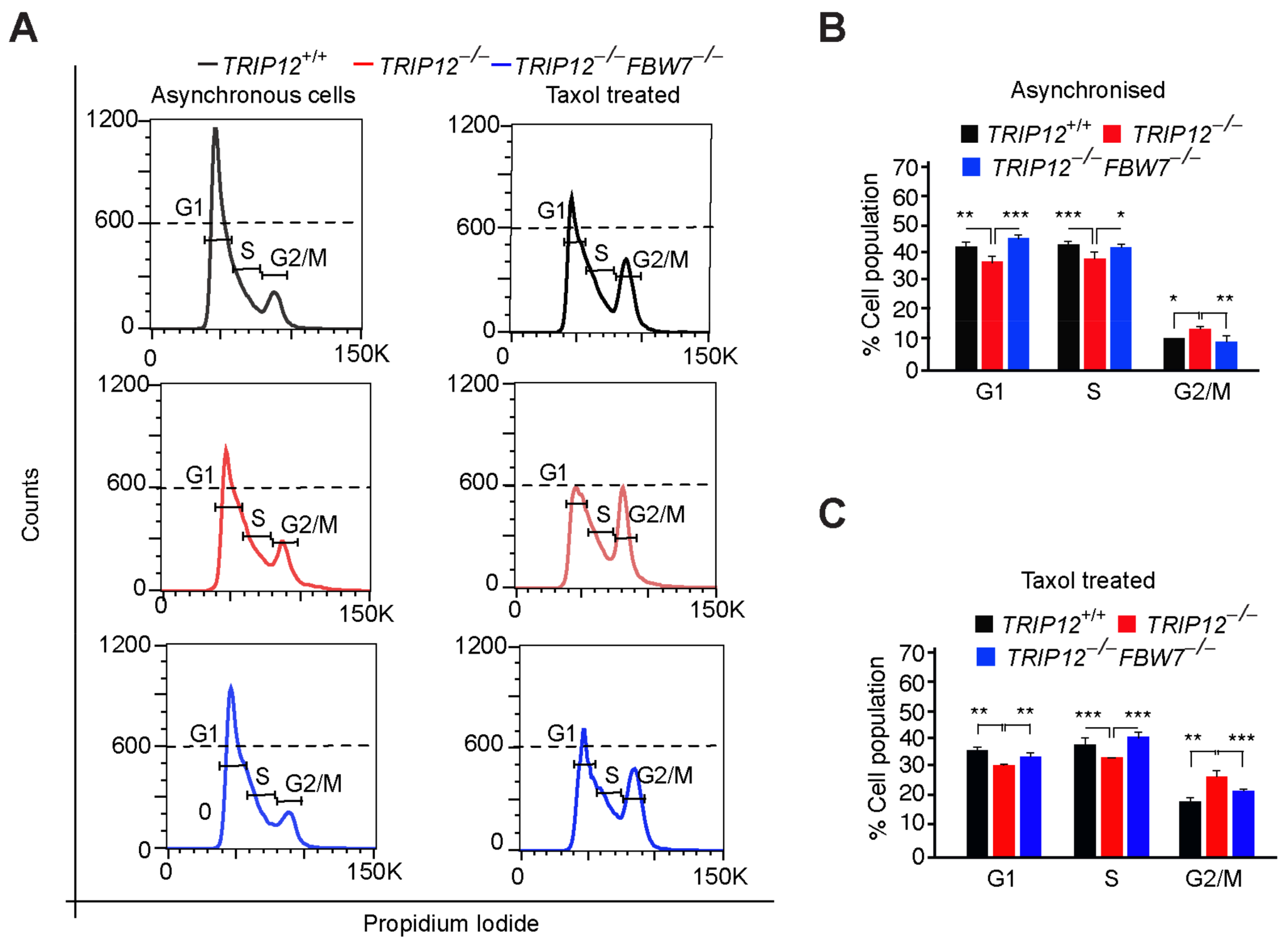
Loss of function FBW7 mutations leads to MCL-1 protein accumulation which culminates in chemotherapy resistance of cancer cells [6]. We previously showed that targeting TRIP12 enhances Taxol-induced cell death in colorectal cancer [17]. To understand the precise molecular mechanism of Taxol-induced death in those cells, we performed cell-cycle analyses of asynchronized as well as Taxol treated HCT116 cells. In comparison to wildtype cells, TRIP12 knockout causes subtle increase in asynchronously growing mitotic cells (Figure 2A,B), indicative of enhanced proliferation or inability of the cells to exit mitosis efficiently. As expected, Taxol treatment led to the sharp induction of mitotic cells which was significantly increased in the absence of TRIP12 (Figure 2A,C). Similar results were obtained in additional HEK293 cells where the mitotic arrest was more pronounced in TRIP12 knockout cells compared to wildtype cells (Supplementary Figure S2A). The increase in mitotic cells in asynchronous as well as Taxol-arrested TRIP12 knockout cells was near completely normalized to wildtype levels in TRIP12−/−FBW7−/− (double knockout) cells. These results suggest two possibilities, either TRIP12 is required for exit from mitosis during Taxol-induced mitotic arrest or TRIP12 knockout cells enter mitosis faster than the controls as previously suggested [13].
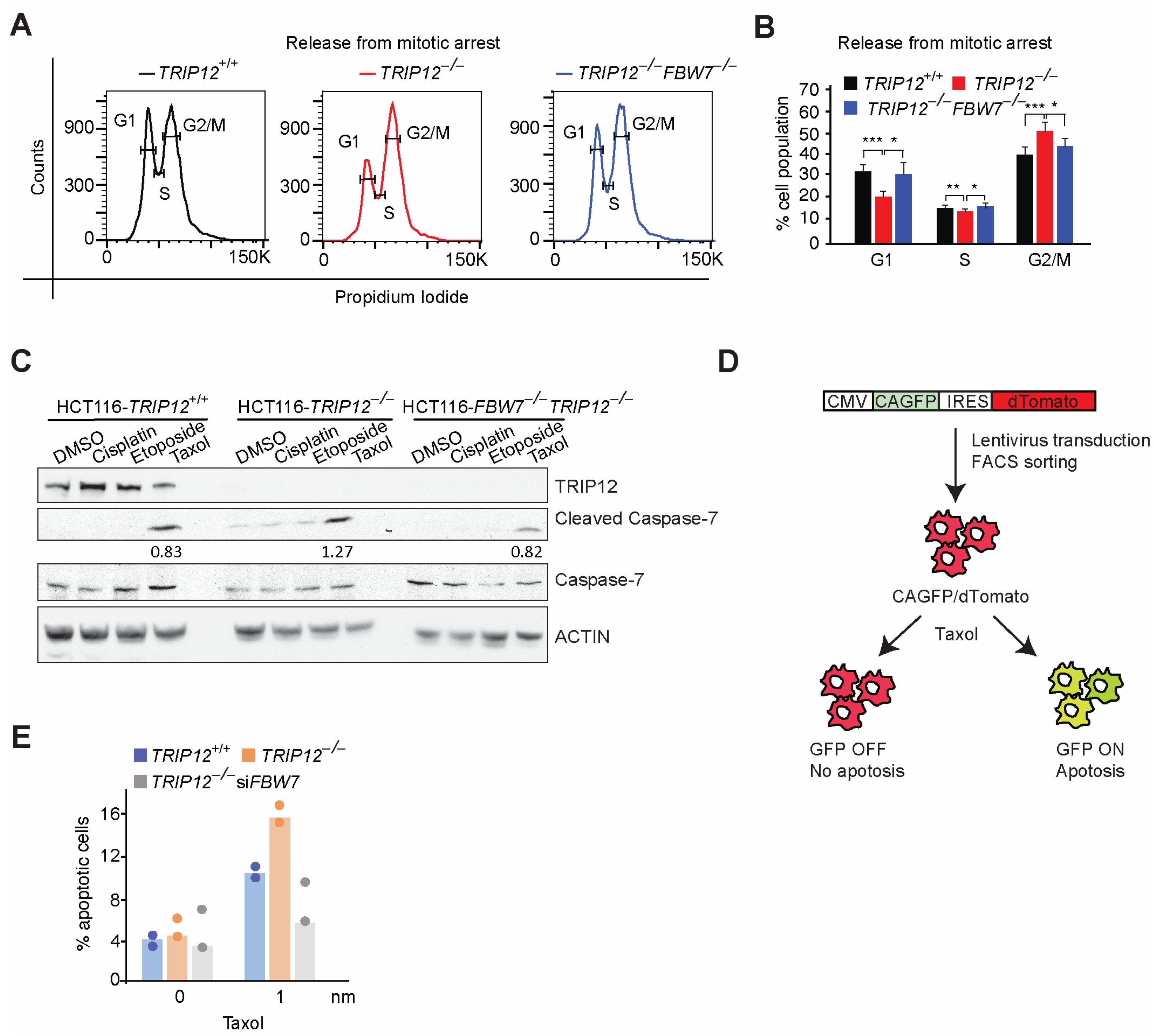
To resolve this apparent bias, we synchronized HCT116 cells in mitosis and then released the cells in fresh media without mitotic blocker nocodazole. Strikingly, TRIP12 deletion delayed exit from mitosis compared to wildtype cells, an effect which was completely normalized to wildtype levels in double knockout cells (Figure 3A,B). Consistent with that, a low dose of Taxol but not of other chemotherapy drugs cisplatin or etoposide, enhanced the cleaved-Caspase-7/total-Caspase-7 ratio in TRIP12-depleted cells compared to wildtype cells (Figure 3C), indicative of higher cell death. This effect was also normalized in double knockout cells to wildtype levels (Figure 3C). Finally, to test if TRIP12 deletion sensitizes cells to Taxol-induced apoptosis, we generated stable cell lines expressing a lentivirus-mediated apoptosis reporter using a previously published Caspase activable GFP (CAGFP) expression plasmid [19]. When the cells are committed to apoptosis, the DEVD peptide is cleaved by caspases and allows for the expression of GFP and monitoring of early apoptosis via immunofluorescence or FACS (Figure 3D). As expected, Taxol treatment enhanced the number of GFP positive cells well above the background in wildtype cells which was roughly doubled in TRIP12 knockout cells, an effect completely blunted by siRNA-mediated knockdown of FBW7 (Figure 3E). Thus, our data suggest that TRIP12 is required for efficient exit from mitosis and TRIP12 knockout sensitizes HCT116 cells to apoptosis and these effects are largely mediated via FBW7.

2.3. Mutating FBW7 Recognizable GSK3β Phosphodegron on MCL-1 Reverses Mitotic Arrest in TRIP12−/− Cells

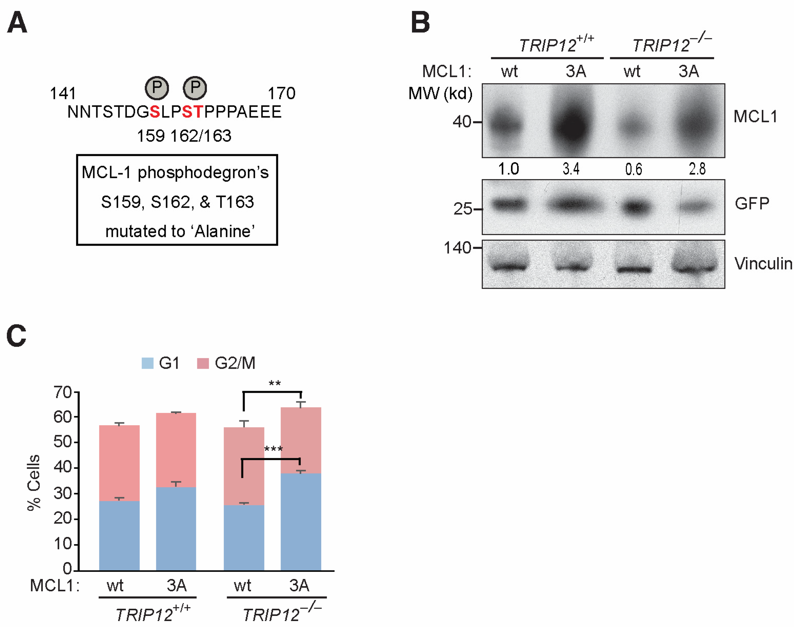
FBW7 recognizes its substrate by their phosphorylated residues within a GSK3β phosphodegron. To check if negative regulation of MCL-1 by FBW7 is mediated through GSK3β-mediated phosphorylation, we mutated Ser159/162 and Thr163 in MCL-1’s phosphodegron (Figure 4A). The triple mutation not only stabilizes MCL-1 (3A-mutant) protein in wildtype cells but also stabilizes the reduced MCL-1 protein levels in TRIP12 knockout cells similar to 3A mutant in wildtype cells as judged by Western blotting and normalization of MCL-1 blots to GFP control blots (Figure 4B), thus confirming the requirement of GSK3β phosphorylation-mediated FBW7 targeting of MCL-1 protein degradation in wildtype as well as TRIP12 knockout cells [6]. To test if enhanced degradation of MCL-1 by FBW7 in TRIP12 knockout cells is responsible for mitotic arrest, we overexpressed MCL-1 wildtype and 3A-mutant plasmids in TRIP12 wildtype and knockout cells, synchronized those cells in mitosis, released them from mitotic arrest for 3–4 h, and then performed the cell-cycle analysis. As previously seen (Figure 3A,B), TRIP12 knockout cells exited mitosis less efficiently than wildtype cells when wildtype MCL-1 was overexpressed (Figure 4C). However, MCL-1 3A-mutant leads to more efficient exit from mitosis in both TRIP12 wildtype and knockout cells. Thus, our data suggest that TRIP12/MCL-1 axis is required for efficient mitotic exit, and FBW7-mediated proteasomal degradation of phosphorylated MCL-1 may block this effect.

2.4. FBW7 Ubiquitylation Resistant Mutant Reduces MCL-1 Protein Levels and Sensitize HCT116 Cells to Taxol

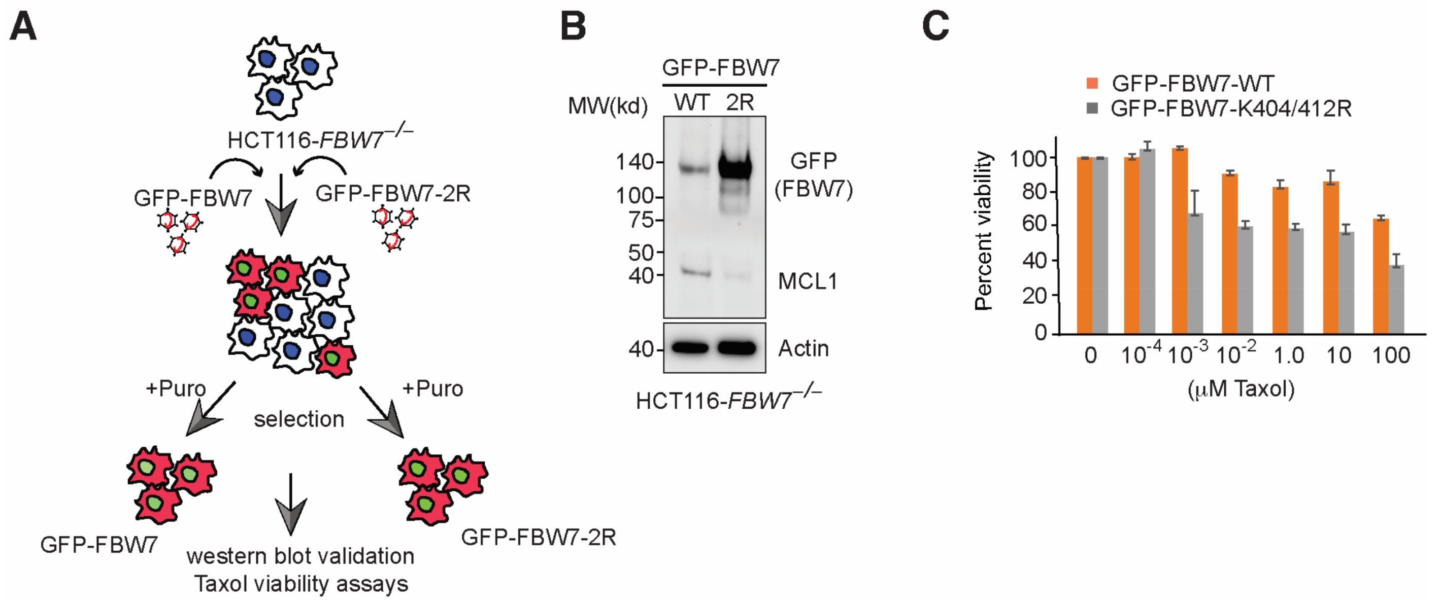
TRIP12 depletion stabilizes FBW7 protein and sensitizes cancer cells in a FBW7-dependent manner (Figure 1A) [17]. In addition to FBW7 regulation, TRIP12 is involved in numerous biological functions [12,13,14,15]. To unequivocally establish that the enhanced FBW7 function in HCT116 cells is responsible for Taxol-induced cell death, we sought to use an alternate approach. We previously showed that the FBW7 K404/K412R double-point mutant is strongly stabilized due to its inability to autoubiquitylate itself on two crucial lysine residues essential for FBW7 protein degradation [17]. First, we cloned FBW7 wildtype and FBW7 K404/412R (2R) mutant in a protein stability reporter plasmid and overexpress the two plasmids in HEK293FT cells. Consistent with previous findings, we noticed the sharp accumulation of the FBW7 2R-mutant compared to FBW7 wildtype protein (Supplementary Figure S2B). Next, to check if FBW7 stabilization alone would sensitize cancer cells and mimic TRIP12 deletion, we used lentivirus-mediated overexpression of wildtype and K404/412R FBW7 (2R) mutant in HCT116FBW7−/− cells which are otherwise resistant to Taxol (Figure 5A) [6,17]. As expected, the FBW7 2R-mutant is strongly stabilized in HCT116FBW7−/− cells and MCL-1 protein levels were sharply reduced in those cells compared to HCT116FBW7−/− cells overexpressing wildtype FBW7 (Figure 5B). Finally, FBW7 2R-mutant and not FBW7 wildtype overexpression sensitized HCT116 cells to increasing doses of Taxol (Figure 5C) and provides proof-of-concept evidence that suggests mitigating FBW7 protein levels in chemotherapy resistance might be an interesting therapeutic target particularly in cancer patients with FBW7 wildtype genotype.

2.5. Targeting TRIP12 Sensitizes Ovarian but Not Breast Cancer Cells to Taxol-Induced Cell Death

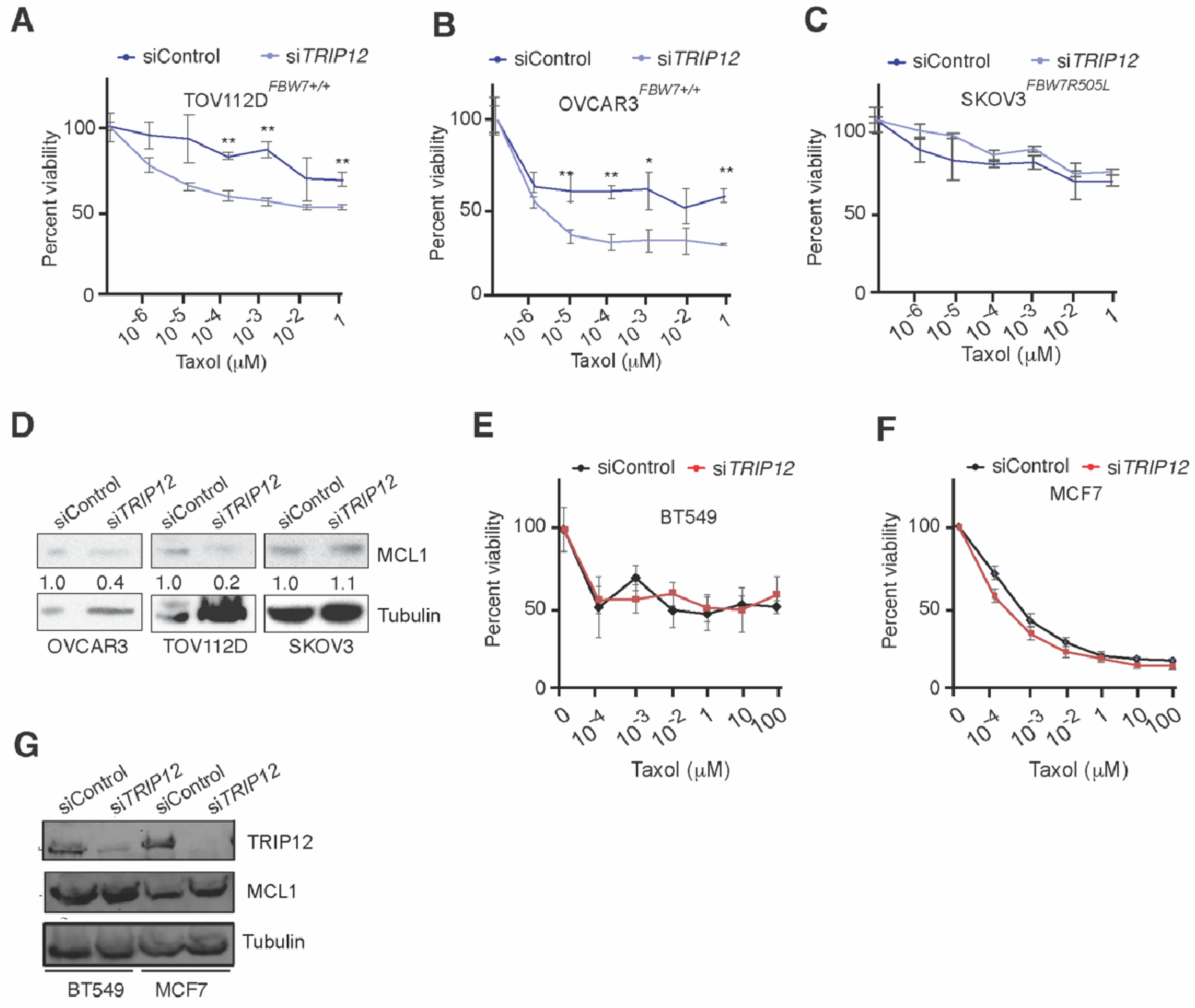
To this end, we described a pathway which could be therapeutically exploited for targeting cancers that are otherwise resistant to anti-mitotic chemotherapy. However, Taxol is not the standard of care in treatment of colorectal cancer in clinics. Thus, to test whether TRIP12 inhibition will sensitize other cancer cells to Taxol, we knockdown TRIP12 in ovarian cancer cells and treated those cells with increasing doses of Taxol. Strikingly, TRIP12 depletion strongly sensitized FBW7 wild type ovarian cancer cells whereas FBW7R505L mutant cell line was resistant to Taxol treatment regardless of TRIP12 status (Figure 6A–C). Consistent with previous results, TRIP12 knockdown reduced MCL-1 protein levels in FBW7 wild type OVCAR3 and TOV112D cells but not in mutant SKOV3 cells (Figure 6D). Thus, our data suggest that TRIP12 targeting might sensitize FBW7 wild type ovarian cancer cells to Taxol therapy.
Next, we tested whether the depletion of TRIP12 in breast cancer cells could achieve similar synergy with Taxol as witnessed with ovarian cancer cells. The triple negative breast cancer BT549 responded weakly to increasing doses of Taxol as judged by LDH cytotoxicity assay (Figure 6E). Although MCF7 cells were more sensitive to Taxol compared to BT549, TRIP12 knockdown marginally enhanced cell death at very high doses of Taxol in those cells (Figure 6F). Interestingly, TRIP12 knockdown in both BT549 and MCF7 cells did not affect MCL-1 protein levels (Figure 6G), providing a possible explanation as to why TRIP12 deletion could not sensitize those cells to low doses of Taxol. Finally, to test the hypothesis that high MCL-1 levels in breast cancer cells help those cells escape mitotic cell death, we used siRNA-mediated targeting of MCL-1 in Taxol resistant BT549 cells and study their response to Taxol (Supplementary Figure S2A). Indeed, siRNA-mediated MCL-1 depletion strongly sensitized BT549 cells to Taxol (Supplementary Figure S2B), these results are consistent with previous findings [20] and highlight the importance of considering MCL-1 protein levels for treatment of triple breast cancer.

3. Discussion

Cancer chemotherapy resistance is a major hurdle in patients’ treatment. However, despite producing nominal benefits in most cases, chemotherapy has been widely used for cancer treatment for decades. The philosophy behind this relentless practice is ‘one treatment for all’ cancer patients. Such an approach often ignores the tumor heterogeneity, patients’ genetic background, de novo resistance to available treatments, cancer stages, molecular diversity, and aggressive metastatic disease. On the contrary, an alternate approach by stratifying patients into groups based on their genetic or molecular profiles might provide a more effective way of cancer management [21]. Thus, understanding the molecular mechanisms of chemotherapy resistance can provide relevant clinical or molecular biomarkers that may help predict chemotherapy response.
Loss of FBW7 function mutation is long associated with cancer chemotherapy resistance and aggressive phenotypes [22]. FBW7 facilitates phosphorylation-dependent proteasomal degradation of multiple proto-oncogenic molecules [5]. In the absence of FBW7 activity, several FBW7 substrates accumulate including antiapoptotic MCL-1. High MCL-1 levels not only prevent cell death of FBW7 mutated cells due to unusually high oncogenic signaling from c-MYC, CyclinE, and c-JUN proteins, but also provide a mechanism to escape chemotherapy-induced cell death [6]. Interestingly, the majority of FBW7 mutations in human cancers are heterozygous with at least one wildtype allele retained by the patient [5]. Additionally, others and we have shown that FBW7 protein is downregulated independent of its mutational status in many cancer patients [8]. Thus, it is reasonable to believe that chemotherapy resistance can be blocked by enhanced FBW7 activity by directly interfering with its protein turnover. However, such a hypothesis has not been tested until recently [17].
We previously established E3 ubiquitin ligase TRIP12 to be a negative regulator of FBW7 protein [17]. Inhibition of TRIP12 not only stabilizes FBW7 protein but also leads to downregulation of MCL-1 protein (Figure 1). The enhanced MCL-1 degradation in TRIP12 knockout cells is largely carried out during mitosis (Figure 1) which culminates in aberrant exit from mitosis and affects cell-cycle re-entry, particularly in response to Taxol, a mechanism most likely responsible for enhanced cell death in TRIP12 knockout cells by Taxol. These results have broader clinical implications, since FBW7 protein is downregulated in cancer and TRIP12 may provide a target for pharmacological intervention to restore FBW7 activity. Additionally, TRIP12/MCL-1 expression might be a useful biomarker for texane-based chemotherapy response in some but not all cancers.
Although we find consistent downregulation of MCL-1 protein in the absence of TRIP12 in multiplate cell lines, exactly what defines substrate specificity of FBW7 in TRIP12 depleted cells is not clear. For example, previous work demonstrated that FBW7 dimerization provides selectivity towards its substrates in a context dependent manner [5]. Yet, we do not find FBW7 dimerization to be affected by TRIP12 inhibition. Moreover, the majority of FBW7 substrates were unaffected upon TRIP12 inhibition including c-MYC, c-Jun, and Notch-1 in HEK293 cells [17]. Contrary to that, we find c-Jun accumulation in U2OS cells upon TRIP12 inhibition (Figure 1A). These differences in c-Jun protein could be due to the tissue specific nature of FBW7 activity towards its substrates or, alternatively, a result of enhanced gene expression and totally unrelated to protein stability.
Our data show remarkable downregulation of MCL-1 protein and near complete reversal of Taxol resistance by overexpression of highly stabilized FBW7 2R-mutant which demonstrates the power of mitigating FBW7 and MCL-1 protein levels in human cancers for efficient chemotherapy response. This could be achieved by pharmacological targeting of TRIP12 to increase endogenous FBW7 protein level; however, to date, there are no specific inhibitors available for TRIP12. Chemical biology screens can be designed to scan and identify TRIP12 specific inhibitors. One caveat to this approach is that TRIP12 contains a highly conserved HECT domain, thus making it largely difficult to specifically inhibit its activity without affecting the function of broader HECT family members involved in diverse cellular functions. Alternatively, small molecule inhibitors of MCL-1 can be exploited against cancer and chemotherapy resistance [23]. Interestingly, some of those inhibitors are already tested against acute myeloid leukemia and Hodgkin’s lymphoma in clinical trials [23]. Once completed, these studies may shed some invaluable light on the utility of MCL-1 inhibitors for cancer treatment and might encourage clinical trials against solid cancers.
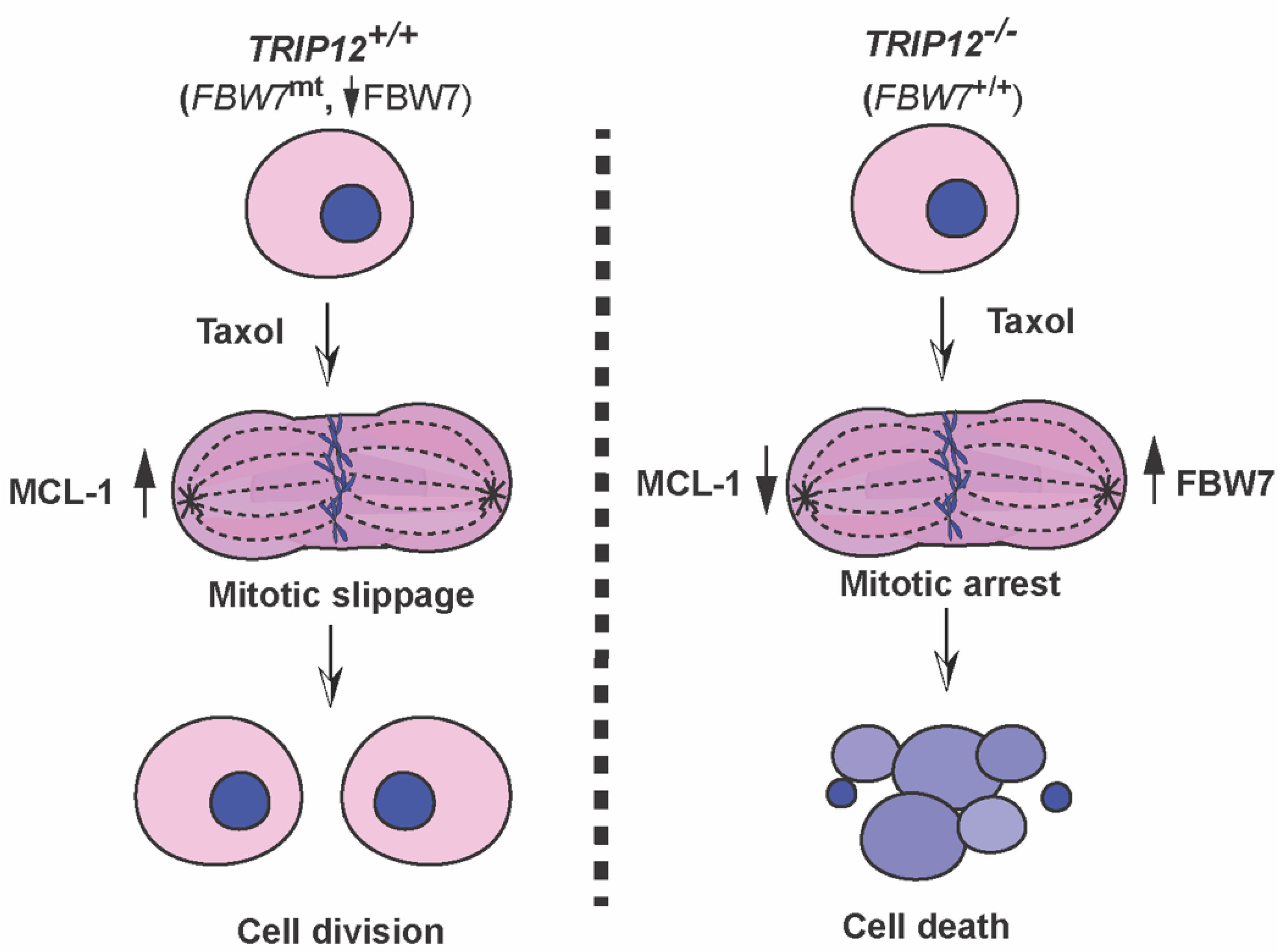
Finally, our data show that colorectal and ovarian cancer cells can be sensitized to Taxol by genetic inhibition of TRIP12 and this effect is largely dependent on FBW7 and MCL-1 proteins. However, this genetic interaction was not seen in breast cancer cells because TRIP12 inhibition in those cells did not enhance Taxol-mediated cell death. Interestingly, MCL-1 amplifications are more frequent in breast cancer compared to colorectal or ovarian cancer which will ultimately nullify the impact of TRIP12 inhibition in those cells. Of note, we used two different breast cancer cell lines MCF7A (ER+/PR+) and BT549 (triple negative), and MCF7A cells that expressed much less MCL-1 protein compared to BT549, were more sensitive to Taxol (Figure 6F). Deletion of TRIP12 in MCF7A cells had negligible effect on Taxol-mediated cell death; albeit only at higher doses did it marginally enhance cell death. However, BT549 that were largely resistant to TRIP12 inhibition and Taxol treatment, were readily sensitized to Taxol by siRNA-mediated MCL-1 knockdown. Thus, our data highlight the importance of stratifying breast cancer patients on their MCL-1 expression for Taxol therapy as previously suggested [20]. Importantly, targeting MCL-1 may provide an alternate treatment option for triple negative breast cancer patients in combination with Taxol. Importantly, our study provides evidence that TRIP12/FBW7/MCL-1 axis is a major determinant of Taxol-mediated mitotic arrest and MCL-1 protein is at the heart of this genetic interaction (Figure 7).

4. Methods and Materials

4.1. Cell Lines

All cell lines were obtained from the cell services of the Francis Crick Institute (London, UK) and were maintained as per the guidelines from ATCC or previously reported [17].

4.2. Site Directed Mutagenesis

MCL-1 serine to alanine mutants were made using the Quick-change lightning site-directed mutagenesis kit (Agilent, Stockport, UK). Mutated plasmid clones were validated by Sanger sequencing, and then used for subsequent overexpression in mammalian cells followed by Western blot or cell-cycle analysis.

4.3. Western Blot Assays

Immunoblotting was carried out as previously described [24]. Briefly, cells were harvested, washed, lysed in 1x cell lysis buffer (#9803, CST), and ran on 7.5 or 10% Tris-HCL SDS-PAGE gels. After the wet-transfer of proteins on nitrocellulose membrane, the membranes were blocked for 1 h at room temperature in 5% non-fat dry milk, and then incubated in primary antibodies overnight at 4 °C.

4.4. Antibodies

Antibodies used for Western blotting were anti-FBW7α (#A301-720A), and anti-TRIP12 (#301-814A) from Bethyl laboratories (US), anti-vinculin (#V9131) from Sigma-Aldrich (St. Louis, MA, USA), anti-Actin HRP conjugated (#ab-49900), anti-GAPDH (#ab9485), anti-c-MYC-Y69 (#ab-32072) from Abcam (Cambridge, UK), anti-CyclinE (#sc-481) from Santa Cruz Biotechnology (Dallas, TX, USA), anti-MCL-1 (#54539) and anti-Cleaved caspase-7 (#9491) from Cell signaling technology (Denver, CO, USA).

4.5. Cell Viability Assays

Cell viability assays were performed using Promega’s Cell-titer blue cell viability assay as per the vendor’s instructions. Briefly, 5000 cells/well were plated on a 96-well plate in triplicates and allowed to adhere overnight. In the next morning, indicated doses of Taxol were added to the cells and plates were further incubated up to 72 h. After 72 h, CTB reagent was diluted in complete DMEM and added on the cells; then the plate was further incubated between 30 to 60 min and read on a TECAN plate reader as per the kit’s (Promega, G8081) protocol.

4.6. LDH Cell Cytotoxicity Assays

Cell Cytotoxicity was measured by LDH kit (#11644793001, Roche/Genentech, Basel, Switzerland). Briefly, cells were treated with Taxol as above and 72 h after Taxol treatment, 100 μL of cell-free supernatant was transferred into another 96-well microplate, spun to remove cell debris, and incubated with 100 μL of LDH-assay reaction mixture for 15–30 min at 37 °C in humidified incubator set at 5% CO2. LDH released in the media due to cytotoxicity was able to reduce NAD+ to NAD+H+ which in turn converted the yellow tetrazolium salt to formazan red salt. Absorbance of the color generated was measured by TECAN microplate reader at 492 nm and 620 nm.

4.7. Cell Cycle Analysis

Cell-cycle analysis was performed as reported before [25]. For synchronizing cells in mitosis, cells were blocked in 2 mM thymidine for 24 h. After 24 h, cells were released in complete DMEM 10% FBS media for 3 h and then arrested in mitosis by adding 100 ng/mL nocodazole overnight. After overnight mitotic block, cells were washed and collected in ice chilled PBS, fixed in 70% ethanol, washed 2X in PBS, treated with 20 μg/mL RNase H, and stained with 50 µg/mL propidium iodide overnight. Cell-cycle profiles were obtained on BD CSamplerTM plus. For Western blots of MCL-1 in mitosis, cells were synchronized by double thymidine block and released in 100 ng/mL nocodazole for the indicated times.

4.8. Generation CA-GFP Apoptosis Reported Plasmid and Cell Lines

CA-GFP plasmid was previously published [19]. We used CA-GFP plasmid sequence from addgene plasmid (#32748) to design a bicistronic expression vector expressing CA-GFP under the CMV promotor separated by an internal ribosome entry site (IRES)-mediated expression of dTomato reporter protein. The plasmid was sourced from vectorbuilder.com. Lentivirus preparation, transduction, and cell selection was performed as before [26].

Supplementary Materials

The following supporting information can be downloaded at: https://www.mdpi.com/article/10.3390/cancers15020505/s1, Figure S1: (A) Western blots against indicated FBW7 substrates in cells incubated with indicated siRNA. (B) Western blot against indicated antibodies from co-immunoprecipitation of differential tag FBW7 protein expressing cellular extracts with indicated siRNA treatments. (C) Western blot confirmation of shRNA-mediated TRIP12 knockdown and accumulation of its substrates FBW7 and RNF168 in HEK293 cells. (D) Western blot against indicated antibodies from cellular extracts with cell synchronization as indicated; Supplementary Figure S2: (A) Western blots confirmation of MCL-1 knockdown in BT549 cells. (B) LDH cell cytotoxicity assay from BT549 cells treated with indicated concentration of Taxol and siRNA.

Author Contributions

O.M.K. designed the study, performed experiments, wrote the manuscript, and assembled the figures. K.S.K. and R.A. performed experiments. A.K. and K.S.K. helped with manuscript writing and figure assembly. A.K. and K.S.K. are graduate students in O.M.K.’s lab. All authors have read and agreed to the published version of the manuscript.

Funding

This work was supported by Qatar National Research Fund grant NPRP13S-0121-200130 and an intramural grant from Hamad bin Khalifa University, Doha (Qatar).

Institutional Review Board Statement

Not applicable.

Informed Consent Statement

Not applicable.

Data Availability Statement

Data sharing not applicable.

Conflicts of Interest

The authors declare that the research was conducted in the absence of any commercial or financial relationships that could be construed as a potential conflict of interest.

References

  1. Nagai, H.; Kim, Y.H. Cancer Prevention from the Perspective of Global Cancer Burden Patterns. J. Thorac. Dis. 2017, 9, 448–451. [Google Scholar] [CrossRef]
  2. Ojima, I.; Lichtenthal, B.; Lee, S.; Wang, C.; Wang, X. Taxane Anticancer Agents: A Patent Perspective. Expert Opin. Ther. Pat. 2016, 26, 1–20. [Google Scholar] [CrossRef] [PubMed] [Green Version]
  3. Hüsemann, L.C.; Reese, A.; Radine, C.; Piekorz, R.P.; Budach, W.; Sohn, D.; Jänicke, R.U. The Microtubule Targeting Agents Eribulin and Paclitaxel Activate Similar Signaling Pathways and Induce Cell Death Predominantly in a Caspase-Independent Manner. Cell Cycle 2020, 19, 464–478. [Google Scholar] [CrossRef]
  4. Choi, Y.H.; Yoo, Y.H. Taxol-Induced Growth Arrest and Apoptosis Is Associated with the Upregulation of the Cdk Inhibitor, P21waf1/Cip1, in Human Breast Cancer Cells. Oncol. Rep. 2012, 28, 2163–2169. [Google Scholar] [CrossRef] [Green Version]
  5. Welcker, M.; Clurman, B.E. FBW7 Ubiquitin Ligase: A Tumour Suppressor at the Crossroads of Cell Division, Growth and Differentiation. Nat. Rev. Cancer 2008, 8, 83–93. [Google Scholar] [CrossRef] [PubMed]
  6. Wertz, I.E.; Kusam, S.; Lam, C.; Okamoto, T.; Sandoval, W.; Anderson, D.J.; Helgason, E.; Ernst, J.A.; Eby, M.; Liu, J.; et al. Sensitivity to Antitubulin Chemotherapeutics Is Regulated by MCL-1 and FBW7. Nature 2011, 471, 110–114. [Google Scholar] [CrossRef] [PubMed]
  7. Inuzuka, H.; Shaik, S.; Onoyama, I.; Gao, D.; Tseng, A.; Maser, R.S.; Zhai, B.; Wan, L.; Gutierrez, A.; Lau, A.W.; et al. Scf(FBW7) Regulates Cellular Apoptosis by Targeting MCL-1 for Ubiquitylation and Destruction. Nature 2011, 471, 104–109. [Google Scholar] [CrossRef] [Green Version]
  8. Khan, O.M.; Carvalho, J.; Spencer-Dene, B.; Mitter, R.; Frith, D.; Snijders, A.P.; Wood, S.A.; Behrens, A. The Deubiquitinase Usp9x Regulates FBW7 Stability and Suppresses Colorectal Cancer. J. Clin. Investig. 2018, 128, 1326–1337. [Google Scholar] [CrossRef] [Green Version]
  9. Min, S.-H.; Lau, A.W.; Lee, T.H.; Inuzuka, H.; Wei, S.; Huang, P.; Shaik, S.; Lee, D.Y.; Finn, G.; Balastik, M.; et al. Negative Regulation of the Stability and Tumor Suppressor Function of FBW7 by the Pin1 Prolyl Isomerase. Mol. Cell 2012, 46, 771–783. [Google Scholar] [CrossRef] [Green Version]
  10. Johnson, E.S.; Ma, P.C.; Ota, I.M.; Varshavsky, A. A Proteolytic Pathway That Recognizes Ubiquitin as a Degradation Signal. J. Biol. Chem. 1995, 270, 17442–17456. [Google Scholar] [CrossRef] [PubMed]
  11. Park, M.J.; Taki, T.; Oda, M.; Watanabe, T.; Yumura-Yagi, K.; Kobayashi, R.; Suzuki, N.; Hara, J.; Horibe, K.; Hayashi, Y. Fbxw7 and Notch1 Mutations in Childhood T Cell Acute Lymphoblastic Leukaemia and T Cell Non-Hodgkin Lymphoma. Br. J. Haematol. 2009, 145, 198–206. [Google Scholar] [CrossRef] [PubMed]
  12. Gudjonsson, T.; Altmeyer, M.; Savic, V.; Toledo, L.; Dinant, C.; Grøfte, M.; Bartkova, J.; Poulsen, M.; Oka, Y.; Bekker-Jensen, S.; et al. TRIP12 and Ubr5 Suppress Spreading of Chromatin Ubiquitylation at Damaged Chromosomes. Cell 2012, 150, 697–709. [Google Scholar] [CrossRef] [PubMed] [Green Version]
  13. Larrieu, D.; Brunet, M.; Vargas, C.; Hanoun, N.; Ligat, L.; Dagnon, L.; Lulka, H.; Pommier, R.M.; Selves, J.; Jády, B.E.; et al. The E3 Ubiquitin Ligase TRIP12 Participates in Cell Cycle Progression and Chromosome Stability. Sci. Rep. 2020, 10, 789. [Google Scholar] [CrossRef] [PubMed] [Green Version]
  14. Seo, B.A.; Kim, D.; Hwang, H.; Kim, M.S.; Ma, S.-X.; Kwon, S.-H.; Kweon, S.H.; Yoo, J.M.; Choi, S.; Kwon, S.H.; et al. TRIP12 Ubiquitination of Glucocerebrosidase Contributes to Neurodegeneration in Parkinson’s Disease. Neuron 2021, 109, 3758–3774.e11. [Google Scholar] [CrossRef]
  15. Chen, D.; Yoon, J.B.; Gu, W. Reactivating the Arf-P53 Axis in Aml Cells by Targeting Ulf. Cell Cycle 2010, 9, 2946–2951. [Google Scholar] [CrossRef] [Green Version]
  16. Gatti, M.; Imhof, R.; Huang, Q.; Baudis, M.; Altmeyer, M. The Ubiquitin Ligase TRIP12 Limits Parp1 Trapping and Constrains Parp Inhibitor Efficiency. Cell Rep. 2020, 32, 107985. [Google Scholar] [CrossRef]
  17. Khan, O.M.; Almagro, J.; Nelson, J.K.; Horswell, S.; Encheva, V.; Keyan, K.S.; Clurman, B.E.; Snijders, A.P.; Behrens, A. Proteasomal Degradation of the Tumour Suppressor FBW7 Requires Branched Ubiquitylation by TRIP12. Nat. Commun. 2021, 12, 2043. [Google Scholar] [CrossRef]
  18. Grim, J.E.; Gustafson, M.P.; Hirata, R.K.; Hagar, A.C.; Swanger, J.; Welcker, M.; Hwang, H.C.; Ericsson, J.; Russell, D.W.; Clurman, B.E. Isoform- and Cell Cycle-Dependent Substrate Degradation by the FBW7 Ubiquitin Ligase. J. Cell Biol. 2008, 181, 913–920. [Google Scholar] [CrossRef] [Green Version]
  19. Nicholls, S.B.; Chu, J.; Abbruzzese, G.; Tremblay, K.D.; Hardy, J.A. Mechanism of a Genetically Encoded Dark-to-Bright Reporter for Caspase Activity. J. Biol. Chem. 2011, 286, 24977–24986. [Google Scholar] [CrossRef] [Green Version]
  20. Campbell, K.J.; Mason, S.M.; Winder, M.L.; Willemsen, R.B.E.; Cloix, C.; Lawson, H.; Rooney, N.; Dhayade, S.; Sims, A.H.; Blyth, K.; et al. Breast Cancer Dependence on MCL-1 Is Due to Its Canonical Anti-Apoptotic Function. Cell Death Differ. 2021, 28, 2589–2600. [Google Scholar] [CrossRef]
  21. Verma, M. Personalized Medicine and Cancer. J. Pers. Med. 2012, 2, 1–14. [Google Scholar] [CrossRef] [PubMed] [Green Version]
  22. Fan, J.; Bellon, M.; Ju, M.; Zhao, L.; Wei, M.; Fu, L.; Nicot, C. Clinical Significance of Fbxw7 Loss of Function in Human Cancers. Mol. Cancer 2022, 21, 87. [Google Scholar] [CrossRef] [PubMed]
  23. Bolomsky, A.; Vogler, M.; Köse, M.C.; Heckman, C.A.; Ehx, G.; Ludwig, H.; Caers, J. MCL-1 Inhibitors, Fast-Lane Development of a New Class of Anti-Cancer Agents. J. Hematol. Oncol. 2020, 13, 173. [Google Scholar] [CrossRef]
  24. Akula, M.K.; Ibrahim, M.X.; Ivarsson, E.G.; Khan, O.M.; Kumar, I.T.; Erlandsson, M.; Karlsson, C.; Xu, X.; Brisslert, M.; Brakebusch, C.; et al. Protein Prenylation Restrains Innate Immunity by Inhibiting Rac1 Effector Interactions. Nat. Commun. 2019, 10, 3975. [Google Scholar] [CrossRef] [PubMed] [Green Version]
  25. Aldaalis, A.; Bengoechea-Alonso, M.T.; Ericsson, J. The Srebp-Dependent Regulation of Cyclin D1 Coordinates Cell Proliferation and Lipid Synthesis. Front. Oncol. 2022, 12, 942386. [Google Scholar] [CrossRef]
  26. Bengoechea-Alonso, M.T.; Aldaalis, A.; Ericsson, J. Loss of the FBW7 Tumor Suppressor Rewires Cholesterol Metabolism in Cancer Cells Leading to Activation of the Pi3k-Akt Signalling Axis. Front. Oncol. 2022, 12, 990672. [Google Scholar] [CrossRef] [PubMed]
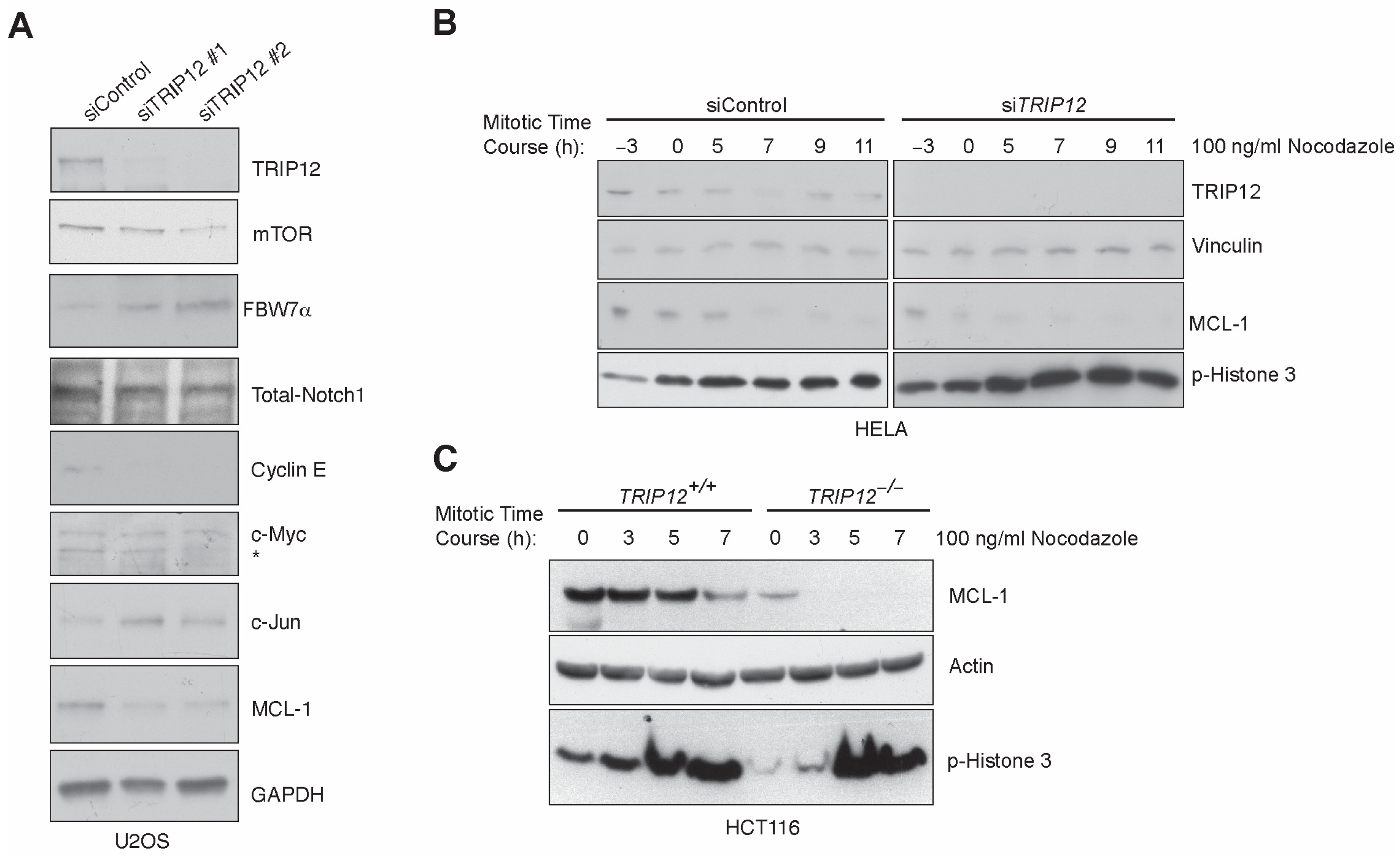
Figure 1. FBW7 protein is stabilized and its substrates mTOR, CyclinE, and MCL-1 are downregu–lated in TRIP12 depleted cells. (A), Western blot validation of FBW7 protein stability and substrates on U2OS cell lysates transfected with two independent siRNAs targeting TRIP12 gene compared to a non-targeting control. Note: * denotes non-specific immunoreactive band. (B,C) Immunoblot showing decreased MCL-1 levels in HELA and HCT116 cells transfected with TRIP12 siRNA followed by a double thymidine block and subsequent release in 100 ng/mL nocodazole for indicated times relative to ‘0’ h, which is an approximate mitotic onset time after release from thymidine block.
Cancers 15 00505 g001
Figure 2. Enhanced mitotic arrest in TRIP12-depleted cells: (A) Histogram of asynchronously grow–ing and Taxol treated HCT116 cells with indicated genotypes. Following treatment with Taxol, cells were collected, fixed, and stained with Propidium iodide (PI), and the DNA content was analyzed by FACS. (B,C) Quantification of fluorescence intensity of the PI-stained DNA in a population of cells within distinct cell-cycle phases from A. Mean ± S.D. of 3 independent experiments is shown. * p < 0.05, ** p < 0.01, and *** p < 0.001.
Cancers 15 00505 g002
Figure 3. Aberrant release from mitosis in TRIP12-depleted cells: (A), Histogram of cells with indi–cated genotypes, synchronized in mitosis and released in normal media. (B) Quantification from experiments in (A). (C) Western blot for indicated antibodies showing cleaved caspase-7/total caspase-7 ratio in cells from indicated genotypes. (D) Schematic for generation of a CA-GFP fluorescent apoptosis reporter. (E) Quantification of FACS data from stable cell lines expressing lentivirus-mediated CA-GFP in cells from indicated genotypes. * p < 0.05, ** p < 0.01, and *** p < 0.001.
Cancers 15 00505 g003
Figure 4. Efficient release from mitotic arrest in cells overexpression MCL-1 3A-mutant. (A) Sche–matic of MCL-1’s GSK3β phosphodegron mutated to alanine. (B) Western blot for indicated antibodies in cells overexpressing MCL-1 wildtype and 3A-mutant. Numbers under MCL-1 blot denotes densitometric quantification normalized to transfection control GFP. Vinculin antibody is used as a loading control. (C) MCL-1 wild type and mutant overexpressing mitotically synchronized TRIP12 depleted cells subjected to cell-cycle analysis after 3 HR of release from mitotic arrest assessed by flow cytometry. ** p < 0.01, and *** p < 0.001.
Cancers 15 00505 g004
Figure 5. FBW7 K404/412R mutant reduces MCL-1 protein and sensitize HCT116-FBW7−/− cells to Taxol. (A) Schematic of generation of FBW7 wildtype and K404/412R mutant cell lines. (B) Immunoblot showing FBW7 K404/412R stability and downregulation of MCL-1 in HCT116-FBW7−/− cells in comparison to wild type controls. (C) Cell titer blue viability assay quantification from HCT116-FBW7−/− overexpressing FBW7 wild type and 2R mutant cells treated with increasing doses of Taxol. Mean of 3 independent experiments is shown as percent viability.
Cancers 15 00505 g005
Figure 6. Analyzing the role of TRIP12 inhibition in sensitizing ovarian and breast cancer cells to Taxol. (AC) Cell titer blue viability assay quantification in ovarian cancer cells of indicated geno–types treatment with indicated doses of Taxol. (D) Western blot for indicated antibodies in ovarian cancer cells incubated with siControl or siTRIP12. Numbers under the MCL1 blot represents densitometric quantification normalized to loading control α-Tubulin. (E,F) Cell titer blue viability assay quantification in breast cancer cells treated with indicated doses of Taxol. (G) Western blot for indicated antibodies in ovarian cancer cells incubated with siControl or siTRIP12. Note: * = p < 0.05 and ** = p < 0.01.
Cancers 15 00505 g006
Figure 7. TRIP12 inhibits mitotic arrest in response to Taxol.
Cancers 15 00505 g007
Disclaimer/Publisher’s Note: The statements, opinions and data contained in all publications are solely those of the individual author(s) and contributor(s) and not of MDPI and/or the editor(s). MDPI and/or the editor(s) disclaim responsibility for any injury to people or property resulting from any ideas, methods, instructions or products referred to in the content.