Simple Summary
Although colorectal cancer is the third most common type of cancer, its treatment strategies still have room for improvement, as current techniques carry risks that aggressively deteriorate patients’ quality of life. Medical physics using nanotechnology tools can significantly contribute to solving this challenge. The high biocompatibility, easy functionalization, and targeting capabilities of plasmonic (gold) nanoparticles, together with their high atomic number, which provides a large X-ray absorption cross-section, makes them potential agents for enhancing cancer therapeutics as photothermal therapy agents and as radiosensitizers. Hence, the auspicious possibility of synergistically combining radio- and phototherapies is of imperative importance and must be explored to enhance their clinical application.
Abstract
Colorectal cancer is the third most common cancer worldwide. Despite recent advances in the treatment of this pathology, which include a personalized approach using radio- and chemotherapies in combination with advanced surgical techniques, it is imperative to enhance the performance of these treatments and decrease their detrimental side effects on patients’ health. Nanomedicine is likely the pathway towards solving this challenge by enhancing both the therapeutic and diagnostic capabilities. In particular, plasmonic nanoparticles show remarkable potential due to their dual therapeutic functionalities as photothermal therapy agents and as radiosensitizers in radiotherapy. Their dual functionality, high biocompatibility, easy functionalization, and targeting capabilities make them potential agents for inducing efficient cancer cell death with minimal side effects. This review aims to identify the main challenges in the diagnosis and treatment of colorectal cancer. The heterogeneous nature of this cancer is also discussed from a single-cell point of view. The most relevant works in photo- and radiotherapy using nanotechnology-based therapies for colorectal cancer are addressed, ranging from in vitro studies (2D and 3D cell cultures) to in vivo studies and clinical trials. Although the results using nanoparticles as a photo- and radiosensitizers in photo- and radiotherapy are promising, preliminary studies showed that the possibility of combining both therapies must be explored to improve the treatment efficiency.
1. Introduction
Colorectal cancer (CRC) caused 1.9 million new cases and 0.9 million fatalities in 2020 [1]. As a result, CRC is considered the second most fatal cancer and third most common malignancy, whose prevalence is projected to alarmingly increase over the next years [1]. Considering population aging, growth, and human development, the latest GLOBOCAN data predict that the number of new CRC cases will reach 3.2 million in 2040 [1]. In high-income nations, the rising incidence of CRC is more prominent, partly attributable to lifestyle decisions, including eating habits and physical activity levels [1,2]. Despite the advances made in the last 40 years, namely, with the introduction of new drugs, radiotherapy, and the use of more accurate surgical procedures, the treatment of CRC continues to be extremely complex, with a survival rate that varies according to the stage of the disease at diagnosis and that usually ranges from 90% in patients with localized cancer to 14% in people with metastatic cancer [3]. Therefore, it is urgent to tackle this disease with different therapeutic strategies that may achieve better survival. A more personalized therapeutic strategy guided by new materials for early and accurate diagnoses and the ability to enhance tumor responses to treatment will probably be the way to achieve this goal [3,4]. The use of nanomaterials is currently a hot topic with a wide range of applications for medical diagnosis and treatment. Nanotechnology plays a major role in both the biotechnology and medical fields, as it offers new techniques to overcome the constraints of conventional medicine. The purpose of this article is to offer a comprehensive evaluation of the most relevant clinical and research findings regarding the use of nanoparticles (NPs) in CRC treatment. With a focus on gold nanostructures, it will address the application of NPs in radiotherapy (acting as radiosensitizers), nanoparticle-induced hyperthermia techniques such as photothermal therapy (PTT), and the synergistic effect that could arise from the combination of both treatments.
2. Challenges in Colorectal Cancer: A Medical Point of View
CRC is currently managed with a set of varied and advanced techniques that are adaptable to the patient and the characteristics of the neoplasm, including radiotherapy (RT), chemotherapy (CT), and surgery [2,5,6]. The staging of the tumor and its location are the most important criteria in determining which therapy is useful and how effective it may be [4,7]. The local extent of the tumor, its spread to nearby tissues, and the involvement of lymph nodes or distant sites (metastasis) are what determine the stage of the disease [4]. In its early stages, pre-cancerous lesions are confined to the inner layer (mucosa) of the colon or rectum (stage 0) [4,8]. As CRC progresses to stage I, the tumor outgrows the muscularis mucosa (thin muscle layer of mucosa) into the submucosa and into the muscularis propria. In stage II, the cancer has spread to the colon’s or rectum’s outermost layers but has not penetrated through them, and in stage III, it has progressed to the neighboring lymph nodes, vessels, or fat deposits surrounding them. In the highest and most aggressive stage (stage IV), CRC has progressed to distant organs, most frequently to the liver or lungs, or to distant parts of the peritoneum (the fascia covering the abdominal cavity) [4,8].
Surgery is frequently the first line of treatment for this cancer in its early stages (stages I-II), while chemotherapy is typically prescribed as the first option for metastatic CRC (stage IV) [9]. In the presence of risk factors for local recurrence, neoadjuvant treatments with the use of RT and CT, i.e., prior to surgery, are the gold standard [10]. In fact, recently, there has been an increase in the use of neoadjuvant treatments for rectal cancer, with the goal of achieving a complete tumor response. This increase has allowed patients to be treated with an organ preservation strategy, i.e., a non-surgical approach called watch-and-wait [10]. This strategy translates into a significant increase in the patient’s quality of life, who would otherwise be referred for surgical procedures with serious consequences, namely, on urinary, sexual, and defecatory functions, and, in many cases, the need for a permanent stoma [11]. However, a major concern in watch-and-wait patients is related to the risk of local regrowth after a complete clinical tumor response. About 30% of patients will require additional surgical procedures with a subsequent loss of quality of life [11]. Improvement is therefore essential on two fronts: (i) increase the response of colorectal cancer to neoadjuvant treatment to achieve higher rates of complete tumor response; (ii) more accurate and earlier diagnosis, in terms of both initial staging and post-treatment surveillance. The upward development of nanotechnology during the last few decades has been seen as a promising approach for cancer diagnosis and treatment, as several NP-based assays demonstrated improved selectivity and sensitivity with new capabilities not possible with the existing conventional techniques [3,4,12,13,14]. These advancements not only may improve cancer patient survival by allowing an early diagnosis but also might be used to track cancer progression in response to treatment [12]. This is particularly relevant for localized tumors, where early diagnosis and treatment allow a much higher rate of survival.
Magnetic nanoparticles (MNPs), quantum dots (QDs), and gold nanoparticles (AuNPs) are some examples of nanomaterials that have already demonstrated the potential to facilitate the diagnosis and prognosis of different cancers, including breast cancer, lung cancer, leukemia, liver cancer, colon cancer, and rectal cancer [3,4,12]. When properly functionalized with the right ligands, QDs can act as biomarkers and recognize specific cancer-associated molecules to allow their visualization by different imaging techniques [15,16]. In parallel to this, iron oxide nanoformulations (MNPs) and AuNPs, taking advantage of their physical properties, can also act as contrast agents in imaging techniques such as magnetic resonance (MRI) and computed tomography [17,18,19]. Beyond their diagnostic capabilities, such NPs can also have an active role in cancer treatment. The utilization of nano-based carrier systems as a means of drug delivery is one of the most promising cancer therapy methods [14,19]. In parallel to this, the strong photoelectric absorption coefficient of gold (its high Z (Z = 79)) provides a large X-ray absorption cross-section that, when combined with high Auger and Coster-Kronig (C-K) electron emission yields, makes AuNPs excellent radiosensitizers, offering significant local radiation dose enhancement [20]. The results show that using nanoparticles in cancer diagnostics and treatment is a promising approach to combating the current difficulties and limitations, unlocking new frontiers and treatment modalities.
3. Heterogeneity in Colorectal Cancer: Single-Cell Point of View
Intratumoral heterogeneity is likely to have implications for cancer treatment, local regrowth, and biomarker discovery, particularly for targeted treatments. In the case of CRC, it presents complex and heterogeneous phenotypes with effects at the epigenomic, genomic, transcriptomic, proteomic, and metabolomic levels [21,22]. Single-cell technologies have contributed to exciting progress in understanding the single-cell heterogeneity of carcinogenesis progression and metastasis, which can be fundamental to knowing the tumor in depth and choosing the most appropriate treatment [23,24]. CRC heterogeneity studies focus on clonal heterogeneity, the microenvironment, spatial organization and crosstalk, cell differentiation, metastasis, the therapy resistance of human T cells, non-immune cells, organoids, in vivo models, and patients [25,26,27]. Given the complexity of CRC and tumor heterogeneity, it is important to integrate patient information into models to predict heterogeneous cancer evolution [28]. Organ-on-a-chip 2D and 3D models have been developed by culturing primary human intestinal epithelial stem cells to mimic and shape the human intestinal structure and mechanical properties [28,29,30,31]. Three-dimensional in vitro platforms have shown that 90% of genetic mutations in patient tumors are present in tumor organoids, corroborating the robust use of organoids to study tumor heterogeneity (as discussed in detail in Section 6.2) [32,33]. The culturing of organoids from metastatic colorectal cancer patients’ biopsies has a success rate of 71%, revealing a new tool for the clinic as a potential platform for treatment decisions for the individual patient [33].
Different single-cell technologies have been developed to study the morphology, plasticity, phenotype, genomics, transcriptomics, and proteomics of tumor heterogeneity [34]. Depending on the study and the sample type, high-throughput technologies have been developed, such as fluorescence-activated cell sorting (FACS), magnetic-activated cell sorting (MACS), microfluidics, and microarray devices (Figure 1) [35,36,37,38,39]. Innovation in high-spatial- and high-temporal-resolution imaging with the ability to make a sort decision is receiving attention to tie cell phenotypes to omics data [35]. Microarray technologies provide high spatial and temporal resolution, providing different approaches to collecting single cells for further genetic analysis [35]. Magnetic collections using microarrays can be accomplished when iron oxide microbeads or nanoparticles are embedded into the individual microcarrier [35,37]. Microarrays have been used for high-quality imaging and studying metastatic tumor cell motility and invasion over time [35,37,40]. Gracz et al. studied how a single intestinal stem cell (ISC) developed into an organoid over time by time-lapse imaging on a microarray platform. Moreover, quantitative polymerase chain reaction (qPCR) was performed to correlate the phenotype with gene expression analysis results. This study observed high levels of active ISC markers at 0 h, while secretory progenitor markers and differentiated cell markers were found at later time points, finally obtaining mature organoids after 240 h [41].
Technologies that provide a high throughput are ideal for large-scale multi-omics data classification to comprehensively characterize CRC at a single-cell level [42]. Among the different single-cell omics technologies, single-cell RNA sequencing (scRNA-seq) is receiving attention because it provides a full transcriptome characterization of single-cell diversity and heterogeneity. scRNA-seq measurements were used to gain insights into the CRC microenvironment composition, revealing the dynamic nature of immune and non-immune cells from CRC at different stages [43,44]. A single-cell triple omics sequencing (scTrio-seq) technique has been developed to characterize multiple layers of molecular features assessing simultaneity, somatic copy number alterations (SCNAs), DNA methylation, and transcriptome information from single cells. This technique has been used in CRC patients, showing that molecular alterations are important in metastases and cancer progression [45]. Different studies have observed molecular phenotype diversity and different mutations in transcriptome profiles from scRNA-seq measurements in colorectal organoids [46,47]. Single-cell DNA sequencing (scDNA-seq) and scRNA-seq techniques have been used to study drug resistance and intratumoral genetic diversity in organoids [48]. This study supports the strength of the characteristics of organoid clone cultures from individual cells. All colorectal cancers in this study presented resistance to the most common drugs used to treat the disease, which highlights the need for new treatments. Moreover, comparing the DNA sequences, they found a greater somatic mutation rate in CRC than in normal colorectal cells [48].
In summary, cell heterogeneity in cancers is one of the greatest challenges in enhancing cancer diagnosis and treatment for the development of effective personalized therapies. Single-cell studies of nanotechnology-based treatments using radiotherapy and phototherapy are also needed to understand the mechanism of interaction between radiation and nanoparticles from a single-cell point of view. Since CRC presents heterogeneous clinical behavior and diverse responses to treatment, the power of single-cell technologies and the creation of new model systems can address the complexity of CRC by developing improved and new strategies for treatment.

Figure 1.
Single-cell separation, sorting and collection (a) micro- and (b) macro-technologies. Reprinted from Ref. [49] with permission from Elsevier. (c) Example of a microarray device. Embedded microstructures arrays (EMAs): scanning electron microscopy cross-section images of a full array (c) and one microcarrier (d). (e) Transparent EMA glued to the cassette where cells will be seeded. (f–j) Since iron oxide microbeads were embedded into each microcarrier, magnetic collections were performed by a microwand to isolate cells and release them into a different container. (k,l) High-quality images of cancer cells on EMAs. Black scale bars: 250 µm; white scale bars: 20 µm. Adapted with permission from Ref. [37]. Copyright 2022, Creative Common CC BY.

4. Nanoparticles in Radiotherapy
Radiotherapy (RT) is one of the most effective and widely used techniques for cancer treatment, and it is used in approximately 50% of cancer patients [50,51,52]. The mechanism behind this type of treatment is cellular damage caused by the interaction between ionizing radiation (IR) and biological tissues [53]. Radiation can be delivered to cancer cells either via an external beam (conventional RT) or through an internally implanted radiation source (brachytherapy) [54]. In external beam radiotherapy, the most frequently used therapies rely on the use of X-rays, where orthovoltage (100 to 500 keV) or megavoltage (>1 MeV) photons are commonly used to treat superficial or deep-seated tumors, respectively. Alternatively, energetic particles can also be employed, though they are less common due to their higher costs [55]. Cell exposure to IR triggers a cascade of processes that can be divided into three phases: physical, chemical, and biological [56,57,58]. The physical phase, which occurs in the first nanoseconds of irradiation, describes the interactions between charged particles and the atoms of which the tissue is composed. IR mainly interacts with orbital electrons, ejecting some of them from atoms (ionization) and raising others to higher energy levels (excitation). If the ejected secondary electrons are energetic enough, they may excite or ionize other surrounding atoms, thus triggering a cascade of ionization events. Because of the deposited energy and fast relaxation processes, various ionized/excited water molecules are generated (water radiolysis), resulting in the breakage of chemical bonds and the formation of free radicals, which are highly unstable. These water radicals, usually called reactive oxygen species (ROS), can be stabilized by other molecules in the medium through oxidation-reduction reactions. The ensemble of processes describing the mechanism of action of free radicals is usually referred to as the chemical phase. Besides the previously described reactions, ROS interact with different biological systems, such as DNA, lipids, and proteins. It is important to note that DNA damage can occur either through the so-called “indirect effect”, caused by ROS, or the “direct effect”, in which DNA is directly injured by the ionization-excitation processes that take place in the physical stage [59,60], as presented in Figure 2. Finally, the biological phase includes a series of cellular processes that are activated to repair radiation-induced damage [61]. Unrepaired damage can possibly result in malignant cell transformation or cell death over a span of seconds to days, or even years [62,63].
As already mentioned, IR damages multiple intracellular components. Despite being effective in damaging tumor cells, radiation therapy presents some critical drawbacks, such as dose heterogeneity, cell radioresistance, and damage to the surrounding healthy tissues. Consequently, the appropriate control of the delivered radiation dose is a crucial aspect to minimize toxicity to normal tissues [64]. To achieve more efficient dose delivery to targeted organs, numerous improvements in RT have emerged in the last few decades, such as intensity-modulated radiation therapy (IMRT), image guidance, stereotactic radiation therapy, and particle therapy [65,66,67]. However, increasing the maximum dose accumulation in cancerogenic tissues while sparing healthy ones remains a great challenge [62,68]. During the last several years, aiming to develop more effective anticancer regimes, a growing community of researchers has been exploiting the unique properties of nanomaterials to resolve the unsolved challenge of classical RT [69,70]. Among nanomaterials, nanoparticles (NPs) have been found to offer unique opportunities for RT [71,72,73] due to their specific properties, such as a high surface-to-volume ratio, enhanced cellular uptake, and ease of surface modification. Moreover, NPs are found to undergo deep tissue penetration, which increases the enhanced permeability and retention (EPR) effect [72]. In the context of radiotherapy treatment, NPs can either be used as radiosensitizers for external radiation beams, such as high-Z elements, or be employed as delivery formulations for therapeutic radionucleotides [55,74].

Figure 2.
Direct and indirect cellular effects of IR. Reprinted with permission from Ref. [75]. Copyright 2022, American Chemical Society.

The concept of radio-sensitization dates to the mid-1970s, when enhanced radiation damage to chromosomal DNA was first reported in patients undergoing iodine angiography [76]. Later, it was discovered that metal implants could alter the delivered doses during radiation therapy treatments [77,78], and in the last decades, several studies have exploited the capability of nanosized high-Z metal particles in promoting dose delivery to cancer tissues as an opportunity to improve RT procedures [75]. In this context, the rationale for using high-Z materials as radiosensitizers was first based on differences in the energy absorption properties of metals compared to soft tissues, i.e., physical interactions. Depending on the nature and energy of the ionizing source, such as X-rays, protons, neutrons, and ions, different mechanisms can occur. When X-rays impinge on a material, photons can be attenuated by four major processes, namely, the photoelectric effect, Compton effect, pair production, and Rayleigh scattering [79], as presented in Figure 2.
In a low regime of ionizing radiation (<500 keV), the photoelectric effect is the dominant interaction process with high-Z atoms (Z = 60–80) [80,81,82,83]. In this framework, the photoelectric cross-section strongly depends on , allowing high-Z NPs to transfer radiation to the medium by photoabsorption and subsequent electronic emission more efficiently than water. In this process, the energy of the incident photon is completely absorbed by the atom, which ejects an electron from its inner shell, also called a photoelectron, which possesses kinetic energy equal to the energy of the primary beam minus its binding energy. To fill the generated inner vacancy, two possible situations can occur, resulting either in the production of characteristic X-rays (also called fluorescent photons or secondary radiation) or in the emission of an Auger electron [84]. Fluorescent photons can travel longer ranges, while Auger electrons travel short distances, being effective in producing very high local ionization density [74,85]. Since the PE has a relatively small contribution to absorption in soft tissues (where the Compton effect dominates), photoelectrons, secondary photons, and Auger electrons emitted from high-Z metal NPs will cause a highly localized dose enhancement in the surrounding cells as illustrated in Figure 3 [55,75,82]. To enhance the biological effects through physical mechanisms, the NPs must be in the proximity of the selected target.
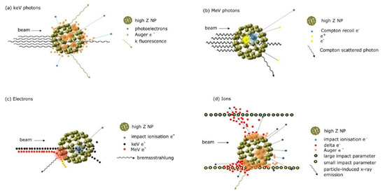
Figure 3.
Mechanisms of interaction of high-Z NPs with (a) keV photons, (b) MeV photons, (c) electrons, and (d) ions. Reprinted with permission from Ref. [82]. Copyright 2022, Creative Commons.
According to theory, NP-enhanced radiotherapy would only be effective with low-energy X-rays. At the same time, no increase in the overall dose deposition would be expected using higher energy or particle radiation, where the interactions between NPs and matter are dominated by the Compton effect, which does not strongly depend on Z [86,87]. However, radiosensitization with high-Z materials has also been observed for clinical MeV radiation [88,89,90,91], proton [92,93], and ion sources [94], indicating that the physical mechanism is not exhaustive in explaining the dose enhancement caused by the presence of such NPs [95,96]. In this regard, several studies have pointed out that other non-physical mechanisms, such as chemical and biological processes, play an important role in producing indirect damage to cancer cells. In fact, the main chemical effect contributing to NP-induced radiosensitization is the formation of ROS. ROS enhancement can also be produced by catalytic processes, apart from the physical effects previously described, which can result in oxidative stress, the interruption of the cell cycle, or the inhibition of DNA repair [97,98]. In this context, several studies have demonstrated that boosted ROS production can also be achieved by using metal oxide NPs with lower atomic numbers (made by Si, Al, Ti, Zn, Fe, and Ce, for instance) because of the high catalytic activities of their surfaces [99].
Over the past few years, NPs have been studied for their use as radiosensitizers to improve the response of cancerogenic tissues to radiotherapy procedures. In this framework, there has been considerable interest in the study of metallic, bimetallic, and metal-oxide-based NPs [100]. The most promising types of NPs are briefly described in the following paragraph.
Among metallic NPs, gold nanoparticles (AuNPs, Z = 79) have been widely studied for either diagnostic or therapeutic applications in cancer therapy due to their high atomic number, good biocompatibility, and strong photoelectric absorption. Regarding RT, the increase in the local radiation dose in the presence of AuNPs is attributed to strong photoelectric interactions because of their high atomic number, but also to the generation of ROS when the NPs are irradiated with particulate/high-energy X-rays [89]. Moreover, the efficiency of Au NPs as radiosensitizers depends not only on the NP size, shape, concentration, and coating but also on the external beam energy and specific cell line [68,87,101]. Ongoing research is focusing on the optimization of NP characteristics to reduce the delivered doses and to combine IR with other treatment modalities [75,102]. Another candidate as a sensitizer is gadolinium (Gd, Z = 64), which is commonly employed as a magnetic resonance imaging (MRI) contrast agent. Studies on Gd-based NPs have confirmed a significant sensitization effect in the presence of ionizing radiation under different conditions, i.e., NP dimensions, coatings, beam energies, and cell lines, allowing AGuIX, a Gd and Si-based NP, to enter clinical trials. Moreover, due to their properties, Gd-based NPs have been considered suitable for use in combination therapies, such as image-guided RT [90,103]. Among metal-based NPs, silver NPs (Ag, Z = 47) have also attracted attention due to their enhanced radiation sensitization [104]. However, their ability to accumulate in tumor cells remains to be improved [100]. As an alternative to full metallic systems, metal oxide NPs have been considered a viable option. In this regard, Hafnium (Hf, Z = 72) oxide NPs (NBTXR3) have been considered an appropriate RT sensitizer because of their high atomic number, electron density, and chemical stability. Moreover, clinical trials concerning different tumor locations, such as head and neck, prostate, and rectal cancer, are being successfully carried out [80]. Besides the aforementioned NPs, other elements, having either high or low atomic numbers, may be employed as radiosensitizers, such as Fe, Ta, Bi, and Ce, for instance [75,82,100,105].
Moreover, although NPs are currently widely studied to directly enhance radiation effects, NPs can also be employed as delivery vehicles for tumor-specific radiosensitivity drugs due to their ability to be simultaneously functionalized with chelators and targeting agents, thus enhancing cellular uptake [55].
In the specific context of RT for colorectal cancer, Au, Ag, and SPIO NPs have shown promising results as radiosensitizers (which will be discussed in detail in Section 6) [5,106,107,108]. In addition to this, several types of NPs are being explored for their use as nanocarriers for targeted therapy, which can be combined with RT to achieve better results [109,110,111]. However, it is worth mentioning that despite the large number of studies, there are clear difficulties in translating preclinical studies into clinical trials. These adversities may be related to the large number of variables that must be investigated to control and optimize the desired outcome. The reported studies have examined different aspects, such as diverging cell lines, NP materials and coatings, and radiation parameters. Consequently, experimental data reporting the radiation enhancement effects produced by several types of NPs on different biological systems have shown large variability. Hence, despite the promising preclinical results of NP-mediated radiosensitization, the exact mechanism of the interaction between NPs and ionizing radiation, as well as the subsequent biochemical and biological effects, remains incomplete, and further studies are therefore required, namely, studies using single-cell models, as discussed in the previous section [80,112,113].
5. Nanoparticle-Based Photothermal Therapy
A potential oncologic therapy method known as hyperthermia involves heating cancer cells up to 40–45 °C to induce apoptosis, or programmed cell death [19,114]. The traditional hyperthermia method, involving an external heating source that generates a temperature gradient to a maximum on the body’s surface that rapidly decreases with distance, has several drawbacks, not only because the energy is dissipated in healthy tissues located between the surface and the tumor but also because there is no differentiation between the targeted tissue and the surrounding normal tissues [115]. The advent of nanotechnology and its recent developments enable these drawbacks to be tackled by providing an appropriate way for localized and differentiated heat delivery to reach local and controlled hyperthermia. This is achieved via functionalized nanostructures that are activated by external/outside stimuli, such as electromagnetic radiation. In this context, functionalized nanoparticles, when concentrated inside the tumor, can absorb the energy coming from the external source to locally enhance the effects of hyperthermia [19,115]. Photothermal therapy (PTT) is an excellent example of innovative nanotechnology-based strategies. It is a nanoparticle-mediated hyperthermia technique that consists of a minimally invasive localized treatment, whose goal is to convert electromagnetic radiation into heat by stimulating photoabsorbing agents that are administrated to the body intravenously or intratumorally [19,114]. Laser light in the near-infrared (NIR) region is the energy source typically used in PTT due to its enhanced tissue penetration capability, with lower absorption in biological tissues [19,116]. In particular, the highest transmittance for NIR is found in tissue components such as hemoglobin and water, which allows NIR radiation to go through 10 cm of subcutaneous tissue, 4 cm of skull/brain tissue, or 4 cm of muscle tissue [116]. Noble metal NPs, carbon-based nanomaterials, metal compounds, and organic nanomaterials are some of the types of photothermal agents currently under development for application in PTT [116]. Among them, gold nanostructures are the most popular and have been extensively explored due to their biocompatibility, versatility, and high light-to-heat conversion efficiency and the fact that by controlling their shape and aspect ratio, it is possible to tune their absorption spectra peaks to match the desired wavelength, including the maximum absorption in the NIR region for optimal tissue penetration [2,19]. In this case, the heat generation phenomenon can be explained by a feature of metallic nanoparticles called surface plasmon resonance (SPR) [19,114,117]. SPR is caused by the presence of conduction electrons that oscillate on the metallic nanoparticle surface. If the incident light wavelength/frequency triggers the resonance of the oscillating electrons, a high-energy state is reached, leading to sequential energy dissipation in the form of heat. Under these circumstances, the light absorption results in optimal heat generation at the NPs that ultimately dissipates from the particle to the surrounding media [19,114].
Phototherapy mediated by gold nanoparticles has already shown promising results in vitro and in vivo (with animal models) for different types of cancer, including breast, prostate, lung, colon, and colorectal cancer. Manivasagan et al. demonstrated that gold nanorods ([AuNR] = 25 µg mL−1), duly modified for biocompatibility purposes, combined with laser irradiation (2 W cm−2, 5 min) induced significant apoptosis (63.3%) when compared with three control groups (0.38%, 1.74%, and 12.01%) in breast cancer cells (MDA-MB-231) [118]. Sangnier et al. studied the photothermal effect of four gold nanoparticle morphologies and the resulting induced cancer cell death on human prostate cancer (PC3) cells. For cancer cells exposed to 808 nm NIR irradiation (0.3 W cm−2, 10 min) with gold nanorods ([AuNR] = 98.5 µg mL−1), the number of viable cancerous cells decreases to 38% in the extracellular condition and to 11% in the intracellular situation [119]. Taking advantage of the ability to functionalize NPs to target specific cancer types, Knights et al. studied the use of pulsed and continuous lasers operating at 854 nm on lung cancer cells (A549) with nanorods functionalized with anti-EGFR antibodies. It was demonstrated that pulsed laser irradiation (pulse repetition frequency: 10 Hz; pulse duration: 7 ns; spot size: 9 mm; radiant exposure: 25 mJ cm−2) resulted in a 93% ± 13% reduction in cell viability when compared to control exposures, highlighting the efficiency of this minimally invasive therapy [120].
Specifically considering CRC, in 2010, Goodrich et al. evaluated the efficacy of photothermal treatment in a murine subcutaneous colon cancer model (CT26) as summarized in Table 1. Here, the solid tumors were infused with PEGylated gold nanorods, followed by the percutaneous irradiation of the tumor with an 808 nm laser (3.5 W, 180 s exposure time). Statistics show that the photothermally treated group’s survival outlasted that of the control groups, and 60 days after the treatment, 44% of the nanorod-treated mice survived with evidence of complete tumor ablation [121]. Three years later, Kirui et al. report the use of immunotargeted hybrid gold-iron oxide NPs for PTT treatment in a xenograft colorectal cancer tumor model (SW 1222 cells in subcutaneous-tumor-bearing mice) [122]. Here, PTT-induced toxicity (808 nm; 5 W/cm2, 20 min every 48 h) was verified by histological assessments of treated tumors, which showed over 65% necrosis in positive controls after 14-day photothermal therapy. In addition, this kind of NP can be used simultaneously as a contrast agent for non-invasive MR imaging: the gold NP portion of NPs works as an hyperthermia agent, while the iron oxide portion acts as an MR imaging agent [122]. Continuing the dual-function theragnostic NP trend, Azhdarzadeh et al. used gold-coated SPIONs (superparamagnetic iron oxide nanoparticles) functionalized with thiol molecules for the MRI and PTT of colon cancer cells (HT-29 cells). They observed that after irradiation (LED, 820 nm, 0.7 W/cm2, 2–8 min), a remarkable 80% of the cancerous cells were dead ([Au@SPIONs = 500 μg/mL]). The harmlessness of this type of irradiation was also proven by the almost null effect verified on the viability of NP-free cells [123]. In 2018, Chen et al. used gold nanorods conjugated with carbonic anhydrase IX (an antibody that specifically binds to a biomarker of hypoxia) to facilitate the targeting of hypoxic tumor areas. In vivo tests on subcutaneous HT29 xenografts revealed that the irradiation (760 nm, 12 W/cm2, 2–3 min) of mouse tumors injected with GNR/anti-CAIX led to complete ablation without tumor regrowth [124]. More recently, Simón et al. reported the effect of silica-gold nanoshells (NS) injected in a murine tumor model (CT26). Here, with fractionated PTT (807 nm, 1.2 W/cm2, 5 min) significant reductions in tumor sizes were found, and even complete regression was observed [125]. They also reported no significant changes in tumor development or survival between the groups receiving single-dose PTT and fractionated PTT.

Table 1.
Summary of highlighted preclinical studies evaluating photothermal therapy mediated by gold nanostructures in colorectal cancer.
Assembling all of these findings, it is noticeable that the most often utilized in vivo model in preclinical studies is the induction of subcutaneous tumors, with CT26, C26, and HCT116 being the most used CRC-related cell lines in BALB/c mice [126]. It is also apparent that metallic nanoformulations are quite popular, and, in several cases, they are also suitable for theragnostic and imaging purposes. Regarding PTT and laser parameters, each group has its own protocol, which makes comparing the effectiveness of NPs for heating a challenge. The only common parameter is that most preclinical studies operate in the first NIR biological window (700–950 nm). Still, different metal concentrations are utilized, power densities range from 0.1 to 6 W/cm2, and irradiation intervals range from a few seconds to minutes [127].
Over the years, PTT has proved its potential to selectively ablate malignant tissue and has been performed successfully on animals with small tumors. However, larger and more clinically significant tumors have been found to be more challenging in their complete removal without recurrence [125,128,129]. One of the main causes of this is that larger tumors frequently have hypoxic zones with poor blood flow, which, along with high interstitial pressure, hinders the transport of nanoparticles. In addition, the uneven nanoparticle distribution and the limited laser penetration depth can lead to heterogeneous intratumoral heat distributions [125]. These are challenging obstacles to overcome, but researchers have been inspired to investigate alternative uses of PTT in combination with other therapies, such as boosting the effectiveness of chemotherapy, releasing anticancer drugs, or inducing an immunological response for immunotherapy [19,130,131,132]. In addition to this, NPs can be exploited as radiosensitizers (Section 4), offering a significant enhancement of radiation damage to tumor tissue with reduced side effects on healthy surrounding cells [133,134,135]. Although not related to colorectal cancer, it is worth mentioning that gold nanoparticle systems have already moved to PTT clinical trials. In this context, AuroLase (photoabsorber-conjugated gold-silica NPs) is being developed for cancer therapy against head and neck (NCT01679470), lung (NCT00848042), and prostate tumors (NCT02680535) [136,137,138].
6. Photo- and Radiotherapy for Colorectal Cancer Treatment
6.1. In Vitro Studies
In vitro studies are fundamental for the proof-of-concept of the therapeutic potential associated with any treatment modality. Consequently, it is crucial to perform these assays for the clinical implementation of new strategies for treating colorectal cancer, namely, the combination of photothermal therapy (PTT) and radiotherapy. A search through the literature reveals that this theme has not yet been addressed and, therefore, represents an eventual new path for future research. Nevertheless, different authors have evaluated the suitability of PTT alone (Section 5) or in combination with chemotherapy, i.e., chemo-photothermal therapy, for treating this type of cancer using different nanomaterials [5,139,140,141]. Promising results were obtained due to the synergy of different treatments, as discussed in this section, which opens a window for future studies combining radiation techniques.
Once again, one example concerns nanomaterials composed of noble metals, such as AuNPs, since it is possible to take advantage of their tunable surface plasmon resonance properties to produce localized photothermal heating [116]. In this context, Hosseinzadeh et al. analyzed the potential of a chemo-photothermal strategy in vitro using SN38-conjugated hyaluronic acid AuNPs in two colon cancer cell lines (HT-29 and SW480) [132]. Here, the authors were able to reduce cell viability to below 10% after irradiation with red light. However, such nanomaterials present an absorption peak in the visible range and, therefore, are not adequate for in vivo applications.
This problem was overcome by White et al., who hybridized AuNPs with ultrasmall superparamagnetic iron oxide nanoparticles, obtaining hybrid magnetic AuNPs with an absorption peak in the near-infrared region (NIR). These nanoparticles were conjugated with a monoclonal antibody that targets CC-531 cells, causing a reduction in cell viability of up to 83% after exposure to NIR radiation [142]. Kirui et al. also produced Au and iron oxide hybrid nanoparticles, having analyzed their suitability for PTT in two colon cancer cell lines (SW1222 and HT-29). These nanomaterials were biofunctionalized with an antibody that targets those cells, and, after NIR irradiation, it achieved a maximum cell death of 65% [143].
An alternative way to tune the absorption peak of Au nanomaterials is by modifying their aspect ratio, resulting in elongated nanostructures. In this context, Seo et al. fabricated methylene blue-loaded Au nanorods with a SiO2 shell, having assessed their suitability for a dual-treatment strategy for colorectal cancer involving photothermal and photodynamic therapy [144]. Here, the authors used the CT26 cell line, and, after a NIR laser irradiation of the nanoparticle-loaded cells, they observed a reduction in cell activity to 11%. Guo et al. also studied this type of nanomaterial and produced fluorescein isothiocyanate/cisplatin-loaded chitosan-Au nanorods and evaluated their potential as chemo-photothermal and contrast agents for real-time cell imaging [131]. Through the use of the LoVo cell line, these authors achieved considerable cell death under NIR irradiation and, simultaneously, optically imaged these cells. Another noble metal nanomaterial suitable for PTT is Au nanoshells, which present an absorption peak that can be tuned by adjusting their size and thickness. In this context, Azhdarzadeh et al. synthesized MUC-1 aptamer-targeted Au-coated superparamagnetic iron oxide nanoparticles and verified that these nanomaterials were able to not only work as contrast agents in MRI but also reduce the HT-29 cell viability upon NIR irradiation (Figure 4), working, therefore, as a theragnostic agent [123]. Regarding theragnostics, Wang et al. also reported the use of assembled phage fusion protein-modified Au-Ag hybrid nanorods for PTT and fluorescence imaging [145]. Here, in vitro studies were performed with SW-620 cells, having verified that it was possible to acquire fluorescence images of the cells and, simultaneously, reduce their viability to 30% through irradiation with a NIR laser.
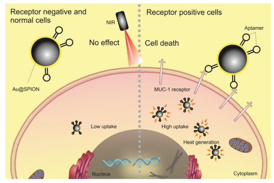
Figure 4.
MUC-1 aptamer-targeted gold-coated superparamagnetic iron oxide nanoparticles for PTT and MRI of colorectal cancer. Reprinted with permission from Ref. [123]. Copyright 2022, Elsevier.
A different type of nanomaterial that also presents an absorption capacity in the NIR region are those based on carbon, such as graphene and carbon nanotubes. In this context, Fiorica et al. demonstrated that it was possible to load graphene oxide nanogels with a chemotherapeutic drug, having assessed their potential for chemo-photothermal therapy using HCT-116 cells (Figure 5) [146]. As a result, the authors observed that, after NIR irradiation of the cells incubated with the nanogels, the cell viability dropped to almost 0%. A different work by Einafshar et al. addressed the fabrication of SN38-conjugated cyclodextrin-coated graphene oxide for chemo-photothermal therapy, as well [147]. Here, the authors reduced the HT-29 cell viability by ~14% after the exposure of cells incubated with this nanomaterial to a NIR laser.
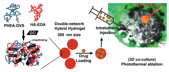
Figure 5.
Drug-loaded graphene oxide nanogels for destruction of HCT-116 cells. Reprinted with permission from Ref. [146]. Copyright 2022, American Chemical Society.
The potential of multiwalled carbon nanotubes for this biomedical application has also been studied by some authors, such as Levi-Polyachenko et al., who analyzed their suitability for hyperthermic chemotherapy delivery using RKO and HCT-116 cells [148]. Here, it was verified that the NIR irradiation of these cells in the presence of multiwalled carbon nanotubes and oxaliplatin led to a significant reduction in their viability. In a different work, Graham et al. produced folic acid-functionalized multiwalled carbon nanotubes and assessed their potential for the PTT of colorectal cancer, considering the same cell lines [149]. As a result, the authors verified that the functionalized multiwalled carbon nanotubes presented 400% to 500% higher affinity for these cells compared to the non-functionalized ones. Additionally, after NIR irradiation, they observed a reduction in cell viability between 50% and 60%. Regarding this theme, Tan et al. produced poly (carbonate-urea) urethane-functionalized multiwalled carbon nanotubes for the thermal ablation of cancer cells through exposure to NIR radiation [150]. This study was performed using HCT-116 cells and, after performing the treatment, achieved a reduction in cell viability of 95.12%.
Besides nanomaterials based on Au and carbon, metal compound nanomaterials have also been studied in the context of colorectal cancer treatment. One example is the work by Koo et al., who fabricated nanocomposites of copper sulfate and assessed their suitability for the in vitro PTT of Caco-2 cells [151]. Here, it was observed that the NIR irradiation of nanocomposite-incubated cells led to a reduction in cell viability as high as about 80%. In another work, Hessel et al. produced amphiphilic polymer-coated copper selenide nanocrystals, which presented a strong absorption in the NIR region, and assessed their potential for the PTT of colon cancer in vitro using the HCT-116 cell line [152]. As a result, the authors verified that, after NIR irradiation, all cells exposed to the fabricated nanocrystals were destroyed.
The suitability of organic nanomaterials for this biomedical application has also been investigated by a few authors. For example, Obiweluozor et al. synthesized polydioxanone nanofibers containing polydopamine nanospheres and bortezomib for chemo-photothermal therapy using the CT-26 cell line [153]. In this study, after NIR irradiation of the nanofiber-incubated cells, there was a verified reduction in cell viability to 5%. Another work by Kelkar et al. evaluated the use of nanoparticles based on a semiconducting conjugated polymer known as poly-[4,4-bis(2-ethylhexyl)-cyclopenta [2,1-b;3,4-b′]dithiophene-2,6-diyl-alt-2,1,3-benzoselenadiazole-4,7-diyl] (PCPDTBSe) in causing cell death through heat generation resulting from irradiation [154]. The authors verified that this nanomaterial possessed a dual absorption in the electromagnetic spectrum’s blue and NIR ranges. Subsequently, they were used in PTT tests with CT-26 cells, achieving a 90% reduction in cell viability. In another study, MacNeill et al. reported the use of a different organic nanomaterial, i.e., low-band-gap donor-acceptor-conjugated polymer nanoparticles, for the PTT of two different colorectal cancer cell lines (RKO and HCT-116) [155]. In this study, the authors achieved a cell viability lower than 10% after irradiation with a NIR laser.
In conclusion, it is verified that various authors have addressed the use of multiple nanomaterials for the PTT or chemo-photothermal therapy of colorectal cancer in vitro, having achieved promising results with different cell lines. Nevertheless, the combination of PTT with radiotherapy for the treatment of this specific cancer has not yet been reported, and, therefore, is a new research path that should be explored in the future.
6.2. Three-dimensional Models
Two-dimensional cultures are useful tools in cancer therapy to study the toxicity and efficacy of different treatment modalities in a reproducible and quick manner. Nevertheless, due to their simplicity, the data obtained with these models may not correlate with subsequent in vivo and clinical studies [156]. In this context, 3D models represent a more realistic approach to studying the outcomes of different therapeutics, including nanomedicines, radiotherapy, and photothermal therapy [157,158], contributing to the reduction in the number of animals when proceeding to in vivo studies. This is a result of the ability of 3D models to better mimic tumor heterogeneity, pH variation, nutrient and drug access, and cell-cell and cell-extracellular-matrix interactions [159,160].
To date, numerous 3D models of colorectal cancer have been developed with increased complexity, ranging from spheroids, composed of either one cell line or a co-culture, to the development of patient-derived organoids [161,162,163]. Spheroids are 3D cellular aggregates that can be produced using scaffold-free methods, such as the hanging drop method, cell culture on low-attachment plates, and forced-floating and agitation methods [161,162,163]. In addition, scaffold-based methods can be used, whereby cells are embedded in ECM constituents, such as collagen, hyaluronic acid, or mixtures such as Matrigel [161,162]. Recently, the need to obtain more complex and close-to-real 3D models led to the development of organoids. These are derived from pluripotent or multipotent stem cells that self-organize into 3D structures, which more closely mimic the anatomical and physiological features of an organ. One of their greatest advantages is the possibility of using patient-derived stem cells, leading to the development of personalized therapies [163,164].
Different studies have developed 3D models of colorectal cancer to better evaluate the toxicity and therapeutic potential of nanoparticles to be used as radiosensitizers. Hau et al. developed a combinatorial strategy based on spherical PEGylated gold nanoparticles (10 nm) and radiotherapy to enhance the treatment efficacy in colorectal cancer. Gold nanoparticles were chosen, as they can enhance the delivery of the radiation dose in the tumor area. Their toxicity was evaluated in both 3D ring models and 3D spheroids of colorectal cancer using the human colorectal cell line LOVO. Three-dimensional models were produced by incubating LOVO cells with Nanoshuttle, followed by aggregation by levitation using a magnet drive overnight in an ultralow-attachment 96-well plate. Nanoparticles were cytocompatible up to concentrations of 25 µg/mL, with 3D models being less susceptible to treatment than 2D models, demonstrating the importance of using the former to assess the toxicity of nanoparticles. Finally, a synergistic effect between gold nanoparticles and ionizing radiation (either kilo- or megavoltage) was observed in LOVO cells using the clonogenic assay [165]. A study aiming to combine small nanoparticles and radiotherapy was also carried out by Goodarzi et al. In this work, sub-5 nm nanoparticles composed of polysiloxane and gadolinium chelates were produced, and their penetration capacity was evaluated on multicellular spheroids of the human colorectal cancer cell line HCT-116. These nanoparticles are potent radiosensitizers and are currently in clinical trials in combination with radiotherapy for the management of different types of cancer. The authors developed a protocol for spheroid preparation using an agarose-based microsystem and performed a detailed study on the penetration, cellular internalization, and organelle distribution of the nanoparticles at different time points and concentrations (Figure 6). These data provide useful information to more effectively study the interaction and distribution of nanoparticles in a three-dimensional environment for future radiosensitization studies [166].
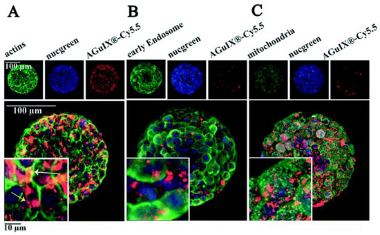
Figure 6.
Penetration of polysiloxane and gadolinium chelate nanoparticles marked with fluorescent probe Cy5.5 (AGuIX -Cy5.5) in HCT-116 colorectal cancer spheroids and co-localization with different cellular structures: actin (A), early endosome (B), and mitochondria (C). Blue channel: cell nucleus; red channel: AGuIX -Cy5.5; green channel: phalloidin (A); EEA1 antibody-labeled early endosomes (B); AIF antibody-labeled mitochondria. Adapted with permission from [166]. Copyright 2022, Creative Commons.
The efficacy of photothermal therapy has also been evaluated in different 3D colorectal cancer models. McCarthy et al. produced Hybrid Donor-Acceptor Polymer Particles (HDAPPs) composed of fluorescent and photothermal polymers and nanoparticles containing only the photothermal polymer (BSe). Nanoparticles (185 nm) were coated with hyaluronic acid (HA), and their interactions and photothermal ablation effects were evaluated in 3D organoids of colorectal cancer. Organoids were produced by mixing the mouse colorectal carcinoma cell line CT26 with a solution of extracellular matrix components (heprasil and collagen solution), followed by cross-linking through UV irradiation. The nanoparticles induced a time-dependent temperature rise after exposure to a continuous laser at 800 nm at 5 W, with BSe nanoparticles demonstrating the highest temperature rise (of more than 60 °C). A reduced penetration capacity of the HA-coated nanoparticles across the organoid was detected, with a higher accumulation in the organoid periphery, in comparison with their non-coated counterparts, in opposition to what occurred in 2D models. This could be related to greater interactions between the nanoparticles and the extracellular matrix. Nevertheless, HA-coated BSe presented the most effective photothermal ablation effect upon laser irradiation for 60 s, achieving almost the full eradication of the organoid. These results highlight the relevance of using 3D models to assess both the penetration and photothermal effects of nanoparticles, as 3D organoids required higher doses and longer incubation and irradiation times in comparison with 2D models [167].
Fiorica et al. developed graphene oxide-embedded nanogels composed of HA and polyaspartamide (300 nm) for the photothermal therapy of colorectal cancer. To improve the efficacy of the treatment, the chemotherapeutic drug irinotecan was incorporated into the nanosystem. Three-dimensional tumor models were obtained by seeding human dermal fibroblasts and HCT-116 cells in Matrigel. The nanosystem induced a time-dependent temperature increase upon irradiation with an 810 nm diode laser (3 × 10⁻³ W mm−3) for up to 150 s, demonstrating a ∆T of approximately 0.011 °C W−1 mg−1 and a mass extinction coefficient of 48.2 L g−1 cm−1. After the injection of the nanosystem into the 3D model and irradiation for 200 s, a necrotic spot was observed, which was enhanced in the presence of irinotecan, demonstrating the efficacy of the thermoablation treatment in combination with chemotherapy [146]. Roma-Rodrigues et al. also designed a strategy combining photothermal therapy and chemotherapy for colorectal cancer using spherical PEGylated gold nanoparticles (18 nm). The authors produced 3D spheroids of doxorubicin-sensitive and resistant colorectal cancer cell lines (HCT116 and HCT116-DoxR, respectively) by culturing the cells in super-low-attachment 96-well plates, followed by orbital shaking and incubation for 7 days. Tumor spheroids were then incubated with the nanoparticles and irradiated for 1 min with a diode-pumped solid-state laser (532 nm) coupled to a 1 mm diameter optical fiber at 3.78 W cm−2. A time-dependent loss of integrity of the tumor spheroid was observed for both 3D models. In addition, real-time data demonstrated that photothermal therapy improved the penetration and therapeutic efficacy of doxorubicin in both colorectal 3D models, demonstrating the relevance of combining localized photothermal therapy and chemotherapy to reduce chemotherapeutic drug doses and enhance the efficacy in cases of tumor drug resistance [168].
Overall, these studies demonstrate that there are large differences in the response to both radiotherapy and photothermal therapy between 2D and 3D models, highlighting the importance of using the latter to better assess treatment outcomes in vivo and in future clinical applications.
6.3. In Vivo Studies
In vivo studies of plasmonic gold nanoparticles (GNPs) of different sizes and shapes have already been performed for concomitant phototherapy and radiotherapy applied to colorectal cancer; however, combined photothermal and radiotherapy can also be chosen in future studies [110]. Most studies use nanoparticles with sizes that fall within 10–100 nm, as nanoparticles with a size below 10 nm are rapidly excreted by the kidneys. In comparison, those with a size above 100 nm are quickly captured by the immune system [169]. Different shapes have been used for in vivo studies, such as spheres [170], which present higher cellular uptake [110]; nanorods [171,172,173], which present high absorption coefficients [174]; and nanocages [6] or half-shells [130] for drug delivery.
Although there exist clinical trials that have investigated the applicability of GNP-based radiotherapy and phototherapy [98,175], current in vivo studies applied to colorectal cancer are limited to preclinical experiments [2]. Most preclinical models consist of xenograft models of human colorectal cancer cell lines injected subcutaneously into the flanks of mice [116,176]. In these models, the tumor is exposed, facilitating the penetrability and absorption of the radiation beam by nanoparticles distributed within the tumor [98]. For phototherapy, more clinically realistic models rely on tumors implanted in deeper organs such as the liver, requiring a catheter carrying a fiber-optic to irradiate the tumor [142,169]. Phototherapy studies make use of a NIR laser (808 or 810 nm [173]) to maximize penetration within the tumor, with irradiation times ranging from 2 to 10 min and laser power ranging from 0.5 to 12 W to achieve heating above 55 °C and induce thermal cytotoxicity [177], while radiotherapy studies make use of doses usually ranging between 4 and 30 Gy. The optimal laser exposure in phototherapy is a compromise between the photothermal conversion efficiency of the specific nanoparticle type [6], its temporal heating curve [122], and the integrity of the healthy tissue surrounding the tumor, as excessive laser exposure may cause undesired damage and apoptosis [121].
The efficacy of phototherapy and/or radiotherapy also depends on the proportion of nanoparticles accumulated within the tumor at a specific dose [121,178]. GNPs are deposited in the tumor upon systemic administration owing to the EPR given by fenestrations in immature blood vessels in the tumor [121]. However, the rate of deposition in the tumor is low [169], and GNPs accumulate in undesired organs of the reticuloendothelial system [121] regardless of their size, while small-sized GNPs accumulate in the rest of the tissues [177]. Several studies claim that the non-specific biodistribution of GNPs does not cause systemic toxicity during photothermal therapy [6,172,179] or radiotherapy [170,180] and that the renal clearance of GNPs is achieved 72 h after intravenous injection [181]; however, the toxicity of GNPs remains unclear and requires further studies [98,182]. Different strategies have been developed to maximize the aggregation of GNPs within the tumor and minimize potential systemic toxicity. These include GNP surface functionalization with hypoxia biomarkers such as CAIX [124], mesenchymal stem cells (MSC) [171], tiopronin [183], or antibodies such as the anti-death receptor-4 (DR4) monoclonal antibody [130] or the PD-ligand-1 antibody [6].
However, in radiotherapy, surface functionalization can have adverse effects due to the scavenging of low-energy electrons [101]. Other strategies involve the use of the pH-mediated aggregation of Au-RRVR complexes to increase tumoral aggregation [178]. The site-selective injection of theranostic GNPs is an invasive although effective alternative [169,176,183]. Theranostic nanoparticles enable both treatment and diagnosis by medical imaging [184] and are usually composed of a contrast material such as iron-platinum (FePt) [184] or gadolinium [169]. Photothermal therapy with GNPs is a promising treatment regimen for colorectal cancer. Many studies have demonstrated an evident recession of the tumoral volume [124,130,172,173,178,184,185]. Others have shown a significant increase in survival time [121,179] and even total survival [6,171], with minimal effects on surrounding healthy tissues. Similarly, GNP-based radiotherapy has shown promising results regarding tumor growth delay [180,183] and the survival rate, with up to 100% survival after 60 days [170]. The application of hyperthermia before radiotherapy produces an increase in perfusion that reduces hypoxia within the tumor, facilitating the radionecrosis of hypoxic-resistant tumoral cells [186]. Several clinical trials were conducted in the 1980s and 1990s to assess the viability of combined radio- and phototherapy, known as thermoradiotherapy [175]. However, the results were not promising due to the difficulty of achieving localized hyperthermic temperatures and the procedures’ invasiveness [186]. With the rise of nanotechnology, thermoradiotherapy studies have been reinitiated, although, to our knowledge, very few have yet been applied to colorectal cancer [186].
7. Conclusions and Future Perspectives
CRC is the third most prevalent cancer worldwide. Despite recent developments in the treatment of this pathology, which include a personalized approach using radio- and chemotherapies in conjunction with cutting-edge surgical techniques, it is critical to improve the effectiveness of these interventions and lessen their negative side effects on patients’ health. The use of NPs as radiosensitizers to enhance the responsiveness of cancerogenic tissues to radiotherapy has been researched throughout the past few years. The study of metallic, bimetallic, and metal-oxide-based NPs has drawn a lot of interest within this paradigm. In parallel, plasmonic-nanoparticle-mediated hyperthermia techniques, such as PTT, have been successfully carried out on animals with tiny tumors over the years, demonstrating its ability to selectively destroy malignant tissue. Considering the complexity of larger and more clinically significant tumors, PTT as a stand-alone treatment might not be viable in terms of complete tumor removal without reoccurrence. As a result, researchers have been motivated to investigate alternative applications of PTT in conjunction with other therapies to concomitantly benefit from its advantages. Enhancing the efficacy of chemotherapy, releasing anticancer medications, inducing an immune response to immunotherapy, and acting as radiosensitizers are some examples of how such NPs can be successfully applied to obtain a synergistic effect between different therapies.
Regarding CRC specifically, this review addressed studies performed from single cells to in vitro (2D and 3D cell cultures), in vivo, and clinical trials. Despite the promising results obtained for both radiotherapy and PTT alone, the combination of PTT with radiotherapy for the treatment of this specific cancer has not yet been reported and is therefore a new research path that should be explored in the future. The radiosensitization effect of hyperthermia needs to be explored and quantified as an enhanced equivalent radiation dose. Both the temperature and time interval between radiation therapy sessions need to be correlated with cell death outcomes, and their effects need to be understood.
Author Contributions
Conceptualization, C.T.d.S., J.H.B. and J.A.; methodology, C.T.d.S., J.H.B., J.A., M.V.G.-G. and B.C.-L.; writing the original draft, S.C.F. (points 1, 2, and 5), B.C.-L. (point 3), S.C. (point 4), R.M. (point 6a), A.G. and S.R. (point 6b), and D.S. (point 6c); writing—review and editing, all authors. All authors have read and agreed to the published version of the manuscript.
Funding
CTS acknowledges the program Atraccion de Talento (CAM), ref. 2020-T1/IND-19889. Ricardo Magalhães and Sofia Caspani acknowledge to Fundação para a Ciência e Tecnologia (FCT) and Programa Operacional Regional Norte (Fundo Social Europeu) for the grants SFRH/BD/148563/2019 and 2021.08212.BD, respectively. This work was financially supported by the European Union’s Horizon 2020 research and innovation program under the Marie Sklodowska-Curie Grant Agreement No. 734801 and Strategic Funding contracts UIDB/04968/2020, UIDB/04968/2021, and UIDP/04968/2021 and also by the Network of Extreme Conditions Laboratories (NECL) under the project NORTE-01- 0145-FEDER-022096. J. H. Belo acknowledges EEA grants for the project FBR_OC1_85 and PARSUK-FCT for the project SMARTX. He also acknowledges FCT for the projects PTDC/FISMAC/31302/2017, CERN/FISTEC/0003/2019, and PTDC/EME-TED/3099/2020 and for his contract DL57/2016 reference SFRH-BPD-87430/2012. A. Granja acknowledges Fundação para a Ciência e Tecnologia and Ministério da Ciência, Tecnologia e Ensino Superior, through the project HEALTH-UNORTE I&D&I project (NORTE-01–0145-FEDER-000039).
Conflicts of Interest
The authors declare no conflict of interest.
References
- Xi, Y.; Xu, P. Global colorectal cancer burden in 2020 and projections to 2040. Transl. Oncol. 2021, 14, 101174. [Google Scholar] [CrossRef]
- Khot, M.I.; Andrew, H.; Svavarsdottir, H.S.; Armstrong, G.; Quyna, A.J.; Jaynea, D.G. A Review on the Scope of Photothermal Therapy–Based Nanomedicines in Preclinical Models of Colorectal Cancer. Clin. Color. Cancer 2019, 18, e200–e209. [Google Scholar] [CrossRef]
- Dessale, M.; Mengistu, G.; Mengist, H.M. Nanotechnology: A Promising Approach for Cancer Diagnosis, Therapeutics and Theragnosis. Int. J. Nanomed. 2022, 17, 3735–3749. [Google Scholar] [CrossRef] [PubMed]
- Brar, B.; Ranjan, K.; Palria, A.; Kumar, R.; Ghosh, M.; Sihag, S.; Minakshi, P. Nanotechnology in Colorectal Cancer for Precision Diagnosis and Therapy. Front. Nanotechnol. 2021, 3, 1–21. [Google Scholar] [CrossRef]
- Soto, K.M.; Mendoza, S.; López-Romero, J.M.; Gasca-Tirado, J.R.; Manzano-Ramírez, A. Gold nanoparticles: Synthesis, application in colon cancer therapy and new approaches-review. Green Chem. Lett. Rev. 2021, 14, 665–678. [Google Scholar] [CrossRef]
- Wang, S.; Song, Y.; Cao, K.; Zhang, L.; Fang, X.; Chen, F.; Feng, S.; Yan, F. Photothermal therapy mediated by gold nanocages composed of anti-PDL1 and galunisertib for improved synergistic immunotherapy in colorectal cancer. Acta Biomater. 2021, 134, 621–632. [Google Scholar] [CrossRef]
- Burt, R.W.; Barthel, J.S.; Dunn, K.B.; David, D.S.; Drelichman, E.; Ford, J.M.; Giardiello, F.M.; Gruber, S.B.; Halverson, A.L.; Hamilton, S.R.; et al. Colorectal Cancer Screening. J. Natl. Compr. Cancer Netw. 2010, 8, 8–61. [Google Scholar] [CrossRef] [PubMed]
- Society, A.C. Colorectal Cancer Early Detection, Diagnosis, and Staging. Available online: Cancer.org (accessed on 10 October 2022).
- Kaveh, S.; Ebrahimi, P.; Rezapour, A.; Mozafari, M.; Sayehmiri, K. Bevacizumab and erlotinib versus bevacizumab for colorectal cancer treatment: Systematic review and meta-analysis. Int. J. Clin. Pharm. 2019, 41, 30–41. [Google Scholar] [CrossRef]
- de Azevedo, J.M.; Vailati, B.B.; Julião, G.P.S.; Fernandez, L.M.; Perez, R.O. Current Surgical Strategies in the Management of Rectal Cancer. Curr. Color. Cancer Rep. 2019, 15, 18–27. [Google Scholar] [CrossRef]
- Nasir, I. Salvage surgery for local regrowths in Watch & Wait-Are we harming our patients by deferring the surgery? Eur. J. Surg. Oncol. 2019, 45, 1559–1566. [Google Scholar]
- Zhang, Y.; Li, M.; Gao, X.; Chen, Y.; Liu, T. Nanotechnology in cancer diagnosis: Progress, challenges and opportunities. J. Hematol. Oncol. 2019, 12, 1–13. [Google Scholar] [CrossRef]
- Reshmitha, T.R.; Shini, V.S.; Nisha, P. Nanotechnology Approaches for Colorectal Cancer Diagnosis and Therapy. In Colon Cancer Diagnosis and Therapy; Vishvakarma, N.K., Nagaraju, G.P., Shukla, D., Eds.; Springer International Publishing: Cham, Switzerland, 2021; Volume 2, pp. 171–186. [Google Scholar]
- Al-Joufi, F.A.; Setia, A.; Salem-Bekhit, M.M.; Sahu, R.K.; Alqahtani, F.Y.; Widyowati, R.; Aleanizy, F.S. Molecular Pathogenesis of Colorectal Cancer with an Emphasis on Recent Advances in Biomarkers, as Well as Nanotechnology-Based Diagnostic and Therapeutic Approaches. Nanomaterials 2022, 12, 169. [Google Scholar] [CrossRef] [PubMed]
- Combes, G.F.; Vučković, A.-M.; Bakulić, M.P.; Antoine, R.; Bonačić-Koutecky, V.; Trajković, K. Nanotechnology in Tumor Biomarker Detection: The Potential of Liganded Nanoclusters as Nonlinear Optical Contrast Agents for Molecular Diagnostics of Cancer. Cancers 2021, 13, 4206. [Google Scholar] [CrossRef] [PubMed]
- Fang, J.; Wang, S.; Li, W.; Yuan, D.; Song, J. Quantitative detection of the tumor-associated antigen large external antigen in colorectal cancer tissues and cells using quantum dot probe. Int. J. Nanomed. 2016, 11, 235–247. [Google Scholar] [CrossRef] [PubMed]
- Caspani, S.; Magalhães, R.; Araújo, J.P.; Sousa, C.T. Magnetic Nanomaterials as Contrast Agents for MRI. Materials 2020, 13, 2586. [Google Scholar] [CrossRef]
- Siddique, S.; Chow, J.C.L. Application of Nanomaterials in Biomedical Imaging and Cancer Therapy. Nanomaterials 2020, 10, 1700. [Google Scholar] [CrossRef]
- Granja, A.; Pinheiro, M.; Sousa, C.T.; Reis, S. Gold nanostructures as mediators of hyperthermia therapies in breast cancer. Biochem. Pharmacol. 2021, 190, 114639. [Google Scholar] [CrossRef]
- Li, W.; Belchior, A.; Beuve, M.; Chen, Y.; Di Maria, S.; Friedland, W.; Gervais, B.; Heide, B.; Hocine, N.; Ipatov, A.; et al. Intercomparison of dose enhancement ratio and secondary electron spectra for gold nanoparticles irradiated by X-rays calculated using multiple Monte Carlo simulation codes. Phys. Medica 2020, 69, 147–163. [Google Scholar] [CrossRef]
- Li, J.; Ma, X.; Chakravarti, D.; Shalapour, S.; DePinho, R.A. Genetic and biological hallmarks of colorectal cancer. Genes Dev. 2021, 35, 787–820. [Google Scholar] [CrossRef]
- AlMusawi, S.; Ahmed, M.; Nateri, A.S. Understanding cell-cell communication and signaling in the colorectal cancer microenvironment. Clin. Transl. Med. 2021, 11, e308. [Google Scholar] [CrossRef]
- Venkatachalapathy, S.; Jokhun, D.S.; Andhari, M.; Shivashankar, G.V. Single cell imaging-based chromatin biomarkers for tumor progression. Sci. Rep. 2021, 11, 1–14. [Google Scholar] [CrossRef]
- Mitra, A.K.; Mukherjee, U.K.; Harding, T.; Jang, J.S.; Stessman, H.; Li, Y.; Abyzov, A.; Jen, J.; Kumar, S.; Rajkumar, V.; et al. Single-cell analysis of targeted transcriptome predicts drug sensitivity of single cells within human myeloma tumors. Leukemia 2015, 30, 1094–1102. [Google Scholar] [CrossRef] [PubMed]
- Qi, J.; Crinier, A.; Escalière, B.; Ye, Y.; Wang, Z.; Zhang, T.; Batista, L.; Liu, H.; Hong, L.; Wu, N.; et al. Single-cell transcriptomic landscape reveals tumor specific innate lymphoid cells associated with colorectal cancer progression. Cell Rep. Med. 2021, 2, 100353. [Google Scholar] [CrossRef] [PubMed]
- Wang, H.; Gong, P.; Chen, T.; Gao, S.; Wu, Z.; Wang, X.; Li, J.; Marjani, S.L.; Costa, J.; Weissman, S.M.; et al. Colorectal Cancer Stem Cell States Uncovered by Simultaneous Single-Cell Analysis of Transcriptome and Telomeres. Adv. Sci. 2021, 8, 2004320. [Google Scholar] [CrossRef] [PubMed]
- Chen, K.-Y.; Srinivasan, T.; Lin, C.; Tung, K.-L.; Gao, Z.; Hsu, D.S.; Lipkin, S.M.; Shen, X. Single-Cell Transcriptomics Reveals Heterogeneity and Drug Response of Human Colorectal Cancer Organoids. In Proceedings of the 2018 40th Annual International Conference of the IEEE Engineering in Medicine and Biology Society (EMBC), Honolulu, HI, USA, 18–21 July 2018; pp. 2378–2381. [Google Scholar] [CrossRef]
- Hinman, S.S.; Wang, Y.; Kim, R.; Allbritton, N.L. In vitro generation of self-renewing human intestinal epithelia over planar and shaped collagen hydrogels. Nat. Protoc. 2020, 16, 352–382. [Google Scholar] [CrossRef] [PubMed]
- Dutton, J.S.; Hinman, S.S.; Kim, R.; Wang, Y.; Allbritton, N.L. Primary Cell-Derived Intestinal Models: Recapitulating Physiology. Trends Biotechnol. 2018, 37, 744–760. [Google Scholar] [CrossRef] [PubMed]
- Wang, Y.; Gunasekara, D.B.; Attayek, P.J.; Reed, M.I.; DiSalvo, M.; Nguyen, D.L.; Dutton, J.S.; Lebhar, M.S.; Bultman, S.J.; Sims, C.E.; et al. In Vitro Generation of Mouse Colon Crypts. ACS Biomater. Sci. Eng. 2017, 3, 2502–2513. [Google Scholar] [CrossRef]
- Wang, Y.; Sims, C.E.; Allbritton, N.L. Human 2D Crypt Model for Assaying Intestinal Stem Cell Proliferation and Differentiation. Anal. Chem. 2022, 94, 9345–9354. [Google Scholar] [CrossRef]
- Van de Wetering, M.; Francies, H.E.; Francis, J.M.; Bounova, G.; Iorio, F.; Pronk, A.; van Houdt, W.; van Gorp, J.; Taylor-Weiner, A.; Kester, L.; et al. Prospective derivation of a living organoid biobank of colorectal cancer patients. Cell 2015, 161, 933–945. [Google Scholar] [CrossRef]
- Weeber, F.; van de Wetering, M.; Hoogstraat, M.; Dijkstra, K.K.; Krijgsman, O.; Kuilman, T.; Gadellaa-van Hooijdonk, C.G.M.; van der Velden, D.L.; Peeper, D.S.; Cuppen, E.P.J.G.; et al. Preserved genetic diversity in organoids cultured from biopsies of human colorectal cancer metastases. Proc. Natl. Acad. Sci. USA 2015, 112, 13308–13311. [Google Scholar] [CrossRef]
- Qian, M.; Wang, D.C.; Chen, H.; Cheng, Y. Detection of single cell heterogeneity in cancer. Semin. Cell Dev. Biol. 2017, 64, 143–149. [Google Scholar] [CrossRef]
- Cortés-Llanos, B.; Wang, Y.; Sims, C.E.; Allbritton, N.L. A technology of a different sort: Microraft arrays. Lab Chip 2021, 21, 3204–3218. [Google Scholar] [CrossRef] [PubMed]
- LaBelle, C.A.; Massaro, A.; Cortés-Llanos, B.; Sims, C.E.; Allbritton, N.L. Image-Based Live Cell Sorting. Trends Biotechnol. 2020, 39, 613–623. [Google Scholar] [CrossRef] [PubMed]
- DiSalvo, M.; Cortés-Llanos, B.; LaBelle, C.A.; Murdoch, D.M.; Allbritton, N.L. Scalable Additive Construction of Arrayed Microstructures with Encoded Properties for Bioimaging. Micromachines 2022, 13, 1392. [Google Scholar] [CrossRef]
- Pan, J.; Wan, J. Methodological comparison of FACS and MACS isolation of enriched microglia and astrocytes from mouse brain. J. Immunol. Methods 2020, 486, 112834. [Google Scholar] [CrossRef] [PubMed]
- Enrico, L.; Veronica, Z.; Domenico, M.; Alessandra, R. FACS Analysis of Memory T Lymphocytes. In T-Cell Differentiation: Methods and Protocols; Luigi, E., Ed.; Springer: New York, NY, USA, 2017; pp. 31–47. [Google Scholar]
- Nowotarski, H.L.; Attayek, P.J.; Allbritton, N.L. Automated platform for cell selection and separation based on four-dimensional motility and matrix degradation. Analyst 2020, 145, 2731–2742. [Google Scholar] [CrossRef]
- Gracz, A.; Williamson, I.; Roche, K.C.; Johnston, M.J.; Wang, F.; Wang, Y.; Attayek, P.; Balowski, J.; Liu, X.F.; Laurenza, R.J.; et al. A high-throughput platform for stem cell niche co-cultures and downstream gene expression analysis. Nat. Cell Biol. 2015, 17, 340–349. [Google Scholar] [CrossRef] [PubMed]
- Frank, M.H.; Wilson, B.J.; Gold, J.S.; Frank, N.Y. Clinical Implications of Colorectal Cancer Stem Cells in the Age of Single-Cell Omics and Targeted Therapies. Gastroenterology 2021, 160, 1947–1960. [Google Scholar] [CrossRef]
- Mei, Y.; Xiao, W.; Hu, H.; Lu, G.; Chen, L.; Sun, Z.; Lü, M.; Ma, W.; Jiang, T.; Gao, Y.; et al. Single-cell analyses reveal suppressive tumor microenvironment of human colorectal cancer. Clin. Transl. Med. 2021, 11, e422. [Google Scholar] [CrossRef]
- Zhang, Y.; Song, J.; Zhao, Z.; Yang, M.; Chen, M.; Liu, C.; Ji, J.; Zhu, D. Single-cell transcriptome analysis reveals tumor immune microenvironment heterogenicity and granulocytes enrichment in colorectal cancer liver metastases. Cancer Lett. 2019, 470, 84–94. [Google Scholar] [CrossRef]
- Bian, S.; Hou, Y.; Zhou, X.; Li, X.; Yong, J.; Wang, Y.; Wang, W.; Yan, J.; Hu, B.; Guo, H.; et al. Single-cell multiomics sequencing and analyses of human colorectal cancer. Science 2018, 362, 1060–1063. [Google Scholar] [CrossRef] [PubMed]
- Yan, H.H.N.; Siu, H.C.; Ho, S.L.; Yue, S.S.K.; Gao, Y.; Tsui, W.Y.; Chan, D.; Chan, A.S.; Wong, J.W.H.; Man, A.H.Y.; et al. Organoid cultures of early-onset colorectal cancers reveal distinct and rare genetic profiles. Gut 2020, 69, 2165–2179. [Google Scholar] [CrossRef] [PubMed]
- Cossarizza, A.; Chang, H.; Radbruch, A.; Abrignani, S.; Addo, R.; Akdis, M.; Andrä, I.; Andreata, F.; Annunziato, F.; Arranz, E.; et al. Guidelines for the use of flow cytometry and cell sorting in immunological studies (third edition). Eur. J. Immunol. 2021, 51, 2708–3145. [Google Scholar] [CrossRef] [PubMed]
- Roerink, S.F.; Sasaki, N.; Lee-Six, H.; Young, M.D.; Alexandrov, L.B.; Behjati, S.; Mitchell, T.J.; Grossmann, S.; Lightfoot, H.; Egan, D.A.; et al. Intra-tumour diversification in colorectal cancer at the single-cell level. Nature 2018, 556, 457–462. [Google Scholar] [CrossRef]
- Radfar, P.; Es, H.A.; Salomon, R.; Kulasinghe, A.; Ramalingam, N.; Sarafraz-Yazdi, E.; Thiery, J.P.; Warkiani, M.E. Single-cell analysis of circulating tumour cells: Enabling technologies and clinical applications. Trends Biotechnol. 2022, 40, 1041–1060. [Google Scholar] [CrossRef]
- Ngwa, W.; Boateng, F.; Kumar, R.; Irvine, D.J.; Formenti, S.; Ngoma, T.; Herskind, C.; Veldwijk, M.R.; Hildenbrand, G.L.; Hausmann, M.; et al. Smart Radiation Therapy Biomaterials. Int. J. Radiat. Oncol. Biol. Phys. 2017, 97, 624–637. [Google Scholar] [CrossRef]
- Kunz-Schughart, L.A.; Dubrovska, A.; Peitzsch, C.; Ewe, A.; Aigner, A.; Schellenburg, S.; Muders, M.H.; Hampel, S.; Cirillo, G.; Iemma, F.; et al. Nanoparticles for radiooncology: Mission, vision, challenges. Biomaterials 2017, 120, 155–184. [Google Scholar] [CrossRef]
- Baskar, R.; Lee, K.A.; Yeo, R.; Yeoh, K.-W. Cancer and Radiation Therapy: Current Advances and Future Directions. Int. J. Med. Sci. 2012, 9, 193–199. [Google Scholar] [CrossRef]
- Song, G.; Cheng, L.; Chao, Y.; Yang, K.; Liu, Z. Emerging Nanotechnology and Advanced Materials for Cancer Radiation Therapy. Adv. Mater. 2017, 29. [Google Scholar] [CrossRef]
- Delaney, G.; Jacob, S.; Featherstone, C.; Barton, M. The role of radiotherapy in cancer treatment. Cancer 2005, 104, 1129–1137. [Google Scholar] [CrossRef]
- Pallares, R.M.; Abergel, R.J. Nanoparticles for targeted cancer radiotherapy. Nano Res. 2020, 13, 1–11. [Google Scholar] [CrossRef]
- West, C.M.L. Introduction to Radiobiology. Int. J. Radiat. Biol. 1992, 62, 125. [Google Scholar] [CrossRef]
- Hendry, J.H. Introduction to Radiobiology; Tubiana, M., Dutreix, J., Wambersie, A., Eds.; Wiley: Hoboken, NJ, USA, 1991; Volume 24, p. 339. [Google Scholar]
- Joiner, M.C.; van der Kogel, A.J. Basic Clinical Radiobiology; CRC Press: London, UK, 2009. [Google Scholar]
- Hall, E.J.; Giaccia, A.J. Radiobiology for the Radiologist; Lippincott Williams & Wilkins: Philadelphia, PA, USA, 2006. [Google Scholar]
- Azzam, E.I.; Jay-Gerin, J.-P.; Pain, D. Ionizing radiation-induced metabolic oxidative stress and prolonged cell injury. Cancer Lett. 2012, 327, 48–60. [Google Scholar] [CrossRef] [PubMed]
- Clement, S.; Campbell, J.M.; Deng, W.; Guller, A.; Nisar, S.; Liu, G.; Wilson, B.C.; Goldys, E.M. Mechanisms for Tuning Engineered Nanomaterials to Enhance Radiation Therapy of Cancer. Adv. Sci. 2020, 7, 2003584. [Google Scholar] [CrossRef]
- Chen, H.H.W.; Kuo, M.T. Improving radiotherapy in cancer treatment: Promises and challenges. Oncotarget 2017, 8, 62742–62758. [Google Scholar] [CrossRef]
- Reisz, J.A.; Bansal, N.; Qian, J.; Zhao, W.; Furdui, C.M. Effects of Ionizing Radiation on Biological Molecules—Mechanisms of Damage and Emerging Methods of Detection. Antioxid. Redox Signal. 2014, 21, 260–292. [Google Scholar] [CrossRef]
- Wang, K.; Tepper, J.E. Radiation therapy-associated toxicity: Etiology, management, and prevention. CA A Cancer J. Clin. 2021, 71, 437–454. [Google Scholar] [CrossRef]
- Baumann, M.; Krause, M.; Overgaard, J.; Debus, J.; Bentzen, S.M.; Daartz, J.; Richter, C.; Zips, D.; Bortfeld, T. Radiation oncology in the era of precision medicine. Nat. Rev. Cancer 2016, 16, 234–249. [Google Scholar] [CrossRef]
- Guckenberger, M.; Lawrenz, I.; Flentje, M. Moderately hypofractionated radiotherapy for localized prostate cancer: Long-term outcome using IMRT and volumetric IGRT. Strahlenther. Onkol. 2014, 190, 48–53. [Google Scholar] [CrossRef]
- Tekatli, H.; Hof, S.V.; Nossent, E.J.; Dahele, M.; Verbakel, W.F.; Slotman, B.J.; Senan, S. Use of Stereotactic Ablative Radiotherapy (SABR) in Non–Small Cell Lung Cancer Measuring More Than 5 cm. J. Thorac. Oncol. 2017, 12, 974–982. [Google Scholar] [CrossRef]
- Chen, Y.; Yang, J.; Fu, S.; Wu, J. Gold Nanoparticles as Radiosensitizers in Cancer Radiotherapy. Int. J. Nanomed. 2020, 15, 9407–9430. [Google Scholar] [CrossRef] [PubMed]
- Mi, Y.; Shao, Z.; Vang, J.; Kaidar-Person, O.; Wang, A.Z. Application of nanotechnology to cancer radiotherapy. Cancer Nanotechnol. 2016, 7, 1–16. [Google Scholar] [CrossRef] [PubMed]
- Boateng, F.; Ngwa, W. Delivery of Nanoparticle-Based Radiosensitizers for Radiotherapy Applications. Int. J. Mol. Sci. 2019, 21, 273. [Google Scholar] [CrossRef]
- Sun, H.; Wang, X.; Zhai, S. The Rational Design and Biological Mechanisms of Nanoradiosensitizers. Nanomaterials 2020, 10, 504. [Google Scholar] [CrossRef] [PubMed]
- Kwatra, D.; Venugopal, A.; Anant, S. Nanoparticles in radiation therapy: A summary of various approaches to enhance radiosensitization in cancer. Transl. Cancer Res. 2013, 2, 330–342. [Google Scholar]
- Gavas, S.; Quazi, S.; Karpiński, T.M. Nanoparticles for Cancer Therapy: Current Progress and Challenges. Nanoscale Res. Lett. 2021, 16, 1–21. [Google Scholar] [CrossRef]
- Adams, F.H.; Norman, A.; Mello, R.S.; Bass, D. Effect of Radiation and Contrast Media on Chromosomes. Radiology 1977, 124, 823–826. [Google Scholar] [CrossRef]
- Goel, S.; Ni, D.; Cai, W. Harnessing the Power of Nanotechnology for Enhanced Radiation Therapy. ACS Nano 2017, 11, 5233–5237. [Google Scholar] [CrossRef]
- Allal, A.S.; Michel Richter, M.D.; Russo, M.; Rouzaud, M.; Dulguerov, P.; Kurtz, J.M. Dose Variation at Bone/Titanium Interfaces Using Titanium Hollow Screw Osseointegrating Reconstruction Plates. Int. J. Radiat. Oncol. Biol. Phys. 1998, 40, 215–219. [Google Scholar] [CrossRef]
- Melian, E.; Fatyga, M.; Lam, P.; Steinberg, M.; Reddy, S.P.; Petruzzelli, G.J.; Glasgow, G.P. Effect of metal reconstruction plates on cobalt-60 dose distribution: A predictive formula and clinical implications. Int. J. Radiat. Oncol. 1999, 44, 725–730. [Google Scholar] [CrossRef]
- Crapanzano, R.; Secchi, V.; Villa, I. Co-Adjuvant Nanoparticles for Radiotherapy Treatments of Oncological Diseases. Appl. Sci. 2021, 11, 7073. [Google Scholar] [CrossRef]
- Hubbell, J.H. Radiation Physics, in Encyclopedia of Physical Science and Technology; Academic Press: Cambridge, MA, USA, 2003. [Google Scholar]
- Tremi, I.; Spyratou, E.; Souli, M.; Efstathopoulos, E.; Makropoulou, M.; Georgakilas, A.; Sihver, L. Requirements for Designing an Effective Metallic Nanoparticle (NP)-Boosted Radiation Therapy (RT). Cancers 2021, 13, 3185. [Google Scholar] [CrossRef] [PubMed]
- Retif, P.; Pinel, S.; Toussaint, M.; Frochot, C.; Chouikrat, R.; Bastogne, T.; Barberi-Heyob, M. Nanoparticles for Radiation Therapy Enhancement: The Key Parameters. Theranostics 2015, 5, 1030–1044. [Google Scholar] [CrossRef]
- Kuncic, Z.; Lacombe, S. Nanoparticle radio-enhancement: Principles, progress and application to cancer treatment. Phys. Med. Biol. 2017, 63, 02TR01. [Google Scholar] [CrossRef]
- Hainfeld, J.F.; Dilmanian, F.A.; Slatkin, D.N.; Smilowitz, H.M. Radiotherapy enhancement with gold nanoparticles. J. Pharm. Pharmacol. 2008, 60, 977–985. [Google Scholar] [CrossRef] [PubMed]
- L’Annunziata, M.F. Atomic Electron Radiation, in Radioactivity; Elsevier: Amsterdam, The Netherlands, 2007. [Google Scholar]
- Ku, A.; Facca, V.J.; Cai, Z.; Reilly, R.M. Auger electrons for cancer therapy—A review. EJNMMI Radiopharm. Chem. 2019, 4, 36. [Google Scholar] [CrossRef] [PubMed]
- McMahon, S.J.; Hyland, W.B.; Muir, M.F.; Coulter, J.; Jain, S.; Butterworth, K.; Schettino, G.; Dickson, G.R.; Hounsell, A.R.; O’Sullivan, J.M.; et al. Biological consequences of nanoscale energy deposition near irradiated heavy atom nanoparticles. Sci. Rep. 2011, 1, 18. [Google Scholar] [CrossRef]
- Butterworth, K.T.; McMahon, S.J.; Currell, F.J.; Prise, K.M. Physical basis and biological mechanisms of gold nanoparticle radiosensitization. Nanoscale 2012, 4, 4830–4838. [Google Scholar] [CrossRef]
- Mousavi, M.; Nedaei, H.A.; Khoei, S.; Eynali, S.; Khoshgard, K.; Robatjazi, M.; Rad, R.I. Enhancement of radiosensitivity of melanoma cells by pegylated gold nanoparticles under irradiation of megavoltage electrons. Int. J. Radiat. Biol. 2016, 93, 214–221. [Google Scholar] [CrossRef]
- Jain, S.; Coulter, J.A.; Hounsell, A.R.; Butterworth, K.T.; McMahon, S.J.; Hyland, W.B.; Muir, M.F.; Dickson, G.R.; Prise, K.M.; Currell, F.J.; et al. Cell-Specific Radiosensitization by Gold Nanoparticles at Megavoltage Radiation Energies. Int. J. Radiat. Oncol. 2011, 79, 531–539. [Google Scholar] [CrossRef]
- Detappe, A.; Kunjachan, S.; Drané, P.; Kotb, S.; Myronakis, M.; Biancur, D.E.; Ireland, T.; Wagar, M.; Lux, F.; Tillement, O.; et al. Key clinical beam parameters for nanoparticle-mediated radiation dose amplification. Sci. Rep. 2016, 6, 34040. [Google Scholar] [CrossRef]
- Chithrani, D.B.; Jelveh, S.; Jalali, F.; van Prooijen, M.; Allen, C.; Bristow, R.G.; Hill, R.P.; Jaffray, D.A. Gold Nanoparticles as Radiation Sensitizers in Cancer Therapy. Radiat. Res. 2010, 173, 719–728. [Google Scholar] [CrossRef] [PubMed]
- Schlathölter, T.; Eustache, P.; Porcel, E.; Salado, D.; Stefancikova, L.; Tillement, O.; Lux, F.; Mowat, P.; Biegun, A.K.; Van Goethem, M.J.; et al. Improving proton therapy by metal-containing nanoparticles: Nanoscale insights. Int. J. Nanomed. 2016, 11, 1549. [Google Scholar] [CrossRef]
- Martínez-Rovira, I.; Prezado, Y. Evaluation of the local dose enhancement in the combination of proton therapy and nanoparticles. Med. Phys. 2015, 42, 6703–6710. [Google Scholar] [CrossRef] [PubMed]
- Wu, C.-Y. Development of theranostic active-targeting boron-containing gold nanoparticles for boron neutron capture therapy (BNCT). Colloids Surf. B Biointerfaces 2019, 183, 110387. [Google Scholar] [CrossRef] [PubMed]
- Rieck, K.; Bromma, K.; Sung, W.; Bannister, A.; Schuemann, J.; Chithrani, D.B. Modulation of gold nanoparticle mediated radiation dose enhancement through synchronization of breast tumor cell population. Br. J. Radiol. 2019, 92, 20190283. [Google Scholar] [CrossRef]
- Butterworth, K.; McMahon, S.; Taggart, L.; Prise, K. Radiosensitization by gold nanoparticles: Effective at megavoltage energies and potential role of oxidative stress. Transl. Cancer Res. 2013, 2, 269–279. [Google Scholar]
- Howard, D.; Sebastian, S.; Le, Q.V.-C.; Thierry, B.; Kempson, I. Chemical Mechanisms of Nanoparticle Radiosensitization and Radioprotection: A Review of Structure-Function Relationships Influencing Reactive Oxygen Species. Int. J. Mol. Sci. 2020, 21, 579. [Google Scholar] [CrossRef]
- Her, S.; Jaffray, D.A.; Allen, C. Gold nanoparticles for applications in cancer radiotherapy: Mechanisms and recent advancements. Adv. Drug Deliv. Rev. 2017, 109, 84–101. [Google Scholar] [CrossRef]
- Guerreiro, A.; Chatterton, N.; Crabb, E.M.; Golding, J.P. A comparison of the radiosensitisation ability of 22 different element metal oxide nanoparticles using clinical megavoltage X-rays. Cancer Nanotechnol. 2019, 10, 1–20. [Google Scholar] [CrossRef]
- Zhang, Y.; Han, X.; Liu, Y.; Wang, S.; Han, X.; Cheng, C. Research progress on nano-sensitizers for enhancing the effects of radiotherapy. Mater. Adv. 2022, 3, 3709–3725. [Google Scholar] [CrossRef]
- Penninckx, S.; Heuskin, A.-C.; Michiels, C.; Lucas, S. Gold Nanoparticles as a Potent Radiosensitizer: A Transdisciplinary Approach from Physics to Patient. Cancers 2020, 12, 2021. [Google Scholar] [CrossRef] [PubMed]
- Khosravi, H.; Doosti-Irani, A.; Bouraghi, H.; Nikzad, S. Investigation of Gold Nanoparticles Effects in Radiation Therapy of Cancer: A Systematic Review. J. Adv. Med. Biomed. Res. 2022, 30, 388–396. [Google Scholar] [CrossRef]
- Kotb, S.; Detappe, A.; Lux, F.; Appaix, F.; Barbier, E.; Tran, V.-L.; Plissonneau, M.; Gehan, H.; Lefranc, F.; Rodriguez-Lafrasse, C.; et al. Gadolinium-Based Nanoparticles and Radiation Therapy for Multiple Brain Melanoma Metastases: Proof of Concept before Phase I Trial. Theranostics 2016, 6, 418–427. [Google Scholar] [CrossRef] [PubMed]
- Liu, P. Silver nanoparticles: A novel radiation sensitizer for glioma? Nanoscale 2013, 5, 11829–11836. [Google Scholar] [CrossRef]
- Jin, J.; Zhao, Q. Engineering nanoparticles to reprogram radiotherapy and immunotherapy: Recent advances and future challenges. J. Nanobiotechnol. 2020, 18, 1–17. [Google Scholar] [CrossRef]
- Mohammadian, M.; Minaei, S.E.; Dezfuli, A.S. Improve the cytotoxic effects of megavoltage radiation treatment by Fe3O4@Cus–PEG nanoparticles as a novel radiosensitizer in colorectal cancer cells. Cancer Nanotechnol. 2022, 13, 1–21. [Google Scholar] [CrossRef]
- Martínez-Esquivias, F.; Gutiérrez-Angulo, M.; Becerra-Ruiz, J.S.; Martinez-Perez, L.A.; de la Cruz-Ahumada, C.J.; Guzmán-Flores, J.M. Bioinformatic Analysis of the Effect of Silver Nanoparticles on Colorectal Cancer Cell Line. BioMed. Res. Int. 2022, 2022, 1–10. [Google Scholar] [CrossRef]
- Habiba, K.; Aziz, K.; Sanders, K.; Santiago, C.M.; Mahadevan, L.S.K.; Makarov, V.; Weiner, B.R.; Morell, G.; Krishnan, S. Enhancing Colorectal Cancer Radiation Therapy Efficacy using Silver Nanoprisms Decorated with Graphene as Radiosensitizers. Sci. Rep. 2019, 9, 1–9. [Google Scholar] [CrossRef]
- Xie, Y.-H.; Chen, Y.-X.; Fang, J.-Y. Comprehensive review of targeted therapy for colorectal cancer. Signal Transduct. Target. Ther. 2020, 5, 1–30. [Google Scholar] [CrossRef]
- Cruz-Nova, P.; Ancira-Cortez, A.; Ferro-Flores, G.; Ocampo-García, B.; Gibbens-Bandala, B. Controlled-Release Nanosystems with a Dual Function of Targeted Therapy and Radiotherapy in Colorectal Cancer. Pharmaceutics 2022, 14, 1095. [Google Scholar] [CrossRef] [PubMed]
- Zhu, R.; Zhang, F.; Peng, Y.; Xie, T.; Wang, Y.; Lan, Y. Current Progress in Cancer Treatment Using Nanomaterials. Front. Oncol. 2022, 12. [Google Scholar] [CrossRef] [PubMed]
- Scher, N.; Bonvalot, S.; Le Tourneau, C.; Chajon, E.; Verry, C.; Thariat, J.; Calugaru, V. Review of clinical applications of radiation-enhancing nanoparticles. Biotechnol. Rep. 2020, 28, e00548. [Google Scholar] [CrossRef] [PubMed]
- Kempson, I. Mechanisms of nanoparticle radiosensitization. WIREs Nanomed. Nanobiotechnol. 2021, 13, e1656. [Google Scholar] [CrossRef] [PubMed]
- Espinosa, A.; Kolosnjaj-Tabi, J.; Abou-Hassan, A.; Sangnier, A.P.; Curcio, A.; Silva, A.K.A.; Di Corato, R.; Neveu, S.; Pellegrino, T.; Liz-Marzán, L.M.; et al. Magnetic (Hyper)Thermia or Photothermia? Progressive Comparison of Iron Oxide and Gold Nanoparticles Heating in Water, in Cells, and In Vivo. Adv. Funct. Mater. 2018, 28, 1803660. [Google Scholar] [CrossRef]
- Beik, J.; Abed, Z.; Ghoreishi, F.S.; Hosseini-Nami, S.; Mehrzadi, S.; Shakeri-Zadeh, A.; Kamrava, S.K. Nanotechnology in hyperthermia cancer therapy: From fundamental principles to advanced applications. J. Control Release 2016, 235, 205–221. [Google Scholar] [CrossRef]
- Chen, J.; Zeng, Z.; Huang, L.; Luo, S.; Dong, J.; Zhou, F.H.; Zhou, K.; Wang, L.; Kang, L. Photothermal therapy technology of metastatic colorectal cancer. Am. J. Transl. Res. 2020, 12, 3089–3115. [Google Scholar]
- Cheng, L.; Wang, C.; Feng, L.; Yang, K.; Liu, Z. Functional Nanomaterials for Phototherapies of Cancer. Chem. Rev. 2014, 114, 10869–10939. [Google Scholar] [CrossRef]
- Manivasagan, P.; Bharathiraja, S.; Moorthy, M.S.; Mondal, S.; Nguyen, T.P.; Kim, H.; Phan, T.T.V.; Lee, K.D.; Oh, J. Biocompatible Chitosan Oligosaccharide Modified Gold Nanorods as Highly Effective Photothermal Agents for Ablation of Breast Cancer Cells. Polymers 2018, 10, 232. [Google Scholar] [CrossRef]
- Plan Sangnier, A.; Van de Walle, A.; Aufaure, R.; Fradet, M.; Motte, L.; Guénin, E.; Lalatonne, Y.; Wilhelm, C. Endosomal Confinement of Gold Nanospheres, Nanorods, and Nanoraspberries Governs Their Photothermal Identity and Is Beneficial for Cancer Cell Therapy. Adv. Biosyst. 2020, 4, 1900284. [Google Scholar] [CrossRef]
- Knights, O.; Freear, S.; McLaughlan, J.R. Improving Plasmonic Photothermal Therapy of Lung Cancer Cells with Anti-EGFR Targeted Gold Nanorods. Nanomaterials 2020, 10, 1307. [Google Scholar] [CrossRef]
- Goodrich, G.P.; Bao, L.; Gill-Sharp, K.; Sang, K.L.; Wang, J.; Payne, J.D. Photothermal therapy in a murine colon cancer model using near-infrared absorbing gold nanorods. J. Biomed. Opt. 2010, 15, 018001. [Google Scholar] [CrossRef] [PubMed]
- Kirui, D.K.; Khalidov, I.; Wang, Y.; Batt, C.A. Targeted near-IR hybrid magnetic nanoparticles for in vivo cancer therapy and imaging. Nanomed. Nanotechnol. Biol. Med. 2013, 9, 702–711. [Google Scholar] [CrossRef] [PubMed]
- Azhdarzadeh, M.; Atyabi, F.; Saei, A.A.; Varnamkhasti, B.S.; Omidi, Y.; Fateh, M.; Ghavami, M.; Shanehsazzadeh, S.; Dinarvand, R. Theranostic MUC-1 aptamer targeted gold coated superparamagnetic iron oxide nanoparticles for magnetic resonance imaging and photothermal therapy of colon cancer. Colloids Surf. B Biointerfaces 2016, 143, 224–232. [Google Scholar] [CrossRef] [PubMed]
- Chen, Y.; Bian, X.; Aliru, M.; Deorukhkar, A.A.; Ekpenyong, O.; Liang, S.; John, J.; Ma, J.; Gao, X.; Schwartz, J.; et al. Hypoxia-targeted gold nanorods for cancer photothermal therapy. Oncotarget 2018, 9, 26556–26571. [Google Scholar] [CrossRef] [PubMed]
- Simón, M.; Norregaard, K.; Jørgensen, J.T.; Oddershede, L.B.; Kjaer, A. Fractionated photothermal therapy in a murine tumor model: Comparison with single dose. Int. J. Nanomed. 2019, 14, 5369–5379. [Google Scholar] [CrossRef] [PubMed]
- Cabeza, L.; Perazzoli, G.; Mesas, C.; Jiménez-Luna, C.; Prados, J.; Rama, A.R.; Melguizo, C. Nanoparticles in Colorectal Cancer Therapy: Latest In Vivo Assays, Clinical Trials, and Patents. AAPS PharmSciTech 2020, 21, 1–15. [Google Scholar] [CrossRef]
- de la Encarnación, C. Multifunctional plasmonic-magnetic nanoparticles for bioimaging and hyperthermia. Adv. Drug Deliv. Rev. 2022, 189, 114484. [Google Scholar] [CrossRef]
- Chatterjee, D.K.; Diagaradjane, P.; Krishnan, S. Nanoparticle-mediated hyperthermia in cancer therapy. Ther. Deliv. 2011, 2, 1001–1014. [Google Scholar] [CrossRef]
- Ayala-Orozco, C.; Urban, C.; Bishnoi, S.; Urban, A.; Charron, H.; Mitchell, T.; Shea, M.; Nanda, S.; Schiff, R.; Halas, N.; et al. Sub-100nm gold nanomatryoshkas improve photo-thermal therapy efficacy in large and highly aggressive triple negative breast tumors. J. Control Release 2014, 191, 90–97. [Google Scholar] [CrossRef]
- Lee, S.-M.; Kim, H.J.; Kim, S.Y.; Kwon, M.-K.; Kim, S.; Cho, A.; Yun, M.; Shin, J.-S.; Yoo, K.-H. Drug-loaded gold plasmonic nanoparticles for treatment of multidrug resistance in cancer. Biomaterials 2014, 35, 2272–2282. [Google Scholar] [CrossRef]
- Guo, R.; Zhang, L.; Qian, H.; Li, R.; Jiang, X.; Liu, B. Multifunctional Nanocarriers for Cell Imaging, Drug Delivery, and Near-IR Photothermal Therapy. Langmuir 2010, 26, 5428–5434. [Google Scholar] [CrossRef]
- Hosseinzadeh, H.; Atyabi, F.; Varnamkhasti, B.S.; Hosseinzadeh, R.; Ostad, S.N.; Ghahremani, M.H.; Dinarvand, R. SN38 conjugated hyaluronic acid gold nanoparticles as a novel system against metastatic colon cancer cells. Int. J. Pharm. 2017, 526, 339–352. [Google Scholar] [CrossRef] [PubMed]
- Mortezaee, K.; Narmani, A.; Salehi, M.; Bagheri, H.; Farhood, B.; Haghi-Aminjan, H.; Najafi, M. Synergic effects of nanoparticles-mediated hyperthermia in radiotherapy/chemotherapy of cancer. Life Sci. 2021, 269, 119020. [Google Scholar] [CrossRef] [PubMed]
- Haume, K.; Rosa, S.; Grellet, S.; Śmiałek, M.A.; Butterworth, K.T.; Solov’Yov, A.V.; Prise, K.M.; Golding, J.; Mason, N.J. Gold nanoparticles for cancer radiotherapy: A review. Cancer Nanotechnol. 2016, 7, 1–20. [Google Scholar] [CrossRef] [PubMed]
- Alhussan, A.; Bozdoğan, E.; Chithrani, D. Combining Gold Nanoparticles with Other Radiosensitizing Agents for Unlocking the Full Potential of Cancer Radiotherapy. Pharmaceutics 2021, 13, 442. [Google Scholar] [CrossRef]
- Monaco, H.; Yokomizo, S.; Choi, H.S.; Kashiwagi, S. Quickly evolving near-infrared photoimmunotherapy provides multifaceted approach to modern cancer treatment. VIEW 2021, 3, 20200110. [Google Scholar] [CrossRef]
- Rastinehad, A.R.; Anastos, H.; Wajswol, E.; Winoker, J.S.; Sfakianos, J.P.; Doppalapudi, S.K.; Carrick, M.R.; Knauer, C.J.; Taouli, B.; Lewis, S.C.; et al. Gold nanoshell-localized photothermal ablation of prostate tumors in a clinical pilot device study. Proc. Natl. Acad. Sci. USA 2019, 116, 18590–18596. [Google Scholar] [CrossRef]
- Balfourier, A.; Kolosnjaj-Tabi, J.; Luciani, N.; Carn, F.; Gazeau, F. Gold-based therapy: From past to present. Proc. Natl. Acad. Sci. USA 2020, 117, 22639–22648. [Google Scholar] [CrossRef]
- Estelrich, J.; Busquets, M.A. Iron Oxide Nanoparticles in Photothermal Therapy. Molecules 2018, 23, 1567. [Google Scholar] [CrossRef]
- Emami, F.; Banstola, A.; Vatanara, A.; Lee, S.; Kim, J.O.; Jeong, J.-H.; Yook, S. Doxorubicin and Anti-PD-L1 Antibody Conjugated Gold Nanoparticles for Colorectal Cancer Photochemotherapy. Mol. Pharm. 2019, 16, 1184–1199. [Google Scholar] [CrossRef] [PubMed]
- Lee, S.-Y.; Shieh, M.-J. Platinum(II) Drug-Loaded Gold Nanoshells for Chemo-Photothermal Therapy in Colorectal Cancer. ACS Appl. Mater. Interfaces 2020, 12, 4254–4264. [Google Scholar] [CrossRef] [PubMed]
- White, S.B.; Kim, D.-H.; Guo, Y.; Li, W.; Yang, Y.; Chen, J.; Gogineni, V.R.; Larson, A.C. Biofunctionalized Hybrid Magnetic Gold Nanoparticles as Catalysts for Photothermal Ablation of Colorectal Liver Metastases. Radiology 2017, 285, 809–819. [Google Scholar] [CrossRef]
- Kirui, D.K.; Rey, D.A.; Batt, C.A. Gold hybrid nanoparticles for targeted phototherapy and cancer imaging. Nanotechnology 2010, 21, 105105. [Google Scholar] [CrossRef] [PubMed]
- Seo, S.H.; Kim, B.M.; Joe, A.; Han, H.W.; Chen, X.; Cheng, Z.; Jang, E.S. NIR-light-induced surface-enhanced Raman scattering for detection and photothermal/photodynamic therapy of cancer cells using methylene blue-embedded gold nanorod@SiO2 nanocomposites. Biomaterials 2014, 35, 3309–3318. [Google Scholar] [CrossRef]
- Wang, F.; Liu, P.; Sun, L.; Li, C.; Petrenko, V.A.; Liu, A. Bio-mimetic Nanostructure Self-assembled from Au@Ag Heterogeneous Nanorods and Phage Fusion Proteins for Targeted Tumor Optical Detection and Photothermal Therapy. Sci. Rep. 2014, 4, 6808. [Google Scholar] [CrossRef]
- Fiorica, C.; Mauro, N.; Pitarresi, G.; Scialabba, C.; Palumbo, F.S.; Giammona, G. Double-Network-Structured Graphene Oxide-Containing Nanogels as Photothermal Agents for the Treatment of Colorectal Cancer. Biomacromolecules 2017, 18, 1010–1018. [Google Scholar] [CrossRef]
- Einafshar, E.; Asl, A.H.; Nia, A.H.; Mohammadi, M.; Malekzadeh, A.; Ramezani, M. New cyclodextrin-based nanocarriers for drug delivery and phototherapy using an irinotecan metabolite. Carbohydr. Polym. 2018, 194, 103–110. [Google Scholar] [CrossRef]
- Levi-Polyachenko, N.H.; Merkel, E.J.; Jones, B.T.; Carroll, D.L.; Stewart, I.J.H. Rapid Photothermal Intracellular Drug Delivery Using Multiwalled Carbon Nanotubes. Mol. Pharm. 2009, 6, 1092–1099. [Google Scholar] [CrossRef]
- Graham, E.G.; MacNeill, C.M.; Levi-Polyachenko, N.H. Quantifying folic acid-functionalized multi-walled carbon nanotubes bound to colorectal cancer cells for improved photothermal ablation. J. Nanoparticle Res. 2013, 15, 1–12. [Google Scholar] [CrossRef]
- Tan, A.; Madani, S.Y.; Rajadas, J.; Pastorin, G.; Seifalian, A.M. Synergistic photothermal ablative effects of functionalizing carbon nanotubes with a POSS-PCU nanocomposite polymer. J. Nanobiotechnol. 2012, 10, 34. [Google Scholar] [CrossRef]
- Koo, J.S.; Lee, S.Y.; Nam, S.; Azad, O.K.; Kim, M.; Kim, K.; Chae, B.-J.; Kang, W.-S.; Cho, H.-J. Preparation of cupric sulfate-based self-emulsifiable nanocomposites and their application to the photothermal therapy of colon adenocarcinoma. Biochem. Biophys. Res. Commun. 2018, 503, 2471–2477. [Google Scholar] [CrossRef]
- Hessel, C.M.; Pattani, V.P.; Rasch, M.; Panthani, M.G.; Koo, B.; Tunnell, J.W.; Korgel, B.A. Copper Selenide Nanocrystals for Photothermal Therapy. Nano Lett. 2011, 11, 2560–2566. [Google Scholar] [CrossRef]
- Obiweluozor, O.F.; Emechebe, A.G.; Tiwari, A.P.; Kim, J.Y.; Park, C.H.; Kim, C.S. Short duration cancer treatment: Inspired by a fast bio-resorbable smart nano-fiber device containing NIR lethal polydopamine nanospheres for effective chemo–photothermal cancer therapy. Int. J. Nanomed. 2018, 13, 6375–6390. [Google Scholar] [CrossRef]
- Kelkar, S.S.; McCabe-Lankford, E.; Albright, R.; Harrington, P.; Levi-Polyachenko, N.H. Dual wavelength stimulation of polymeric nanoparticles for photothermal therapy. Lasers Surg. Med. 2016, 48, 893–902. [Google Scholar] [CrossRef]
- MacNeill, C.M.; Coffin, R.C.; Carroll, D.L.; Levi-Polyachenko, N.H. Low Band Gap Donor-Acceptor Conjugated Polymer Nanoparticles and their NIR-mediated Thermal Ablation of Cancer Cells. Macromol. Biosci. 2012, 13, 28–34. [Google Scholar] [CrossRef]
- Wang, H.; Brown, P.C.; Chow, E.C.; Ewart, L.; Ferguson, S.S.; Fitzpatrick, S.; Freedman, B.S.; Guo, G.L.; Hedrich, W.; Heyward, S.; et al. 3D cell culture models: Drug pharmacokinetics, safety assessment, and regulatory consideration. Clin. Transl. Sci. 2021, 14, 1659–1680. [Google Scholar] [CrossRef]
- Darrigues, E.; Nima, Z.A.; Griffin, R.J.; Anderson, J.M.; Biris, A.S.; Rodriguez, A. 3D cultures for modeling nanomaterial-based photothermal therapy. Nanoscale Horiz. 2019, 5, 400–430. [Google Scholar] [CrossRef] [PubMed]
- Brüningk, S.C.; Rivens, I.; Box, C.; Oelfke, U.; ter Haar, G. 3D tumour spheroids for the prediction of the effects of radiation and hyperthermia treatments. Sci. Rep. 2020, 10, 1–13. [Google Scholar] [CrossRef] [PubMed]
- Aggarwal, V.; Miranda, O.; Johnston, P.A.; Sant, S. Three dimensional engineered models to study hypoxia biology in breast cancer. Cancer Lett. 2020, 490, 124–142. [Google Scholar] [CrossRef] [PubMed]
- Nunes, A.S.; Barros, A.S.; Costa, E.C.; Moreira, A.F.; Correia, I.J. 3D tumor spheroids as in vitro models to mimic in vivo human solid tumors resistance to therapeutic drugs. Biotechnol. Bioeng. 2018, 116, 206–226. [Google Scholar] [CrossRef]
- Reidy, E.; Leonard, N.; Treacy, O.; Ryan, A. A 3D View of Colorectal Cancer Models in Predicting Therapeutic Responses and Resistance. Cancers 2021, 13, 227. [Google Scholar] [CrossRef] [PubMed]
- Simelane, N.W.N.; Abrahamse, H. Nanoparticle-Mediated Delivery Systems in Photodynamic Therapy of Colorectal Cancer. Int. J. Mol. Sci. 2021, 22, 12405. [Google Scholar] [CrossRef]
- Kiwaki, T.; Kataoka, H. Patient-Derived Organoids of Colorectal Cancer: A Useful Tool for Personalized Medicine. J. Pers. Med. 2022, 12, 695. [Google Scholar] [CrossRef] [PubMed]
- Pasch, C.A.; Favreau, P.F.; Yueh, A.E.; Babiarz, C.P.; Gillette, A.A.; Sharick, J.T.; Karim, M.R.; Nickel, K.P.; DeZeeuw, A.K.; Sprackling, C.M.; et al. Patient-Derived Cancer Organoid Cultures to Predict Sensitivity to Chemotherapy and Radiation. Clin. Cancer Res. 2019, 25, 5376–5387. [Google Scholar] [CrossRef]
- Hau, H.; Khanal, D.; Rogers, L.; Suchowerska, N.; Kumar, R.; Sridhar, S.; McKenzie, D.; Chrzanowski, W. Dose enhancement and cytotoxicity of gold nanoparticles in colon cancer cells when irradiated with kilo- and mega-voltage radiation. Bioeng. Transl. Med. 2016, 1, 94–102. [Google Scholar] [CrossRef] [PubMed]
- Goodarzi, S.; Prunet, A.; Rossetti, F.; Bort, G.; Tillement, O.; Porcel, E.; Lacombe, S.; Wu, T.-D.; Guerquin-Kern, J.-L.; Delanoë-Ayari, H.; et al. Quantifying nanotherapeutic penetration using a hydrogel-based microsystem as a new 3D in vitro platform. Lab Chip 2021, 21, 2495–2510. [Google Scholar] [CrossRef]
- McCarthy, B.; Cudykier, A.; Singh, R.; Levi-Polyachenko, N.; Soker, S. Semiconducting polymer nanoparticles for photothermal ablation of colorectal cancer organoids. Sci. Rep. 2021, 11, 1–12. [Google Scholar] [CrossRef]
- Roma-Rodrigues, C.; Pombo, I.; Fernandes, A.; Baptista, P. Hyperthermia Induced by Gold Nanoparticles and Visible Light Photothermy Combined with Chemotherapy to Tackle Doxorubicin Sensitive and Resistant Colorectal Tumor 3D Spheroids. Int. J. Mol. Sci. 2020, 21, 8017. [Google Scholar] [CrossRef]
- Parchur, A.K.; Sharma, G.; Jagtap, J.M.; Gogineni, V.R.; LaViolette, P.S.; Flister, M.J.; White, S.B.; Joshi, A. Vascular Interventional Radiology-Guided Photothermal Therapy of Colorectal Cancer Liver Metastasis with Theranostic Gold Nanorods. ACS Nano 2018, 12, 6597–6611. [Google Scholar] [CrossRef]
- Mirrahimi, M.; Khateri, M.; Beik, J.; Ghoreishi, F.S.; Dezfuli, A.S.; Ghaznavi, H.; Shakeri-Zadeh, A. Enhancement of chemoradiation by co-incorporation of gold nanoparticles and cisplatin into alginate hydrogel. J. Biomed. Mater. Res. Part B Appl. Biomater. 2019, 107, 2658–2663. [Google Scholar] [CrossRef]
- Yun, W.S.; Shim, M.K.; Lim, S.; Song, S.; Kim, J.; Yang, S.; Hwang, H.S.; Kim, M.R.; Yoon, H.Y.; Lim, D.-K.; et al. Mesenchymal Stem Cell-Mediated Deep Tumor Delivery of Gold Nanorod for Photothermal Therapy. Nanomaterials 2022, 12, 3410. [Google Scholar] [CrossRef] [PubMed]
- Ye, L.; Chen, Y.; Mao, J.; Lei, X.; Yang, Q.; Cui, C. Dendrimer-modified gold nanorods as a platform for combinational gene therapy and photothermal therapy of tumors. J. Exp. Clin. Cancer Res. 2021, 40, 1–16. [Google Scholar] [CrossRef]
- Licciardi, M.; Varvarà, P.; Tranchina, L.; Puleio, R.; Cicero, L.; Cassata, G.; Giammona, G. In vivo efficacy of verteporfin loaded gold nanorods for combined photothermal/photodynamic colon cancer therapy. Int. J. Pharm. 2022, 625, 25. [Google Scholar] [CrossRef] [PubMed]
- Gerosa, C.; Crisponi, G.; Nurchi, V.M.; Saba, L.; Cappai, R.; Cau, F.; Faa, G.; Van Eyken, P.; Scartozzi, M.; Floris, G.; et al. Gold Nanoparticles: A New Golden Era in Oncology? Pharmaceuticals 2020, 13, 192. [Google Scholar] [CrossRef] [PubMed]
- Moros, E.G.; Corry, P.M.; Orton, C.G. Thermoradiotherapy is underutilized for the treatment of cancer. Med. Phys. 2006, 34, 1–4. [Google Scholar] [CrossRef] [PubMed]
- Charest, G.; Tippayamontri, T.; Shi, M.; Wehbe, M.; Anantha, M.; Bally, M.; Sanche, L. Concomitant Chemoradiation Therapy with Gold Nanoparticles and Platinum Drugs Co-Encapsulated in Liposomes. Int. J. Mol. Sci. 2020, 21, 4848. [Google Scholar] [CrossRef]
- Lasagna-Reeves, C.; Gonzalez-Romero, D.; Barria, M.; Olmedo, I.; Clos, A.; Ramanujam, V.S.; Urayama, A.; Vergara, L.; Kogan, M.; Soto, C. Bioaccumulation and toxicity of gold nanoparticles after repeated administration in mice. Biochem. Biophys. Res. Commun. 2010, 393, 649–655. [Google Scholar] [CrossRef]
- Cheng, X.; Zhou, X.; Xu, J.; Sun, R.; Xia, H.; Ding, J.; Chin, Y.E.; Chai, Z.; Shi, H.; Gao, M. Furin Enzyme and pH Synergistically Triggered Aggregation of Gold Nanoparticles for Activated Photoacoustic Imaging and Photothermal Therapy of Tumors. Anal. Chem. 2021, 93, 9277–9285. [Google Scholar] [CrossRef]
- Mulens-Arias, V.; Nicolás-Boluda, A.; Pinto, A.; Balfourier, A.; Carn, F.; Silva, A.K.A.; Pocard, M.; Gazeau, F. Tumor-Selective Immune-Active Mild Hyperthermia Associated with Chemotherapy in Colon Peritoneal Metastasis by Photoactivation of Fluorouracil–Gold Nanoparticle Complexes. ACS Nano 2021, 15, 3330–3348. [Google Scholar] [CrossRef]
- Qin, X.; Yang, C.; Xu, H.; Zhang, R.; Zhang, D.; Tu, J.; Guo, Y.; Niu, B.; Kong, L.; Zhang, Z. Cell-Derived Biogenetic Gold Nanoparticles for Sensitizing Radiotherapy and Boosting Immune Response against Cancer. Small 2021, 17, e2103984. [Google Scholar] [CrossRef] [PubMed]
- Miladi, I.; Alric, C.; Dufort, S.; Mowat, P.; Dutour, A.; Mandon, C.; Laurent, G.; Bräuer-Krisch, E.; Herath, N.; Coll, J.-L.; et al. The In Vivo Radiosensitizing Effect of Gold Nanoparticles Based MRI Contrast Agents. Small 2014, 10, 1116–1124. [Google Scholar] [CrossRef] [PubMed]
- Aldahhan, R.; Almohazey, D.; Alam Khan, F. Emerging trends in the application of gold nanoformulations in colon cancer diagnosis and treatment. Semin. Cancer Biol. 2022, 86, 1056–1065. [Google Scholar] [CrossRef] [PubMed]
- Shi, M.; Paquette, B.; Thippayamontri, T.; Gendron, L.; Guérin, B.; Sanche, L. Increased radiosensitivity of colorectal tumors with intra-tumoral injection of low dose of gold nanoparticles. Int. J. Nanomed. 2016, 11, 5323–5333. [Google Scholar] [CrossRef]
- Zhang, R.; Cheng, K.; Antaris, A.L.; Ma, X.; Yang, M.; Ramakrishnan, S.; Liu, G.; Lu, A.; Dai, H.; Tian, M.; et al. Hybrid anisotropic nanostructures for dual-modal cancer imaging and image-guided chemo-thermo therapies. Biomaterials 2016, 103, 265–277. [Google Scholar] [CrossRef]
- Wang, S.; Yang, Y.; Wu, H.; Li, J.; Xie, P.; Xu, F.; Zhou, L.; Zhao, J.; Chen, H. Thermosensitive and tum or microenvironment activated nanotheranostics for the chemodynamic/photothermal therapy of colorectal tumor. J. Colloid Interface Sci. 2021, 612, 223–234. [Google Scholar] [CrossRef]
- Diagaradjane, P.; Shetty, A.; Wang, J.C.; Elliott, A.M.; Schwartz, J.; Shentu, S.; Park, H.C.; Deorukhkar, A.; Stafford, R.J.; Cho, S.H.; et al. Modulation of in Vivo Tumor Radiation Response via Gold Nanoshell-Mediated Vascular-Focused Hyperthermia: Characterizing an Integrated Antihypoxic and Localized Vascular Disrupting Targeting Strategy. Nano Lett. 2008, 8, 1492–1500. [Google Scholar] [CrossRef]


Disclaimer/Publisher’s Note: The statements, opinions and data contained in all publications are solely those of the individual author(s) and contributor(s) and not of MDPI and/or the editor(s). MDPI and/or the editor(s) disclaim responsibility for any injury to people or property resulting from any ideas, methods, instructions or products referred to in the content. |
© 2023 by the authors. Licensee MDPI, Basel, Switzerland. This article is an open access article distributed under the terms and conditions of the Creative Commons Attribution (CC BY) license (https://creativecommons.org/licenses/by/4.0/).