Review

Molecular Basis and Natural History of Medullary Thyroid Cancer: It is (Almost) All in the RET

by
Nicolas Sahakian
1,
Frédéric Castinetti
1,† and
Pauline Romanet
2,*,†
1
Aix Marseille Univ, APHM, INSERM, MMG, La Conception University Hospital, Department of Endocrinology, Marseille, France
2
Aix Marseille Univ, APHM, INSERM, MMG, La Conception University Hospital, Laboratory of Molecular Biology, Marseille, France
*
Author to whom correspondence should be addressed.
These authors contributed equally to this work.
Cancers 2023, 15(19), 4865; https://doi.org/10.3390/cancers15194865
Submission received: 28 August 2023 / Revised: 20 September 2023 / Accepted: 29 September 2023 / Published: 5 October 2023
(This article belongs to the Special Issue The Molecular Basis of Thyroid Cancer)

Abstract

:

Simple Summary

Medullary thyroid carcinoma (MTC) is a rare neoplasm supported by a strong genetic determinism. This review summarizes the genetic landscape of MTC at both germline and somatic levels to understand the molecular basis and the natural history of the tumour, mainly but not exclusively, linked to RET proto-oncogene genetic abnormalities. RET is a tyrosine kinase receptor that represents a therapeutic target with encouraging results. However, some RET genetic variations could lead to treatment resistance.

Abstract

Medullary thyroid cancer (MTC) is a rare disease, which can be either sporadic (roughly 75% of cases) or genetically determined (multiple endocrine neoplasia type 2, due to REarranged during Transfection RET germline mutations, 25% of cases). Interestingly, RET pathogenic variants (mainly M918T) have also been reported in aggressive forms of sporadic MTC, suggesting the importance of RET signalling pathways in the pathogenesis of MTC. The initial theory of RET codon-related MTC aggressiveness has been recently questioned by studies suggesting that this would only define the age at disease onset rather than the aggressiveness of MTC. Other factors might however impact the natural history of the disease, such as RET polymorphisms, epigenetic factors, environmental factors, MET (mesenchymal–epithelial transition) alterations, or even other genetic alterations such as RAS family (HRAS, KRAS, NRAS) genetic alterations. This review will detail the molecular bases of MTC, focusing on RET pathways, and the potential mechanisms that explain the phenotypic intra- and interfamilial heterogeneity.

1. Introduction

Thyroid carcinoma is the most frequently occurring endocrine cancer, with a reported worldwide prevalence of about 1.9 million cases and an estimated age-standardized incidence rate of 6.6/100,000 [1]. Different from the most common papillary thyroid carcinoma (PTC), which accounts for over 90% cases, medullary thyroid carcinoma (MTC) is a rare neoplasm originating from the C-cells of the thyroid first described by Hazard et al. in the late 1960s [2]. With an incidence of 0.19/100,000 per year and a prevalence of 3.8/100,000 [3], MTC represents up to 5% of all thyroid gland carcinomas and shows a strong genetic determinism, at both the germline and somatic levels. While roughly 75% cases are sporadic, 25% are hereditary and are a part of multiple endocrine neoplasia type 2 (MEN2), which is caused by REarranged during Transfection (RET) germline mutations [4,5]. MTCs are encountered in nearly 100% of MEN2 cases. RET is a tyrosine kinase receptor [6] and plays a central role in MTC development, not only in hereditary MTCs but also in sporadic cases, in which somatic RET pathogenic variants have also been reported in aggressive forms and been shown to correlate with worse outcomes, suggesting the importance of RET signalling pathways in the pathogenesis of MTC [6]. RET is greatly involved in all aspects of MTC management: (1) discovery of a RET pathogenic germline variant leads to the diagnosis of MEN2, (2) presence of a RET pathogenic variant, at both the germline and somatic levels, predicts tumour aggressiveness and, lastly, (3) presence of a RET pathogenic variant enables specific therapy such as tyrosine kinase inhibitor to be employed. Other factors can however impact the natural history of the disease, including RET polymorphisms, epigenetic factors, environmental factors, or even other genetic alterations, such as MET (mesenchymal–epithelial transition), RAS family (HRAS, KRAS, NRAS), or CDKN family (cyclin dependant kinase inhibitor) genetic alterations. This review will detail the molecular bases of MTC, focusing on the RET pathways and potential mechanisms that explain phenotypic heterogeneity.

2. Methodology

The authors identified articles published in English up to June 2023, using the U.S. National Library of Medicine (PubMed®; https://www.ncbi.nlm.nih.gov/pubmed (accessed on 30 June 2023) database, performing a systematic search using the following terms: “multiple endocrine neoplasia type 2”, “MEN2”, “medullary thyroid carcinoma”, “MTC”, “hereditary”, “familial”, “sporadic”, “REarranged during Transfection”, “RET”, “structure”, “function”, “mutation”, “variant”, “polymorphism”, “germline”, “somatic”, “RAS”, “HRAS”, “KRAS”, “NRAS”, “MET”, “CDKN”, “epigenetic”, “outcome”, “prognosis”, “mortality”, “metastasis”, “cabozantinib”, “vandetanib”, “selpercatinib”, “pralsetinib”, “zeletinib”, “tyrosine kinase inhibitor”, “TKI”, “RET specific TKI”, “resistance”, and “TKI resistance”. A secondary review of the reference lists and subsequent manuscripts citing previously published papers led to the identification of additional relevant articles.

3. Molecular Basis of the RET Pathway

  • RET proto-oncogene and RET protein structure
The REarranged during Transfection (RET) proto-oncogene was identified in 1985 as a novel transforming gene in NIH3T3 cells upon transfection with DNA isolated from human lymphoma cells [7]. RET is located on chromosome 10q11.2 and is composed of 20 exons (48 Kb). It encodes a transmembrane protein belonging to the cadherin superfamily and acts as a tyrosine kinase (TK) receptor [8]. As a membrane receptor, RET has three domains: an extracellular domain, a transmembrane domain, and an intracellular domain, which contains the tyrosine kinase domain (Figure 1) [7,9,10]. The extracellular part consists of a region homologous to the cadherin family of cell adhesion molecules (four cadherin-like domains) and a large cysteine-rich domain (with 16 cysteine residues in a stretch of 120 amino acids). The intracellular domain is divided into two tyrosine kinase subdomains separated by 28 amino acids (TK1 and TK2). These subdomains contain the tyrosine residues that are phosphorylated during receptor activation and are involved in the activation of downstream intracellular pathways. RET is subject to alternative splicing in the 3′ region, producing three different protein isoforms that contain 9 (RET9), 43 (RET43), and 51 (RET51) additional amino acids in the C-terminal tail, downstream from glycine 1063 (RET9: 1072 amino acids, RET43: 1106 amino acids, and RET51: 1114 amino acids) [9]. Both RET9 and RET51 are the major forms found in vivo and have molecular weights between 150 and 170 KDa, depending on the degree of N-glycosylation [10].
b.
RET canonical signalling pathway
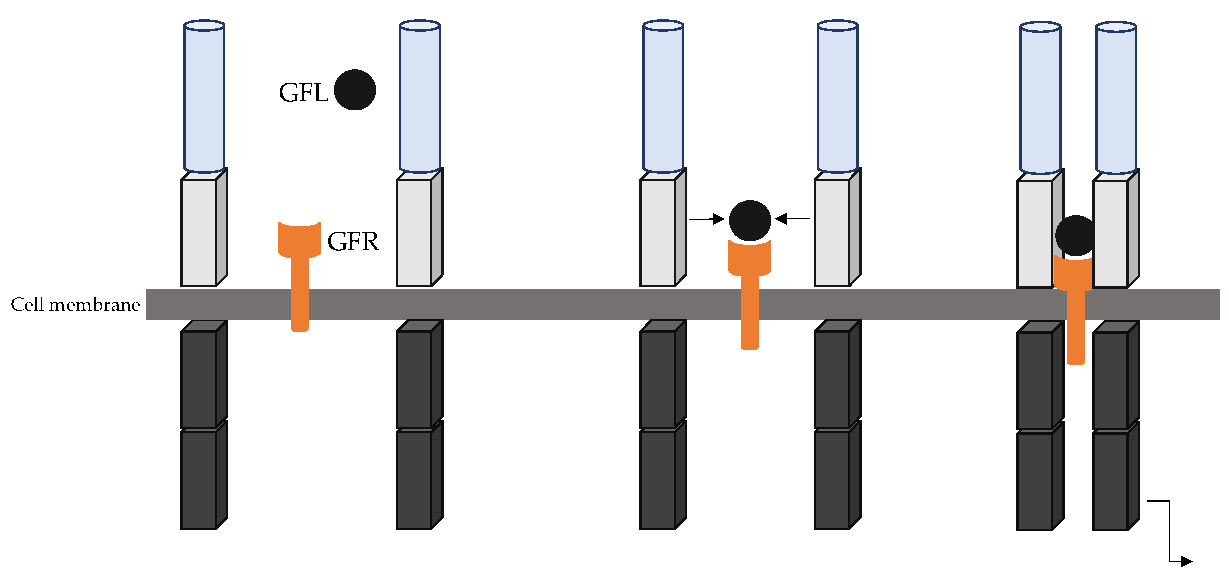
RET is mainly expressed in cells derived from the neural crest, including thyroid C-cells, chromaffin cells of the adrenal medulla, and the enteric autonomic plexus. RET is essential for various biological functions including enteric nervous system development, kidney and lower urinary tract development, as well as spermatogenesis [11]. The RET receptor is activated through a complex formed by the glial cell line-derived neurotrophic factor (GDNF) family of ligand (GFL) (GDNF family, such as Neurturin, Artemin, and Persephin) and co-receptors. The GFR co-receptors are classically bound to the plasma membranes within lipid rich rafts, but also occur in a soluble form and both can then activate RET in two distinct forms of activation. Under normal conditions, RET ligand-dependent activation is mediated by Ca2+ ions binding to cadherin-like domains and the interaction of GFR co-receptors and their respective GFL ligand. The GFR ligand complex, together with the extracellular domain of RET leads to RET dimerization and autophosphorylation of the intracellular tyrosine residue (Figure 2) [12,13]. Once activated, RET initiates different intracellular downstream pathways involved in the regulation of crucial processes such as differentiation, survival, proliferation, and migration [11].
c.
RET oncogene
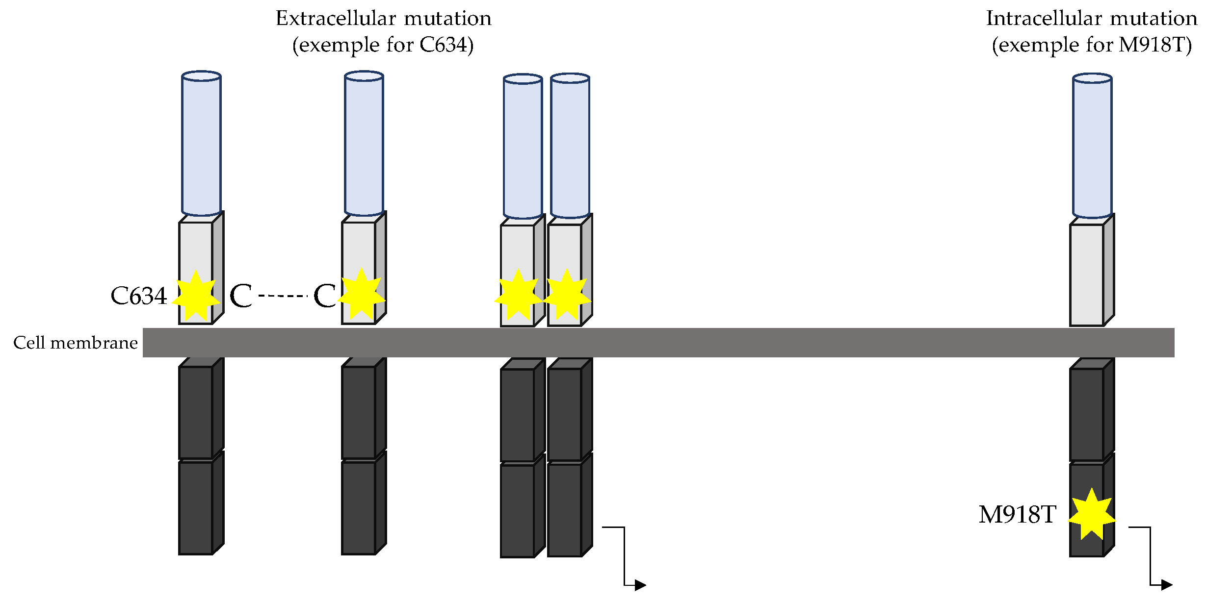
In MTC, several activating mutations can confer oncogenic properties to RET, located in both intra- and extra-cellular domains. The molecular mechanism by which RET activating mutations drive the neoplastic process was determined by Santoro and co-workers [14]. Under normal conditions, RET is only activated in the presence of GFR/GFL complex, which on binding to the RET receptor drives its dimerization and autophosphorylation of the intracellular signalling pathway. In vivo studies have highlighted that RET mutations C634R and M918T lead to constitutive phosphorylation of tyrosine, and their in vitro kinase activity was significantly higher than that of the wild-type protein [15]. Most mutations that occur in the extracellular domain are located in the cysteine-rich domain on exon 10 and exon 11. This domain contains cysteine residues, leading to formation of disulphide bonds necessary for the correct conformation of RET receptors. Mutations occurring in codons 609, 611, 618, 620, 630, and 634, which all code for a cysteine residue, lead to the misconformation of RET and allow self-dimerization without a ligand. Mutations affecting one of six cysteines located in the juxtamembrane domain of the RET receptor led to the replacement of the Cys residue with an alternate amino acid. Functional studies have shown that each of these mutations induced constitutive catalytic activity due to aberrant disulphide homodimerization of RET [16,17]. RET proteins with Cys 634 mutation were constitutively phosphorylated on tyrosine and their in vitro kinase activity was significantly higher than that of the WT protein [15]. Mutations in exon 11 (codon 630 and 634) lead to higher RET activation compared to mutations within exon 10 (codons 609, 611, 618, and 620) [16]. While extracellular mutations still required RET dimerization for signal transduction, intracellular mutations within exon 16 (RET M918T) did not [15,18,19] (Figure 3). Moreover, studies have highlighted that M918T is phosphorylated early and interacts with partners inducing downstream signalling before reaching the cell membrane [20].

4. Genetic Drivers of Hereditary and Sporadic MTC

  • Hereditary MTC
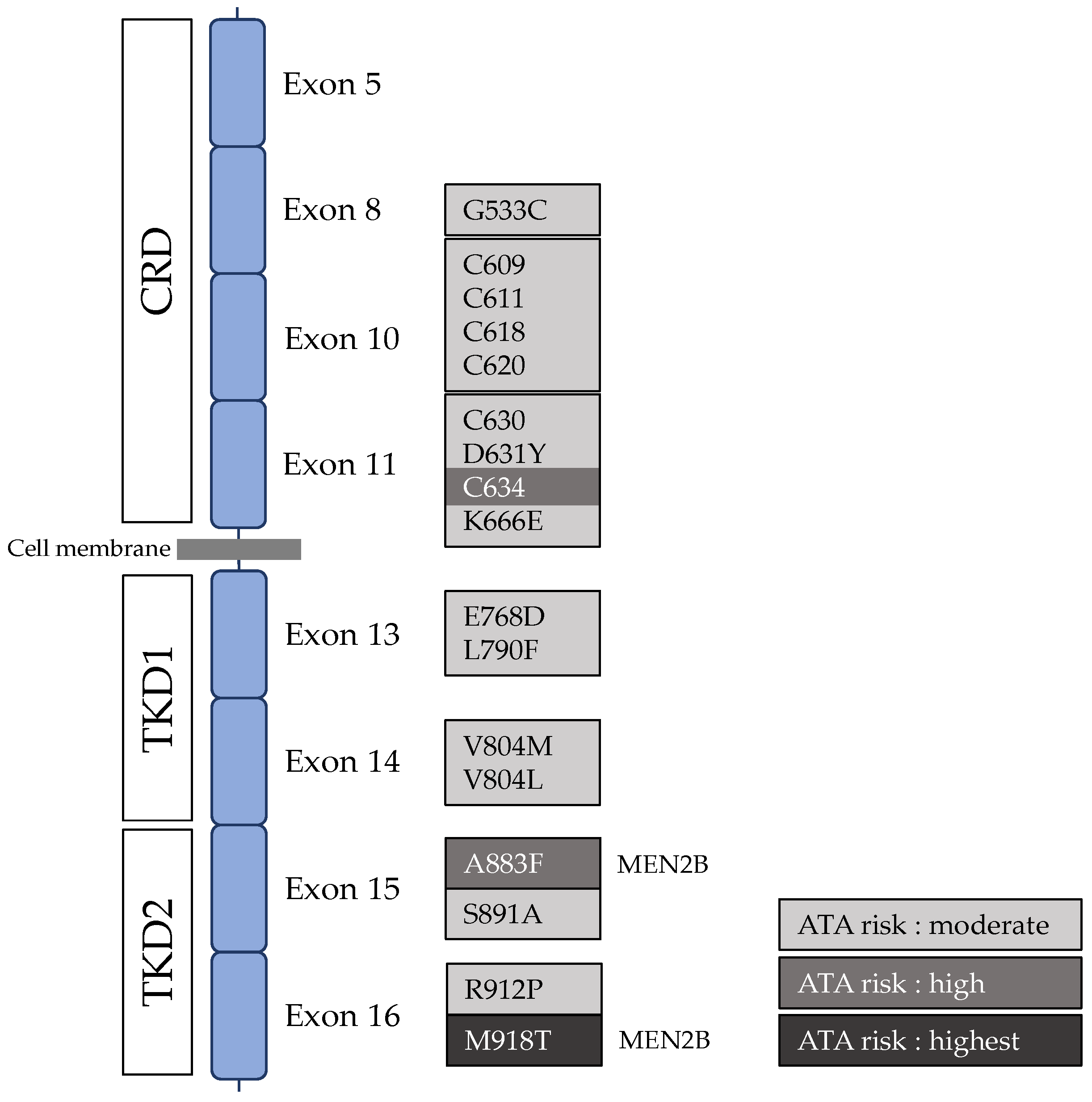
The concurrence of thyroid carcinoma and pheochromocytoma was first reported in 1961 by Sipple [21]. Later in 1968, Steiner et al. reported a family with an association of thyroid carcinoma, pheochromocytoma, hyperparathyroidism, and Cushing’s syndrome [22]. To describe this syndrome, and mirroring the term previously used by Wermer to characterise multiple adenomatosis of endocrine glands [23], the term multiple endocrine neoplasia type 2 (MEN2) was first coined. As previously described, the RET gene was identified as responsible for MEN2 in the late 1990s [24]. At first, a genotype–phenotype correlation was suggested [22]. The majority of MEN2A (OMIM #171400) results from missense mutations within the extracellular domain, particularly in the cysteine-rich domain and mainly involving codon 634 [4,24,25,26], whereas MEN2B (OMIM #162300) is almost exclusively caused by a specific exon 16 mutation (c.2753T > C, p.Met918Thr) [4,25,26,27]. Apart from these most commonly reported mutations, several other variants both in the extracellular [28,29,30,31,32,33] and intracellular domains [34,35,36,37,38] have been reported as being responsible for MEN2 (RET variants and exon localisation are summarized in Figure 4).
At the germline level, rare indel mutations can also be found to be responsible for MEN2-associated disease. Pathogenic mutations in the RET proto-oncogene have been successively reported since 2009 in an open access database [39] and are now available from the online ARUP MEN2 database [40].
MTC occurs in roughly 100% of MEN2 cases and may be more or less aggressive, depending on the RET mutation that leads to MEN2. RET mutations are thus classified into three risk grades (moderate, high, and highest risk), according to the risk of aggressive MTC [41]. This classification is currently the basis for international guidelines for MEN2-related MTC management proposed by the American Thyroid Association (ATA) [4]. Though the genotype–phenotype correlation is well-accepted, the theory of RET codon-related MTC aggressiveness has recently been questioned. In a large monocentric observational study involving 262 patients with moderate-risk (n = 127) and high-risk (n = 135) germline RET mutations, Voss et al. reported that both moderate- and high-risk patients had similar overall survival (OS), metastatic disease after MTC diagnosis, and clinical outcomes, suggesting that moderate- and high-risk RET mutations do not predict MTC aggressiveness [42]. In this study, lymph node invasion was frequently described in patients with moderate- and high-risk RET mutations (53/127, 42%, and 48/135, 36%, respectively), but not distant metastatic disease (10/127, 8%, and 7/135, 5%, respectively). Finally, this study demonstrates that metastatic disease is not more frequent in MEN2 patients with high-risk RET mutations, i.e., mutations in codon C634, than in MEN2 patients with moderate-risk RET variants. Thus, the authors suggested that RET mutation would only define the age at disease onset rather than the aggressiveness of MTC [42]. However, this study was limited by differences in the duration of follow-up between the two groups, as patients with a moderate-risk RET variant had a shorter follow-up compared to patients with a high-risk variant (median follow-up 6.5 vs. 11.5 years, respectively). Moreover, age at last follow-up was lower in patients with high-risk variants compared to patients with moderate risk (34.5 vs. 48.5 years, respectively) [43]. Two other independent studies have published similar findings. In a cohort comparing 122 moderate-risk and 120 high-risk MTC, Raue et al. reported that both had the same disease stage at the time of diagnosis (in particular, five patients in both groups (4%) exhibited stage IV disease at onset). At the last follow-up, both moderate-risk and high-risk groups showed the same remission rate (56% vs. 61%), the same rate of metastatic disease (14% vs. 13%), and the same disease-specific death (7% vs. 7%). Interestingly, while high-risk patients were younger at the time of diagnosis of stage III disease compared to moderate-risk patients (30.4 ± 13.0 years vs. 41.7 ± 15.3 years), the mean interval from stage I to III did not differ (12.7 years vs. 10.1 years). These results support the previously proposed hypothesis that MEN2A RET pathogenic variants can determine the age of onset of MTC, but that MTC is an equally aggressive tumour in both moderate- and high-risk groups after its initial onset. However, the age at surgery and the duration of follow-up differed between moderate- and high-risk groups, as being younger (23.0 ± 15.7 years vs. 35.3 years) and longer (15.8 ± 9.5 years vs. 10.8 ± 9.0 years), respectively. Moreover, successive therapeutic lines employed in the patients were not available and possibly varied between the two groups [44]. Machens et al. investigated the progression from node-negative to node-positive MTC according to the RET risk category. Considering 387 patients, 201 with node-negative and 186 with node-positive MTC, the author showed that tumour progression to node metastasis was similar in both groups, taking 8.6 to 9.1 years in the moderate-risk group and 13.6 years in the high-risk MTC patients [45]. However, this study compared the age at thyroidectomy between the groups and not the time between thyroidectomy and node progression in the same patient. Other authors have reported atypical cases of late onset MEN2-related tumours despite carrying the high-risk RET variant C634 [46,47], highlighting that other factors might modify the natural history of MEN2-related MTC.
b.
Sporadic MTC
Sporadic MTC is defined as MTC in patients without the germline RET mutation, other MEN2-related manifestations, such as pheochromocytoma and hyperparathyroidism, or familial history suggestive of MEN2.
  • Somatic RET mutations are a major factor in sporadic MTC tumourigenesis.
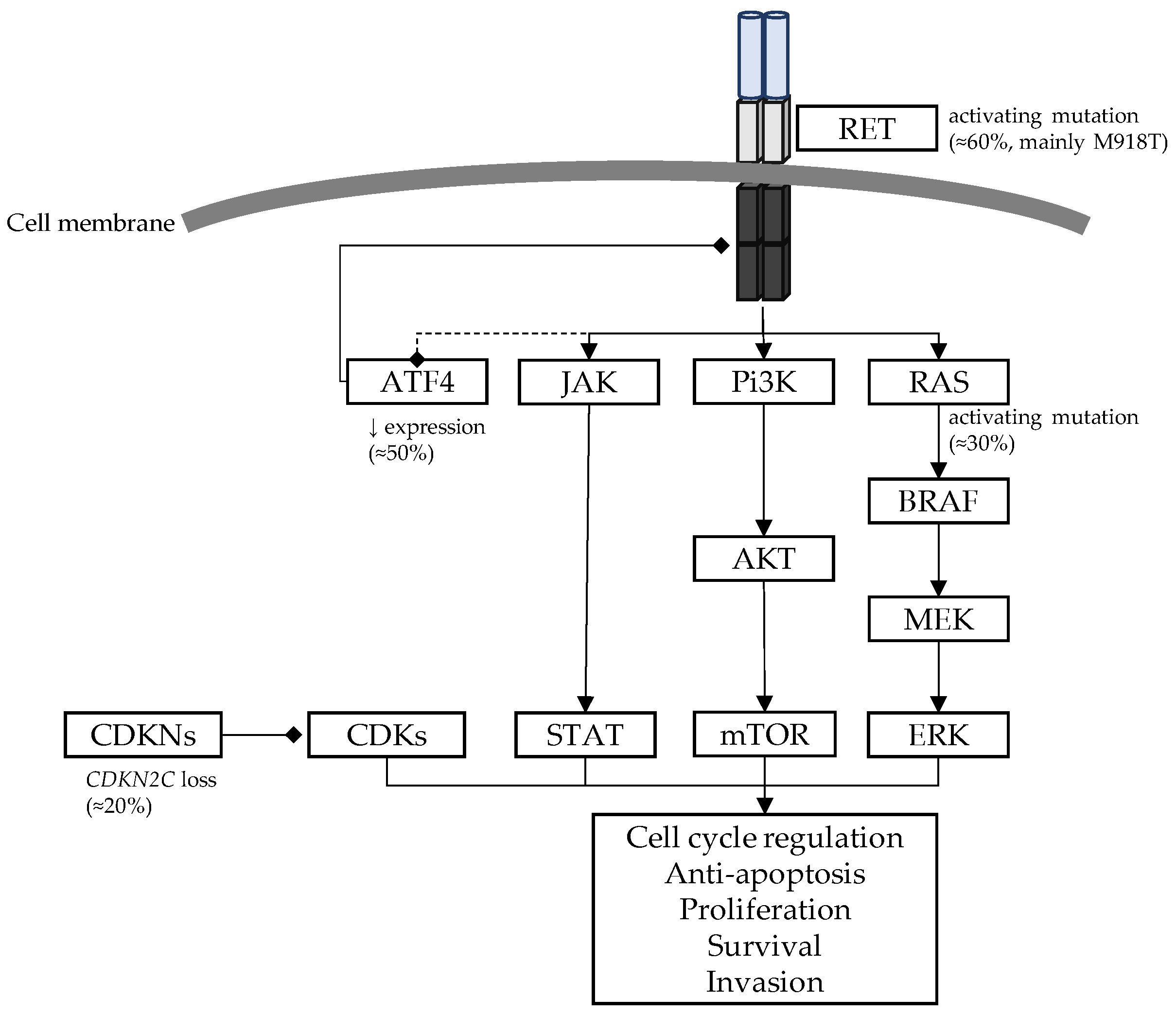
In the late 1990s, Marsh et al. showed that two thirds of sporadic MTCs exhibited a somatic mutation at codon 918 (M918T) [48]. These data were recently confirmed by NGS-targeted sequencing in a large cohort of 148 sporadic MTCs. Apart from codon 918 (RET M918T) [6,48,49], somatic events may occur in codon 634 [49,50] and other MEN2-related variants [48,51,52]. Other kinds of mutation, such as indels involving critical cysteine residues, may lead to RET oncogenic activation that is responsible for MTC (Table 1). Somatic RET mutations, particularly RET M918T, are related to the MTC phenotype. RET M918T mutated MTCs present a more advanced stage at diagnosis and have worse outcomes [6,49,53]. The presence of RET mutations, both together and when considering M918T alone, correlates to an advanced stage of the disease (p = 0.0025), a higher tumour (T) category (p < 0.0001), and the presence of both lymph-node (N) (p = 0.0021) and distant metastases (M) (p = 0.0073) [54]. In a large meta-analysis including 964 sporadic MTCs from 23 studies, the sporadic MTCs with somatic RET mutation were associated with a younger age at diagnosis, a higher proportion of pT3/T4 (OR = 2.31, 95%CI [1.55–3.45], I2 = 0%), with an increased risk of lymph node metastasis (OR = 3.61, 95%CI [2.33–5.60, I2 = 0%) and with a higher propensity for distant metastasis (OR = 2.85, 95%CI [1.64–4.94], I2 = 0%) compared to MTCs with wild-type RET [55]. Finally, in sporadic MTCs, RET mutation was found to be an indicator of poor prognosis, with an increased risk of both tumour recurrence (OR = 3.01, 95%CI [1.65–5.48], I2 = 41%) and patient death (OR = 2.43, 95%CI [1.06–5.57], I2 = 33%) [55]. Romei et al. reported that the majority of sporadic MTCs that were advanced and metastatic carried a RET mutation, mainly M918T (93.8%) [56]. The prevalence of somatic RET M918T mutation frequency increases with tumour size, from 11.3% in microMTCs to 58.8% in MTCs larger than 30 mm [57]. Even though the role of missense mutations is well-known, the role of other molecular alterations remains uncertain. A recent study revealed that RET indels are not uncommon, occurring in up to 17.4% of MTCs, and that these correlate with aggressive disease [58].
RET copy number alterations (CNAs) are found in 13 to 30% of hereditary and sporadic MTCs [66,67]. The presence of RET mutation at both the germline or somatic level was more frequent in tumours with RET CNAs compared to those without (83.3% vs. 42.5%, p = 0.003) [68]. Another study showed that RET amplifications only affect tumours carrying mutations, not playing a role in tumourigenesis but potentiating the transforming activity of the RET oncogene [69]. Furthermore, the prevalence of RET CNAs was significantly higher in advanced stages of the disease compared with lower stages (66.7% vs. 33.3%, p = 0.003) (p = 0.003) and was associated with poorer outcomes (p = 0.005) [68].
Taken together, these data highlight that somatic RET mutations appear to be the main oncogenic driver in sporadic MTCs, with RET M918T being the most common variant and being correlated with MTC aggressiveness. However, in addition to RET somatic mutations and CNAs, other oncogenes commonly involved in cancer pathogenesis have also been investigated in sporadic MTCs.
  • RAS oncogene in sporadic MTC
Several studies have shown that the human RAS gene family plays a crucial role in the development of MTCs. The RAS oncogene is the second main oncogenic driver after RET in sporadic MTCs. The three RAS oncogenes encode three 21 KDa monomeric distinct proteins (H-RAS, N-RAS, K-RAS), which act as GTPases that control key signal transduction cascades including the Raf/MEK/ERK and PI3Kinase/Akt pathways [70,71]. Activating mutations in specific hotspots on the RAS genes are found in about 30% of all human cancers. In thyroid neoplasia, RAS gene point mutations, mainly in NRAS, are detected in benign and malignant tumours that arise from the follicular epithelium. Through a large exome study, Agrawal et al. performed tumoural whole exome sequencing on a total of 57 MTCs (36 sporadic) and reported that overall 91% of all tumours had RET (75%), HRAS (12%), or KRAS (4%) mutations [72]. While somatic RAS mutations were found in 10 to 70% of RET-negative sporadic MTCs, the association of both RET and RAS somatic mutations is rare [73,74,75]. Moreover, no RAS mutations were found in hereditary MTCs [76], suggesting that activation of RAS and RET genes are mutually exclusive and represent alternative genetic events in sporadic MTC tumourigenesis. Most RAS mutations correspond to mutation hot spots in exons 2 and 3, but a smaller proportion of them were detected in exon 4. Compared to WT RAS sporadic MTCs, patients with RAS-mutant MTCs showed no significant differences between the age at diagnosis, tumour size, lymph node invasion, and distant metastasis. Though rarely reported in studies, the presence of RAS mutations in MTCs was not an indicator for tumour relapse (OR = 0.57, 95%IC [0.23–1.42], I2 = 0%) [55]. Thus, patients harbouring a sporadic MTC carrying a RAS mutation appear to have a better prognosis than patients harbouring a RET mutation [54]. While RET and RAS oncogenes are present in about 50% to 90% of sporadic MTCs, most of the remaining cases appear to still lack a genetic driver [54,77].
  • CDKNs and tumour suppressor genes in sporadic MTCs
The members of the CDKN2 family (CDKN2A [p15], CDKN2B [p16], CDKN2C [p18], and CDKN2D [p19]) are cyclin dependant kinase inhibitors that block cell cycle progression by interacting with CDK4 or CDK6 to prevent activation of the Cyclin D–CDK4/6 complex. The loss of CDKNs induces increased and uncontrolled phosphorylation of retinoblastoma protein (RB) and unregulated progression through the S phase of the cell cycle. The loss of chromosome regions 1p32 and 19p13, encoding CDKNs, are relatively frequently found in sporadic MTCs (39% and 24%, respectively). Thus, several studies have investigated the putative role of the CDKN/RB pathway in MTC. In a study including 62 sporadic MTCs, CDKN2C loss was observed in 19% (12/62) of cases, including six MTCs with a somatic RET M918T mutation. CDKN2C loss was associated with a higher stage MTC at diagnosis (p = 0.0009) and a worse outcome compared to both MTC with or without somatic RET M918T mutation and exhibited a lower median overall and disease specific survival. Interestingly, overall survival was lower in patients with a combination of both somatic RET M918T and somatic CDKN2C copy loss, suggesting a synergistic effect [78]. In a recent study involving 58 sporadic MTCs (40 with somatic RET mutation and 10 with somatic RAS mutation), CDKN2C loss correlated with shorter time to distant metastatic disease (1.59 years vs. 5.18 years, p = 0.03) while RET mutations did not reach significance (2.83 years vs. 6.60 years, p = 0.5) [79]. In vivo preclinical models have assessed the role of loss of CDKN in RET-mutant MTC. MEN2B (RET M918T) mice with Cdkn2c knock-out (Cdkn2c−/−) developed MTC at a greatly increased incidence compared to MEN2B (RET M918T) Cdkn2c+/− mice and with a higher proliferation rate, suggesting that Cdkn2c loss increases the risk of MTC and enhances MTC progression, in synergy with the RET oncogene [80]. Aside from CDKN2C copy loss, Van Veelen and colleagues identified somatic mutations in CDKN2C in four MTCs. In vitro analysis showed that the CKDN2C mutant is less stable than the wild type and its interaction with CDK4/6 is impaired, leading to reduced inhibition of cell growth [81].
Polymorphisms in CDKN1B and CDKN2A were found to be over- and under-represented, respectively, in 45 sporadic MTCs compared to 98 controls (62.2% vs. 40.2%, p = 0.038 and 15.6% vs. 32.6%, p = 0.0075, respectively) [82]. In this study, CDKN1B and CDKN2A polymorphisms were found to contribute 11% of the total risk of developing a sporadic MTC and, when combined with CDKN2C polymorphisms, to be associated with aggressiveness. Interestingly in this study, only patients with CDKN2B polymorphisms exhibited metastatic disease (36.3% vs. 0%, p = 0.0261) [82]. In non-hereditary tumours, WT CDKN2C displays a higher rate of lymph node metastasis compared to the homozygous variant genotype (83.8% vs. 40%, p = 0.03) [83]. CDKN1BWT/RETmut MTCs also show more aggressive behaviour, while the CDKN1B V109G polymorphism is associated with better outcomes after initial surgery compared to the WT allele [84].
Finally, a protective effect of CDKN1A SNP (p.Ser31Arg) was found in both hereditary (n = 77) and non-hereditary MTCs (n = 361) (OR = 0.53, 95%CI [0.36–0.78], p = 0.001) [85].
  • Other genetic abnormalities in sporadic MTC
c-MET (mesenchymal–epithelial transition factor) and its ligand, hepatocyte growth factor (HGF), belong to the MET family and act as a receptor tyrosine kinase that is expressed on the membrane of various epithelial cells. The HGF/c-MET axis, which can interact and cooperate with other types of tyrosine kinases, can stimulate various downstream signalling pathways in tumour cells, such as PI3K/AKT, JAK/STAT, Ras/MAPK, SRC, and Wnt/β-catenin [86]. The HGF/c-MET pathway is activated in most papillary thyroid cancers [87,88,89,90,91] and is correlated with PTC aggressiveness [91,92]. Activation of MET mutations, at both the germline and somatic levels, and gene amplification have been reported in several cancers [93,94], including thyroid neoplasms (in which the main mutation is MET T1010I in exon 14) [95]. Two studies have reported that MET T1010I variant of uncertain significance was identified in about 3 to 5% of sporadic MTCs [54,95]. Ciampi et al., in a study involving 148 sporadic MTCs, reported that five patients had a MET variant and two patients had no other identified driver of tumourigenesis [54]. Nevertheless, HGF and c-MET are expressed in most MTC cases, generally co-expressed [96], but do not show a clear correlation with clinical or pathological features [96]. In view of these data, MET mutation does not seem to play a crucial role in MTC tumourigenesis though MET gene amplification may act as an additional driver.
Activating transcription factor 4 (ATF4) is a crucial negative regulator of RET tyrosine kinase receptor in MTC. In mice, Atf4-targeted deletion has been shown to result in C-cell hyperplasia (CCH), Atf4 over-expression, and decreased MTC cell survival, and it inhibited the activation of the RET downstream pathway [97]. RET negatively regulates ATF4-mediated apoptosis [98]. ATF4 expression is decreased or lost in about half of human MTCs, compared with normal thyroid follicular cells [97] and inversely correlates with an increase in MTC stage [99]. Low ATF4 expression is associated with poor MTC outcomes [97] and has a significant negative impact on median survival when compared to high protein expression (p < 0.001). The combination of somatic RETM918T and low ATF4 protein levels further decreased overall survival [97,100] and was associated with increased 5-year recurrence, while higher ATF4 levels were significantly correlated to better 5-year survival [99].
The BRAF gene belongs to the RAF family of protein kinase that are key components of the MAPK signalling pathway, mediating cell growth, differentiation, and survival. While BRAF (mainly BRAF V600E) is a well-known oncogenic variant involved in various cancers, such as melanoma [101] and papillary thyroid cancer [102], in sporadic MTCs, BRAF V600E has rarely been reported [73,76,77,103,104,105,106] and appears to be a distant marginal driver.
In an independent series of 51 MTCs, Araujo et al. reported focal recurrent copy number gains involving the DLK1 (delta like non-canonical Notch ligand 1) and AIFM3 (apoptosis inducing factor mitochondria associated 3) genes. The functional relevance of CNA was also assessed by in silico analysis, and CNA status was found to be correlated with protein expression (DLK1, p = 0.01), tumour size (DLK1, p = 0.04), and tumour staging (AIFM3, p = 0.01 and DLK1, p = 0.05). These data provide a novel insight into MTC biology and suggest a common CNA landscape, regardless of whether the tumour is a sporadic or hereditary MTC [107].
Other mutations, such as AKT1 or CTNNB1, are unlikely to be mutation drivers in sporadic MTC [106].
The main oncogenic drivers involved in sporadic MTCs are summarized in Figure 5.

5. Modifiers of the Natural History of MTC

  • RET single nucleotide polymorphisms and MTC
In recent years, several RET single nucleotide polymorphisms have been suspected to be involved in RET-mutated MTCs, principally the G691S (exon11, c.2071G>A, p.Gly691Ser, rs1799939), L769L (exon 13, c.2307G>T, p.Leu769=, rs1800861), S836S (exon 14, c.2508C>T, pSer836=, rs1800862), and S904S (exon 15, 2712C>G, p.Ser904=, rs180086) alleles (allelic frequencies from gnomADv2.1 are listed in Table 2). The G691S and S904S RET polymorphisms are in linkage disequilibrium and co-segregate, so these results have been grouped together and referred to as the RET G691S/S904S haplotype [108,109,110].
Their effect on the development and progression of MTC is not totally convincing, and the explanation of their mechanism remains speculative. For example, RET mRNA levels in MTC specimens showed no difference between patients with and without RET G691S/S904S variants [108]. In silico analysis revealed that the RET G691S variant did not modify RET mRNA [111] but appears to introduce two new phosphorylation targets at Serine 686 and 691 and abolishes a phosphorylation site at Tyrosine 687, increasing activation of the RAS/ERK pathway [112]. Functional studies of the RET G691S variant showed that it does not exhibit transforming activity per se, but appeared to enhance the oncogenic properties of pathogenic RET mutations [113,114,115]. In transfection studies, the RET double mutant (G691S/S891A or G691S/K666E) activated the ERK1/2 pathway more than a single pathogenic mutant [114,115]. Other authors have suggested that the RET G691S polymorphism could have a cooperative action on RET dimerization in MEN2A, with the pathogenic RET mutation affecting the extracellular cysteine-rich domain and particularly codon 634 [114]. The clinical relevance of RET polymorphisms has been examined in several studies with inconsistent results.
  • RET G691S/S904S (rs1799939/rs1800863)
RET G691S polymorphism was shown to have a modifier effect on the age at onset of MTC in MEN2A families [114,115,116]. In a large Indian study, Mishra et al. identified that the RET S904S synonymous variant was over-represented in hereditary MTCs (53.2%) when compared to controls (41.7%) (OR = 2.82, 95%CI [1.64–4.86], p < 0.001) [85].
In sporadic MTCs, RET G691S polymorphism allele frequency was found to be higher compared to controls (27.83% vs. 18.86%, p = 0.029) [108,111] and associated with higher preoperative calcitonin levels (p < 0.001) and more frequent lymph node metastases (p < 0.05) compared to patients without RET G691S [117]; however, these data were not confirmed in other studies [112,118,119,120].
Finally, Lantieri et al. conducted a meta-analysis including 968 cases (among which 849 were sporadic MTCs) and 2115 healthy individuals. The allelic frequency of RET G691S polymorphism was 19.6% in the control subjects and 23.5% among MTC cases, and patients with RET G691S polymorphisms exhibited an increased risk of MTC compared to controls (OR = 1.22, 95%CI [1.06–1.39], p = 0.0049) [112].
  • RET L769L (rs1800861)
Several studies speculated that the synonymous RET L769L variant may be associated with an earlier age of MTC onset in MEN2 [121]. While some studies have suggested a putative role of the RET L769L variant with a higher risk of MTC and younger age of onset [121,122,123,124] as well as a higher risk of lymph node invasion [125], in contrast, a large case-control association study carried out on 585 sporadic MTCs, 1529 non-medullary thyroid cancers, and 989 healthy controls, Gemignani et al. reported that RET L769L allele frequency was lower in patients with MTC (OR = 0.70 95%CI [0.58–0.84], p = 1.9 × 10⁴) and rare homozygotes showed a lower OR = 0.32 (95%CI [0.17–0.60], p = 2.3 × 10⁴) [126]. Finally, Zhang et al. conducted a meta-analysis involving 1117 cases (among which a majority were sporadic MTCs) and 1916 controls from 13 studies and showed a similar frequency of L769L polymorphism in MTC patients and controls, suggesting no association between this variant and an elevated risk of MTC (OR = 1.06, 95%CI [0.94–1.19]) [127].
  • RET S836S (rs1800862)
Similarly, RET S836S polymorphism was found to be more frequent in MEN2A and sporadic MTC patients compared to controls and suspected to be associated with earlier onset and earlier lymph node and distant metastases [128,129,130]. These data were not confirmed by the meta-analysis of Zhang et al., which again found similar frequencies of S836S polymorphism in MTC patients and controls, suggesting no association between this variant and an elevated MTC risk (OR = 1.20, 95%CI [0.97–1.49]) [127].
  • Multiple RET polymorphisms
Interestingly, RET polymorphisms may have an additive effect [131]. Indeed, the RET L769L and S836S SNPs were associated with an increased risk of MTC, and individuals harbouring more than three polymorphic alleles exhibited a higher risk of MTC and worse MTC outcomes, suggesting an additive effect of these variants. Additionally, the latter had an increased risk of lymph node and distant metastases at diagnosis [131]. However, another study highlighted contrasting data and showed that in carriers and non-carriers of the RET variants (G691S, L767L, S836S, and S904S), sporadic MTCs were clinically and histologically indistinguishable [120].
  • Other RET polymorphisms
Apart from these variants in the coding sequence, some intronic polymorphisms have been suspected to be associated with a high risk of sporadic MTCs and more aggressive behaviour: IVS1-126G>T (intron 1, c.74-126G>T, rs2565206) [118,132], IVS1-126, and IVS14-24 (intron 14, c.2608-24G>A, rs2472737) [113]. In G533C MEN2A patients, Tamahana et al. showed that IVS14-126G>T was associated with a younger age at diagnosis and IVS8+82A>G (intron 8, c.1648+84G>A, rs3026750); 85–86 insC (intron 6, c.1648+88delC, rs754105711) was associated with the presence of lymph node metastases at diagnosis [133]. In silico analysis predicted that the IVS8+82A>G; 85–86 insC could alter splicing and create a site for NFAT transcription factor (nuclear factor activated T-cells), belonging to a protein family that has been found to be involved in cell cycle regulation, differentiation, survival, and invasion [133]. However, since no in vivo or in vitro experiments are so far available, these data remain inconclusive.
Cebrian and colleagues reported that STOP+388bp G>A (c.*388G>A, rs3026782) was associated with an increased risk of MTC while non intronic RET A45A (c.135A>G, p.Ala45=, rs1800858, gnomAD allelic frequency 73.6%) was associated with a protective effect [111], but these results were not replicated in another study [134].
Taken together, these data highlight that RET polymorphisms are potentially involved in MTC development, but to date only association studies are available and data concerning putative mechanisms remain elusive [135].
b.
Multiple mutations
Yang and colleagues reported that the likely benign RET V292M variant may modify the natural history of MTC in the RET C634Y family. All three carriers of the RET V292M variant were asymptomatic and did not display any manifestation of MTC despite being aged 58, 62, and 82 years. Among RET C634Y mutation carriers, patients with the RET V292M variant exhibited more extensive disease including lymph node invasion. However, RET C634Y belongs to the high-risk grade variants and is thus sufficient to explain this phenotype alone. In vitro studies showed that the RET V292M/C634Y double mutant had a faster rate of migration than either single RET C634Y mutant. Thus, the authors suggested that the RET V292M variant could modify the natural history of MEN2A-related MTC [136]. This mutation has been previously reported in a whole exome study performed by Qi et al. In this study, the authors showed that the combined RET mutation p.C634Y/V292M/R67H/R982C exhibited a more aggressive clinical phenotype than p.C364Y or p.V292M/R67H/R982C [136]. The RET variant V292M has already been reported in a 44-year-old man with unilateral MTC and unilateral pheochromocytoma, suggesting MEN2 syndrome [137]. However, the allele frequency of V292M is very common (gnomAD frequency 0.06% and up to 0.7% in East Asia) for an uncommon disease. Taken together, the RET V292M variant is still therefore considered as a variant of unknown significance (VUS) [40].
c.
Somatic mutation in MEN2-associated MTC
  • RET somatic alterations in MEN2-associated MTCs
While germline and sporadic RET mutations lead to MEN2 and sporadic MTC, respectively, both germline RET variants and additional somatic RET mutation, especially RET M918T, in the same patient have been only rarely reported. The impact of this mutation on the clinical course is unknown, and, in practice, a somatic event is rarely looked for in MEN2A patients. Marsh et al. reported, in 1996, cases of MEN2A patients harbouring a germline heterozygous RET variant and a second RET M918T mutation at the somatic level. Among the thyroid specimens from 16 MEN2A patients (15 MTCs and 1 HCC without MTC), they found four patients with a somatic RET M918T mutation: two had germline high-risk variants, C634R and C634S, and the other two had germline moderate-risk variants, C618R and 620R. Three of the four patients, the exception being a young patient with CCH at 11 years old, who was germline-tested through family genetic testing, had metastatic disease. In the 12 thyroid specimens without a somatic RET M918T mutation, no other somatic variants were found [138]. In the same year, Eng et al. reported a somatic RET M918T mutation in a MEN2 patient with a RET C634R germline mutation and with metastatic disease [139]. In these cases, however, the involvement of the M918T somatic variant in the metastatic phenotype could not be confirmed as patients with C634 variants frequently present with aggressive MTC [4]. Lombardo et al. described a 12-year-old girl exhibiting an extensive and invasive MTC despite carrying a germline moderate-risk 804 RET variant, leading to the identification of a somatic RET M918T mutation [140]. Our team also reported the case of a 35-year-old woman presenting with a particularly aggressive MTC with RET L790F germline mutation and in which a RET M918T mutation was found at the somatic level [141]. In 2001, Quadro et al. reported a somatic deletion of RET exons 4 to 16 in an 18-year-old woman harbouring a locally advanced MTC in which germline RET C634R was identified [142]. She died at the age of 27 years due to the progression of lung metastases. This event was located on the WT allele and was limited to the metastatic tumours. Even though the molecular effect of this structural RET alteration is unknown (loss or gain of function), it may explain the dramatic outcome in this patient.
Aside from somatic mutations, other somatic alterations may contribute to MTC tumourigenesis in MEN2. As RET acts as an oncogenic driver, RET copy number alterations have also been assessed in MEN2-related MTC. Koch et al. reported an allelic imbalance of the mutated and wild-type RET alleles in 6 of 19 samples and suggested that allelic imbalance could act as a “second activating hit” in RET mutant tumours [66]. Similar results were reported later [143,144]. Recently, Bim et al. reported somatic RET CNA in the form of retroposed copies in MTC samples and cells lines, suggesting that the generation of somatic RET retrocopies may have implications for MTC tumourigenesis and progression [145].
  • Non-RET mutations could modify MEN2-related MTC outcome
Recently, Mathiesen et al. reported the case of a young woman with the RET L790F variant presenting with MTC and lymph node metastases at 14 years of age. She was subsequently treated with surgery and cytotoxic chemotherapy. After a further increase in calcitonin more than ten years after the initial surgery, she presented with histologically-confirmed metastases in mediastinal lymph nodes and probable metastatic lung, mammary, liver, and abdominal lesions. No RET somatic mutation was identified, but a variant of FTL3 was identified. As FTL3 encodes a transcription factor involved in growth and differentiation, the authors suggested that this variant could have modified the natural history of MTC in this patient [146].
d.
Epigenetic alterations in MTC
Epigenetic control of gene expression plays a major role in tumourigenesis and progression in many cancers. In cells, gene expression is under the control of epigenetic regulation that includes DNA methylation, histone modifications, and expression of miRNAs. Epigenetic disturbances can deregulate gene expression and lead to tumourigenesis. The epigenetic landscape in MTCs remains poorly understood [147]. In colorectal cancer, patients with RET-hypermethylated tumours showed significantly worse overall survival [148]. On the opposite, RET hypomethylation was found in RET-WT MTCs compared to normal tissue [149]. Global methylation levels were reported to be higher in MTCs as compared to PTCs, but were not associated with clinical and/or oncological characteristics of the disease [150]. The expression of EZH2 and SMYD3 histone methyltransferase was found to be increased in an aggressive MTC [151]. Several studies have also shown dysregulation of miRNAs in MTC, including an increase in hsa-miR-375 expression. The hsa-miR-375 microRNA is suspected to be anti-proliferative and, in some cases, to have a pro-apoptotic action in some cancers. In vitro, hsa-miR-375 increased the sensitivity of Nthy-ori 3-1 cells to Vandetanib, a tyrosine kinase inhibitor (see below), and may be considered a biomarker of sensitivity [152].
e.
Parent-of-origin effects as phenotype modifiers in MEN2-related MTC
There is little information on the impact of parent-of-origin effects on the phenotypic expression of MEN. In the 1990s, Carlson et al. showed that de novo RET M918T mutation always occurred on the paternally-derived chromosome, suggesting a differential susceptibility of RET to mutation in parental-derived DNA, but showed at the same time that RET does not map to a known imprinted locus and revealed that both parental RET alleles are expressed in MEN2-related tumours [153]. Recently, in an observational study involving 405 patients carrying heterozygous MEN2A RET missense mutation, Machens et al. highlighted that offspring who inherited the trait from the father developed node metastases (p = 0.007) at a significantly younger age than offspring who inherited the trait from the mother [154], but the two groups were heterogeneous with regard to the number of C634 high-risk variant carriers. In another study involving 169 MEN2A relatives carrying C634 variants with information available on the gender of the parent who transmitted the familial RET variant, the same team reported similar results. Particularly in terms of MTC, they showed that offspring from affected fathers more often showed lymph node metastases (45 vs. 19%, p = 0.006) compared to offspring from affected mothers [155]. However, these data should be treated with caution due to the heterogeneity in long-term follow-up between the “paternal-affected” and “maternal-affected” groups [155].

6. Targeting RET in MTC: A New Therapeutic Paradigm

Historically, advanced progressive MTC had very poor outcomes due to the low response rate to available therapy. Since MTC is derived from C-cells, which do not take up iodine, treatment with radioactive iodine (131I) is pointless. Covalent linkage to meta-iodobenzylguanidine could potentially increase the uptake by some MTCs, however this does not offer much in terms of efficacy. Other systemic treatments, such as internal vectorised radiotherapy, using either bispecific antibodies targeting CEA associated with 131I or octreotide analogues labelled with yttrium 90, seem only to reduce progression, at the frequent cost of renal and haematological toxicity [156]. Cytotoxic chemotherapy, doxorubicin as a simple drug or in combination with cisplatin, has provided a response rate of between 0 and 20%, though the best response was only partial and median survival was low [156,157]. The association of 5-fluorouracil with dacarbazine seems to be the most effective and the least toxic combination, with a response rate of about 20% [156,157].
Thus, the emergence of tyrosine kinase inhibitors represented a game changer in the management of patients harbouring a progressive advanced MTC, particularly in RET-mutant MTCs.
  • Multikinase inhibitors
  • Vandetanib (ZD6474)
Vandetanib was the first treatment to obtain FDA approval in 2012 for the treatment of symptomatic or progressive MTC in patients with locally advanced or metastatic disease [158]. Vandetanib, a selective oral inhibitor of VEGFR, EGFR, and RET [159,160], has been reported to block the enzymatic activity of RET-derived oncoproteins and decrease the proliferation of a RET-mutant MTC cell line in vitro [161,162]. It was first assessed in patients with unresectable, locally advanced, or metastatic hereditary MTC, where Vandetanib led to either stable disease or a partial response in around 75% of cases. Further studies demonstrated that Vandetanib resulted in a disease control rate of 68% to 73%, with a safety profile consistent with the initial study. Specifically, treatment-associated adverse effects occurred in about 65% of cases, the most common being diarrhoea, rash, asthenia, and nausea [163,164]. Wells et al., in the phase III ZETA trial, reported that Vandetanib at a dose of 300 mg/d increased progression-free survival (PFS) to 30.5 months, compared to 19.3 months in the placebo arm, in patients with locally advanced or metastatic MTC [165]. Interestingly, a superior treatment-related response was observed in RET-positive MTCs, both hereditary and somatic. Somatic level RET M918T mutant MTCs exhibited a higher response rate [165]. Real-world data has since shown results consistent with the phase III trial, both in terms of efficacy and safety [166,167,168].
  • Cabozantinib (XL184)
Cabozantinib is a multikinase inhibitor targeting MET, VEGFR2, FLT3, c-KIT, and RET, which has exhibited robust antiangiogenic, antitumour, and anti-invasive effects in preclinical models [169] and clinical activity in patients with solid carcinomas. Early observations suggested that Cabozantinib was effective in MTCs and had an acceptable safety profile [170]. Further studies demonstrated that Cabozantinib at a daily dose of 140 mg increased both OS and PFS [171,172,173]. In the phase III EXAM trial, which evaluated locally or distant progressive MTC, Cabozantinib at a daily dose of 140 mg increased PFS by 11.2 months compared to 4.0 months in the placebo group. The 1-year PFS rate slightly increased from 7.2% on placebo to 47.3% with treatment [171]. In a phase III trial, tumour responses confirmed that Cabozantinib is clinically active in all MTC subgroups, regardless of the genotype, with ORRs ranging from 34% (RET M918T positive subgroup) to 20% (RET M918T negative subgroup), while no objective responses were noticed in patients who received the placebo [172,173]. Cabozantinib-associated adverse events included diarrhoea, palmar–plantar erythrodysesthesia, weight loss, anorexia, nausea, and fatigue which led to dose reduction and treatment discontinuation in 79% and 16% cases, respectively [171]. Though Cabozantinib appears to outperform Vandetanib, the results of phase III studies are not directly comparable since the ZETA trial also included patients without progression and permitted cross-over if progression occurred in the placebo arm, both of which were not permitted in the EXAM trial. Moreover, about 20% of patients in the EXAM trial were previously treated with TKI, including the half who were treated with Vandetanib.
In order to decrease treatment toxicity and related side effects leading to treatment discontinuation, a recent phase IV randomized non-inferiority trial was performed. Patients with progressive metastatic MTC were randomized to Cabozantib at 60 mg/day or 140 mg/d. Severe adverse events, dose reduction, and treatment discontinuation due to side effects were lower for the 60 mg/d group compared to 140 mg/d (63% vs. 72%; 69% vs. 81% and 23% vs. 36%). However, PFS non-inferiority of Cabozantinib at 60 mg/d vs. 140 mg/d was not met (median PFS 11.0 vs. 13.9 months, HR 1.24, p = 0.19) [174]. Koehler et al. retrospectively reported real-practice clinical outcomes from 48 patients with advanced MTCs who were treated with TKI. Used as a first-line treatment (n = 7), Cabozantinib led to stable disease in 43% of cases, with a PFS of 9 months. The 6- and 12-months survival rates were 100% and 100%, respectively. As a second-line treatment (n = 16), Cabozantinib achieved a partial response in 31%, with a PFS of 4 months and an OS of 19 months, with 6- and 12-month survival rates of 77% and 70%, respectively. Analysing first- and second-line treatments together, the median PFS was 4 months, and the 6- and 12- months OR rates were 78% and 70%, respectively. These data suggests that Cabozantinib performed less well than Vandetanib. However, only one patient received the recommended dose of 140 mg/d of Cabozantinib compared to over half the patients for Vandetanib [168].
b.
RET specific inhibitor
  • Pralsetinib (BLU-667)
Pralsetinib is a selective RET inhibitor approved in 2020 for the treatment of various RET-associated cancers, targeting both wild-type and kinase-activating RET mutations [175,176]. It has shown a potent and durable antitumour activity in RET-mutated MTCs in the ARROW trial. In this early phase I/II multicentric open-labelled study involving 122 patients with advanced RET-mutant MTCs, Pralsetinib at a dose of 400 mg/day led to an overall response rate of 60% in 55 previously treated patients and 71% in 21 treatment-naïve patients. Disease control rates were 93% and 100%, respectively. Response rates were consistent, with a response rate at 12 months of 92% and 84%, in the two patient groups, respectively. Common adverse effects, which included anaemia, neutropenia, musculoskeletal pain, constipation, increased aspartate amino-transferase, and hypertension, were often mild and led to treatment discontinuation in only 4% of cases. Unfortunately, one patient died, secondary to interstitial pneumonitis, and this was recognized as a treatment-related adverse event [177].
  • Selpercatinib (LOXO-292)
Selpercatinib received FDA approval in 2020 for RET-associated cancers [178] and may well become a cornerstone in the management of advanced RET-positive MTC. Selpercatinib, a highly selective RET-specific inhibitor, demonstrated potent activity in preclinical studies against human cancer cell lines harbouring endogenous RET gene alterations [179]. This compound was then evaluated in RET-associated human cancers in the LIBRETTO-001 trial. In this phase I/II trial, 143 participants harbouring a RET-mutant MTC (55 previously treated with a multikinase inhibitor) received a dose of 20 to 240 mg twice daily (phase 1) or 160 mg twice daily (phase 2). In treatment-naïve patients (n = 88), the objective response rate was 73% and the 1-year sustained objective response rate was 92%, slightly better than 69% and 82%, respectively, that was found in previously treated patients (n = 55). The most common grade 3 or higher adverse events were hypertension, liver enzyme increase, hyponatremia and diarrhoea. The drug-related discontinuation of the treatment remained low (2%) [180]. LIBRETTO-0531, a head-to-head phase III comparison study, is currently underway and will assess both the efficacy and the safety of Selpercatinib at a dose of 160 mg twice daily, compared to Cabozantinib (140 mg/d) or Vandetanib (300 mg/d) [181].
  • Zeteletinib (BOS172738)
The most recent compound, Zeteletinib, formerly BOS172738, is a next-generation inhibitor with nanomolar potency against RET and far higher selectivity against VEGF2. This molecule is currently in a phase 1 dose-escalation trial involving patients with advanced solid tumours with RET gene alteration, including MTC. The first results have shown that BOS172738 had a favourable safety profile in 67 patients with long-term administration, with the most frequent adverse effects classified as mild. The overall response rate was 44% in MTC (n = 16), including one complete response [182]. This trial is still underway and results should be available in the course of 2024 [183].
c.
TKI limitations
While these therapies have shown very promising results, recent case reports have suggested a risk of acquired resistance to these drugs, mainly driven by acquired somatic RET variants. Bruce et al. reported the case of a patient with metastatic sporadic MTC with a somatic RET M918T mutation, who was treated using several therapeutic lines. After Vandetanib and Cabozantinib, he was given Selpercatinib for 12 months and showed a partial response. After 12 months, progression occurred and he was then enrolled in a phase I/II Pralsetinib trial, but after 6 weeks showed rapid deterioration with worsening liver metastases observed on imaging, consistent with progressive disease. Genetic screening performed on a tumour biopsy specimen after Selpercatinib but before Pralsetinib revealed RET M918T, RET V804M, RET G810S mutations, and lastly a CDKN2A LOH in addition to the original RET mutation [184]. Subbiah et al. published a case report of a 49-year-old man presenting with metastatic sporadic RET M918T MTC, treated with several TKI lines, who developed RET variants leading to resistance. After consecutive therapy with Sorafenib, Vandetanib, Cabozantinib, and RXDX-105, the authors identified a RET V804M variant. The patient was treated with Selpercatinib with PR over 24 months before progression occurred. Genetic analysis performed on blood tumoural DNA identified a RET Y806C variant at 2 months, which was missing on subsequent analyses up to 29 months, at which time it was again present. At 22 and 24 months, RET G810C and G810S variants were identified, respectively [185].
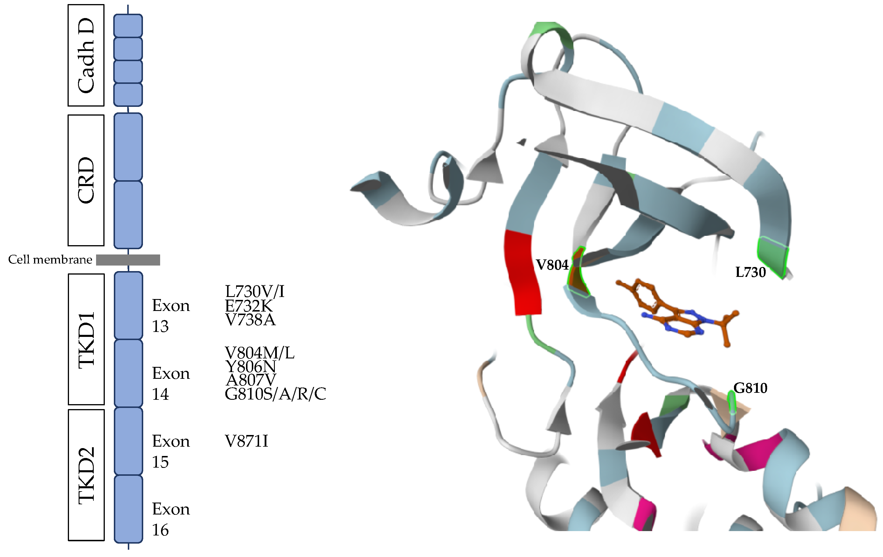
Most mutations involved in TKI resistance are located at the RET catalytic site. The structure of this site is as follows: the N-lobe is composed of beta sheets and an important alpha C helix, which forms the roof and is important for positioning and orientation of the ATP molecule in the catalytic site. An important motif on the N-lobe is the so-called glycine-rich loop that is involved in accommodating the nucleotide in the active site. The region connecting the N- and C-lobes is the hinge. Beyond its structural function, the hinge is involved in ATP binding through hydrogen bonds. This motif is not only important for binding and positioning of the nucleotide but also for the binding of tyrosine kinase inhibitors (TKIs) that compete with ATP. This residue, known as the gatekeeper residue, can restrict nucleotide access to the active site, which in the case of RET substitution by a bulkier residue (such as methionine or in some cases leucine) leads to steric hindrance and is associated with resistance to several TKIs, while retaining nucleotide accessibility [186]. The RET V804M variant has been reported in early studies as being potentially involved in tyrosine kinase inhibitor resistance. Vitagliano et al. reported that the RET V804M variant partially rescued MTC cell proliferation and the MAPK pathway in a MTC cell line harbouring RET C634W or M918T mutations treated with Vandetanib [162]. In vitro studies showed that both Cabozantinib and Vandetanib activities are impaired due to the RET V804M/L variant [187]. While RET-specific inhibitors do not seem to be significantly affected by the RET V804 variant in vitro, the RET G810 variant appears to markedly reduce Selpercatinib sensitivity and impair Pralsetinib action in RET M918T-positive MTC cell lines [185]. Being located at the solvent front, the G810 residue plays a crucial role in nucleotide binding, and replacement of glycine with a larger residue (such as serine, alanine, arginine, or cysteine) leads to steric hindrance and TKI resistance. Apart from the RET G810 variant, several other variants in the catalytic site appear to decrease RET inhibitor efficacy in vivo and in vitro, including RET L730, RET V738, RET Y806, and lastly RET A807 [185,187,188]. In vitro studies have shown that this kinase domain variant impaired the molecule interaction with the catalytic site and decreased RET non-specific and specific activity. RET variants leading to TKI resistance are outlined in Figure 6.
Other RET alterations, such as indels, have been reported as potential modifiers of the treatment response. In the large cohort presented by Elisei et al., two patients harbouring RET indels exhibited an excellent response to Selpercatinib [58]. On the other hand, another study reported contrasting data. Wijewardene et al. reported a 35-year-old man presenting with an aggressive metastatic MTC harbouring p.632_633del RET that was poorly responsive to Selpercatinib. Structural modelling revealed a paucity of disulphide bonds between C630 and C634 in p.632_633del RET sequences, while an intermolecular disulphide bond was formed between two C656 residues. In vitro experiments confirmed a reduction in the efficacy of Selpercatinib upon p.632_633del RET compared with wild-type RET control [65]. Better understanding and characterisation of RET variants leading to TKI resistance is important in order to provide patients with better personalized care, as shown in this report in which liquid biopsy and tissue NGS analyses allowed the author to identify six different RET mutations and one KRAS mutations, leading to modification of the treatment with another TKI and achieving disease control [189]. Possible factors predisposing to TKI resistance in both sporadic and hereditary MTCs have recently been reported in a study of a cohort of 54 metastatic MTCs with a median follow-up of 10.5 years. Interestingly, a more favourable disease course was found in patients who exhibited serious adverse events under treatment [190].
d.
Future perspectives and alternative therapies in MTC
Other drugs and systemic therapies which have shown promising preclinical efficacy are currently being evaluated in phase I/II trials. Among them, new-generation RET inhibitors, such as TPX-0046 and HA121-28, and multikinase drug repositioning with Regorafenib, Sorafenib, or even Sunitinib are being evaluated. As MTCs express the somatostatin receptor (SSTR), vectorized internal radiotherapy using 177Lu-SSTR analogue may represent a putative therapy in progressive metastatic tumour. Immune co-inhibitory molecules, such as programmed cell death protein-1 (PD-1/PD-L1) and cytotoxic T-lymphocyte antigen 4 (CTLA-4), are also expressed in MTC tumour cells, thus immune checkpoint inhibitors could also be a possible therapeutic option in progressive MTC. The small molecule ONC201 (imipridone), which inhibits both AKT and ERK, causes apoptotic cell death, decreased transcription of RET, VEGFR2, and IGFBP2, and increased transcription of ATF4 and ATF4 target genes. Since advanced MTCs show low ATF4 expression and ATF4 negatively regulates RET, this drug could be an alternative in RET inhibitor-resistant progressive MTC. These compounds and the results of the main studies on their use are summarized by Saltiki et al. [191].

7. Conclusions

Medullary thyroid carcinoma is a rare endocrine neoplasm, which occurs in both hereditary (25%) and sporadic (75%) forms. The RET proto-oncogene plays a central role in both forms. In hereditary MTC, a germline RET mutation leads to MTC development and determines its behaviour. Several putative factors have been suspected to modify the outcome of hereditary MTC, such as RET polymorphisms or RET somatic mutations. In sporadic MTC, RET acts as a tumourigenesis driver in roughly 50% of cases, mainly due to the RET M918T somatic mutation. This mutation correlates with MTC aggressiveness and the disease outcome. Regarding MTC therapy, targeting RET with non-specific or specific tyrosine kinase inhibitors has produced promising results, leading to FDA approval for four molecules. Follow-up studies will allow us to determine whether new somatic mutations and alternative pathway activation might decrease the efficacy of these drugs on a long-term basis. Aside from RET, other molecular markers such as RAS, CDKN, MET, or ATF4 could also play a key role in both MTC tumourigenesis and therapies. Further studies are now needed in order to improve our understanding of these second line mechanisms.

Author Contributions

N.S.: writing—original draft (lead), review & editing (equal); F.C.: writing—original draft (supporting), review & editing (lead); P.R.: writing—original draft (supporting), review & editing (equal). All authors have read and agreed to the published version of the manuscript.

Funding

Nicolas Sahakian received financial support from the French Endocrine Society (Société Française d’Endocrinologie, SFE). Frederic Castinetti and Pauline Romanet have no funding information to declare.

Institutional Review Board Statement

Not applicable.

Informed Consent Statement

Not applicable.

Data Availability Statement

No new data were created or analysed in this study. Data sharing is not applicable to this article.

Conflicts of Interest

The authors declare no conflict of interest.

References

  1. Cancer Today. Available online: https://gco.iarc.fr/today/home (accessed on 10 August 2023).
  2. Hazard, J.B.; Hawk, W.A.; Crile, G. Medullary (Solid) Carcinoma of the Thyroid—A Clinicopathologic Entity. J. Clin. Endocrinol. Metab. 1959, 19, 152–161. [Google Scholar] [CrossRef]
  3. Mathiesen, J.S.; Kroustrup, J.P.; Vestergaard, P.; Stochholm, K.; Poulsen, P.L.; Rasmussen, Å.K.; Feldt-Rasmussen, U.; Schytte, S.; Londero, S.C.; Pedersen, H.B.; et al. Incidence and Prevalence of Sporadic and Hereditary MTC in Denmark 1960–2014: A Nationwide Study. Endocr. Connect. 2018, 7, 829–839. [Google Scholar] [CrossRef]
  4. Wells, S.A.; Asa, S.L.; Dralle, H.; Elisei, R.; Evans, D.B.; Gagel, R.F.; Lee, N.; MacHens, A.; Moley, J.F.; Pacini, F.; et al. Revised American Thyroid Association Guidelines for the Management of Medullary Thyroid Carcinoma. Thyroid 2015, 25, 567–610. [Google Scholar] [CrossRef]
  5. Marsh, D.J.; Mulligan, L.M.; Eng, C. RET Proto-Oncogene Mutations in Multiple Endocrine Neoplasia Type 2 and Medullary Thyroid Carcinoma. Horm. Res. Paediatr. 1997, 47, 168–178. [Google Scholar] [CrossRef]
  6. Elisei, R.; Cosci, B.; Romei, C.; Bottici, V.; Renzini, G.; Molinaro, E.; Agate, L.; Vivaldi, A.; Faviana, P.; Basolo, F.; et al. Prognostic Significance of Somatic RET Oncogene Mutations in Sporadic Medullary Thyroid Cancer: A 10-Year Follow-up Study. J. Clin. Endocrinol. Metab. 2008, 93, 682–687. [Google Scholar] [CrossRef]
  7. Takahashi, M.; Ritz, J.; Cooper, G.M. Activation of a Novel Human Transforming Gene, Ret, by DNA Rearrangement. Cell 1985, 42, 581–588. [Google Scholar] [CrossRef] [PubMed]
  8. Takahashi, M.; Cooper, G.M. Ret Transforming Gene Encodes a Fusion Protein Homologous to Tyrosine Kinases. Mol. Cell. Biol. 1987, 7, 1378–1385. [Google Scholar] [CrossRef] [PubMed]
  9. Myers, S.M.; Eng, C.; Ponder, B.A.; Mulligan, L.M. Characterization of RET Proto-Oncogene 3′ Splicing Variants and Polyadenylation Sites: A Novel C-Terminus for RET. Oncogene 1995, 11, 2039–2045. [Google Scholar]
  10. Takahashi, M.; Asai, N.; Iwashita, T.; Isomura, T.; Miyazaki, K.; Matsuyama, M. Characterization of the RET Proto-Oncogene Products Expressed in Mouse L Cells. Oncogene 1993, 8, 2925–2929. [Google Scholar] [PubMed]
  11. Kawai, K.; Takahashi, M. Intracellular RET Signaling Pathways Activated by GDNF. Cell Tissue Res. 2020, 382, 113–123. [Google Scholar] [CrossRef]
  12. Jing, S.; Wen, D.; Yu, Y.; Holst, P.L.; Luo, Y.; Fang, M.; Tamir, R.; Antonio, L.; Hu, Z.; Cupples, R.; et al. GDNF-Induced Activation of the RET Protein Tyrosine Kinase Is Mediated by GDNFR-Alpha, a Novel Receptor for GDNF. Cell 1996, 85, 1113–1124. [Google Scholar] [CrossRef] [PubMed]
  13. Kjær, S.; Ibáñez, C.F. Identification of a Surface for Binding to the GDNF-GFR Alpha 1 Complex in the First Cadherin-like Domain of RET. J. Biol. Chem. 2003, 278, 47898–47904. [Google Scholar] [CrossRef] [PubMed]
  14. Santoro, M.; Carlomagno, F.; Romano, A.; Bottaro, D.P.; Dathan, N.A.; Grieco, M.; Fusco, A.; Vecchio, G.; Maťoškova, B.; Kraus, M.H.; et al. Activation of RET as a Dominant Transforming Gene by Germline Mutations of MEN2A and MEN2B. Science 1995, 267, 381–383. [Google Scholar] [CrossRef] [PubMed]
  15. Borrello, M.G.; Smith, D.P.; Pasini, B.; Bongarzone, I.; Greco, A.; Lorenzo, M.J.; Arighi, E.; Miranda, C.; Eng, C.; Alberti, L. RET Activation by Germline MEN2A and MEN2B Mutations. Oncogene 1995, 11, 2419–2427. [Google Scholar]
  16. Chappuis-Flament, S.; Pasini, A.; De Vita, G.; Ségouffin-Cariou, C.; Fusco, A.; Attié, T.; Lenoir, G.M.; Santoro, M.; Billaud, M. Dual Effect on the RET Receptor of MEN 2 Mutations Affecting Specific Extracytoplasmic Cysteines. Oncogene 1998, 17, 2851–2861. [Google Scholar] [CrossRef]
  17. Rossel, M.; Pasini, A.; Chappuis, S.; Geneste, O.; Fournier, L.; Schuffenecker, I.; Takahashi, M.; Van Grunsven, L.A.; Urdiales, J.L.; Rudkin, B.B.; et al. Distinct Biological Properties of Two RET Isoforms Activated by MEN 2A and MEN 2B Mutations. Oncogene 1997, 14, 265–275. [Google Scholar] [CrossRef]
  18. Carlson, K.M.; Dou, S.; Chi, D.; Scavarda, N.; Toshima, K.; Jackson, C.E.; Wells, S.A.; Goodfellow, P.J.; Donis-Keller, H. Single Missense Mutation in the Tyrosine Kinase Catalytic Domain of the RET Protooncogene Is Associated with Multiple Endocrine Neoplasia Type 2B. Proc. Natl. Acad. Sci. USA 1994, 91, 1579–1583. [Google Scholar] [CrossRef]
  19. Gujral, T.S.; Singh, V.K.; Jia, Z.; Mulligan, L.M. Molecular Mechanisms of RET Receptor-Mediated Oncogenesis in Multiple Endocrine Neoplasia 2B. Cancer Res. 2006, 66, 10741–10749. [Google Scholar] [CrossRef]
  20. Runeberg-Roos, P.; Virtanen, H.; Saarma, M. RET(MEN 2B) Is Active in the Endoplasmic Reticulum before Reaching the Cell Surface. Oncogene 2007, 26, 7909–7915. [Google Scholar] [CrossRef] [PubMed]
  21. Sipple, J.H. The Association of Pheochromocytoma with Carcinoma of the Thyroid Gland. Am. J. Med. 1961, 31, 163–166. [Google Scholar] [CrossRef]
  22. Steiner, A.L.; Goodman, A.D.; Powers, S.R. Study of a Kindred with Pheochromocytoma, Medullary Thyroid Carcinoma, Hyperparathyroidism and Cushing’s Disease: Multiple Endocrine Neoplasia, Type 2. Medicine 1968, 47, 371–409. [Google Scholar] [CrossRef]
  23. Wermer, P. Genetic Aspects of Adenomatosis of Endocrine Glands. Am. J. Med. 1954, 16, 363–371. [Google Scholar] [CrossRef]
  24. Mulligan, L.M.; Kwok, J.B.J.; Healey, C.S.; Elsdon, M.J.; Eng, C.; Gardner, E.; Love, D.R.; Mole, S.E.; Moore, J.K.; Papi, L.; et al. Germ-Line Mutations of the RET Proto-Oncogene in Multiple Endocrine Neoplasia Type 2A. Nature 1993, 363, 458–460. [Google Scholar] [CrossRef]
  25. Mulligan, L.M.; Marsh, D.J.; Robinson, B.G.; Schuffenecker, I.; Zedenius, J.; Lips, C.J.M.; Gagel, R.F.; Takai, S.I.; Noll, W.W.; Fink, M.; et al. Genotype-Phenotype Correlation in Multiple Endocrine Neoplasia Type 2: Report of the International RET Mutation Consortium. J. Intern. Med. 1995, 238, 343–346. [Google Scholar] [CrossRef] [PubMed]
  26. Eng, C.; Clayton, D.; Schuffenecker, I.; Lenoir, G.; Cote, G.; Gagel, R.F.; Amstel, H.K.P.v.; Lips, C.J.M.; Nishisho, I.; Takai, S.-I.; et al. The Relationship Between Specific RET Proto-Oncogene Mutations and Disease Phenotype in Multiple Endocrine Neoplasia Type 2: International RET Mutation Consortium Analysis. JAMA 1996, 276, 1575–1579. [Google Scholar] [CrossRef] [PubMed]
  27. Hofstra, R.M.W.; Landsvater, R.M.; Ceccherini, I.; Stulp, R.P.; Stelwagen, T.; Luo, Y.; Pasini, B.; Hoppener, J.W.M.; Van Amstel, H.K.P.; Romeo, G.; et al. A Mutation in the RET Proto-Oncogene Associated with Multiple Endocrine Neoplasia Type 2B and Sporadic Medullary Thyroid Carcinoma. Nature 1994, 367, 375–376. [Google Scholar] [CrossRef] [PubMed]
  28. Donis-Keller, H.; Dou, S.; Chi, D.; Carlson, K.M.; Toshima, K.; Lairmore, T.C.; Howe, J.R.; Moley, J.F.; Goodfellow, P.; Wells, S.A. Mutations in the RET Proto-Oncogene Are Associated with MEN 2A and FMTC. Hum. Mol. Genet. 1993, 2, 851–856. [Google Scholar] [CrossRef]
  29. Qi, X.P.; Jin, B.Y.; Li, P.F.; Wang, S.; Zhao, Y.H.; Cao, Z.L.; Yu, X.H.; Cheng, J.; Fang, X.D.; Zhao, J.Q. RET S409Y Germline Mutation and Associated Medullary Thyroid Carcinoma. Thyroid 2019, 29, 1447–1456. [Google Scholar] [CrossRef]
  30. Álvares Da Silva, A.M.; Maciel, R.M.B.; Dias Da Silva, M.R.; Toledo, S.R.C.; De Carvalho, M.B.; Cerutti, J.M. A Novel Germ-Line Point Mutation in RET Exon 8 (Gly(533)Cys) in a Large Kindred with Familial Medullary Thyroid Carcinoma. J. Clin. Endocrinol. Metab. 2003, 88, 5438–5443. [Google Scholar] [CrossRef]
  31. Ahmed, S.A.; Snow-Bailey, K.; Highsmith, W.E.; Sun, W.; Fenwick, R.G.; Mao, R. Nine Novel Germline Gene Variants in the RET Proto-Oncogene Identified in Twelve Unrelated Cases. J. Mol. Diagn. 2005, 7, 283–288. [Google Scholar] [CrossRef]
  32. Bae, S.J.; Kim, D.J.; Kim, J.Y.; Park, S.Y.; Choi, S.H.; Song, Y.D.; Ki, C.S.; Chung, J.H. A Rare Extracellular D631Y Germline Mutation of the RET Proto-Oncogene in Two Korean Families with Multiple Endocrine Neoplasia 2A. Thyroid 2006, 16, 609–614. [Google Scholar] [CrossRef]
  33. Peppa, M.; Boutati, E.; Kamakari, S.; Pikounis, V.; Peros, G.; Panayiotides, I.G.; Economopoulos, T.; Raptis, S.A.; Hadjidakis, D. Multiple Endocrine Neoplasia Type 2A in Two Families with the Familial Medullary Thyroid Carcinoma Associated G533C Mutation of the RET Proto-Oncogene. Eur. J. Endocrinol. 2008, 159, 767–771. [Google Scholar] [CrossRef] [PubMed]
  34. Eng, C.; Smith, D.P.; Mulligan, L.M.; Healey, C.S.; Zvelebil, M.J.; Stonehouse, T.J.; Ponder, M.A.; Jackson, C.E.; Waterfield, M.D.; Ponder, B.A. A Novel Point Mutation in the Tyrosine Kinase Domain of the RET Proto-Oncogene in Sporadic Medullary Thyroid Carcinoma and in a Family with FMTC—PubMed. Oncogene 1995, 10, 509–513. [Google Scholar]
  35. Fattoruso, O.; Quadro, L.; Libroia, A.; Verga, U.; Lupoli, G.; Cascone, E.; Colantuoni, V. A GTG to ATG Novel Point Mutation at Codon 804 in Exon 14 of the RET Proto-Oncogene in Two Families Affected by Familial Medullary Thyroid Carcinoma. Hum. Mutat. 1998, 11 (Suppl. S1), S167–S171. [Google Scholar] [CrossRef] [PubMed]
  36. Bolino, A.; Schuffenecker, I.; Luo, Y.; Seri, M.; Silengo, M.; Tocco, T.; Chabrier, G.; Houdent, C.; Murat, A.; Schlumberger, M.; et al. RET Mutations in Exons 13 and 14 of FMTC Patients. Oncogene 1995, 10, 2415–2419. [Google Scholar]
  37. Berndt, I.; Reuter, M.; Saller, B.; Frank-Raue, K.; Groth, P.; Grußendorf, M.; Raue, F.; Ritter, M.M.; Höppner, W. A New Hot Spot for Mutations in the RET Protooncogene Causing Familial Medullary Thyroid Carcinoma and Multiple Endocrine Neoplasia Type 2A. J. Clin. Endocrinol. Metab. 1998, 83, 770–774. [Google Scholar] [CrossRef]
  38. Smith, D.P.; Houghton, C.; Ponder, B.A.J. Germline Mutation of RET Codon 883 in Two Cases of de Novo MEN 2B. Oncogene 1997, 15, 1213–1217. [Google Scholar] [CrossRef] [PubMed]
  39. Margraf, R.L.; Crockett, D.K.; Krautscheid, P.M.F.; Seamons, R.; Calderon, F.R.O.; Wittwer, C.T.; Mao, R. Multiple Endocrine Neoplasia Type 2 RET Protooncogene Database: Repository of MEN2-Associated RET Sequence Variation and Reference for Genotype/Phenotype Correlations. Hum. Mutat. 2009, 30, 548–556. [Google Scholar] [CrossRef] [PubMed]
  40. ARUP Scientific Resource for Research and Education: MEN2 Database | University of Utah. Available online: https://arup.utah.edu/database/MEN2/MEN2_welcome.php (accessed on 7 June 2023).
  41. Frank-Raue, K.; Raue, F. Hereditary Medullary Thyroid Cancer Genotype-Phenotype Correlation. Recent Results Cancer Res. 2015, 204, 139–156. [Google Scholar] [CrossRef]
  42. Voss, R.K.; Feng, L.; Lee, J.E.; Perrier, N.D.; Graham, P.H.; Hyde, S.M.; Nieves-Munoz, F.; Cabanillas, M.E.; Waguespack, S.G.; Cote, G.J.; et al. Medullary Thyroid Carcinoma in MEN2A: ATA Moderate- or High-Risk RET Mutations Do Not Predict Disease Aggressiveness. J. Clin. Endocrinol. Metab. 2017, 102, 2807–2813. [Google Scholar] [CrossRef] [PubMed]
  43. Castinetti, F.; Whollk, N. Medullary Thyroid Carcinoma in MEN2A: ATA Moderate- or High-Risk RET Mutations Do Not Predict Disease Aggressiveness. J. Clin. Endocrinol. Metab. 2017, 102, 3557–3558. [Google Scholar] [CrossRef]
  44. Raue, F.; Bruckner, T.; Frank-Raue, K. Long-Term Outcomes and Aggressiveness of Hereditary Medullary Thyroid Carcinoma: 40 Years of Experience at One Center. J. Clin. Endocrinol. Metab. 2019, 104, 4264–4272. [Google Scholar] [CrossRef]
  45. Machens, A.; Lorenz, K.; Weber, F.; Dralle, H. Lymph Node Metastasis in Hereditary Medullary Thyroid Cancer Is Independent of the Underlying RET Germline Mutation. Eur. J. Surg. Oncol. 2021, 47, 920–923. [Google Scholar] [CrossRef] [PubMed]
  46. Walczyk, A.; Zgubieński, K.; Chmielewski, G.; Hińcza-Nowak, K.; Kowalik, A.; Jaskulski, J.; Kowalska, A. Late-Onset Medullary Thyroid Cancer in a Patient with a Germline RET Codon C634R Mutation. Diagnostics 2021, 11, 1448. [Google Scholar] [CrossRef] [PubMed]
  47. Zou, H.J.; Li, Y.S.; Shan, Z.Y. Genetic Analysis and Clinical Investigation of a Pedigree with Multiple Endocrine Neoplasia Type 2A: A Case Report. Oncol. Lett. 2016, 12, 2657–2659. [Google Scholar] [CrossRef]
  48. Marsh, D.J.; Learoyd, D.L.; Andrew, S.D.; Krishnan, L.; Pojer, R.; Richardson, A.L.; Delbridge, L.; Eng, C.; Robinson, B.G. Somatic Mutations in the RET Proto-Oncogene in Sporadic Medullary Thyroid Carcinoma. Clin. Endocrinol. 1996, 44, 249–257. [Google Scholar] [CrossRef]
  49. Romei, R.; Elisei, R.; Pinchera, A.; Ceccherini, I.; Molinaro, E.; Mancusi, F.; Martino, E.; Romeo, G.; Pacini, F. Somatic Mutations of the RET Protooncogene in Sporadic Medullary Thyroid Carcinoma Are Not Restricted to Exon 16 and Are Associated with Tumor Recurrence. J. Clin. Endocrinol. Metab. 1996, 81, 1619–1622. [Google Scholar] [CrossRef] [PubMed]
  50. Kimura, T.; Yokogoshi, Y.; Saito, S.; Yoshimoto, K. Mutations in the Cysteine-Rich Region of the RET Proto-Oncogene in Patients Diagnosed as Having Sporadic Medullary Thyroid Carcinoma. Endocr. J. 1995, 42, 517–525. [Google Scholar] [CrossRef]
  51. Dvorakova, S.; Vaclavikova, E.; Sykorova, V.; Vcelak, J.; Novak, Z.; Duskova, J.; Ryska, A.; Laco, J.; Cap, J.; Kodetova, D.; et al. Somatic Mutations in the RET Proto-Oncogene in Sporadic Medullary Thyroid Carcinomas. Mol. Cell. Endocrinol. 2008, 284, 21–27. [Google Scholar] [CrossRef]
  52. Dvorǎká, Š.; Václavíková, E.; Sýkorová, V.; Dušková, J.; Vlček, P.; Ryška, A.; Novák, Z.; Bendlová, B. New Multiple Somatic Mutations in the RET Proto-Oncogene Associated with a Sporadic Medullary Thyroid Carcinoma. Thyroid 2006, 16, 311–316. [Google Scholar] [CrossRef]
  53. Schilling, T.; Bürck, J.; Sinn, H.-P.; Clemens, A.; Otto, H.F.; Höppner, W.; Herfarth, C.; Ziegler, R.; Schwab, M.; Raue, F. Prognostic Value of Codon 918 (ATG→ACG) RET Proto-Oncogene Mutations in Sporadic Medullary Thyroid Carcinoma. Int. J. Cancer 2001, 95, 62–66. [Google Scholar] [CrossRef] [PubMed]
  54. Ciampi, R.; Romei, C.; Ramone, T.; Prete, A.; Tacito, A.; Cappagli, V.; Bottici, V.; Viola, D.; Torregrossa, L.; Ugolini, C.; et al. Genetic Landscape of Somatic Mutations in a Large Cohort of Sporadic Medullary Thyroid Carcinomas Studied by Next-Generation Targeted Sequencing. iScience 2019, 20, 324–336. [Google Scholar] [CrossRef]
  55. Vuong, H.G.; Odate, T.; Ngo, H.T.T.; Pham, T.Q.; Tran, T.T.K.; Mochizuki, K.; Nakazawa, T.; Katoh, R.; Kondo, T. Clinical Significance of RET and RAS Mutations in Sporadic Medullary Thyroid Carcinoma: A Meta-Analysis. Endocr. Relat. Cancer 2018, 25, 633–641. [Google Scholar] [CrossRef] [PubMed]
  56. Romei, C.; Casella, F.; Tacito, A.; Bottici, V.; Valerio, L.; Viola, D.; Cappagli, V.; Matrone, A.; Ciampi, R.; Piaggi, P.; et al. New Insights in the Molecular Signature of Advanced Medullary Thyroid Cancer: Evidence of a Bad Outcome of Cases with Double RET Mutations. J. Med. Genet. 2016, 53, 729–734. [Google Scholar] [CrossRef] [PubMed]
  57. Romei, C.; Ugolini, C.; Cosci, B.; Torregrossa, L.; Vivaldi, A.; Ciampi, R.; Tacito, A.; Basolo, F.; Materazzi, G.; Miccoli, P.; et al. Low Prevalence of the Somatic M918T RET Mutation in Micro-Medullary Thyroid Cancer. Thyroid 2012, 22, 476–481. [Google Scholar] [CrossRef] [PubMed]
  58. Elisei, R.; Ciampi, R.; Matrone, A.; Prete, A.; Gambale, C.; Ramone, T.; Simeakis, G.; Materazzi, G.; Torregrossa, L.; Ugolini, C.; et al. Somatic RET Indels in Sporadic Medullary Thyroid Cancer: Prevalence and Response to Selpercatinib. J. Clin. Endocrinol. Metab. 2022, 107, 2195–2202. [Google Scholar] [CrossRef]
  59. Jhiang, S.M.; Fithian, L.; Weghorst, C.M.; Clark, O.H.; Falko, J.M.; O’Dorisio, T.M.; Mazzaferri, E.L. RET Mutation Screening in MEN2 Patients and Discovery of a Novel Mutation in a Sporadic Medullary Thyroid Carcinoma. Thyroid 1996, 6, 115–121. [Google Scholar] [CrossRef]
  60. Alemi, M.; Lucas, S.D.; Sällström, J.F.; Akerström, G.; Wilander, E. A Novel Deletion in the RET Proto-Oncogene Found in Sporadic Medullary Thyroid Carcinoma. Anticancer Res. 1996, 16, 2619–2622. [Google Scholar]
  61. Alemi, M.; Lucas, S.D.; Sällström, J.F.; Bergholm, U.; Åkerström, G.; Wilander, E. A Complex Nine Base Pair Deletion in RET Exon 11 Common in Sporadic Medullary Thyroid Carcinoma. Oncogene 1997, 14, 2041–2045. [Google Scholar] [CrossRef]
  62. Ceccherini, I.; Pasini, B.; Pacini, F.; Gullo, M.; Bongarzone, I.; Romei, C.; Santamaria, G.; Matera, I.; Mondellini, P.; Scopsi, L.; et al. Somatic in Frame Deletions Not Involving Juxtamembranous Cysteine Residues Strongly Activate the RET Proto-Oncogene. Oncogene 1997, 14, 2609–2612. [Google Scholar] [CrossRef]
  63. Marsh, D.J.; Andrew, S.D.; Learoyd, D.L.; Pojer, R.; Eng, C.; Robinson, B.G. Deletion-Insertion Mutation Encompassing RET Codon 634 Is Associated with Medullary Thyroid Carcinoma. Hum. Mutat. 1998, 11 (Suppl. S1), S3–S4. [Google Scholar] [CrossRef] [PubMed]
  64. Kalinin, V.; Frilling, A. 27-Bp Deletion in the RET Proto-Oncogene as a Somatic Mutation Associated with Medullary Thyroid Carcinoma. J. Mol. Med. 1998, 76, 365–367. [Google Scholar] [CrossRef] [PubMed]
  65. Wijewardene, A.; Bastard, K.; Wang, B.; Gild, M.; Luxford, C.; Gill, A.; Robinson, B.; Bullock, M.; Clifton-Bligh, R. A Case Report of Poor Response to Selpercatinib in the Presence of a 632_633 RET Deletion. Thyroid 2023, 33, 119–125. [Google Scholar] [CrossRef]
  66. Koch, C.A.; Huang, S.C.; Moley, J.F.; Azumi, N.; Chrousos, G.P.; Gagel, R.F.; Zhuang, Z.; Pacak, K.; Vortmeyer, A.O. Allelic Imbalance of the Mutant and Wild-Type RET Allele in MEN 2A-Associated Medullary Thyroid Carcinoma. Oncogene 2001, 20, 7809–7811. [Google Scholar] [CrossRef] [PubMed]
  67. Ramone, T.; Mulè, C.; Ciampi, R.; Bottici, V.; Cappagli, V.; Prete, A.; Matrone, A.; Piaggi, P.; Torregrossa, L.; Basolo, F.; et al. RET Copy Number Alteration in Medullary Thyroid Cancer Is a Rare Event Correlated with RET Somatic Mutations and High Allelic Frequency. Genes 2020, 12, 35. [Google Scholar] [CrossRef]
  68. Ciampi, R.; Romei, C.; Cosci, B.; Vivaldi, A.; Bottici, V.; Renzini, G.; Ugolini, C.; Tacito, A.; Basolo, F.; Pinchera, A.; et al. Chromosome 10 and RET Gene Copy Number Alterations in Hereditary and Sporadic Medullary Thyroid Carcinoma. Mol. Cell. Endocrinol. 2012, 348, 176–182. [Google Scholar] [CrossRef] [PubMed]
  69. Mulè, C.; Ciampi, R.; Ramone, T.; Prete, A.; Matrone, A.; Cappagli, V.; Torregrossa, L.; Basolo, F.; Elisei, R.; Romei, C. Higher RET Gene Expression Levels Do Not Represent AnAlternative RET Activation Mechanism in Medullary Thyroid Carcinoma. Biomolecules 2021, 11, 1542. [Google Scholar] [CrossRef]
  70. Plowman, S.J.; Hancock, J.F. Ras Signaling from Plasma Membrane and Endomembrane Microdomains. Biochim. Biophys. Acta 2005, 1746, 274–283. [Google Scholar] [CrossRef]
  71. Lau, K.S.; Haigis, K.M. Non-Redundancy within the RAS Oncogene Family: Insights into Mutational Disparities in Cancer. Mol. Cells 2009, 28, 315–320. [Google Scholar] [CrossRef]
  72. Agrawal, N.; Jiao, Y.; Sausen, M.; Leary, R.; Bettegowda, C.; Roberts, N.J.; Bhan, S.; Ho, A.S.; Khan, Z.; Bishop, J.; et al. Exomic Sequencing of Medullary Thyroid Cancer Reveals Dominant and Mutually Exclusive Oncogenic Mutations in RET and RAS. J. Clin. Endocrinol. Metab. 2013, 98, E364–E369. [Google Scholar] [CrossRef] [PubMed]
  73. Ciampi, R.; Romei, C.; Pieruzzi, L.; Tacito, A.; Molinaro, E.; Agate, L.; Bottici, V.; Casella, F.; Ugolini, C.; Materazzi, G.; et al. Classical Point Mutations of RET, BRAF and RAS Oncogenes Are Not Shared in Papillary and Medullary Thyroid Cancer Occurring Simultaneously in the Same Gland. J. Endocrinol. Invest. 2017, 40, 55–62. [Google Scholar] [CrossRef]
  74. Moura, M.M.; Cavaco, B.M.; Pinto, A.E.; Leite, V. High Prevalence of RAS Mutations in RET-Negative Sporadic Medullary Thyroid Carcinomas. J. Clin. Endocrinol. Metab. 2011, 96, E863–E868. [Google Scholar] [CrossRef]
  75. Oczko-Wojciechowska, M.; Pfeifer, A.; Rusinek, D.; Pawlaczek, A.; Zebracka-Gala, J.; Kowalska, M.; Kowal, M.; Swierniak, M.; Krajewska, J.; Gawlik, T.; et al. The Prevalence of Somatic RAS Mutations in Medullary Thyroid Cancer—A Polish Population Study. Endokrynol. Pol. 2015, 66, 121–125. [Google Scholar] [CrossRef]
  76. Boichard, A.; Croux, L.; Al Ghuzlan, A.; Broutin, S.; Dupuy, C.; Leboulleux, S.; Schlumberger, M.; Bidart, J.M.; Lacroix, L. Somatic RAS Mutations Occur in a Large Proportion of Sporadic RET-Negative Medullary Thyroid Carcinomas and Extend to a Previously Unidentified Exon. J. Clin. Endocrinol. Metab. 2012, 97, E2031–E2035. [Google Scholar] [CrossRef]
  77. Shi, X.; Sun, Y.; Shen, C.; Zhang, Y.; Shi, R.; Zhang, F.; Liao, T.; Lv, G.; Zhu, Z.; Jiao, L.; et al. Integrated Proteogenomic Characterization of Medullary Thyroid Carcinoma. Cell Discov. 2022, 8, 120. [Google Scholar] [CrossRef]
  78. Grubbs, E.G.; Williams, M.D.; Scheet, P.; Vattathil, S.; Perrier, N.D.; Lee, J.E.; Gagel, R.F.; Hai, T.; Feng, L.; Cabanillas, M.E.; et al. Role of CDKN2C Copy Number in Sporadic Medullary Thyroid Carcinoma. Thyroid 2016, 26, 1553–1562. [Google Scholar] [CrossRef]
  79. Maxwell, J.E.; Gule-Monroe, M.K.; Subbiah, V.; Hu, M.; Perrier, N.D.; Cabanillas, M.E.; Lee, J.E.; Graham, P.H.; Cote, G.J.; Busaidy, N.L.; et al. Novel Use of a Clinical Laboratory Improvements Amendments (CLIA)-Certified Cyclin-Dependent Kinase N2C (CDKN2C) Loss Assay in Sporadic Medullary Thyroid Carcinoma. Surgery 2020, 167, 80–86. [Google Scholar] [CrossRef] [PubMed]
  80. Van Veelen, W.; Van Gasteren, C.J.R.; Acton, D.S.; Franklin, D.S.; Berger, R.; Lips, C.J.M.; Höppener, J.W.M. Synergistic Effect of Oncogenic RET and Loss of P18 on Medullary Thyroid Carcinoma Development. Cancer Res. 2008, 68, 1329–1337. [Google Scholar] [CrossRef]
  81. Van Veelen, W.; Klompmaker, R.; Gloerich, M.; Van Gasteren, C.J.R.; Kalkhoven, E.; Berger, R.; Lips, C.J.M.; Medema, R.H.; Höppener, J.W.M.; Acton, D.S. P18 Is a Tumor Suppressor Gene Involved in Human Medullary Thyroid Carcinoma and Pheochromocytoma Development. Int. J. Cancer 2009, 124, 339–345. [Google Scholar] [CrossRef]
  82. Barbieri, R.B.; Bufalo, N.E.; Secolin, R.; Assumpção, L.V.M.; Maciel, R.M.B.; Cerutti, J.M.; Ward, L.S. Polymorphisms of Cell Cycle Control Genes Influence the Development of Sporadic Medullary Thyroid Carcinoma. Eur. J. Endocrinol. 2014, 171, 761–767. [Google Scholar] [CrossRef]
  83. Mishra, V.; Kowtal, P.; Rane, P.; Sarin, R. Modulatory Role of Single Nucleotide Polymorphisms of Distinct Genetic Pathways on Clinical Behavior of Medullary Thyroid Carcinoma. Asian Pac. J. Cancer Prev. 2020, 21, 1289–1293. [Google Scholar] [CrossRef]
  84. Pasquali, D.; Circelli, L.; Faggiano, A.; Pancione, M.; Renzullo, A.; Elisei, R.; Romei, C.; Accardo, G.; Coppola, V.R.; De Palma, M.; et al. CDKN1B V109G Polymorphism a New Prognostic Factor in Sporadic Medullary Thyroid Carcinoma. Eur. J. Endocrinol. 2011, 164, 397–404. [Google Scholar] [CrossRef]
  85. Mishra, V.; Kowtal, P.; Rane, P.; Sarin, R. Genetic Risk Association of CDKN1A and RET Gene SNPs with Medullary Thyroid Carcinoma: Results from the Largest MTC Cohort and Meta-Analysis. Cancer Med. 2019, 8, 6151–6161. [Google Scholar] [CrossRef]
  86. Zhang, Y.; Xia, M.; Jin, K.; Wang, S.; Wei, H.; Fan, C.; Wu, Y.; Li, X.; Li, X.; Li, G.; et al. Function of the C-Met Receptor Tyrosine Kinase in Carcinogenesis and Associated Therapeutic Opportunities. Mol. Cancer 2018, 17, 45. [Google Scholar] [CrossRef]
  87. Trovato, M.; Villari, D.; Bartolone, L.; Spinella, S.; Simone, A.; Violi, M.A.; Trimarchi, F.; Batolo, D.; Benvenga, S. Expression of the Hepatocyte Growth Factor and C-Met in Normal Thyroid, Non-Neoplastic, and Neoplastic Nodules. Thyroid 1998, 8, 125–131. [Google Scholar] [CrossRef]
  88. Ruco, L.P.; Stoppacciaro, A.; Ballarini, F.; Prat, M.; Scarpino, S. Met Protein and Hepatocyte Growth Factor (HGF) in Papillary Carcinoma of the Thyroid: Evidence for a Pathogenetic Role in Tumourigenesis. J. Pathol. 2001, 194, 4–8. [Google Scholar] [CrossRef]
  89. Nardone, H.C.; Ziober, A.F.; LiVolsi, V.A.; Mandel, S.J.; Baloch, Z.W.; Weber, R.S.; Mick, R.; Ziober, B.L. C-Met Expression in Tall Cell Variant Papillary Carcinoma of the Thyroid. Cancer 2003, 98, 1386–1393. [Google Scholar] [CrossRef]
  90. Ruco, L.; Scarpino, S. The Pathogenetic Role of the HGF/c-Met System in Papillary Carcinoma of the Thyroid. Biomedicines 2014, 2, 263–274. [Google Scholar] [CrossRef]
  91. Pekova, B.; Sykorova, V.; Dvorakova, S.; Vaclavikova, E.; Moravcova, J.; Katra, R.; Astl, J.; Vlcek, P.; Kodetova, D.; Vcelak, J.; et al. RET, NTRK, ALK, BRAF, and MET Fusions in a Large Cohort of Pediatric Papillary Thyroid Carcinomas. Thyroid 2020, 30, 1771–1780. [Google Scholar] [CrossRef]
  92. Ramirez, R.; Hsu, D.; Patel, A.; Fenton, C.; Dinauer, C.; Tuttle, R.M.; Francis, G.L. Over-Expression of Hepatocyte Growth Factor/Scatter Factor (HGF/SF) and the HGF/SF Receptor (CMET) Are Associated with a High Risk of Metastasis and Recurrence for Children and Young Adults with Papillary Thyroid Carcinoma. Clin. Endocrinol. 2000, 53, 635–644. [Google Scholar] [CrossRef]
  93. Schmidt, L.; Duh, F.-M.; Chen, F.; Kishida, T.; Glenn, G.; Choyke, P.; Scherer, S.W.; Zhuang, Z.; Lubensky, I.; Dean, M.; et al. Germline and Somatic Mutations in the Tyrosine Kinase Domain of the MET Proto-Oncogene in Papillary Renal Carcinomas. Nat. Genet. 1997, 16, 68–73. [Google Scholar] [CrossRef]
  94. Ma, P.C.; Tretiakova, M.S.; MacKinnon, A.C.; Ramnath, N.; Johnson, C.; Dietrich, S.; Seiwert, T.; Christensen, J.G.; Jagadeeswaran, R.; Krausz, T.; et al. Expression and Mutational Analysis of MET in Human Solid Cancers. Genes Chromosomes Cancer 2008, 47, 1025–1037. [Google Scholar] [CrossRef] [PubMed]
  95. Wasenius, V.M.; Hemmer, S.; Karjalainen-Lindsberg, M.L.; Nupponen, N.N.; Franssila, K.; Joensuu, H. MET Receptor Tyrosine Kinase Sequence Alterations in Differentiated Thyroid Carcinoma. Am. J. Surg. Pathol. 2005, 29, 544–549. [Google Scholar] [CrossRef]
  96. Papotti, M.; Olivero, M.; Volante, M.; Negro, F.; Prat, M.; Comoglio, P.M.; Direnzo, M.F. Expression of Hepatocyte Growth Factor (HGF) and Its Receptor (MET) in Medullary Carcinoma of the Thyroid. Endocr. Pathol. 2000, 11, 19–30. [Google Scholar] [CrossRef]
  97. Bagheri-Yarmand, R.; Williams, M.D.; Grubbs, E.G.; Gagel, R.F. ATF4 Targets RET for Degradation and Is a Candidate Tumor Suppressor Gene in Medullary Thyroid Cancer. J. Clin. Endocrinol. Metab. 2017, 102, 933–941. [Google Scholar] [CrossRef] [PubMed]
  98. Bagheri-Yarmand, R.; Sinha, K.M.; Gururaj, A.E.; Ahmed, Z.; Rizvi, Y.Q.; Huang, S.C.; Ladbury, J.E.; Bogler, O.; Williams, M.D.; Cote, G.J.; et al. A Novel Dual Kinase Function of the RET Proto-Oncogene Negatively Regulates Activating Transcription Factor 4-Mediated Apoptosis. J. Biol. Chem. 2015, 290, 11749–11761. [Google Scholar] [CrossRef] [PubMed]
  99. Ma, S.; Wang, H.; Li, W.; Yan, Z.; Luo, X.; Lu, P. The Correlation between the Expression of ATF4 and Procalcitonin Combined with the Detection of RET Mutation and the Pathological Stage and Clinical Prognosis of Medullary Thyroid Carcinoma. Can. J. Physiol. Pharmacol. 2022, 100, 19–25. [Google Scholar] [CrossRef] [PubMed]
  100. Williams, M.D.; Ma, J.; Grubbs, E.G.; Gagel, R.F.; Bagheri-Yarmand, R. ATF4 Loss of Heterozygosity Is Associated with Poor Overall Survival in Medullary Thyroid Carcinoma. Am. J. Cancer Res. 2021, 11, 3227. [Google Scholar]
  101. Davies, H.; Bignell, G.R.; Cox, C.; Stephens, P.; Edkins, S.; Clegg, S.; Teague, J.; Woffendin, H.; Garnett, M.J.; Bottomley, W.; et al. Mutations of the BRAF Gene in Human Cancer. Nature 2002, 417, 949–954. [Google Scholar] [CrossRef]
  102. Ciampi, R.; Nikiforov, Y.E. Alterations of the BRAF Gene in Thyroid Tumors. Endocr. Pathol. 2005, 16, 163–171. [Google Scholar] [CrossRef]
  103. Robbins, R.J.; Thomas, J.S.; Osuna, P.M.; Shakil, J. A BRAF V600E Mutation in RET-Negative Medullary Thyroid Cancer. Case Rep. Endocrinol. 2020, 2020, 7641940. [Google Scholar] [CrossRef] [PubMed]
  104. Goutas, N.; Vlachodimitropoulous, D.; Bouka, M.; Lazaris, A.C.; Nasioulas, G.; Gazouli, M. BRAF and K-RAS Mutation in a Greek Papillary and Medullary Thyroid Carcinoma Cohort. Anticancer. Res. 2008, 28, 305–308. [Google Scholar]
  105. Fibbi, B.; Pinzani, P.; Salvianti, F.; Rossi, M.; Petrone, L.; De Feo, M.L.; Panconesi, R.; Vezzosi, V.; Bianchi, S.; Simontacchi, G.; et al. Synchronous Occurrence of Medullary and Papillary Carcinoma of the Thyroid in a Patient with Cutaneous Melanoma: Determination of BRAFV600E in Peripheral Blood and Tissues. Report of a Case and Review of the Literature. Endocr. Pathol. 2014, 25, 324–331. [Google Scholar] [CrossRef]
  106. Schulten, H.J.; Al-Maghrabi, J.; Al-Ghamdi, K.; Salama, S.; Al-muhayawi, S.; Chaudhary, A.; Hamour, O.; Abuzenadah, A.; Gari, M.; Al-Qahtani, M. Mutational Screening of RET, HRAS, KRAS, NRAS, BRAF, AKT1, and CTNNB1 in Medullary Thyroid Carcinoma. Anticancer Res. 2011, 31, 4179–4183. [Google Scholar]
  107. Araujo, A.N.; Camacho, C.P.; Mendes, T.B.; Lindsey, S.C.; Moraes, L.; Miyazawa, M.; Delcelo, R.; Pellegrino, R.; Mazzotti, D.R.; Maciel, R.M.d.B.; et al. Comprehensive Assessment of Copy Number Alterations Uncovers Recurrent AIFM3 and DLK1 Copy Gain in Medullary Thyroid Carcinoma. Cancers 2021, 13, 218. [Google Scholar] [CrossRef]
  108. Elisei, R.; Cosci, B.; Romei, C.; Bottici, V.; Sculli, M.; Lari, R.; Barale, R.; Pacini, F.; Pinchera, A. RET Exon 11 (G691S) Polymorphism Is Significantly More Frequent in Sporadic Medullary Thyroid Carcinoma than in the General Population. J. Clin. Endocrinol. Metab. 2004, 89, 3579–3584. [Google Scholar] [CrossRef] [PubMed]
  109. Robledo, M.; Gil, L.; Polla, M.; Cebria, A.; Rui, S.; Azan, M.; Benitez, J.; Mena, J.; Rojas, M. Polymorphisms G691S/S904S of RET as Genetic Modifiers of MEN 2A. Cancer Res. 2003, 63, 1814–1817. [Google Scholar]
  110. Sheikholeslami, S.; Yeganeh, M.Z.; Rad, L.H.; Ghadaksaz, H.G.; Hedayati, M. Haplotype Frequency of G691S/S904S in the RET Proto-Onco-Gene in Patients with Medullary Thyroid Carcinoma. Iran. J. Public Health 2014, 43, 235–240. [Google Scholar] [PubMed]
  111. Cebrian, A.; Lesueur, F.; Martin, S.; Leyland, J.; Ahmed, S.; Luccarini, C.; Smith, P.L.; Luben, R.; Whittaker, J.; Pharoah, P.D.; et al. Polymorphisms in the Initiators of RET (Rearranged during Transfection) Signaling Pathway and Susceptibility to Sporadic Medullary Thyroid Carcinoma. J. Clin. Endocrinol. Metab. 2005, 90, 6268–6274. [Google Scholar] [CrossRef] [PubMed]
  112. Lantieri, F.; Caroli, F.; Ceccherini, I.; Griseri, P. The Involvement of the RET Variant G691S in Medullary Thyroid Carcinoma Enlightened by a Meta-Analysis Study. Int. J. Cancer 2013, 132, 2808–2819. [Google Scholar] [CrossRef]
  113. Fugazzola, L.; Muzza, M.; Mian, C.; Cordella, D.; Barollo, S.; Alberti, L.; Cirello, V.; Dazzi, D.; Girelli, M.E.; Opocher, G.; et al. RET Genotypes in Sporadic Medullary Thyroid Cancer: Studies in a Large Italian Series. Clin. Endocrinol. 2008, 69, 418–425. [Google Scholar] [CrossRef]
  114. Borrello, M.G.; Aiello, A.; Peissel, B.; Rizzetti, M.G.; Mondellini, P.; Degl’Innocenti, D.; Catalano, V.; Gobbo, M.; Collini, P.; Bongarzone, I.; et al. Functional Characterization of the MTC-Associated Germline RET-K666E Mutation: Evidence of Oncogenic Potential Enhanced by the G691S Polymorphism. Endocr. Relat. Cancer 2011, 18, 519–527. [Google Scholar] [CrossRef] [PubMed]
  115. Colombo, C.; Minna, E.; Rizzetti, M.G.; Romeo, P.; Lecis, D.; Persani, L.; Mondellini, P.; Pierotti, M.A.; Greco, A.; Fugazzola, L.; et al. The Modifier Role of RET-G691S Polymorphism in Hereditary Medullary Thyroid Carcinoma: Functional Characterization and Expression/Penetrance Studies. Orphanet J. Rare Dis. 2015, 10, 25. [Google Scholar] [CrossRef] [PubMed]
  116. Shifrin, A.L.; Ogilvie, J.B.; Stang, M.T.; Fay, A.M.; Kuo, Y.H.; Matulewicz, T.; Xenachis, C.Z.; Vernick, J.J. Single Nucleotide Polymorphisms Act as Modifiers and Correlate with the Development of Medullary and Simultaneous Medullary/Papillary Thyroid Carcinomas in 2 Large, Non-Related Families with the RET V804M Proto-Oncogene Mutation. Surgery 2010, 148, 1274–1281. [Google Scholar] [CrossRef] [PubMed]
  117. Cardot-Bauters, C.; Leteurtre, E.; Leclerc, L.; Vantyghem, M.C.; Do Cao, C.; Wemeau, J.L.; D’Herbomez, M.; Carnaille, B.; Barbu, V.; Pinson, S.; et al. Does the RET Variant G691S Influence the Features of Sporadic Medullary Thyroid Carcinoma? Clin. Endocrinol. 2008, 69, 506–510. [Google Scholar] [CrossRef]
  118. Fernández, R.M.; Peciña, A.; Antiñolo, G.; Navarro, E.; Borrego, S. Analysis of RET Polymorphisms and Haplotypes in the Context of Sporadic Medullary Thyroid Carcinoma. Thyroid 2006, 16, 411–417. [Google Scholar] [CrossRef]
  119. Sharma, B.P.; Saranath, D. RET Gene Mutations and Polymorphisms in Medullary Thyroid Carcinomas in Indian Patients. J. Biosci. 2011, 36, 603–611. [Google Scholar] [CrossRef]
  120. Machens, A.; Frank-Raue, K.; Lorenz, K.; Rondot, S.; Raue, F.; Dralle, H. Clinical Relevance of RET Variants G691S, L769L, S836S and S904S to Sporadic Medullary Thyroid Cancer. Clin. Endocrinol. 2012, 76, 691–697. [Google Scholar] [CrossRef]
  121. Magalhães, P.K.R.; De Castro, M.; Elias, L.L.K.; Soares, E.G.; Maciel, L.M.Z. Polymorphisms in the RET Proto-Oncogene and the Phenotypic Presentation of Familial Medullary Thyroid Carcinoma. Thyroid 2004, 14, 848–852. [Google Scholar] [CrossRef]
  122. Wiench, M.; Wygoda, Z.; Gubala, E.; Wloch, J.; Lisowska, K.; Krassowski, J.; Scieglinska, D.; Fiszer-Kierzkowska, A.; Lange, D.; Kula, D.; et al. Estimation of Risk of Inherited Medullary Thyroid Carcinoma in Apparent Sporadic Patients. J. Clin. Oncol. 2001, 19, 1374–1380. [Google Scholar] [CrossRef]
  123. Sromek, M.; Czetwertyńska, M.; Skasko, E.; Zielińska, J.; Czapczak, D.; Steffen, J. The Frequency of Selected Polymorphic Variants of the RET Gene in Patients with Medullary Thyroid Carcinoma and in the General Population of Central Poland. Endocr. Pathol. 2010, 21, 178–185. [Google Scholar] [CrossRef]
  124. Baumgartner-Parzer, S.M.; Lang, R.; Wagner, L.; Heinze, G.; Niederle, B.; Kaserer, K.; Waldhäasl, W.; Vierhapper, H. Polymorphisms in Exon 13 and Intron 14 of the RET Protooncogene: Genetic Modifiers of Medullary Thyroid Carcinoma? J. Clin. Endocrinol. Metab. 2005, 90, 6232–6236. [Google Scholar] [CrossRef]
  125. Gursoy, A.; Erdogan, M.F.; Erdogan, G. Significance of the RET Proto-Oncogene Polymorphisms in Turkish Sporadic Medullary Thyroid Carcinoma Patients. J. Endocrinol. Invest. 2006, 29, 858–862. [Google Scholar] [CrossRef]
  126. Gemignani, F.; Romei, C.; Ciampi, R.; Corrado, A.; Melaiu, O.; Figlioli, G.; Bonotti, A.; Foddis, R.; Cristaudo, A.; Pellegrini, G.; et al. Polymorphisms Within the RET Proto-Oncogene and Risk of Sporadic Medullary Thyroid Carcinoma. Thyroid 2020, 30, 1579–1588. [Google Scholar] [CrossRef] [PubMed]
  127. Zhang, Y.; Wang, S.; Chen, X.; Huang, S.; Li, J. Quantitative Assessment of the Association between L769L and S836S Polymorphisms at RET Gene and Medullary Thyroid Carcinoma Risk. Tumor Biol. 2014, 35, 6641–6647. [Google Scholar] [CrossRef] [PubMed]
  128. Gimm, O.; Neuberg, D.S.; Marsh, D.J.; Dahia, P.L.M.; Hoang-Vu, C.; Raue, F.; Hinze, R.; Dralle, H.; Eng, C. Over-Representation of a Germline RET Sequence Variant in Patients with Sporadic Medullary Thyroid Carcinoma and Somatic RET Codon 918 Mutation. Oncogene 1999, 18, 1369–1373. [Google Scholar] [CrossRef]
  129. Ruiz, A.; Antiñolo, G.; Fernández, R.M.; Eng, C.; Marcos, I.; Borrego, S. Germline Sequence Variant S836S in the RET Proto-Oncogene Is Associated with Low Level Predisposition to Sporadic Medullary Thyroid Carcinoma in the Spanish Population. Clin. Endocrinol. 2001, 55, 399–402. [Google Scholar] [CrossRef] [PubMed]
  130. Siqueira, D.R.; Romitti, M.; Da Rocha, A.P.; Ceolin, L.; Meotti, C.; Estivalet, A.; Puñales, M.K.; Maia, A.L. The RET Polymorphic Allele S836S Is Associated with Early Metastatic Disease in Patients with Hereditary or Sporadic Medullary Thyroid Carcinoma. Endocr. Relat. Cancer 2010, 17, 953–963. [Google Scholar] [CrossRef]
  131. Ceolin, L.; Siqueira, D.R.; Ferreira, C.V.; Romitti, M.; Maia, S.C.; Leiria, L.; Crispim, D.; Prolla, P.A.; Maia, A.L. Additive Effect of RET Polymorphisms on Sporadic Medullary Thyroid Carcinoma Susceptibility and Tumor Aggressiveness. Eur. J. Endocrinol. 2012, 166, 847–854. [Google Scholar] [CrossRef] [PubMed]
  132. Fernández, R.M.; Robledo, M.; Antiñolo, G.; Peciña, A.; Ruiz-Llorente, S.; Eng, C.; Borrego, S. The RET IVS1-126G>T Variant Is Strongly Associated with the Development of Sporadic Medullary Thyroid Cancer. Thyroid 2004, 14, 329–331. [Google Scholar] [CrossRef]
  133. Tamanaha, R.; Camacho, C.P.; Pereira, A.C.; Da Silva, A.M.Á.; MacIel, R.M.B.; Cerutti, J.M. Evaluation of RET Polymorphisms in a Six-Generation Family with G533C RET Mutation: Specific RET Variants May Modulate Age at Onset and Clinical Presentation. Clin. Endocrinol. 2009, 71, 56–64. [Google Scholar] [CrossRef] [PubMed]
  134. Fitze, G.; Schierz, M.; Kuhlisch, E.; Schreiber, M.; Ziegler, A.; Roesner, D.; Schackert, H.K. Novel Intronic Polymorphisms in the RET Proto-Oncogene and Their Association with Hirschsprung Disease. Hum. Mutat. 2003, 22, 177. [Google Scholar] [CrossRef]
  135. Ceolin, L.; Siqueira, D.R.; Romitti, M.; Ferreira, C.V.; Maia, A.L. Molecular Basis of Medullary Thyroid Carcinoma: The Role of RET Polymorphisms. Int. J. Mol. Sci. 2012, 13, 221–239. [Google Scholar] [CrossRef] [PubMed]
  136. Yang, Z.; Qi, X.; Gross, N.; Kou, X.; Bai, Y.; Feng, Y.; Wang, B.; Zafereo, M.E.; Li, G.; Sun, C.; et al. The Synergy of Germline C634Y and V292M RET Mutations in a Northern Chinese Family with Multiple Endocrine Neoplasia Type 2A. J. Cell. Mol. Med. 2020, 24, 13163–13170. [Google Scholar] [CrossRef] [PubMed]
  137. Castellone, M.D.; Verrienti, A.; Magendra Rao, D.; Sponziello, M.; Fabbro, D.; Muthu, M.; Durante, C.; Maranghi, M.; Damante, G.; Pizzolitto, S.; et al. A Novel de Novo Germline V292M Mutation in the Extracellular Region of RET in a Patient with Phaeochromocytoma and Medullary Thyroid Carcinoma: Functional Characterization. Clin. Endocrinol. 2010, 73, 529–534. [Google Scholar] [CrossRef]
  138. Marsh, D.J.; Andrew, S.D.; Eng, C.; Learoyd, D.L.; Capes, A.G.; Pojer, R.; Richardson, A.L.; Houghton, C.; Mulligan, L.M.; Ponder, B.A.J.; et al. Germline and Somatic Mutations in an Oncogene: RET Mutations in Inherited Medullary Thyroid Carcinoma. Cancer Res. 1996, 56, 1241–1243. [Google Scholar]
  139. Eng, C.; Mulligan, L.M.; Healey, C.S.; Houghton, C.; Frilling, A.; Raue, F.; Thomas, G.A.; Ponder, B.A. Heterogeneous Mutation of the RET Proto-Oncogene in Subpopulations of Medullary Thyroid Carcinoma. Cancer Res. 1996, 56, 2167–2170. [Google Scholar] [PubMed]
  140. Lombardo, F.; Baudin, E.; Chiefari, E.; Arturi, F.; Bardet, S.; Caillou, B.; Conte, C.; Dallapiccola, B.; Giuffrida, D.; Bidart, J.M.; et al. Familial Medullary Thyroid Carcinoma: Clinical Variability and Low Aggressiveness Associated with RET Mutation at Codon 804. J. Clin. Endocrinol. Metab. 2002, 87, 1674–1680. [Google Scholar] [CrossRef]
  141. Sahakian, N.; Romanet, P.; Mirebeau-Prunier, D.; Paladino, C.; Castinetti, F.; Barlier, A. Letter to the Editor: The Somatic RET M918T Variant May Modify the Natural History of Germline RET L790F MEN2-Related Medullary Thyroid Carcinoma. Thyroid 2023. In press. [Google Scholar] [CrossRef]
  142. Quadro, L.; Fattoruso, O.; Cosma, M.P.; Verga, U.; Porcellini, A.; Libroia, A.; Colantuoni, V. Loss of Heterozygosity at the RET Protooncogene Locus in a Case of Multiple Endocrine Neoplasia Type 2A. J. Clin. Endocrinol. Metab. 2001, 86, 239–244. [Google Scholar] [CrossRef]
  143. Huang, S.C.; Torres-Cruz, J.; Pack, S.D.; Koch, C.A.; Vortmeyer, A.O.; Mannan, P.; Lubensky, I.A.; Gagel, R.F.; Zhuang, Z. Amplification and Overexpression of Mutant RET in Multiple Endocrine Neoplasia Type 2-Associated Medullary Thyroid Carcinoma. J. Clin. Endocrinol. Metab. 2003, 88, 459–463. [Google Scholar] [CrossRef] [PubMed]
  144. Koch, C.A.; Brouwers, F.M.; Vortmeyer, A.O.; Tannapfel, A.; Libutti, S.K.; Zhuang, Z.; Pacak, K.; Neumann, H.P.H.; Paschke, R. Somatic VHL Gene Alterations in MEN2-Associated Medullary Thyroid Carcinoma. BMC Cancer 2006, 6, 131. [Google Scholar] [CrossRef]
  145. Bim, L.V.; Navarro, F.C.P.; Valente, F.O.F.; Lima-Junior, J.V.; Delcelo, R.; Dias-Da-Silva, M.R.; Maciel, R.M.B.; Galante, P.A.F.; Cerutti, J.M. Retroposed Copies of RET Gene: A Somatically Acquired Event in Medullary Thyroid Carcinoma. BMC Med. Genomics 2019, 12, 104. [Google Scholar] [CrossRef] [PubMed]
  146. Mathiesen, J.S.; Nielsen, S.G.; Rasmussen, Å.K.; Kiss, K.; Wadt, K.; Hermann, A.P.; Nielsen, M.F.; Larsen, S.R.; Brusgaard, K.; Frederiksen, A.L.; et al. Variability in Medullary Thyroid Carcinoma in RET L790F Carriers: A Case Comparison Study of Index Patients. Front. Endocrinol. 2020, 11, 251. [Google Scholar] [CrossRef]
  147. Joo, L.J.S.; Zhao, J.T.; Gild, M.L.; Glover, A.R.; Sidhu, S.B. Epigenetic Regulation of RET Receptor Tyrosine Kinase and Non-Coding RNAs in MTC. Mol. Cell. Endocrinol. 2018, 469, 48–53. [Google Scholar] [CrossRef] [PubMed]
  148. Draht, M.X.G.; Smits, K.M.; Tournier, B.; Jooste, V.; Chapusot, C.; Carvalho, B.; Cleven, A.H.G.; Derks, S.; Wouters, K.A.D.; Belt, E.J.T.; et al. Promoter CpG Island Methylation of RET Predicts Poor Prognosis in Stage II Colorectal Cancer Patients. Mol. Oncol. 2014, 8, 679–688. [Google Scholar] [CrossRef]
  149. Shakiba, E.; Movahedi, M.; Majd, A.; Hedayati, M. Investigating the Expression and Promoter Methylation of RET Gene in Patients with Medullary Thyroid Cancer with Unmutated RET. J. Cell. Physiol. 2019, 234, 16304–16311. [Google Scholar] [CrossRef]
  150. Ceolin, L.; Goularte, A.P.P.; Ferreira, C.V.; Romitti, M.; Maia, A.L. Global DNA Methylation Profile in Medullary Thyroid Cancer Patients. Exp. Mol. Pathol. 2018, 105, 110–114. [Google Scholar] [CrossRef]
  151. Sponziello, M.; Durante, C.; Boichard, A.; Dima, M.; Puppin, C.; Verrienti, A.; Tamburrano, G.; Di Rocco, G.; Redler, A.; Lacroix, L.; et al. Epigenetic-Related Gene Expression Profile in Medullary Thyroid Cancer Revealed the Overexpression of the Histone Methyltransferases EZH2 and SMYD3 in Aggressive Tumours. Mol. Cell. Endocrinol. 2014, 392, 8–13. [Google Scholar] [CrossRef] [PubMed]
  152. Lassalle, S.; Zangari, J.; Popa, A.; Ilie, M.; Hofman, V.; Long, E.; Patey, M.; Tissier, F.; Belléannée, G.; Trouette, H.; et al. MicroRNA-375/SEC23A as Biomarkers of the in Vitro Efficacy of Vandetanib. Oncotarget 2016, 7, 30461–30478. [Google Scholar] [CrossRef] [PubMed]
  153. Carlson, K.M.; Bracamontes, J.; Jackson, C.E.; Clark, R.; Lacroix, A.; Wells, S.A.; Goodfellow, P.J. Parent-of-Origin Effects in Multiple Endocrine Neoplasia Type 2B. Am. J. Hum. Genet. 1994, 55, 1076–1082. [Google Scholar]
  154. Machens, A.; Lorenz, K.; Weber, F.; Dralle, H. Sex Differences in MEN 2A Penetrance and Expression According to Parental Inheritance. Eur. J. Endocrinol. 2022, 186, 469–476. [Google Scholar] [CrossRef]
  155. Machens, A.; Lorenz, K.; Weber, F.; Dralle, H. Medullary Thyroid Cancer and Pheochromocytoma in MEN2A: Are There Parent of Origin Effects on Disease Expression? Fam. Cancer 2022, 21, 473–478. [Google Scholar] [CrossRef]
  156. Hadoux, J.; Pacini, F.; Tuttle, R.M.; Schlumberger, M. Management of Advanced Medullary Thyroid Cancer. Lancet Diabetes Endocrinol. 2016, 4, 64–71. [Google Scholar] [CrossRef]
  157. Hadoux, J.; Schlumberger, M. Chemotherapy and Tyrosine-Kinase Inhibitors for Medullary Thyroid Cancer. Best Pract. Res. Clin. Endocrinol. Metab. 2017, 31, 335–347. [Google Scholar] [CrossRef]
  158. Thornton, K.; Kim, G.; Maher, V.E.; Chattopadhyay, S.; Tang, S.; Moon, Y.J.; Song, P.; Marathe, A.; Balakrishnan, S.; Zhu, H.; et al. Vandetanib for the Treatment of Symptomatic or Progressive Medullary Thyroid Cancer in Patients with Unresectable Locally Advanced or Metastatic Disease: U.S. Food and Drug Administration Drug Approval Summary. Clin. Cancer Res. 2012, 18, 3722–3730. [Google Scholar] [CrossRef]
  159. Wedge, S.R.; Ogilvie, D.J.; Dukes, M.; Kendrew, J.; Chester, R.; Jackson, J.A.; Boffey, S.J.; Valentine, P.J.; Curwen, J.O.; Musgrove, H.L.; et al. ZD6474 Inhibits Vascular Endothelial Growth Factor Signaling, Angiogenesis, and Tumor Growth Following Oral Administration. Cancer Res. 2002, 62, 4645–4655. [Google Scholar] [PubMed]
  160. Ryan, A.J.; Wedge, S.R. ZD6474—A Novel Inhibitor of VEGFR and EGFR Tyrosine Kinase Activity. Br. J. Cancer 2005, 92 (Suppl. S1), S6–S13. [Google Scholar] [CrossRef] [PubMed]
  161. Carlomagno, F.; Vitagliano, D.; Guida, T.; Ciardiello, F.; Tortora, G.; Vecchio, G.; Ryan, A.J.; Fontanini, G.; Fusco, A.; Santoro, M. ZD6474, an Orally Available Inhibitor of KDR Tyrosine Kinase Activity, Efficiently Blocks Oncogenic RET Kinases. Cancer Res. 2002, 62, 7284–7290. [Google Scholar] [PubMed]
  162. Vitagliano, D.; De Falco, V.; Tamburrino, A.; Coluzzi, S.; Troncone, G.; Chiappetta, G.; Ciardiello, F.; Tortora, G.; Fagin, J.A.; Ryan, A.J.; et al. The Tyrosine Kinase Inhibitor ZD6474 Blocks Proliferation of RET Mutant Medullary Thyroid Carcinoma Cells. Endocr. Relat. Cancer 2010, 18, 1–11. [Google Scholar] [CrossRef]
  163. Wells, S.A.; Gosnell, J.E.; Gagel, R.F.; Moley, J.; Pfister, D.; Sosa, J.A.; Skinner, M.; Krebs, A.; Vasselli, J.; Schlumberger, M. Vandetanib for the Treatment of Patients with Locally Advanced or Metastatic Hereditary Medullary Thyroid Cancer. J. Clin. Oncol. 2010, 28, 767–772. [Google Scholar] [CrossRef] [PubMed]
  164. Robinson, B.G.; Paz-Ares, L.; Krebs, A.; Vasselli, J.; Haddad, R. Vandetanib (100 Mg) in Patients with Locally Advanced or Metastatic Hereditary Medullary Thyroid Cancer. J. Clin. Endocrinol. Metab. 2010, 95, 2664–2671. [Google Scholar] [CrossRef] [PubMed]
  165. Wells, S.A.; Robinson, B.G.; Gagel, R.F.; Dralle, H.; Fagin, J.A.; Santoro, M.; Baudin, E.; Elisei, R.; Jarzab, B.; Vasselli, J.R.; et al. Vandetanib in Patients with Locally Advanced or Metastatic Medullary Thyroid Cancer: A Randomized, Double-Blind Phase III Trial. J. Clin. Oncol. 2012, 30, 134–141. [Google Scholar] [CrossRef] [PubMed]
  166. Chougnet, C.N.; Borget, I.; Leboulleux, S.; De La Fouchardiere, C.; Bonichon, F.; Criniere, L.; Niccoli, P.; Bardet, S.; Schneegans, O.; Zanetta, S.; et al. Vandetanib for the Treatment of Advanced Medullary Thyroid Cancer Outside a Clinical Trial: Results from a French Cohort. Thyroid 2015, 25, 386–391. [Google Scholar] [CrossRef]
  167. Kim, M.; Yoon, J.H.; Ahn, J.; Jeon, M.J.; Kim, H.K.; Lim, D.J.; Kang, H.-C.; Kim, I.J.; Shong, Y.K.; Kim, T.Y.; et al. Vandetanib for the Management of Advanced Medullary Thyroid Cancer: A Real-World Multicenter Experience. Endocrinol. Metab. 2020, 35, 587–594. [Google Scholar] [CrossRef]
  168. Koehler, V.F.; Adam, P.; Frank-Raue, K.; Raue, F.; Berg, E.; Hoster, E.; Allelein, S.; Schott, M.; Kroiss, M.; Spitzweg, C. Real-World Efficacy and Safety of Cabozantinib and Vandetanib in Advanced Medullary Thyroid Cancer. Thyroid 2021, 31, 459–469. [Google Scholar] [CrossRef]
  169. Bentzien, F.; Zuzow, M.; Heald, N.; Gibson, A.; Shi, Y.; Goon, L.; Yu, P.; Engst, S.; Zhang, W.; Huang, D.; et al. In Vitro and in Vivo Activity of Cabozantinib (XL184), an Inhibitor of RET, MET, and VEGFR2, in a Model of Medullary Thyroid Cancer. Thyroid 2013, 23, 1569–1577. [Google Scholar] [CrossRef]
  170. Kurzrock, R.; Sherman, S.I.; Ball, D.W.; Forastiere, A.A.; Cohen, R.B.; Mehra, R.; Pfister, D.G.; Cohen, E.E.W.; Janisch, L.; Nauling, F.; et al. Activity of XL184 (Cabozantinib), an Oral Tyrosine Kinase Inhibitor, in Patients with Medullary Thyroid Cancer. J. Clin. Oncol. 2011, 29, 2660–2666. [Google Scholar] [CrossRef] [PubMed]
  171. Elisei, R.; Schlumberger, M.J.; Müller, S.P.; Schöffski, P.; Brose, M.S.; Shah, M.H.; Licitra, L.; Jarzab, B.; Medvedev, V.; Kreissl, M.C.; et al. Cabozantinib in Progressive Medullary Thyroid Cancer. J. Clin. Oncol. 2013, 31, 3639–3646. [Google Scholar] [CrossRef]
  172. Schlumberger, M.; Elisei, R.; Müller, S.; Schöffski, P.; Brose, M.; Shah, M.; Licitra, L.; Krajewska, J.; Kreissl, M.C.; Niederle, B.; et al. Overall Survival Analysis of EXAM, a Phase III Trial of Cabozantinib in Patients with Radiographically Progressive Medullary Thyroid Carcinoma. Ann. Oncol. 2017, 28, 2813–2819. [Google Scholar] [CrossRef] [PubMed]
  173. Sherman, S.I.; Clary, D.O.; Elisei, R.; Schlumberger, M.J.; Cohen, E.E.W.; Schöffski, P.; Wirth, L.J.; Mangeshkar, M.; Aftab, D.T.; Brose, M.S. Correlative Analyses of RET and RAS Mutations in a Phase 3 Trial of Cabozantinib in Patients with Progressive, Metastatic Medullary Thyroid Cancer. Cancer 2016, 122, 3856–3864. [Google Scholar] [CrossRef]
  174. Capdevila, J.; Klochikhin, A.; Leboulleux, S.; Isaev, P.; Badiu, C.; Robinson, B.; Hughes, B.G.M.; Keam, B.; Parnis, F.; Elisei, R.; et al. A Randomized, Double-Blind Noninferiority Study to Evaluate the Efficacy of the Cabozantinib Tablet at 60 Mg Per Day Compared with the Cabozantinib Capsule at 140 Mg Per Day in Patients with Progressive, Metastatic Medullary Thyroid Cancer. Thyroid 2022, 32, 515–524. [Google Scholar] [CrossRef] [PubMed]
  175. Markham, A. Pralsetinib: First Approval. Drugs 2020, 80, 1865–1870. [Google Scholar] [CrossRef]
  176. Kim, J.; Bradford, D.; Larkins, E.; Pai-Scherf, L.H.; Chatterjee, S.; Mishra-Kalyani, P.S.; Wearne, E.; Helms, W.S.; Ayyoub, A.; Bi, Y.; et al. FDA Approval Summary: Pralsetinib for the Treatment of Lung and Thyroid Cancers With RET Gene Mutations or Fusions. Clin. Cancer Res. 2021, 27, 5452–5456. [Google Scholar] [CrossRef] [PubMed]
  177. Subbiah, V.; Hu, M.I.; Wirth, L.J.; Schuler, M.; Mansfield, A.S.; Curigliano, G.; Brose, M.S.; Zhu, V.W.; Leboulleux, S.; Bowles, D.W.; et al. Pralsetinib for Patients with Advanced or Metastatic RET-Altered Thyroid Cancer (ARROW): A Multi-Cohort, Open-Label, Registrational, Phase 1/2 Study. Lancet Diabetes Endocrinol. 2021, 9, 491–501. [Google Scholar] [CrossRef] [PubMed]
  178. Markham, A. Selpercatinib: First Approval. Drugs 2020, 80, 1119–1124. [Google Scholar] [CrossRef]
  179. Subbiah, V.; Velcheti, V.; Tuch, B.B.; Ebata, K.; Busaidy, N.L.; Cabanillas, M.E.; Wirth, L.J.; Stock, S.; Smith, S.; Lauriault, V.; et al. Selective RET Kinase Inhibition for Patients with RET-Altered Cancers. Ann. Oncol. 2018, 29, 1869–1876. [Google Scholar] [CrossRef]
  180. Wirth, L.J.; Sherman, E.; Robinson, B.; Solomon, B.; Kang, H.; Lorch, J.; Worden, F.; Brose, M.; Patel, J.; Leboulleux, S.; et al. Efficacy of Selpercatinib in RET-Altered Thyroid Cancers. N. Engl. J. Med. 2020, 383, 825–835. [Google Scholar] [CrossRef]
  181. Wirth, L.J.; Brose, M.S.; Elisei, R.; Capdevila, J.; Hoff, A.O.; Hu, M.I.; Tahara, M.; Robinson, B.; Gao, M.; Xia, M.; et al. LIBRETTO-531: A Phase III Study of Selpercatinib in Multikinase Inhibitor-Naïve RET-Mutant Medullary Thyroid Cancer. Future Oncol. 2022, 18, 3143–3150. [Google Scholar] [CrossRef]
  182. Schoffski, P.; Cho, B.C.; Italiano, A.; Loong, H.H.F.; Massard, C.; Medina Rodriguez, L.; Shih, J.-Y.; Subbiah, V.; Verlingue, L.; Andreas, K.; et al. BOS172738, a Highly Potent and Selective RET Inhibitor, for the Treatment of RET -Altered Tumors Including RET -Fusion+ NSCLC and RET -Mutant MTC: Phase 1 Study Results. J. Clin. Oncol. 2021, 39, 3008. [Google Scholar] [CrossRef]
  183. Safety, Efficacy, and Tolerability of BOS172738 in Patients with Advanced Rearranged During Transfection (RET) Gene-Altered Tumors—Full Text View. Available online: clinicaltrials.gov (accessed on 29 June 2023).
  184. Bruce, J.Y.; Bible, K.C.; Chintakuntlawar, A.V. Emergence of Resistant Clones in Medullary Thyroid Cancer May Not Be Rescued by Subsequent Salvage Highly Selective Rearranged During Transfection-Inhibitor Therapy. Thyroid 2021, 31, 332–333. [Google Scholar] [CrossRef]
  185. Subbiah, V.; Shen, T.; Terzyan, S.S.; Liu, X.; Hu, X.; Patel, K.P.; Hu, M.; Cabanillas, M.; Behrang, A.; Meric-Bernstam, F.; et al. Structural Basis of Acquired Resistance to Selpercatinib and Pralsetinib Mediated by Non-Gatekeeper RET Mutations. Ann. Oncol. 2021, 32, 261–268. [Google Scholar] [CrossRef]
  186. Plaza-Menacho, I. Structure and Function of RET in Multiple Endocrine Neoplasia Type 2. Endocr. Relat. Cancer 2018, 25, T79–T90. [Google Scholar] [CrossRef]
  187. Liu, X.; Shen, T.; Mooers, B.H.M.; Hilberg, F.; Wu, J. Drug Resistance Profiles of Mutations in the RET Kinase Domain. Br. J. Pharmacol. 2018, 175, 3504–3515. [Google Scholar] [CrossRef] [PubMed]
  188. Solomon, B.J.; Tan, L.; Lin, J.J.; Wong, S.Q.; Hollizeck, S.; Ebata, K.; Tuch, B.B.; Yoda, S.; Gainor, J.F.; Sequist, L.V.; et al. RET Solvent Front Mutations Mediate Acquired Resistance to Selective RET Inhibition in RET-Driven Malignancies. J. Thorac. Oncol. 2020, 15, 541–549. [Google Scholar] [CrossRef] [PubMed]
  189. Gumà, J.; Peña, K.B.; Riu, F.; Lucia-Gozálvez, C.; Vidaller, A.; Maldonado, V.; Parada, D. Blood Liquid Biopsy in an Advanced Medullary Thyroid Carcinoma: A Case Study with Rearranged during Transfection Heterogeneity. Pathobiology 2023, 90, 281–288. [Google Scholar] [CrossRef] [PubMed]
  190. Saltiki, K.; Simeakis, G.; Karapanou, O.; Paschou, S.A.; Alevizaki, M. Metastatic Medullary Thyroid Carcinoma (MTC): Disease Course, Treatment Modalities and Factors Predisposing for Drug Resistance. Endocrine 2023, 80, 570–579. [Google Scholar] [CrossRef]
  191. Saltiki, K.; Simeakis, G.; Karapanou, O.; Alevizaki, M. Management Of Endocrine Disease: Medullary Thyroid Cancer: From Molecular Biology and Therapeutic Pitfalls to Future Targeted Treatment Perspectives. Eur. J. Endocrinol. 2022, 187, R53–R63. [Google Scholar] [CrossRef] [PubMed]
Figure 1. RET structure. Soft blue cylinder represents the cadherin domain, and soft grey boxes represent the cysteine-rich domain, including exons 5, 8, 10, and 11. The two grey lines represent the cell membrane. Grey boxes represent the intracellular part, with both tyrosine kinase domain 1 (exons 13 and 14) and tyrosine kinase domain 2 (exons 15 and 16). Cadh domain: cadherin domain; CRD: cysteine-rich domain; TKD: tyrosine kinase domain.
Cancers 15 04865 g001
Figure 2. RET canonical pathway. Soft blue cylinder represents the cadherin domain and soft grey boxes represent the cysteine-rich domain. The two grey lines represent the cell membrane. Grey boxes represent the tyrosine kinase domain. GLF: glial ligand; GFR: glial receptor.
Cancers 15 04865 g002
Figure 3. RET oncogenic activation in both extracellular and intracellular RET mutant responsible for MEN2. Soft blue cylinder represents the cadherin domain and soft grey boxes represent the cysteine-rich domain. The two grey lines represent the cell membrane. Grey boxes represent the tyrosine kinase domain. Extracellular mutation occurring in cysteine-rich domain leads to aberrant intermolecular disulphide bond, leading to RET dimerization and activation without ligand. Intracellular mutation occurring in tyrosine kinase domain leads to RET autophosphorylation, particularly in RET M918T in which RET is activated as a monomer. Yellow stars represent the RET mutation localisation.
Cancers 15 04865 g003
Figure 4. Representation of the most common reported RET mutations and their localization. CRD: cysteine-rich domain; TKD1: tyrosine kinase domain 1; TKD2: tyrosine kinase domain 2.
Cancers 15 04865 g004
Figure 5. Main oncogenic drivers in sporadic MTC with percentages of most commonly involved genes. MEK: mitogen-activated protein kinase; ERK: extracellular signal-regulated kinases; Pi3K: phosphoinositide 3-kinases; AKT: protein kinase B; mTOR: mammalian target of rapamycin; ATF4: activating transcription factor 4; CDKs: cyclin dependent kinases; CDKNs: cyclin dependent kinase inhibitors.
Cancers 15 04865 g005
Figure 6. RET mutations responsible for tyrosine kinase resistance. CRD: cysteine-rich domain; TKD1: tyrosine kinase domain 1; TKD2: tyrosine kinase domain 2. The left panel represents the different mutations across the RET protooncogene. The right panel is a 3D representation of the catalytic site of RET, with ligand (1-(tert-Butyl)-3-(p-tolyl)-1H-pyrazolo [3,4-d]pyrimidin-4-amine) located in the active site. The three residues highlighted in green are the residues V804 (gatekeeper-residue), L730 (rooftop of the pocket), and G810 (solvent front residue). The 3D representation was made by using the Miztli application (https://miztli.biokerden.eu/ (accessed on 23 August 2023).
Cancers 15 04865 g006
Table 1. RET somatic indels reported in sporadic MTCs and associated clinical features.
RefRET MutationClinical Features
[28]6-bp del involving codon 630No clinical data
[50]6-bp del involving codon 632 to 63494 yo man, T3N0M0 MTC (post mortem data)
[36]6-bp del in exon 11 (codons 630–631)No clinical data
[34]6-bp del in exon 11 (codons 630–631)No clinical data
[59]6-bp del in exon 11 (codons 632–633)22 yo man, TxN1 MTC,
[60]9-bp del in exon 11 (codons 633 to 635)No clinical data
[61]9-bp del in exon 11 (codons 633 to 635)58 yo man, no MTC data
[62]48-bp del in exon 10 (codons 592 to 607) 28 yo woman, T3N1M0 MTC, node progression
[62]6-bp del in exon 11 (codons 632–633)32 yo, T4N1b MTC, metastatic progression
[63]24-bp deletion including
codon 634 combined with a 6-bp insertion
No clinical data
[64]27-bp somatic heterozygous deletion (codons 611–619 or 612–620) in exon 1046 yo woman, T1NxMx MTC
[51]6-bp del in exon 11 (codons 632–633)61 yo woman, T4N1Mx MTC, metastatic progression at 6 months
43 yo man, T4N1Mx MTC, node progression at 12 months
59 yo woman, T4N1Mx MTC, node progression at 36 months
[54]T636_V637delinsCRT
E898_E901del
L629_D631delinsH
E632_C634del
No clinical data
[58]c.2694_2705delTGTTTATGAAGA
c.2647_2648delGCinsT
c.1894_1899delGAGCTG
c.1899_1900delGTinsTG
31 patients. MTC showed a rather aggressive clinical behaviour at the time of diagnosis and more frequent metastatic disease during the follow-up.
2 MTC exhibited an excellent response to TKI
[65]6-bp del in exon 11 (codons 632–633)35 yo man, aggressive metastatic MTC, poor response to TKI
TKI: tyrosine kinase inhibitor.
Table 2. RET single nucleotide polymorphisms suspected to be involved in MTC.
# dbSNPGenomic Coordinates
(GRCh37)
HGVS Nomenclature (NM_020975.4)Total Allelic Frequency (gnomAD v2.1, Last Accessed 4 July 2023)
rs2565206Chr10:43595781Intron 1 c.74-126G>T32.5%
rs1800858Chr10:43595968exon 2 c.135A>G, p.Ala45=73.6%
rs3026782Chr10:436241053′ UTR c. * 388G>A18.4%
rs754105711Chr10:43607759Intron 8 c.1648+88delC *74.9%
rs3026750Chr10:43607756Intron 8 c.1648+84G>A74.0%
rs1799939Chr10:43610119exon 11, c.2071G>A, p.Gly691Ser20.5%
rs1800861Chr10:43613843exon 13, c.2307G>T, p.Leu769=74.4%
rs1800862Chr10:43615094exon 14, c.2508C>T, pSer836=4.4%
rs2472737Chr10:43615505Intron 14 c.2608-24G>A20.4%
rs1800863Chr10:43615633exon 15, c.2712C>G, p.Ser904=20.6%
* Referred to as IVS8 85_86insC rs3482797 in the literature.
Disclaimer/Publisher’s Note: The statements, opinions and data contained in all publications are solely those of the individual author(s) and contributor(s) and not of MDPI and/or the editor(s). MDPI and/or the editor(s) disclaim responsibility for any injury to people or property resulting from any ideas, methods, instructions or products referred to in the content.