Article

Management of Small Bowel Neuroendocrine Tumours: 10 Years’ Experience at a Tertiary Referral Centre

1
Department of Surgery and Cancer, Imperial College London, Hammersmith Campus, Du Cane Road, London W12 0HS, UK
2
CRUK Oxford Centre, Department of Oncology, University of Oxford, Oxford OX1 2JD, UK
3
Department of Surgery, Ealing Hospital, London North West University Healthcare NHS Trust, London HA1 3UJ, UK
4
Department of Surgery, University College London Hospitals NHS Foundation Trust London, London NW1 2BU, UK
5
Department of Gastroenterology, St Mark’s Hospital, London North West University Health Care NHS Trust, London HA1 3UJ, UK
6
Department of Nuclear Medicine, University College London Hospitals NHS Foundation Trust London, London NW1 2BU, UK
7
MRC Centre for Neurodevelopmental Disorders, King’s College London, London SE1 8WA, UK
*
Author to whom correspondence should be addressed.
Cancers 2023, 15(18), 4438; https://doi.org/10.3390/cancers15184438
Submission received: 25 July 2023 / Revised: 24 August 2023 / Accepted: 1 September 2023 / Published: 6 September 2023
(This article belongs to the Section Cancer Therapy)

Abstract

:

Simple Summary

Neuroendocrine tumours in the small bowel are often diagnosed when they are at an advanced stage. Treatment for these tumours can be a challenge, and there are multiple types of treatment available, such as surgery, medical options, and targeted radiotherapy. This study sought to report the outcomes of patients with small bowel neuroendocrine tumours treated at a specialist centre, where combined treatment strategies have been increasingly used.

Abstract

Background: Neuroendocrine tumours (NET) arising from the small bowel are clinically challenging and are often diagnosed at advanced stages. Disease control with surgery alone can be demanding. Multimodal treatment concepts integrating surgery and non-surgical modalities could be of benefit. Method: Retrospective review of consecutive adult patients with SB NET treated at Imperial College Healthcare NHS Trust between 1 January 2010 and 31 December 2019. Data regarding clinicopathological characteristics, treatments, and disease trajectory were extracted and summarised. Overall and progression/recurrence-free survival were estimated at 5 and 10 years. Results: 154 patients were identified, with a median age of 64 years (range 33–87); 135/154 (87.7%) had stage III/IV disease at diagnosis. Surgery was used in 125 individuals (81.2%), typically with either segmental small bowel resection (60.8%) or right hemicolectomy (33.6%) and mesenteric lymphadenectomy for the primary tumour. Systemic and/or liver-directed therapies were used in 126 (81.8%); 60 (47.6%) had more than one line of non-surgical treatment. Median follow-up was 67.2 months (range 3.1–310.4); overall survival at 5 and 10 years was 91.0% (95% CI: 84.9–94.7%) and 82.5% (95% CI: 72.9–88.9%), respectively. Imaging-based median progression-free survival was 42.7 months (95% CI: 24.7 to 72.4); 5-year progression-free survival was 63.4% (95% CI: 55.0–70.6%); 10-year progression-free survival was 18.7% (95% CI: 12.4–26.1). Nineteen patients (12.3%) reached 10 years follow-up without disease recurrence and therefore were considered cured. Conclusions: Most patients with SB NET present in a metastasised stage. Multimodal treatment concepts may be associated with excellent clinical outcomes. Future work should explore optimal approaches to treatment sequencing and patient selection.

1. Introduction

Neuroendocrine tumours (NET) of the small bowel (SB), recently renamed under the umbrella term neuroendocrine neoplasms (NEN), present manifold clinical challenges: their incidence is steadily rising (0.2 per 100,000 in 1973 compared to 1.05 per 100,000 in 2012) [1,2], they are multifocal in 30–56% of cases [3,4,5], they frequently metastasise, and up to 70% of patients with SB NET are under-staged on morphologic imaging [6]. Somatostatin receptor (SSTR) based functional imaging, preferably in positron emission tomography (PET)/computed tomography (CT) techniques have evolved to become gold standard imaging tools since they yield significantly higher diagnostic sensitivity in the detection of both locoregional disease and distant metastases [7,8]. Whilst the vast majority of SB NET are well-differentiated grade (G) 1 or G2 lesions, approximately 90% still have mesenteric lymph node metastases and 45–70% have liver metastases at diagnosis [9,10,11].
Radical resection with an aim to eliminate the primary tumour/s including locoregional mesenteric metastases (and, if applicable, complete resection of liver metastases) is the cornerstone of efficient treatment, yielding long-term survival of patients with SB NET [11]. An analysis of the Surveillance, Epidemiology, and End Results (SEER) database v18 spanning the period 2000–2014 revealed a 5-year overall survival of 76% for SB NET patients who had any tumour-directed surgery, compared to 45.30% for those who were never treated surgically [12]. In the largest single-centre series to date on 603 SB NET patients who were diagnosed between 1985 and 2010 at Uppsala University, the 5-year overall survival was 67% [11]. Therein, locoregional resection was reported as a significant positive prognostic factor associated with increased survival compared to no surgery or explorative laparotomy only.
Complete resection of the primary tumour and attendant locoregional disease may be technically demanding [13]. Not only are the primary lesions frequently very small and difficult to identify even on gold standard imaging, but lymphadenectomy also presents challenges to surgery aiming to follow small intestine-sparing principles due to frequent encasement of vital mesenteric vascularity accompanied by desmoplastic reaction or mesenteric fibrosis [9,14]. In SB NET patients with liver metastases, gross bilobar multifocal spread accompanied by microscopic deposits is a common finding, reducing the chance of complete elimination of hepatic disease burden to less than 30% [11,15,16,17]. As treatment of metastatic disease occupies a pivotal role in the management of patients with SB NET, outcomes of recent landmark randomised controlled trials on somatostatin analogues (PROMID [18], CLARINET [19]), mTOR inhibitors (RADIANT IV [20]), and peptide receptor radionuclide therapy (NETTER-1 [21]) have contributed to the incremental implementation of multimodal treatment concepts combining surgery with non-surgical therapeutic strategies.
In this retrospective study, we report the outcomes of patients with SB NET in the era of multimodal treatment concepts and precision medicine in a tertiary referral centre and highlight the benefits and pitfalls of surgical approaches.

2. Materials and Methods

2.1. Study Population

A prospectively maintained database of consecutive patients with SB NET referred to the Imperial College Health Care NHS Trust (an ENETS Center of Excellence) between 1 January 2010 and 31 December 2019 was used for this retrospective case series study. Small bowel NET were identified as those with a primary tumour site from the duodeno-jejunal junction to the ileo-caecal junction. The diagnosis of a NEN was established histologically on surgical specimens or on biopsy; in most instances, this was on material from liver metastases. All but one patient had a follow-up of at least six months. All patients were discussed in local multidisciplinary team meetings. Patients consented to participation in clinical research. Regarding ethical approval, the institutional approval for the prospectively maintained patient database is under GEN_16; the collection and research use of integrated imaging, genomic, and other clinico-biological data is under 07/MRE09/54 (REC 22/WA/2836), and this specific study was approved by the HRA (IRAS ref: 35117, REC: 23/EM/0102).

2.2. Diagnostics

At baseline and during follow-up, a panel of diagnostic procedures including clinical assessment (e.g., investigations for carcinoid heart disease), imaging (cardiac ultrasonography, computed tomography (CT), CT colonography, magnetic resonance imaging [MRI], 68Gallium(68Ga)-DOTA-D-Phe1-Tyr3-Thr8-octreotide (DOTATATE) positron emission tomography (PET)/CT, and 18Fluorine-2-deoxyglucose (18F-FDG) PET/CT), endoscopic procedures (colonoscopy, video capsule endoscopy, and double balloon enteroscopy), and biochemical tests (serum gut hormones, chromogranin A and B, and 5-hydroxyindoleacetic acid (5-HIAA) in 24h urine) were used. The procedures were performed according to standard protocols as described previously [22].

2.3. Staging and Grading

Tumour staging and grading were according to the European Neuroendocrine Tumour Society (ENETS) tumour-node-metastasis (TNM) staging and grading system [23]. The World Health Organisation (WHO) classifications 2017 and 2019, respectively, were considered [24,25]. Liver metastases were considered synchronous if present at the time of primary tumour diagnosis and metachronous if diagnosed at least 6 months after primary tumour diagnosis.

2.4. Surgical Procedures

Emergency and elective cases were included, as were patients who were referred to us after incomplete/futile surgeries performed elsewhere. Our standard surgical approach included open laparotomy followed by systematic palpation of the entire small bowel starting at the Treitz ligament and ending at the caecal valve, assessment of mesenteric lymph node metastases involvement (stage I–IV) [26], and segmental small bowel resection/s including mesenteric loco-regional disease. A lymph node-first, intestinal-sparing principle was followed. For tumours localised in the terminal ileum, concomitant oncologic right hemicolectomy was performed. Some patients who had their initial surgery at our referring institutions had ileocecal resections. In recent years we have applied a modified approach in selected patients by starting the procedure laparoscopically, mobilising the bowel, and manually exploring the intestine after longitudinal enlargement of the port site incision for the camera and evisceration of all small bowel loops and mesentery. Cytoreduction of peritoneal carcinomatosis was applied as appropriate (peritoneal stripping and/or local electrocautery). In patients considered for liver surgery, the panel of procedures included resections according to Brisbane nomenclature [27], ≥ 70% resections (debulking), and segmental resections combined with intraoperative radiofrequency ablation (RFA). Liver resection was performed as an isolated measure or in combination with primary tumour resection. Selected patients with non-resectable liver metastases and no extrahepatic disease manifestation were evaluated for liver transplantation. Selected patients with advanced loco-regional disease were considered for multivisceral resections. In some cases, the tumour was discovered incidentally at the time of laparotomy for non-NET-related disorders. A patient with stage IV non-resectable mesenteric lymph node metastases and a multifocal primary tumour was investigated for multivisceral intestinal transplantation.
All patients considered for elective surgery were treated with octreotide (50 microgram/hour intravenously) for 12 h prior to surgery and 24 h thereafter to minimise the risk of carcinoid crisis. The definition of carcinoid crisis was according to criteria set by Fouche et al. [28]. Resected specimens were subjected to immuno-histochemical examination including assessment of Ki67% for tumour grading. Surgical morbidity (90 days) was assessed according to the Clavien–Dindo classification [29] and 90-day mortality was recorded.

2.5. Non-Surgical Treatments

Patients recommended treatment with somatostatin analogues were administered either 30 mg octreotide long-acting release (LAR) every 28 days, or 90 mg or 120 mg (preferably) lanreotide every 28 days. Those considered for mTOR inhibitors were prescribed 10 mg everolimus per day (starting dose). For high-grade NEN considered for chemotherapy, 5 fluorouracil (FU) and platinum-based regimens for poorly differentiated cancers were applied. Peptide receptor radionuclide therapy (PRRT) with Lutetium-177 (177Lu)-DOTA0-Try3-octreotate (177Lu-DOTATATE) (Lutathera®) followed protocols reported by us previously [30]. Selected patients were recruited for treatment in the NETTER-1 trial and the RADIANT IV trial. Liver-directed therapies included percutaneous CT or microbubble ultrasound-guided radiofrequency ablation (RFA), selective internal radiotherapy (protocol previously reported [31]), and, in the early phase of the study period, transarterial chemoembolisation (TACE).

2.6. Monitoring of Response to Treatment and Follow-Up

Regular blood tests for assessment of adverse events and imaging were carried out. Final restaging and assessment of response to treatment were performed with SSTR-PET/CT at 6 months after the last PRRT cycle. Responses were evaluated with the European Organisation for Research and Therapy of Cancer (EORTC) criteria [32] (PET component of PET/CT) as well as by the Response Evaluation Criteria in Solid Tumours (RECIST) [33] (CT component of PET/CT or MRI). Adverse events were assessed from laboratory data at the time of occurrence and graded according to the Common Terminology Criteria for Adverse Events (CTCAE) version 4.0 (NCI, Bethesda, Rockville, MD, USA). Patients were followed up in our clinic until death or study end date (31 December 2020). Follow-up encompassed standard biochemistry, assessment of blood and urinary standard tumour markers for NEN, morphologic imaging every 3–6 months, and SSTR-PET/CT-based imaging every 6–12 months or anytime earlier if suspicious findings on morphologic imaging were evident.

2.7. Statistical Analysis

Patient demographics and tumour characteristics were reported with descriptive statistics. Recurrence-free survival (RFS), progression-free survival (PFS), and overall survival (OS) were assessed by using the Kaplan–Meier methodology. Recurrence-free survival was calculated from the date of surgery to the date of disease recurrence evident on imaging. Progression-free survival was calculated from the date of initial diagnosis to the date of progression evident on imaging on first-line systemic treatment or liver-directed treatment. Overall survival was calculated from initial diagnosis to the date of death or the last follow-up visit, respectively. According to a statistical definition of cure previously described [34], patients alive and recurrence-free after 10 years were deemed cured. Log-rank tests were used to compare Kaplan–Meier curves, with p < 0.05 set as the threshold for significance.
We performed univariable and multivariable analyses of factors potentially associated with OS and PFS/RFS using Cox proportional hazards regression; however, these were purely exploratory due to the low number of events for most analyses. Stata V17 was used for all analyses.

3. Results

In total, 154 patients, of them 81 females, were included in the study (Table 1). The median age of the study cohort was 64 years (range: 33–87 years). One patient had hereditary multifocal small bowel NET (with multiple siblings affected). Two patients had more than one primary NET: one a metachronous pancreatic NET and the other a synchronous appendix NET. Both were negative on genetic screening for multiple endocrine neoplasia type 1 and 4, respectively.
Fifteen patients (9.7%) had the primary tumour resected in their local hospital prior to referral to our centre for further management. Tissue blocks of all of them were re-examined by a dedicated NET pathologist. Symptoms of carcinoid syndrome were present in 46 patients (29.9%). Various symptoms caused by local effect of the primary tumour such as pain, bleeding, and/or occlusion were recorded in 137/154 (89.0%). In 10/154 cases (6.5%), carcinoid heart disease was evident. In patients with the primary tumour still in place at the time of referral, the site of the primary tumour (small intestine) was identified on imaging and/or endoscopy in 87.1% (121/139). In total, 135 cases (87.5%) were in metastatic stage. Of them, 44 (28.6%) had nodal disease only, 2 (1.3%) had distant metastases only, and in 89 (57.8%) nodal disease and distant metastases were present. Liver metastases were evident in 88 patients (57.1%). The ‘classical’ tumour markers chromogranin A and 5-HIAA proved non-informative for confirmation of the diagnosis, disease stage, and/or follow-up. Eight patients (5.2%) had synchronous double malignancies, a NET, and an adenocarcinoma. The initial diagnosis of a NET was made in 13 patients (10.4%) at emergency laparotomy for intestinal obstruction or bleeding.

3.1. Surgical Treatment

Surgery for the primary tumour including loco-regional lymphadenectomy (and, in some cases, liver resection) took place in 125 patients (81.2%) (Table 2). In all patients with a unifocal tumour, the primary was in the ileum. Multifocal primary tumours were found in 46 cases (36.5%). The median number of tumours in multifocal cases was eight (range, 3–29). In all but two cases, multifocal primaries were missed on preoperative imaging. The number of primary tumours detected intraoperatively by surgeons corresponded with the numbers from the histologic analysis.
In the vast majority of cases, either segmental small bowel resection (60.8%) or right hemicolectomy (33.6%) was carried out. The grade of the primary tumour was G1 in 102 cases (81.6%), G2 in 22 cases (17.6%), and G3 in 1 case (0.8%). In cases of multifocal tumours with inter-tumoural grade variation, the highest grade was considered. Multivisceral resections were required in three cases to achieve an R0 situation. Thirteen patients (10.4%) underwent emergency laparotomy for intestinal obstruction caused by widespread metastases and/or peritoneal carcinomatosis. In four patients, a NET was discovered incidentally at the time of abdominal surgery for non-NET conditions. One patient had explorative laparotomy and biopsy only. In one patient with a multifocal ileal primary and extensive conventionally unresectable metastatic bulk in the mesentery, a radical resection followed by multivisceral liver-free intestinal transplantation took place after neoadjuvant peptide receptor radionuclide therapy (for more details see previous reports [35]).
In 90 patients (72.0%) locoregional lymph node metastases were present. Eighty-five patients (55.2%) and sixty patients (39.0%) had lymphovascular and perineural invasion, respectively. Complete resection of lymph node metastases was carried out in 84/90 (93.3%). In six patients with extensive mesenteric lymph node metastases (all also had liver metastases), only an incomplete resection of lymph nodes circumferentially encasing the mesenteric root (stage IV) was technically feasible without taking a high risk of postoperative short bowel syndrome. Two patients with an initial R0 resection of locoregionally limited disease had a reoperation for recurrence of mesenteric lymph node metastases after 12 months and 23 months, respectively.
Of patients with liver metastases, 18 (20.5%) (18/88 with LM) were considered as suitable for liver resection, either concomitant with primary tumour resection or as a separate procedure. All had type II liver metastases. Four patients with type II and type III liver metastases, respectively, were evaluated for liver transplantation. None passed the evaluation process as they did not meet the Milan criteria for liver transplantation of neuroendocrine liver metastases.
Five patients were referred to us after laparoscopic procedures with an intent to resect NET at hospitals elsewhere. In two, no tumour was found at the index procedure and in three only incomplete tumour resections were performed. The residual disease was evident on 68Ga-DOTATATE PET/CT performed for staging purposes after referral. All five patients underwent laparotomy and radical resection of primary tumours and locoregional lymph node metastases. Both patients in whom initially no tumour was identified had multifocal NET with 9 and 12 primary ileal tumours, respectively.
Two patients had cardiac surgery for advanced carcinoid heart disease. In both, multiple valve replacement was required. In one of them, right hemicolectomy, mesenteric lympadenectomy, and liver debulking followed cardiac surgery, and the other remained on systemic treatment with somatostatin analogues.
Postoperative 90-day morbidity was 5.6% (7/125); all were grade 1 according to the Clavien–Dindo classification. The 90-day mortality was 0%. No patient experienced a peri-operative carcinoid crisis.

3.2. Non-Surgical Treatment

Systemic and/or liver-directed therapies were administered to 125 patients (81.2%) during the study period, either as an adjunct to surgical treatment for control of residual non-resectable disease and/or for disease progression (the results are not presented in detail since they were published previously either as single centre series or within multicentre trials) [20,21,31]. In one of the patients, PRRT was used in the neoadjuvant setting prior to multivisceral transplantation. In 60/125 surgically treated patients (48.0%) more than one line of non-surgical treatment was required.
Liver-directed procedures included percutaneous RFA in 10 patients, SIRT in 31 patients, and TACE in 2 patients. Systemic targeted therapies encompassed somatostatin analogues (SSA), PRRT, and mTOR inhibitors in 89, 45, and 5 patients, respectively. Of the patients who had SSA as first-line systemic treatment, 64.1% (57/89) experienced disease progression within 24 months.

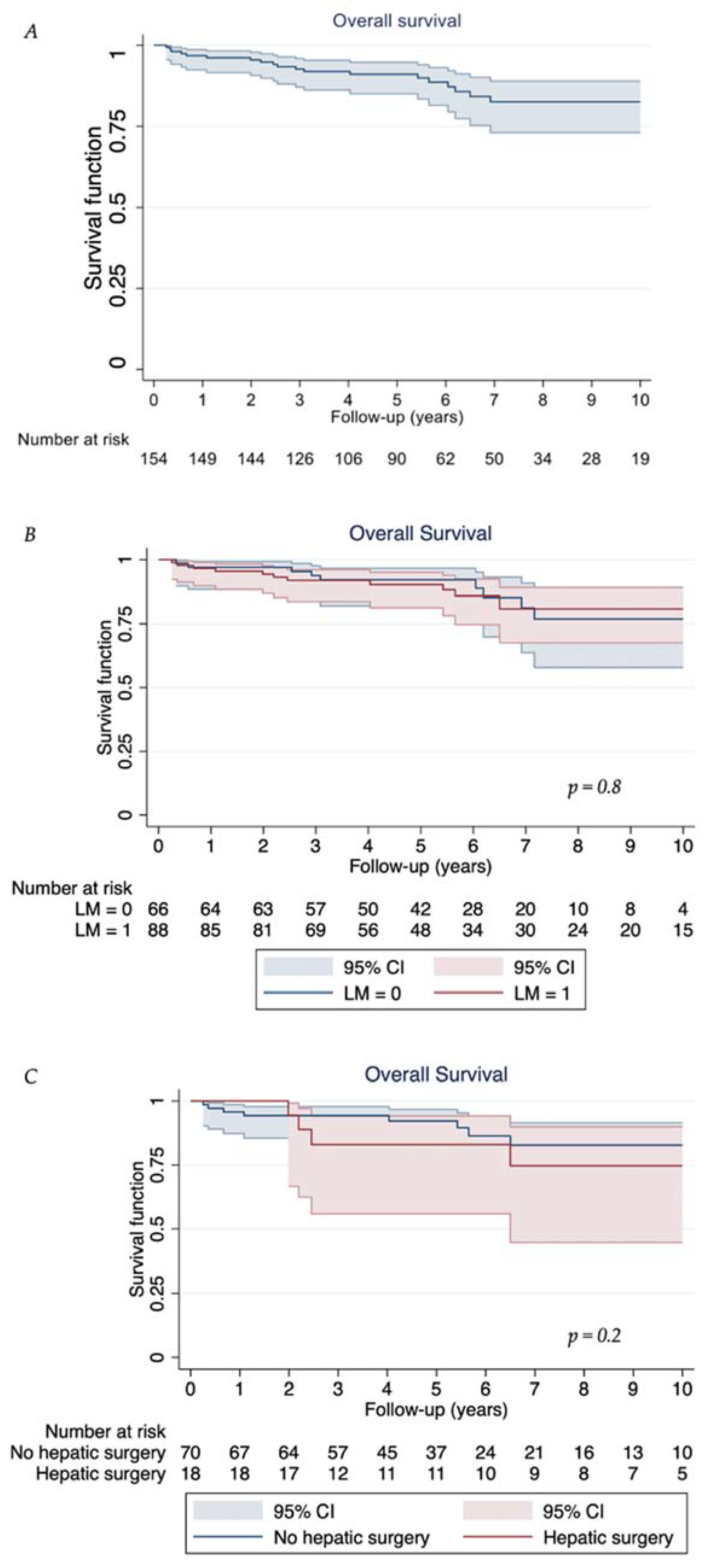
3.3. Follow-Up and Survival Outcomes

The median follow-up was 67.2 months (range, 3.1–310.4 months). No patient was lost to follow-up. At the last follow-up, 29 patients (18.8%) were alive with no evidence of NET, 102 (66.2%) were alive with NET, 18 (11.7%) died of NET, and 5 (all of them with stable disease, 3.3%) died of non-NET related causes.
The median overall survival was not reached. Overall survival at 1 year, 3 years, 5 years, and 10 years was 96.8% (95% CI: 92.4–98.5%), 92.6% (95% CI: 87.0–95.9%), 91.0% (95% CI: 84.9–94.7%), and 82.5% (95% CI: 72.9–88.9%), respectively (Table 3, Figure 1A). When discriminating between patients with no liver metastases and those with liver metastases, the 3-year, 5-year, and 10-year OS was 93.4% (95% CI: 83.3–97.5%), 91.6% (95% CI: 81.0–96.4%), and 79.8% (95% CI: 61.3–90.2%), respectively, and 92.1% (95% CI: 84.2–96.2%), 90.6% (95% CI: 82.0–95.2%), and 84.1% (95% CI: 72.1–91.2%), respectively (p = 0.8, Figure 1B). In those with liver metastases, there was no significant difference observed between those who underwent liver resection and those who did not (p = 0.2, Figure 1C).
Imaging-based median progression-free survival was 42.7 months (95% CI: 24.7 to 72.4 months). Progression-free survival at 1 year, 3 years, 5 years, and 10 years was 96.7% (95% CI: 92.4–98.6%), 84.3% (95 CI: 77.5–89.2%), 63.4% (95% CI: 55.0–70.6%), and 18.7% (95% CI: 12.4–26.1%), respectively. Recurrence-free survival was not calculated due to the small number of patients who developed recurrent disease (n = 2). The patient who underwent multivisceral transplantation was alive and tumour-free at 9 years following surgery [35]. Nineteen patients were (15.2%) alive and tumour-free at 10 years after surgery and were considered cured.
The univariate and multivariable analyses of factors associated with OS and PFS are reported in Table 4 and Table 5, respectively. The only significant associations (i.e., p-value < 0.05) identified were those for grade 2 tumours having reduced OS and PFS, but this must be interpreted in the context of the low numbers of events affecting precision and the extensive use of multiple lines of therapy.

4. Discussion

In this study comprising 154 consecutive patients with SB NET, we have demonstrated the following: (a) the vast majority of SB NET is present in stage III or IV although classified as low-grade neoplasia in 70% of cases; (b) despite high tumour stages and high hepatic tumour burden, radical surgery embedded within multimodal treatment concepts achieves favourable long-term outcomes with acceptable treatment-associated morbidity and nil mortality. The 5-year and 10-year OS of 91.0% and 82.5%, respectively, achieved in our cohort support the notion that cytoreductive surgery should be considered in all SB NET patients fit for surgical intervention [36,37,38]. Of note, a cure was likely achieved in 15%. Overall survival did not differ between patients with liver metastases and those with no hepatic involvement. Overlapping confidence intervals of the two groups likely reflect heavy use of successful, continued lines of systemic and/or liver-directed treatment in combination with surgery. No ‘predictors’ of survival were identified—we hypothesise that multimodal treatment approaches contributed to the favourable long-term survival in this series compared with the 5-year survival (all stages) in the range between 53% and 63% noted in earlier reports [39,40] and the 5-year and 10-year OS of 67% and 37%, respectively, reported in a systematic review in patients in metastasised stages [41].
The outcome of randomised controlled trials for systemic treatment of SBNET, PROMID [18], CLARINET [19], RADIANT IV [20], and NETTER-1 [42], which all failed to show impact on OS, further underpins the pivotal role of surgery.
When planning surgery for SB NET, several issues should be considered. Preoperative imaging frequently understages the disease, both in terms of the primary tumour and the metastases. In our series, 36.5% had multifocal primaries that were only detectable in their full extent by meticulous intra-operative palpation. In the series reported by Pasquer et al. [10], multicentric primaries were diagnosed in 33% and missed in 61% on preoperative imaging. Keck et al. found multifocal primaries in 56% of them, with 72% located within 100 cm of the ileocolic valve [3]. Clinical behaviour and survival are comparable between multifocal tumours and unifocal counterparts [4,5]. Based on the results of complete genome sequencing of tumours from patients with multicentric NET, Elias et al. suggested an independent clonal origin of such lesions [43].
Other underestimated findings on preoperative imaging are mesenteric tumour deposits and peritoneal carcinomatosis. The latter was observed at laparotomy in 12.8% of our patients and in 20–30% in other series [11,44]. Fata et al. [45] found mesenteric tumour deposits described as discrete but irregular mesenteric tumour nodules frequently located adjacent to neurovascular bundles and discontinuous from the primary neoplasm in 68% of 132 resected small bowel NET. Since both peritoneal carcinomatosis and mesenteric tumour deposits have been reported as independent factors of prognostic relevance, they should be considered when planning cytoreduction [10,11,44,45].
In contrast to NET originating from the pancreas or lung, high-grade NET or neuroendocrine carcinoma (NEC) are rarely seen in the small intestine. In line with observations made by others, the majority of our patients had G1 or G2 with low Ki67 tumours [11,36,45]. When assessing the Ki67 index, intra-tumoural and inter-tumoural grade heterogeneity should be considered. In our previous study, we have shown discrepancies in grading between primary gastroenteropancreatic NET and their metastatic sites in 35.3% of cases [46]. In 77.8% of patients with liver metastases, the Ki67 % was higher in liver lesions than in the primary tumour.
An open approach as recommended in current guidelines [13,47] is still considered to be a gold standard since it facilitates extensive manual palpation of the entire small intestine, secure access to lymph node metastases at the root of the mesentery, and synchronous liver debulking in selected cases. Laparoscopic resection is feasible [48,49]; however, solid evidence for the true benefit for the patient is still lacking. Conversion rates of 30% [50] and as high as 80% in patients with stage III or stage IV mesenteric lymph node metastases [51] have been reported.
Abdominal lymph node metastases are present in 75–90% of SB NET at initial diagnosis [9,10,52]. Their complete resection is pivotal for favourable long-term survival; however, this is challenging when there is an encasement of the mesenteric vessel origin and/or retropancreatic space, particularly in the presence of retractile fibrotic mesenteritis and/or desmoplastic reaction [11,53]. An optimal lympadenectomy should comprise at least eight lymph nodes under consideration of limited resection of the small intestine and utilisation of intestinal sparing techniques [54,55,56]. In patients with inadequate primary resection, a reintervention irrespective of negative imaging results should be considered [57]. Although 72% of patients in our series had lymph node metastases, 93.3% of these had them completely resected, and only two showed evidence of recurrence in the lymph nodes thereafter. This is in contrast to the published experience in patients with neuroendocrine liver metastases, which recur in virtually all cases [58].
The present study has limitations. The study sample comprises those treated at a tertiary referral centre; this may affect the transportability of findings to other settings, such as the high incidence of distantly metastatic disease in this group, and that all patients underwent elective surgery. Further, due to being managed in a tertiary centre, all patients underwent 68GA DOTATATE PET/CT prior to treatment decisions, which could have increased disease detection (e.g., identifying deposits that could have been ‘missed’ by other imaging modalities) and therefore exerted influence on treatment selection. The retrospective nature of this case series is also unable to ascertain the effects of specific treatment decisions on outcomes.
A substantial number of reports has shown that hepatic resection for neuroendocrine liver metastases provides favourable outcome with 5-year overall survival rates of 60–100% [15,16,38,59]. As shown in this series and in the experience of others, only about 30% are good candidates for hepatic resection (< 45% liver involvement, ≥ 70% cytoreduction, resectable/limited extrahepatic spread). In a systematic review and meta-analysis of outcomes of liver resection in patients with stage IV pancreatic NET, the median 1-year, 3-year, and 5-year overall survival in the resected group was 92.7%, 76.9%, and 67.5%, respectively, compared with 77.3%, 40.9%, and 26.6%, respectively, in the non-resected group [60]. As the authors rightly highlight, these data must be taken with caution since there were no randomised controlled trials, the selection for patients was biased by their suitability for surgery, and the treatment in the no liver-resection group was heterogeneous, ranging from primary tumour resection as the only measure to multimodal treatment concepts. Not achieving hepatic resection was the only independent factor of survival in the retrospective series in patients with SB NET and synchronous liver metastases reported by Addeo et al. [61].
In the light of high recurrence rates [62,63,64] and apparently no difference in the long-term survival outcome between patients who had R0 liver resection and those with R1 or R2 status [65,66], a shift from classical major resections [67] to more parenchyma preserving procedures [66,68] and acceptance of a debulking threshold of 70% in contrast to previous 90% has evolved over the last decade. In the series of Scott et al. encompassing 184 patients who had 188 cytoreductive surgeries, the median overall survival and progression free-survival was 89.4 months and 22.5 months, respectively [69]. In addition, 70% debulking was associated with better OS compared with < 70%, and 90% cytoreductive surgery was not associated with improved OS when compared with 70–90%.
The appropriateness of primary tumour resection in asymptomatic SB NET patients with non-resectable liver metastases is debated controversially. While proponents argue that primary tumour resection avoids local complications and prolongs survival, opponents suggest resection in this setting only if symptoms occur. The results of a systematic review were in favour of a proactive approach; median overall survival was 112 months in the primary tumour resection group compared with 60 months in the conservatively managed group. Five-year overall survival rates were 74% and 44%, respectively [70]. A study performed by Bennet et al. demonstrated that upfront small bowel resection was associated with lower rates of readmission and intervention compared to non-operative management [71]. In a single-centre series reported by Daskalakis et al., prophylactic upfront locoregional surgery had no survival advantage [72]. Moreover, delayed surgery was associated with fewer laparotomies for intestinal obstruction. Of note, emergency procedures were included in the latter group. In the experience of Ahmed et al. [15], in contrast, primary tumour resection and the Ki67 index were the only independent predictors of survival in the group of 360 patients with SBNET metastasised to the liver. Results reported by Kaemmerer et al. in patients with stage IV SBNET considered for peptide receptor radionuclide therapy supported upfront primary tumour resection leading to a median overall survival and progression-free survival of 134 months and 18 months, respectively, compared to 67 months and 18 months, respectively [73].
Frequently metastatic disease, high recurrence rates after hepatic resections, and promising results of systemic medical therapies and interventional liver directed procedures call for the consideration of multimodal concepts (combined surgical and non-surgical modalities) in the management of SB NET. In this series, multimodal treatment was applied in more than two third of patients. Of them, 87% had somatostatin receptor targeted therapy. Although in NEN, a neoadjuvant approach utilising peptide receptor radionuclide therapy has been mainly used in those originating from the pancreas [74], results in small bowel NET have been shown in few reports [30,75,76]. The recent case series of Fisher and colleagues comprised 17 patients with metastatic ileal NET and found a commendable 93% 5-year overall survival, and a 70% progression-free survival with the use of SSAs after surgery [76].
Genomic, transcriptomic, metabonomic, and epigenetic studies of SB NET have contributed to the delineation of novel molecular subgroups with differing risks of metastases and aggressive behaviour [77,78,79]. Future avenues of research should focus on the selection of treatment based on individual patient and tumour characteristics, optimal sequence of treatment modalities, and effective monitoring of treatment response utilising molecular-based markers [80,81].

5. Conclusions

Multimodal treatment strategies may be associated with excellent outcomes in SBNET, even though patients often present in advanced stages of the disease. Future study in this tumour type should focus on ascertaining the optimal sequencing and combinations of different therapies and explore their impact on overall survival.

Author Contributions

Conceptualization, A.K.C., P.D., A.v.R., A.H., R.G., J.B., S.L., H.W., N.H. and A.F.; data curation, A.K.C., P.D., A.v.R., A.H., R.G., J.B., S.L., H.W., N.H. and A.F.; formal analysis, A.K.C. and A.F.; investigation, A.K.C., P.D., A.v.R., A.H., R.G., J.B., S.L., H.W., N.H. and A.F.; writing—original draft, A.K.C. and A.F.; writing—review and editing, P.D., A.v.R., A.H., R.G., J.B., S.L., H.W. and N.H. All authors have read and agreed to the published version of the manuscript.

Funding

This study was supported by a research grant to A.F. by the Heinz-Horst Deichmann Foundation (P31294). P.D. was supported by a European Union FP7 grant (FP7-MC-IEF). The funding sources had no role in the design and conduct of the study; collection, management, analysis, and interpretation of the data; and preparation, review, or approval of the manuscript.

Institutional Review Board Statement

This study was conducted in accordance with the Declaration of Helsinki, and approved by the Health Research Authority (IRAS ref: 35117, Research Ethics Committee reference: 23/EM/0102, date of approval 17 May 2023).

Informed Consent Statement

Informed consent was obtained from all individual subjects involved in the study. Regarding ethical approval: the institutional approval for the prospectively maintained patient database is under GEN_16; the collection and research use of integrated imaging, genomic and other clinico-biological data is under 07/MRE09/54 (REC 22/WA/2836), and this specific study was approved by the Health Research Authority (IRAS ref: 35117, REC: 23/EM/0102).

Data Availability Statement

Due to the nature of the clinical data, information governance considerations, and the remit of the consent gained from individual participants for inclusion in the institutional research database, the raw data used for this study cannot be made available to other investigators.

Acknowledgments

We would like to thank James Kinross, Paul Ziprin, Adam Haycock, Janindra Warusawitarne, Omar Faiz, Adil Al Nahhas, and Paul Tait for their input in the management of our patients with SB NET. Claudia von Arx and Anne-Marie Feeney are acknowledged for their help with data collection.

Conflicts of Interest

The authors declare no conflict of interest.

References

  1. Dasari, A.; Shen, C.; Halperin, D.; Zhao, B.; Zhou, S.; Xu, Y.; Shih, T.; Yao, J.C. Trends in the Incidence, Prevalence, and Survival Outcomes in Patients With Neuroendocrine Tumors in the United States. JAMA Oncol. 2017, 3, 1335. [Google Scholar] [CrossRef] [PubMed]
  2. Modlin, I.M.; Sandor, A. An analysis of 8305 cases of carcinoid tumors. Cancer 1997, 79, 813–829. [Google Scholar] [CrossRef]
  3. Keck, K.J.; Maxwell, J.E.; Menda, Y.; Bellizzi, A.; Dillon, J.; O’Dorisio, T.M.; Howe, J.R. Identification of primary tumors in patients presenting with metastatic gastroenteropancreatic neuroendocrine tumors. Surgery 2017, 161, 272–279. [Google Scholar] [CrossRef] [PubMed]
  4. Gangi, A.; Siegel, E.; Barmparas, G.; Lo, S.; Jamil, L.H.; Hendifar, A.; Nissen, N.N.; Wolin, E.M.; Amersi, F. Multifocality in Small Bowel Neuroendocrine Tumors. J. Gastrointest. Surg. 2018, 22, 303–309. [Google Scholar] [CrossRef]
  5. Choi, A.B.; Maxwell, J.E.; Keck, K.J.; Bellizzi, A.J.; Dillon, J.S.; O’Dorisio, T.M.; Howe, J.R. Is Multifocality an Indicator of Aggressive Behavior in Small Bowel Neuroendocrine Tumors? Pancreas 2017, 46, 1115–1120. [Google Scholar] [CrossRef] [PubMed]
  6. Clift, A.K.; Faiz, O.; Al-Nahhas, A.; Bockisch, A.; Liedke, M.O.; Schloericke, E.; Wasan, H.; Martin, J.; Ziprin, P.; Moorthy, K.; et al. Role of Staging in Patients with Small Intestinal Neuroendocrine Tumours. J. Gastrointest. Surg. 2016, 20, 180–188. [Google Scholar] [CrossRef] [PubMed]
  7. Frilling, A.; Sotiropoulos, G.C.; Radtke, A.; Malago, M.; Bockisch, A.; Kuehl, H.; Li, J.; Broelsch, C.E. The Impact of 68Ga-DOTATOC positron emission tomography/computed tomography on the multimodal management of patients with neuroendocrine tumors. Ann. Surg. 2010, 252, 850–856. [Google Scholar] [CrossRef] [PubMed]
  8. Sadowski, S.M.; Neychev, V.; Millo, C.; Shih, J.; Nilubol, N.; Herscovitch, P.; Pacak, K.; Marx, S.J.; Kebebew, E. Prospective study of 68Ga-DOTATATE positron emission tomography/computed tomography for detecting gastro-entero-pancreatic neuroendocrine tumors and unknown primary sites. J. Clin. Oncol. 2016, 34, 588–597. [Google Scholar] [CrossRef]
  9. Zaidi, M.Y.; Lopez-Aguiar, A.G.; Dillhoff, M.; Beal, E.; Poultsides, G.; Makris, E.; Rocha, F.; Crown, A.; Idrees, K.; Marincola Smith, P.; et al. Prognostic Role of Lymph Node Positivity and Number of Lymph Nodes Needed for Accurately Staging Small-Bowel Neuroendocrine Tumors. JAMA Surg. 2019, 154, 134–140. [Google Scholar] [CrossRef]
  10. Pasquer, A.; Walter, T.; Hervieu, V.; Forestier, J.; Scoazec, J.-Y.; Lombard-Bohas, C.; Poncet, G. Surgical Management of Small Bowel Neuroendocrine Tumors: Specific Requirements and Their Impact on Staging and Prognosis. Ann. Surg. Oncol. 2015, 22 (Suppl. 3), S742–S749. [Google Scholar] [CrossRef] [PubMed]
  11. Norlén, O.; Stålberg, P.; Öberg, K.; Eriksson, J.; Hedberg, J.; Hessman, O. Long-term results of surgery for small intestinal neuroendocrine tumors at a tertiary referral center. World J. Surg. 2012, 36, 1419–1431. [Google Scholar] [CrossRef] [PubMed]
  12. Shah, C.P.; Mramba, L.K.; Bishnoi, R.; Unnikrishnan, A.; Duff, J.M.; Chandana, S.R. Survival trends of metastatic small intestinal neuroendocrine tumor: A population-based analysis of SEER database. J. Gastrointest. Oncol. 2019, 10, 869–877. [Google Scholar] [CrossRef] [PubMed]
  13. Howe, J.R.; Cardona, K.; Fraker, D.L.; Kebebew, E.; Untch, B.R.; Wang, Y.Z.; Law, C.H.; Liu, E.H.; Kim, M.K.; Menda, Y.; et al. The surgical management of small bowel neuroendocrine tumors. Pancreas 2017, 46, 715–731. [Google Scholar] [CrossRef] [PubMed]
  14. Hallet, J.; Law, C.; Hallet, J.; Pasieka, J.; Koea, J.; Meyer-Rochow, W. Role of Primary Tumor Resection for Metastatic Small Bowel Neuroendocrine Tumors. World J. Surg. 2021, 45, 213–218. [Google Scholar] [CrossRef] [PubMed]
  15. Ahmed, A.; Turner, G.; King, B.; Jones, L.; Culliford, D.; McCance, D.; Ardill, J.; Johnston, B.T.; Poston, G.; Rees, M.; et al. Midgut neuroendocrine tumours with liver metastases: Results of the UKINETS study. Endocr. Relat. Cancer 2009, 16, 885–894. [Google Scholar] [CrossRef]
  16. Fairweather, M.; Swanson, R.; Wang, J.; Brais, L.K.; Dutton, T.; Kulke, M.H.; Clancy, T.E. Management of Neuroendocrine Tumor Liver Metastases: Long-Term Outcomes and Prognostic Factors from a Large Prospective Database. Ann. Surg. Oncol. 2017, 24, 2319–2325. [Google Scholar] [CrossRef] [PubMed]
  17. Linecker, M.; Kambakamba, P.; Raptis, D.A.; Malagó, M.; Ratti, F.; Aldrighetti, L.; Robles-Campos, R.; Lehwald-Tywuschik, N.; Knoefel, W.; Balci, D.; et al. ALPPS in neuroendocrine liver metastases not amenable for conventional resection—Lessons learned from an interim analysis of the International ALPPS Registry. HPB 2020, 22, 537–544. [Google Scholar] [CrossRef] [PubMed]
  18. Rinke, A.; Müller, H.-H.; Schade-Brittinger, C.; Klose, K.-J.; Barth, P.; Wied, M.; Mayer, C.; Aimnossadati, B.; Pape, U.-F.; Blaker, M.; et al. Placebo-controlled, double-blind, prospective, randomized study on the effect of octreotide LAR in the control of tumor growth in patients with metastatic neuroendocrine midgut tumors: A report from the PROMID Study Group. J. Clin. Oncol. 2009, 27, 4656–4663. [Google Scholar] [CrossRef] [PubMed]
  19. Caplin, M.E.; Pavel, M.; Ćwikła, J.B.; Phan, A.T.; Raderer, M.; Sedláčková, E.; Cadiot, G.; Wolin, E.M.; Capdevila, J.; Wall, L.; et al. Lanreotide in metastatic enteropancreatic neuroendocrine tumors. N. Engl. J. Med. 2014, 371, 224–233. [Google Scholar] [CrossRef] [PubMed]
  20. Yao, J.C.; Shah, M.H.; Ito, T.; Bohas, C.L.; Wolin, E.M.; Van Cutsem, E.; Hobday, T.J.; Okusaka, T.; Capdevila, J.; de Vries, E.G.E.; et al. Everolimus for advanced pancreatic neuroendocrine tumors. N. Engl. J. Med. 2011, 364, 514–523. [Google Scholar] [CrossRef] [PubMed]
  21. Strosberg, J.; El-Haddad, G.; Wolin, E.; Hendifar, A.; Yao, J.; Chasen, B.; Mittra, E.; Kunz, P.L.; Hulke, M.H.; Jacene, H.; et al. Phase 3 Trial of177Lu-Dotatate for Midgut Neuroendocrine Tumors. N. Engl. J. Med. 2017, 376, 125–135. [Google Scholar] [CrossRef]
  22. Clift, A.K.; Kidd, M.; Bodei, L.; Toumpanakis, C.; Baum, R.P.; Oberg, K.; Modlin, I.M.; Frilling, A. At the Cutting Edge Neuroendocrine Neoplasms of the Small Bowel and Pancreas. Neuroendocrinology 2020, 110, 444–476. [Google Scholar] [CrossRef]
  23. Rindi, G.; Klöppel, G.; Couvelard, A.; Komminoth, P.; Körner, M.; Lopes, J.M.; McNicol, A.-M.; Nilsson, O.; Perren, A.; Scarpa, A.; et al. TNM staging of midgut and hindgut (neuro) endocrine tumors: A consensus proposal including a grading system. Virchows Arch. 2007, 451, 757–762. [Google Scholar] [CrossRef]
  24. Lloyd, R.V.; Osamura, R.Y.; Kloppel, G.; Rosai, J.; World Health Organization; International Agency for Research on Cancer. WHO Classification of Tumours of Endocrine Organs, 4th ed.; International Agency for Research on Cancer: Lyon, France, 2017; Volume 10. [Google Scholar]
  25. Klimstra, D.S. Classification of neuroendocrine neoplasms of the digestive system. In WHO Classification of Tumours: Digestive System Tumours, 5th ed.; International Agency for Research on Cancer: Lyon, France, 2019; p. 16. [Google Scholar]
  26. Ohrvall, U.; Eriksson, B.; Juhlin, C.; Karacagil, S.; Rastad, J.; Hellman, P.; Akerstrom, G. Method for dissection of mesenteric metastases in mid-gut carcinoid tumors. World J. Surg. 2000, 24, 1402–1408. [Google Scholar] [CrossRef]
  27. Strasberg, S.M. Nomenclature of hepatic anatomy and resections: A review of the Brisbane 2000 system. J. Hepatobiliary Pancreat. Surg. 2005, 12, 351–355. [Google Scholar] [CrossRef]
  28. Fouché, M.; Bouffard, Y.; Le Goff, M.C.; Prothet, J.; Malavieille, F.; Sagnard, P.; Christin, F.; Hayi-Slayman, D.; Pasquer, A.; Poncet, G.; et al. Intraoperative carcinoid syndrome during small-bowel neuroendocrine tumour surgery. Endocr. Connect. 2018, 7, 1245–1250. [Google Scholar] [CrossRef]
  29. Dindo, D.; Demartines, N.; Clavien, P.-A. Classification of surgical complications: A new proposal with evaluation in a cohort of 6336 patients and results of a survey. Ann. Surg. 2004, 240, 205–213. [Google Scholar] [CrossRef]
  30. Frilling, A.; Clift, A.K.; Frampton, A.E.; Bomanji, J.; Kaemmerer, D.; Al-Nahhas, A.; Alsafi, A.; Kidd, M.; Modlin, I.M.; Horsch, D.; et al. A combination of surgery, theranostics, and liquid biopsy—A personalised oncologic approach to treatment of patients with advanced metastatic neuroendocrine neoplasms. Int. J. Med. Sci. 2021, 18, 2166–2175. [Google Scholar] [CrossRef]
  31. Frilling, A.; Clift, A.K.; Braat, A.J.A.T.; Alsafi, A.; Wasan, H.S.; Al-Nahhas, A.; Thomas, R.; Drymousis, P.; Habib, N.; Tait, P.N. Radioembolisation with 90Y microspheres for neuroendocrine liver metastases: An institutional case series, systematic review and meta-analysis. HPB 2019, 21, 773–783. [Google Scholar] [CrossRef]
  32. Young, H.; Baum, R.; Cremerius, U.; Herholz, K.; Hoekstra, O.; Lammertsma, A.A.; Pruim, J.; Price, P. Measurement of clinical and subclinical tumour response using [18F]- fluorodeoxyglucose and positron emission tomography: Review and 1999 EORTC recommendations. Eur. J. Cancer 1999, 35, 1773–1782. [Google Scholar] [CrossRef]
  33. Eisenhauer, E.A.A.; Therasse, P.; Bogaerts, J.; Schwartz, L.H.H.; Sargent, D.; Ford, R.; Dancey, J.; Arbuck, S.; Gwyther, S.; Mooney, M.; et al. New response evaluation criteria in solid tumours: Revised RECIST guideline (version 1.1). Eur. J. Cancer 2009, 45, 228–247. [Google Scholar] [CrossRef]
  34. Jakobsen, L.H.; Andersson, T.M.L.; Biccler, J.L.; Poulsen, L.; Severinsen, M.T.; El-Galaly, T.C.; Bogsted, M. On estimating the time to statistical cure. BMC Med. Res. Methodol. 2020, 20, 71. [Google Scholar] [CrossRef]
  35. Clift, A.K.; Giele, H.; Reddy, S.; MacEdo, R.; Al-Nahhas, A.; Wasan, H.S.; Gondolesi, G.E.; Vianna, R.M.; Friend, P.; Vaidya, A.; et al. Neoadjuvant peptide receptor radionuclide therapy and modified multivisceral transplantation for an advanced small intestinal neuroendocrine neoplasm: An updated case report. Innov. Surg. Sci. 2017, 2, 247–253. [Google Scholar] [CrossRef]
  36. Wu, L.; Fu, J.; Wan, L.; Pan, J.; Lai, S.; Zhong, J.; Chung, D.C.; Wang, L. Survival outcomes and surgical intervention of small intestinal neuroendocrine tumors: A population based retrospective study. Oncotarget 2017, 8, 4935–4947. [Google Scholar] [CrossRef]
  37. Habbe, N.; Fendrich, V.; Heverhagen, A.; Ramaswamy, A.; Bartsch, D.K. Outcome of surgery for ileojejunal neuroendocrine tumors. Surg. Today 2013, 43, 1168–1174. [Google Scholar] [CrossRef]
  38. Boudreaux, J.P.; Wang, Y.-Z.; Diebold, A.E.; Frey, D.J.; Anthony, L.; Uhlhorn, A.P.; Ryan, P.; Woltering, E.A. A single institution’s experience with surgical cytoreduction of stage IV, well-differentiated, small bowel neuroendocrine tumors. J. Am. Coll. Surg. 2014, 218, 837–844. [Google Scholar] [CrossRef]
  39. Burke, A.P.; Sobin, L.H.; Federspiel, B.H.; Shekitka, K.M.; Helwig, E.B. Carcinoid tumors of the duodenum. A clinicopathologic study of 99 cases. Arch. Pathol. Lab. Med. 1990, 114, 700–704. [Google Scholar]
  40. Modlin, I.M.; Lye, K.D.; Kidd, M. A 5-decade analysis of 13,715 carcinoid tumors. Cancer 2003, 97, 934–959. [Google Scholar] [CrossRef]
  41. Van Den Heede, K.; Chidambaram, S.; Van Slycke, S.; Brusselaers, N.; Warfvinge, C.F.; Ohlsson, H.; Gistafsson, R.; Nordenstrom, E.; Almquist, M. Long-term survival of metastatic small intestine neuroendocrine tumors: A meta-analysis. Endocr. Relat. Cancer 2022, 29, 163–173. [Google Scholar] [CrossRef]
  42. Strosberg, J.R.; Caplin, M.E.; Kunz, P.L.; Ruszniewski, P.B.; Bodei, L.; Hendifar, A.; Mittra, E.; Wolin, E.M.; Yao, J.C.; Pavel, M.; et al. 177 Lu-Dotatate plus long-acting octreotide versus high-dose long-acting octreotide in patients with midgut neuroendocrine tumours (NETTER-1): Final overall survival and long-term safety results from an open-label, randomised, controlled, phase 3 trial. Lancet Oncol. 2021, 22, 1752–1763. [Google Scholar] [CrossRef]
  43. Elias, E.; Ardalan, A.; Lindberg, M.; Reinsbach, S.E.; Muth, A.; Nilsson, O.; Arvisson, Y.; Larsson, E. Independent somatic evolution underlies clustered neuroendocrine tumors in the human small intestine. Nat. Commun. 2021, 12, 6367. [Google Scholar] [CrossRef]
  44. Wonn, S.M.; Limbach, K.E.; Pommier, S.E.J.; Ratzlaff, A.N.; Leon, E.J.; McCully, B.H.; Pommier, R.F. Outcomes of cytoreductive operations for peritoneal carcinomatosis with or without liver cytoreduction in patients with small bowel neuroendocrine tumors. Surgery 2021, 169, 168–174. [Google Scholar] [CrossRef]
  45. Fata, C.R.; Gonzalez, R.S.; Liu, E.; Cates, J.M.; Shi, C. Mesenteric Tumor Deposits in Midgut Small Intestinal Neuroendocrine Tumors Are a Stronger Indicator Than Lymph Node Metastasis for Liver Metastasis and Poor Prognosis. Am. J. Surg. Pathol. 2017, 41, 128–133. [Google Scholar] [CrossRef]
  46. Miller, H.C.; Drymousis, P.; Flora, R.; Goldin, R.; Spalding, D.; Frilling, A. Role of Ki-67 proliferation index in the assessment of patients with neuroendocrine neoplasias regarding the stage of disease. World J. Surg. 2014, 38, 1353–1361. [Google Scholar] [CrossRef]
  47. Partelli, S.; Bartsch, D.K.; Capdevila, J.; Chen, J.; Knigge, U.; Niederle, B.; Nieveen van Dijkum, E.J.M.; Pape, U.-F.; Pascher, A.; Ramage, J.; et al. ENETS Consensus Guidelines for the Standards of Care in Neuroendocrine Tumours: Surgery for Small Intestinal and Pancreatic Neuroendocrine Tumours. Neuroendocrinology 2017, 105, 255–265. [Google Scholar] [CrossRef]
  48. Reissman, P.; Shmailov, S.; Grozinsky-Glasberg, S.; Gross, D.J. Laparoscopic resection of primary midgut carcinoid tumors. Surg. Endosc. 2013, 27, 3678–3682. [Google Scholar] [CrossRef]
  49. Figueiredo, M.N.; Maggiori, L.; Gaujoux, S.; Couvelard, A.; Guedj, N.; Ruszniewski, P.; Panis, Y. Surgery for small-bowel neuroendocrine tumors: Is there any benefit of the laparoscopic approach? Surg. Endosc. 2014, 28, 1720–1726. [Google Scholar] [CrossRef]
  50. Kaçmaz, E.; Van Eeden, S.; Koppes, J.C.C.; Klümpen, H.J.; Bemelman, W.A.; Nieveen Van Dijkum, E.J.M.; Engelsman, A.F.; Tanis, P.J. Value of Laparoscopy for Resection of Small-Bowel Neuroendocrine Neoplasms Including Central Mesenteric Lymphadenectomy. Dis. Colon. Rectum. 2021, 64, 1240–1248. [Google Scholar] [CrossRef]
  51. Kasai, Y.; Mahuron, K.; Hirose, K.; Corvera, C.U.; Kim, G.E.; Hope, T.A.; Shih, B.E.; Warren, R.S.; Bergsland, E.K.; Nakakura, E.K. A novel stratification of mesenteric mass involvement as a predictor of challenging mesenteric lymph node dissection by minimally invasive approach for ileal neuroendocrine tumors. J. Surg. Oncol. 2020, 122, 204–211. [Google Scholar] [CrossRef]
  52. Kaçmaz, E.; Sarasqueta, A.F.; van Eeden, S.; Dreijerink, K.M.A.; Klümpen, H.J.; Tanis, P.J.; Nieveen van Dikjum, E.J.M.; Engelsman, A.F. Update on Incidence, Prevalence, Treatment and Survival of Patients with Small Bowel Neuroendocrine Neoplasms in the Netherlands. World J. Surg. 2021, 45, 2482–2491. [Google Scholar] [CrossRef]
  53. Pasquer, A.; Walter, T.; Rousset, P.; Hervieu, V.; Forestier, J.; Lombard-Bohas, C.; Poncet, G. Lymphadenectomy during Small Bowel Neuroendocrine Tumor Surgery: The Concept of Skip Metastases. Ann. Surg. Oncol. 2016, 23, 804–808. [Google Scholar] [CrossRef]
  54. Lardière-Deguelte, S.; De Mestier, L.; Appéré, F.; Vullierme, M.P.; Zappa, M.; Hoeffel, C.; Noaves, M.; Brixi, H.; Hentic, O.; Ruszniewski, P.; et al. Toward a preoperative classification of lymph node metastases in patients with small intestinal neuroendocrine tumors in the era of intestinal-sparing surgery. Neuroendocrinology 2016, 103, 552–559. [Google Scholar] [CrossRef]
  55. Motz, B.M.; Lorimer, P.D.; Boselli, D.; Hill, J.S.; Salo, J.C. Optimal Lymphadenectomy in Small Bowel Neuroendocrine Tumors: Analysis of the NCDB. J. Gastrointest. Surg. 2018, 22, 117–123. [Google Scholar] [CrossRef]
  56. Hallet, J.; Law, C. Extent of Lymph Node Dissection for Small Bowel Neuroendocrine Tumors. World J. Surg. 2021, 45, 197–202. [Google Scholar] [CrossRef]
  57. Deguelte, S.; Hammoutene, C.; Poncet, G.; Brunaud, L.; Perrier, M.; Kianmanesh, R.; Cadiot, G. Concept of reintervention with thorough lymphadenectomy after suboptimal resection of small-intestine neuroendocrine neoplasms: A multicentre preliminary study. J. Neuroendocrinol. 2022, 34, e13117. [Google Scholar] [CrossRef]
  58. Saxena, A.; Chua, T.C.; Perera, M.; Chu, F.; Morris, D.L. Surgical resection of hepatic metastases from neuroendocrine neoplasms: A systematic review. Surg. Oncol. 2012, 21, e131–e141. [Google Scholar] [CrossRef]
  59. Frilling, A.; Li, J.; Malamutmann, E.; Schmid, K.-W.K.-W.; Bockisch, A.; Broelsch, C.E. Treatment of liver metastases from neuroendocrine tumours in relation to the extent of hepatic disease. Br. J. Surg. 2009, 96, 175–184. [Google Scholar] [CrossRef]
  60. Yu, X.; Gu, J.; Wu, H.; Fu, D.; Li, J.; Jin, C. Resection of Liver Metastases: A Treatment Provides a Long-Term Survival Benefit for Patients with Advanced Pancreatic Neuroendocrine Tumors: A Systematic Review and Meta-Analysis. J. Oncol. 2018, 2018, 6273847. [Google Scholar] [CrossRef]
  61. Addeo, P.; Bertin, J.B.; Imperiale, A.; Averous, G.; Terrone, A.; Goichot, B.; Bachellier, P. Outcomes of Simultaneous Resection of Small Bowel Neuroendocrine Tumors with Synchronous Liver Metastases. World J. Surg. 2020, 44, 2377–2384. [Google Scholar] [CrossRef]
  62. Spolverato, G.; Bagante, F.; Aldrighetti, L.; Poultsides, G.A.; Bauer, T.W.; Fields, R.C.; Kumar Maithel, S.; Marques, H.P.; Weiss, M.; Pawlik, T.M. Management and outcomes of patients with recurrent neuroendocrine liver metastasis after curative surgery: An international multi-institutional analysis. J. Surg. Oncol. 2017, 116, 298–306. [Google Scholar] [CrossRef]
  63. Mayo, S.C.; de Jong, M.C.; Pulitano, C.; Clary, B.M.; Reddy, S.K.; Gamblin, T.C.; Celinski, S.A.; Kooby, D.A.; Staley, C.A.; Stokes, J.B.; et al. Surgical management of hepatic neuroendocrine tumor metastasis: Results from an international multi-institutional analysis. Ann. Surg. Oncol. 2010, 17, 3129–3136. [Google Scholar] [CrossRef] [PubMed]
  64. Scigliano, S.; Lebtahi, R.; Maire, F.; Stievenart, J.L.; Kianmanesh, R.; Sauvanet, A.; Vullierme, M.P.; Coulevard, A.; Belghiti, J.; Ruszniewski, P.; et al. Clinical and imaging follow-up after exhaustive liver resection of endocrine metastases: A 15-year monocentric experience. Endocr. Relat. Cancer 2009, 16, 977–990. [Google Scholar] [CrossRef]
  65. Glazer, E.S.; Tseng, J.F.; Al-Refaie, W.; Solorzano, C.C.; Liu, P.; Willborn, K.A.; Abdalla, E.K.; Vauthey, J.-N.; Curley, S.A. Long-term survival after surgical management of neuroendocrine hepatic metastases. HPB 2010, 12, 427–433. [Google Scholar] [CrossRef] [PubMed]
  66. Graff-Baker, A.N.; Sauer, D.A.; Pommier, S.J.; Pommier, R.F. Expanded criteria for carcinoid liver debulking: Maintaining survival and increasing the number of eligible patients. Surgery 2014, 156, 1369–1377. [Google Scholar] [CrossRef] [PubMed]
  67. Sarmiento, J.M.; Heywood, G.; Rubin, J.; Ilstrup, D.M.; Nagorney, D.M.; Que, F.G. Surgical treatment of neuroendocrine metastases to the liver: A plea for resection to increase survival. J. Am. Coll. Surg. 2003, 197, 29–37. [Google Scholar] [CrossRef] [PubMed]
  68. Maxwell, J.E.; Sherman, S.K.; O’Dorisio, T.M.; Bellizzi, A.M.; Howe, J.R. Liver-directed surgery of neuroendocrine metastases: What is the optimal strategy? Surgery 2016, 159, 320–335. [Google Scholar] [CrossRef]
  69. Scott, A.T.; Breheny, P.J.; Keck, K.J.; Bellizzi, A.M.; Dillon, J.S.; O’Dorisio, T.M.; Howe, J.R. Effective cytoreduction can be achieved in patients with numerous neuroendocrine tumor liver metastases (NETLMs). Surgery 2019, 165, 166–175. [Google Scholar] [CrossRef]
  70. Van Den Heede, K.; Chidambaram, S.; Van Slycke, S.; Brusselaers, N.; Warfvinge, C.F.; Ohlsson, H.; Nordenstrom, E.; Almquist, M. Effect of primary tumour resection without curative intent in patients with metastatic neuroendocrine tumours of the small intestine and right colon: Meta-analysis. Br. J. Surg. 2022, 109, 191–199. [Google Scholar] [CrossRef]
  71. Bennett, S.; Coburn, N.; Law, C.; Mahar, A.; Zhao, H.; Singh, S.; Zuk, V.; Myrehaug, S.; Gupta, V.; Levy, J.; et al. Upfront Small Bowel Resection for Small Bowel Neuroendocrine Tumors with Synchronous Metastases: A Propensity-Score Matched Comparative Population-Based Analysis. Ann. Surg. 2020, 276, e450–e458. [Google Scholar] [CrossRef]
  72. Daskalakis, K.; Karakatsanis, A.; Hessman, O.; Stuart, H.C.; Welin, S.; Tiensuu Janson, E.; Oberg, K.; Hellman, P.; Norlen, O.; Stalberg, P. Association of a Prophylactic Surgical Approach to Stage IV Small Intestinal Neuroendocrine Tumors With Survival. JAMA Oncol. 2018, 4, 183–189. [Google Scholar] [CrossRef]
  73. Kaemmerer, D.; Twrznik, M.; Kulkarni, H.R.; Hörsch, D.; Sehner, S.; Baum, R.P.; Hommann, M. Prior Resection of the Primary Tumor Prolongs Survival After Peptide Receptor Radionuclide Therapy of Advanced Neuroendocrine Neoplasms. Ann. Surg. 2021, 274, e45–e53. [Google Scholar] [CrossRef] [PubMed]
  74. Partelli, S.; Bertani, E.; Bartolomei, M.; Perali, C.; Muffatti, F.; Grana, C.M.; Lena, S.M.; Doglioni, C.; Crippa, S.; Farzio, N.; et al. Peptide receptor radionuclide therapy as neoadjuvant therapy for resectable or potentially resectable pancreatic neuroendocrine neoplasms. Surgery 2018, 163, 761–767. [Google Scholar] [CrossRef] [PubMed]
  75. Parghane, R.V.; Bhandare, M.; Chaudhari, V.; Ostwal, V.; Ramaswamy, A.; Talole, S.; Shrikhande, S.V.; Basu, B. Surgical Feasibility, Determinants, and Overall Efficacy of Neoadjuvant 177 Lu-DOTATATE PRRT for Locally Advanced Unresectable Gastroenteropancreatic Neuroendocrine Tumors. J. Nucl. Med. 2021, 62, 1558–1563. [Google Scholar] [CrossRef] [PubMed]
  76. Fisher, A.T.; Titan, A.L.; Foster, D.S.; Worth, P.J.; Poultsides, G.A.; Visser, B.C.; Dua, M.M.; Norton, J.A. Management of Ileal Neuroendocrine Tumors with Liver Metastases. J. Gastrointest. Surg. 2020, 24, 1530–1539. [Google Scholar] [CrossRef]
  77. Karpathakis, A.; Dibra, H.; Pipinikas, C.; Feber, A.; Morris, T.; Francis, J.; Oukrif, D.; Mandair, D.; Pericleous, M.; Mohmaduvesh, M.; et al. Prognostic Impact of Novel Molecular Subtypes of Small Intestinal Neuroendocrine Tumor. Clin. Cancer Res. 2016, 22, 250–258. [Google Scholar] [CrossRef]
  78. Scarpa, A. The landscape of molecular alterations in pancreatic and small intestinal neuroendocrine tumours. Ann. Endocrinol. 2019, 80, 153–158. [Google Scholar] [CrossRef]
  79. Kidd, M.; Modlin, I.M.; Drozdov, I. Gene network-based analysis identifies two potential subtypes of small intestinal neuroendocrine tumors. BMC Genom. 2014, 15, 595. [Google Scholar] [CrossRef]
  80. Modlin, I.M.; Kidd, M.; Frilling, A.; Falconi, M.; Filosso, P.L.; Malczewska, A.; Kitz, A. Molecular Genomic Assessment Using a Blood-based mRNA Signature (NETest) is Cost-effective and Predicts Neuroendocrine Tumor Recurrence with 94% Accuracy. Ann. Surg. 2021, 274, 481–490. [Google Scholar] [CrossRef]
  81. Malczewska, A.; Frampton, A.E.; Mato Prado, M.; Ameri, S.; Dabrowska, A.F.; Zagorac, S.; Clift, A.K.; Kos-Kudla, B.; Faiz, O.; Stebbing, J.; et al. Circulating MicroRNAs in Small-bowel Neuroendocrine Tumors: A Potential Tool for Diagnosis and Assessment of Effectiveness of Surgical Resection. Ann. Surg. 2021, 274, e1–e9. [Google Scholar] [CrossRef]
Figure 1. Overall survival for (A) (top) the study sample; (B) (middle) stratified by liver metastases status; (C) (bottom) whether or not patients with liver metastases underwent hepatic surgery. The p-values for parts (B,C) were obtained from a log-rank test.
Cancers 15 04438 g001
Table 1. Demographic and clinicopathological characteristics of the study cohort. The stage is as per the AJCC staging system; the grade is as per the ENETS/WHO grading system.
ParameterN (Column % unless Otherwise Specified)
Total patients154
Age at initial diagnosisMedian 64 years (range 33–87)
<50 years24
50 to 59 years35
60 to 69 years50
70 to 74 years21
75+ years24
Sex
Female81 (52.6)
Male73 (47.4)
Stage at initial diagnosis (on imaging)
Stage I/II—localised disease only19 (12.3)
Stage III—nodal metastases only44 (28.6)
Stage IV—distant metastases (with or without nodal metastases)91 (59.1)
Tumour grade
G1107 (69.5)
G235 (22.7)
G31 (0.7)
Not available11 (7.1)
Site of distant metastases
Liver only70 (45.5)
Liver + bone7 (4.5)
Liver + mesenteric mass3 (2.0)
Liver + peritoneum or liver + omentum4 (2.6)
Peritoneum + bone2 (1.3)
Mesenteric mass2 (1.3)
Bone only2 (1.3)
Liver + lung + bone1 (0.6)
Primary tumour location
Ileum154 (100)
Jejunum (all multifocal, also lesions in ileum)3 (1.9)
Carcinoid syndrome present46 (29.9)
Carcinoid heart disease present10 (6.5)
Surgery used as first line treatment125 (81.2)
Non-surgical treatment use (at least once during clinical course)
Somatostatin analogues89 (57.8)
Peptide receptor radionuclide therapy45 (29.2)
Selective internal radiotherapy31 (20.1)
Radiofrequency ablation10 (6.5)
mTOR inhibitor5 (3.3)
Transarterial chemoembolisation2 (1.3)
Table 2. Surgical and histological characteristics of patients with small bowel neuroendocrine tumours. All individuals undergoing emergency surgery did so prior to referral to the tertiary centre. ‘Not available’ for pT staging refers to the unavailability of the full pathological report of surgical specimens at data extraction. * Surgery was performed at referring hospitals (while the pathologist at our centre re-reviewed the histopathology slides to confirm the diagnosis of well-differentiated NET, there was insufficient tissue remaining for Ki67 staining).
ParameterN (Column % unless Otherwise Specified)
Total patients undergoing surgical treatment125
Type of surgery
Small bowel resection76 (49.4)
Right hemicolectomy42 (27.3)
Multivisceral resection3 (2.0)
Incidental finding during other abdominal surgery4 (2.6)
Resection + modified multivisceral transplantation1 (0.7)
Mesenteric lymphadenectomy (complete)84 (67.2)
Mesenteric lymphadenectomy (incomplete)2 (1.4)
Repeat lymphadenectomy after lymph node recurrence2 (1.4)
Emergency surgery
Emergency index surgery13 (10.4)
Exploratory laparotomy and biopsy only1 (1.4)
Tumour grade (as per ENETS-WHO system)
G192 (73.6)
G128 (22.4)
G31 (0.8)
Not available *4 (3.2)
Primary tumour stage (as per ENETS-UICC system)
pT13 (2.4)
pT215 (12.0)
pT326 (20.8)
pT442 (33.6)
Not available39 (31.2)
Lymph node metastases
N035 (28.0)
N190 (72.0)
Multifocal primary tumour46 (36.8)
Mean number of lymph nodes resected per patient18 (range 7–46)
Mesenteric tumour deposits present6 (4.8)
Perineural invasion present60 (48.0)
Lympho-angioinvasion present85 (68.0)
90-days surgical morbidity7 (5.6)
90-days surgical mortality0 (0)
Table 3. Kaplan–Meier estimates of survival functions at 1 year and 3, 5, and 10 years after initial diagnosis.
Overall SurvivalKaplan–Meier Estimate (95% CI)
1-year96.8% (92.4 to 98.5)
3-year92.6% (87.1 to 95.9)
5-year91.0% (85.0 to 94.7)
10-year82.5% (73.0 to 88.9)
Progression-free survival
1-year96.7% (92.4 to 98.6)
3-year84.3% (77.5 to 89.2)
5-year63.4% (55.0 to 70.6)
10-year18.7% (12.4 to 26.1)
Table 4. Associations between selected clinicopathological characteristics and 10-year overall survival were assessed using hazard ratios estimated using Cox proportional hazards modelling. Not estimable: non-convergence of the Cox proportional hazards model (small cell counts). N/A: not applicable.
Univariable Multivariable
ParameterHazard Ratio (95% CI)p-ValueParameterHazard Ratio (95% CI)p-Value
Age (per year)1.04 (1.00 to 1.08)0.038Age (per year)1.04 (1.00 to 1.10)0.099
Sex (female vs. male)1.05 (0.43 to 2.59)0.916Sex (female vs. male)0.77 (0.29 to 2.02)0.591
Stage Stage
Stage I/II1 (reference) Stage I/II1 (reference)
Stage III1.24 (0.25 to 6.14)0.796Stage III0.65 (0.11 to 3.82)0.631
Stage IV1.20 (0.29 to 5.40)0.808Stage IV0.53 (0.09 to 3.26)0.497
Grade Grade
11 (reference) 11 (reference)
22.79 (1.08 to 6.75)0.03323.22 (1.20 to 8.62)0.020
3Not estimableN/A3Not estimableN/A
Multifocal primary Multifocal primary
No1 (reference) No1 (reference)
Yes1.15 (0.44 to 2.99)0.779Yes1.03 (0.35 to 2.97)0.959
Carcinoid syndrome Carcinoid syndrome
No1 (reference) No1 (reference)
Yes0.93 (0.36 to 2.40)1.00Yes1.03 (0.35 to 2.99)0.958
Surgery as 1st line treatment Surgery as 1st line treatment
No1 (reference) No1 (reference)
Yes0.93 (0.31 to 2.77)0.897Yes0.57 (0.13 to 2.47)0.454
Lymphovascular invasion0.94 (0.30 to 2.80)0.899Lymphovascular invasionNot estimableN/A
Perineural invasion1.74 (0.72 to 4.19)0.215Perineural invasion2.25 (0.69 to 7.29)0.177
Table 5. Associations between selected clinicopathological characteristics and 10-year progression-free survival were assessed using hazard ratios estimated using Cox proportional hazards modelling. Not estimable: non-convergence of the Cox proportional hazards model (small cell counts). N/A: not applicable.
Univariable Multivariable
ParameterHazard Ratio (95% CI)p-ValueParameterHazard Ratio (95% CI)p-Value
Age (per year)1.00 (1.00 to 1.02)0.188Age (per year)1.00 (0.99 to 1.02)0.369
Sex (female vs. male)1.26 (0.88 to 1.88)0.205Sex (female vs. male)1.03 (0.69 to 1.54)0.867
Stage Stage
Stage I/II1 (reference) Stage I/II1 (reference)
Stage III1.17 (0.59 to 2.33)0.656Stage III1.10 (0.53 to 2.33)0.785
Stage IV1.32 (0.70 to 2.49)0.389Stage IV1.32 (0.63 to 2.77)0.464
Grade Grade
11 (reference) 11 (reference)
21.61 (1.07 to 2.43)0.02421.71 (1.10 to 2.67)0.018
31.17 (0.16 to 8.48)0.87331.69 (0.22 to 13.18)0.615
Multifocal primary Multifocal primary
No1 (reference) No1 (reference)
Yes1.32 (0.89 to 1.97)0.159Yes1.37 (0.90 to 2.08)0.146
Carcinoid syndrome Carcinoid syndrome
No1 (reference) No1 (reference)
Yes1.07 (0.72 to 1.56)0.743Yes0.73 (0.45 to 1.17)0.190
Surgery as 1st line treatment Surgery as 1st line treatment
No1 (reference) No1 (reference)
Yes0.87 (0.56 to 1.34)0.519Yes0.95 (0.54 to 1.69)0.865
Lymphovascular invasion0.80 (0.51 to 1.37)0.500Lymphovascular invasionNot estimableN/A
Perineural invasion0.95 (0.65 to 1.41)0.815Perineural invasion0.95 (0.59 to 1.52)
Disclaimer/Publisher’s Note: The statements, opinions and data contained in all publications are solely those of the individual author(s) and contributor(s) and not of MDPI and/or the editor(s). MDPI and/or the editor(s) disclaim responsibility for any injury to people or property resulting from any ideas, methods, instructions or products referred to in the content.

