Article

The First Cold Atmospheric Plasma Phase I Clinical Trial for the Treatment of Advanced Solid Tumors: A Novel Treatment Arm for Cancer

1
Department of Translational Research, Jerome Canady Research Institute for Advanced Biological and Technological Sciences, Takoma Park, MD 20912, USA
2
Department of Mechanical and Aerospace Engineering, The George Washington University, Washington, DC 20052, USA
3
Department of Surgery, University of Maryland, Capital Regional Medical Center, Largo, MD 21044, USA
4
Department of Surgery, Rush University Medical Center, Chicago, IL 60612, USA
5
Department of Surgical Oncology/General Surgery, Chaim Sheba Medical Center, Ramat Gan 52621, Israel
6
Department of Surgery, University of Massachusetts Chan Medical School, Worcester, MA 01854, USA
*
Author to whom correspondence should be addressed.
Cancers 2023, 15(14), 3688; https://doi.org/10.3390/cancers15143688
Submission received: 16 June 2023 / Revised: 10 July 2023 / Accepted: 17 July 2023 / Published: 20 July 2023
(This article belongs to the Section Clinical Research of Cancer)

Abstract

:

Simple Summary

It is estimated that 65% of solid tumor resections result in residual microscopic tumor cells at the surgical margin, which contributes to local recurrence and poor survival despite advancements in cancer therapies. Cold Atmospheric Plasma (CAP), a unique form of physical plasma, has emerged as a promising medical technology. Canady Helios Cold Plasma (CHCP) is a novel CAP device investigated in the first phase I clinical study with the primary goal of demonstrating safety. Promising findings demonstrated the device’s ability to control residual disease and improve patient survival. Ex vivo experiments on patient tissue samples showed CHCP-induced cancer cell death without harming normal cells. These results present CHCP as a safe and effective treatment in combination with surgery, providing a new avenue for controlling microscopic residual cancerous cells at the surgical margin.

Abstract

Local regional recurrence (LRR) remains the primary cause of treatment failure in solid tumors despite advancements in cancer therapies. Canady Helios Cold Plasma (CHCP) is a novel Cold Atmospheric Plasma device that generates an Electromagnetic Field and Reactive Oxygen and Nitrogen Species to induce cancer cell death. In the first FDA-approved Phase I trial (March 2020–April 2021), 20 patients with stage IV or recurrent solid tumors underwent surgical resection combined with intra-operative CHCP treatment. Safety was the primary endpoint; secondary endpoints were non-LRR, survival, cancer cell death, and the preservation of surrounding healthy tissue. CHCP did not impact intraoperative physiological data (p > 0.05) or cause any related adverse events. Overall response rates at 26 months for R0 and R0 with microscopic positive margin (R0-MPM) patients were 69% (95% CI, 19–40%) and 100% (95% CI, 100–100.0%), respectively. Survival rates for R0 (n = 7), R0-MPM (n = 5), R1 (n = 6), and R2 (n = 2) patients at 28 months were 86%, 40%, 67%, and 0%, respectively. The cumulative overall survival rate was 24% at 31 months (n = 20, 95% CI, 5.3–100.0). CHCP treatment combined with surgery is safe, selective towards cancer, and demonstrates exceptional LRR control in R0 and R0-MPM patients. (Clinical Trials identifier: NCT04267575).

1. Introduction

Cancer remains a significant global health challenge, with over 1.9 million new cases, and is the second leading cause of death in the US, with 608,570 cancer-related fatalities in 2021 [1,2]. Malignant solid tumors are characterized by high local regional recurrence (LRR) and poor five-year survival rates [3]. Despite advancements in surgery, chemotherapy, radiation, immunotherapy, and molecular-targeted therapy regimes, residual disease can persist and lead to LRR. Today, the two fundamental challenges that remain after macroscopic tumor removal are the complete eradication of residual microscopic tumor cells and the preservation of noncancerous surrounding tissue.
Surgery remains a primary treatment for most solid tumors. However, complete removal (R0) of cancerous tissue is challenging, and residual microscopic tumors (R1) or macroscopic tumors (R2) at the surgical site can lead to LRR and poor prognosis. Solid tumor LRR for microscopic positive margins at the surgical site ranges from 5% to 65% [4]. Negative pathology findings do not guarantee the absence of LRR, which often requires re-operation, radiation, chemotherapy, or biological agents [5,6].
Plasma is the fourth state of matter formed by ionizing a neutral gas, i.e., (argon, helium) with electromagnetic fields [7]. High-temperature argon plasma coagulation has been used in endoscopy and surgery for over 40 years [8,9]. Cold atmosphere plasma (CAP), a non-equilibrium ionized gas composed of many different species, has made significant progress in clinical medicine since its introduction by physicists over 10 years ago [10,11]. Three-dimensional non-contact bioelectric pulse electromagnetic fields created by CAP cause a biophysical phenomenon, “Irreversible Electroporation (IRE)”, that increases cancer cell membrane permeability to CAP species such as reactive oxygen species (ROS) and reactive nitrogen species (RNS), resulting in apoptosis [12,13].
The efficacies of CAP devices for cancer treatment have been observed on many tumors, including sarcoma [14], glioblastoma [15,16], melanoma [17], breast [18,19], prostate [20], colorectal [21], lung [22], and many others [23,24,25,26,27]. The Canady Helios Cold Plasma (CHCP) system is a novel, non-thermal (26–30 °C), non-contact plasma jet [18] that generates a plasma-treated electromagnetic field (PTEF)™, ROS, RNS, and other species that eradicate microscopic tumor cells while sparing the surrounding noncancerous tissue by previously reported mechanism [28,29,30]. Standard CHCP treatment dosages for various cancer types have been developed in our laboratory based on previously published in vivo, in vitro, and in silico data that demonstrated CHCP-induced apoptosis reduced cell viability of cancer cells (80–90%) [14,18,19,23,30,31,32,33].
We report the first Phase I FDA-approved IDE clinical trial for the application of CAP in combination with surgical resection for the eradication of residual microscopic tumor cells.

2. Materials and Methods

2.1. Trial Oversight

The CHCP Trial was a multicenter, open-labeled, prospective controlled trial that enrolled eligible subjects with Stage IV or recurrent solid tumors. Patients were treated with CHCP intra-operatively at the surgical margin site after macroscopic tumor resection. The trial period was between March 2020–April 2021, with a follow-up ranging from 21–32 months (Figure 1). Twenty patients were recruited from Rush University Medical Center (RUMC), Chicago, IL, USA, and Sheba Medical Center (SMC), Tel HaShomer, Israel. Molecular biological studies were performed at the Jerome Canady Research Institute for Advanced Biological and Technological Sciences (JCRI-ABTS), Takoma Park, MD, USA. The trial was conducted in accordance with International Council for Harmonization Good Clinical Practice guidelines, and all patients provided written informed consent prior to enrollment. The authors vouch for the completeness and accuracy of the data and for the fidelity of the study to the protocol.

2.2. Patients

Inclusion criteria: Stage IV or recurrent solid tumors, male and female patients ≥18 years, biopsy verified histopathology or cytology diagnosis of a malignant solid tumor as defined by the World Health Organization (WHO) or by cross-sectional imaging reviewed by a board-certified radiologist, good performance status (Eastern Cooperative Oncology Group (ECOG) ≤ 2, Karnofsky > 60% and American Society of Anesthesiology (ASA)) score of ≤3, and scheduled for complete surgical resection. Additional eligibility criteria can be found at ClinicalTrials.gov identifier: NCT04267575.
Prior treatment evaluation: History and physical examination, baseline review, Chest X-ray, MRI or CT of the tumor site, and Chest and Abdominal CT and PET scan when appropriate. Surgical respectability and postoperative chemotherapy, radiation, or immunotherapy were determined between the surgeon and multidisciplinary management team.

2.3. Safety Assessment

The primary endpoint was safety. Intra-operatively, continuous monitoring of blood pressure (BP), pulse, body temperature, End Tidal CO2, and O2 saturation was performed while also recording the CHCP beam temperature using a Forward Looking InfraRed (FLIR) thermal camera (Supplementary Figure S1). Biopsies of CHCP-treated margins of normal tissues were sent to RUMC or SMC Pathology Department for the detection of tissue damage. Patients were evaluated post-operatively up to 30 days from discharge according to Common Terminology Criteria for Adverse Events (CTCAE, version 4.03 to 5.0).
The secondary endpoint was sustained clinical complete response 12 months after CHCP combined with surgery. Pathological complete response was defined as the absence of residual cancer on the histologic examination of surgical specimens based on Response Evaluation Criteria in Solid Tumors (RECIST) [34]: Progressive disease, stable disease, partial response, near complete response, or complete response.
Overall Response Rate (ORR) is defined as no recurrence from the time of CHCP treatment to LRR in R0 resected patients. ORR was evaluated by postoperative T2 weighted MRI, PET, or CT scans. LRR-free survival includes events of LRR or death from the time of CHCP treatment. Overall Survival (OS) was defined as the duration from the intraoperative CHCP treatment to death.

2.4. Pathology

H&E staining: Fresh tissue specimens with/without ex vivo CHCP treatment were fixed in 10% neutral buffered formalin for 24–48 h, followed by dehydration in graded ethanol solutions and clearing with xylene and embedded in paraffin wax. Thin sections (6–7 μm thick) were obtained and underwent H&E staining involving deparaffinization, hematoxylin staining, differentiation, eosin staining, dehydration, and mounting. The stained slides were analyzed under a light microscope to evaluate cellular structures, tissue architecture, and pathological changes.
Terminal deoxynucleotidyl transferase dUTP nick-end labeling (TUNEL) Assay: Formalin-Fixed Paraffin-Embedded (FFPE) sections were deparaffinized using xylene and graded ethanol solutions and rehydration with distilled water. The TUNEL reaction mixture (Abcam, Cambridge, MA, USA), composed of the TUNEL enzyme and nucleotide mixture, was applied to the tissue sections, followed by incubation at 37 °C. Chromogenic development involved washing the sections and applying enzyme-conjugated substrate solution diaminobenzidine (DAB). Sections were counterstained with a methylene green, followed by dehydration, and mounted for analysis.

2.5. Primary Culture

Ex vivo CHCP-treated tumors samples were dissociated and processed using Miltenyi Biotec GentleMACS Dissociator (Miltenyi Biotech, Gaithersburg, MD, USA), and tumor cells were isolated using the Tumor Cell Isolation Kit (Miltenyi Biotech, NO. 130-108-339, Gaithersburg, MD, USA). The tissues were sliced into small fragments (2–4 mm3) in RPMI-1640 medium (Life Technologies, Grand Island, NY, USA) supplemented with Penicillin–Streptomycin (100 IU/mL) (Life Technologies, Grand Island, NY, USA) and Amphotericin B (0.25 ug/mL) (Life Technologies, Grand Island, NY, USA) followed by filtration and centrifugation. Cell pellets were plated on collagen-coated culture plates in DMEM-F12 (Life Technologies, Grand Island, NY, USA) or RPMI-1640 supplemented with 10% fetal bovine serum (Sigma 12306C, Saint Louis, MO, USA) and incubated at 37 °C with 5% CO2 for 3 to 16 days. The cells were imaged and counted for analysis. Cell count or growth area was plotted as Mean ± Standard Error of the Mean (SEM) (n = 3).

2.6. Quantitative Confocal Immunofluorescence Analysis

FFPE sections were deparaffinized and permeabilized with permeabilization buffer (0.2% Triton X-100, Milwaukee, WI, USA in PBS) and blocked with a blocking (5% bovine serum albumin (BSA) (Sigma 12306C, Saint Louis, MO, USA)). Fluorescently labeled primary antibodies (specific CD44 and BID) (Cell Signalling, Danvers, MA, USA) at dilution of 1:100 in dilution buffer were incubated with the samples overnight. After washing thoroughly, the samples were then mounted with DAPI mounting media (Vector Laboratories, Inc., Newark, CA, USA) The stained sections were imaged under confocal microscope, high-resolution images were acquired, and fluorescence intensity was quantified.

2.7. Statistical Analysis
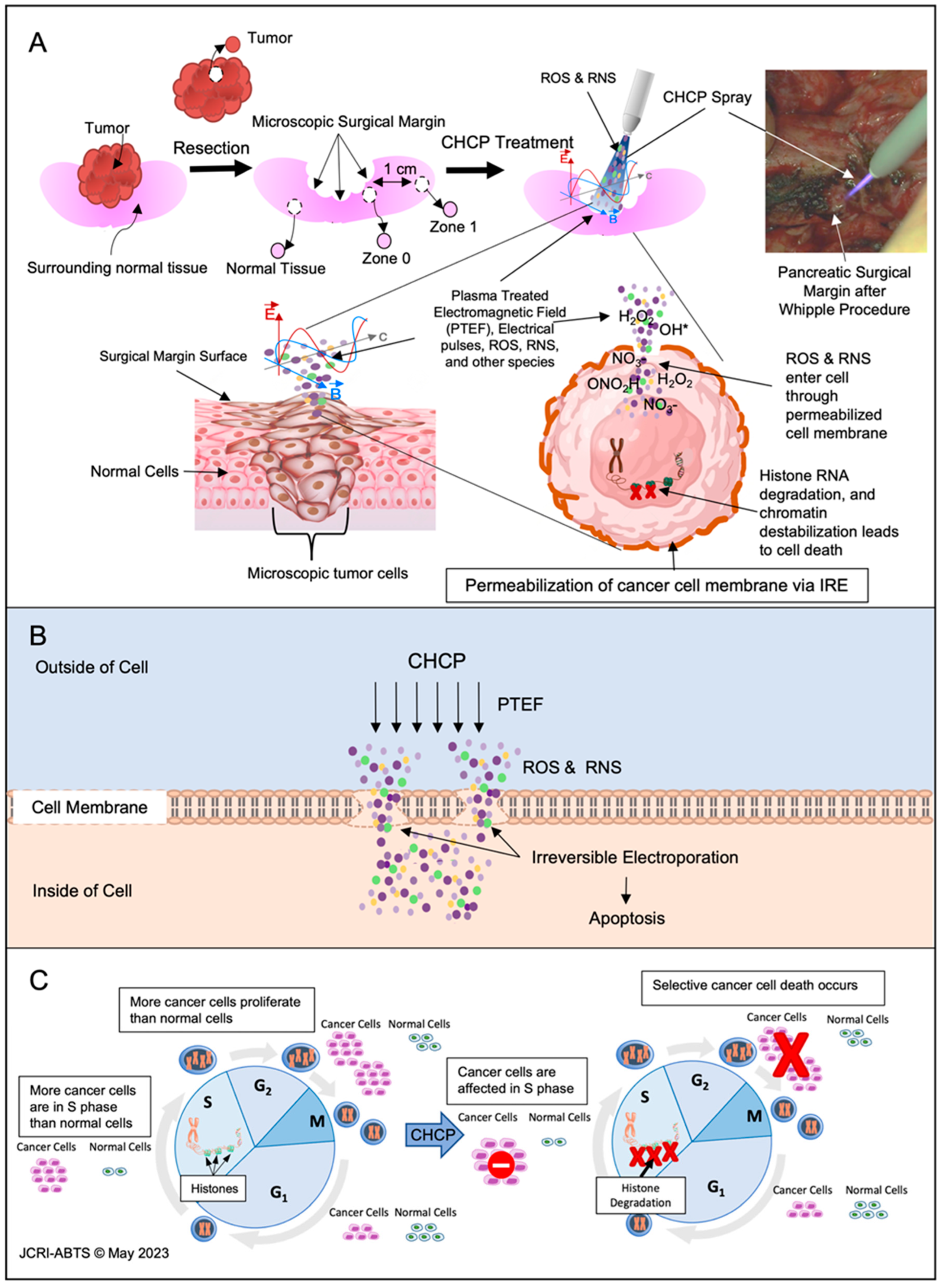
Kaplan–Meier curves with 95% confidence intervals (CI) were generated with RStudio, Version 1.4.1106. Quantitative data were analyzed by Student’s t-test with differences considered statistically significant for * p < 0.05, ** p < 0.01, and *** p < 0.001.

3. Results

3.1. Patient Demographics and Characteristics

Five patients were treated at SMC, and 15 patients at RUMC. Patient details are shown in Table 1.

3.2. Safety Outcomes

3.2.1. Intra-Operative Physiological Data and CHCP Temperature

BP, pulse, body temperature, End Tidal CO2, and O2 saturation were recorded before, during, and after CHCP treatment, which demonstrated no significant changes (Supplementary Figures S2 and S3, p > 0.05).

3.2.2. Post-Operative Adverse Events

There were no adverse events related to CHCP treatment. Two patients had surgical complications (Clavien Dindo Grade III and IIIb); refer to Table 1.

3.3. Efficacy Outcomes

3.3.1. Recurrence and Overall Survival

Twelve patients underwent R0 resection. JCRI-ABTS reported 5/12 (42%) R0 patients with microscopic positive margins (R0-MPM). Two out of seven (29%) R0 patients exhibited LRR between 1–20 months, and no R0-MPM patients exhibited LRR to date. The 26-month ORR for R0 and R0-MPM patients was 69% (95% CI, 19–40%) and 100%, respectively (95% CI, 100–100.0%) (Figure 2A). Comparison of ORR and LRR-free survival between R0 patients and those with R0-MPM did not reveal any statistically significant differences, with median durations of 21 and 25 months, respectively (Figure 2A,B). As of 13 February 2023, 10/20 (50%) patients succumbed to their disease within a range of 3 to 32 months. The Kaplan–Meier analysis (Figure 2C) indicated a 31-month overall survival (OS) rate of 24% (95% CI, 5.3–100.0%) with a median survival of 23 months. Survival rates at 28 months varied among patient groups: R0 (86%), R0-MPM (40%), R1 (67%), and R2 (0%) (p < 0.013) (Figure 2C). Only R0 and R0-MPM patients showed significantly higher survival rates compared to R2 patients at 10 months (p < 0.0019 and p < 0.0083, respectively). No significant difference in survival rate was found among R1 and R2 patients at 10 months or among R0, R0-MPM, and R1 at 28 months. The median survival for R0, R0-MPM, R1, and R2 patients were 23, 25, 25.5, and 6.5 months, respectively.

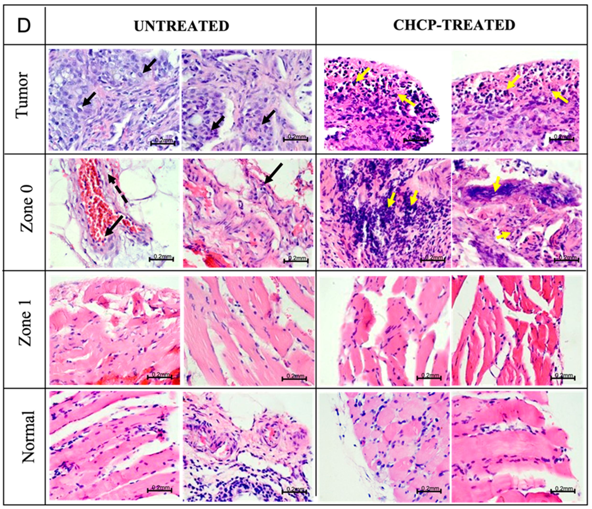
3.3.2. Histopathology Evaluation

RUMC and SMC Pathology Department reported no thermal damage or histological changes in 19/21 (90%) samples of ex vivo CHCP-treated normal tissue. Two samples demonstrated thermal damage, but a Bovie electrocautery device was used to dissect the tissue prior to CHCP treatment.

3.3.3. Histopathology Findings

A total of 12 patients (60%) achieved R0 status. Tissue specimens were collected as illustrated in Figure 3A: Tumor (resected tumor tissue), Zone 0 (surgical margin), Zone 1 (1 cm away from the surgical margin), and Normal (surrounding normal tissue of the tumor). Resected tissues were CHCP treated ex vivo. The electroporation and cellular mechanism of CHCP in cancer cells is illustrated in Figure 3B,C and reported in our previous studies [30,33]. H&E stained tissue samples demonstrated CHCP-induced tumor cell death in 15/15 samples (100%) and histological damages on normal tissue in 0/15 samples (0%). Tumor cells were detected in the following areas: Zone 0 in 5/9 (55%), Zone 1 in 0/6 (0%), and normal tissue in patient R0010 (Tensor Fasciae Latae) 1/13 (7.7%). No thermal damage in normal tissue was found after CHCP treatment (Figure 3D). A complete overview of H&E-stained CHCP-treated and untreated tissues is available in Supplementary Figure S4.

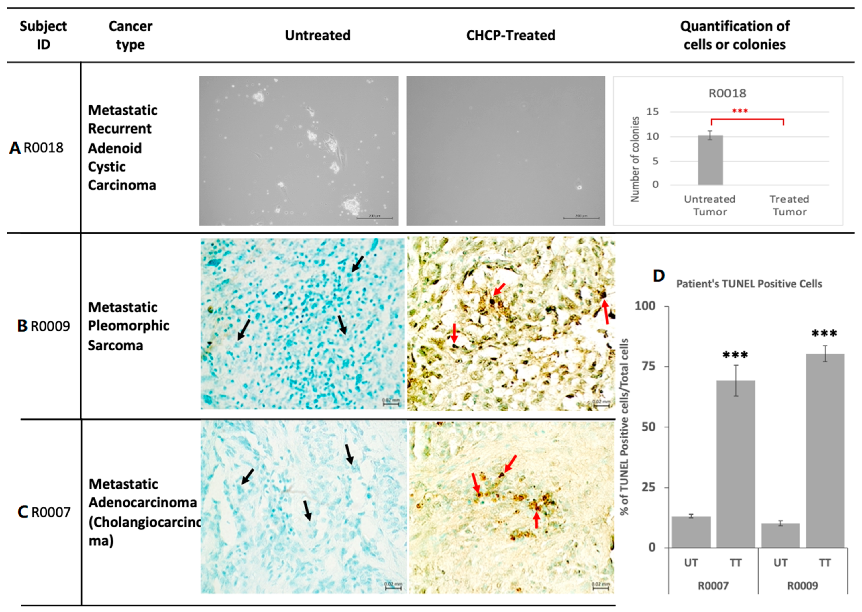
3.3.4. Primary Tissue Culture

The primary culture was established successfully from 10/15 (67%) tumor samples (Figure 4A). Tumor cell survival from the CHCP-treated tissues was significantly lower compared to the untreated tumor tissues in 8/10 (80%) cases, no cancer cells survived in 3/10 (30%) cases, and significantly fewer cells survived in 5/10 (50%) cases (Supplementary Figure S5). CHCP did not significantly reduce cancer cell survival in 2/10 cases (20%) due to suboptimal treatment doses.

3.3.5. Apoptotic DNA Damage Analysis by TUNEL Assay

Ex vivo CHCP-treated samples were analyzed for apoptosis by TUNEL assay, and positive cells were measured (Figure 4). Less than 1% of the untreated and treated normal cells showed TUNEL staining (p > 0.05). CHCP-treated tumor tissue showed a significant increase in TUNEL-positive cells compared to untreated tumor tissue (54–88%), demonstrating CHCP-induced DNA damage and apoptosis in cancer cells (p < 0.001). A complete comparison table demonstrating all patient tissue samples with TUNEL staining of CHCP-treated and untreated tumor samples is available in Supplementary Figure S6.

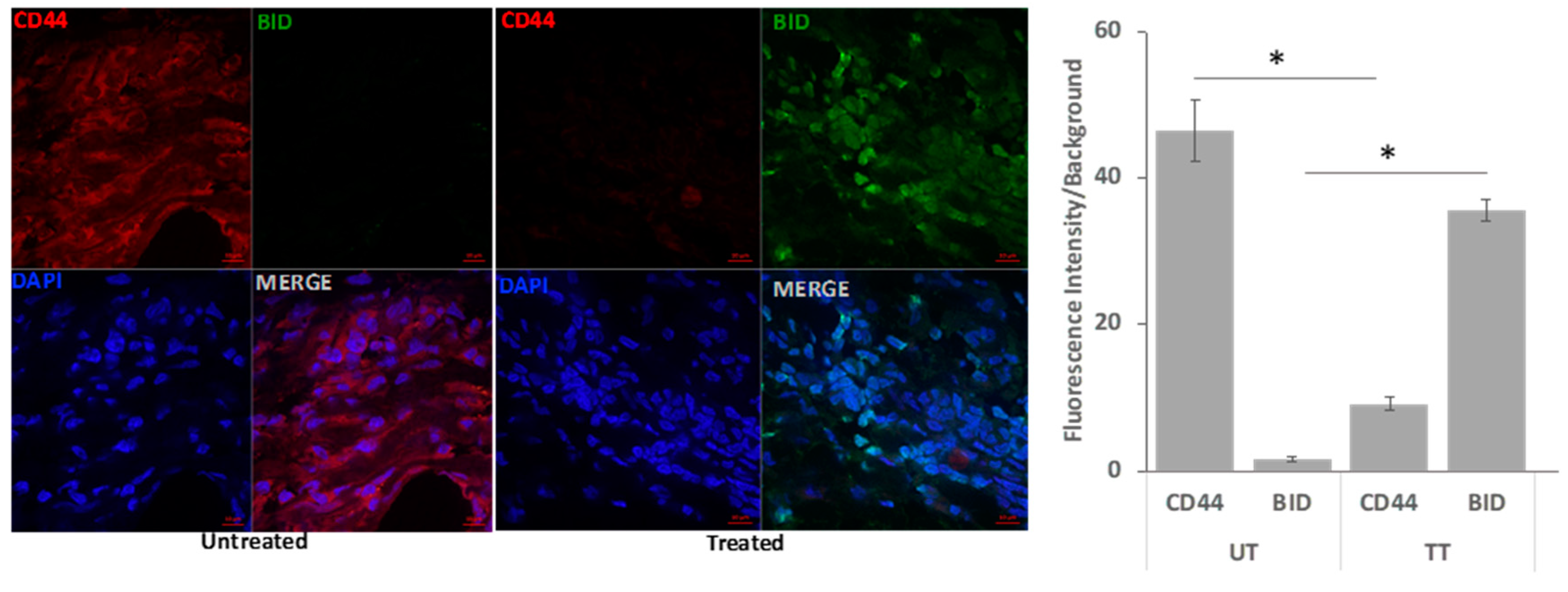
3.3.6. Confocal Immunofluorescence Analysis

Quantitative confocal immunofluorescence analysis showed that tumor stem cell marker CD44 was significantly decreased, and the apoptotic marker BID protein expression was increased in CHCP-treated tumor samples compared to untreated tumor samples (Figure 5). Significant morphological alterations such as shrinkage or fragmentation in the nuclear morphology of cells in the CHCP-treated tumor tissues were observed, suggesting chromatin destabilization during apoptosis.

3.3.7. Gene Expression Analysis

CHCP-treated tumor samples were analyzed for gene expression using real-time qPCR to evaluate 111 genes related to apoptosis, oxidative stress, immune checkpoint, T-cell regulation, and tumor promotion. Complete details of the study, including a table of differentially regulated genes, are provided in Supplementary Table S1.

3.3.8. Regulation of Protein Expression

CHCP-treated and control tissue samples were analyzed using Western blot for the expression of apoptotic protein markers (BAD, BCL2, PUMA, and BID). Some cancer types exhibited significant up-regulation of pro-apoptotic proteins and down-regulation of the anti-apoptotic marker BCL2 following CHCP treatment. Supplementary Figure S7 provides detailed data and analysis.

4. Discussion

The LRR rate for solid tumors remains as high as 65%, even in the absence of distant disease [4]. Historically, stage IV solid tumors are typically not resected due to the high risk of palliative surgery [35]. We demonstrated that CHCP treatment in combination with surgery is a viable option for controlling LRR and possibly improving overall survival.
CHCP is a safe and potentially effective treatment option for a wide range of Stage IV or Recurrent solid tumors. Although R0 resection was achieved in 60% of patients (n = 12), microscopic tumor cells were identified at the surgical margin site (Zone 0) in 5 patients (R0-MPM, 42%) (R0004, R0005, R0009, R0011, and R0012). Despite the OS rate of 40% at 28 months observed in R0-MPM patients, none of the patients exhibited any signs of recurrence (LRR) during the follow-up period. The absence of LRR in these five R0-MPM patients following the CHCP therapeutic approach could completely eradicate undetectable microscopic tumor cells at the surgical margin site, independent of the cancer type.
Ex vivo observations of H&E and TUNEL staining showed significant cell death at Zone 0. Molecular and cell biology analyses demonstrated that CHCP treatment induced differential expression of pro-apoptotic and anti-apoptotic genes. These findings provide compelling evidence of CHCP-induced apoptosis in cancer cells, underscoring its potential to eliminate microscopic tumor cells at the surgical margin.
The 26-month non-LRR rate of R0 and R0-MPM patients reached 57% and 80%, respectively. There was no significant difference in OS rate between R0, R0-MPM, and R1 patients at 28 months. Furthermore, overwhelming significance in OS was observed when comparing R0, R0-MPM, R1, and R2 resection, with rates of 86%, 40%, 67%, and 0% at 28 months, respectively. These data underscore the heightened therapeutic efficacy of CHCP treatment when combined with R0, R0-MPM, and R1 surgical resection. Consequently, our study highlights the potential of expanding CHCP treatment to encompass less invasive solid tumors, as it offers promise for enhancing local control.
CHCP is a non-invasive and non-contact electroporation device that creates PTEF™, ROS, RNS, and other species which can be personalized to the patient’s cancer or subtype. CHCP treatment takes only 5–7 min intra-operatively and preserves the surrounding noncancerous tissue with no side effects in contrast to contact-based electroporation devices such as Optune (Novocure), NanoKnife (Angiodynamics), and Aliya System (Galvanize Therapeutics). CHCP seamlessly integrates into existing standard-of-care protocols without adding extra burden or disrupting the patient’s overall treatment regimen.
The JCRI-ABTS laboratory discovered two mechanisms underlying the effects of CHCP-generated electromagnetic fields on membrane permeability: electroporation and alteration of membrane lipid structure [33], consistent with previous studies on CAP [12,13]. We provide valuable insight into the biological effects of PTEF™ and the mechanisms underlying its ability to induce membrane permeability in living cells [36,37,38].
Furthermore, our laboratory discovered that CHCP-treated cancer cells undergo histone mRNA oxidation and degradation, leading to the upregulation of DNA damage response genes and apoptosis [30]. Compared to normal cells, cancer cells have frequent cell cycles and a greater percentage of cells in the S-phase, and higher levels of intracellular ROS and RNS [39]. Additional CAP-generated species may overwhelm the system and switch the ROS and RNS effect from tumor-promoting to tumor-suppressing [40].
Studies on CAP treatment of various cancer types have demonstrated the abscopal effect in tumors, suggesting its potential to induce an immune response by releasing cytokines and DAMPs from apoptotic cancer cells [41]. However, immunosuppressants may hinder this effect, possibly contributing to early LRR in patient R0013 with a history of immunosuppressant use.
We acknowledge the study limitation of 20 patients, and a phase II study will be conducted with a larger racially, ethnically diverse cohort of patients to determine CHCP efficacy on different cancer molecular subtypes.

5. Conclusions

This clinical trial opens an important window into the potential use of this technology following Stage IV solid tumor cytoreduction and primary tumorectomy. These results highlight the safety, selectivity, and significant reduction of local regional recurrence achieved through the synergistic approach of CHCP treatment and surgery. Our comprehensive findings underscore the remarkable capacity of CHCP to eradicate the possibility of recurrence, even when microscopic tumor cells are detected at the surgical margin site in R0 patients. The combination of CHCP potency on cancer cells and the benign interaction with normal tissue renders it an exceptionally enticing therapeutic approach for addressing surgical margins, thereby mitigating the risk of recurrence.

Supplementary Materials

The following supporting information can be downloaded at: https://www.mdpi.com/article/10.3390/cancers15143688/s1, Figure S1: CHCP Treatment Temperature Measurements; Figure S2: Patient vital signs during surgery; Figure S3: Patient vital signs during surgery; Figure S4: Histopathology of Patient Tissue Samples; Figure S5: Primary Cell Culture of Patient Tumor Samples; Figure S6: TUNEL Assay Analysis; Figure S7: Western blot analysis for apoptotic markers after CHCP treatment on patient tumor samples; Figure S8: Tumor Cell Death at the surgical margin; Table S1: Gene profiling analysis; Excel File: Western Blot image source file.

Author Contributions

Conceptualization, J.C., T.Z., S.G., M.A., M.K., G.B. and A.N.; methodology, J.C., T.Z., A.N., S.G., M.A., M.K., G.B. and S.R.K.M.; software, T.Z.; validation, J.C., T.Z., S.R.K.M., S.G., A.N., L.L., O.Z.J., M.K., G.B. and X.C.; formal analysis, J.C., S.R.K.M., T.Z., L.L., O.Z.J., M.A., G.B. and X.C.; investigation, J.C., A.N., S.G., M.A., A.T.B., M.W.C., K.M., C.O., K.M.S. and S.R.K.M.; resources, J.C.; data curation, J.C., S.R.K.M., L.L., O.Z.J., K.O., G.S. and X.C.; writing—original draft preparation, J.C., S.R.K.M., L.L. and O.Z.J.; writing—review and editing, J.C., S.R.K.M., L.L., O.Z.J., M.A., A.T.B., M.W.C., K.M., C.O., K.M.S., K.O., G.S., M.K., S.G., A.N. and G.B.; visualization, J.C., T.Z. and S.R.K.M.; supervision, J.C.; project administration, J.C., T.Z., S.G., A.N., K.O., G.S. and S.R.K.M.; funding acquisition, J.C. All authors have read and agreed to the published version of the manuscript.

Funding

This research was funded by US Medical Innovations for #G19065.

Institutional Review Board Statement

The study was conducted in accordance with the Declaration of Helsinki and approved by the Institutional Review Board of RUMC and SMC after receiving approval from the FDA (IDE #G19065) and the Israeli Ministry of Health. Sheba IRB Number 6224-19; Rush Institutional Review Board FWA #: 00000482, ORA Number: 19062503-IRB01-CR03.

Informed Consent Statement

Informed consent was obtained from all subjects involved in the study.

Data Availability Statement

The data presented in this study are available in this article (and supplementary material).

Acknowledgments

We thank the participating patients and their families, previous compassionate use patients, surgical teams performing these advanced high-risk surgical procedures, and allowance by SMC and RUMC and the study team at JCRI-ABTS. Independent reviewers Norman Lepor, M.D., and Clyde Callender, M.D., and all authors approved the final version of the manuscript.

Conflicts of Interest

The authors declare no conflict of interest.

References

  1. Siegel, R.L.; Miller, K.D.; Fuchs, H.E.; Jemal, A. Cancer Statistics, 2021. CA Cancer J. Clin. 2021, 71, 7–33. [Google Scholar] [CrossRef] [PubMed]
  2. Miller, K.D.; Nogueira, L.; Devasia, T.; Mariotto, A.T.B.; Yabroff, K.R.; Jemal, A.; Kramer, J.; Siegel, R.L. Cancer treatment and survivorship statistics, 2022. CA Cancer J. Clin. 2022, 72, 409–436. [Google Scholar] [CrossRef] [PubMed]
  3. Sanoff, H.K. Improving Treatment Approaches for Rectal Cancer. N. Engl. J. Med. 2022, 386, 2425–2426. [Google Scholar] [CrossRef] [PubMed]
  4. Mahvi, D.A.; Liu, R.; Grinstaff, M.W.; Colson, Y.L.; Raut, C.P. Local Cancer Recurrence: The Realities, Challenges, and Opportunities for New Therapies. CA Cancer J. Clin. 2018, 68, 488–505. [Google Scholar] [CrossRef] [PubMed] [Green Version]
  5. Buchholz, T.A.; Ali, S.; Hunt, K.K. Multidisciplinary Management of Locoregional Recurrent Breast Cancer. J. Clin. Oncol. 2020, 38, 2321–2328. [Google Scholar] [CrossRef]
  6. Cihoric, N.; Tsikkinis, A.; Filipovic, N.; Jeremic, B. Treatment options for isolated locoregional recurrences of nonsmall cell lung cancer after surgery: Yes, radiation therapy too! Eur. Respir. J. 2016, 48, 276–278. [Google Scholar] [CrossRef] [Green Version]
  7. Laroussi, M.; Lu, X.; Keidar, M. Perspective: The physics, diagnostics, and applications of atmospheric pressure low temperature plasma sources used in plasma medicine. J. Appl. Phys. 2017, 122. [Google Scholar] [CrossRef]
  8. Charles, F.; Morrison, J. Electrosurgical Method and Apparatus for Initiating An Electrical Discharge in An Inert Gas Flow. U.S. Patent 4,040,426, 9 August 1977. [Google Scholar]
  9. Canady, J. Surgical Coagulation Device. U.S. Patent 5,207,675, 4 May 1993. [Google Scholar]
  10. Babington, P.; Rajjoub, K.; Canady, J.; Siu, A.; Keidar, M.; Sherman, J.H. Use of cold atmospheric plasma in the treatment of cancer. Biointerphases 2015, 10, 029403. [Google Scholar] [CrossRef] [PubMed] [Green Version]
  11. Laroussi, M. Low Temperature Plasma-Based Sterilization: Overview and State-of-the-Art. Plasma Process. Polym. 2005, 2, 391–400. [Google Scholar] [CrossRef]
  12. Yan, D.; Wang, Q.; Adhikari, M.; Malyavko, A.; Lin, L.; Zolotukhin, D.B.; Yao, X.; Kirschner, M.; Sherman, J.H.; Keidar, M. A Physically Triggered Cell Death via Transbarrier Cold Atmospheric Plasma Cancer Treatment. ACS Appl. Mater. Interfaces 2020, 12, 34548–34563. [Google Scholar] [CrossRef]
  13. Haralambiev, L.; Nitsch, A.; Einenkel, R.; Muzzio, D.O.; Gelbrich, N.; Burchardt, M.; Zygmunt, M.; Ekkernkamp, A.; Stope, M.B.; Gumbel, D. The Effect of Cold Atmospheric Plasma on the Membrane Permeability of Human Osteosarcoma Cells. Anticancer Res. 2020, 40, 841–846. [Google Scholar] [CrossRef]
  14. Ly, L.; Cheng, X.; Murthy, S.R.K.; Jones, O.Z.; Zhuang, T.; Gitelis, S.; Blank, A.T.; Nissan, A.; Adileh, M.; Colman, M.; et al. Canady Cold Helios Plasma Reduces Soft Tissue Sarcoma Viability by Inhibiting Proliferation, Disrupting Cell Cycle, and Inducing Apoptosis: A Preliminary Report. Molecules 2022, 27, 4168. [Google Scholar] [CrossRef]
  15. Gjika, E.; Pal-Ghosh, S.; Kirschner, M.E.; Lin, L.; Sherman, J.H.; Stepp, M.A.; Keidar, M. Combination Therapy of Cold Atmospheric Plasma (CAP) With Temozolomide in the Treatment of U87MG Glioblastoma Cells. Sci. Rep. 2020, 10, 16495. [Google Scholar] [CrossRef] [PubMed]
  16. Chen, Z.; Simonyan, H.; Cheng, X.; Gjika, E.; Lin, L.; Canady, J.; Sherman, J.H.; Young, C.; Keidar, M. A Novel Micro Cold Atmospheric Plasma Device for Glioblastoma Both In Vitro and In Vivo. Cancers 2017, 9, 61. [Google Scholar] [CrossRef] [PubMed] [Green Version]
  17. Liu, J.-R.; Wu, Y.-M.; Xu, G.-M.; Gao, L.-G.; Ma, Y.; Shi, X.-M.; Zhang, G.-J. Low-temperature plasma induced melanoma apoptosis by triggering a p53/PIGs/caspase-dependent pathway in vivo and in vitro. J. Phys. D Appl. Phys. 2019, 52, 315204. [Google Scholar] [CrossRef]
  18. Cheng, X.; Rowe, W.; Ly, L.; Shashurin, A.; Zhuang, T.; Wigh, S.; Basadonna, G.; Trink, B.; Keidar, M.; Canady, J. Treatment of Triple-Negative Breast Cancer Cells with the Canady Cold Plasma Conversion System: Preliminary Results. Plasma 2018, 1, 218–228. [Google Scholar] [CrossRef] [Green Version]
  19. Ly, L.; Cheng, X.; Murthy, S.R.K.; Zhuang, T.; Jones, O.Z.; Basadonna, G.; Keidar, M.; Canady, J. Canady Cold Plasma Conversion System Treatment: An effective Inhibitor of Cell Viability In Breast Cancer Molecular Subtypes. Clin. Plasma Med. 2020, 19, 100109. [Google Scholar] [CrossRef]
  20. Sun, C.; Chesnokov, V.; Larson, G.; Itakura, K. Glucosamine Enhances TRAIL-Induced Apoptosis in the Prostate Cancer Cell Line DU145. Medicines 2019, 6, 104. [Google Scholar] [CrossRef] [Green Version]
  21. Griseti, E.; Kolosnjaj-Tabi, J.; Gibot, L.; Fourquaux, I.; Rols, M.P.; Yousfi, M.; Merbahi, N.; Golzio, M. Pulsed Electric Field Treatment Enhances the Cytotoxicity of Plasma-Activated Liquids in a Three-Dimensional Human Colorectal Cancer Cell Model. Sci. Rep. 2019, 9, 7583. [Google Scholar] [CrossRef] [Green Version]
  22. Ma, J.; Yu, K.N.; Cheng, C.; Ni, G.; Shen, J.; Han, W. Targeting Nrf2-mediated heme oxygenase-1 enhances non-thermal plasma-induced cell death in non-small-cell lung cancer A549 cells. Arch. Biochem. Biophys. 2018, 658, 54–65. [Google Scholar] [CrossRef] [PubMed]
  23. Rowe, W.; Cheng, X.; Ly, L.; Zhuang, T.; Basadonna, G.; Trink, B.; Keidar, M.; Canady, J. The Canady Helios Cold Plasma Scalpel Significantly Decreases Viability in Malignant Solid Tumor Cells in a Dose-Dependent Manner. Plasma 2018, 1, 177–188. [Google Scholar] [CrossRef] [Green Version]
  24. Feil, L.; Koch, A.; Utz, R.; Ackermann, M.; Barz, J.; Stope, M.; Kramer, B.; Wallwiener, D.; Brucker, S.Y.; Weiss, M. Cancer-Selective Treatment of Cancerous and Non-Cancerous Human Cervical Cell Models by a Non-Thermally Operated Electrosurgical Argon Plasma Device. Cancers 2020, 12, 1037. [Google Scholar] [CrossRef] [PubMed] [Green Version]
  25. Guerrero-Preston, R.; Ogawa, T.; Uemura, M.; Shumulinsky, G.; Valle, B.L.; Pirini, F.; Ravi, R.; Sidransky, D.; Keidar, M.; Trink, B. Cold Atmospheric Plasma Treatment Selectively Targets Head and Neck Squamous Cell Carcinoma Cells. Int. J. Mol. Med. 2014, 34, 941–946. [Google Scholar] [CrossRef] [Green Version]
  26. Yan, D.; Sherman, J.H.; Keidar, M. Cold atmospheric plasma, a novel promising anti-cancer treatment modality. Oncotarget 2017, 8, 15977–15995. [Google Scholar] [CrossRef] [Green Version]
  27. Vaquero, J.; Judée, F.; Vallette, M.; Decauchy, H.; Arbelaiz, A.; Aoudjehane, L.; Scatton, O.; Gonzalez-Sanchez, E.; Merabtene, F.; Augustin, J.; et al. Cold-Atmospheric Plasma Induces Tumor Cell Death in Preclinical In Vivo and In Vitro Models of Human Cholangiocarcinoma. Cancers 2020, 12, 1280. [Google Scholar] [CrossRef] [PubMed]
  28. Ly, L.; Jones, S.; Shashurin, A.; Zhuang, T.; Rowe, W.; Cheng, X.; Wigh, S.; Naab, T.; Keidar, M.; Canady, J. A New Cold Plasma Jet: Performance Evaluation of Cold Plasma, Hybrid Plasma and Argon Plasma Coagulation. Plasma 2018, 1, 189–200. [Google Scholar] [CrossRef] [Green Version]
  29. Canady, J.; Shashurin, A.; Keidar, M.; Zhuang, T. Integrated cold plasma and high frequency plasma electrosurgical system and method. U.S. Patent No. 9,999,462, 19 June 2018. [Google Scholar]
  30. Cheng, X.; Murthy, S.R.K.; Zhuang, T.; Ly, L.; Jones, O.; Basadonna, G.; Keidar, M.; Kanaan, Y.; Canady, J. Canady Helios Cold Plasma Induces Breast Cancer Cell Death by Oxidation of Histone mRNA. Int. J. Mol. Sci. 2021, 22, 9578. [Google Scholar] [CrossRef]
  31. Elbedour, A.; Cheng, X.; Murthy, S.R.K.; Zhuang, T.; Ly, L.; Jones, O.; Basadonna, G.; Keidar, M.; Canady, J. The Granger Causal Effects of Canady Helios Cold Plasma on the Inhibition of Breast Cancer Cell Proliferation. Appl. Sci. 2022, 12, 4622. [Google Scholar] [CrossRef]
  32. Jones, O.; Cheng, X.; Murthy, S.R.K.; Ly, L.; Zhuang, T.; Basadonna, G.; Keidar, M.; Canady, J. The Synergistic Effect of Canady Helios Cold Atmospheric Plasma and a FOLFIRINOX Regimen for the Treatment of Cholangiocarcinoma In Vitro. Sci. Rep. 2021, 11, 8967. [Google Scholar] [CrossRef]
  33. Murthy, S.R.K.; Cheng, X.; Zhuang, T.; Ly, L.; Jones, O.; Basadonna, G.; Keidar, M.; Canady, J. BCL2A1 regulates Canady Helios Cold Plasma-induced cell death in triple-negative breast cancer. Sci. Rep. 2022, 12, 4038. [Google Scholar] [CrossRef]
  34. Schwartz, L.H.; Litiere, S.; de Vries, E.; Ford, R.; Gwyther, S.; Mandrekar, S.; Shankar, L.; Bogaerts, J.; Chen, A.; Dancey, J.; et al. RECIST 1.1-Update and clarification: From the RECIST committee. Eur. J. Cancer 2016, 62, 132–137. [Google Scholar] [CrossRef] [PubMed] [Green Version]
  35. Badgwell, B.; Krouse, R.; Cormier, J.; Guevara, C.; Klimberg, V.S.; Ferrell, B. Frequent and early death limits quality of life assessment in patients with advanced malignancies evaluated for palliative surgical intervention. Ann. Surg. Oncol. 2012, 19, 3651–3658. [Google Scholar] [CrossRef] [PubMed]
  36. Neumann, E.; Rosenheck, K. Permeability changes induced by electric impulses in vesicular membranes. J. Membr. Biol. 1972, 10, 279–290. [Google Scholar] [CrossRef] [PubMed] [Green Version]
  37. Escoffre, J.M.; Portet, T.; Wasungu, L.; Teissié, J.; Dean, D.; Rols, M.P. What is (still not) known of the mechanism by which electroporation mediates gene transfer and expression in cells and tissues. Mol. Biotechnol. 2009, 41, 286–295. [Google Scholar] [CrossRef]
  38. Teissie, J.; Golzio, M.; Rols, M.P. Mechanisms of cell membrane electropermeabilization: A minireview of our present (lack of ?) knowledge. Biochim. Biophys. Acta 2005, 1724, 270–280. [Google Scholar] [CrossRef]
  39. Volotskova, O.; Hawley, T.S.; Stepp, M.A.; Keidar, M. Targeting the cancer cell cycle by cold atmospheric plasma. Sci. Rep. 2012, 2, 636. [Google Scholar] [CrossRef] [Green Version]
  40. Semmler, M.L.; Bekeschus, S.; Schafer, M.; Bernhardt, T.; Fischer, T.; Witzke, K.; Seebauer, C.; Rebl, H.; Grambow, E.; Vollmar, B.; et al. Molecular Mechanisms of the Efficacy of Cold Atmospheric Pressure Plasma (CAP) in Cancer Treatment. Cancers 2020, 12, 269. [Google Scholar] [CrossRef] [Green Version]
  41. Mizuno, K.; Yonetamari, K.; Shirakawa, Y.; Akiyama, T.; Ono, R. Anti-tumor immune response induced by nanosecond pulsed streamer discharge in mice. J. Phys. D Appl. Phys. 2017, 50, 12LT01. [Google Scholar] [CrossRef]
Figure 1. (A) Device setup of Canady Helios™ Cold Plasma System. (B) Study protocol showing pre-operative, intra-operative, and post-operative treatments. The patients included in the trial underwent multiple rounds of chemotherapy and radiation before being considered for Canady Helios Cold Plasma (CHCP) treatment. FDA approval was specifically granted for the use of CHCP at the surgical margins after macroscopic removal of solid tumors in patients with Stage 4 metastatic or recurrent solid tumors. Patient selection was based on a comprehensive evaluation conducted by a multi-disciplinary team at each institution. Eligible patients had a treatment history including chemotherapy, radiation, immunotherapy, surgery, and Hyperthermic Intraperitoneal Chemotherapy (HIPEC). Notably, these patients would not traditionally have been offered surgery due to the advanced stage of their disease. The primary objective of the CHCP trial was to demonstrate the safety of the treatment. Achieving a complete (R0) or microscopic residual (R1) surgical resection with CHCP treatment was to decrease the likelihood of local regional recurrence (LRR).
Cancers 15 03688 g001
Figure 2. Kaplan–Meier survival curves as of February 13, 2023. (AC) Symbol ‘+’ indicates the last follow-up of each individual patient. (A) ORR of patients with R0 (n = 7) and R0-MPM status (n = 5) (p > 0.05). Early recurrence (<20 months) occurred in two R0 patients: R0013 had a previous kidney transplant on immunosuppression, and R0002 with metastatic myxofibrosarcoma. (B) LRR-free survival rate (including events for recurrence or death) for patients with R0 (n = 7) and R0-MPM status (n = 5) (p > 0.05). (C) OS rates of all patients (n = 20) and among the subset of R0 (n = 7), R0-MPM (n = 5), R1 (n = 6), and R2 (n = 2) patients: R0 vs. R2 (p < 0.0019), and R0-MPM vs. R2 (p < 0.0083) showed significant difference at 10 months.
Cancers 15 03688 g002
Figure 3. (A) Schematic images of the surgery and CHCP treatment procedure: resection of the tumor using standard surgical procedures, collection of specimens (Tumor, Zone 0, Zone 1, and Normal tissue), and application of CHCP spray to the surgical margin and a representative picture of CHCP treatment performed on pancreatic surgical margin after Whipple procedure for cancer. The OH* denotes free form of hydroxyl; (PTEF: E is the electrical field, B is the magnetic field, C is the 3D reference of the magnetic field.) (B) Illustration of CHCP produced PTEF™, ROS, and RNS entering a cancer cell membrane through “pores” generated by electroporation. (C) Illustration of the selective induction of cancer cell death by CHCP treatment resulting from 8-oxoG modification and degradation of histone mRNA during the early S phase. (D) Light micrographs displaying H&E staining of Patient R0004 (metastatic recurrent non-small cell lung adenocarcinoma to the left hip/upper end of femur) Tumor, Zone 0, Zone 1, and Normal (vastus lateralis muscle) tissue samples with or without CHCP treatment. Black arrows and yellow arrows point to untreated tumor cells and treated dead tumor cells, respectively (scale bars = 0.2 mm).
Cancers 15 03688 g003aCancers 15 03688 g003b
Figure 4. (A) Representative phase contrast images and quantification of primary culture established with Patient R0018 Tumor Samples, untreated and post-CHCP treatment (scale bars = 200 μm). (B,C) represents light microscope images of apoptosis analysis by TUNEL assay staining for patient R0009 (B) and R0007 (C) tumor tissue samples that were untreated (UT) or treated (TT). Black arrows and red arrows point to untreated tumor cells and treated dead tumor cells, respectively. Apoptotic cells were labeled with TdT, and signals were developed using DAB with Methylene Green counter stain. The majority of CHCP untreated tissue sections (0.1–24%) did not show TdT signal. Light Micrographs displaying the morphological Spectrum at 63×. (D) Quantitative analysis of apoptosis demonstrated by TUNEL assay. Percentage of TUNEL-positive cells (54–88%) signified DNA damage due to apoptosis after CHCP treatment. There was a significant increase in TUNEL-positive cells in TT compared to UT. Semi-quantitative analysis was performed on TUNEL-stained FFPE sections by manually counting cells under a light microscope. The data are represented by the SEM (n = 3) (*** p < 0.001, Student’s t-test).
Cancers 15 03688 g004
Figure 5. Expression of apoptotic marker protein BID After CHCP treatment in metastatic recurrent colon cancer patient tumor tissue: Representative multicolor fluorescence confocal images showing tumor cells stained for CD44 (red), BID (green), and DAPI (blue). The CHCP-treated tumor tissue shows increasing protein staining for apoptotic protein BID, and untreated samples show increased staining for tumor marker CD44. Statistical analysis by SEM (n = 12) (* CD44: UT vs. TT p < 0.00643−6, BID: UT vs. TT p < 0.00156−12 Student’s t-test).
Cancers 15 03688 g005
Table 1. Table for patient demographics, characteristics, and surgery details.
Patient ID (Sex, Age)Post-Operative
Diagnosis
R0 Post
Operation
ECOG ≤ 2 ASA ≤ 3
LOS
(Days)
OS Post
Surgical Resection
Adverse Events
with CHCP
Preoperative Staging
Stage IV
(Metastatic or
Recurrent)
Intra- and
Post-Operative Outcomes
Neo-Adjuvant
Chemo-Therapy
Intra-Operative
Chemo-Therapy
Adjuvant
Chemo-Therapy
S0001
(F, 49)
Metastatic recurrent colon cancerYesYes7Died of disease (32 months)
Lost follow-up (26 months)
NoneYesNo complicationsCTHIPEC, IORTCT
S0002
(F, 44)
Metastatic recurrent ovarian cancerNoYes5Died of disease
(29 months)
NoneYesNo complicationsCTNoneCT
S0003
(F, 61)
Metastatic anal cancer to the liver NoYes5Died of disease (3 months)NoneYesNo complicationsCTNoneIMT
S0004
(M, 51)
Recurrent epithelioid peritoneal MesotheliomaNoYes1Died of disease (10 months)NoneYesNo complicationsCTHIPECNone
S0005
(M, 60)
Recurrent Colon CancerNoYes6Died of disease
(31 months)
NoneYesNo complicationsCTHIPECCT
R0002
(F, 85)
Metastatic myxofibrosarcoma left chest wallYesYes1Alive with disease
(30 months)
Recurrence of disease (20 months) at left chest wall resection site
NoneYesNo complicationsRT, RT (internal), CT NoneRT
R0003
(F, 63)
Recurrent metastatic breast carcinoma to the right pelvisNoYes5Alive with disease
(29 months)
FDRS9
NoneYesNo complicationsRT, HTNoneRT, HT, IMT, CT
R0004
(F, 76)
Metastatic non-small cell lung carcinoma (NSCLC) to the left proximal femurYesYes4Died of disease
(26 months)
NoneYesNo complicationsNoneNoneRT, IMT
R0005
(M, 61)
Metastatic renal cell carcinoma to the left clavicleYesYes2Alive
(28 months)
FDRS
NoneYesNo complicationsRTNoneIMT
R0007
(M, 59)
Metastatic cholangiocarcinoma to the left distal humerusNoYes3Died of disease
(8 months)
NoneYesNo complicationsNoneNoneCT, CRT
R0008
(M, 58)
Metastatic Non-small cell Lung Carcinoma (NSCLC) to the right hip and spine.YesYes7Died of disease
(8 months)
NoneYesNo complicationsCRT, IMT, RT, CTNoneCT
R0009
(F, 77)
Pleomorphic sarcoma of left distal femurYesYes2Alive (25 months)
FDRS
NoneYesNo complicationsNoneNoneRT
R0011
(M, 67)
Metastatic Chordoma to right glutealYesYes11Died of disease
(20 months)
NoneYesNo complicationsRTNoneRT
R0010
(M, 53)
Metastatic melanoma to the left pelvisNoYes18Died of disease
(4 months)
NoneYesIntraoperative venous bleeding.
Emergent angiogram
IR embolization 1st re-admission to hospital for concern of active bleeding in the surgical area. Treatment embolization of left illiac artery.
2nd re-admission to hospital for continued surgical incision drainage observation. New bleed ruled out. Treatment with silvadene dressing changes.
IMTNoneNone
R0012
(M, 41)
Metastatic Pleomorphic Spindle cell sarcomaYesYes1Alive (23 months)
FDRS
NoneYesNo complicationsCRTNoneRT, CT
R0013
(F, 55)
Squamous cell Carcinoma (SCC)
(History of Kidney Transplant)
YesYes2Alive with disease (23 months)
Recurrence of disease (1 month) in the hand and axillary lymph node. Receiving anti-rejection immunosuppression therapy for kidney transplant.
NoneYesNo complicationsIMTNoneCT
R0014
(F, 77)
Metastatic angiosarcoma to contralateral breast right AxillaYesYes1Alive (23 months)
FDRS
NoneYesNo complicationsRT, HT, CTNoneCT
R0016
(F, 50)
Metastatic Non-Small Cell Lung Carcinoma (NSCLC) to BoneNoYes2Alive (22 months)
FDRS
NoneYesNo complicationsInternal RT (radioactive iodine); RT, CRT, and CT/IMT for NSCLC.NoneRT/IMT
R0017
(M, 26)
Metastatic Desmoplastic Small Round Cell Sarcoma of left inguinal soft tissue and testicleYesYes1Alive (22 months)
FDRS
NoneYesNo complicationsCTNoneCRT
R0018
(M, 69)
Metastatic Adenoid Cystic Carcinoma of the
left submandibular gland
YesYes28Alive (21 months)
FDRS
NoneYesSalivary leak. Return to the OR for Repair of floor of the mouth defect with buccal fat flapRT, CTNoneNone
R0 resection—No gross or microscopic tumor at the surgical margin; ECOG—Eastern Cooperative Oncology Group (Performance Status); LOS—Length of Stay; OS—Overall Survival after surgical resection; CHCP—Canady Helios Cold Plasma; HIPEC—Hyperthermic Intraperitoneal Chemotherapy; IMT—Immunotherapy; HT—Hormone Therapy; RT—Radiation Therapy; CT—Chemotherapy; IORT—Intraoperative Radiation Therapy; FDRS—Free of Disease at Resection Site; Region of Enrolment: S00## SMC, R00## RUMC.
Disclaimer/Publisher’s Note: The statements, opinions and data contained in all publications are solely those of the individual author(s) and contributor(s) and not of MDPI and/or the editor(s). MDPI and/or the editor(s) disclaim responsibility for any injury to people or property resulting from any ideas, methods, instructions or products referred to in the content.

