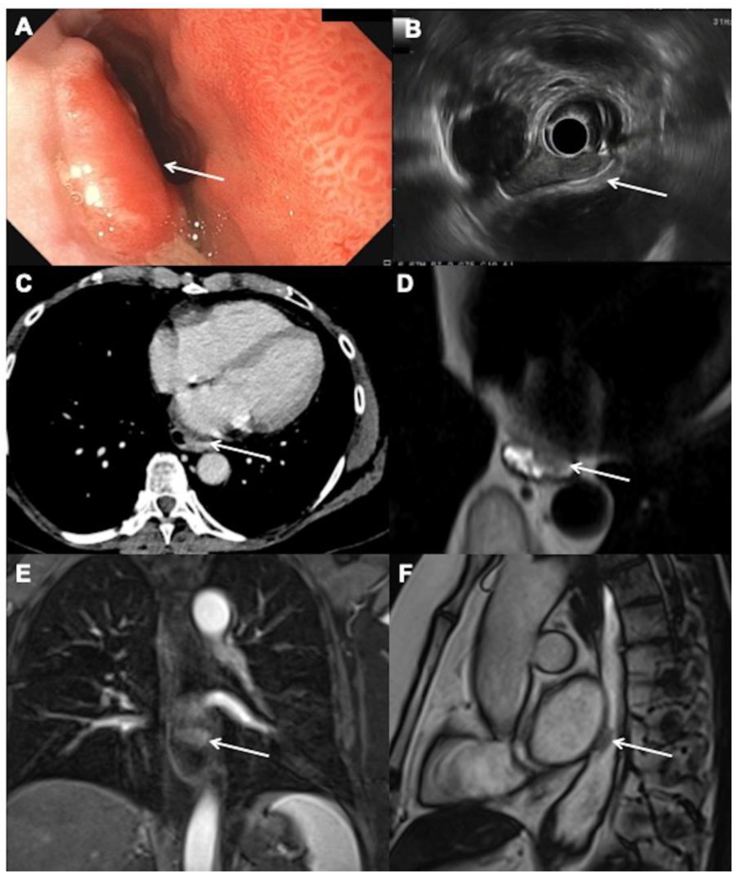
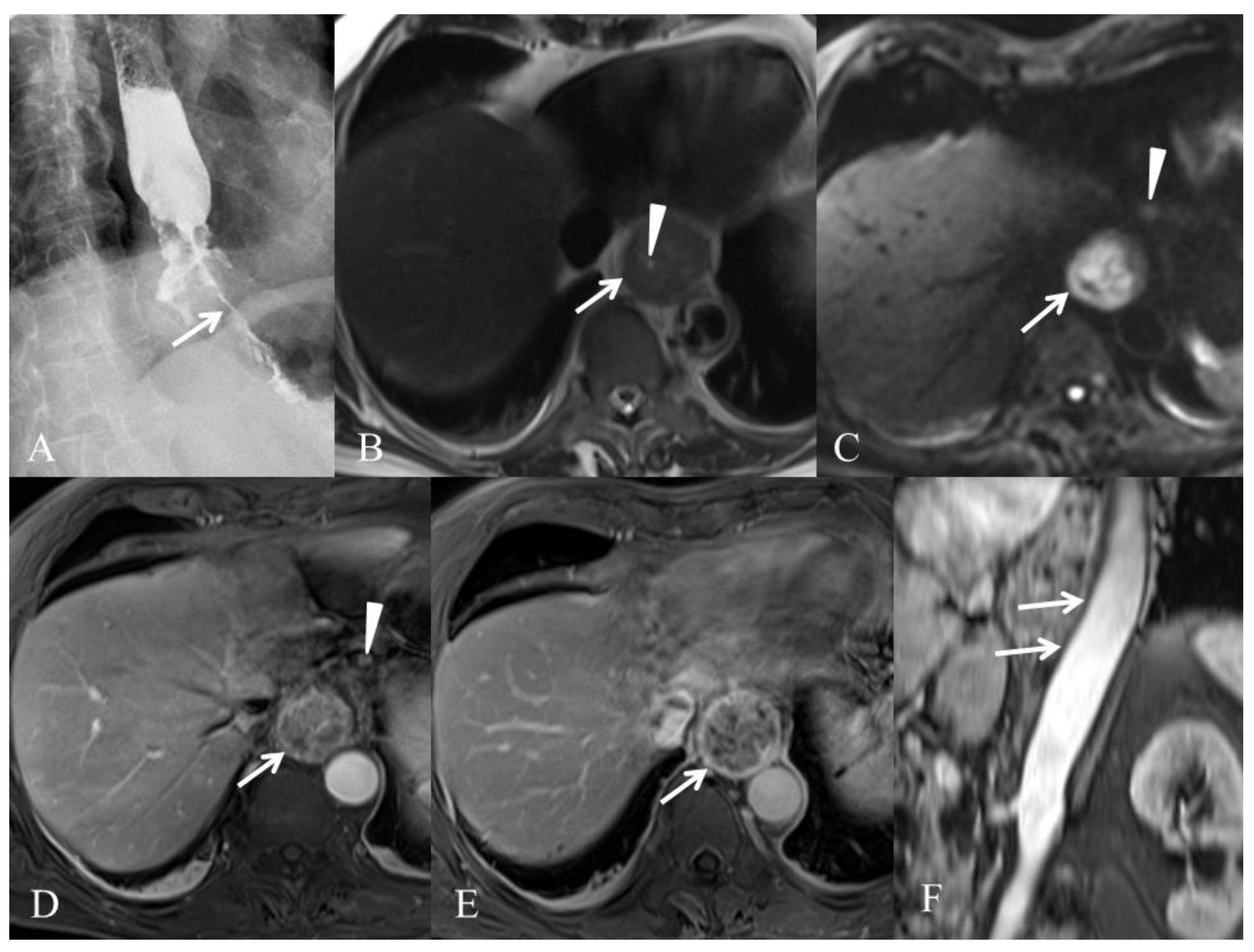
The Role of Magnetic Resonance Imaging in the Management of Esophageal Cancer
Abstract
Simple Summary
Abstract
1. Introduction
2. MRI Modalities for the Esophagus
2.1. Major Technical Developments in MRI of the Esophagus Overtime
2.2. How We Do It in Our Center
3. MRI and Esophageal Cancer (EC) Diagnosis and Staging
3.1. Initial Tumor (T) Staging
3.2. Node (N) Staging
3.3. Metastases (M) Staging
3.4. Target Volume Delineation before Irradiation
4. MRI in Assessment of Treatment Response and Prediction of Recurrence
5. Discussion and Perspectives
6. Conclusions
Author Contributions
Funding
Conflicts of Interest
References
- Arnold, M.; Abnet, C.C.; Neale, R.E.; Vignat, J.; Giovannucci, E.L.; McGlynn, K.A.; Bray, F. Global Burden of 5 Major Types of Gastrointestinal Cancer. Gastroenterology 2020, 159, 335–349. [Google Scholar] [CrossRef] [PubMed]
- Kelly, R.J.; Ajani, J.A.; Kuzdzal, J.; Zander, T.; Van Cutsem, E.; Piessen, G.; Mendez, G.; Feliciano, J.; Motoyama, S.; Lièvre, A.; et al. Adjuvant Nivolumab in Resected Esophageal or Gastroesophageal Junction Cancer. N. Engl. J. Med. 2021, 384, 1191–1203. [Google Scholar] [CrossRef]
- Janjigian, Y.Y.; Shitara, K.; Moehler, M.; Garrido, M.; Salman, P.; Shen, L.; Wyrwicz, L.; Yamaguchi, K.; Skoczylas, T.; Campos Bragagnoli, A.; et al. First-Line Nivolumab plus Chemotherapy versus Chemotherapy Alone for Advanced Gastric, Gas-tro-Oesophageal Junction, and Oesophageal Adenocarcinoma (CheckMate 649): A Randomised, Open-Label, Phase 3 Trial. Lancet 2021, 398, 27–40. [Google Scholar] [CrossRef]
- Sun, J.-M.; Shen, L.; Shah, M.A.; Enzinger, P.; Adenis, A.; Doi, T.; Kojima, T.; Metges, J.-P.; Li, Z.; Kim, S.-B.; et al. Pembrolizumab plus Chemotherapy versus Chemotherapy Alone for First-Line Treatment of Advanced Oesophageal Cancer (KEYNOTE-590): A Randomised, Placebo-Controlled, Phase 3 Study. Lancet 2021, 398, 759–771. [Google Scholar] [CrossRef]
- Lordick, F.; Mariette, C.; Haustermans, K.; Obermannová, R.; Arnold, D. ESMO Guidelines Committee Oesophageal Cancer: ESMO Clinical Practice Guidelines for Diagnosis, Treatment and Follow-Up. Ann. Oncol. 2016, 27, v50–v57. [Google Scholar] [CrossRef] [PubMed]
- Veziant, J.; Gaillard, M.; Barat, M.; Dohan, A.; Barret, M.; Manceau, G.; Karoui, M.; Bonnet, S.; Fuks, D.; Soyer, P. Imaging of Postoperative Complications Following Ivor-Lewis Esophagectomy. Diagn. Interv. Imaging 2021, 103, 67–78. [Google Scholar] [CrossRef] [PubMed]
- Bosset, J.F.; Gignoux, M.; Triboulet, J.P.; Tiret, E.; Mantion, G.; Elias, D.; Lozach, P.; Ollier, J.C.; Pavy, J.J.; Mercier, M.; et al. Chemoradiotherapy Followed by Surgery Compared with Surgery Alone in Squamous-Cell Cancer of the Esophagus. N. Engl. J. Med. 1997, 337, 161–167. [Google Scholar] [CrossRef]
- Rice, T.W.; Blackstone, E.H.; Rusch, V.W. 7th Edition of the AJCC Cancer Staging Manual: Esophagus and Esophagogastric Junction. Ann. Surg. Oncol. 2010, 17, 1721–1724. [Google Scholar] [CrossRef] [PubMed]
- Manabe, T.; Kawamitsu, H.; Higashino, T.; Lee, H.; Fujii, M.; Hoshi, H.; Sugimura, K. Esophageal Magnetic Resonance Fluoroscopy: Optimization of the Sequence. J. Comput. Assist. Tomogr. 2004, 28, 697–703. [Google Scholar] [CrossRef] [PubMed]
- Riddell, A.M.; Richardson, C.; Scurr, E.; Brown, G. The Development and Optimization of High Spatial Resolution MRI for Imaging the Oesophagus Using an External Surface Coil. Br. J. Radiol. 2006, 79, 873–879. [Google Scholar] [CrossRef] [PubMed]
- Riddell, A.M.; Davies, D.C.; Allum, W.H.; Wotherspoon, A.C.; Richardson, C.; Brown, G. High-Resolution MRI in Evaluation of the Surgical Anatomy of the Esophagus and Posterior Mediastinum. AJR Am. J. Roentgenol. 2007, 188, W37–W43. [Google Scholar] [CrossRef] [PubMed]
- Riddell, A.M.; Allum, W.H.; Thompson, J.N.; Wotherspoon, A.C.; Richardson, C.; Brown, G. The Appearances of Oesophageal Carcinoma Demonstrated on High-Resolution, T2-Weighted MRI, with Histopathological Correlation. Eur. Radiol. 2007, 17, 391–399. [Google Scholar] [CrossRef] [PubMed]
- Yamada, I.; Miyasaka, N.; Hikishima, K.; Tokairin, Y.; Kawano, T.; Ito, E.; Kobayashi, D.; Eishi, Y.; Okano, H. Ultra-High-Resolution MR Imaging of Esophageal Carcinoma at Ultra-High Field Strength (7.0T) Ex Vivo: Correlation with Histopathologic Findings. Magn. Reason. Imaging 2015, 33, 413–419. [Google Scholar] [CrossRef] [PubMed]
- Weijs, T.J.; Goense, L.; van Rossum, P.S.N.; Meijer, G.J.; van Lier, A.L.H.M.W.; Wessels, F.J.; Braat, M.N.G.; Lips, I.M.; Ruurda, J.P.; Cuesta, M.A.; et al. The Peri-Esophageal Connective Tissue Layers and Related Compartments: Visualization by Histology and Magnetic Resonance Imaging. J. Anat. 2017, 230, 262–271. [Google Scholar] [CrossRef] [PubMed]
- Kulinna-Cosentini, C.; Schima, W.; Cosentini, E.P. Dynamic MR Imaging of the Gastroesophageal Junction in Healthy Volunteers during Bolus Passage. J. Magn. Reson. Imaging 2007, 25, 749–754. [Google Scholar] [CrossRef] [PubMed]
- Pavone, P.; Cardone, G.P.; Cisternino, S.; Di Girolamo, M.; Aytan, E.; Passariello, R. Gadopentetate Dimeglumine-Barium Paste for Opacification of the Esophageal Lumen on MR Images. AJR Am. J. Roentgenol. 1992, 159, 762–764. [Google Scholar] [CrossRef]
- Ogawa, Y.; Noda, Y.; Morio, K.; Nishioka, A.; Inomata, T.; Yoshida, S.; Toki, T.; Ogoshi, S. Ferric Ammonium Citrate-Cellulose Paste for Opacification of the Esophageal Lumen on MRI. J. Comput. Assist. Tomogr. 1996, 20, 455–459. [Google Scholar] [CrossRef]
- Faletti, R.; Gatti, M.; Di Chio, A.; Fronda, M.; Anselmino, M.; Ferraris, F.; Gaita, F.; Fonio, P. Concentrated Pineapple Juice for Visualisation of the Oesophagus during Magnetic Resonance Angiography before Atrial Fibrillation Radiofrequency Catheter Ablation. Eur. Radiol. Exp. 2018, 2, 39. [Google Scholar] [CrossRef]
- Luo, L.-N.; He, L.-J.; Gao, X.-Y.; Huang, X.-X.; Shan, H.-B.; Luo, G.-Y.; Li, Y.; Lin, S.-Y.; Wang, G.-B.; Zhang, R.; et al. Endoscopic Ultrasound for Preoperative Esophageal Squamous Cell Carcinoma: A Meta-Analysis. PLoS ONE 2016, 11, e0158373. [Google Scholar] [CrossRef]
- Subasinghe, D.; Samarasekera, D.N. A Study Comparing Endoscopic Ultrasound (EUS) and Computed Tomography (CT) in Staging Oesophageal Cancer and Their Role in Clinical Decision Making. J. Gastrointest. Cancer 2010, 41, 38–42. [Google Scholar] [CrossRef]
- Puli, S.-R.; Reddy, J.-B.; Bechtold, M.-L.; Antillon, D.; Ibdah, J.-A.; Antillon, M.-R. Staging Accuracy of Esophageal Cancer by Endoscopic Ultrasound: A Meta-Analysis and Systematic Review. World J. Gastroenterol. 2008, 14, 1479–1490. [Google Scholar] [CrossRef]
- Wakelin, S.J.; Deans, C.; Crofts, T.J.; Allan, P.L.; Plevris, J.N.; Paterson-Brown, S. A Comparison of Computerised Tomography, Laparoscopic Ultrasound and Endoscopic Ultrasound in the Preoperative Staging of Oesophago-Gastric Carcinoma. Eur. J. Radiol. 2002, 41, 161–167. [Google Scholar] [CrossRef]
- Kelly, S.; Harris, K.M.; Berry, E.; Hutton, J.; Roderick, P.; Cullingworth, J.; Gathercole, L.; Smith, M.A. A Systematic Review of the Staging Performance of Endoscopic Ultrasound in Gastro-Oesophageal Carcinoma. Gut 2001, 49, 534–539. [Google Scholar] [CrossRef] [PubMed]
- Quint, L.E.; Bogot, N.R. Staging Esophageal Cancer. Cancer Imaging 2008, 8, S33–S42. [Google Scholar] [CrossRef] [PubMed]
- Rice, T.W. Clinical Staging of Esophageal Carcinoma. CT, EUS, and PET. Chest Surg. Clin. N. Am. 2000, 10, 471–485. [Google Scholar] [PubMed]
- van Westreenen, H.L.; Westerterp, M.; Bossuyt, P.M.M.; Pruim, J.; Sloof, G.W.; van Lanschot, J.J.B.; Groen, H.; Plukker, J.T.M. Systematic Review of the Staging Performance of 18F-Fluorodeoxyglucose Positron Emission Tomography in Esophageal Cancer. J. Clin. Oncol. 2004, 22, 3805–3812. [Google Scholar] [CrossRef]
- Choi, J.; Kim, S.G.; Kim, J.S.; Jung, H.C.; Song, I.S. Comparison of Endoscopic Ultrasonography (EUS), Positron Emission Tomography (PET), and Computed Tomography (CT) in the Preoperative Locoregional Staging of Resectable Esophageal Cancer. Surg. Endosc. 2010, 24, 1380–1386. [Google Scholar] [CrossRef] [PubMed]
- Van Vliet, E.P.M.; Eijkemans, M.J.C.; Poley, J.-W.; Steyerberg, E.W.; Kuipers, E.J.; Siersema, P.D. Staging of Esophageal Carcinoma in a Low-Volume EUS Center Compared with Reported Results from High-Volume Centers. Gastrointest. Endosc. 2006, 63, 938–947. [Google Scholar] [CrossRef]
- Yamada, I.; Murata, Y.; Izumi, Y.; Kawano, T.; Endo, M.; Kuroiwa, T.; Shibuya, H. Staging of Esophageal Carcinoma in Vitro with 4.7-T MR Imaging. Radiology 1997, 204, 521–526. [Google Scholar] [CrossRef]
- Yamada, I.; Izumi, Y.; Kawano, T.; Yoshino, N.; Tetsumura, A.; Kumagai, J.; Shibuya, H. Esophageal Carcinoma: Evaluation with High-Resolution Three-Dimensional Constructive Interference in Steady State MR Imaging in Vitro. J. Magn. Reason. Imaging 2006, 24, 1326–1332. [Google Scholar] [CrossRef]
- Yamada, I.; Hikishima, K.; Miyasaka, N.; Tokairin, Y.; Kawano, T.; Ito, E.; Kobayashi, D.; Eishi, Y.; Okano, H.; Shibuya, H. Diffusion-Tensor MRI and Tractography of the Esophageal Wall Ex Vivo. J. Magn. Reason. Imaging 2014, 40, 567–576. [Google Scholar] [CrossRef] [PubMed]
- Yamada, I.; Hikishima, K.; Miyasaka, N.; Kawano, T.; Tokairin, Y.; Ito, E.; Kobayashi, D.; Eishi, Y.; Okano, H. Esophageal Carcinoma: Ex Vivo Evaluation with Diffusion-Tensor MR Imaging and Tractography at 7 T. Radiology 2014, 272, 164–173. [Google Scholar] [CrossRef]
- Wei, Y.; Wu, S.; Gao, F.; Sun, T.; Zheng, D.; Ning, P.; Zhao, C.; Li, Z.; Li, X.; Li, L.; et al. Esophageal Carcinoma: Ex Vivo Evaluation by High-Spatial-Resolution T2 -Mapping MRI Compared with Histopathological Findings at 3.0T. J. Magn. Reason. Imaging 2017, 45, 1609–1616. [Google Scholar] [CrossRef] [PubMed]
- van Rossum, P.S.N.; Goense, L.; Meziani, J.; Reitsma, J.B.; Siersema, P.D.; Vleggaar, F.P.; van Vulpen, M.; Meijer, G.J.; Ruurda, J.P.; van Hillegersberg, R. Endoscopic Biopsy and EUS for the Detection of Pathologic Complete Response after Neoadjuvant Chemoradiotherapy in Esophageal Cancer: A Systematic Review and Meta-Analysis. Gastrointest. Endosc. 2016, 83, 866–879. [Google Scholar] [CrossRef]
- Lee, S.L.; Yadav, P.; Starekova, J.; Christensen, L.; Chandereng, T.; Chappell, R.; Reeder, S.B.; Bassetti, M.F. Diagnostic Performance of MRI for Esophageal Carcinoma: A Systematic Review and Meta-Analysis. Radiology 2021, 299, 583–594. [Google Scholar] [CrossRef] [PubMed]
- Zhang, F.; Qu, J.; Zhang, H.; Liu, H.; Qin, J.; Ding, Z.; Li, Y.; Ma, J.; Zhang, Z.; Wang, Z.; et al. Preoperative T Staging of Potentially Resectable Esophageal Cancer: A Comparison between Free-Breathing Radial VIBE and Breath-Hold Cartesian VIBE, with Histopathological Correlation. Transl. Oncol. 2017, 10, 324–331. [Google Scholar] [CrossRef]
- Kayani, B.; Zacharakis, E.; Ahmed, K.; Hanna, G.B. Lymph Node Metastases and Prognosis in Oesophageal Carcinoma—A Systematic Review. Eur. J. Surg. Oncol. 2011, 37, 747–753. [Google Scholar] [CrossRef]
- Zhang, J.; Hu, W.; Zang, L.; Yao, Y.; Tang, Y.; Qian, Z.; Gao, P.; Wu, X.; Li, S.; Xie, Z.; et al. Clinical Investigation on Application of Water Swallowing to MR Esophagography. Eur. J. Radiol. 2012, 81, 1980–1985. [Google Scholar] [CrossRef]
- Tang, Y.-L.; Zhang, X.-M.; Yang, Z.-G.; Huang, Y.-C.; Chen, T.-W.; Chen, Y.-L.; Chen, F.; Zeng, N.-L.; Li, R.; Hu, J. The Blood Oxygenation T2* Values of Resectable Esophageal Squamous Cell Carcinomas as Measured by 3T Magnetic Resonance Imaging: Association with Tumor Stage. Korean J. Radiol. 2017, 18, 674–681. [Google Scholar] [CrossRef] [PubMed]
- Wu, L.; Ou, J.; Chen, T.-W.; Li, R.; Zhang, X.-M.; Chen, Y.-L.; Jiang, Y.; Yang, J.-Q.; Cao, J.-M. Tumour Volume of Resectable Oesophageal Squamous Cell Carcinoma Measured with MRI Correlates Well with T Category and Lymphatic Metastasis. Eur. Radiol. 2018, 28, 4757–4765. [Google Scholar] [CrossRef] [PubMed]
- Chen, Y.-L.; Li, R.; Chen, T.-W.; Ou, J.; Zhang, X.-M.; Chen, F.; Wu, L.; Jiang, Y.; Laws, M.; Shah, K.; et al. Whole-Tumour Histogram Analysis of Pharmacokinetic Parameters from Dynamic Contrast-Enhanced MRI in Resectable Oesophageal Squamous Cell Carcinoma Can Predict T-Stage and Regional Lymph Node Metastasis. Eur. J. Radiol. 2019, 112, 112–120. [Google Scholar] [CrossRef]
- Rice, T.W.; Rusch, V.W.; Ishwaran, H.; Blackstone, E.H. Worldwide Esophageal Cancer Collaboration Cancer of the Esophagus and Esophagogastric Junction: Data-Driven Staging for the Seventh Edition of the American Joint Committee on Cancer/International Union Against Cancer Cancer Staging Manuals. Cancer 2010, 116, 3763–3773. [Google Scholar] [CrossRef] [PubMed]
- Lerut, T.E.; de Leyn, P.; Coosemans, W.; Van Raemdonck, D.; Cuypers, P.; Van Cleynenbreughel, B. Advanced Esophageal Carcinoma. World J. Surg. 1994, 18, 379–387. [Google Scholar] [CrossRef] [PubMed]
- Waterman, T.A.; Hagen, J.A.; Peters, J.H.; DeMeester, S.R.; Taylor, C.R.; Demeester, T.R. The Prognostic Importance of Immunohistochemically Detected Node Metastases in Resected Esophageal Adenocarcinoma. Ann. Thorac. Surg. 2004, 78, 1161–1169. [Google Scholar] [CrossRef] [PubMed]
- Bhamidipati, C.M.; Stukenborg, G.J.; Thomas, C.J.; Lau, C.L.; Kozower, B.D.; Jones, D.R. Pathologic Lymph Node Ratio Is a Predictor of Survival in Esophageal Cancer. Ann. Thorac. Surg. 2012, 94, 1643–1651. [Google Scholar] [CrossRef]
- van Vliet, E.P.M.; Heijenbrok-Kal, M.H.; Hunink, M.G.M.; Kuipers, E.J.; Siersema, P.D. Staging Investigations for Oesophageal Cancer: A Meta-Analysis. Br. J. Cancer 2008, 98, 547–557. [Google Scholar] [CrossRef] [PubMed]
- Eloubeidi, M.A.; Wallace, M.B.; Reed, C.E.; Hadzijahic, N.; Lewin, D.N.; Van Velse, A.; Leveen, M.B.; Etemad, B.; Matsuda, K.; Patel, R.S.; et al. The Utility of EUS and EUS-Guided Fine Needle Aspiration in Detecting Celiac Lymph Node Metastasis in Patients with Esophageal Cancer: A Single-Center Experience. Gastrointest. Endosc. 2001, 54, 714–719. [Google Scholar] [CrossRef]
- Vazquez-Sequeiros, E.; Norton, I.D.; Clain, J.E.; Wang, K.K.; Affi, A.; Allen, M.; Deschamps, C.; Miller, D.; Salomao, D.; Wiersema, M.J. Impact of EUS-Guided Fine-Needle Aspiration on Lymph Node Staging in Patients with Esophageal Carcinoma. Gastrointest. Endosc. 2001, 53, 751–757. [Google Scholar] [CrossRef]
- Quint, L.E.; Glazer, G.M.; Orringer, M.B. Esophageal Imaging by MR and CT: Study of Normal Anatomy and Neoplasms. Radiology 1985, 156, 727–731. [Google Scholar] [CrossRef]
- Petrillo, R.; Balzarini, L.; Bidoli, P.; Ceglia, E.; D’Ippolito, G.; Tess, J.D.; Musumeci, R. Esophageal Squamous Cell Carcinoma: MRI Evaluation of Mediastinum. Gastrointest. Radiol. 1990, 15, 275–278. [Google Scholar] [CrossRef]
- Takashima, S.; Takeuchi, N.; Shiozaki, H.; Kobayashi, K.; Morimoto, S.; Ikezoe, J.; Tomiyama, N.; Harada, K.; Shogen, K.; Kozuka, T. Carcinoma of the Esophagus: CT vs MR Imaging in Determining Resectability. AJR Am. J. Roentgenol. 1991, 156, 297–302. [Google Scholar] [CrossRef] [PubMed]
- Mizowaki, T.; Nishimura, Y.; Shimada, Y.; Nakano, Y.; Imamura, M.; Konishi, J.; Hiraoka, M. Optimal Size Criteria of Malignant Lymph Nodes in the Treatment Planning of Radiotherapy for Esophageal Cancer: Evaluation by Computed Tomography and Magnetic Resonance Imaging. Int. J. Radiat. Oncol. Biol. Phys. 1996, 36, 1091–1098. [Google Scholar] [CrossRef]
- Wu, L.-F.; Wang, B.-Z.; Feng, J.-L.; Cheng, W.-R.; Liu, G.-R.; Xu, X.-H.; Zheng, Z.-C. Preoperative TN Staging of Esophageal Cancer: Comparison of Miniprobe Ultrasonography, Spiral CT and MRI. World J. Gastroenterol. 2003, 9, 219–224. [Google Scholar] [CrossRef] [PubMed]
- Nishimura, H.; Tanigawa, N.; Hiramatsu, M.; Tatsumi, Y.; Matsuki, M.; Narabayashi, I. Preoperative Esophageal Cancer Staging: Magnetic Resonance Imaging of Lymph Node with Ferumoxtran-10, an Ultrasmall Superparamagnetic Iron Oxide. J. Am. Coll. Surg. 2006, 202, 604–611. [Google Scholar] [CrossRef] [PubMed]
- Weissleder, R.; Elizondo, G.; Wittenberg, J.; Lee, A.S.; Josephson, L.; Brady, T.J. Ultrasmall Superparamagnetic Iron Oxide: An Intravenous Contrast Agent for Assessing Lymph Nodes with MR Imaging. Radiology 1990, 175, 494–498. [Google Scholar] [CrossRef]
- Zhang, F.; Zhu, L.; Huang, X.; Niu, G.; Chen, X. Differentiation of Reactive and Tumor Metastatic Lymph Nodes with Diffusion-Weighted and SPIO-Enhanced MRI. Mol. Imaging Biol. 2013, 15, 40–47. [Google Scholar] [CrossRef]
- Pultrum, B.B.; van der Jagt, E.J.; van Westreenen, H.L.; van Dullemen, H.M.; Kappert, P.; Groen, H.; Sietsma, J.; Oudkerk, M.; Plukker, J.T.M.; van Dam, G.M. Detection of Lymph Node Metastases with Ultrasmall Superparamagnetic Iron Oxide (USPIO)-Enhanced Magnetic Resonance Imaging in Oesophageal Cancer: A Feasibility Study. Cancer Imaging 2009, 9, 19–28. [Google Scholar] [CrossRef]
- Sakurada, A.; Takahara, T.; Kwee, T.C.; Yamashita, T.; Nasu, S.; Horie, T.; Van Cauteren, M.; Imai, Y. Diagnostic Performance of Diffusion-Weighted Magnetic Resonance Imaging in Esophageal Cancer. Eur. Radiol. 2009, 19, 1461–1469. [Google Scholar] [CrossRef]
- Shuto, K.; Kono, T.; Shiratori, T.; Akutsu, Y.; Uesato, M.; Mori, M.; Narushima, K.; Imanishi, S.; Nabeya, Y.; Yanagawa, N.; et al. Diagnostic Performance of Diffusion-Weighted Magnetic Resonance Imaging in Assessing Lymph Node Metastasis of Esophageal Cancer Compared with PET. Esophagus 2020, 17, 239–249. [Google Scholar] [CrossRef]
- Alper, F.; Turkyilmaz, A.; Kurtcan, S.; Aydin, Y.; Onbas, O.; Acemoglu, H.; Eroglu, A. Effectiveness of the STIR Turbo Spin-Echo Sequence MR Imaging in Evaluation of Lymphadenopathy in Esophageal Cancer. Eur. J. Radiol. 2011, 80, 625–628. [Google Scholar] [CrossRef]
- Jiang, Y.; Chen, Y.-L.; Chen, T.-W.; Wu, L.; Ou, J.; Li, R.; Zhang, X.-M.; Yang, J.-Q.; Cao, J.-M. Is There Association of Gross Tumor Volume of Adenocarcinoma of Oesophagogastric Junction Measured on Magnetic Resonance Imaging with N Stage? Eur. J. Radiol. 2019, 110, 181–186. [Google Scholar] [CrossRef]
- Qu, J.; Shen, C.; Qin, J.; Wang, Z.; Liu, Z.; Guo, J.; Zhang, H.; Gao, P.; Bei, T.; Wang, Y.; et al. The MR Radiomic Signature Can Predict Preoperative Lymph Node Metastasis in Patients with Esophageal Cancer. Eur. Radiol. 2019, 29, 906–914. [Google Scholar] [CrossRef]
- Kroese, T.E.; Goense, L.; van Hillegersberg, R.; de Keizer, B.; Mook, S.; Ruurda, J.P.; van Rossum, P.S.N. Detection of Distant Interval Metastases after Neoadjuvant Therapy for Esophageal Cancer with 18F-FDG PET(/CT): A Systematic Review and Meta-Analysis. Dis. Esophagus 2018, 31, doy055. [Google Scholar] [CrossRef]
- Gong, J.; Cao, W.; Zhang, Z.; Deng, Y.; Kang, L.; Zhu, P.; Liu, Z.; Zhou, Z. Diagnostic Efficacy of Whole-Body Diffusion-Weighted Imaging in the Detection of Tumour Recurrence and Metastasis by Comparison with 18F-2-Fluoro-2-Deoxy-D-Glucose Positron Emission Tomography or Computed Tomography in Patients with Gastrointestinal Cancer. Gastroenterol. Rep. 2015, 3, 128–135. [Google Scholar] [CrossRef] [PubMed][Green Version]
- Malik, V.; Harmon, M.; Johnston, C.; Fagan, A.J.; Claxton, Z.; Ravi, N.; O’Toole, D.; Muldoon, C.; Keogan, M.; Reynolds, J.V.; et al. Whole Body MRI in the Staging of Esophageal Cancer--A Prospective Comparison with Whole Body 18F-FDG PET-CT. Dig. Surg. 2015, 32, 397–408. [Google Scholar] [CrossRef]
- Hou, D.-L.; Shi, G.-F.; Gao, X.-S.; Asaumi, J.; Li, X.-Y.; Liu, H.; Yao, C.; Chang, J.Y. Improved Longitudinal Length Accuracy of Gross Tumor Volume Delineation with Diffusion Weighted Magnetic Resonance Imaging for Esophageal Squamous Cell Carcinoma. Radiat. Oncol. 2013, 8, 169. [Google Scholar] [CrossRef] [PubMed]
- Sillah, K.; Williams, L.R.; Laasch, H.-U.; Saleem, A.; Watkins, G.; Pritchard, S.A.; Price, P.M.; West, C.M.; Welch, I.M. Computed Tomography Overestimation of Esophageal Tumor Length: Implications for Radiotherapy Planning. World J. Gastrointest. Oncol. 2010, 2, 197–204. [Google Scholar] [CrossRef]
- Konski, A.; Doss, M.; Milestone, B.; Haluszka, O.; Hanlon, A.; Freedman, G.; Adler, L. The Integration of 18-Fluoro-Deoxy-Glucose Positron Emission Tomography and Endoscopic Ultrasound in the Treatment-Planning Process for Esophageal Carcinoma. Int J. Radiat. Oncol. Biol. Phys. 2005, 61, 1123–1128. [Google Scholar] [CrossRef]
- Vollenbrock, S.E.; Nowee, M.E.; Voncken, F.E.M.; Kotte, A.N.T.J.; Goense, L.; van Rossum, P.S.N.; van Lier, A.L.H.M.W.; Heijmink, S.W.; Bartels-Rutten, A.; Wessels, F.J.; et al. Gross Tumor Delineation in Esophageal Cancer on MRI Compared With 18F-FDG-PET/CT. Adv. Radiat. Oncol. 2019, 4, 596–604. [Google Scholar] [CrossRef]
- Lee, S.L.; Bassetti, M.; Meijer, G.J.; Mook, S. Review of MR-Guided Radiotherapy for Esophageal Cancer. Front. Oncol. 2021, 11, 628009. [Google Scholar] [CrossRef]
- Boonstra, J.J.; Kok, T.C.; Wijnhoven, B.P.; van Heijl, M.; van Berge Henegouwen, M.I.; Ten Kate, F.J.; Siersema, P.D.; Dinjens, W.N.; van Lanschot, J.J.; Tilanus, H.W.; et al. Chemotherapy Followed by Surgery versus Surgery Alone in Patients with Resectable Oesophageal Squamous Cell Carcinoma: Long-Term Results of a Randomized Controlled Trial. BMC Cancer 2011, 11, 181. [Google Scholar] [CrossRef] [PubMed]
- Sjoquist, K.M.; Burmeister, B.H.; Smithers, B.M.; Zalcberg, J.R.; Simes, R.J.; Barbour, A.; Gebski, V. Australasian Gastro-Intestinal Trials Group Survival after Neoadjuvant Chemotherapy or Chemoradiotherapy for Resectable Oesophageal Carcinoma: An Updated Meta-Analysis. Lancet Oncol. 2011, 12, 681–692. [Google Scholar] [CrossRef]
- van Hagen, P.; Hulshof, M.C.C.M.; van Lanschot, J.J.B.; Steyerberg, E.W.; van Berge Henegouwen, M.I.; Wijnhoven, B.P.L.; Richel, D.J.; Nieuwenhuijzen, G.a.P.; Hospers, G.a.P.; Bonenkamp, J.J.; et al. Preoperative Chemoradiotherapy for Esophageal or Junctional Cancer. N. Engl. J. Med. 2012, 366, 2074–2084. [Google Scholar] [CrossRef] [PubMed]
- Heneghan, H.M.; Donohoe, C.; Elliot, J.; Ahmed, Z.; Malik, V.; Ravi, N.; Reynolds, J.V. Can CT-PET and Endoscopic Assessment Post-Neoadjuvant Chemoradiotherapy Predict Residual Disease in Esophageal Cancer? Ann. Surg. 2016, 264, 831–838. [Google Scholar] [CrossRef]
- Ngamruengphong, S.; Sharma, V.K.; Nguyen, B.; Das, A. Assessment of Response to Neoadjuvant Therapy in Esophageal Cancer: An Updated Systematic Review of Diagnostic Accuracy of Endoscopic Ultrasonography and Fluorodeoxyglucose Positron Emission Tomography. Dis. Esophagus 2010, 23, 216–231. [Google Scholar] [CrossRef] [PubMed]
- Eyck, B.M.; Onstenk, B.D.; Noordman, B.J.; Nieboer, D.; Spaander, M.C.W.; Valkema, R.; Lagarde, S.M.; Wijnhoven, B.P.L.; van Lanschot, J.J.B. Accuracy of Detecting Residual Disease After Neoadjuvant Chemoradiotherapy for Esophageal Cancer: A Systematic Review and Meta-Analysis. Ann. Surg. 2020, 271, 245–256. [Google Scholar] [CrossRef] [PubMed]
- de Gouw, D.J.J.M.; Klarenbeek, B.R.; Driessen, M.; Bouwense, S.A.W.; van Workum, F.; Fütterer, J.J.; Rovers, M.M.; Ten Broek, R.P.G.; Rosman, C. Detecting Pathological Complete Response in Esophageal Cancer after Neoadjuvant Therapy Based on Imaging Techniques: A Diagnostic Systematic Review and Meta-Analysis. J. Thorac. Oncol. 2019, 14, 1156–1171. [Google Scholar] [CrossRef]
- van Heijl, M.; Phoa, S.S.K.S.; van Berge Henegouwen, M.I.; Omloo, J.M.T.; Mearadji, B.M.; Sloof, G.W.; Bossuyt, P.M.M.; Hulshof, M.C.C.M.; Richel, D.J.; Bergman, J.J.G.H.M.; et al. Accuracy and Reproducibility of 3D-CT Measurements for Early Response Assessment of Chemoradiotherapy in Patients with Oesophageal Cancer. Eur. J. Surg. Oncol. 2011, 37, 1064–1071. [Google Scholar] [CrossRef][Green Version]
- Zuccaro, G.; Rice, T.W.; Goldblum, J.; Medendorp, S.V.; Becker, M.; Pimentel, R.; Gitlin, L.; Adelstein, D.J. Endoscopic Ultrasound Cannot Determine Suitability for Esophagectomy after Aggressive Chemoradiotherapy for Esophageal Cancer. Am. J. Gastroenterol. 1999, 94, 906–912. [Google Scholar] [CrossRef] [PubMed]
- Griffin, J.M.; Reed, C.E.; Denlinger, C.E. Utility of Restaging Endoscopic Ultrasound after Neoadjuvant Therapy for Esophageal Cancer. Ann. Thorac. Surg. 2012, 93, 1855–1859. [Google Scholar] [CrossRef]
- Imanishi, S.; Shuto, K.; Aoyagi, T.; Kono, T.; Saito, H.; Matsubara, H. Diffusion-Weighted Magnetic Resonance Imaging for Predicting and Detecting the Early Response to Chemoradiotherapy of Advanced Esophageal Squamous Cell Carcinoma. Dig. Surg. 2013, 30, 240–248. [Google Scholar] [CrossRef] [PubMed]
- Aoyagi, T.; Shuto, K.; Okazumi, S.; Shimada, H.; Kazama, T.; Matsubara, H. Apparent Diffusion Coefficient Values Measured by Diffusion-Weighted Imaging Predict Chemoradiotherapeutic Effect for Advanced Esophageal Cancer. Dig. Surg. 2011, 28, 252–257. [Google Scholar] [CrossRef] [PubMed]
- van Rossum, P.S.N.; van Lier, A.L.H.M.W.; van Vulpen, M.; Reerink, O.; Lagendijk, J.J.W.; Lin, S.H.; van Hillegersberg, R.; Ruurda, J.P.; Meijer, G.J.; Lips, I.M. Diffusion-Weighted Magnetic Resonance Imaging for the Prediction of Pathologic Response to Neoadjuvant Chemoradiotherapy in Esophageal Cancer. Radiother. Oncol. 2015, 115, 163–170. [Google Scholar] [CrossRef]
- Fang, P.; Musall, B.C.; Son, J.B.; Moreno, A.C.; Hobbs, B.P.; Carter, B.W.; Fellman, B.M.; Mawlawi, O.; Ma, J.; Lin, S.H. Multimodal Imaging of Pathologic Response to Chemoradiation in Esophageal Cancer. Int. J. Radiat. Oncol. Biol. Phys. 2018, 102, 996–1001. [Google Scholar] [CrossRef] [PubMed]
- Borggreve, A.S.; Heethuis, S.E.; Boekhoff, M.R.; Goense, L.; van Rossum, P.S.N.; Brosens, L.A.A.; van Lier, A.L.H.M.W.; van Hillegersberg, R.; Lagendijk, J.J.W.; Mook, S.; et al. Optimal Timing for Prediction of Pathologic Complete Response to Neoadjuvant Chemoradiotherapy with Diffusion-Weighted MRI in Patients with Esophageal Cancer. Eur. Radiol. 2020, 30, 1896–1907. [Google Scholar] [CrossRef] [PubMed]
- Leandri, C.; Soyer, P.; Oudjit, A.; Guillaumot, M.-A.; Chaussade, S.; Dohan, A.; Barret, M. Contribution of Magnetic Resonance Imaging to the Management of Esophageal Diseases: A Systematic Review. Eur. J. Radiol. 2019, 120, 108684. [Google Scholar] [CrossRef] [PubMed]
- Li, F.P.; Wang, H.; Hou, J.; Tang, J.; Lu, Q.; Wang, L.L.; Yu, X.P. Utility of Intravoxel Incoherent Motion Diffusion-Weighted Imaging in Predicting Early Response to Concurrent Chemoradiotherapy in Oesophageal Squamous Cell Carcinoma. Clin. Radiol. 2018, 73, 756.e17–756.e26. [Google Scholar] [CrossRef]
- Heethuis, S.E.; Goense, L.; van Rossum, P.S.N.; Borggreve, A.S.; Mook, S.; Voncken, F.E.M.; Bartels-Rutten, A.; Aleman, B.M.P.; van Hillegersberg, R.; Ruurda, J.P.; et al. DW-MRI and DCE-MRI Are of Complementary Value in Predicting Pathologic Response to Neoadjuvant Chemoradiotherapy for Esophageal Cancer. Acta Oncol. 2018, 57, 1201–1208. [Google Scholar] [CrossRef] [PubMed]
- Lei, J.; Han, Q.; Zhu, S.; Shi, D.; Dou, S.; Su, Z.; Xu, X. Assessment of Esophageal Carcinoma Undergoing Concurrent Chemoradiotherapy with Quantitative Dynamic Contrast-Enhanced Magnetic Resonance Imaging. Oncol. Lett. 2015, 10, 3607–3612. [Google Scholar] [CrossRef]
- Defize, I.L.; Boekhoff, M.R.; Borggreve, A.S.; van Lier, A.L.H.M.W.; Takahashi, N.; Haj Mohammad, N.; Ruurda, J.P.; van Hillegersberg, R.; Mook, S.; Meijer, G.J. Tumor Volume Regression during Neoadjuvant Chemoradiotherapy for Esophageal Cancer: A Prospective Study with Weekly MRI. Acta Oncol. 2020, 59, 753–759. [Google Scholar] [CrossRef] [PubMed]
- Cheng, B.; Yu, J. Predictive Value of Diffusion-Weighted MR Imaging in Early Response to Chemoradiotherapy of Esophageal Cancer: A Meta-Analysis. Dis. Esophagus 2019, 32, doy065. [Google Scholar] [CrossRef] [PubMed]
- Borggreve, A.S.; Mook, S.; Verheij, M.; Mul, V.E.M.; Bergman, J.J.; Bartels-Rutten, A.; Ter Beek, L.C.; Beets-Tan, R.G.H.; Bennink, R.J.; van Berge Henegouwen, M.I.; et al. Preoperative Image-Guided Identification of Response to Neoadjuvant Chemoradiotherapy in Esophageal Cancer (PRIDE): A Multicenter Observational Study. BMC Cancer 2018, 18, 1006. [Google Scholar] [CrossRef] [PubMed]
- Lee, G.; Hoseok, I.; Kim, S.-J.; Jeong, Y.J.; Kim, I.J.; Pak, K.; Park, D.Y.; Kim, G.H. Clinical Implication of PET/MR Imaging in Preoperative Esophageal Cancer Staging: Comparison with PET/CT, Endoscopic Ultrasonography, and CT. J. Nucl. Med. 2014, 55, 1242–1247. [Google Scholar] [CrossRef] [PubMed]
- Chidambaram, S.; Sounderajah, V.; Maynard, N.; Markar, S.R. Diagnostic Performance of Artificial Intelligence-Centred Systems in the Diagnosis and Postoperative Surveillance of Upper Gastrointestinal Malignancies Using Computed Tomography Imaging: A Systematic Review and Meta-Analysis of Diagnostic Accuracy. Ann. Surg. Oncol. 2021, 29, 1977–1990. [Google Scholar] [CrossRef] [PubMed]
- Chassagnon, G.; Dohan, A. Artificial Intelligence: From Challenges to Clinical Implementation. Diagn. Interv. Imaging 2020, 101, 763–764. [Google Scholar] [CrossRef] [PubMed]
- Nakaura, T.; Higaki, T.; Awai, K.; Ikeda, O.; Yamashita, Y. A Primer for Understanding Radiology Articles about Machine Learning and Deep Learning. Diagn. Interv. Imaging 2020, 101, 765–770. [Google Scholar] [CrossRef] [PubMed]


| Sequence Parameter | T2-Single Shot TSE | Steady States | Diffusion (EPI) | T1-Weighted before and after Gadolinium Chelate |
|---|---|---|---|---|
| Plane | Axial and coronal | Oblique | Axial | Axial and coronal |
| TE/TR (ms) | 93/100 | 1.71/433 | 80/7900 | 2.19/4.85 |
| Flip angle (°) | 150 | 60 | 90 | 10 |
| FOV (mm) | 450 × 450 | 360 × 360 | 420 × 380 | 380 × 308 |
| Matrix size | 384 × 269 | 256 × 256 | 200 × 200 | 320 × 240 |
| Slice thickness (mm) | 6 | 10 | 7 | 2 |
| Voxel size (mm3) | 1.2 × 1.2 × 6 | 0.7 × 0.7 × 10 | 1.1 × 1.1 × 7 | 1.2 × 1.2 × 2 |
| Number of slices | 23 | 1 | 40 | 80 |
| Inter-slices gap (%) | 30 | NA | 20 | 20 |
Publisher’s Note: MDPI stays neutral with regard to jurisdictional claims in published maps and institutional affiliations. |
© 2022 by the authors. Licensee MDPI, Basel, Switzerland. This article is an open access article distributed under the terms and conditions of the Creative Commons Attribution (CC BY) license (https://creativecommons.org/licenses/by/4.0/).
Share and Cite
Pellat, A.; Dohan, A.; Soyer, P.; Veziant, J.; Coriat, R.; Barret, M. The Role of Magnetic Resonance Imaging in the Management of Esophageal Cancer. Cancers 2022, 14, 1141. https://doi.org/10.3390/cancers14051141
Pellat A, Dohan A, Soyer P, Veziant J, Coriat R, Barret M. The Role of Magnetic Resonance Imaging in the Management of Esophageal Cancer. Cancers. 2022; 14(5):1141. https://doi.org/10.3390/cancers14051141
Chicago/Turabian StylePellat, Anna, Anthony Dohan, Philippe Soyer, Julie Veziant, Romain Coriat, and Maximilien Barret. 2022. "The Role of Magnetic Resonance Imaging in the Management of Esophageal Cancer" Cancers 14, no. 5: 1141. https://doi.org/10.3390/cancers14051141
APA StylePellat, A., Dohan, A., Soyer, P., Veziant, J., Coriat, R., & Barret, M. (2022). The Role of Magnetic Resonance Imaging in the Management of Esophageal Cancer. Cancers, 14(5), 1141. https://doi.org/10.3390/cancers14051141


