Review

KRAS and RAS-MAPK Pathway Deregulation in Mature B Cell Lymphoproliferative Disorders

Clinical and Experimental Onco-Hematology Unit, Centro di Riferimento Oncologico di Aviano (CRO) IRCCS, 33081 Aviano, Italy
*
Author to whom correspondence should be addressed.
These authors contributed equally to this work.
Cancers 2022, 14(3), 666; https://doi.org/10.3390/cancers14030666
Submission received: 23 December 2021 / Revised: 24 January 2022 / Accepted: 26 January 2022 / Published: 28 January 2022
(This article belongs to the Special Issue Targeting KRAS: Elucidating Mechanisms of Sensitivity and Resistance)

Abstract

:

Simple Summary

KRAS and genes in the RAS-MAPK pathway are among the most frequently deregulated genes in solid tumors and in this context a large amount of KRAS and RAS-MAPK targeting molecules have been developed and tested. The improved understanding of genomic variation in mature B cell neoplasms revealed a considerable portion of patients presenting with KRAS or RAS-MAPK pathway aberrations. These patients could potentially benefit from the use of RAS-RAF-MEK-ERK inhibitors, and in most of the cases, clinical investigation is only in the beginning. Here, we review the incidence of KRAS and RAS-MAPK mutations in mature B cell lymphoproliferative disorders, their association with progression and survival, and current therapeutic strategies targeting RAS-MAPK signaling.

Abstract

KRAS mutations account for the most frequent mutations in human cancers, and are generally correlated with disease aggressiveness, poor prognosis, and poor response to therapies. KRAS is required for adult hematopoiesis and plays a key role in B cell development and mature B cell proliferation and survival, proved to be critical for B cell receptor-induced ERK pathway activation. In mature B cell neoplasms, commonly seen in adults, KRAS and RAS-MAPK pathway aberrations occur in a relevant fraction of patients, reaching high recurrence in some specific subtypes like multiple myeloma and hairy cell leukemia. As inhibitors targeting the RAS-MAPK pathway are being developed and improved, it is of outmost importance to precisely identify all subgroups of patients that could potentially benefit from their use. Herein, we review the role of KRAS and RAS-MAPK signaling in malignant hematopoiesis, focusing on mature B cell lymphoproliferative disorders. We discuss KRAS and RAS-MAPK pathway aberrations describing type, incidence, mutual exclusion with other genetic abnormalities, and association with prognosis. We review the current therapeutic strategies applied in mature B cell neoplasms to counteract RAS-MAPK signaling in pre-clinical and clinical studies, including most promising combination therapies. We finally present an overview of genetically engineered mouse models bearing KRAS and RAS-MAPK pathway aberrations in the hematopoietic compartment, which are valuable tools in the understanding of cancer biology and etiology.

1. Introduction

The RAS-MAPK pathway is one of the most deregulated and extensively characterized pathways in human cancer, with KRAS being the most frequently mutated gene. The RAS-MAPK pathway plays a crucial role in the control of cell proliferation, differentiation and survival, and its aberrant activation in the oncogenic context is frequently associated with invasion, transformation, disease aggressiveness and drug resistance [1].
Inhibitory molecules, targeting single or multiple components of the RAS-MAPK signaling, have been developed and made available for clinical use. Many RAS-MAPK inhibitors are being evaluated in clinical trials with encouraging results, although the extent and duration of efficacy differ depending on tumor type and pattern of genetic aberrations. As strategies to counteract aberrant RAS-MAPK signaling keep improving, such as by the combinatorial use of multiple agents, it is of the utmost importance to precisely identify all subgroups of patients that could potentially benefit from their use.
The first evidence of RAS mutations in mature B cell malignancies date back to 1989, when Neri and colleagues identified KRAS and NRAS mutations in a subgroup of multiple myeloma cases [2]. But it is only with the advent of high-throughput sequencing techniques that the mutational landscape of mature B cell neoplasms has been disclosed, revealing its extreme heterogeneity. Intensive investigation approaches based on whole-genome and whole-exome sequencing (WGS/WES) and the contextual analysis of paired normal samples allowed the identification of a broad spectrum of aberrations. So far, aberrations in genes that are part of the RAS-MAPK signaling have been identified in most of the mature B cell neoplasms, but they present with very different occurrence, and are associated to distinct biological and clinical significance.
In this review we outline the incidence and the main features of KRAS and RAS-MAPK aberrations in the context of mature B cell malignancies. We discuss the main clinical and pre-clinical therapeutic approaches to counteract aberrant RAS-MAPK signaling in the different B cell neoplasms. Finally, we present an overview of conditional mouse models that through expression of RAS-MAPK aberrations in different hematopoietic compartments may help in the understanding of their oncogenic role.

2. KRAS and RAS-MAPK Pathway in B Lymphocytes

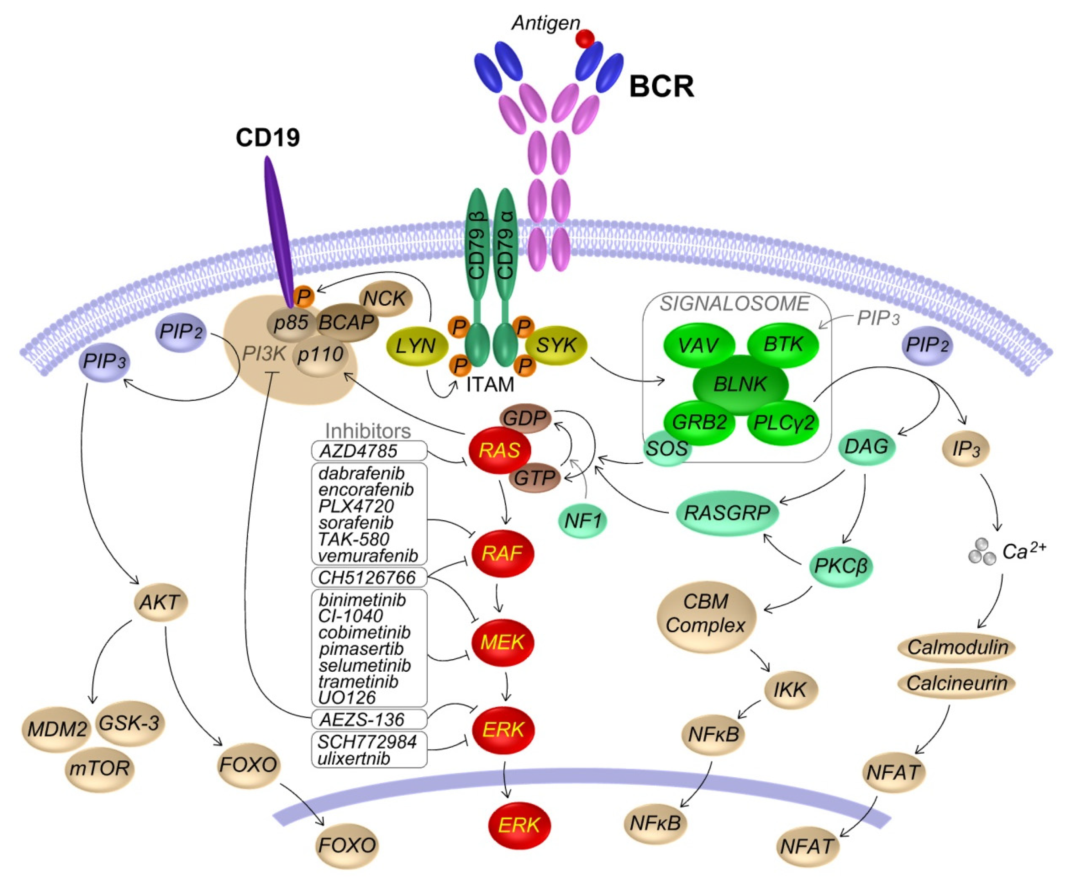
2.1. RAS-MAPK Signaling

RAS proteins are ubiquitously expressed membrane-bound small GTPases that, transducing signals from cell surface receptors to intracellular effector pathways, modulate numerous cellular processes including cell growth, differentiation, and survival. In humans, three RAS genes, KRAS, NRAS and HRAS, encode for four RAS proteins, namely KRAS4A, KRAS4B, NRAS and HRAS. These four isoforms are highly homologous and differ mostly in the C-terminal region, relevant for RAS protein maturation, membrane binding and function. RAS proteins act as molecular switches and cycles between an inactive GDP-bound state and an active GTP-bound state. RAS activity is regulated by two groups of proteins, the guanine nucleotide exchange factors (GEFs) and the GTPase activating proteins (GAPs). GEFs, such as son of sevenless (SOS) and Ras guanyl nucleotide releasing proteins (RasGRPs), promote GDP to GTP exchange, leading to GTP-bound RAS active state. GAPs, such as NF1 and RasGAP, stimulate the RAS GTPase activity promoting the switch back to GDP-bound RAS inactive state [1].
Among the numerous RAS activated effector pathways, the RAF-MEK-ERK (MAPK, mitogen-activated protein kinase) pathway is the one best characterized and more strongly involved in human cancer. RAF is a family of serine/threonine kinases comprising ARAF, BRAF and CRAF/RAF1. RAS activation promotes cytosolic RAF recruitment to plasma membrane, dimerization and phosphorylation. Activated RAF phosphorylates MEK, which in turn phosphorylates ERK. Activated ERK phosphorylates hundreds of targets in the cytoplasm and nucleus regulating many cellular processes including growth, proliferation and survival. Among RAF proteins, BRAF is the prevalent isoform, being more easily activated by RAS and having higher basal kinase activity. However, increasing attention is arising around RAF1 and its regulatory role on RAF kinases, as observed in BRAF inhibitor-mediated paradoxical ERK activation [3].
Another well-known RAS effector is the phosphatidylinositol 3 kinase (PI3K) pathway that is activated through active RAS binding to the p110 catalytic subunit and the enhancing of its catalytic activity [4].

2.2. Role of the BCR Induced RAS-MAPK Pathway Activation in B Cells

The BCR guides and controls every stages of B cell life. The BCR is a surface membrane molecular complex composed of a pair of identical immunoglobulin heavy (IgH) and light (IgL) chains whose unique variable region determines the specificity for antigen recognition, associated to the heterodimer of signal transduction subunits CD79A (Igα) and CD79B (Igβ) that allow the transduction of BCR signals.
Upon antigen binding, LYN or other SRC kinases phosphorylate CD79A/B in their intracellular immunoreceptor tyrosine-based activation motifs (ITAMs). Phosphorylated ITAMs recruit and activate SYK, which in turn activates BTK, PLCγ2, VAV, GRB2, the adaptor protein BLNK, and promote the formation of signalosome. This results in the activation of NF-kB, NFAT and MAPK signaling pathways [5]. In particular, the RAS-MAPK pathway is activated downstream of both PLCγ2/PKCβ/RasGRP and GRB2/SOS and the two activation mechanisms are involved in a positive feedback loop [6]. LYN phosphorylation of the co-receptor CD19 activates PI3K and its downstream targets including AKT and BTK [5]. A scheme of the BCR pathway is depicted in Figure 1.

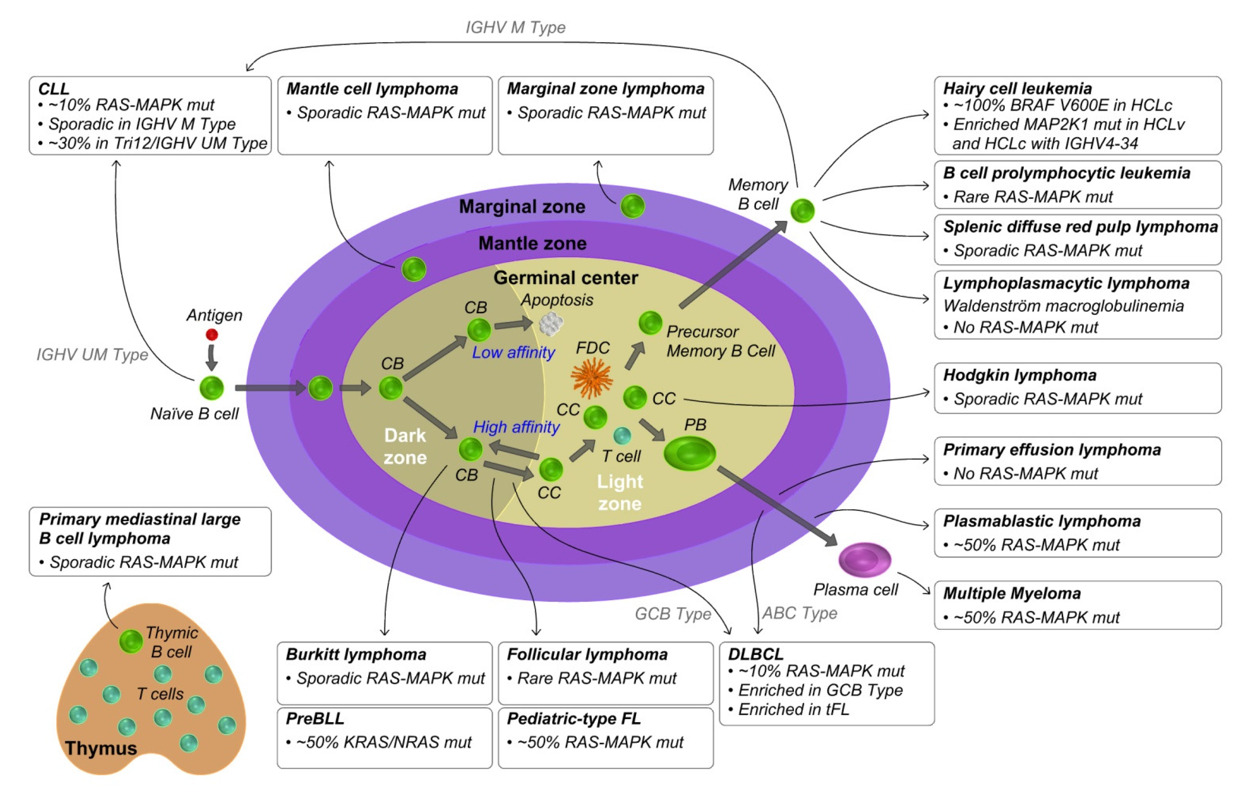
3. KRAS and RAS-MAPK Pathway Aberrations in Mature B Cell Lymphoproliferative Disorders

Mature B cell neoplasms are a heterogeneous group of hematopoietic disease, presenting with very different morphological, immunophenotypic and clinical features that include B cell prolymphocytic leukemia (B-PLL), Hodgkin’s Lymphoma (HL) and Non-Hodgkin’s Lymphoma (NHL). For each B cell neoplasm it is possible to define a “normal B cell counterpart”, also called a “cell of origin”, on the basis of its phenotypic features reminiscent of B cells at a particular stage of differentiation (Figure 2). Immunoglobulin heavy-chain variable region gene (IGHV) mutational status and gene expression profile of malignant cells mostly take part to the definition of their normal B cell counterpart [7,8].
Virtually all mature B cell lymphoproliferative disorders have been investigated by high-throughput sequencing, and in most of the cases recurrent genetic hallmarks have been identified. Despite this, the depth of genetic knowledge varies by disease, with some malignancies having more than one thousand of cases investigated by WGS or WES, and others limited to less than a dozen cases sequenced, or analyzed primarily by targeted resequencing. Indeed, several aspects of disease presentation might influence the extent of its genomic characterization, including the incidence among the population, the anatomical site of presentation, the portion of non-malignant cells in the tumor milieu, and the degree of genomic instability and complexity. This disparity, which penalizes less frequently mutated genes, will be bridged by the constant improvement in genomic characterization. Grouping genetic aberrations in the main cellular pathways allows us to appreciate the contribution of less frequent mutated genes to the oncogenic process. This is the case of aberrations in the RAS-MAPK pathway, initially identified individually as the hallmark of some lymphoproliferative disorders, that have now emerged as a group of aberrations frequently observed in mature B cell neoplasms.
In this section we review the occurrence and key features of RAS-MAPK aberrations in the context of the main mature B cell neoplasms, focusing on focal genomic aberrations, mostly point mutations, but also small insertions/deletions that represent the genetic events most often involved in RAS-MAPK deregulation. The mutation incidence in each different malignancy is summarized in Figure 2, whereas focal aberrations occurring in the most frequently mutated RAS-MAPK pathway genes, including those encoding for RAS (KRAS, NRAS, HRAS), RAF (BRAF, RAF1), MEK1/2 (MAP2K1 and MAP2K2), ERK1/2 (MAPK3 and MAPK1) and NF1, are listed in Table 1.

3.1. Mantle Cell Lymphoma

Mantle cell lymphoma (MCL) is an uncommon and aggressive NHL that usually affects elderly patients. Nodal MCL (nMCL) is the prevalent MCL subtype, accounting for 80–90% of cases, that mainly presents with the accumulation of malignant cells in the lymph nodes and extranodal sites, and is characterized by unmutated (UM) or minimally mutated IGHV, high expression of the SOX11 gene, and an aggressive clinical course. The less common MCL form, called non-nodal leukemic MCL (nnMCL), is characterized by leukemic manifestation, mutated IGHV, absent or very low SOX11 expression and indolent clinical behavior [53]. MCL is historically thought to arise from naïve B cells that reside in the mantle zone of lymphoid follicles [54]; while nMCL maintains the feature of naïve-like mature B cells, nnMCL has features of memory-like B cells and is thought to originate from cells that have passed through the germinal center (GC) [55].
The genetic hallmark of MCL is the chromosomal translocation t(11;14)(q13;q32), leading to high expression of cyclin D1 and constitutive cell cycle deregulation [53]. While nMCL is characterized by very high genomic instability, with numerous structural variants (SV), somatic copy number alterations (SCNA) and recurrent somatic mutations, nnMCL presents with a lower number of genomic alterations and is considered genetically stable [9].
RAS-MAPK mutations are uncommon in MCL, with a few BRAF or NRAS mutated cases found by Nadeu and colleagues [9], one MAPK3 mutation found in a relapsed MCL case [10] and some KRAS, BRAF and NF1 mutations identified in a cohort of MCL patients who discontinued ibrutinib treatment [11].

3.2. Diffuse Large B Cell Lymphoma

Diffuse large B-cell lymphoma (DLBCL) is an aggressive NHL characterized by high genetic, phenotypic and clinical heterogeneity [56]. Based on their gene expression profiles, DLBCLs are divided in activated B cell-like (ABC) and GC B cell-like (GCB) subtypes, while 10–20% of cases remain unclassified [57,58]. ABC DLBCL is thought to derive from antigen-exposed B cells passed through the GC and committed to plasmablastic differentiation [56]. Accordingly, this subtype is characterized by constitutive NF-kB activation; mutations in genes involved in BCR and toll-like receptor signaling and impaired terminal B cell differentiation [59,60,61]. GCB DLBCL is thought to originate from GC light zone B cells [56], frequently presents with activation of PI3K signaling, the alteration of chromatin-modifying enzymes and GC B cell migration, and with the translocation of BCL2 [62,63,64,65]. The ABC subtype is associated with worse overall survival when compared to GCB DLBCL [14,57].
Multiple mechanisms perturb driver genes, with candidate cancer genes frequently residing in focal SCNA [12]. IGH, BCL2, BCL6 and MYC are frequently involved in chromosomal rearrangement, and more than 300 recurrently mutated genes have been identified in DLBCL [12,14,66].
Mutations in RAS-MAPK genes in DLBCL were firstly observed in the 1990s [67] and then in 2012 with the advent of next generation sequencing [13]. Shortly after, Reddy and colleagues reported the WES of 1001 DLBCL cases and 400 paired germline samples. KRAS (n = 17), HRAS (n = 20), BRAF (n = 19), and MAP2K1 (n = 16) were among the 150 putative driver genes recurrently mutated in DLBCL identified in the study [14]. Overall, 72 cases (~7%) had one RAS-MAPK mutation and no co-occurrence was observed in this dataset. Several KRAS and HRAS mutations occurring on exon 5 were observed, and no NRAS mutations were found in this cohort. Furthermore, NF1 (n = 33) was among the 150 putative driver genes and, in a few cases, NF1 mutations were co-occurring with HRAS, KRAS or MAP2K1 mutations. In this study, mutations of NF1 and HRAS were associated with more favorable prognosis [14]. In a further large study, that involved 574 DLBCLs, an association of BRAF (6.1% GCB vs. 1.4% ABC) and MAP2K1 (6.8% GCB vs. 0.7% ABC) mutations with the GCB subtype was identified. In this study, RAS-MAPK mutations were mostly clonal (defined as >10% allele frequency) [15]. Accordingly, Chapuy and colleagues identified BRAF (6% incidence), KRAS (3%) and MAP2K1 (3%) among candidate cancer genes, and mutations in these genes were mostly clonal. By integrating recurrent mutations, SCNAs and SVs, five distinct DLBCL subsets were defined. In subset 4, composed primarily by GCB subtype, BRAF alterations were frequently identified and were associated with a favorable outcome [12]. In a study investigating the genomic profile of de novo DLBCL, relapse/refractory (r/r) DLBCL and DLBCL transformed from follicular lymphoma, aberrations of genes affecting the RAS-MAPK pathway were found in 18.4%, 13.8% and 23.1% of cases, respectively [66].
Genes in the RAS-MAPK signaling are further involved in amplification (e.g., BRAF, RAF1, KRAS, HRAS) or deletions (e.g., NRAS, MAP2K2) [12]. Moreover, a mutagenesis screening identified the proto-oncogene Ras-responsive element-binding protein 1 (RREB1) as a genetic driver in DLBCL, with an impact on proliferation and KRAS signaling regulation. The study suggests RREB1 upregulation as a mechanism of RAS-MAPK activation in DLBCL [68]. Lastly, BRAF and NRAS point mutations were identified in primary extranodal DLBCL of the thyroid, suggesting a role of the RAS-MAPK pathway alteration in the pathogenesis of DLBCL developing in the thyroid gland [69,70].
The RAS-MAPK pathway does not come up among the most relevant pathways deregulated in DLBCL, however, the definition of RAS-MAPK mutations still present in a considerable fraction of patients at the clonal level, may be useful in the perspective of targeted therapy.

3.3. Follicular Lymphoma

Follicular lymphoma (FL) is the most common and indolent NHL derived from GC B cells. FL usually presents in lymph nodes and ultimately disseminates in the bone marrow (BM) and extranodal sites. Tumor cells grow in GC-like structures in enlarged lymphoid follicles, consist of centroblasts and centrocytes, and the centroblast to centrocyte ratio is informative of the FL pathological grade. FL cells express GC B cell markers, have mutated IGHV genes and resemble normal GC light zone B cells by gene expression profile. The great majority of FL are characterized by the t(14;18)(q32;q21) translocation that results in BCL2 constitutive expression. FL cells go through long preclinical and subclinical phases before overt FL, in which they cycle between GC cell and memory cell states. Multiple GC re-entry cause accumulation of AID-induced genomic aberrations, mainly in histone and chromatin modifying enzymes, that further hijack the GC molecular program with promotion of FL lymphomagenesis and complex subclonal heterogeneity [71,72]. Most patients initially respond to therapy but a large proportion of FL patients experience relapse and a fraction of FL undergoes histological transformation. In most of the cases FL transform to clonally related DLBCL, mainly of the GCB subtype [71].
Mutations in the RAS-MAPK pathway genes are a rare event in FL [16,73,74]. However, a high frequency of mutations, including oncogenic hotspots in KRAS, BRAF and MAP2K1, were observed in cases of secondary histiocytic/dendritic neoplasms, developed either concurrently or subsequently to clonally related FL, suggesting a role for RAS-MAPK pathway alteration in the transdifferentiation process [75,76]. Moreover, enrichment of aberrations of genes affecting the RAS-MAPK pathway [66] and NRAS upregulation were observed in FL transformed to DLBCL, supporting a role of RAS-MAPK signaling in FL transformation [77].
In the context of an FL variant, the pediatric-type follicular lymphoma (PTFL), more than half of cases present with mutations in RAS-MAPK pathways. PTFL, mostly occurring in pediatric age and in young adults, and only sporadically in older adults, is characterized by localized lymphadenopathy and excellent prognosis. As opposed to typical FL, PTFL lacks BCL2 gene rearrangements and mutations in epigenetic modifier genes. The most frequently mutated gene is MAP2K1, with approximately half of PTFL patients carrying known missense activating mutations, but also MAPK1 and RRAS activating mutations were observed. The high recurrence and mutually exclusivity of mutations in the RAS-MAPK genes suggest a role of RAS-MAPK signaling activation in the unique biology of PTFL [17,78].

3.4. Burkitt Lymphoma

Burkitt lymphoma (BL) is an aggressive B cell NHL characterized by fast growing cells and strong invasiveness. BL may present in three different variants, namely the endemic, sporadic, and immunodeficiency-related. The endemic variant is prevalent in young children from malaria-endemic areas of Africa and is associated with Epstein-Barr virus (EBV) infection. Sporadic BL is the predominant variant in non-malarial areas, mostly diagnosed in childhood and less common in adults, and is associated with EBV in 30% of cases. BL of the immunodeficiency-related type is associated with HIV infection or organ transplantation, and 40% of cases present EBV infection [79]. According to its gene expression profile, BL is thought to originate from GC dark zone B cells, even though origin from memory B cells has been proposed for EBV-positive cases [79,80]. MYC translocation is the genetic hallmark of BL, generated via aberrant somatic hypermutation or class switch recombination. The translocation is present in 90% of cases, involves the immunoglobulin gene loci (mainly IGH but also IGL and IGK) and results in constitutive MYC activation.
Genomic aberrations in RAS-MAPK pathway genes are only sporadic in BL, with few missense point mutations identified in BRAF [18,19], MAP2K1 [81], MAP2K2 [20] and RAF1 [21]. Instead, genes in the RAS-MAPK pathway are quite often involved in amplification, including KRAS, RAF1 and gain of 7q32-q36 hosting BRAF gene [79].
Of interest, a rare BL variant characterized by a precursor B cell immunophenotype, called precursor B cell phenotype Burkitt leukemia (preBLL), was shown to present with frequent (50%) KRAS (G13D, A146T) and NRAS (G13D/R) aberrations [82]. However, it should be noted that several discrepancies with BL, including MYC translocations being generated in premature B cells via V(D)J recombination, the frequent lack of functional BCR, and the mutation and DNA methylation patterns closely resembling preB lymphoblastic leukemia/lymphoma (pB-ALL/LBL), call into question the classification of preBLL among BL subtypes [82].

3.5. Hodgkin Lymphoma

Hodgkin’s lymphoma (HL) is a mature B cell neoplasm commonly diagnosed in young adults. Classical Hodgkin’s lymphoma (cHL) is the prevalent form and is marked by large mononuclear cells and multinucleate giant cells called Hodgkin and Reed-Sternberg (HRS) cells. HRS cells lack functional BCR and most of the B cell markers, are typically rare (<5% of cells) in the affected lymph nodes, and are embedded in an inflammatory background. Classical HL is further subdivided into nodular sclerosis, mixed cellularity, lymphocyte-depleted and lymphocyte-rich subtypes. The less common form, accounting for 10% of HL, is called nodular lymphocyte-predominant HL (NLPHL) and is characterized by lymphocyte-predominant (LP) cells, also known as lymphocytic and histiocytic cells, that express a GC B cell phenotype. Both HL forms arise from mature B cells; HRS cells are derived from GC B cells that have acquired disadvantageous mutations in the IGHV genes and have failed apoptosis, while LP cells are derived from positively selected GC B cells [83,84].
Sporadic mutations in the RAS-MAPK pathway genes were found, including NRAS, BRAF, MAP2K1 and NF1 [22,23,85,86]. A single case with KRAS L19F mutation was reported in the literature [24]. In a cHL case, KRAS mutation (G60D) was found in cells from clonal hematopoiesis but not in HRS cells; indeed, clonal hematopoiesis of indeterminate potential was shown to occur in a fraction (12.5%) of cHL cases, including in young patients [87].

3.6. Primary Mediastinal Large B Cell Lymphoma

Primary mediastinal large B-cell lymphoma (PMBL) is a rare aggressive B cell NHL mostly diagnosed in young women and typically presenting with large mediastinal mass. PMBL cells present as large cells expressing typical B cell antigens but lacking surface immunoglobulins. PMBLs are thought to arise in the thymus from transformed thymic medullary B cells [88]. While sharing morphological features with DLBCL, PMBL transcriptional and molecular features are closely related to cHL [88].
Only sporadic mutations in the RAS-MAPK genes were identified [89,90].

3.7. Chronic Lymphocytic Leukemia

Chronic lymphocytic leukemia (CLL) is an indolent mature B cell neoplasm representing the most common leukemia in the Western world. CLL is characterized by the accumulation of mature clonal B lymphocytes in the blood, BM and other secondary lymphoid tissues. CLL is characterized by remarkable clinical heterogeneity, ranging from an indolent disease with no requirement for treatment in some patients to rapid disease progression and subsequent treatment refractoriness in others [91]. CLL may undergo histologic transformation into an aggressive B cell lymphoma, commonly DLBCL or HL, a process termed Richter’s transformation (RT), associated with a very dismal clinical outcome [92]. A plethora of prognostic factors, both clinical and biological parameters, has been identified to allow a better prediction of the individual prognosis of a given patient, including disease stage, the presence of chromosomal abnormalities (see below), IGHV mutational status, gene mutations (such as TP53, NOTCH1, SF3B1, BIRC3), and surface antigen expression (CD49d, CD38, ZAP-70) [93,94,95]. While the chromosomal abnormalities trisomy 12 (tri12), 13q14 deletion (del13) and 11q22 deletion (del11) have been suggested as early driver events in CLL onset observed to remain stably clonal during disease evolution, the deletion of 17p13 (del17) and recurrent gene mutations are considered late driver events [25]. Most frequently mutated genes include NOTCH1, TP53, ATM, SF3B1, BIRC3, CHD2, MYD88, POT1 and XPO1 [25,26,31].
Early observation of RAS mutations in CLL date back to the 1990s [67]. More recently, in a large WES study investigating a CLL cohort of 538 cases, Landau and colleagues revealed an 8.7% mutation incidence in RAS-MAPK pathway genes, including KRAS and NRAS (4.1%), BRAF (3.7%) and MAP2K1 (2%). Mutations were mostly subclonal and defined late driver events in CLL [25]. A comparable mutation incidence in these genes was observed in other studies [26,27,28,96]. With a lower frequency, mutations were found in additional genes of the RAS-MAPK pathway (MAPK1, MAP2K2, RAF1 and NF1) or coding for upstream regulatory proteins, such as KITLG, KIT, PTPN11, GNB1 [29]. KRAS and NRAS mutations largely occur in the hotspots codons G12, G13, Q61 and A146, while BRAF mutations only rarely involve the canonical V600E mutation and mainly occur nearby, in the activation segment of kinase domain [25,26,30,31]. Of note, BRAF V600E mutation was found to be enriched in a cohort of patients that underwent RT [97]. MAP2K1 mutations were enriched in previously treated patients, suggesting that these mutations might be selected by the therapy [25].
RAS-MAPK mutations were mostly associated to IGHV UM, tri12 and tri12-associated features like high expression of ZAP-70, CD49d and CD38 [27,29]. Of note, the KRAS gene, one of the more frequently mutated genes in the pathway, is located on chromosome 12. Furthermore, RAS-MAPK mutations were associated with worse treatment free survival (TFS) [29]. In a CLL cohort, purposely enriched in IGHV UM and tri12 CLL cases we showed that up to 90% of RAS/RAF aberrations occur in IGHV UM CLL, and up to 80% occur in cases bearing tri12 [30]. In particular, RAS/RAF mutations were found in roughly 30% of patients with IGHV UM and tri12 as sole chromosomal aberration (tri12-only, without co-occurring del13q, del11q or del17p), gaining a place among the most frequently mutated genes in the group alongside NOTCH1 [98,99]. Of note, RAS/RAF mutation frequency was much lower in other cytogenetic defined groups, with up to 16% incidence found among IGHV UM patients bearing tri12 plus other cytogenetic aberrations, mostly del13q but also del11q and del17p, and only 4% incidence detected in patients with del13q as sole chromosomal aberration, highlighting how the type of genomic structural variants strongly influenced RAS/RAF mutation incidence in CLL. A tendency toward mutually exclusivity of RAS/RAF mutations with NOTCH1 and BIRC3 aberrations was noted. In the context of the tri12/IGHV UM group, the association of RAS/RAF mutations with high expression of ZAP-70, CD49d, CD38 was lost. Indeed, the association of RAS mutations with a shorter TFS was retained in the whole cohort and in the contest of the most heavily mutated group, defined as tri12-only/IGHV UM/NOTCH1 UM [30]. BRAF mutations showed no prognostic value in some studies [25,30], whereas they were associated to shorter TFS or overall survival as independent prognostic drivers in some other studies [26,31,96]
Impaired response to therapy and drug resistance were frequently observed in RAS-MAPK mutated CLL; in particular, KRAS mutations were associated with worse overall response to lenalidomide-based therapy [28], and with no response to chlorambucil/CD20-antibody chemoimmunotherapy [27]. BRAF mutations were associated with acquired resistance to the BCL2 inhibitor venetoclax [100] and with fludarabine refractoriness [101].
Gene expression analysis of CLL cases carrying RAS-MAPK mutations revealed the upregulation of genes of the MAPK pathway. Accordingly, high levels of endogenous phospho ERK was found in mutated CLL, proving activation of the RAS-MAPK pathway in the group [29]. Enrichment of KRAS, BRAF and MAP2K1 activating mutations were observed in CLL patients who did not respond to PI3K inhibitors, and persistent ERK activation was shown to mediate PI3K inhibitor resistance in CLL [102]. In this study, combination of MEK inhibitors (CI-1040 and trametinib) with the PI3K inhibitor idelalisib proved to overcome PI3K inhibitor resistance in vitro [102].

3.8. Hairy Cell Leukemia

Hairy cell leukemia (HCL) is a chronic lymphoproliferative disorder characterized by peculiar leukemic cells with abundant cytoplasm and hairy-looking projections. HCL cells mostly present features of post-GC memory B cells, with expression of CD19, surface immunoglobulin and with clonal rearrangements of immunoglobulin genes. HCL cells are typically found in the BM, spleen, liver and, in lower amounts, in the blood [103]. Classic HCL (HCLc) is characterized by the expression of CD11c, CD103, CD123 and CD25 in the surface of leukemic cells, the indolent course of disease and good response to purine nucleoside analog therapy. Ten percent of patients lack the specific surface immunophenotype and present a more aggressive disease with worse response to standard therapies; this is known as the HCL variant (HCLv) [104].
Near all HCLc patients harbour the BRAF V600E mutation [32]. The BRAF V600E mutation is a disease-defining genetic aberration, shown to be somatic, clonal and stable during the course of disease. BRAF V600E mutations are mostly heterozygous, with the exception of patients in which co-occurring 7q deletion causes the loss of the wild-type allele [103]. The BRAF V600E mutation, which occurs on exon 15, constitutively activates the BRAF kinase activity and the downstream MEK/ERK signaling pathway [105], primarily contributing to HCL pathobiology. Sustained MEK/ERK signaling promotes HCL cells survival [106], but more than this, BRAF V600E signaling shapes all main tracts of HCL biological identity, as shown in the BRAF inhibition experiments. Indeed, the use of BRAF inhibitors in vitro determines the loss of typical gene expression, immunophenotype and morphologic characteristics of hairy cells, with induction of apoptosis [107]. BRAF V600E mutations are absent in the subgroup of HCLc cases with IGHV4-34 rearrangement and in HCLv cases [108]. Patients in these two groups are enriched with mutations in the MAP2K1 gene that result in MEK/ERK pathway activation. Unfortunately, the majority of these MAP2K1 mutations make cells insensitive to current MEK inhibitors due to the disruption of the inhibitor binding domain [33]. Alternative mutations in exon 11 of the BRAF gene, namely F468C and D449E, were identified in two HCL cases without the canonical V600E aberration, and in a single case with BRAF V600E, a second BRAF exon 15 aberration was identified (namely S602T) [35].
Although many HCL cases have been screened for the presence or absence of the BRAF V600E point mutation, most frequently the assays design strictly included the hotspot V600 codon, and the BRAF exon 15 and exon 11 were sequenced at a deep level in only a minority of cases. In sharp contrast with other NHL subtypes, less than 20 HCL cases have been analyzed by WES in the reviewed literature [32,33,109,110], and in one study a large panel of tumour-related genes has been investigated by target sequencing in a relatively large cohort (61 HCL patients) [34]. In this latter study, KRAS and NRAS activating mutations were identified. Since these mutations are known to cooperate with the BRAF inhibitor-mediated paradoxical ERK activation and resistance mechanisms, assessing their presence may be of value for therapeutic choices.

3.9. Other Post Germinal Center Lymphomas

Marginal zone lymphomas (MZL) originate from the marginal zone B cells and may involve the spleen, defined splenic MZL (SMZL), the lymph nodes, defined nodal MZL (NMZL), or the extranodal sites, defined extranodal marginal zone lymphoma (EMZL) of mucosa-associated lymphoid tissue (MALT). SMZL and NMZL are characterized by aberrations of the NOTCH2, KLF2 and NF-kB signaling genes involved in the commitment of mature B cells to the marginal zone [111]. Furthermore, NMZL is associated with recurrent molecular lesions of PTPRD [112]. Sporadic somatic point mutations of RAS-MAPK genes were identified in SMZL, including KRAS, BRAF, RAF1, MAP2K1, MAP2K2 and NF1, as reported in a systematic review including WES and targeted resequencing studies [39]. In NMZL only BRAF mutations, including V600E, have been reported so far [40].
MALT lymphoma occurs at diverse anatomic sites and is associated with chronic inflammatory disorders, such as chronic infections and autoimmune disorders. MALT lymphoma originates from the marginal zone B cells of the acquired MALT, and the pathogenesis is centered on activation of the NF-kB pathway triggered by genetic changes and immunological stimulation, including chronic BCR activation by infectious agents like Helicobacter pylori or autoantigens [113]. Recurrent chromosomal translocations t(11;18), t(14;18), and t(1;14) are detected in MALT lymphomas, affecting BIRC3/MALT1, MALT1, and BCL10 genes, respectively. Deregulation of the RAS-MAPK pathway seems not to be involved in the disease, and RAS-MAPK aberrations are only sporadically reported [41,42].
Splenic diffuse red pulp lymphoma (SDRPL) is a rare small B cell lymphoma characterized by villous lymphocytes circulating in the peripheral blood, BM and spleen. SDRPL presents similarity with SMZL, HCL and HCLv. In the literature, only a single study presented deep sequencing data from SDRPL cases, highlighting the recurrence of BCL6 corepressor (BCOR) aberrations in these cases [38]. The study further revealed sporadic mutations targeting several genes in the RAS-MAPK pathway [38]. Similarly, two MAP2K1 mutated cases and one BRAF mutated case were previously identified in a cohort of 19 SDRPL patients analyzed by Sanger sequencing [43], confirming that RAS-MAPK mutations, although infrequent, are part of the genomic landscape of SDRPL.
B cell prolymphocytic leukemia (B-PLL) is a rare mature B cell malignancy, characterized by the accumulation of clonal prolymphocytes in the peripheral blood. B-PLL mostly presents as aggressive disease with poor prognosis, and mostly affects elderly patients. B-PLL closely resembles CLL and MCL; the leukemic cells have clonal IGHV gene rearrangements and present with both mutated or UM IGHV genes. B-PLL is characterized by a complex karyotype, including trisomy 12, and frequent aberrations of TP53 and MYC [114,115]. The rarity of the disease prevented genomic characterization in large series, and a study reporting WES analysis on 16 B-PLL cases revealed the absence of recurrent RAS-MAPK mutations [116]. Only one case of B-PLL with a BRAF V600E mutation in heterozygosis was reported to date, associated to trisomy 12 [44].
Waldenström macroglobulinemia (WM) is the most frequent form of lymphoplasmacytic lymphoma (LPL), and is characterized by the accumulation, in the BM, lymph nodes, and spleen, of clonally related lymphocytes, lymphoplasmacytic cells and plasma cells, which secrete monoclonal IgM proteins. Less frequent forms of LPL include IgA secreting, IgG secreting and nonsecreting LPL [117,118]. WM cells generally express post-GC features and likely originate from memory B cells that have experienced GC reaction and retain the capacity to undergo plasma cell differentiation [119]. No mutations in the RAS-MAPK pathway were reported in WM [120,121,122] though limited WGS, and WES data are available in the literature.

3.10. Multiple Myeloma

Monoclonal expansion of plasma cells in the BM and the abnormal production of immunoglobulins, called M proteins, characterize multiple myeloma (MM), also known as plasma cell myeloma. MM progresses from the early pre-malignant stage defined by monoclonal gammopathy of undetermined significance (MGUS) to the asymptomatic smoldering MM (SMM), to the overt symptomatic MM, until reaching the most aggressive stage characterized by plasma cell leukemia (PCL) and extra medullary disease. Primary cytogenetic aberrations are thought to be an early event in the transformation from polyclonal to monoclonal plasma cells. The acquisition of trisomies of odd-numbered chromosomes (3, 5, 7, 9, 11, 15, 19 and 21) characterizes the hyperdiploid MM subtype, whereas translocations of the IGH locus at chromosome 14q32 with various partner chromosomes (most frequently 4, 6, 11, 16, and 20) characterizes the nonhyperdiploid subtype [123]. MM is characterized by extreme genetic heterogeneity both among patients, presenting their own composite of chromosomal rearrangements and gene mutations, and intraclonally, with most of the patients presenting a complex subclonal structure [46].
The identification of KRAS and NRAS mutations in MM dates back to the 1980 and was the first description of the involvement of RAS aberrations in mature B cell malignancies [2].
To date, with more than one thousand MM cases sequenced by WGS or WES, the RAS-MAPK pathway appears to be the most frequently mutated pathway in MM. With minor variation among the different studies, the reported mutation incidence is 22–25% for KRAS, 20–25% for NRAS and 6–15% for BRAF, with up to 50% of newly diagnosed MM cases affected by RAS-MAPK pathway aberration on the whole [45,46,47,124]. The most frequent hotspot mutations are codons 12, 13 and 61 in the KRAS and NRAS genes, with a prevalence of Q61 mutations for NRAS [125], and codon V600 in the BRAF gene. KRAS, NRAS and BRAF mutations were frequently identified in high cancer cell fractions [47] and considered driver mutations in MM. Mutations were found to be both clonal in some patients and subclonal in others, suggesting that even driver mutations can be acquired late in the disease progression [46]. A study on an MM cohort with long-term follow-up data revealed BRAF mutations being associated with an adverse outcome. While the most frequent BRAF V600E mutation results in constitutive BRAF activation, a substantial part of non-V600E BRAF mutations consists of hypoactive or kinase-dead mutations. Notably, patients with inactivating BRAF mutations had worse outcomes than those with BRAF activating mutations [126].
The large amount of genomic data available for newly diagnosed MM, allowed the identification of the association and mutual exclusion between driver aberrations. KRAS mutations were found to positively correlate with t(11;14) [47], whereas NRAS mutations negatively correlate with t(4;14) [124,125]. BRAF mutations are enriched in t(14;16) group where a prevalence of mutations in the codon D594 is observed [125]. Less frequently, mutations are found in other genes part of the RAS-MAPK signaling, like NF1 and RASA2, both negative regulators of RAS proteins, and PTPN11, PRKD2, FGFR3, positive RAS regulators [125]. FGFR3 and PRKD2 aberrations were associated to t(4;14), whereas PTPN11 and RASA2 aberrations were associated to hyperdiploidy [125]. Furthermore, fusion genes involving BRAF, NTRK3, ALK, FGFR1, and ROS1, shown to activate MEK/ERK pathway in other cancers, were described [127].
Activation of the MEK/ERK pathway mediates MM cell proliferation, survival and migration [128], and accumulation of RAS/RAF pathway aberrations is deeply involved in the progression, relapse and drug resistance in MM. Indeed, the frequency of KRAS, NRAS and BRAF mutations increases from 24% in SMM [129], to 50% in newly diagnosed MM, and up to 72% among MM cases refractory to proteasome inhibitors (PIs) and/or immunomodulatory drugs (IMiDs) [130].
KRAS mutations have a critical role in MGUS to MM progression, with KRAS mutations rarely identified in MGUS cases [131] but being the most frequently mutated gene in SMM, and shown to be independently associated with a shorter time to progression from SMM to MM [129]. Likewise, NRAS mutations are less frequent in SMM when compared to MM, consistent with a role as drivers of progression [129].
NRAS mutation seems to be crucial in the promotion of drug resistance, indeed a prevalence of NRAS mutations are observed in r/r MM [48]. Accordingly, NRAS mutations are associated with reduced sensitivity to PI bortezomib in relapsed MM [132].

3.11. Other Plasma Cell Related Diseases

Plasmablastic lymphoma (PBL) is an aggressive B cell lymphoma with poor prognosis, characterized by large neoplastic cells with plasmablast morphology and plasma cell phenotype. PBL mostly occurs in adult patients with HIV infection or with iatrogenic immunodeficiency, and predominantly have extranodal presentation. It is considered a rare disease, but cases increase dramatically in the world regions with high HIV incidence [49]. PBL is supposed to derive from post-GC B cells in the transition toward plasma cell differentiation. PBL cells are characterized by the downregulation of B cell antigens and BCR signaling genes, and by the high expression of plasma cell markers. PBL is frequently associated with EBV infection and MYC rearrangements. MYC mutations that can be ascribed to an aberrant somatic hypermutation pattern were also reported [49].
The mutational profile of PBL appeared only very recently in the literature, and revealed up to 49% of cases carrying RAS-MAPK pathway aberrations [49,50,51,52]. NRAS gene mutations were prevalent, detected in up to 33% of cases and mostly occurring in G12, G13 and Q61 hotspots. KRAS mutations were found in up to 12% of cases, BRAF and MAP2K1 gene mutations each occurred in up to 7% of PBL cases, and HRAS had 2% frequency. Occasionally, mutations in other RAS-MAPK pathway related genes like RAF1, MAPK1, MAPK3 or NF1 were found.
Co-occurrence of mutations in multiple RAS-MAPK genes were observed, with the exception of KRAS and NRAS mutations that were mutually exclusive. Most of the mutations lead to constitutive activation of the RAS-MAPK pathway, and despite the identification of subclonal aberrations, most of the RAS-MAPK mutations were clonal and probably drivers in the oncogenic process [52]. In the cases investigated by Liu et al., RAS/RAF mutations were found in heterozygosis and mutant alleles were actively expressed [49]. Genes in the RAS-MAPK pathway are further involved in chromosome gain or amplification [49,50,51] in PBL, and recurrent amplification of the 8q24.13 region was reported, encompassing the TRIB1 gene, which participates in MEK/ERK signaling activation [52]. Specific associations or mutual exclusions of RAS-MAPK mutations with other molecular events are not highlighted in the current literature but could be of interest, especially in the context of the NOTCH1 pathway aberrations recurrently observed in PBL [49]. In regard to the association with prognosis, Frontzek et al. reported a trend toward unfavorable outcomes in PBL patients with NRAS aberrations [52]. Recurrent RAS/RAF aberrations (55% of mutation incidence) were also identified in post-transplant PBLs, a rare type of post-transplant lymphoproliferative disorder accounting for 5–14% of PBL cases [133]. The ctivation of RAS-MAPK signaling emerges as a pathogenetic mechanism in plasma cell dyscrasias, [49] and the therapeutic potential of RAS/RAF/MEK/ERK inhibition should be addressed in PBL [52].
Primary effusion lymphoma (PEL) is a rare and aggressive B cell lymphoma that commonly presents as malignant effusions in the body cavities without detectable tumor masses. A rare subtype of this disease, called extracavitary PEL, presents with solid tumor mass in extranodal sites. PEL mostly affects immunocompromised patients, prevalently with HIV infection, or elderly patients, and is generally associated with poor prognosis [134]. PEL is thought to originate from post-GC B cells with plasmablastic differentiation, malignant cells usually lack B cell markers and immunoglobulin expression, while hold markers of plasma cell differentiation
Very little is known about the genomic landscape of PEL, mostly due to the rarity of the disease. PEL is characterized by complex karyotypes with many chromosomal aberrations. Gaidano et al. observed the frequent occurrence of complete or partial trisomy of chromosome 12 and chromosome 7, hosting KRAS and BRAF, respectively, in PEL cell lines [135]. Furthermore, amplification of RAF1 was reported in EBV negative PEL cell lines [136]. WES of PEL cell lines revealed mutations mostly ascribed to the APOBEC mutational signature, and no mutations in the RAS-MAPK pathway genes were reported [137]. Similarly, in an additional target deep sequencing analysis of 12 primary PEL cases, no mutations were found in the BRAF gene, the only member of the RAS-MAPK pathway included in the 36-genes lymphopanel investigated [138].

4. RAS-MAPK Pathway Inhibitors

The hyperactivation of RAS-MAPK signaling has long been regarded as a key therapeutic target in cancer. First-generation RAF inhibitors (such as vemurafenib, dabrafenib, encorafenib), selective for the BRAF V600E mutant, showed efficacy in BRAF V600E mutated cancers. Unfortunately, resistance mechanisms inevitably emerged, mostly based on ERK pathway reactivation. New-generation RAF inhibitors (such as TAK-580), and numerous RAS, MEK, and ERK small molecule inhibitors allow the targeting of the RAS-MAPK pathway at different levels, as shown in Figure 1. Their use as single agents or in combination, such as dual RAF/MEK inhibition, is giving promising results [3]. Recurrent aberrant activation of RAS-MAPK signaling in mature B cell neoplasms, frequently associated with disease progression and drug resistance, has encouraged exploration of the efficacy of RAS/RAF/MEK/ERK inhibitors in many of these diseases. In this section we outline the main strategies to counteract the aberrant RAS-MAPK signaling that have been evaluated in the different malignancies of the mature B cell. The subclonal nature of leukemias and lymphomas adds a degree of difficulty to the targeting of RAS-MAPK, with single-agent therapy often promoting the emergence of resistant subclones. Several combinatorial therapeutic strategies are currently under evaluation. Reviewed clinical and pre-clinical therapeutic approaches are summarized in Table 2.

4.1. RAF Inhibitors

4.1.1. Clinical Setting

Currently, three BRAF inhibitors are approved in Europe and US for the treatment of patients with BRAF-mutant cancers, namely vemurafenib, dabrafenib and encorafenib. These drugs are orally bioavailable, ATP-competitive, small-molecule inhibitors of BRAF V600E kinase [186].
Early case reports on the use of the BRAF inhibitor vemurafenib in MM patients with the BRAF V600E mutation has shown some clinical activity [145,146]. In particular, a case report on the use of vemurafenib in a MM patient with BRAF V600E mutation and the absence of RAS aberrations showed durable response with no signs of progressive disease or secondary malignancies after eight months [143]. Follow up of the same case was lately reported showing the acquisition of NRAS mutations. In this patient, the use of vemurafenib in combination with PI bortezomib proved to be efficacious [144]. The rationale for using the PI and RAF/MEK inhibitor combinations in patients with RAS-MAPK pathway constitutive activation come from two relevant observations. One is that PI bortezomib has been shown to counteract RAS-MAPK signaling while decreasing ERK phosphorylation levels [187]. In addition, activating MAPK pathway mutations increases proteasome capacity while boosting PI resistance. Pharmacologic RAF or MEK inhibition was shown to decrease proteasome activity and sensitized myeloma cells to PIs. Accordingly, in an MM in vivo murine model carrying an Nras activating mutation, the PI/MEK inhibitor combination showed enhanced activity [174].
Besides these initial reports, the use of vemurafenib in clinical trials had only a limited effect among MM BRAF V600E mutated patients. In the phase II NCT01524978 basket trial, three out of five MM patients initially enrolled reached disease stabilization [141]. A subsequent report on the same study revealed that two out of nine MM patients had encouraging and long-lasting responses to treatment, and one additional patient had a shorter response [142]. This latter patient, who relapsed after achieving a partial remission, had acquired an NRAS mutation and was switched to a clinical trial that test the combination of BRAF inhibitor dabrafenib and MEK inhibitor trametinib (NCT03091257).
In patients with HCL, the use of vemurafenib induces complete or partial response in 96–100% of cases [139]. Unfortunately, all patients retain residual disease and persistence of ERK phosphorylation at the end of treatment with both vemurafenib or dabrafenib, and the vast majority experience relapse [139,151,188].
The mechanisms of resistance to BRAF inhibitor therapy in HCL are associated with the acquisition of aberrations within the MEK/ERK signaling pathway (KRAS, MAP2K1 and IRS1 activating mutations or NF1 and NF2 deletions) that reactivate the pathway despite BRAF blockade [34,139,147]. Furthermore, a case of acute myeloid lymphoma (AML) development during vemurafenib treatment was reported, and a PI3K E545K activating mutation was identified in the AML clone, suggesting a potential additional mechanism of MAPK pathway reactivation [188]. Currently, the use of BRAF/MEK inhibitors (dabrafenib plus trametinib) are being evaluated in clinical trial in r/r HCL patients, with promising results [152]. An impressive response was obtained from the combination of vemurafenib plus the anti-CD20 monoclonal antibody rituximab that showed a durable complete response in most patients with r/r HCL [150].
Sorafenib, an RAF and multi-target kinase inhibitor, has shown a very limited activity as a single agent in unselected patients with r/r MM, leading to stable disease for several months in a minority of patients [154,155]. Similarly, the evaluation of sorafenib as a single agent in patients with r/r DLBCL revealed acceptable tolerability but very low activity [160,161]. In CLL, two phase II clinical trials evaluating sorafenib in relapsed patients (NCT00303966, NCT01510756) have been terminated early with only very few recruited patients (five and four, respectively) and no data available in the literature.
In a phase II clinical trial, the combination of sorafenib and the AKT inhibitor perifosine induced a noteworthy response rate in cHL patients, though the disease regression was not durable in the majority of these patients, and induced a partial response in one out of four CLL patients included in the study [169].

4.1.2. Preclinical Studies

Sorafenib was also investigated in several preclinical studies. The combination of sorafenib with MEK inhibitors synergistically potentiated apoptosis in DLBCL cells [162]. In the context of cHL, sorafenib combined with the HDAC inhibitor Givinostat, synergistically inhibited growth, and induced necroptosis in the cHL cell line and xenograft model [167]. In MCL cell lines and primary samples, sorafenib induced apoptosis and impairment of BCR signaling, showing a strong synergism with the SYK inhibitor R406. Moreover, sorafenib modulated cells response to signals from the microenvironment, overcoming stroma-mediated resistance to bortezomib. Accordingly, the combination of sorafenib plus bortezomib synergistically reduced tumor growth in an in vivo xenograft MCL model [165].
In another preclinical study using the FL, DLBCL and BL cell lines, sorafenib induced apoptosis and inhibition of proliferation and had a synergistic cytotoxic effect in combination with the mTOR inhibitor rapamycin [166].
Other strategies demonstrated to be effective in preclinical models. AZD4785, an antisense oligonucleotide which selectively targets and downregulates all KRAS isoforms, has shown good preclinical efficacy both in in vitro and in vivo models of KRAS mutated MM, both alone or in a combinatory regimen with the PI bortezomib [170]. TAK-580, a novel pan-RAF inhibitor that acts by disrupting RAF homo- or heterodimerization, has shown response either alone or in combination with PIs and IMiDs in MM cell lines [153].

4.2. MEK Inhibitors

4.2.1. Clinical Setting

Since MAPK-pathway reactivation is a frequent resistance mechanism in BRAF inhibitor monotherapy, combined BRAF and MEK inhibition strategies have been developed. Currently, three MEK inhibitors, namely binimetinib, cobimetinib and trametinib, are approved in Europe and US for the treatment of cancer patients [186].
The combined BRAF and MEK blockade has been evaluated in MM cases, and in the first case report on the combination of vemurafenib and cobimetinib, a patient, with highly resistant MM and harboring the BRAF V600E mutation, achieved rapid and complete response [148]. Similarly, another case report described prolonged progression free survival in a MM patients, with BRAF V600E mutation and disease progression despite multiple lines of therapy, treated with the vemurafenib/cobimetinib combination [149]. Two clinical trials are currently addressing the effectiveness of combined BRAF/MEK inhibition in r/r MM, evaluating encorafenib plus binimetinib (NCT02834364) and dabrafenib plus trametinib (NCT03091257).
Furthermore, the opportunity to target RAF and MEK with a single drug has been explored. Treatment of a subgroup of heavily pretreated MM patients with CH5126766, a dual RAF-MEK inhibitor, induced durable partial response in one patient and durable disease stabilization in another, both patients carrying KRAS mutations [171].
MEK inhibitors used as single-agents were also evaluated in MM. The MEK inhibitor selumetinib (AZD6244) resulted in only minimal responses in r/r MM patients, and the response rate was not linked to the RAS/RAF mutational status [179]. Also in r/r DLBCL patients, the use of the single agent selumetinib demonstrated low tolerability and very limited efficacy [180].
Conversely, in a retrospective study, the MEK inhibitor trametinib showed a 40% response rate among 40 patients with MAPK pathway-activated MM [173]. A preliminary report on the MEK inhibitor cobimetinib, evaluated in the clinical trial NCT03312530, showed no response when used as single agent in MM. Moderate activity was described for the combinations of cobimetinib with the BCL2 inhibitor venetoclax, and particular high activity was shown in t(11;14) MM patients [176].

4.2.2. Preclinical Studies

Very interesting results were obtained from the use of selumetinib in combination with pan-HDAC or class I HDAC inhibitors in MM cells. Indeed, combining selumetinib with LBH589 (panobinostat) or FK228 (romidepsin) induced synergistic apoptosis in RAS/RAF mutated MM cell lines [182].
In DLBCL cell line models, a synergistic effect was observed for the MEK inhibitor trametinib and the BTK inhibitor tirabrutinib [175]. Similarly, strong synergism was observed for the MEK inhibitor pimasertib combined with the PI3K-delta inhibitor idelalisib or with the BTK inhibitor ibrutinib in DLBCL (particularly the ABC type), in MCL cell lines, and in an ABC DLBCL in vivo xenograft model [183].
In CLL, an in vitro drugs screening revealed increased sensitivity to MEK (selumetinib, cobimetinib, trametinib) and ERK (SCH772984) inhibitors among tri12 and KRAS mutated cells [189]. In another study, the MEK inhibitor binimetinib reduced cell proliferation and survival in CLL cells under conditions that mimic the tumour microenvironment, and sensitized CLL cells to the BH3-mimetics ABT-737 and venetoclax [177]. Furthermore, a synergistic effect in reducing CLL cells survival and proliferation was reported for binimetinib in combination with the AKT inhibitor MK2206 [178]. The rationale for dual inhibition of the RAS-MAPK and PI3K/AKT/mTOR pathways arise from the recurrent observation that the inhibition of only one of the pathways can result in the in paradoxical activation of the other pathway [178].
Similarly, in cHL, the complex cross-talks between PI3K/AKT and MAPK signaling, both active in malignant cells, prompted the use of the PI3K/ERK dual inhibitor AEZS-136 and demonstrated efficacy in impairing cell proliferation and inducing necroptosis in cHL cell lines and xenograft models [185].
In MCL, the opportunity to block the RAS-MAPK pathway downstream to ERK was investigated. RSK2, a kinase downstream to RAS-MAPK signaling, was frequently found to be constitutively active in the MCL cell line and primary samples, independently from RAS/MEK/ERK activation. BI-D1870, a specific inhibitor of RSK2 N-terminal kinase domain (NTKD), induced growth inhibition and apoptosis in MCL lines, suggesting RSK2 NTKD as a potential therapeutic target in MCL [190].

5. Oncogenic RAS/RAF Mouse Model

Genetically engineered mouse models are precious tools for the comprehension of cancer biology, that allow to evaluate tumor interactions with the microenvironment, to understand the relevance of specific genetic aberrations, and to test the efficacy of therapeutic agents. In this section we present some examples of constitutive or conditional mouse models recapitulating genetic lesions in the RAS-MAPK pathway genes frequently observed in mature B cell malignancies. The conditional expression of an oncogenic version of Ras/Raf genes at specific developmental stages or in specific hematopoietic compartments adds to their role in the development and progression of cancers, allowing for the definition of early and late events in disease pathogenesis. Reviewed mouse models are summarized in Table 3.

5.1. KRAS

The expression of oncogenic Kras G12D has been extensively modeled in the different hematopoietic compartments of transgenic mice. Conditional Kras G12D expression in hematopoietic stem cell (HSC), under the control of Mx1-Cre, lead to myeloproliferative disease (MPD) reminiscent of chronic myelomonocytic leukemia (CMML) or juvenile myelomonocytic leukemia (JMML) in all mice, and to concomitant T cell leukemia in a small fraction of mice. Expression restricted to BM compartment, upon BM transplantation in recipient mice, lead to aggressive T acute lymphoblastic leukemia (ALL) enriched with Notch1 mutations [192].
Conditional Kras G12D expression in post-GC B cells (Cγ1-Cre) and in B cells undergoing GC reaction (AID-Cre) failed to induce MM or other B cell malignancies, and only minimal B cells perturbation was induced when Kras G12D was expressed in the context of Arf pathway inactivation, a tumor-prone condition known to cooperate with Kras mutations [193].
Conditional Kras A146T expression in HSC (Mx1-Cre) lead to myelodysplastic syndrome/myeloproliferative neoplasm (MDS/MPN) with the expansion of immature myeloid cells in the BM and spleen. The onset of disease was delayed and the death occurred at an older age in comparison to Kras G12D mice, in line with a weak tumorigenic potential of the A146T mutant form [194]. Constitutive expression of Kras V14I mutation that recapitulates the most phenotypic feature of Noonan syndrome leads to the development of MPD reminiscent of human JMML. Transplantation of BM cells from Kras V14I/+ mice leads to the development of MPD in recipient mice [195].

5.2. NRAS

Similarly to Kras G12D, Nras G12D has been extensively investigated in mouse models. In particular, Wang and colleagues investigated the effect of oncogenic Nras G12D signaling in different cellular contexts and in a gradient of expression levels [196]. When expressed at early embryonic stage, Nras G12D/+ was sufficient to cause embryonic lethality, whereas expression below the endogenous level did not cause abnormalities and cancers. When expressed in post-natal hematopoietic cells under the control of Mx1-Cre, Nras G12D/+ leads to either histiocytic sarcoma or a chronic MPD resembling CMML in 50% of mice, whereas Nras G12D/G12D leads to acute MPD with the expansion of myeloid compartments in all mice. When expression was restricted to the BM compartment, upon BM transplantation in recipient mice, Nras G12D/+ lead to CMML in 95% of mice, in some of which the up-regulation of oncogenic Nras allele through uniparental disomy (UPD) was observed, and to acute T ALL in 8% of mice, in some of which up-regulation of Nras wild type allele was observed. Nras G12D/G12D expression restricted to BM compartment lead to 100% acute T ALL enriched with Notch1 aberrations [196].
The oncogenic potential of Nras Q61R aberration frequently observed in MM was also investigated. Nras Q61R/+ expression restricted to GC B cells lead to MM or other lymphoid diseases in only a minority of cases. On the contrary, Nras Q61R/+ expression in the context of indolent MM mouse model (Vκ*MYC) with concomitant activation of human MYC in GC B cells, resulted in high malignant MM with hyperactivation of the ERK and AKT pathways [198].

5.3. BRAF

The study of Braf V600E mutation in the murine hematopoietic compartment was prompted by the almost universal presence of this mutation in HCL cases, found in leukemic cells but also in the HSC and B progenitor cells of patients. The expression of Braf V600E in murine HSC resulted in a lethal disorder reminiscent of HCL, characterized by increased self-renewal capacity of early B lineage cells and impairment of myeloid and erythroid differentiation; the use of a BRAF inhibitor reversed the phenotype. Of note, the typical morphologic phenotype of hairy cells was not seen. On the contrary, the expression of Braf V600E in fetal hematopoietic cells resulted in embryonic lethality, whereas its expression in B lineage cells did not result in HCL or other malignant phenotypes, suggesting that specific alterations in the HSC compartment drive the HCL onset [200].
The Braf V600E aberration was also studied in the context of CLL. Braf V600E expression in B cells of Eμ-TCL1 mice, a well-established CLL mouse model, resulted in the acceleration of CLL onset and the shortening of mice survival. Braf V600E leukemia was characterized by reduced apoptosis and enhanced immune suppressive effects on the cells of the microenvironment. This model hints towards potential benefits of using RAF/MEK inhibitors and checkpoint inhibitors, and might be applied to test the activity of different drug combinatorial strategies [201].

6. Conclusions

KRAS mutations and mutations in the RAS-MAPK pathway show heterogeneous incidence among mature B cell neoplasms without apparent association with specific B cell differentiation stages or with specific signaling dependencies (e.g., BCR signaling or microenvironment). KRAS, NRAS, and BRAF are the most frequently mutated RAS-MAPK genes, with KRAS and NRAS aberrations being mostly mutually exclusive, and, when co-occurring, mainly affecting different subclones. HRAS is only rarely mutated, as observed in solid tumors and in line with the suggested function of a weak oncogene. As opposed to what was reported in T ALL and in other malignant contexts [202,203], no cooperation between NOTCH1 and RAS aberrations have been observed in mature B cell malignancies.
RAS-MAPK mutations are mostly considered late driver events in mature B cell neoplasms, found both clonally and subclonally, and are often associated with disease progression and drug resistance. The only notable exception is represented by BRAF V600E mutation in HCL, occurring early in the HSC compartment and driving the onset of disease. Accordingly, with the exception of Braf V600E, which proved to induce HCL-like disorder when expressed in the HSC compartment of transgenic mice, most of the RAS-MAPK genetic aberrations fail to induce B cell lineage oncogenic transformation when expressed in conditional murine models. On the contrary, when expressed in a malignant B cell context, RAS-MAPK genetic aberrations accelerate and sharpen the phenotype, as observed for Nras Q61R expression in the Vκ*MYC MM mouse model and Braf V600E expression in the Eμ-TCL1 CLL mouse model. This observation suggests that RAS-MAPK aberrations mostly need a cooperative genetic event to exert their oncogenic role in the onset and progression of B cell malignancies.
The clinical evaluation of RAS-RAF-MAPK inhibitors are advanced in some neoplasms, such as MM and HCL, while completely missing in others. The careful assessment of RAS-MAPK aberrations is not always a prerequisite in the use of RAS-RAF-MAPK inhibitors in the clinic, although it may help to anticipate response or resistance onset. Indeed, RAS-MAPK aberrations play a major role in MAPK pathway reactivation when inhibitors are used as single agents. Multi-agent therapies, like combining RAF/MEK inhibitors in HCL and MM, are appearing as a strategy to be preferred, achieving very promising results in clinical trials.
In conclusion, while considered an exclusive hallmark of MM for many years, RAS-MAPK mutations turned out to be prominent players in the genomic landscape of mature B cell malignancies, affecting a consistent fraction of patients. As extensive genomic characterization improves, particularly in rare neoplasms, this fraction could further expand. Careful characterization is needed to fully elucidate the meaning of these aberrations in mature B cell neoplasms, also in view of the well-established role of KRAS aberrations in promoting an immunosuppressive and tumor protective microenvironment in the context of solid tumors.
Lastly, additional attention needs to be paid to genetic events involving RAS/RAF pseudogenes, frequently involved in copy number gains and transcriptional activation. The aberrant activation of the Braf pseudogene, proved to upregulate Braf and downstream MAPK pathways through competitive endogenous RNA mechanisms, was shown to induce B cell lymphoma (DLBCL) in mice [204].
Thus, in the bustling era for RAS-RAF-MAPK targeting molecules, the KRAS and RAS-MAPK signaling represents a crucial therapeutic target in mature B cell neoplasms that must be completely exploited.

Author Contributions

E.V. wrote the manuscript. A.Z., R.B., T.B., E.T., F.P. and V.G. revised the manuscript. All authors have read and agreed to the published version of the manuscript.

Funding

This research was funded by Fondazione Umberto Veronesi, Post-doctoral Fellowships-year 2021 (to E.V.); Progetto Ricerca Finalizzata PE-2016-02362756 (to V.G.), and RF-2018-12365790 (to A.Z.), Italian Ministry of Health, Rome, Italy; Associazione Italiana Ricerca Cancro (AIRC), Investigator Grant IG-21687 (to V.G.); Bando giovani ricercatori 2020, Fondazione Italiana Linfomi (to F.P.).

Acknowledgments

Authors wish to thank Maurizio Faginali for professional illustrations.

Conflicts of Interest

The authors declare that they have no conflicts of interest.

References

  1. Uprety, D.; Adjei, A.A. KRAS: From undruggable to a druggable Cancer Target. Cancer Treat. Rev. 2020, 89. [Google Scholar] [CrossRef] [PubMed]
  2. Neri, A.; Murphy, J.P.; Cro, L.; Ferrero, D.; Tarella, C.; Baldini, L.; Dalla-Favera, R. Ras oncogene mutation in multiple myeloma. J. Exp. Med. 1989, 170, 1715–1725. [Google Scholar] [CrossRef] [PubMed] [Green Version]
  3. Degirmenci, U.; Yap, J.; Sim, Y.R.M.; Qin, S.; Hu, J. Drug resistance in targeted cancer therapies with RAF inhibitors. Cancer Drug Resist. 2021, 4, 665–683. [Google Scholar] [CrossRef]
  4. Cuesta, C.; Arevalo-Alameda, C.; Castellano, E. The Importance of Being PI3K in the RAS Signaling Network. Genes 2021, 12, 1094. [Google Scholar] [CrossRef] [PubMed]
  5. Tanaka, S.; Baba, Y. B Cell Receptor Signaling. Adv. Exp. Med. Biol. 2020, 1254, 23–36. [Google Scholar] [CrossRef]
  6. McLaurin, J.D.; Weiner, O.D. Multiple sources of signal amplification within the B-cell Ras/MAPK pathway. Mol. Biol. Cell 2019, 30, 1610–1620. [Google Scholar] [CrossRef]
  7. Shaffer, A.L., 3rd; Young, R.M.; Staudt, L.M. Pathogenesis of human B cell lymphomas. Annu. Rev. Immunol. 2012, 30, 565–610. [Google Scholar] [CrossRef]
  8. Basso, K.; Dalla-Favera, R. Germinal centres and B cell lymphomagenesis. Nat. Rev. Immunol. 2015, 15, 172–184. [Google Scholar] [CrossRef]
  9. Nadeu, F.; Martin-Garcia, D.; Clot, G.; Diaz-Navarro, A.; Duran-Ferrer, M.; Navarro, A.; Vilarrasa-Blasi, R.; Kulis, M.; Royo, R.; Gutierrez-Abril, J.; et al. Genomic and epigenomic insights into the origin, pathogenesis, and clinical behavior of mantle cell lymphoma subtypes. Blood 2020, 136, 1419–1432. [Google Scholar] [CrossRef]
  10. Wu, C.; de Miranda, N.F.; Chen, L.; Wasik, A.M.; Mansouri, L.; Jurczak, W.; Galazka, K.; Dlugosz-Danecka, M.; Machaczka, M.; Zhang, H.; et al. Genetic heterogeneity in primary and relapsed mantle cell lymphomas: Impact of recurrent CARD11 mutations. Oncotarget 2016, 7, 38180–38190. [Google Scholar] [CrossRef]
  11. Jain, P.; Kanagal-Shamanna, R.; Zhang, S.; Ahmed, M.; Ghorab, A.; Zhang, L.; Ok, C.Y.; Li, S.; Hagemeister, F.; Zeng, D.; et al. Long-term outcomes and mutation profiling of patients with mantle cell lymphoma (MCL) who discontinued ibrutinib. Br. J. Haematol. 2018, 183, 578–587. [Google Scholar] [CrossRef] [PubMed] [Green Version]
  12. Chapuy, B.; Stewart, C.; Dunford, A.J.; Kim, J.; Kamburov, A.; Redd, R.A.; Lawrence, M.S.; Roemer, M.G.M.; Li, A.J.; Ziepert, M.; et al. Molecular subtypes of diffuse large B cell lymphoma are associated with distinct pathogenic mechanisms and outcomes. Nat. Med. 2018, 24, 679–690. [Google Scholar] [CrossRef] [PubMed]
  13. Lohr, J.G.; Stojanov, P.; Lawrence, M.S.; Auclair, D.; Chapuy, B.; Sougnez, C.; Cruz-Gordillo, P.; Knoechel, B.; Asmann, Y.W.; Slager, S.L.; et al. Discovery and prioritization of somatic mutations in diffuse large B-cell lymphoma (DLBCL) by whole-exome sequencing. Proc. Natl. Acad. Sci. USA 2012, 109, 3879–3884. [Google Scholar] [CrossRef] [Green Version]
  14. Reddy, A.; Zhang, J.; Davis, N.S.; Moffitt, A.B.; Love, C.L.; Waldrop, A.; Leppa, S.; Pasanen, A.; Meriranta, L.; Karjalainen-Lindsberg, M.L.; et al. Genetic and Functional Drivers of Diffuse Large B Cell Lymphoma. Cell 2017, 171, 481–494.e415. [Google Scholar] [CrossRef] [PubMed] [Green Version]
  15. Schmitz, R.; Wright, G.W.; Huang, D.W.; Johnson, C.A.; Phelan, J.D.; Wang, J.Q.; Roulland, S.; Kasbekar, M.; Young, R.M.; Shaffer, A.L.; et al. Genetics and Pathogenesis of Diffuse Large B-Cell Lymphoma. N. Engl. J. Med. 2018, 378, 1396–1407. [Google Scholar] [CrossRef] [PubMed]
  16. Okosun, J.; Bodor, C.; Wang, J.; Araf, S.; Yang, C.Y.; Pan, C.; Boller, S.; Cittaro, D.; Bozek, M.; Iqbal, S.; et al. Integrated genomic analysis identifies recurrent mutations and evolution patterns driving the initiation and progression of follicular lymphoma. Nat. Genet. 2014, 46, 176–181. [Google Scholar] [CrossRef] [PubMed]
  17. Louissaint, A., Jr.; Schafernak, K.T.; Geyer, J.T.; Kovach, A.E.; Ghandi, M.; Gratzinger, D.; Roth, C.G.; Paxton, C.N.; Kim, S.; Namgyal, C.; et al. Pediatric-type nodal follicular lymphoma: A Biologically distinct lymphoma with frequent MAPK pathway mutations. Blood 2016, 128, 1093–1100. [Google Scholar] [CrossRef] [Green Version]
  18. Love, C.; Sun, Z.; Jima, D.; Li, G.; Zhang, J.; Miles, R.; Richards, K.L.; Dunphy, C.H.; Choi, W.W.; Srivastava, G.; et al. The genetic landscape of mutations in Burkitt lymphoma. Nat. Genet. 2012, 44, 1321–1325. [Google Scholar] [CrossRef] [Green Version]
  19. Grande, B.M.; Gerhard, D.S.; Jiang, A.; Griner, N.B.; Abramson, J.S.; Alexander, T.B.; Allen, H.; Ayers, L.W.; Bethony, J.M.; Bhatia, K.; et al. Genome-wide discovery of somatic coding and noncoding mutations in pediatric endemic and sporadic Burkitt lymphoma. Blood 2019, 133, 1313–1324. [Google Scholar] [CrossRef]
  20. Schmitz, R.; Young, R.M.; Ceribelli, M.; Jhavar, S.; Xiao, W.; Zhang, M.; Wright, G.; Shaffer, A.L.; Hodson, D.J.; Buras, E.; et al. Burkitt lymphoma pathogenesis and therapeutic targets from structural and functional genomics. Nature 2012, 490, 116–120. [Google Scholar] [CrossRef]
  21. Richter, J.; Schlesner, M.; Hoffmann, S.; Kreuz, M.; Leich, E.; Burkhardt, B.; Rosolowski, M.; Ammerpohl, O.; Wagener, R.; Bernhart, S.H.; et al. Recurrent mutation of the ID3 gene in Burkitt lymphoma identified by integrated genome, exome and transcriptome sequencing. Nat. Genet. 2012, 44, 1316–1320. [Google Scholar] [CrossRef] [PubMed]
  22. Tiacci, E.; Ladewig, E.; Schiavoni, G.; Penson, A.; Fortini, E.; Pettirossi, V.; Wang, Y.; Rosseto, A.; Venanzi, A.; Vlasevska, S.; et al. Pervasive mutations of JAK-STAT pathway genes in classical Hodgkin lymphoma. Blood 2018, 131, 2454–2465. [Google Scholar] [CrossRef] [Green Version]
  23. Spina, V.; Bruscaggin, A.; Cuccaro, A.; Martini, M.; Di Trani, M.; Forestieri, G.; Manzoni, M.; Condoluci, A.; Arribas, A.; Terzi-Di-Bergamo, L.; et al. Circulating tumor DNA reveals genetics, clonal evolution, and residual disease in classical Hodgkin lymphoma. Blood 2018, 131, 2413–2425. [Google Scholar] [CrossRef] [PubMed] [Green Version]
  24. Greil, R.; Pleyer, L.; Jansko, B.; Feierabend, C.; Rettenbacher, L.; Stiefel, O.; Rass, C.; Morre, P.; Neureiter, D.; Greil-Ressler, S. Sequential immunotherapy in a patient with primary refractory Hodgkin lymphoma and novel mutations. Oncotarget 2018, 9, 20928–20940. [Google Scholar] [CrossRef] [PubMed] [Green Version]
  25. Landau, D.A.; Tausch, E.; Taylor-Weiner, A.N.; Stewart, C.; Reiter, J.G.; Bahlo, J.; Kluth, S.; Bozic, I.; Lawrence, M.; Bottcher, S.; et al. Mutations driving CLL and their evolution in progression and relapse. Nature 2015, 526, 525–530. [Google Scholar] [CrossRef] [Green Version]
  26. Puente, X.S.; Bea, S.; Valdes-Mas, R.; Villamor, N.; Gutierrez-Abril, J.; Martin-Subero, J.I.; Munar, M.; Rubio-Perez, C.; Jares, P.; Aymerich, M.; et al. Non-coding recurrent mutations in chronic lymphocytic leukaemia. Nature 2015, 526, 519–524. [Google Scholar] [CrossRef]
  27. Herling, C.D.; Klaumunzer, M.; Rocha, C.K.; Altmuller, J.; Thiele, H.; Bahlo, J.; Kluth, S.; Crispatzu, G.; Herling, M.; Schiller, J.; et al. Complex karyotypes and KRAS and POT1 mutations impact outcome in CLL after chlorambucil-based chemotherapy or chemoimmunotherapy. Blood 2016, 128, 395–404. [Google Scholar] [CrossRef] [Green Version]
  28. Takahashi, K.; Hu, B.; Wang, F.; Yan, Y.; Kim, E.; Vitale, C.; Patel, K.P.; Strati, P.; Gumbs, C.; Little, L.; et al. Clinical implications of cancer gene mutations in patients with chronic lymphocytic leukemia treated with lenalidomide. Blood 2018, 131, 1820–1832. [Google Scholar] [CrossRef] [Green Version]
  29. Gimenez, N.; Martinez-Trillos, A.; Montraveta, A.; Lopez-Guerra, M.; Rosich, L.; Nadeu, F.; Valero, J.G.; Aymerich, M.; Magnano, L.; Rozman, M.; et al. Mutations in the RAS-BRAF-MAPK-ERK pathway define a specific subgroup of patients with adverse clinical features and provide new therapeutic options in chronic lymphocytic leukemia. Haematologica 2019, 104, 576–586. [Google Scholar] [CrossRef]
  30. Vendramini, E.; Bomben, R.; Pozzo, F.; Benedetti, D.; Bittolo, T.; Rossi, F.M.; Dal Bo, M.; Rabe, K.G.; Pozzato, G.; Zaja, F.; et al. KRAS, NRAS, and BRAF mutations are highly enriched in trisomy 12 chronic lymphocytic leukemia and are associated with shorter treatment-free survival. Leukemia 2019, 33, 2111–2115. [Google Scholar] [CrossRef] [Green Version]
  31. Nadeu, F.; Clot, G.; Delgado, J.; Martin-Garcia, D.; Baumann, T.; Salaverria, I.; Bea, S.; Pinyol, M.; Jares, P.; Navarro, A.; et al. Clinical impact of the subclonal architecture and mutational complexity in chronic lymphocytic leukemia. Leukemia 2018, 32, 645–653. [Google Scholar] [CrossRef]
  32. Tiacci, E.; Trifonov, V.; Schiavoni, G.; Holmes, A.; Kern, W.; Martelli, M.P.; Pucciarini, A.; Bigerna, B.; Pacini, R.; Wells, V.A.; et al. BRAF mutations in hairy-cell leukemia. N. Engl. J. Med. 2011, 364, 2305–2315. [Google Scholar] [CrossRef] [Green Version]
  33. Waterfall, J.J.; Arons, E.; Walker, R.L.; Pineda, M.; Roth, L.; Killian, J.K.; Abaan, O.D.; Davis, S.R.; Kreitman, R.J.; Meltzer, P.S. High prevalence of MAP2K1 mutations in variant and IGHV4-34-expressing hairy-cell leukemias. Nat. Genet. 2014, 46, 8–10. [Google Scholar] [CrossRef]
  34. Durham, B.H.; Getta, B.; Dietrich, S.; Taylor, J.; Won, H.; Bogenberger, J.M.; Scott, S.; Kim, E.; Chung, Y.R.; Chung, S.S.; et al. Genomic analysis of hairy cell leukemia identifies novel recurrent genetic alterations. Blood 2017, 130, 1644–1648. [Google Scholar] [CrossRef] [Green Version]
  35. Tschernitz, S.; Flossbach, L.; Bonengel, M.; Roth, S.; Rosenwald, A.; Geissinger, E. Alternative BRAF mutations in BRAF V600E-negative hairy cell leukaemias. Br. J. Haematol. 2014, 165, 529–533. [Google Scholar] [CrossRef]
  36. Parry, M.; Rose-Zerilli, M.J.; Ljungstrom, V.; Gibson, J.; Wang, J.; Walewska, R.; Parker, H.; Parker, A.; Davis, Z.; Gardiner, A.; et al. Genetics and Prognostication in Splenic Marginal Zone Lymphoma: Revelations from Deep Sequencing. Clin. Cancer Res. 2015, 21, 4174–4183. [Google Scholar] [CrossRef] [Green Version]
  37. Rossi, D.; Trifonov, V.; Fangazio, M.; Bruscaggin, A.; Rasi, S.; Spina, V.; Monti, S.; Vaisitti, T.; Arruga, F.; Fama, R.; et al. The coding genome of splenic marginal zone lymphoma: Activation of NOTCH2 and other pathways regulating marginal zone development. J. Exp. Med. 2012, 209, 1537–1551. [Google Scholar] [CrossRef] [Green Version]
  38. Jallades, L.; Baseggio, L.; Sujobert, P.; Huet, S.; Chabane, K.; Callet-Bauchu, E.; Verney, A.; Hayette, S.; Desvignes, J.P.; Salgado, D.; et al. Exome sequencing identifies recurrent BCOR alterations and the absence of KLF2, TNFAIP3 and MYD88 mutations in splenic diffuse red pulp small B-cell lymphoma. Haematologica 2017, 102, 1758–1766. [Google Scholar] [CrossRef] [Green Version]
  39. Jaramillo Oquendo, C.; Parker, H.; Oscier, D.; Ennis, S.; Gibson, J.; Strefford, J.C. Systematic Review of Somatic Mutations in Splenic Marginal Zone Lymphoma. Sci. Rep. 2019, 9, 10444. [Google Scholar] [CrossRef] [Green Version]
  40. Pillonel, V.; Juskevicius, D.; Ng, C.K.Y.; Bodmer, A.; Zettl, A.; Jucker, D.; Dirnhofer, S.; Tzankov, A. High-throughput sequencing of nodal marginal zone lymphomas identifies recurrent BRAF mutations. Leukemia 2018, 32, 2412–2426. [Google Scholar] [CrossRef] [Green Version]
  41. Hyeon, J.; Lee, B.; Shin, S.H.; Yoo, H.Y.; Kim, S.J.; Kim, W.S.; Park, W.Y.; Ko, Y.H. Targeted deep sequencing of gastric marginal zone lymphoma identified alterations of TRAF3 and TNFAIP3 that were mutually exclusive for MALT1 rearrangement. Mod. Pathol. 2018, 31, 1418–1428. [Google Scholar] [CrossRef] [PubMed]
  42. Cascione, L.; Rinaldi, A.; Bruscaggin, A.; Tarantelli, C.; Arribas, A.J.; Kwee, I.; Pecciarini, L.; Mensah, A.A.; Spina, V.; Chung, E.Y.L.; et al. Novel insights into the genetics and epigenetics of MALT lymphoma unveiled by next generation sequencing analyses. Haematologica 2019, 104, e558–e561. [Google Scholar] [CrossRef] [PubMed]
  43. Martinez, D.; Navarro, A.; Martinez-Trillos, A.; Molina-Urra, R.; Gonzalez-Farre, B.; Salaverria, I.; Nadeu, F.; Enjuanes, A.; Clot, G.; Costa, D.; et al. NOTCH1, TP53, and MAP2K1 Mutations in Splenic Diffuse Red Pulp Small B-cell Lymphoma Are Associated With Progressive Disease. Am. J. Surg. Pathol. 2016, 40, 192–201. [Google Scholar] [CrossRef] [PubMed]
  44. Langabeer, S.E.; Quinn, F.; O’Brien, D.; McElligott, A.M.; Kelly, J.; Browne, P.V.; Vandenberghe, E. Incidence of the BRAF V600E mutation in chronic lymphocytic leukaemia and prolymphocytic leukaemia. Leuk. Res. 2012, 36, 483–484. [Google Scholar] [CrossRef] [PubMed]
  45. Lohr, J.G.; Stojanov, P.; Carter, S.L.; Cruz-Gordillo, P.; Lawrence, M.S.; Auclair, D.; Sougnez, C.; Knoechel, B.; Gould, J.; Saksena, G.; et al. Widespread genetic heterogeneity in multiple myeloma: Implications for targeted therapy. Cancer Cell 2014, 25, 91–101. [Google Scholar] [CrossRef] [PubMed] [Green Version]
  46. Bolli, N.; Avet-Loiseau, H.; Wedge, D.C.; Van Loo, P.; Alexandrov, L.B.; Martincorena, I.; Dawson, K.J.; Iorio, F.; Nik-Zainal, S.; Bignell, G.R.; et al. Heterogeneity of genomic evolution and mutational profiles in multiple myeloma. Nat. Commun. 2014, 5, 2997. [Google Scholar] [CrossRef] [Green Version]
  47. Walker, B.A.; Boyle, E.M.; Wardell, C.P.; Murison, A.; Begum, D.B.; Dahir, N.M.; Proszek, P.Z.; Johnson, D.C.; Kaiser, M.F.; Melchor, L.; et al. Mutational Spectrum, Copy Number Changes, and Outcome: Results of a Sequencing Study of Patients With Newly Diagnosed Myeloma. J. Clin. Oncol. 2015, 33, 3911–3920. [Google Scholar] [CrossRef]
  48. Xu, J.; Pfarr, N.; Endris, V.; Mai, E.K.; Md Hanafiah, N.H.; Lehners, N.; Penzel, R.; Weichert, W.; Ho, A.D.; Schirmacher, P.; et al. Molecular signaling in multiple myeloma: Association of RAS/RAF mutations and MEK/ERK pathway activation. Oncogenesis 2017, 6, e337. [Google Scholar] [CrossRef] [Green Version]
  49. Liu, Z.; Filip, I.; Gomez, K.; Engelbrecht, D.; Meer, S.; Lalloo, P.N.; Patel, P.; Perner, Y.; Zhao, J.; Wang, J.; et al. Genomic characterization of HIV-associated plasmablastic lymphoma identifies pervasive mutations in the JAK-STAT pathway. Blood Cancer Discov. 2020, 1, 112–125. [Google Scholar] [CrossRef]
  50. Ramis-Zaldivar, J.E.; Gonzalez-Farre, B.; Nicolae, A.; Pack, S.; Clot, G.; Nadeu, F.; Mottok, A.; Horn, H.; Song, J.Y.; Fu, K.; et al. MAP-kinase and JAK-STAT pathways dysregulation in plasmablastic lymphoma. Haematologica 2021, 10, 2682. [Google Scholar] [CrossRef]
  51. Garcia-Reyero, J.; Martinez Magunacelaya, N.; Gonzalez de Villambrosia, S.; Loghavi, S.; Gomez Mediavilla, A.; Tonda, R.; Beltran, S.; Gut, M.; Perena Gonzalez, A.; d’Amore, E.; et al. Genetic lesions in MYC and STAT3 drive oncogenic transcription factor overexpression in plasmablastic lymphoma. Haematologica 2021, 106, 1120–1128. [Google Scholar] [CrossRef] [PubMed] [Green Version]
  52. Frontzek, F.; Staiger, A.M.; Zapukhlyak, M.; Xu, W.; Bonzheim, I.; Borgmann, V.; Sander, P.; Baptista, M.J.; Heming, J.N.; Berning, P.; et al. Molecular and functional profiling identifies therapeutically targetable vulnerabilities in plasmablastic lymphoma. Nat. Commun. 2021, 12, 5183. [Google Scholar] [CrossRef] [PubMed]
  53. Navarro, A.; Bea, S.; Jares, P.; Campo, E. Molecular Pathogenesis of Mantle Cell Lymphoma. Hematol. Oncol. Clin. N. Am. 2020, 34, 795–807. [Google Scholar] [CrossRef] [PubMed]
  54. Jares, P.; Colomer, D.; Campo, E. Molecular pathogenesis of mantle cell lymphoma. J. Clin. Investig. 2012, 122, 3416–3423. [Google Scholar] [CrossRef] [PubMed] [Green Version]
  55. Navarro, A.; Clot, G.; Royo, C.; Jares, P.; Hadzidimitriou, A.; Agathangelidis, A.; Bikos, V.; Darzentas, N.; Papadaki, T.; Salaverria, I.; et al. Molecular subsets of mantle cell lymphoma defined by the IGHV mutational status and SOX11 expression have distinct biologic and clinical features. Cancer Res. 2012, 72, 5307–5316. [Google Scholar] [CrossRef] [PubMed] [Green Version]
  56. Pasqualucci, L.; Dalla-Favera, R. Genetics of diffuse large B-cell lymphoma. Blood 2018, 131, 2307–2319. [Google Scholar] [CrossRef]
  57. Alizadeh, A.A.; Eisen, M.B.; Davis, R.E.; Ma, C.; Lossos, I.S.; Rosenwald, A.; Boldrick, J.C.; Sabet, H.; Tran, T.; Yu, X.; et al. Distinct types of diffuse large B-cell lymphoma identified by gene expression profiling. Nature 2000, 403, 503–511. [Google Scholar] [CrossRef]
  58. Rosenwald, A.; Wright, G.; Chan, W.C.; Connors, J.M.; Campo, E.; Fisher, R.I.; Gascoyne, R.D.; Muller-Hermelink, H.K.; Smeland, E.B.; Giltnane, J.M.; et al. The use of molecular profiling to predict survival after chemotherapy for diffuse large-B-cell lymphoma. N. Engl. J. Med. 2002, 346, 1937–1947. [Google Scholar] [CrossRef]
  59. Davis, R.E.; Ngo, V.N.; Lenz, G.; Tolar, P.; Young, R.M.; Romesser, P.B.; Kohlhammer, H.; Lamy, L.; Zhao, H.; Yang, Y.; et al. Chronic active B-cell-receptor signalling in diffuse large B-cell lymphoma. Nature 2010, 463, 88–92. [Google Scholar] [CrossRef] [Green Version]
  60. Ngo, V.N.; Young, R.M.; Schmitz, R.; Jhavar, S.; Xiao, W.; Lim, K.H.; Kohlhammer, H.; Xu, W.; Yang, Y.; Zhao, H.; et al. Oncogenically active MYD88 mutations in human lymphoma. Nature 2011, 470, 115–119. [Google Scholar] [CrossRef] [Green Version]
  61. Mandelbaum, J.; Bhagat, G.; Tang, H.; Mo, T.; Brahmachary, M.; Shen, Q.; Chadburn, A.; Rajewsky, K.; Tarakhovsky, A.; Pasqualucci, L.; et al. BLIMP1 is a tumor suppressor gene frequently disrupted in activated B cell-like diffuse large B cell lymphoma. Cancer Cell 2010, 18, 568–579. [Google Scholar] [CrossRef] [PubMed] [Green Version]
  62. Pfeifer, M.; Grau, M.; Lenze, D.; Wenzel, S.S.; Wolf, A.; Wollert-Wulf, B.; Dietze, K.; Nogai, H.; Storek, B.; Madle, H.; et al. PTEN loss defines a PI3K/AKT pathway-dependent germinal center subtype of diffuse large B-cell lymphoma. Proc. Natl. Acad. Sci. USA 2013, 110, 12420–12425. [Google Scholar] [CrossRef] [PubMed] [Green Version]
  63. Morin, R.D.; Johnson, N.A.; Severson, T.M.; Mungall, A.J.; An, J.; Goya, R.; Paul, J.E.; Boyle, M.; Woolcock, B.W.; Kuchenbauer, F.; et al. Somatic mutations altering EZH2 (Tyr641) in follicular and diffuse large B-cell lymphomas of germinal-center origin. Nat. Genet. 2010, 42, 181–185. [Google Scholar] [CrossRef] [PubMed]
  64. Muppidi, J.R.; Schmitz, R.; Green, J.A.; Xiao, W.; Larsen, A.B.; Braun, S.E.; An, J.; Xu, Y.; Rosenwald, A.; Ott, G.; et al. Loss of signalling via Galpha13 in germinal centre B-cell-derived lymphoma. Nature 2014, 516, 254–258. [Google Scholar] [CrossRef] [Green Version]
  65. Iqbal, J.; Sanger, W.G.; Horsman, D.E.; Rosenwald, A.; Pickering, D.L.; Dave, B.; Dave, S.; Xiao, L.; Cao, K.; Zhu, Q.; et al. BCL2 translocation defines a unique tumor subset within the germinal center B-cell-like diffuse large B-cell lymphoma. Am. J. Pathol. 2004, 165, 159–166. [Google Scholar] [CrossRef] [Green Version]
  66. Intlekofer, A.M.; Joffe, E.; Batlevi, C.L.; Hilden, P.; He, J.; Seshan, V.E.; Zelenetz, A.D.; Palomba, M.L.; Moskowitz, C.H.; Portlock, C.; et al. Integrated DNA/RNA targeted genomic profiling of diffuse large B-cell lymphoma using a clinical assay. Blood Cancer J. 2018, 8, 60. [Google Scholar] [CrossRef]
  67. Nedergaard, T.; Guldberg, P.; Ralfkiaer, E.; Zeuthen, J. A one-step DGGE scanning method for detection of mutations in the K-, N-, and H-ras oncogenes: Mutations at codons 12, 13 and 61 are rare in B-cell non-Hodgkin’s lymphoma. Int. J. Cancer 1997, 71, 364–369. [Google Scholar] [CrossRef]
  68. Rahrmann, E.P.; Wolf, N.K.; Otto, G.M.; Heltemes-Harris, L.; Ramsey, L.B.; Shu, J.; LaRue, R.S.; Linden, M.A.; Rathe, S.K.; Starr, T.K.; et al. Sleeping Beauty Screen Identifies RREB1 and Other Genetic Drivers in Human B-cell Lymphoma. Mol. Cancer Res. 2019, 17, 567–582. [Google Scholar] [CrossRef] [Green Version]
  69. Knief, J.; Gebauer, N.; Bernard, V.; Schemme, J.; Reddemann, K.; Gebauer, J.; Rades, D.; Brabant, G.; Lehnert, H.; Feller, A.C.; et al. Oncogenic mutations and chromosomal aberrations in primary extranodal diffuse large B-cell lymphomas of the thyroid--a study of 21 cases. J. Clin. Endocrinol. Metab. 2015, 100, 754–762. [Google Scholar] [CrossRef]
  70. Aggarwal, N.; Swerdlow, S.H.; Kelly, L.M.; Ogilvie, J.B.; Nikiforova, M.N.; Sathanoori, M.; Nikiforov, Y.E. Thyroid carcinoma-associated genetic mutations also occur in thyroid lymphomas. Mod. Pathol. 2012, 25, 1203–1211. [Google Scholar] [CrossRef] [Green Version]
  71. Milpied, P.; Gandhi, A.K.; Cartron, G.; Pasqualucci, L.; Tarte, K.; Nadel, B.; Roulland, S. Follicular lymphoma dynamics. Adv. Immunol. 2021, 150, 43–103. [Google Scholar] [CrossRef] [PubMed]
  72. Carbone, A.; Roulland, S.; Gloghini, A.; Younes, A.; von Keudell, G.; Lopez-Guillermo, A.; Fitzgibbon, J. Follicular lymphoma. Nat. Rev. Dis. Primers 2019, 5, 83. [Google Scholar] [CrossRef] [PubMed]
  73. Pastore, A.; Jurinovic, V.; Kridel, R.; Hoster, E.; Staiger, A.M.; Szczepanowski, M.; Pott, C.; Kopp, N.; Murakami, M.; Horn, H.; et al. Integration of gene mutations in risk prognostication for patients receiving first-line immunochemotherapy for follicular lymphoma: A retrospective analysis of a prospective clinical trial and validation in a population-based registry. Lancet Oncol. 2015, 16, 1111–1122. [Google Scholar] [CrossRef]
  74. Green, M.R.; Kihira, S.; Liu, C.L.; Nair, R.V.; Salari, R.; Gentles, A.J.; Irish, J.; Stehr, H.; Vicente-Duenas, C.; Romero-Camarero, I.; et al. Mutations in early follicular lymphoma progenitors are associated with suppressed antigen presentation. Proc. Natl. Acad. Sci. USA 2015, 112, E1116–E1125. [Google Scholar] [CrossRef] [PubMed] [Green Version]
  75. Egan, C.; Lack, J.; Skarshaug, S.; Pham, T.A.; Abdullaev, Z.; Xi, L.; Pack, S.; Pittaluga, S.; Jaffe, E.S.; Raffeld, M. The mutational landscape of histiocytic sarcoma associated with lymphoid malignancy. Mod. Pathol. 2021, 34, 336–347. [Google Scholar] [CrossRef] [PubMed]
  76. Pericart, S.; Waysse, C.; Siegfried, A.; Struski, S.; Delabesse, E.; Laurent, C.; Evrard, S. Subsequent development of histiocytic sarcoma and follicular lymphoma: Cytogenetics and next-generation sequencing analyses provide evidence for transdifferentiation of early common lymphoid precursor-a case report and review of literature. Virchows Arch. 2020, 476, 609–614. [Google Scholar] [CrossRef] [PubMed]
  77. Elenitoba-Johnson, K.S.J.; Jenson, S.D.; Abbott, R.T.; Palais, R.A.; Bohling, S.D.; Lin, Z.; Tripp, S.; Shami, P.J.; Wang, L.Y.; Coupland, R.W.; et al. Involvement of multiple signaling pathways in follicular lymphoma transformation: p38-mitogen-activated protein kinase as a target for therapy. Proc. Natl. Acad. Sci. USA 2003, 100, 7259–7264. [Google Scholar] [CrossRef] [Green Version]
  78. Schmidt, J.; Ramis-Zaldivar, J.E.; Nadeu, F.; Gonzalez-Farre, B.; Navarro, A.; Egan, C.; Montes-Mojarro, I.A.; Marafioti, T.; Cabecadas, J.; van der Walt, J.; et al. Mutations of MAP2K1 are frequent in pediatric-type follicular lymphoma and result in ERK pathway activation. Blood 2017, 130, 323–327. [Google Scholar] [CrossRef] [Green Version]
  79. Dozzo, M.; Carobolante, F.; Donisi, P.M.; Scattolin, A.; Maino, E.; Sancetta, R.; Viero, P.; Bassan, R. Burkitt lymphoma in adolescents and young adults: Management challenges. Adolesc. Health Med. 2017, 8, 11–29. [Google Scholar] [CrossRef] [Green Version]
  80. Molyneux, E.M.; Rochford, R.; Griffin, B.; Newton, R.; Jackson, G.; Menon, G.; Harrison, C.J.; Israels, T.; Bailey, S. Burkitt’s lymphoma. Lancet 2012, 379, 1234–1244. [Google Scholar] [CrossRef] [Green Version]
  81. Zhang, J.; Meng, L.; Jiang, W.; Zhang, H.; Zhou, A.; Zeng, N. Identification of clinical molecular targets for childhood Burkitt lymphoma. Transl. Oncol. 2020, 13, 100855. [Google Scholar] [CrossRef]
  82. Wagener, R.; Lopez, C.; Kleinheinz, K.; Bausinger, J.; Aukema, S.M.; Nagel, I.; Toprak, U.H.; Seufert, J.; Altmuller, J.; Thiele, H.; et al. IG-MYC (+) neoplasms with precursor B-cell phenotype are molecularly distinct from Burkitt lymphomas. Blood 2018, 132, 2280–2285. [Google Scholar] [CrossRef] [Green Version]
  83. Mathas, S.; Hartmann, S.; Kuppers, R. Hodgkin lymphoma: Pathology and Biology. Semin. Hematol. 2016, 53, 139–147. [Google Scholar] [CrossRef]
  84. Kuppers, R. The biology of Hodgkin’s lymphoma. Nat. Rev. Cancer 2009, 9, 15–27. [Google Scholar] [CrossRef]
  85. Reichel, J.; Chadburn, A.; Rubinstein, P.G.; Giulino-Roth, L.; Tam, W.; Liu, Y.; Gaiolla, R.; Eng, K.; Brody, J.; Inghirami, G.; et al. Flow sorting and exome sequencing reveal the oncogenome of primary Hodgkin and Reed-Sternberg cells. Blood 2015, 125, 1061–1072. [Google Scholar] [CrossRef] [Green Version]
  86. Wienand, K.; Chapuy, B.; Stewart, C.; Dunford, A.J.; Wu, D.; Kim, J.; Kamburov, A.; Wood, T.R.; Cader, F.Z.; Ducar, M.D.; et al. Genomic analyses of flow-sorted Hodgkin Reed-Sternberg cells reveal complementary mechanisms of immune evasion. Blood Adv. 2019, 3, 4065–4080. [Google Scholar] [CrossRef] [Green Version]
  87. Venanzi, A.; Marra, A.; Schiavoni, G.; Milner, S.G.; Limongello, R.; Santi, A.; Pettirossi, V.; Ultimo, S.; Tasselli, L.; Pucciarini, A.; et al. Dissecting Clonal Hematopoiesis in Tissues of Classical Hodgkin Lymphoma Patients. Blood Cancer Discov. 2021, 2, 216–225. [Google Scholar] [CrossRef]
  88. Ahmed, Z.; Afridi, S.S.; Shahid, Z.; Zamani, Z.; Rehman, S.; Aiman, W.; Khan, M.; Mir, M.A.; Awan, F.T.; Anwer, F.; et al. Primary Mediastinal B-Cell Lymphoma: A 2021 Update on Genetics, Diagnosis, and Novel Therapeutics. Clin. Lymphoma Myeloma Leuk. 2021, 21, e865–e875. [Google Scholar] [CrossRef]
  89. Chapuy, B.; Stewart, C.; Dunford, A.J.; Kim, J.; Wienand, K.; Kamburov, A.; Griffin, G.K.; Chen, P.H.; Lako, A.; Redd, R.A.; et al. Genomic analyses of PMBL reveal new drivers and mechanisms of sensitivity to PD-1 blockade. Blood 2019, 134, 2369–2382. [Google Scholar] [CrossRef]
  90. Mottok, A.; Hung, S.S.; Chavez, E.A.; Woolcock, B.; Telenius, A.; Chong, L.C.; Meissner, B.; Nakamura, H.; Rushton, C.; Vigano, E.; et al. Integrative genomic analysis identifies key pathogenic mechanisms in primary mediastinal large B-cell lymphoma. Blood 2019, 134, 802–813. [Google Scholar] [CrossRef]
  91. Fabbri, G.; Dalla-Favera, R. The molecular pathogenesis of chronic lymphocytic leukaemia. Nat. Rev. Cancer 2016, 16, 145–162. [Google Scholar] [CrossRef]
  92. Tadmor, T.; Levy, I. Richter Transformation in Chronic Lymphocytic Leukemia: Update in the Era of Novel Agents. Cancers 2021, 13, 5141. [Google Scholar] [CrossRef]
  93. Guieze, R.; Wu, C.J. Genomic and epigenomic heterogeneity in chronic lymphocytic leukemia. Blood 2015, 126, 445–453. [Google Scholar] [CrossRef] [Green Version]
  94. Dal Bo, M.; Bulian, P.; Bomben, R.; Zucchetto, A.; Rossi, F.M.; Pozzo, F.; Tissino, E.; Benedetti, D.; Bittolo, T.; Nanni, P.; et al. CD49d prevails over the novel recurrent mutations as independent prognosticator of overall survival in chronic lymphocytic leukemia. Leukemia 2016, 30, 2011–2018. [Google Scholar] [CrossRef]
  95. Bulian, P.; Shanafelt, T.D.; Fegan, C.; Zucchetto, A.; Cro, L.; Nuckel, H.; Baldini, L.; Kurtova, A.V.; Ferrajoli, A.; Burger, J.A.; et al. CD49d is the strongest flow cytometry-based predictor of overall survival in chronic lymphocytic leukemia. J. Clin. Oncol. 2014, 32, 897–904. [Google Scholar] [CrossRef]
  96. Leeksma, A.C.; Taylor, J.; Wu, B.; Gardner, J.R.; He, J.; Nahas, M.; Gonen, M.; Alemayehu, W.G.; Te Raa, D.; Walther, T.; et al. Clonal diversity predicts adverse outcome in chronic lymphocytic leukemia. Leukemia 2019, 33, 390–402. [Google Scholar] [CrossRef]
  97. Sellar, R.S.; Fend, F.; Akarca, A.U.; Agostinelli, C.; Shende, V.; Quintanilla-Martinez, L.; Stein, H.; Pileri, S.A.; Linch, D.; Marafioti, T. BRAF(V600E) mutations are found in Richter syndrome and may allow targeted therapy in a subset of patients. Br. J. Haematol. 2015, 170, 282–285. [Google Scholar] [CrossRef] [Green Version]
  98. Roos-Weil, D.; Nguyen-Khac, F.; Chevret, S.; Touzeau, C.; Roux, C.; Lejeune, J.; Cosson, A.; Mathis, S.; Feugier, P.; Lepretre, S.; et al. Mutational and cytogenetic analyses of 188 CLL patients with trisomy 12: A retrospective study from the French Innovative Leukemia Organization (FILO) working group. Genes Chromosomes Cancer 2018, 57, 533–540. [Google Scholar] [CrossRef]
  99. Del Giudice, I.; Rossi, D.; Chiaretti, S.; Marinelli, M.; Tavolaro, S.; Gabrielli, S.; Laurenti, L.; Marasca, R.; Rasi, S.; Fangazio, M.; et al. NOTCH1 mutations in +12 chronic lymphocytic leukemia (CLL) confer an unfavorable prognosis, induce a distinctive transcriptional profiling and refine the intermediate prognosis of +12 CLL. Haematologica 2012, 97, 437–441. [Google Scholar] [CrossRef] [Green Version]
  100. Herling, C.D.; Abedpour, N.; Weiss, J.; Schmitt, A.; Jachimowicz, R.D.; Merkel, O.; Cartolano, M.; Oberbeck, S.; Mayer, P.; Berg, V.; et al. Clonal dynamics towards the development of venetoclax resistance in chronic lymphocytic leukemia. Nat. Commun. 2018, 9, 727. [Google Scholar] [CrossRef]
  101. Pandzic, T.; Larsson, J.; He, L.; Kundu, S.; Ban, K.; Akhtar-Ali, M.; Hellstrom, A.R.; Schuh, A.; Clifford, R.; Blakemore, S.J.; et al. Transposon Mutagenesis Reveals Fludarabine Resistance Mechanisms in Chronic Lymphocytic Leukemia. Clin. Cancer Res. 2016, 22, 6217–6227. [Google Scholar] [CrossRef] [Green Version]
  102. Murali, I.; Kasar, S.; Naeem, A.; Tyekucheva, S.; Khalsa, J.K.; Thrash, E.M.; Itchaki, G.; Livitz, D.; Leshchiner, I.; Dong, S.; et al. Activation of the MAPK pathway mediates resistance to PI3K inhibitors in chronic lymphocytic leukemia. Blood 2021, 138, 44–56. [Google Scholar] [CrossRef]
  103. Tiacci, E.; Pettirossi, V.; Schiavoni, G.; Falini, B. Genomics of Hairy Cell Leukemia. J. Clin. Oncol. 2017, 35, 1002–1010. [Google Scholar] [CrossRef]
  104. Grever, M.R.; Blachly, J.S.; Andritsos, L.A. Hairy cell leukemia: Update on molecular profiling and therapeutic advances. Blood Rev. 2014, 28, 197–203. [Google Scholar] [CrossRef] [Green Version]
  105. Tiacci, E.; Schiavoni, G.; Martelli, M.P.; Boveri, E.; Pacini, R.; Tabarrini, A.; Zibellini, S.; Santi, A.; Pettirossi, V.; Fortini, E.; et al. Constant activation of the RAF-MEK-ERK pathway as a diagnostic and therapeutic target in hairy cell leukemia. Haematologica 2013, 98, 635–639. [Google Scholar] [CrossRef]
  106. Kamiguti, A.S.; Harris, R.J.; Slupsky, J.R.; Baker, P.K.; Cawley, J.C.; Zuzel, M. Regulation of hairy-cell survival through constitutive activation of mitogen-activated protein kinase pathways. Oncogene 2003, 22, 2272–2284. [Google Scholar] [CrossRef] [Green Version]
  107. Pettirossi, V.; Santi, A.; Imperi, E.; Russo, G.; Pucciarini, A.; Bigerna, B.; Schiavoni, G.; Fortini, E.; Spanhol-Rosseto, A.; Sportoletti, P.; et al. BRAF inhibitors reverse the unique molecular signature and phenotype of hairy cell leukemia and exert potent antileukemic activity. Blood 2015, 125, 1207–1216. [Google Scholar] [CrossRef] [Green Version]
  108. Xi, L.; Arons, E.; Navarro, W.; Calvo, K.R.; Stetler-Stevenson, M.; Raffeld, M.; Kreitman, R.J. Both variant and IGHV4-34-expressing hairy cell leukemia lack the BRAF V600E mutation. Blood 2012, 119, 3330–3332. [Google Scholar] [CrossRef] [Green Version]
  109. Weston-Bell, N.J.; Tapper, W.; Gibson, J.; Bryant, D.; Moreno, Y.; John, M.; Ennis, S.; Kluin-Nelemans, H.C.; Collins, A.R.; Sahota, S.S. Exome Sequencing in Classic Hairy Cell Leukaemia Reveals Widespread Variation in Acquired Somatic Mutations between Individual Tumours Apart from the Signature BRAF V(600)E Lesion. PLoS ONE 2016, 11, e0149162. [Google Scholar] [CrossRef] [Green Version]
  110. Dietrich, S.; Hullein, J.; Lee, S.C.; Hutter, B.; Gonzalez, D.; Jayne, S.; Dyer, M.J.; Oles, M.; Else, M.; Liu, X.; et al. Recurrent CDKN1B (p27) mutations in hairy cell leukemia. Blood 2015, 126, 1005–1008. [Google Scholar] [CrossRef] [Green Version]
  111. Spina, V.; Rossi, D. Molecular pathogenesis of splenic and nodal marginal zone lymphoma. Best Pract. Res. Clin. Haematol. 2017, 30, 5–12. [Google Scholar] [CrossRef] [PubMed]
  112. Spina, V.; Khiabanian, H.; Messina, M.; Monti, S.; Cascione, L.; Bruscaggin, A.; Spaccarotella, E.; Holmes, A.B.; Arcaini, L.; Lucioni, M.; et al. The genetics of nodal marginal zone lymphoma. Blood 2016, 128, 1362–1373. [Google Scholar] [CrossRef] [PubMed]
  113. Du, M.Q. MALT lymphoma: Genetic abnormalities, Immunol.ogical stimulation and molecular mechanism. Best Pract. Res. Clin. Haematol. 2017, 30, 13–23. [Google Scholar] [CrossRef] [PubMed]
  114. Cross, M.; Dearden, C. B and T cell prolymphocytic leukaemia. Best Pract. Res. Clin. Haematol. 2019, 32, 217–228. [Google Scholar] [CrossRef]
  115. Collignon, A.; Wanquet, A.; Maitre, E.; Cornet, E.; Troussard, X.; Aurran-Schleinitz, T. Prolymphocytic Leukemia: New Insights in Diagnosis and in Treatment. Curr. Oncol. Rep. 2017, 19, 29. [Google Scholar] [CrossRef]
  116. Chapiro, E.; Pramil, E.; Diop, M.; Roos-Weil, D.; Dillard, C.; Gabillaud, C.; Maloum, K.; Settegrana, C.; Baseggio, L.; Lesesve, J.F.; et al. Genetic characterization of B-cell prolymphocytic leukemia: A prognostic model involving MYC and TP53. Blood 2019, 134, 1821–1831. [Google Scholar] [CrossRef] [Green Version]
  117. Treon, S.P.; Xu, L.; Guerrera, M.L.; Jimenez, C.; Hunter, Z.R.; Liu, X.; Demos, M.; Gustine, J.; Chan, G.; Munshi, M.; et al. Genomic Landscape of Waldenstrom Macroglobulinemia and Its Impact on Treatment Strategies. J. Clin. Oncol. 2020, 38, 1198–1208. [Google Scholar] [CrossRef]
  118. Hunter, Z.R.; Yang, G.; Xu, L.; Liu, X.; Castillo, J.J.; Treon, S.P. Genomics, Signaling, and Treatment of Waldenstrom Macroglobulinemia. J. Clin. Oncol. 2017, 35, 994–1001. [Google Scholar] [CrossRef]
  119. Garcia-Sanz, R.; Jimenez, C.; Puig, N.; Paiva, B.; Gutierrez, N.C.; Rodriguez-Otero, P.; Almeida, J.; San Miguel, J.; Orfao, A.; Gonzalez, M.; et al. Origin of Waldenstrom’s macroglobulinaemia. Best Pract. Res. Clin. Haematol. 2016, 29, 136–147. [Google Scholar] [CrossRef]
  120. Treon, S.P.; Xu, L.; Yang, G.; Zhou, Y.; Liu, X.; Cao, Y.; Sheehy, P.; Manning, R.J.; Patterson, C.J.; Tripsas, C.; et al. MYD88 L265P somatic mutation in Waldenstrom’s macroglobulinemia. N. Engl. J. Med. 2012, 367, 826–833. [Google Scholar] [CrossRef] [Green Version]
  121. Hunter, Z.R.; Xu, L.; Tsakmaklis, N.; Demos, M.G.; Kofides, A.; Jimenez, C.; Chan, G.G.; Chen, J.; Liu, X.; Munshi, M.; et al. Insights into the genomic landscape of MYD88 wild-type Waldenstrom macroglobulinemia. Blood Adv. 2018, 2, 2937–2946. [Google Scholar] [CrossRef] [PubMed]
  122. Roos-Weil, D.; Decaudin, C.; Armand, M.; Della-Valle, V.; Diop, M.K.; Ghamlouch, H.; Ropars, V.; Herate, C.; Lara, D.; Durot, E.; et al. A recurrent Activating Missense Mutation in Waldenstrom Macroglobulinemia Affects the DNA Binding of the ETS Transcription Factor SPI1 and Enhances Proliferation. Cancer Discov. 2019, 9, 796–811. [Google Scholar] [CrossRef] [PubMed] [Green Version]
  123. Awada, H.; Thapa, B.; Awada, H.; Dong, J.; Gurnari, C.; Hari, P.; Dhakal, B. A Comprehensive Review of the Genomics of Multiple Myeloma: Evolutionary Trajectories, Gene Expression Profiling, and Emerging Therapeutics. Cells 2021, 10, 1961. [Google Scholar] [CrossRef]
  124. Hoang, P.H.; Dobbins, S.E.; Cornish, A.J.; Chubb, D.; Law, P.J.; Kaiser, M.; Houlston, R.S. Whole-genome sequencing of multiple myeloma reveals oncogenic pathways are targeted somatically through multiple mechanisms. Leukemia 2018, 32, 2459–2470. [Google Scholar] [CrossRef] [Green Version]
  125. Walker, B.A.; Mavrommatis, K.; Wardell, C.P.; Ashby, T.C.; Bauer, M.; Davies, F.E.; Rosenthal, A.; Wang, H.; Qu, P.; Hoering, A.; et al. Identification of novel mutational drivers reveals oncogene dependencies in multiple myeloma. Blood 2018, 132, 587–597. [Google Scholar] [CrossRef]
  126. Boyle, E.M.; Ashby, C.; Tytarenko, R.G.; Deshpande, S.; Wang, H.; Wang, Y.; Rosenthal, A.; Sawyer, J.; Tian, E.; Flynt, E.; et al. BRAF and DIS3 Mutations Associate with Adverse Outcome in a Long-term Follow-up of Patients with Multiple Myeloma. Clin. Cancer Res. 2020, 26, 2422–2432. [Google Scholar] [CrossRef] [Green Version]
  127. Morgan, G.J.; He, J.; Tytarenko, R.; Patel, P.; Stephens, O.W.; Zhong, S.; Deshpande, S.; Bauer, M.; Weinhold, N.; Schinke, C.; et al. Kinase domain activation through gene rearrangement in multiple myeloma. Leukemia 2018, 32, 2435–2444. [Google Scholar] [CrossRef] [Green Version]
  128. Podar, K.; Chauhan, D.; Anderson, K.C. Bone marrow microenvironment and the identification of new targets for myeloma therapy. Leukemia 2009, 23, 10–24. [Google Scholar] [CrossRef] [Green Version]
  129. Boyle, E.M.; Deshpande, S.; Tytarenko, R.; Ashby, C.; Wang, Y.; Bauer, M.A.; Johnson, S.K.; Wardell, C.P.; Thanendrarajan, S.; Zangari, M.; et al. The molecular make up of smoldering myeloma highlights the evolutionary pathways leading to multiple myeloma. Nat. Commun. 2021, 12, 293. [Google Scholar] [CrossRef]
  130. Kortum, K.M.; Mai, E.K.; Hanafiah, N.H.; Shi, C.X.; Zhu, Y.X.; Bruins, L.; Barrio, S.; Jedlowski, P.; Merz, M.; Xu, J.; et al. Targeted sequencing of refractory myeloma reveals a high incidence of mutations in CRBN and Ras pathway genes. Blood 2016, 128, 1226–1233. [Google Scholar] [CrossRef] [Green Version]
  131. van Nieuwenhuijzen, N.; Spaan, I.; Raymakers, R.; Peperzak, V. From MGUS to Multiple Myeloma, a Paradigm for Clonal Evolution of Premalignant Cells. Cancer Res. 2018, 78, 2449–2456. [Google Scholar] [CrossRef] [PubMed] [Green Version]
  132. Mulligan, G.; Lichter, D.I.; Di Bacco, A.; Blakemore, S.J.; Berger, A.; Koenig, E.; Bernard, H.; Trepicchio, W.; Li, B.; Neuwirth, R.; et al. Mutation of NRAS but not KRAS significantly reduces myeloma sensitivity to single-agent bortezomib therapy. Blood 2014, 123, 632–639. [Google Scholar] [CrossRef] [PubMed] [Green Version]
  133. Leeman-Neill, R.J.; Soderquist, C.R.; Montanari, F.; Raciti, P.; Park, D.; Radeski, D.; Mansukhani, M.M.; Murty, V.V.; Hsiao, S.; Alobeid, B.; et al. Phenogenomic heterogeneity of post-transplant plasmablastic lymphomas. Haematologica, 2020; online ahead of print. [Google Scholar] [CrossRef]
  134. Shimada, K.; Hayakawa, F.; Kiyoi, H. Biology and management of primary effusion lymphoma. Blood 2018, 132, 1879–1888. [Google Scholar] [CrossRef] [PubMed] [Green Version]
  135. Gaidano, G.; Capello, D.; Cilia, A.M.; Gloghini, A.; Perin, T.; Quattrone, S.; Migliazza, A.; Lo Coco, F.; Saglio, G.; Ascoli, V.; et al. Genetic characterization of HHV-8/KSHV-positive primary effusion lymphoma reveals frequent mutations of BCL6: Implications for disease pathogenesis and histogenesis. Genes Chromosomes Cancer 1999, 24, 16–23. [Google Scholar] [CrossRef]
  136. Roy, D.; Sin, S.H.; Damania, B.; Dittmer, D.P. Tumor suppressor genes FHIT and WWOX are deleted in primary effusion lymphoma (PEL) cell lines. Blood 2011, 118, e32–e39. [Google Scholar] [CrossRef] [Green Version]
  137. Wagener, R.; Alexandrov, L.B.; Montesinos-Rongen, M.; Schlesner, M.; Haake, A.; Drexler, H.G.; Richter, J.; Bignell, G.R.; McDermott, U.; Siebert, R. Analysis of mutational signatures in exomes from B-cell lymphoma cell lines suggest APOBEC3 family members to be involved in the pathogenesis of primary effusion lymphoma. Leukemia 2015, 29, 1612–1615. [Google Scholar] [CrossRef]
  138. Calvani, J.; Gerard, L.; Fadlallah, J.; Poullot, E.; Galicier, L.; Robe, C.; Garzaro, M.; Bertinchamp, R.; Boutboul, D.; Cuccuini, W.; et al. A Comprehensive Clinicopathologic and Molecular Study of 19 Primary Effusion Lymphomas in HIV-infected Patients. Am. J. Surg. Pathol. 2021. [Google Scholar] [CrossRef]
  139. Tiacci, E.; Park, J.H.; De Carolis, L.; Chung, S.S.; Broccoli, A.; Scott, S.; Zaja, F.; Devlin, S.; Pulsoni, A.; Chung, Y.R.; et al. Targeting Mutant BRAF in Relapsed or Refractory Hairy-Cell Leukemia. N. Engl. J. Med. 2015, 373, 1733–1747. [Google Scholar] [CrossRef]
  140. Dietrich, S.; Glimm, H.; Andrulis, M.; von Kalle, C.; Ho, A.D.; Zenz, T. BRAF inhibition in refractory hairy-cell leukemia. N. Engl. J. Med. 2012, 366, 2038–2040. [Google Scholar] [CrossRef]
  141. Hyman, D.M.; Puzanov, I.; Subbiah, V.; Faris, J.E.; Chau, I.; Blay, J.Y.; Wolf, J.; Raje, N.S.; Diamond, E.L.; Hollebecque, A.; et al. Vemurafenib in Multiple Nonmelanoma Cancers with BRAF V600 Mutations. N. Engl. J. Med. 2015, 373, 726–736. [Google Scholar] [CrossRef]
  142. Raje, N.; Chau, I.; Hyman, D.M.; Ribrag, V.; Blay, J.Y.; Tabernero, J.; Elez, E.; Wolf, J.; Yee, A.J.; Kaiser, M.; et al. Vemurafenib in Patients with Relapsed Refractory Multiple Myeloma Harboring BRAF (V600) Mutations: A Cohort of the Histology-Independent VE-BASKET Study. JCO Precis. Oncol. 2018, 2, PO.18.00070. [Google Scholar] [CrossRef] [PubMed]
  143. Andrulis, M.; Lehners, N.; Capper, D.; Penzel, R.; Heining, C.; Huellein, J.; Zenz, T.; von Deimling, A.; Schirmacher, P.; Ho, A.D.; et al. Targeting the BRAF V600E mutation in multiple myeloma. Cancer Discov. 2013, 3, 862–869. [Google Scholar] [CrossRef] [PubMed] [Green Version]
  144. Raab, M.S.; Lehners, N.; Xu, J.; Ho, A.D.; Schirmacher, P.; Goldschmidt, H.; Andrulis, M. Spatially divergent clonal evolution in multiple myeloma: Overcoming resistance to BRAF inhibition. Blood 2016, 127, 2155–2157. [Google Scholar] [CrossRef] [PubMed] [Green Version]
  145. Sharman, J.P.; Chmielecki, J.; Morosini, D.; Palmer, G.A.; Ross, J.S.; Stephens, P.J.; Stafl, J.; Miller, V.A.; Ali, S.M. Vemurafenib response in 2 patients with posttransplant refractory BRAF V600E-mutated multiple myeloma. Clin. Lymphoma Myeloma Leuk. 2014, 14, e161–e163. [Google Scholar] [CrossRef]
  146. Bohn, O.L.; Hsu, K.; Hyman, D.M.; Pignataro, D.S.; Giralt, S.; Teruya-Feldstein, J. BRAF V600E mutation and clonal evolution in a patient with relapsed refractory myeloma with plasmablastic differentiation. Clin. Lymphoma Myeloma Leuk. 2014, 14, e65–e68. [Google Scholar] [CrossRef]
  147. Caeser, R.; Collord, G.; Yao, W.Q.; Chen, Z.; Vassiliou, G.S.; Beer, P.A.; Du, M.Q.; Scott, M.A.; Follows, G.A.; Hodson, D.J. Targeting MEK in vemurafenib-resistant hairy cell leukemia. Leukemia 2019, 33, 541–545. [Google Scholar] [CrossRef]
  148. Mey, U.J.M.; Renner, C.; von Moos, R. Vemurafenib in combination with cobimetinib in relapsed and refractory extramedullary multiple myeloma harboring the BRAF V600E mutation. Hematol. Oncol. 2017, 35, 890–893. [Google Scholar] [CrossRef]
  149. Otieno, S.B.; Nasir, S.; Weir, A.; Johnson, R. Rapid Response in a Patient with Relapsed/Refractory Multiple Myeloma Treated with BRAF/MEK Inhibitors. Case Rep. Hematol. 2020, 2020, 8821415. [Google Scholar] [CrossRef]
  150. Tiacci, E.; De Carolis, L.; Simonetti, E.; Capponi, M.; Ambrosetti, A.; Lucia, E.; Antolino, A.; Pulsoni, A.; Ferrari, S.; Zinzani, P.L.; et al. Vemurafenib plus Rituximab in Refractory or Relapsed Hairy-Cell Leukemia. N. Engl. J. Med. 2021, 384, 1810–1823. [Google Scholar] [CrossRef]
  151. Tiacci, E.; De Carolis, L.; Simonetti, E.; Merluzzi, M.; Bennati, A.; Perriello, V.M.; Pucciarini, A.; Santi, A.; Venanzi, A.; Pettirossi, V.; et al. Safety and efficacy of the BRAF inhibitor dabrafenib in relapsed or refractory hairy cell leukemia: A pilot phase-2 clinical trial. Leukemia 2021, 35, 3314–3318. [Google Scholar] [CrossRef]
  152. Kreitman, R.J.; Moreau, P.; Hutchings, M.; Gazzah, A.; Blay, J.-Y.; Wainberg, Z.A.; Stein, A.; Dietrich, S.; de Jonge, M.J.A.; Willenbacher, W.; et al. Treatment with Combination of Dabrafenib and Trametinib in Patients with Recurrent/Refractory BRAF V600E-Mutated Hairy Cell Leukemia (HCL). Blood 2018, 132 (Suppl. 1), 391. [Google Scholar] [CrossRef]
  153. Suzuki, R.; Kitamura, Y.; Nakamura, Y.; Akashi, H.; Ogawa, Y.; Kawada, H.; Ando, K. Anti-tumor activities of the new oral pan-RAF inhibitor, TAK-580, used as monotherapy or in combination with novel agents in multiple myeloma. Oncotarget 2020, 11, 3984–3997. [Google Scholar] [CrossRef] [PubMed]
  154. Srkalovic, G.; Hussein, M.A.; Hoering, A.; Zonder, J.A.; Popplewell, L.L.; Trivedi, H.; Mazzoni, S.; Sexton, R.; Orlowski, R.Z.; Barlogie, B. A phase II trial of BAY 43-9006 (sorafenib) (NSC-724772) in patients with relapsing and resistant multiple myeloma: SWOG S0434. Cancer Med. 2014, 3, 1275–1283. [Google Scholar] [CrossRef] [PubMed]
  155. Yordanova, A.; Hose, D.; Neben, K.; Witzens-Harig, M.; Gutgemann, I.; Raab, M.S.; Moehler, T.; Goldschmidt, H.; Schmidt-Wolf, I.G. Sorafenib in patients with refractory or recurrent multiple myeloma. Hematol. Oncol. 2013, 31, 197–200. [Google Scholar] [CrossRef] [PubMed]
  156. Messmer, D.; Fecteau, J.F.; O’Hayre, M.; Bharati, I.S.; Handel, T.M.; Kipps, T.J. Chronic lymphocytic leukemia cells receive RAF-dependent survival signals in response to CXCL12 that are sensitive to inhibition by sorafenib. Blood 2011, 117, 882–889. [Google Scholar] [CrossRef] [PubMed] [Green Version]
  157. Huber, S.; Oelsner, M.; Decker, T.; zum Buschenfelde, C.M.; Wagner, M.; Lutzny, G.; Kuhnt, T.; Schmidt, B.; Oostendorp, R.A.; Peschel, C.; et al. Sorafenib induces cell death in chronic lymphocytic leukemia by translational downregulation of Mcl-1. Leukemia 2011, 25, 838–847. [Google Scholar] [CrossRef] [Green Version]
  158. Fecteau, J.F.; Bharati, I.S.; O’Hayre, M.; Handel, T.M.; Kipps, T.J.; Messmer, D. Sorafenib-induced apoptosis of chronic lymphocytic leukemia cells is associated with downregulation of RAF and myeloid cell leukemia sequence 1 (Mcl-1). Mol. Med. 2012, 18, 19–28. [Google Scholar] [CrossRef]
  159. Lopez-Guerra, M.; Xargay-Torrent, S.; Perez-Galan, P.; Saborit-Villarroya, I.; Rosich, L.; Villamor, N.; Aymerich, M.; Roue, G.; Campo, E.; Montserrat, E.; et al. Sorafenib targets BCR kinases and blocks migratory and microenvironmental survival signals in CLL cells. Leukemia 2012, 26, 1429–1432. [Google Scholar] [CrossRef] [Green Version]
  160. Greenwald, D.R.; Li, H.; Luger, S.M.; Go, R.S.; King, D.; Patel, T.; Gascoyne, R.D.; Kolesar, J.; Kahl, B.S.; Horning, S. A phase II study of sorafenib (BAY 43-9006) in recurrent diffuse large B cell lymphoma: An eastern cooperative oncology group study (E1404). J. Hematol. Oncol. 2013, 6, 46. [Google Scholar] [CrossRef] [Green Version]
  161. Guidetti, A.; Carlo-Stella, C.; Locatelli, S.L.; Malorni, W.; Pierdominici, M.; Barbati, C.; Mortarini, R.; Devizzi, L.; Matteucci, P.; Marchiano, A.; et al. Phase II study of sorafenib in patients with relapsed or refractory lymphoma. Br. J. Haematol. 2012, 158, 108–119. [Google Scholar] [CrossRef]
  162. Nguyen, T.K.; Jordan, N.; Friedberg, J.; Fisher, R.I.; Dent, P.; Grant, S. Inhibition of MEK/ERK1/2 sensitizes lymphoma cells to sorafenib-induced apoptosis. Leuk. Res. 2010, 34, 379–386. [Google Scholar] [CrossRef] [PubMed] [Green Version]
  163. Ullrich, K.; Wurster, K.D.; Lamprecht, B.; Kochert, K.; Engert, A.; Dorken, B.; Janz, M.; Mathas, S. BAY 43-9006/Sorafenib blocks CSF1R activity and induces apoptosis in various classical Hodgkin lymphoma cell lines. Br. J. Haematol. 2011, 155, 398–402. [Google Scholar] [CrossRef] [PubMed]
  164. Holz, M.S.; Janning, A.; Renne, C.; Gattenlohner, S.; Spieker, T.; Brauninger, A. Induction of endoplasmic reticulum stress by sorafenib and activation of NF-kappaB by lestaurtinib as a novel resistance mechanism in Hodgkin lymphoma cell lines. Mol. Cancer 2013, 12, 173–183. [Google Scholar] [CrossRef] [Green Version]
  165. Xargay-Torrent, S.; Lopez-Guerra, M.; Montraveta, A.; Saborit-Villarroya, I.; Rosich, L.; Navarro, A.; Perez-Galan, P.; Roue, G.; Campo, E.; Colomer, D. Sorafenib inhibits cell migration and stroma-mediated bortezomib resistance by interfering B-cell receptor signaling and protein translation in mantle cell lymphoma. Clin. Cancer Res. 2013, 19, 586–597. [Google Scholar] [CrossRef] [PubMed] [Green Version]
  166. Ramakrishnan, V.; Timm, M.; Haug, J.L.; Kimlinger, T.K.; Halling, T.; Wellik, L.E.; Witzig, T.E.; Rajkumar, S.V.; Adjei, A.A.; Kumar, S. Sorafenib, a multikinase inhibitor, is effective in vitro against non-Hodgkin lymphoma and synergizes with the mTOR inhibitor rapamycin. Am. J. Hematol. 2012, 87, 277–283. [Google Scholar] [CrossRef] [PubMed] [Green Version]
  167. Locatelli, S.L.; Cleris, L.; Stirparo, G.G.; Tartari, S.; Saba, E.; Pierdominici, M.; Malorni, W.; Carbone, A.; Anichini, A.; Carlo-Stella, C. BIM upregulation and ROS-dependent necroptosis mediate the antitumor effects of the HDACi Givinostat and Sorafenib in Hodgkin lymphoma cell line xenografts. Leukemia 2014, 28, 1861–1871. [Google Scholar] [CrossRef]
  168. Locatelli, S.L.; Giacomini, A.; Guidetti, A.; Cleris, L.; Mortarini, R.; Anichini, A.; Gianni, A.M.; Carlo-Stella, C. Perifosine and sorafenib combination induces mitochondrial cell death and antitumor effects in NOD/SCID mice with Hodgkin lymphoma cell line xenografts. Leukemia 2013, 27, 1677–1687. [Google Scholar] [CrossRef] [Green Version]
  169. Guidetti, A.; Carlo-Stella, C.; Locatelli, S.L.; Malorni, W.; Mortarini, R.; Viviani, S.; Russo, D.; Marchiano, A.; Sorasio, R.; Dodero, A.; et al. Phase II study of perifosine and sorafenib dual-targeted therapy in patients with relapsed or refractory lymphoproliferative diseases. Clin. Cancer Res. 2014, 20, 5641–5651. [Google Scholar] [CrossRef] [Green Version]
  170. Sacco, A.; Federico, C.; Todoerti, K.; Ziccheddu, B.; Palermo, V.; Giacomini, A.; Ravelli, C.; Maccarinelli, F.; Bianchi, G.; Belotti, A.; et al. Specific targeting of the KRAS mutational landscape in myeloma as a tool to unveil the elicited anti-tumor activity. Blood 2021, 138, 1705–1720. [Google Scholar] [CrossRef]
  171. Guo, C.; Chenard-Poirier, M.; Roda, D.; de Miguel, M.; Harris, S.J.; Candilejo, I.M.; Sriskandarajah, P.; Xu, W.; Scaranti, M.; Constantinidou, A.; et al. Intermittent schedules of the oral RAF-MEK inhibitor CH5126766/VS-6766 in patients with RAS/RAF-mutant solid tumours and multiple myeloma: A single-centre, open-label, phase 1 dose-escalation and basket dose-expansion study. Lancet Oncol. 2020, 21, 1478–1488. [Google Scholar] [CrossRef]
  172. Andritsos, L.A.; Grieselhuber, N.R.; Anghelina, M.; Rogers, K.A.; Roychowdhury, S.; Reeser, J.W.; Timmers, C.D.; Freud, A.G.; Blachly, J.S.; Lucas, D.M.; et al. Trametinib for the treatment of IGHV4-34, MAP2K1-mutant variant hairy cell leukemia. Leuk. Lymphoma 2018, 59, 1008–1011. [Google Scholar] [CrossRef] [PubMed]
  173. Heuck, C.J.; Jethava, Y.; Khan, R.; van Rhee, F.; Zangari, M.; Chavan, S.; Robbins, K.; Miller, S.E.; Matin, A.; Mohan, M.; et al. Inhibiting MEK in MAPK pathway-activated myeloma. Leukemia 2016, 30, 976–980. [Google Scholar] [CrossRef] [PubMed]
  174. Shirazi, F.; Jones, R.J.; Singh, R.K.; Zou, J.; Kuiatse, I.; Berkova, Z.; Wang, H.; Lee, H.C.; Hong, S.; Dick, L.; et al. Activating KRAS, NRAS, and BRAF mutants enhance proteasome capacity and reduce endoplasmic reticulum stress in multiple myeloma. Proc. Natl. Acad. Sci. USA 2020, 117, 20004–20014. [Google Scholar] [CrossRef] [PubMed]
  175. Kozaki, R.; Vogler, M.; Walter, H.S.; Jayne, S.; Dinsdale, D.; Siebert, R.; Dyer, M.J.S.; Yoshizawa, T. Responses to the Selective Bruton’s Tyrosine Kinase (BTK) Inhibitor Tirabrutinib (ONO/GS-4059) in Diffuse Large B-cell Lymphoma Cell Lines. Cancers 2018, 10, 127. [Google Scholar] [CrossRef] [PubMed] [Green Version]
  176. Schjesvold, F.; Ribrag, V.; Rodriguez-Otero, P.; Robak, P.J.; Hansson, M.; Hajek, R.; Amor, A.A.; Martinez-López, J.; Onishi, M.; Gallo, J.D.; et al. Safety and Preliminary Efficacy Results from a Phase Ib/II Study of Cobimetinib As a Single Agent and in Combination with Venetoclax with or without Atezolizumab in Patients with Relapsed/Refractory Multiple Myeloma. Blood 2020, 136 (Suppl. 1), 45–46. [Google Scholar] [CrossRef]
  177. Crassini, K.; Shen, Y.; Stevenson, W.S.; Christopherson, R.; Ward, C.; Mulligan, S.P.; Best, O.G. MEK1/2 inhibition by binimetinib is effective as a single agent and potentiates the actions of Venetoclax and ABT-737 under conditions that mimic the chronic lymphocytic leukaemia (CLL) tumour microenvironment. Br. J. Haematol. 2018, 182, 360–372. [Google Scholar] [CrossRef]
  178. Shen, Y.; Crassini, K.; Sandhu, S.; Fatima, N.; Christopherson, R.I.; Mulligan, S.P.; Best, O.G. Dual inhibition of MEK1/2 and AKT by binimetinib and MK2206 induces apoptosis of chronic lymphocytic leukemia cells under conditions that mimic the tumor microenvironment. Leuk. Lymphoma 2019, 60, 1632–1643. [Google Scholar] [CrossRef]
  179. Holkova, B.; Zingone, A.; Kmieciak, M.; Bose, P.; Badros, A.Z.; Voorhees, P.M.; Baz, R.; Korde, N.; Lin, H.Y.; Chen, J.Q.; et al. A Phase II Trial of AZD6244 (Selumetinib, ARRY-142886), an Oral MEK1/2 Inhibitor, in Relapsed/Refractory Multiple Myeloma. Clin. Cancer Res. 2016, 22, 1067–1075. [Google Scholar] [CrossRef] [Green Version]
  180. Galanina, N.; Smith, S.M.; Liao, C.; Petrich, A.; Libao, B.; Gartenhaus, R.; Westin, J.R.; Cohen, K.S.; Knost, J.A.; Stadler, W.M.; et al. University of Chicago phase II consortium trial of selumetinib (MEKi) demonstrates low tolerability and efficacy in relapsed DLBCL. Br. J. Haematol. 2018, 181, 264–267. [Google Scholar] [CrossRef] [Green Version]
  181. Bhalla, S.; Evens, A.M.; Dai, B.; Prachand, S.; Gordon, L.I.; Gartenhaus, R.B. The novel anti-MEK small molecule AZD6244 induces BIM-dependent and AKT-independent apoptosis in diffuse large B-cell lymphoma. Blood 2011, 118, 1052–1061. [Google Scholar] [CrossRef] [Green Version]
  182. Ramakrishnan, V.G.; Miller, K.C.; Macon, E.P.; Kimlinger, T.K.; Haug, J.; Kumar, S.; Gonsalves, W.I.; Rajkumar, S.V.; Kumar, S.K. Histone deacetylase inhibition in combination with MEK or BCL-2 inhibition in multiple myeloma. Haematologica 2019, 104, 2061–2074. [Google Scholar] [CrossRef] [PubMed]
  183. Gaudio, E.; Tarantelli, C.; Kwee, I.; Barassi, C.; Bernasconi, E.; Rinaldi, A.; Ponzoni, M.; Cascione, L.; Targa, A.; Stathis, A.; et al. Combination of the MEK inhibitor pimasertib with BTK or PI3K-delta inhibitors is active in preclinical models of aggressive lymphomas. Ann. Oncol. 2016, 27, 1123–1128. [Google Scholar] [CrossRef] [PubMed]
  184. Zheng, B.; Fiumara, P.; Li, Y.V.; Georgakis, G.; Snell, V.; Younes, M.; Vauthey, J.N.; Carbone, A.; Younes, A. MEK/ERK pathway is aberrantly active in Hodgkin disease: A signaling pathway shared by CD30, CD40, and RANK that regulates cell proliferation and survival. Blood 2003, 102, 1019–1027. [Google Scholar] [CrossRef] [Green Version]
  185. Locatelli, S.L.; Careddu, G.; Stirparo, G.G.; Castagna, L.; Santoro, A.; Carlo-Stella, C. Dual PI3K/ERK inhibition induces necroptotic cell death of Hodgkin Lymphoma cells through IER3 downregulation. Sci. Rep. 2016, 6, 35745. [Google Scholar] [CrossRef] [PubMed] [Green Version]
  186. Roskoski, R., Jr. Targeting oncogenic Raf protein-serine/threonine kinases in human cancers. Pharm. Res. 2018, 135, 239–258. [Google Scholar] [CrossRef] [PubMed]
  187. Hideshima, T.; Chauhan, D.; Richardson, P.; Mitsiades, C.; Mitsiades, N.; Hayashi, T.; Munshi, N.; Dang, L.; Castro, A.; Palombella, V.; et al. NF-kappa B as a therapeutic target in multiple myeloma. J. Biol. Chem. 2002, 277, 16639–16647. [Google Scholar] [CrossRef] [Green Version]
  188. Dietrich, S.; Pircher, A.; Endris, V.; Peyrade, F.; Wendtner, C.M.; Follows, G.A.; Hullein, J.; Jethwa, A.; Ellert, E.; Walther, T.; et al. BRAF inhibition in hairy cell leukemia with low-dose vemurafenib. Blood 2016, 127, 2847–2855. [Google Scholar] [CrossRef] [Green Version]
  189. Dietrich, S.; Oles, M.; Lu, J.; Sellner, L.; Anders, S.; Velten, B.; Wu, B.; Hullein, J.; da Silva Liberio, M.; Walther, T.; et al. Drug-perturbation-based stratification of blood cancer. J. Clin. Investig. 2018, 128, 427–445. [Google Scholar] [CrossRef] [Green Version]
  190. Matsumura-Kimoto, Y.; Tsukamoto, T.; Shimura, Y.; Chinen, Y.; Tanba, K.; Kuwahara-Ota, S.; Fujibayashi, Y.; Nishiyama, D.; Isa, R.; Yamaguchi, J.; et al. Serine-227 in the N-terminal kinase domain of RSK2 is a potential therapeutic target for mantle cell lymphoma. Cancer Med. 2020, 9, 5185–5199. [Google Scholar] [CrossRef]
  191. Tuveson, D.A.; Shaw, A.T.; Willis, N.A.; Silver, D.P.; Jackson, E.L.; Chang, S.; Mercer, K.L.; Grochow, R.; Hock, H.; Crowley, D.; et al. Endogenous oncogenic K-ras(G12D) stimulates proliferation and widespread neoplastic and developmental defects. Cancer Cell 2004, 5, 375–387. [Google Scholar] [CrossRef] [Green Version]
  192. Kindler, T.; Cornejo, M.G.; Scholl, C.; Liu, J.; Leeman, D.S.; Haydu, J.E.; Frohling, S.; Lee, B.H.; Gilliland, D.G. K-RasG12D-induced T-cell lymphoblastic lymphoma/leukemias harbor Notch1 mutations and are sensitive to gamma-secretase inhibitors. Blood 2008, 112, 3373–3382. [Google Scholar] [CrossRef] [PubMed] [Green Version]
  193. Mullins, C.D.; Su, M.Y.; Hucthagowder, V.; Chu, L.; Lu, L.; Kulkarni, S.; Novack, D.; Vij, R.; Tomasson, M.H. Germinal center B-cells resist transformation by Kras independently of tumor suppressor Arf. PLoS ONE 2013, 8, e67941. [Google Scholar] [CrossRef] [PubMed]
  194. Poulin, E.J.; Bera, A.K.; Lu, J.; Lin, Y.J.; Strasser, S.D.; Paulo, J.A.; Huang, T.Q.; Morales, C.; Yan, W.; Cook, J.; et al. Tissue-Specific Oncogenic Activity of KRAS(A146T). Cancer Discov. 2019, 9, 738–755. [Google Scholar] [CrossRef] [Green Version]
  195. Hernandez-Porras, I.; Fabbiano, S.; Schuhmacher, A.J.; Aicher, A.; Canamero, M.; Camara, J.A.; Cusso, L.; Desco, M.; Heeschen, C.; Mulero, F.; et al. K-RasV14I recapitulates Noonan syndrome in mice. Proc. Natl. Acad. Sci. USA 2014, 111, 16395–16400. [Google Scholar] [CrossRef] [PubMed] [Green Version]
  196. Wang, J.; Liu, Y.; Li, Z.; Wang, Z.; Tan, L.X.; Ryu, M.J.; Meline, B.; Du, J.; Young, K.H.; Ranheim, E.; et al. Endogenous oncogenic Nras mutation initiates hematopoietic malignancies in a dose- and cell type-dependent manner. Blood 2011, 118, 368–379. [Google Scholar] [CrossRef] [Green Version]
  197. Wang, J.; Liu, Y.; Li, Z.; Du, J.; Ryu, M.J.; Taylor, P.R.; Fleming, M.D.; Young, K.H.; Pitot, H.; Zhang, J. Endogenous oncogenic Nras mutation promotes aberrant GM-CSF signaling in granulocytic/monocytic precursors in a murine model of chronic myelomonocytic leukemia. Blood 2010, 116, 5991–6002. [Google Scholar] [CrossRef] [PubMed] [Green Version]
  198. Wen, Z.; Rajagopalan, A.; Flietner, E.D.; Yun, G.; Chesi, M.; Furumo, Q.; Burns, R.T.; Papadas, A.; Ranheim, E.A.; Pagenkopf, A.C.; et al. Expression of NrasQ61R and MYC transgene in germinal center B cells induces a highly malignant multiple myeloma in mice. Blood 2021, 137, 61–74. [Google Scholar] [CrossRef]
  199. Mercer, K.; Giblett, S.; Green, S.; Lloyd, D.; DaRocha Dias, S.; Plumb, M.; Marais, R.; Pritchard, C. Expression of endogenous oncogenic V600EB-raf induces proliferation and developmental defects in mice and transformation of primary fibroblasts. Cancer Res. 2005, 65, 11493–11500. [Google Scholar] [CrossRef] [Green Version]
  200. Chung, S.S.; Kim, E.; Park, J.H.; Chung, Y.R.; Lito, P.; Teruya-Feldstein, J.; Hu, W.; Beguelin, W.; Monette, S.; Duy, C.; et al. Hematopoietic stem cell origin of BRAFV600E mutations in hairy cell leukemia. Sci. Transl. Med. 2014, 6, 238ra71. [Google Scholar] [CrossRef] [Green Version]
  201. Tsai, Y.T.; Lakshmanan, A.; Lehman, A.; Harrington, B.K.; Lucas, F.M.; Tran, M.; Sass, E.J.; Long, M.; Flechtner, A.D.; Jaynes, F.; et al. BRAF(V600E) accelerates disease progression and enhances immune suppression in a mouse model of B-cell leukemia. Blood Adv. 2017, 1, 2147–2160. [Google Scholar] [CrossRef] [Green Version]
  202. Kong, G.; Du, J.; Liu, Y.; Meline, B.; Chang, Y.I.; Ranheim, E.A.; Wang, J.; Zhang, J. Notch1 gene mutations target KRAS G12D-expressing CD8+ cells and contribute to their leukemogenic transformation. J. Biol. Chem. 2013, 288, 18219–18227. [Google Scholar] [CrossRef] [PubMed] [Green Version]
  203. Kong, G.; You, X.; Wen, Z.; Chang, Y.I.; Qian, S.; Ranheim, E.A.; Letson, C.; Zhang, X.; Zhou, Y.; Liu, Y.; et al. Downregulating Notch counteracts Kras(G12D)-induced ERK activation and oxidative phosphorylation in myeloproliferative neoplasm. Leukemia 2019, 33, 671–685. [Google Scholar] [CrossRef] [PubMed]
  204. Karreth, F.A.; Reschke, M.; Ruocco, A.; Ng, C.; Chapuy, B.; Leopold, V.; Sjoberg, M.; Keane, T.M.; Verma, A.; Ala, U.; et al. The BRAF pseudogene functions as a competitive endogenous RNA and induces lymphoma in vivo. Cell 2015, 161, 319–332. [Google Scholar] [CrossRef] [PubMed] [Green Version]
Figure 1. BCR signaling, RAS-MAPK pathway and RAS-MAPK inhibitors. Upon B cell receptor (BCR) antigen binding, LYN (or other SRC kinases) phosphorylates the ITAM domain in the signal transduction subunits CD79A/B. This event promotes the recruitment and activation of spleen tyrosine kinase (SYK), phosphorylation of BLNK and formation of the signalosome. SYK activation of proteins in the signalosome, namely Bruton’s tyrosine kinase (BTK), phospholipase Cγ2 (PLCγ2), VAV and the growth factor receptor bound protein 2 (GRB2), eventually results in the activation of the nuclear factor of activated T cells (NFAT), the nuclear factor κ B (NFκB) and the RAS-MAPK signaling pathways. LYN phosphorylation of the co-receptor CD19 activates phosphatidylinositol 3 kinase (PI3K) and its downstream targets including AKT, FOXO and BTK. RAS activation downstream both PLCγ2/PKCβ/RASGRP and GRB2/SOS, promotes the recruitment, dimerization and phosphorylation of RAF, which phosphorylates MEK, which in turn phosphorylates ERK. Activated ERK phosphorylates hundreds of targets in the cytoplasm and nucleus. Active RAS further promotes PI3K signaling through binding to the p110 catalytic subunit and enhancing of its catalytic activity. The main RAS/RAF/MEK/ERK small-molecules inhibitors used to counteract RAS-MAPK signaling in B cell lymphoproliferative disorders are listed next to their main targets. ITAM, immunoreceptor tyrosine-based activation motif; PIP2, phosphatidylinositol-4,5-bisphosphate; PIP3, phosphatidylinositol-3,4,5-triphosphate; IP3, inositol-1, 4,5-triposphate; DAG, diacylglycerol; CBM, CARMA1-BCL10-MALT1; GDP, guanosine diphosphate; GTP, guanosine triphosphate.
Cancers 14 00666 g001
Figure 2. Origin of mature B cell lymphoproliferative disorders and RAS-MAPK pathway deregulation. In the T cell-dependent immune response, the encounter of naïve B cells with the cognate antigens promotes their activation and the formation of germinal centers (GC). In the GC, B cells undergo B cell receptor (BCR) editing through somatic hypermutation (SHM) and class-switch recombination (CSR) to generate B cells with high affinity antibodies of different isotype classes. B cells will differentiate in memory B cells and long-lived plasma cells. In the GC, B cell cycles several times between the dark zone, where proliferation and SHM occur, and the light zone, where interaction with follicular dendritic cells and T cells allow B cell activation and selection on the basis of affinity of their BCR. Acquisition of genetic lesions disrupts normal B cells development and differentiation, leading to malignant transformation. Each mature B cell malignancy is reminiscent of B cells at a particular stage of differentiation, defined as the normal B cell counterpart. The reviewed B cell neoplasms are depicted at the site of their normal counterpart, and incidence of RAS-MAPK aberrations is shown. CB, centroblast; CC, centrocyte; FDC, follicular dendritic cell; PB, plasmablast; mut, mutations; CLL, chronic lymphocytic leukemia; IGHV, immunoglobulin heavy-chain variable region gene; M, mutated; UM, unmutated; Tri12, trisomy 12; HCLc, classic hairy cell leukemia; HCLv, variant hairy cell leukemia; DLBCL, diffuse large B cell lymphoma; GCB, germinal center B cell-like; ABC, activated B cell-like; FL, follicular lymphoma; tFL, DLBCL transformed from follicular lymphoma; PreBLL, precursor B cell phenotype Burkitt leukemia.
Cancers 14 00666 g002
Table 1. RAS-MAPK pathway aberrations in mature B cell lymphoproliferative disorders.
Gene (RefSeq Accession Number)
DiseaseKRASNRASHRASBRAFRAF1MAP2K1MAP2K2MAPK3MAPK1NF1References
(NM_033360)(NM_002524)(NM_001130442)(NM_004333)(NM_002880)(NM_002755)(NM_030662)(NM_002746)(NM_138957)(NM_001042492)
MCLG12D, L19FF156L L485W, L485_V487del, N581I V162L G661fs[9,10,11]
DLBCLL6P, G12A/S/R/D, G13D, V14I, L19F, Q22K, L23R, E31K, T58I, Q61H, A83V, M111V, K117N, D119G/N, A146T, V160M, G174S (NM_004985), P178T, P178Vfs*2, C180XG13D, I24N, Q61R, N116S, E153KR41W, S145L, R153C/H (NM_176795), H166Y, P166S (NM_176795), R169Q, K170X D22N, V120I, H223P, R260C, G327_splice, Q344L, R347Q, D352E, R360Q, N378S, R389H, R424Q, T440P, G466R, S467T, F468S, G469A, K483E, L485F, L525R, N581S/H, D594G/A/N, F595L, L597R/Q, T599R, V600E, K601E/N, R671Q, R719CH105D, L149F, R333C, T353I, R391S, L476F, E478KA26V, Q46L, R47Q, F53L/S/V/C/Y, K57T/E, D67N, A106T, C121S, N122D, Y130H/C, G176S, A283V, T292I, R291K, R305QQ60P, D71N, V202ME367G, E203KY131H, I240V, E322A, A327SS15N, Q28H, G57S, R156H, C245_E247del, R262H, Q282R, G312E, R681Q, R711H, E924X, R1066_splice, G1166_splice, I1186L, A1202S, R1276X, N1338Y, G1403S, R1462Q, V1674I, I1755V, A1858T, L1892X, H1962Pfs, I2026T, I2057V, I2127V, S2152T, T2204I, T2222A, I2267R, N2432S, V2511L, V2657_splice, Y2702F, P2717L, I2739T, Y2742S, T2773A, G2793E[12,13,14,15]
FL F53Y a, Q56P a, K57E/R a, C121S a N297D a, D321G aM1539Wfs*35[16,17]
BL E501A, T508I, D594G, L597R/QE478K S248L [18,19,20,21]
HLL19FV114L D454G, S335Y D380_splice L30F, L342X C324S, R765C[22,23,24]
CLLV7G, G12D/C/V/S/R, G13D/C, V14I, A18V, L19F, Q22K/E, T50I, T58I, Q61H/L/R, K117N/R, A146T/V/PG12C/D, G13D/R/V, S17T, Q61K/R/H/L, A146T G466E, F468S, G469A/V/E/R, K483E, L485F, N486_P491del, E501K, G534R, N581S/I, D594G/N, F595L, L597R/Q, V600E, K601N/E, D638ER391WF53L/V/C, K57N, C121S, P124L, G128V/D, R201H, E203KQ60P, Y134C D291GY489C, T676fs, R1276Q, K1444N, R2594P[25,26,27,28,29,30,31]
HCLG12D, K117NG12C V600E, D449E, F468C, S602T F53L, Q56P, K57E/T/N, I103N, C121S, L42_K57del [32,33,34,35]
SMZLE174K V157I, V600ER282X, S605FK57E, F53LR371Q L1208F[36,37,38,39]
NMZL N58I, L597Q, V600E [40]
EMZL-MALT Y141CQ709_splice F2830L, I941V, W267X[41,42]
SDRPL G12VV187AG469A, K601QP261SK57E/N, V60E, I103N, C121S [38,43]
B-PLL V600E [44]
MMG12A/V/D/C/R/S, G13D/V, V14I, A18D/V, L19F, Q22K/E, I24N, N26K, I36M, A59G, G60R, Q61H/L/R/K/P, E63K, Y64D/N, I84M, K117N/R, D119H, A146T/V, K147EG12D/A/V/S/C/R, G13D/R/C/V, I46M, Q61H/L/R/K/P, Y64D/N, F82L, A91V, A146T, K147N T241M, D287_splice, D380Y, G464V, G466E/V/A, S467L, G469V/A/R/E, K483Q, N486_T491 > K, I554T, N581S/I, E586K, D594G/N/H/E/A, G596V, L597Q, V600E, K601E R47Q T25K, Y49H, F51V, S168P, L1631fs*1, G1649E, S1938_splice, E2729fs*10[45,46,47,48]
PBLV7G, G12R/V/D, G13D/C, A59G, Q61H/R, E63K, K117N, D119N, A146T/VG12D/R/V, G13D/C/R/V, A59D, Q61H/K/R, K117R, A146TR123P, Q61KG464E, G466V/R G469V/A/R, V471F, T599TT, V600E, K601N, M689VS257L, V512EK57N, C121S, P124R/L, T378N M310Cfs*2G16VA2321D, S620I, A928S, R2637Q[49,50,51,52]
For each gene, codons annotation refer to the RefSeq transcript listed on the top of the table, unless otherwise specified. a The reported mutations were identified in the pediatric-type follicular lymphoma (PTFL). MCL, mantle cell lymphoma; DLBCL, diffuse large B cell lymphoma; FL, follicular lymphoma; BL, Burkitt lymphoma; HL, Hodgkin lymphoma; CLL, chronic lymphocytic leukemia; HCL, hairy cell leukemia; SMZL, splenic marginal zone lymphoma; NMZL, nodal marginal zone lymphoma; EMZL-MALT, extranodal marginal zone lymphoma of mucosa-associated lymphoid tissue; SDRPL, splenic diffuse red pulp lymphoma; B-PLL, B cell prolymphocytic leukaemia; MM, multiple myeloma; PBL, plasmablastic lymphoma.
Table 2. RAS-MAPK pathway inhibitors in lymphoproliferative disorders.
CompoundCombination TherapyTarget MechanismDiseaseMolecular Inclusion Criteria aClinical TrialClinical Inclusion CriteriaOther SettingsReferences
vemurafenibnoneBRAF V600E inhHCL BRAF V600Ephase II: EudraCT 2011-005487-13 and NCT01711632r/r case report[139,140]
MMBRAF V600E/Kphase II: NCT01524978r/r case report[141,142,143,144,145,146]
cobimetinibBRAF V600E inh + MEK inhHCLBRAF V600E case report[147]
MMBRAF V600phase II: NCT03297606r/r case report[148,149]
other B- NHLBRAF V600phase II: NCT03297606r/r NA
rituximabBRAF V600E inh + anti-CD20 mabHCLBRAF V600Ephase II: EudraCT-2014-003046-27r/r [150]
obinutuzumabBRAF V600E inh + anti-CD20 mabHCLBRAF V600Ephase II: NCT03410875untreated NA
dabrafenibnoneBRAF V600E/K inhHCLBRAF V600Ephase II: EudraCT-2014-001379-29r/r [151]
CLLRAS-MAPK pre-clinical in vitro model[29]
trametinibBRAF V600E/K inh + MEK inhHCLBRAF V600Ephase II: NCT02034110 r/r [152]
MMKRAS, NRAS, BRAFphase I: NCT03091257r/r NA
encorafenibbinimetinibBRAF V600E/K/D inh + MEK inhMMBRAF V600E/Kphase II: NCT02834364r/r NA
TAK-580 pan-RAF inhMM pre-clinical in vitro model[153]
sorafenibnoneRAF/multikinase inhMM phase II: NSC-724772r/r [154,155]
CLL phase II: NCT00303966; NCT01510756r/r pre-clinical in vitro model[156,157,158,159]
DLBCL phase II: eastern cooperative oncology group study (E1404)r/r pre-clinical in vitro model[160,161,162]
HL pre-clinical in vitro model[163,164]
everolimusRAF/multikinase inh + mTOR inhMM /other B-NHL phase I/II: NCT00474929 r/r NA
bortezomid, R406RAF/multikinase inh + Proteasome inh or Syk inhMCL pre-clinical in vitro and in vivo model[165]
rapamycin RAF/multikinase inh + mTOR inhFL/other B-NHL pre-clinical in vitro model[166]
GivinostatRAF/multikinase inh + HDAC inhHL pre-clinical in vitro and in vivo model[167]
perifosineRAF/multikinase inh + AKT inhHL/CLL phase IIr/r pre-clinical in vitro and in vivo model[168,169]
AZD4785 antisense oligonucleotide targeting KRASMMKRAS pre-clinical in vitro model[170]
CH5126766 MEK-pan-RAF inhMMKRAS and BRAFphase I: NCT02407509r/r [171]
trametinibnoneMEK inhHCLMAP2K1 case report[172]
MMKRAS, NRAS, BRAF r/r retrospective review of trametinib treated patients[173]
bortezomibMEK inh + Proteasome inhMMNRAS pre-clinical in vivo model[174]
tirabrutinibMEK inh + BTK inhDLBCL pre-clinical in vitro model[175]
cobimetinibvenetoclax, atezolizumabMEK inh + BCL2 inh + anti-PD-L1 mabMM phase I/II: NCT03312530r/r [176]
binimetinibABT-737, venetoclaxMEK inh + BH3-mimetic or BCL2 inhCLL pre-clinical in vitro model[177]
MK2206, idelalisibMEK inh + AKT inh or PI3K delta inhCLL pre-clinical in vitro model[178]
selumetinibnoneMEK inhMM phase II: NCT01085214r/r [179]
DLBCL phase II: NCT01278615r/r pre-clinical in vitro and in vivo model[180,181]
LBH589, FK228 MEK inh + HDAC inhMMKRAS, NRAS, BRAF pre-clinical in vitro model[182]
pimasertibibrutinib, idelalisibMEK inh + BTK inh or PI3K-delta inhDLBCL/other B-NHL pre-clinical in vitro model[183]
UO126 MEK inhHL pre-clinical in vitro model[184]
AEZS-136 PI3K/ERK dual inhHL pre-clinical in vitro and in vivo model[185]
ulixertinib ERK inhCLLRAS-MAPK pre-clinical in vitro model[29]
SCH772984CI-1040, trametinib, idelalisibERK inh or MEK inh and/or PI3K- delta inhCLLMAP2K1 pre-clinical in vitro model[102]
a Molecular inclusion criteria refer to RAS-MAPK aberrations required for inclusion in the reported clinical trials, or mutations present in the in vivo/in vitro models used in the indicated pre-clinical studies. NHL, non-Hodgkin lymphoma; HL, Hodgkin lymphoma; HCL, hairy cell leukemia; MM, multiple myeloma; CLL, chronic lymphocytic leukemia; DLBCL, diffuse large B cell lymphoma; MCL, mantle cell lymphoma; FL, follicular lymphoma; HDAC, histone deacetylase; inh, inhibitor; NA, not available.
Table 3. Oncogenic RAS/RAF mouse models.
Target GeneMouse ModelGenetic BackgroundModel TypePhenotypeReference
KRASCMV-cre; LSL-KrasG12DC57BL/6Conditional KrasG12D expression in all tissues (mosaic pattern) at early embryonic stageEmbryonic lethality[191]
Mx1-Cre; LSL-KrasG12DC57BL/6Conditional KrasG12D expression in HSCDevelopment of MPD closely resembling CMML/JMML in all mice. Co-occurrence of T-ALL in minor fraction of mice. BM cell transplantation in primary recipient mice lead mostly to acute T-ALL enriched with Notch1 mutations[192]
Cγ1-Cre; LSL-KrasG12DC57BL/6Conditional KrasG12D expression in post-GC B cellsDevelopment of thymic lymphomas and lung adenomas[193]
AID-Cre-YFP; LSL-KrasG12D129/SvJ x C57BL/6Conditional KrasG12D expression in B cells undergoing GC reactionNo hematopoietic phenotype, development of focal epidermal papillomas[193]
AID-Cre-YFP; LSL-KrasG12D; Arf-/-129/SvJ x C57BL/6Conditional KrasG12D expression in B cells undergoing GC reaction in the context of tumor-prone Arf-null background (KRAS cooperating mutation).Impairment of splenic architecture with deficiency of GC, increased polyclonal antibody responses over time. Development of fatal epidermal papillomas and cutaneous sarcomas[193]
Mx1-Cre; LSL-KrasA146TC57BL/6Conditional KrasA146T expression in HSCDevelopment of MDS/MPN with expansion of immature myeloid cells in the BM and spleen. Delayed disease onset and death in comparison to Mx1-Cre; KrasG12D mice[194]
Kras+/V14I or KrasV14I/V14I129S2/Sv, C57BL/6J or mixed B6/129Constitutive KrasV14I expression Noonan syndrome phenotype and development of MPD reminiscent of human JMML in KrasV14I/V14I; milder phenotype in the heterozygous model[195]
NRASMox2-Cre/+; LSL-NrasG12D/+C57BL/6Conditional NrasG12D expression in epiblasts beginning at E5Embryonic lethality[196]
Mox2-Cre/+; LSL-Nras G12Dhypo/+ or LSL-Nras G12Dhypo/G12DhypoC57BL/6Conditional NrasG12D hypomorphic allele expression (equivalent to 25–40% of single copy Nras wild type allele) in epiblasts beginning at E5No hematopoietic phenotype[196]
Mx1-Cre; LSL-NrasG12D/+ or LSL-NrasG12D/G12DC57BL/6Conditional NrasG12D expression in HSCIn the homozygous model, development of acute MPD with ERK hyperactivation at 12 months. BM cells transplantation in primary recipient mice lead to 100% acute T-ALL enriched with Notch1 mutations; milder phenotype in the heterozygous model: development of histiocytic sarcoma (predominant) or chronic MPD (occasional) resembling CMML at 12 months. BM cells transplantation in primary recipient mice lead to 95% CMML and 8% acute T ALL.[196,197]
IgG1-Cre; LSL-NrasQ61R/+C57BL/6Conditional NrasQ61R expression in GC B cellsDevelopment of MM or other lymphoid disease in a fraction of mice[198]
Vκ*MYC; IgG1-Cre; LSL-NrasQ61R/+C57BL/6Conditional NrasQ61R expression in GC B cells of Vκ*MYC mice (indolent MM mouse model)Development of highly malignant MM characterized by high proliferation index, hyperactivation of ERK and AKT signaling, impaired hematopoiesis, extramedullary disease and expression of human MM gene signatures.[198]
BRAFCMV-Cre; LSL-BrafV600EC57BL/6Conditional BrafV600E expression in all tissue (mosaic pattern) at early embryonic stageEmbryonic lethality[199]
Vav-cre; LSL-BrafV600EC57BL/6Conditional BrafV600E expression in prenatal hematopoietic cellsIn utero hematopoietic transformation and embryonic lethality beyond day 12.5[200]
Mx1-cre; LSL-BrafV600EC57BL/6Conditional BrafV600E expression in HSCDevelopment of HCL-like disorder characterized by extramedullary hematopoiesis, impaired erythroid differentiation, increased clonogenic capacity of B lineage cells and increased circulating soluble CD25. No hairy cells morphologic phenotype[200]
Cd19-cre; LSL-BrafV600EC57BL/6Conditional BrafV600E expression in B lineage cellsNo hematopoietic phenotype. MAPK signaling activation in B lineage cells, minimal elevation of soluble CD25[200]
Eµ-TCL1; Cd19-cre; LSL-BrafV600E Conditional BrafV600E expression in B lineage cells of Eµ-TCL1 mice (CLL mouse model)Acceleration of CLL onset with decreased spontaneous apoptosis, enhanced immune suppression and shortening of mice survival[201]
HSC, hematopoietic stem cells; BM, bone marrow; GC, germinal center; MPD, myeloproliferative disorder; CMML, chronic myelomonocytic leukemia; JMML, juvenile myelomonocytic leukemia; ALL, acute lymphoblastic leukemia; MDS, myelodysplastic syndrome; MPN, myeloproliferative neoplasm; MM, multiple myeloma; HCL, hairy cell leukemia; CLL, chronic lymphocytic leukemia.
Publisher’s Note: MDPI stays neutral with regard to jurisdictional claims in published maps and institutional affiliations.

