Review

Targeted Drug Delivery and Theranostic Strategies in Malignant Lymphomas

1
Institute of Macromolecular Chemistry, Czech Academy of Sciences, 162 06 Prague, Czech Republic
2
Institute of Nuclear Medicine, General University Hospital and First Faculty of Medicine, Charles University in Prague, 128 08 Prague, Czech Republic
3
Department of Radiology, General University Hospital and First Faculty of Medicine, Charles University in Prague, 128 08 Prague, Czech Republic
4
First Faculty of Medicine, Institute of Pathological Physiology, Charles University, 121 08 Prague, Czech Republic
5
First Department of Internal Medicine-Hematology, General University Hospital and First Faculty of Medicine, Charles University in Prague, 128 08 Prague, Czech Republic
*
Author to whom correspondence should be addressed.
Cancers 2022, 14(3), 626; https://doi.org/10.3390/cancers14030626
Submission received: 28 December 2021 / Revised: 17 January 2022 / Accepted: 24 January 2022 / Published: 26 January 2022
(This article belongs to the Special Issue Advances in Prognosis and Theranostics of Cancer)

Abstract

:

Simple Summary

The concept of targeted drug delivery (TDD) represents an innovative and effective treatment approach, which was developed with an attempt to minimize damage toward healthy tissues. Radioimmunotherapy (RIT) with radioimmunoconjugates and TDD with antibody–drug conjugates (ADC) both represent drug delivery systems (DDS) based on monoclonal antibody-mediated delivery of toxic payloads toward the lymphoma tissue. Other modalities of TDD are based on new formulations of “old” cytostatic agents and their passive trapping in the tumor bulk by means of enhanced permeability and retention (EPH) effect. These comprise several clinically approved liposomal formulations of anthracyclines and many investigational nanomedicines including pegylated and non-pegylated liposomes, or polymer-based nanoparticles. Currently, the diagnostic and restaging procedures in aggressive lymphomas are based on nuclear imaging, predominantly on 2-[F18] fluoro-2-deoxy-D-glucose (FDG) positron emission tomography (PET). On a preclinical level, it has been repeatedly demonstrated that the assessment of response and therapy delivery can be fused. Such a theranostic approach that would combine the diagnostic or restaging imaging procedure with a targeted therapy represents an appealing innovative strategy in personalized medicine in hemato-oncology.

Abstract

Malignant lymphomas represent the most common type of hematologic malignancies. The first clinically approved TDD modalities in lymphoma patients were anti-CD20 radioimmunoconjugates (RIT) 131I-tositumomab and 90Y-ibritumomab-tiuxetan. The later clinical success of the first approved antibody–drug conjugate (ADC) for the treatment of lymphomas, anti-CD30 brentuximab vedotin, paved the path for the preclinical development and clinical testing of several other ADCs, including polatuzumab vedotin and loncastuximab tesirine. Other modalities of TDD are based on new formulations of “old” cytostatic agents and their passive trapping in the lymphoma tissue by means of the enhanced permeability and retention (EPR) effect. Currently, the diagnostic and restaging procedures in aggressive lymphomas are based on nuclear imaging, namely PET. A theranostic approach that combines diagnostic or restaging lymphoma imaging with targeted treatment represents an appealing innovative strategy in personalized medicine. The future of theranostics will require not only the capability to provide suitable disease-specific molecular probes but also expertise on big data processing and evaluation. Here, we review the concept of targeted drug delivery in malignant lymphomas from RIT and ADC to a wide array of passively and actively targeted nano-sized investigational agents. We also discuss the future of molecular imaging with special focus on monoclonal antibody-based and monoclonal antibody-derived theranostic strategies.

1. Introduction

1.1. Malignant Lymphomas

Malignant lymphomas are heterogeneous clonal lymphoid tumors that develop from a malignant transformation of precursor or peripheral lymphocytes during various states of their differentiation [1]. Malignant lymphomas comprise Hodgkin lymphomas (HL), rare aggressive lymphomas of B-cell origin, and non-Hodgkin lymphomas (NHL). NHL represent the most common type of hematologic malignancies with an incidence of approx. 13 per 100 thousand inhabitants in the Western Hemisphere. Aggressive B-NHL comprise diffuse large B-cell lymphoma (DLBCL), the most common type of NHL, and many other lymphoma subtypes, including mantle cell lymphoma (MCL), or Burkitt lymphoma. Indolent lymphomas include follicular lymphoma (FL), the second most prevalent lymphoma subtype, marginal zone lymphoma (MZL), and many other lymphoma subtypes. T-NHL consist of a heterogeneous group of malignancies with usually aggressive biological behavior. T-NHL can be roughly divided into systemic (nodal), leukemic, and cutaneous subtypes [2,3].
Front-line therapy of malignant lymphomas is still based on chemotherapy, i.e., on standard genotoxic cytostatic drugs, e.g., alkylating agents (cyclophosphamide, cisplatin), anthracyclines (doxorubicin), nucleoside analogues (cytarabine, fludarabine), vinca alkaloids (vincristine), topoisomerase inhibitors (etoposide), and other DNA-damaging agents (bleomycin). In the case of CD20-positive B-NHL, the chemotherapy is combined with anti-CD20 monoclonal antibodies, rituximab or obinutuzumab, while CD30-positive T-cell lymphomas are treated with the combination of conventional chemotherapy and anti-CD30 antibody–drug conjugate brentuximab vedotin [4,5].
Salvage therapy of aggressive NHL also largely relies on standard salvage polychemotherapy regimens, in indicated cases followed by the consolidation with high-dose therapy and autologous stem cell transplantation [6]. In recent years, several new treatment options emerged for the patients with advanced relapses of aggressive NHL. New anti-CD19 antibody tafasitamab and new antibody–drug conjugates, anti-CD79B polatuzumab vedotin and anti-CD19 loncastuximab tesirine, were approved for patients with relapsed or refractory (R/R) DLBCL [7,8]. Bruton tyrosine-kinase inhibitor ibrutinib has significantly prolonged survival parameters of patients with R/R MCL [9,10]. Immunomodulatory agent lenalidomide has been approved for patients with FL or MCL [11]. Anti-CD30 antibody–drug conjugate brentuximab vedotin and immune check point inhibitors (nivolumab, pembrolizumab) have been approved for salvage therapy of R/R HL [12,13,14]. Most importantly, patients with R/R DLBCL or MCL can be offered adoptive immunotherapy based on ex vivo expanded autologous T-cells with genetically engineered chimeric antigen receptors (CARs) targeted against CD19 antigens (CAR19 T-cells) [15,16,17,18]. CAR19 T-cell-based therapy can induce long-term remissions in heavily pre-treated patients with adverse morphology or molecular-cytogenetic aberrations.

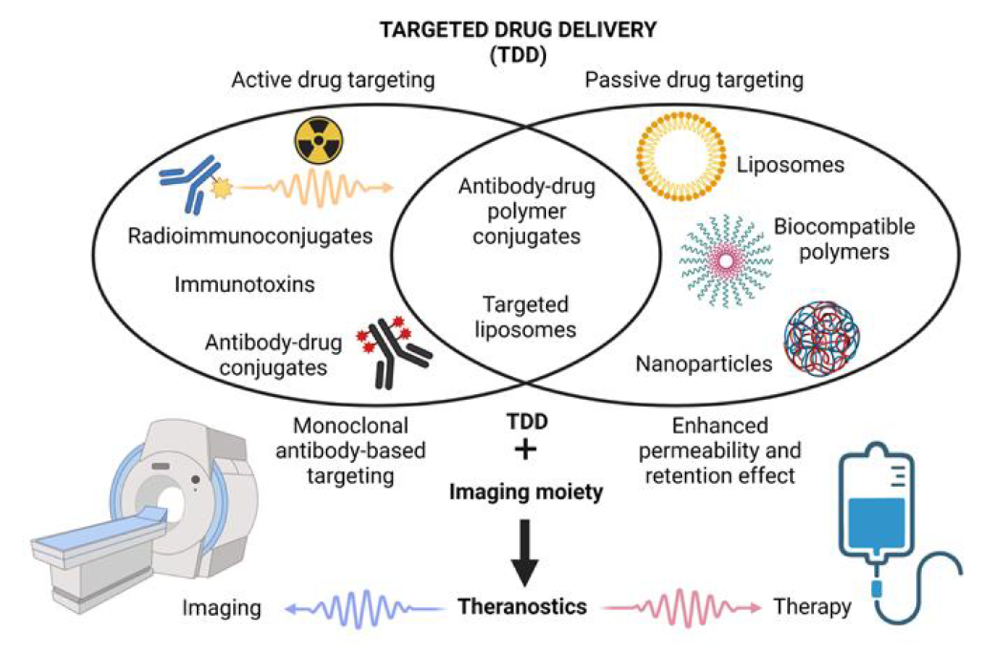
1.2. The Concept of Targeted Drug Delivery and Controlled Drug Release

In 1906, a German pathologist, immunologist, and Nobel Prize laureate Paul Ehrlich formulated the requirements for a new type of an ideal drug called “a magic bullet”, which would deliver the anti-cancer drug directly to the tumor with minimal release of the free drug in the bloodstream during transport [19,20]. Currently, three established large groups of anti-cancer agents fulfill the original idea of the magic bullet proposed by Paul Ehrlich: (1.) actively targeted biologicals based on monoclonal antibody-mediated delivery of toxic warheads (ADC, RIT), (2.) passively targeted DDS based on the enhanced permeability and retention (EPR) effect (liposomes, polymeric nanocarriers, and other types of nanomedicines), and (3.) overlapping agents (various types of actively targeted nanomedicines). The addition of an imaging moiety besides the toxic warheads can convert these agents into theranostics (Figure 1).

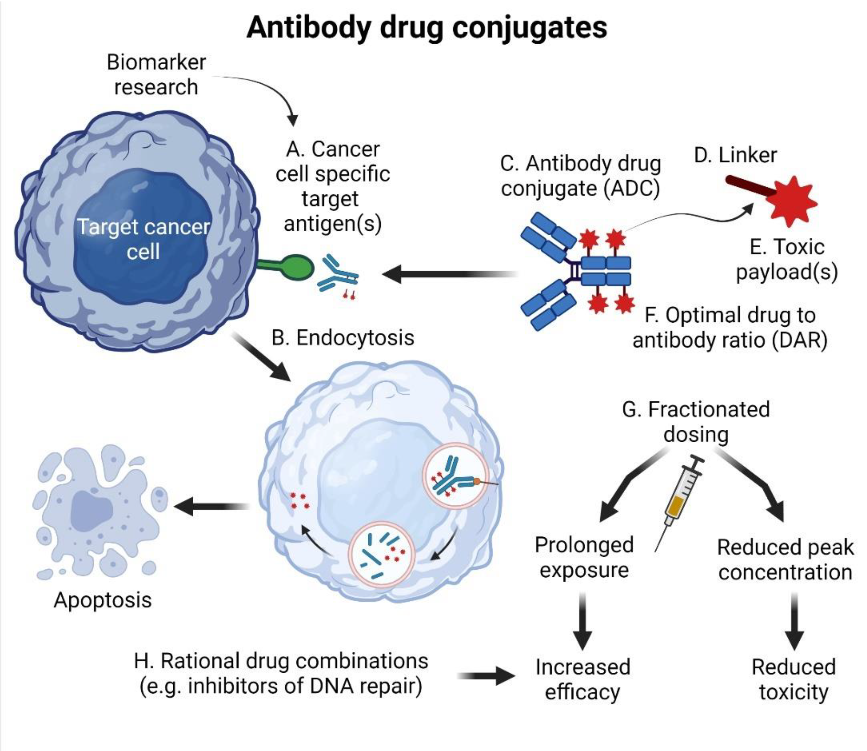
2. Antibody–Drug Conjugates (ADCs) and Immunotoxins: MAb-Mediated TDD

Monoclonal antibodies (MAbs) were the first targeted biologicals approved for the clinical practice in hemato-oncology [21,22]. The unprecedented success of anti-CD20 rituximab paved the way for the development of next-generation and glycoengineered monoclonal antibodies [8,23]. Naked MAbs exert their anti-tumor activity predominantly via activation of host immunity, namely complement (complement-dependent cytotoxicity, CDC) and immune cells, including macrophages and natural killer (NK) cells (antibody-dependent cell-mediated cytotoxicity, ADCC, antibody-dependent cell-mediated phagocytosis, ADCP) [24]. Besides that, however, MAbs represent ideal biological moieties for TDD including RIT and ADC [25]. The concept of ADC is depicted in Figure 2.

2.1. The Structure of ADCs

For the best clinical activity and minimal toxicity, the ADC must fulfill several criteria [26]. First, the target antigen should be highly (ideally exclusively) expressed on tumor cells, and the binding of ADC to the antigen should trigger receptor-mediated endocytosis of the ADC–antigen complex(es) (Figure 2A,B). The low rate of receptor-mediated endocytosis is one of the reasons why the CD20 antigen is not a suitable target for ADC-based strategy (but remains a suitable target for RIT).
Second, to minimize adverse side effects, the linker (Figure 2C,D) between the antibody and the toxic payload should be leak-proof [20]. Specifically, it should ensure a controlled drug release at the tumor site but not during transport in the bloodstream. This is achieved, for example, by enzymatic cleavage by cathepsin B, which recognizes dipeptide sequences such as valine–citrulline or by pH-sensitive hydrolytic cleavage of the hydrazone (acid-labile) linkers [27].
The toxic payloads (Figure 2E) comprise various small molecules with high cytostatic potency, usually antimitotic and alkylating agents. The mitotic poisons include maytansine derivatives derived from the plant Maytenus ovatus DM1 (emtansine, mertansine) and DM4 (soravtansine, ravtansine) and auristatins derived from the marine gastropod Dolabella auricularia (monomethylauristatin E/vedotin/MMAE, monomethylauristatin F/mafodotin/MMAF)). Alkylating agents include calicheamycin, pyrrolobenzodiazepine dimer (PBD), doxorubicin, and other agents [28].
Third, the ADC should be loaded with optimal numbers of toxic payloads, which is expressed as a drug-to-antibody ratio (DAR) (Figure 2F). DAR impacts key physico-chemical, pharmacokinetic, and pharmacodynamic features of the ADC. The currently approved ADCs have a DAR between three and four.
Due to the intentional modulation of the MAb molecule, the immune functions of the antibody carriers within ADCs usually have suppressed immune functions (compared to the parental free antibodies). The other reason why ADCs act as poor immunotherapeuticals is the dosing, which is on average 5–10 times lower compared to that of naked MAbs (e.g., anti-CD20 rituximab 375–500 mg/m2, anti-CD20 obinutuzumab 1000 mg flat dose, anti-CD79b polatuzumab vedotin 1.8 mg/kg). As a result of their modified structure and lower dosing, the mode of action of ADCs is almost exclusively based on TDD, while their roles as mediators of CDC, ADCC, and ADCP are to a great extent suppressed.

2.2. Gemtuzumab Ozogamicin (GO): The First Global Approval

The first ADC approved in clinical practice was gemtuzumab ozogamicin (GO), an anti-CD33 antibody conjugated with calicheamicin [29]. GO was granted accelerated approval in 2000 for the therapy of R/R acute myeloid leukemia (AML) at the dose of 9 mg/m2 at days one and 15, but in 2010 the marketing approval was voluntarily withdrawn because of reported high systemic toxicity. In 2017, GO was regranted approval with modified dosing at a lower dose “fractionated” schedule of 3 mg/m2 days at one, four, and seven [30]. GO is currently being evaluated in numerous clinical trials in combination with chemotherapy or targeted agents for patients with newly diagnosed and treatment-refractory AML, including CPX-351 (NCT03904251), venetoclax (NCT04070768), arsenic trioxide plus all trans-retinoic acid (NCT01409161), and many others.

2.3. Brentuximab Vedotin (BV): The First Clinical Approval for the Therapy of Lymphomas

The second clinically approved ADC was anti-CD30 brentuximab vedotin for the therapy of lymphomas (Table 1).
BV is conjugated with the synthetic antitubulin molecule monomethyl auristatin E (MMAE). In 2011, BV was approved for the therapy of relapsed/refractory (R/R) Hodgkin lymphomas and R/R anaplastic large T-cell lymphomas (ALCL), both characterized by aberrant (over)expression of CD30 [31]. In 2017, BV was approved for the therapy of R/R cutaneous T-cell lymphomas based on the results of the ALCANZA phase III clinical trial, which demonstrated significant improvement in objective response rates achieved in patients treated with BV versus physician’s choice of methotrexate or bexarotene [32]. In 2018, BV in combination with chemotherapy was approved for the therapy of patients with so far untreated Hodgkin lymphoma based on the results of the ECHELON-1 study [33]. Based on the ECHELON-2 phase III clinical trial, BV in combination with cyclophosphamide, doxorubicin, and prednisone was approved for first-line therapy of CD30+ peripheral T-cell lymphomas [5]. The success of BV clearly demonstrated that the concept of ADC can be effective even in the case when the naked antibody (anti-CD30 SGN-30) exerted virtually no clinical activity [34]. The loss of immune-mediated activity of SGN-35 (i.e., SGN-30 conjugated with MMAE) did not hamper the anti-tumor activity of BV, because the MoA of BV is fundamentally different compared to the naked SGN-30 MAb. It must be underlined that CD30 represents an ideal target antigen for the ADC-mediated delivery of a mitotic poison for two reasons. First, CD30 is aberrantly expressed almost exclusively on lymphoma cells but not on healthy tissues. Second, CD30 is readily internalized upon binding of BV, thus delivering the toxic payload specifically within lymphoma cells. Currently, BV is being evaluated in clinical trials for patients with T-cell lymphomas in combination with chemotherapy (NCT05006664), pembrolizumab (NCT04795869), lenalidomide (NCT03302728), and other drug combinations.

2.4. Inotuzumab Ozogamicin: Targeting B-Cell Acute Lymphoblastic Leukemia/Lymphoma

CD22 is a glycoprotein expressed on B-cell acute lymphoblastic leukemia (B-ALL) blasts. Inotuzumab ozogamicin (InO) is a humanized anti-CD22 monoclonal antibody conjugated to calicheamicin [35]. In June 2017, InO was granted approval for the treatment of patients with R/R CD22-positive B-ALL who have failed at least one tyrosine kinase inhibitor-based therapy [36,37]. Of note, InO is effective also in challenging populations of patients with high baseline disease burden including extramedullary disease or lymphoblastic lymphoma [38]. However, a phase III randomized trial in patients with R/R CD20/CD22-positive aggressive B-NHL failed to demonstrate a benefit of InO plus rituximab compared to the investigator’s choice (R-bendamustine or R-gemcitabine) [39].

2.5. Polatuzumab Vedotin (PV): The Game Changer in DLBCL?

CD79B is expressed on B-cells as a part of the B-cell receptor. Polatuzumab vedotin (PV), in combination with bendamustine and rituximab (PV-BR), was approved in 2019 for the therapy of R/R DLBCL based on the results of a phase II clinical trial (NCT02257567) [40]. The ORR in the PV-BR and BR arms was 63% and 25%, respectively. The currently running POLARIX phase III trial is comparing the efficacy of PV in combination with rituximab and chemotherapy (cyclophosphamide, doxorubicin, and prednisone (CHP)) versus rituximab and CHOP (CHP plus vincristine) in previously untreated patients with DLBCL patients. A total of 879 patients were randomized 1:1 to receive PV-R-CHP (+vincristine placebo) or R-CHOP (+PV placebo). The PV-R-CHP combination demonstrated a 27% reduction in the relative risk of disease progression, relapse, or death compared with R-CHOP, with a similar safety profile [41]. PV-R-CHP is thus the first regimen in the last 20 years to significantly improve outcomes in previously untreated DLBCL. PV is currently being evaluated in combination with salvage chemotherapy for patients with R/R DLBCL (NCT04182204, NCT04665765). PV-BR, and PV in combination with lenalidomide and rituximab, is being evaluated in two clinical trials in patients with R/R MCL (NCT04913103, NCT04659044). PV-R-CHP in combination with a bispecific antibody glofitamab is being studied in patients with newly diagnosed high-risk DLBCL (NCT04914741), while a dose-reduced PV-R-CHP regimen is currently being tested in elderly patients with so far untreated DLBCL (NCT04594798).

2.6. Loncastuximab Tesirine (LT): Another Player in CD19-Directed Strategies

CD19-directed therapies became a mainstream of T-cell engaging immunotherapies including adoptive cellular therapy with CD19-directed T-cells with chimeric antigen receptors (axicabtagene ciloleucel, tisagenlecleucel, lisocabtagene maraleucel, brexucabtagene autoleucel), bispecific T-cell engagers (blinatumomab), or CD19-directed naked monoclonal antibodies (tafasitamab) [8,15,16,17,18,42]. In addition, CD19 represents an appealing target antigen for TDD [43]. First, although CD19 is expressed on all mature B-cells, it is not targeted by currently used front-line immunotherapy approaches based on anti-CD20 antibodies. Second, contrary to CD20, CD19 antigens are internalized upon the binding of anti-CD19 antibodies. CD19 antibody conjugated to tesirine, a pyrrolobenzodiazepine dimer (PBDD) cytotoxin, loncastuximab-tesirine (LT), demonstrated promising clinical activity in prognostically adverse R/R DLBCL including double-hit and transformed lymphoma in the LOTIS-2 clinical trial [44]. The overall response rate was 48.3% with 24.1% complete remissions (CRs). PBDD-associated adverse events including oedema, effusions, rash, and liver enzyme elevations were successfully managed by improved supportive care and dose reduction. LT is currently being tested in several clinical trials in patients with R/R NHL (in combination with venetoclax, NCT05053659), with R/R follicular lymphoma (in combination with rituximab, NCT04998669), in R/R MCL and DLBCL (in combination with ibrutinib, NCT03684694), in R/R B-NHL (in combination with salvage chemotherapy, LOTIS-7, NCT04970901), in newly diagnosed DLBCL (in combination with R-CHP, LOTIS-8, NCT03684694), or in newly diagnosed frail/unfit (elderly) DLBCL patients (in combination with rituximab, LOTIS-9, NCT05144009).

2.7. Next-Generation ADCs

The ADCs were developed as sophisticated targeted drug delivery biologicals that improved the therapeutic index of various toxic substances [45]. Besides targeted drug delivery, further improvement of the therapeutic index was achieved by tumor-mediated linker cleavage, e.g., by cathepsin B, which recognizes dipeptide sequences such as valine–citrulline [46]. From this perspective, ADCs can be regarded as prodrugs, actively delivered into the tumor, and fully activated by the tumor. Further development (or evolution) of ADC biotechnology will focus on all parts of the ADC macromolecule, including the MAb carrier, the linker, and the toxic payload (Figure 2). Besides the ADC itself, two other critical factors will impact the efficacy and safety of next-generation ADCs: (1.) selection of suitable tumor-specific or tumor-associated antigen(s), and (2.) optimal dosing schedules including fractionated dosing of the currently approved ADCs [47].
Receptor tyrosine kinase-like orphan receptor 1 (ROR1) represents a promising target antigen, because it is expressed on leukemia and lymphoma cells but not on healthy tissues [48]. Several anti-ROR1 antibodies and ADCs are currently in various stages of preclinical development. VLS-101, anti-ROR1 humanized IgG1 MAb conjugated with MMAE via a standard maleimidocaproyl-valine-citrulline-para-aminobenzoate linker, demonstrated anti-tumor efficacy on patient-derived xenograft models of Richter syndrome (transformation of chronic lymphocytic leukemia to aggressive lymphoma) [49].
Decreased systemic toxicity may be achieved by genetic modification of the Fc fragment of the MAb carrier with an attempt to attenuate the non-specific binding of the Fc fragment to Fcγ receptors expressed on the cells of the reticuloendothelial system, which is responsible for some of the side effects including liver toxicity and myelosuppression. Conditional activation of the binding moiety of the ADC in the tumor tissue by active antibody masking can further increase efficacy and decrease the toxicity of the innovative ADC platforms (e.g., SAFEbody) [50].
Innovative linker technologies will allow bioengineering of ADC constructs with significantly increased DAR (from the usual 3–4 to >10–20). For example, the fleximer linker used in upifitamab rilsodotin (XMT-1536), an ADC directed against sodium-dependent phosphate transport protein 2 (BNaPi2b) and loaded with auristatin, is a biodegradable, highly biocompatible, water-soluble polymer, to which are attached multiple molecules of the auristatin drug yielding a DAR between 12 and 15 [51]. The toxic warheads will broaden beyond antimicrotubule agents to other potent cytotoxic or targeted drugs including topoisomerase inhibitors (e.g., exatecan), alkylating agents (e.g., pyrrolobenzodiazepines), BH3 mimetics (e.g., BCL2 inhibitors), or immunostimulants (e.g., toll-like receptor agonists) [52]. For example, huXBR1-402-G5-PNU, a novel anti-ROR1 ADC, is loaded with a highly potent anthracycline derivative of nemorubicin (PNU-159682) and demonstrated promising clinical activity in preclinical models of MCL after the failure of CAR19 T-cell-based therapy [53].

2.8. Immunotoxins: From Denileukin Diftitox to Moxetumomab Pasudotox and Beyond

Recombinant immunotoxins (IT) are genetically engineered proteins composed of a targeting moiety fused to a bacterial toxin, usually an enzymatically active portion of the diphtheria toxin fragments. The most frequently used targeting moieties of recombinant ITs comprise the fragment variable (Fv), the antibody-binding (Fab) portion of a monoclonal antibody, or the receptor-binding domain of human interleukin 2 (IL-2).
Denileukin diftitox is a diphtheria fusion protein composed of the truncated diphtheria toxin and human IL-2. Denileukin diftitox was approved by the FDA in 1999 for the treatment of cutaneous T-NHL, but it is not available on the market [54]. Next-generation IL-2 cytotoxic fusion protein E7777 has shown clinical activity in Japanese patients with R/R T-NHL [55].
Moxetumomab pasudotox is composed of the Fv fragment of an anti-CD22 monoclonal antibody fused to a 38 kDa fragment of Pseudomonas exotoxin A, PE38 [56]. In 2018, the FDA approved moxetumomab pasudotox-tdfk for patients with hairy cell leukemia who failed two lines of systemic therapy [57].

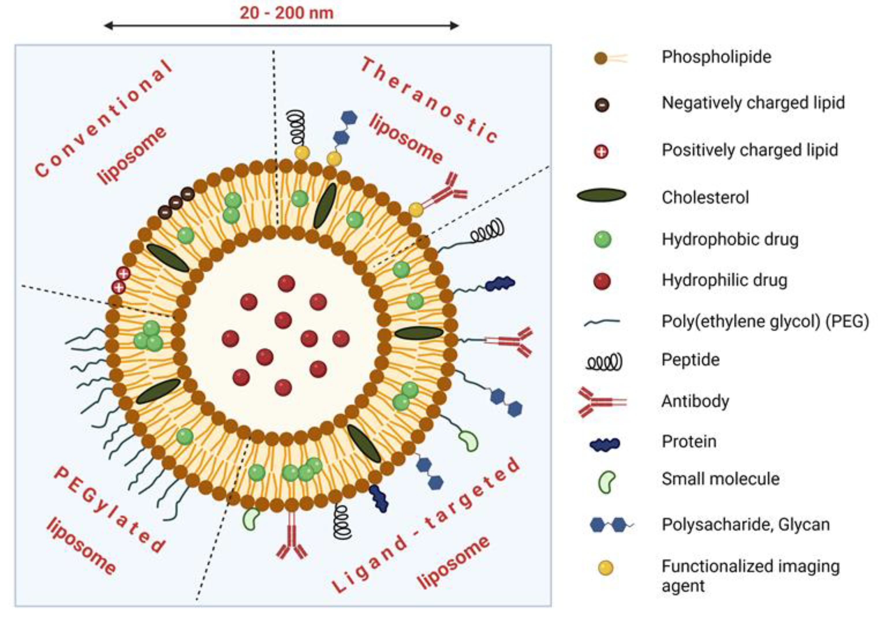
3. Liposomes, Polymeric Nanocarriers and Other Types of Nanomedicines

3.1. Nanosystems for TDD: Structure, Passive and Active Targeting

Thanks to huge progress in the field of biomedical polymer research, Ehrlich’s original idea of the magic bullet gave rise to a new scientific discipline called controlled drug delivery, or controlled drug release. Various types of targeted DDS, such as liposomes, polymeric nanocarriers, polymeric micelles, solid lipid nanocarriers, protein-based nanocarriers, dendrimers, carbon nanotubes, or magnetic nanoparticles, are currently being studied with the aim to improve and enhance the effectiveness of the treatment of serious diseases such as cancer, inflammatory diseases, or bacterial infections. The anti-cancer activity, as well as toxic side effects of these therapeutics, is strongly dependent on their molecular weight, structure, hydrodynamic size, charge, surface properties and functionality, type of bond used for the drug attachment (enzymatically or hydrolytically cleavable), rate of cellular uptake, and the biological behavior, such as biocompatibility, uptake by the immune system (macrophages), and circulation time in the bloodstream.
One of the unique properties of all TDD nanosystems is their passive tumor targeting mediated by the enhanced permeability and retention (EPR) effect, first described by Matsumura and Maeda in 1986. The EPR effect is based on the accumulation of macromolecules within solid tumors due to anatomical differences between the structure of the healthy (organized) and tumor (disorganized) vasculature and lymphatic drainage [58]. Active targeting of nanosystems can be achieved by the attachment of specific ligands (e.g., antibodies, peptides, vitamins, or hormones) with the ability to bind to cognate receptors highly (or exclusively) expressed on the surface of target cells (Figure 1). Nanosystems can also serve as prodrugs that are activated by unique features of the tumor microenvironment including pH, enzymatic activity, temperature, or osmolality.

3.2. Liposomal Formulations of Cytostatics

Liposomes were one of the earliest nanocarriers used for passive targeting of drugs and belong to the most clinically established nanocarriers for therapeutic agent delivery up to now. Liposomes are artificial spherical bilayer vesicles prepared from insoluble polar lipids, e.g., naturally derived phospholipids [59,60]. Under aqueous conditions, they assemble into highly organized membrane-forming aggregates with a structure such as biological membranes, where an aqueous core domain is surrounded by the lipid bilayer [61,62]. Due to the extreme versatility of liposomes and their amphiphilic character, the loaded drugs can be entrapped within the liposome not only in the hydrophilic cavity but also in the lipid bilayer (see Figure 3) [63,64,65,66].
Uptake of the drug inside the liposome contributes significantly to an increased therapeutic index of the loaded drugs, mainly due to (i) protection of the drug against enzymatic degradation, immunologic and chemical inactivation; (ii) prevention of its metabolization before reaching target tissue; (iii) reduced drug exposure to healthy tissues; (iv) increased blood circulation half-life [67,68]. According to the number of their layers (also called lamellae), the liposomes can be divided into multilamellar (>500 nm), small unilamellar (20–100 nm), or large unilamellar vesicles (>100 nm) [59]. In general, the lipids utilized for liposome preparation are natural and synthetic double-chain lipids (phosphorus polar head and glycerol backbone) and sterols, especially cholesterol [69].
Non-coated liposomes are often cleared by the cells of the reticuloendothelial system, which is the reason why there was an effort to modify their surface with an inert biocompatible hydrophilic polymer. A polyethylene glycol (PEG) is predominantly employed as a non-toxic, non-ionic, biocompatible molecule for PEG modification, so-called PEGylation [66]. PEGylation protects liposomes from immune-mediated clearance and confers higher stability, which leads to prolonged circulation of the PEGylated liposomes in the organism [70]. A coating of the liposomes with glycoproteins, oligosaccharides, and polysaccharides was also studied with the aim to prevent the liposomal blood clearance [71]. The surface-modified liposomes are called stealth liposomes and can be prepared with PEG chains of various length covalently attached to the hydrocarbon chain anchors.

3.3. Liposomal Formulations of Anthracyclines

3.3.1. Liposomal Formulations of Doxorubicin—The First Nanomedicines Approved for the Treatment of Cancer

Anthracyclines including doxorubicin, daunorubicin, or epirubicin belong to backbone cytostatic agents for the treatment of both solid tumors and hematological malignancies [65,72]. Liposomes have been primarily studied as anthracycline carriers to reduce the anthracycline-associated cardiotoxicity and increase their therapeutic index [73]. Two liposomal formulations of doxorubicin have been granted marketing approval up to the present: non-PEGylated liposomal doxorubicin (NPLD; Dox citrate-encapsulated formulation; Myocet®), and PEGylated liposomal doxorubicin (PLD; Caelyx® /Doxyl®/LipoDox®) [66,68,69,74]. In 1995, PLD was the first liposomal drug class of agent approved for the treatment of AIDS-related Kaposi’s sarcoma [75]. Besides Kaposi’s sarcoma, PLD was approved for the treatment of patients with ovarian cancer (in 1998) and metastatic breast cancer (in 2003). In 2007, PLD (in combination with bortezomib) was approved for patients with MM who have received at least one prior therapy not including bortezomib [74].
The second approved liposomal formulation of doxorubicin, NPLD (Myocet), was approved in the year 2000 in combination with cyclophosphamide for first-line therapy of metastatic breast cancer.
The non-PEGylated liposomal formulation of daunorubicin, DaunoXome® (DNX), is indicated as a first-line cytotoxic therapy for advanced HIV-associated Kaposi’s sarcoma.
CPX-351 (Vyxeos®) is a liposome-based nanocarrier encapsulating daunorubicin and cytarabine in 1:5 molar ratio [76]. Therapy with CPX-351 was associated with significantly prolonged survival compared to the standard of care “7 + 3” regimen (conventional cytarabine and daunorubicin) in the elderly patients with so far untreated AML, while its safety profile was comparable [77,78]. In 2021, Vyxeos liposomal® was approved also for the treatment of secondary AML in pediatric patients [79].

3.3.2. Liposomal Formulations of Doxorubicin in the Therapy of Aggressive Lymphomas

The rationale for the use of liposomal formulations of doxorubicin in patients with aggressive lymphoma is based on two presumptions. First, doxorubicin is a key component of the R-CHOP-like regimens, which still represent the current golden standard of care for first-line therapy of patients with most types of aggressive lymphomas but has been associated with cardiotoxicity, which limits its use in the cohort of frail/unfit patients. Second, replacement of free doxorubicin with the liposomal nanodrug would decrease cardiotoxicity of free doxorubicin, thereby offering access to the standard of care to more patients including the elderly and comorbid ones. Importantly, in a large meta-analysis of patients with DLBCL treated with NPLD, Visco et al. endorsed the value of NPLD both in terms of response and survival [80]. In contrast, although Sancho et al. confirmed the non-inferiority of NPLD to conventional doxorubicin as part of frontline immunochemotherapy in patients with so far untreated aggressive lymphomas, the substitution with NPLD was not associated with less early cardiotoxicity, although some reduced cardiac safety signals were observed [81]. Similarly, NPLD did not reduce cardiotoxicity, although cardiac safety signals were elevated in R-CHOP compared to R-COMP in an Austrian randomized phase III study [82]. So, the cardiotoxicity issue of the liposomal formulations of doxorubicin remains a matter of debate. Liposomal formulations of doxorubicin as part of various polychemo/immunotherapy regimens demonstrated promising activity with an acceptable safety profile in patients with classical Hodgkin lymphoma, primary mediastinal B-cell lymphoma, and other types of aggressive lymphomas [83,84]. NPLD in combination with romidepsin, a histone deacetylase inhibitor, demonstrated an acceptable safety profile and promising clinical efficacy with deep skin responses in relapsed/refractory cutaneous T-cell lymphomas [85]. Excellent anti-tumor efficacy was reported in a small cohort of R/R DLBCL patients treated with the combination of two liposomal nanodrugs: NPLD and albumin-bound paclitaxel (nabpaclitaxel, Abraxane) [86].

3.4. Liposomal Formulations of Vinca Alkaloids and Cytarabine

Like anthracyclines, vinca alkaloids belong to classical cytostatics broadly used in diverse hematologic malignancies. In 2012, the liposomal formulation of vincristine (vincristine sulphate liposome injection (VSLI), Marquibo®) was approved for therapy of Philadelphia chromosome negative (Ph-) acute lymphoblastic leukemias. This liposomal formulation reduced the side effects of free vincristine, especially neuropathy. VSLI was tested also in patients with aggressive lymphomas, where it was substituted for the conventional vincristine in the R-CHOP regimen (becoming R-CHMP). According to the published data, the R-CHMP combination was highly active, generally well tolerated, and compared favorably to historical trials with R-CHOP in DLBCL [87,88].
Liposomal cytarabine (DepoCyt®) was a liposomal formulation of cytosine arabinoside (ara-C), which was derived to overcome the main drawback of the free drug: its rapid degradation to non-active uracil derivatives during the circulation. In 1999, DepoCyt® was approved for intrathecal treatment of lymphomatous neoplastic meningitis. In 2017, however, Pacira Pharmaceuticals announced the permanent discontinuation of DepoCyt injection due to persistent technical issues related to the product’s manufacturing process (https://professionals.optumrx.com/publications/library/drugwithdrawal-depocyt-2017-0706.html; accessed on 23 January 2022). Despite numerous attempts to develop a clinically approved cytarabine nanomedicine, the only ara-C nanodrug with marketing authorization is the above-mentioned liposomal combination of ara-C and daunorubicin (CPX-351) in patients with AML.

3.5. Targeted Liposomes

Similar to other nanoparticles, liposomes can be equipped with specific coupling ligands for targeted delivery to the tumor tissues. The active targeting of the liposomal nanomedicines should increase the efficacy of the tumor accumulation and consequently also the treatment efficacy of these formulations. Immunoliposomes that use monoclonal antibodies or their fragments for active targeting belong to the most promising nanodrugs [89]. Currently, transferrin receptor, epidermal growth factor receptor, and folate receptor belong to the most studied and employed cell targets for immunoliposomes. The targeting moiety (i.e., antibody, antibody fragment, or single-chain fragment variable (scFv)), the coupling method, and the rate of internalization of the liposome-receptor system all belong to critical factors that impact the anti-tumor efficacy of immunoliposomes. Recently, the clinical trials of immunoliposomes were reviewed in detail [90]. The results obtained so far demonstrate that immunoliposomes represent promising multivalent liposome-antibody constructs with unique therapeutic properties that can be used not only for the therapy but also for diagnostic or restaging procedures, e.g., in vivo imaging of circulating leukemia cells or visualization of tumor endothelium [90].
Beside the monoclonal antibodies, oligonucleotide-derived aptamers represent another class of promising targeting ligands. Aptamers, sometimes called “chemical antibodies” are small nucleic acid ligands composed of RNA or single-stranded DNA oligonucleotides that possess high specificity and affinity for their targets [91]. Their interaction with various targets is similar to that of antibodies: by recognizing a specific three-dimensional structure.

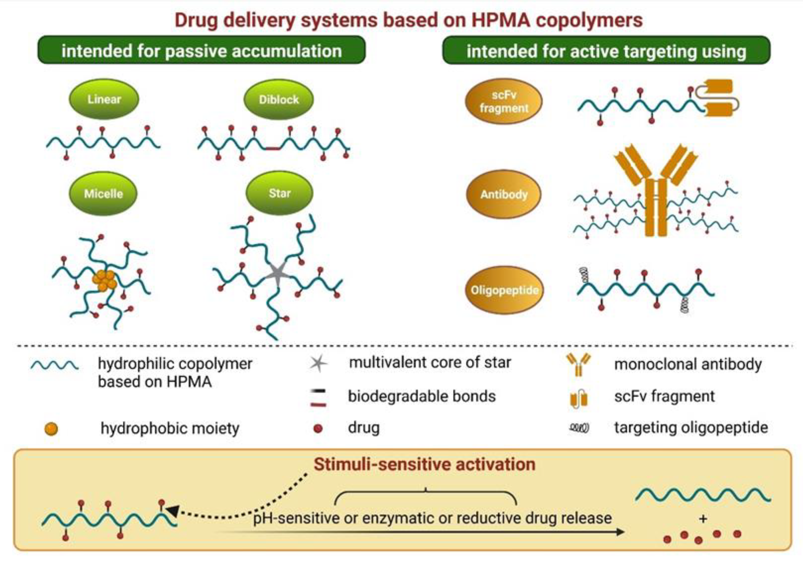
4. Polymer-Based Nanomedicines

4.1. PEG-Based Polymers

The most studied non-degradable water-soluble polymer used for the synthesis of polymer–drug conjugates is poly(ethylene glycol) (PEG) [92]. Monofunctional methoxy PEG is widely used for surface modification of proteins or liposomes to stealth them against biodegradation and improve their pharmacokinetics. Some of these conjugates successfully passed clinical evaluations and were granted marketing authorization, including pegaspargase (Oncaspar®) or peginterferon alfa-2a (Pegasys®). Pegaspargase is a polymer–drug conjugate used in the treatment of acute lymphoblastic leukemia (ALL) [93]. PEG stabilizes the asparaginase enzyme in the conjugate, which prolongs its circulation and reduces the blood levels of the amino acid asparagine. As an alternative to PEG, various poly(2-oxazoline) polymers (POx) were proposed and synthesized [94]. The use of POx enabled the introduction of various functional groups along the polymer backbone. Although POx are very promising materials for biomedical applications, they remain a domain of preclinical research [95,96].

4.2. HPMA-Based Polymers

Among others, water-soluble polymer carriers based on N-(2-hydroxypropyl) methacrylamide (HPMA) copolymers and their drug conjugates have been extensively studied because of their excellent in vitro and in vivo properties. The biocompatibility, non-toxicity, non-fouling properties, and apyrogenic character of HPMA copolymers were demonstrated and described in numerous studies [97]. Because of the limited clinical activity of first-generation HPMA-based nanomedicines, next-generation high-molecular-weight HPMA-based nanotherapeutics were designed with improved pharmacokinetic and biological properties. Various structures including diblocks, multiblocks, grafts, or star-like shapes were developed (Figure 4), and their enhanced treatment efficacy compared to the first-generation HPMA copolymers was confirmed in vivo on animal models of both solid tumors and hematological malignancies [98]. Besides the passive accumulation of the attached drugs achieved predominantly by the EPR effect, HPMA copolymers can be effectively modified as active targeting systems, most frequently by the conjugation of various monoclonal antibodies or their fragments [99] (Figure 4). In addition, HPMA-based nanomedicines can serve as effective theranostics [100].

5. Theranostics

5.1. The Origins and Evolution of the Theranostic Concept

Theranostics (or theragnostics) is a term with multiple definitions and interpretations. Its first use dates back to 1998 [101]. In nuclear medicine, it refers to a concept of combined targeted imaging and therapy delivery. The imaging allows both visualization of the tumor cells and estimation of the effectiveness of the administered therapy. The binding part of a radiopharmaceutical is targeted to a specific structure on tumor cells. The targeting vector is labeled with a beta-plus emitting radionuclide for PET (or a gamma emitter for a single-photon emission computed tomography, SPECT) for the purpose of characterization of the tumor tissue and for the confirmation of presence of the target molecule in the tumor mass. This can also provide more accurate information about disease spread than conventional imaging alone. If the expression of the target is sufficient, the same binding molecule can be labeled with a beta-minus or alpha emitter and used for therapy [102]. The imaging can eventually be used for dosimetric calculations.
Although the term “theranostics” has been used frequently in the last two decades, the practical use of this approach is much older. The first administration of the radioactive iodine 131I for the treatment of thyroid cancer was performed in 1946, and after the imaging technology was implemented into clinical practice, it became an integral part of thyroid cancer management [103,104]. Theranostics has emerged as a promising strategy for the treatment of selected solid tumors, and it is becoming a routine option in their management [105,106]. Theranostic strategies have a great potential in the management of patients with hematologic malignancies because of their predominant radiosensitivity and multifocal presentation [107].

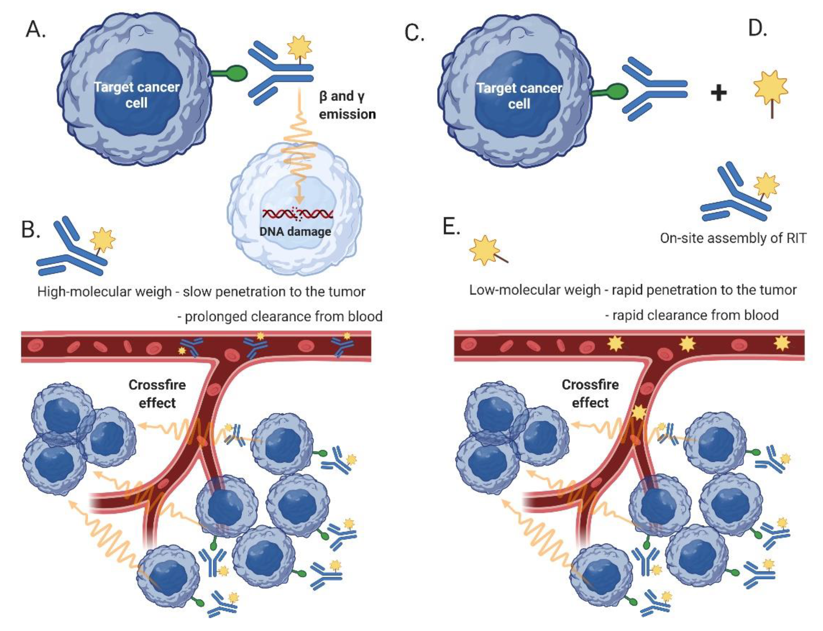
5.2. Radioimmunotherapy (RIT)

Even though a plethora of theranostic pairs have been tested preclinically, the number of approved clinical applications remain limited. The concept of radioimmunotherapy (RIT) represents an example of a successful theranostics application in hematology. Compared to external-beam radiotherapy, RIT achieves a targeted delivery of a radionuclide bound to the monoclonal antibody directly to the lymphoma cells at distant (and distinct) anatomical sites. The so-called crossfire effect, mediated by the emitted electrons and gamma rays, leads also to eradication of lymphoma cells with no/low expression of the target antigen. There are two anti-CD20 radiopharmaceuticals approved for the treatment of B-cell lymphoma: A combined beta- and gamma-emitter iodine-131 tositumomab (Bexxar®) with a half-life of approx. 8 days and a pure beta-emitter yttrium-90 ibritumomab tiuxetan (Zevalin®) with a half-life of 2.7 days [108,109]. Both radiopharmaceuticals originally included a pre-therapeutic scintigraphic imaging step with lower activity for dosimetric purposes (labeling with 131I in the case of tositumomab, and ibritumomab was labeled with 111In). This step was later omitted from the ibritumomab therapy. However, due to complicated manufacture and administration procedures and with the advent of innovative targeted and immunotherapy agents, the clinical use of RIT in the management of B-NHL has become limited.

5.3. Pretargeted RIT

Compared to a single-step RIT, pretargeted RIT (PRIT) is a two-step process developed to address limitations of RIT, namely, to prevent long-term circulation of a radioactive RIT in the blood (Figure 5). In the first step of PRIT, a naked (targeting, cold) antibody is administered to the patient with the principal goal to bind all targeted antigens present on the tumor cells at all involved sites. In the second step (after the unbound targeting antibody has been cleared from the circulation) the patients are injected with a low-molecular-weight radioactive agent with high affinity to the targeting antibody. The small size of the radioactive agent is responsible both for rapid penetration of the agent to the involved organs and tissues (where it is bound to the targeting antibody) and for the rapid clearance of the free (unbound) agent from the circulation. The improved pharmacokinetics translate into improved efficacy and fewer side effects. Experimental PRIT targeting CD38 has demonstrated promising preclinical activity in vivo in experimental models of B-NHL [110,111].

5.4. Immuno-PET

The main limitation of the standard 18F-FDG-based PET is its incapability to detect metabolically inactive lymphoma cells. In contrast, MAb-based PET imaging (immuno-PET) approaches allow detection of all lymphoma cells irrespective of their metabolic activity [112]. An immuno-PET imaging probe consists of a targeting vector (usually a monoclonal antibody or its fragment) conjugated with a radiometal. Small-sized vectors (antibody fragments, nanobodies) are preferable due to their better tumor penetration and accelerated clearance from the bloodstream. The radiometals are loaded to the vector using either a random or site-specific conjugation [113]. Long half-life isotopes, such as cuprum-64 (64Cu, half-life 12 h) or zirconium-89 (89Zr, half-life 78 h), belong among the most commonly used radionuclides for this purpose [114]. In the context of NHL, immuno-PET was studied using antibodies targeting CD20 (e.g., rituximab, obinutuzumab), CD38 (daratumumab), CD30 (SGN-30), and others. It was repeatedly demonstrated that anti-CD20 antibodies or antibody fragments labeled with radiotracers enabled dynamic visualization of lymphoma cells and measurement of the delivered therapeutic effect in patients with NHL [115].

5.5. CXCR4-Targeted Therapy and Other Theranostic Concepts

Other published clinically tested approaches in hemato-oncology are rather scarce. The C-X-C chemokine receptor 4 (CXCR4, CD184) appears to be one of the most promising targets in hematologic malignancies and some solid tumors. The CXCR4-targeted peptide receptor radionuclide therapy employs the small cyclic pentapeptide 68Ga-Pentixafor for the sensitive and high-contrast imaging, while 90Y- or Lutetium-177 (177Lu)-pentixather (3-iodo-D-Tyr1-Pentixafor) with longer circulation times and delayed whole-body clearance is used for CXCR4-targeted therapy [116]. Clinical activity was demonstrated in small groups of patients with acute leukemias, advanced DLBCL, and MM [117].
Paul et al. performed an interesting pilot study that analyzed the treatment of four patients with malignancy (one with DLBCL) with 18F-fluorodeoxyglucose alone [118]. This concept breaks the traditional view of FDG as an option used for imaging only. Positrons emitted by 18F have similar features to electrons and can deliver cytotoxic energy to lymphoma cells. Although the primary objective of this study was safety, anti-tumor activity was documented.
Several other radiopharmaceuticals with a theranostic potential have been tested in numerous preclinical studies.
In conclusion, theranostics is an attractive approach to personalized medicine that is relatively well established in the management of some solid tumors. Except for RIT, however, other theranostic concepts in the field of hemato-oncology are confined mostly to the experimental (preclinical) phase.

6. Theranostic Strategies Based on Magnetic Resonance Imaging

Unlike in nuclear medicine, the tracer (contrast material) in magnetic resonance (MR) imaging (MRI) does not itself confer any anti-cancer effect whatsoever. Instead, it is used to visualize the distribution of the injected carrier substance, which may be loaded with an anti-cancer drug and enter the tumoral mass through passive or active targeting referred to as “tumor-targeted, MRI traceable nanotheranostics” [119]. Smart MR contrast agents result in selective enhancement in the tumor microenvironment not only by increased permeability but also by acid pH [120]. Contrast materials in MRI act by shortening longitudinal relaxation time (T1) or shortening transverse relaxation time (T2). The latter group is represented by superparamagnetic iron oxide nanoparticles. T1 contrast agents are typically gadolinium (Gd) chelates, some ultra-small superparamagnetic iron oxide nanoparticles (SPIONs), and manganese oxide nanoparticles.

6.1. Gadolinium-Based Tracers and Manganese Oxide Nanoparticles

Gadolinium-based contrast materials are used to visualize tissue enhancement by shortening T1 relaxation time and increasing signal intensity (bright signal). While Gd-based contrast materials are widespread, the preclinical evidence on theranostics using Gd imaging is very limited.
Shortening of T1 relaxation time can be achieved not only by Gd but also by manganese (Mn) oxide nanoparticles (MON) [121]. Recently, MONs became widely studied due to their negligible toxicity and bright signal on T1. Another advantage of MONs is that they can also interact with the tumor microenvironment in a non-specific manner (pH, reactive oxygen species, phagocytosis) [122]. MONs with triphenylphosphonium (PPh3) were experimentally used in HeLa cells to target mitochondrial function and induce cell death [6]. Manganese oxide together with chemotherapeutic drugs can also be loaded into micellar or polymerous nanospheres [123,124]. Preparation and experimental use of other Mn oxides in both MR imaging and targeted therapy have been also studied.

6.2. Magnetic Nanoparticles

Magnetic nanoparticles are based on ferromagnetic or ferrimagnetic metals, most commonly iron but also cobalt, nickel, and their compounds and oxides. These nanoparticles exhibit superparamagnetism. In the absence of an external magnetic field, their magnetization randomly flips its direction, and they rest in a paramagnetic state but have higher magnetic susceptibility. With the application of external magnetization, a single high magnetic moment is recruited from the moments of the whole ensemble of nanoparticles.
Superparamagnetic iron oxide nanoparticles (SPIONs) are widely studied due to their minimal toxicity. SPIONs have important properties suitable for targeted therapy. They can be heated by an alternating magnetic field or laser light, regionally distributed using an external magnetic field, and visualized by MRI. SPIONs also exhibit a degree of passive targeting through the EPR effect and can be coupled with epitopes for active targeting. Importantly, SPION nanoparticles can be loaded with chemotherapeutic or immunotherapeutic drugs. Although SPION compounds have demonstrated activity in the targeted anti-cancer treatment in cell cultures and animal models, their clinical use remains limited [125,126].

7. Conclusions

In the last two decades, the concept of anti-lymphoma therapy has dramatically changed with a gradual decline of standard cytostatic agents and rapid implementation of innovative targeted, biological, and immunotherapy agents with better anti-tumor efficacy and fewer side effects. Despite that, genotoxic agents still represent an indispensable backbone of anti-lymphoma therapy, especially of aggressive lymphoma subtypes. It appears that new formulation of “old” conventional cytostatics, with their passive and/or active delivery and controlled release, might represent the future of these highly effective anti-tumor drugs by improving their pharmacokinetics, increasing the therapeutic index, enhancing on-target efficacy, and reducing toxic side effects. Moreover, the binding of imaging probes to these nanomedical biomolecules might in the near future serve not only for more efficacious therapy but also for dynamic restaging procedures or the detection and eradication of the minimal residual disease.

Author Contributions

T.E., A.B., D.Z., L.L., N.R. and P.K.: writing—original draft preparation, writing—review and editing; T.E. and P.K.: visualization, P.K.: supervision, project administration, funding acquisition. All authors have read and agreed to the published version of the manuscript.

Funding

The manuscript was supported by the Grant Agency of the Czech Republic grant GACR19-01417S, by the Ministry of Health of the Czech Republic grant AZV NU21-03-00386, and by the Charles University Center of Excellence UNCE/MED/016.

Conflicts of Interest

The authors declare no conflict of interest.

Abbreviations

ADCntibody drug conjugate
ADCCantibody-dependent cell-mediated cytotoxicity
ADPCantibody-dependent cell-mediated phagocytosis
ALLacute lymphoblastic leukemia
AMLacute myeloid leukemia
BCMAB-cell maturation antigen
BMbelantamab mafodotin
B-NHLB-cell NHL
BVbrentuximab vedotin
CARchimeric antigen receptor
CAR19CAR against CD19-positive cells
CDCcomplement-dependent cytotoxicity
DARdrug to antibody ration
DDSdrug delivery systems
DLBCLdiffuse large B-cell lymphoma
EPRenhanced permeability and retention
FDG2-[F18] fluoro-2-deoxy-D-glucose
FLfollicular lymphoma
Fvfragment variable (of the monoclonal antibody)
Gdgadolinium
GFLGglycylphenylalanylleucylglycine
GOgemtuzumab ozogamicin
HLHodgkin lymphoma
HPMAN-(2-hydroxypropyl)methacrylamide
InOinotuzumab ozogamicin
ITimmunotoxin
LTloncastuximab tesirine
MCLmantle cell lymphoma
MDR-1multi-drug resistance protein 1
MMmultiple myeloma
MMAEmonomethyl auristatin E
MoAmode of action
MRImagnetic resonance imaging
NHLnon-Hodgkin lymphoma
NPLDnon-pegylated liposomal doxorubicin
MONmanganese oxide nanoparticles
MZLmarginal zone lymphoma
Mabmonoclonal antibody
NKnatural killer
PEGpolyethylene glycol
PGApoly(L-glutamic acid)
PLDpegylated liposomal doxorubicin
PETpositron emission tomography
PBDDpyrrolobenzodiazepine dimer
Poxpoly(2-oxazoline) polymers
PRITpretargeted RIT
PSMAprostate specific membrane antigen
PVpolatuzumab vedotin
R-CHOPrituximab + cyclophosphamide + doxorubicin + vincristine + prednisone
RITradioimmunotherapy
R/Rrelapsed/refractory
scFcsingle-chain fragment variable
SPECTsingle-photon emission computed tomography
SPIONsuperparamagnetic iron oxide nanoparticles
TDDtargeted drug delivery
T-NHLT-cell NHL
VSLIvincristine sulphate liposome injection

References

  1. Nogai, H.; Dorken, B.; Lenz, G. Pathogenesis of non-Hodgkin’s lymphoma. J. Clin. Oncol. 2011, 29, 1803–1811. [Google Scholar] [CrossRef] [PubMed]
  2. Swerdlow, S.H.; Campo, E.; Pileri, S.A.; Harris, N.L.; Stein, H.; Siebert, R.; Advani, R.; Ghielmini, M.; Salles, G.A.; Zelenetz, A.D.; et al. The 2016 revision of the World Health Organization classification of lymphoid neoplasms. Blood 2016, 127, 2375–2390. [Google Scholar] [CrossRef] [PubMed] [Green Version]
  3. Swerdlow, S.H.; Cook, J.R. As the world turns, evolving lymphoma classifications-past, present and future. Hum. Pathol. 2019, 95, 55–77. [Google Scholar] [CrossRef]
  4. Salles, G.; Barrett, M.; Foà, R.; Maurer, J.; O’Brien, S.; Valente, N.; Wenger, M.; Maloney, D.G. Rituximab in B-Cell Hematologic Malignancies: A Review of 20 Years of Clinical Experience. Adv. Ther. 2017, 34, 2232–2273. [Google Scholar] [CrossRef] [PubMed] [Green Version]
  5. Horwitz, S.; O’Connor, O.A.; Pro, B.; Illidge, T.; Fanale, M.; Advani, R.; Bartlett, N.L.; Christensen, J.H.; Morschhauser, F.; Domingo-Domenech, E.; et al. Brentuximab vedotin with chemotherapy for CD30-positive peripheral T-cell lymphoma (ECHELON-2): A global, double-blind, randomised, phase 3 trial. Lancet 2019, 393, 229–240. [Google Scholar] [CrossRef] [Green Version]
  6. Crump, M.; Neelapu, S.S.; Farooq, U.; Van Den Neste, E.; Kuruvilla, J.; Westin, J.; Link, B.K.; Hay, A.; Cerhan, J.R.; Zhu, L.; et al. Outcomes in refractory diffuse large B-cell lymphoma: Results from the international SCHOLAR-1 study. Blood 2017, 130, 1800–1808. [Google Scholar] [CrossRef] [PubMed]
  7. Hoy, S.M. Tafasitamab: First Approval. Drugs 2020, 80, 1731–1737. [Google Scholar] [CrossRef]
  8. Salles, G.; Duell, J.; González Barca, E.; Tournilhac, O.; Jurczak, W.; Liberati, A.M.; Nagy, Z.; Obr, A.; Gaidano, G.; André, M.; et al. Tafasitamab plus lenalidomide in relapsed or refractory diffuse large B-cell lymphoma (L-MIND): A multicentre, prospective, single-arm, phase 2 study. Lancet Oncol. 2020, 21, 978–988. [Google Scholar] [CrossRef]
  9. Wang, M.L.; Rule, S.; Martin, P.; Goy, A.; Auer, R.; Kahl, B.S.; Jurczak, W.; Advani, R.H.; Romaguera, J.E.; Williams, M.E.; et al. Targeting BTK with ibrutinib in relapsed or refractory mantle-cell lymphoma. N. Engl. J. Med. 2013, 369, 507–516. [Google Scholar] [CrossRef] [Green Version]
  10. Dreyling, M.; Jurczak, W.; Jerkeman, M.; Silva, R.S.; Rusconi, C.; Trneny, M.; Offner, F.; Caballero, D.; Joao, C.; Witzens-Harig, M.; et al. Ibrutinib versus temsirolimus in patients with relapsed or refractory mantle-cell lymphoma: An international, randomised, open-label, phase 3 study. Lancet 2016, 387, 770–778. [Google Scholar] [CrossRef] [Green Version]
  11. Trneny, M.; Lamy, T.; Walewski, J.; Belada, D.; Mayer, J.; Radford, J.; Jurczak, W.; Morschhauser, F.; Alexeeva, J.; Rule, S.; et al. Lenalidomide versus investigator’s choice in relapsed or refractory mantle cell lymphoma (MCL-002; SPRINT): A phase 2, randomised, multicentre trial. Lancet Oncol. 2016, 17, 319–331. [Google Scholar] [CrossRef]
  12. Ansell, S.M.; Lesokhin, A.M.; Borrello, I.; Halwani, A.; Scott, E.C.; Gutierrez, M.; Schuster, S.J.; Millenson, M.M.; Cattry, D.; Freeman, G.J.; et al. PD-1 blockade with nivolumab in relapsed or refractory Hodgkin’s lymphoma. N. Engl. J. Med. 2015, 372, 311–319. [Google Scholar] [CrossRef] [PubMed] [Green Version]
  13. Chen, R.; Zinzani, P.L.; Fanale, M.A.; Armand, P.; Johnson, N.A.; Brice, P.; Radford, J.; Ribrag, V.; Molin, D.; Vassilakopoulos, T.P.; et al. Phase II Study of the Efficacy and Safety of Pembrolizumab for Relapsed/Refractory Classic Hodgkin Lymphoma. J. Clin. Oncol. 2017, 35, 2125–2132. [Google Scholar] [CrossRef] [PubMed]
  14. Kuruvilla, J.; Ramchandren, R.; Santoro, A.; Paszkiewicz-Kozik, E.; Gasiorowski, R.; Johnson, N.A.; Fogliatto, L.M.; Goncalves, I.; de Oliveira, J.S.R.; Buccheri, V.; et al. Pembrolizumab versus brentuximab vedotin in relapsed or refractory classical Hodgkin lymphoma (KEYNOTE-204): An interim analysis of a multicentre, randomised, open-label, phase 3 study. Lancet Oncol. 2021, 22, 512–524. [Google Scholar] [CrossRef]
  15. Wang, M.; Munoz, J.; Goy, A.; Locke, F.L.; Jacobson, C.A.; Hill, B.T.; Timmerman, J.M.; Holmes, H.; Jaglowski, S.; Flinn, I.W.; et al. KTE-X19 CAR T-Cell Therapy in Relapsed or Refractory Mantle-Cell Lymphoma. N. Engl. J. Med. 2020, 382, 1331–1342. [Google Scholar] [CrossRef]
  16. Neelapu, S.S.; Locke, F.L.; Bartlett, N.L.; Lekakis, L.J.; Miklos, D.B.; Jacobson, C.A.; Braunschweig, I.; Oluwole, O.O.; Siddiqi, T.; Lin, Y.; et al. Axicabtagene Ciloleucel CAR T-Cell Therapy in Refractory Large B-Cell Lymphoma. N. Engl. J. Med. 2017, 377, 2531–2544. [Google Scholar] [CrossRef]
  17. Abramson, J.S.; Palomba, M.L.; Gordon, L.I.; Lunning, M.A.; Wang, M.; Arnason, J.; Mehta, A.; Purev, E.; Maloney, D.G.; Andreadis, C.; et al. Lisocabtagene maraleucel for patients with relapsed or refractory large B-cell lymphomas (TRANSCEND NHL 001): A multicentre seamless design study. Lancet 2020, 396, 839–852. [Google Scholar] [CrossRef]
  18. Schuster, S.J.; Bishop, M.R.; Tam, C.S.; Waller, E.K.; Borchmann, P.; McGuirk, J.P.; Jäger, U.; Jaglowski, S.; Andreadis, C.; Westin, J.R.; et al. Tisagenlecleucel in Adult Relapsed or Refractory Diffuse Large B-Cell Lymphoma. N. Engl. J. Med. 2019, 380, 45–56. [Google Scholar] [CrossRef]
  19. DeVita, V.T., Jr.; Chu, E. A history of cancer chemotherapy. Cancer Res. 2008, 68, 8643–8653. [Google Scholar] [CrossRef] [Green Version]
  20. Schwartz, R.S. Paul Ehrlich’s magic bullets. N. Engl. J. Med. 2004, 350, 1079–1080. [Google Scholar] [CrossRef]
  21. Maloney, D.G.; Grillo-Lopez, A.J.; Bodkin, D.J.; White, C.A.; Liles, T.M.; Royston, I.; Varns, C.; Rosenberg, J.; Levy, R. IDEC-C2B8: Results of a phase I multiple-dose trial in patients with relapsed non-Hodgkin’s lymphoma. J. Clin. Oncol. 1997, 15, 3266–3274. [Google Scholar] [CrossRef] [PubMed]
  22. Maloney, D.G.; Grillo-Lopez, A.J.; White, C.A.; Bodkin, D.; Schilder, R.J.; Neidhart, J.A.; Janakiraman, N.; Foon, K.A.; Liles, T.M.; Dallaire, B.K.; et al. IDEC-C2B8 (Rituximab) anti-CD20 monoclonal antibody therapy in patients with relapsed low-grade non-Hodgkin’s lymphoma. Blood 1997, 90, 2188–2195. [Google Scholar] [CrossRef] [PubMed]
  23. Marcus, R.; Davies, A.; Ando, K.; Klapper, W.; Opat, S.; Owen, C.; Phillips, E.; Sangha, R.; Schlag, R.; Seymour, J.F.; et al. Obinutuzumab for the First-Line Treatment of Follicular Lymphoma. N. Engl. J. Med. 2017, 377, 1331–1344. [Google Scholar] [CrossRef] [PubMed]
  24. Weiner, L.M.; Dhodapkar, M.V.; Ferrone, S. Monoclonal antibodies for cancer immunotherapy. Lancet 2009, 373, 1033–1040. [Google Scholar] [CrossRef] [Green Version]
  25. Tong, J.T.W.; Harris, P.W.R.; Brimble, M.A.; Kavianinia, I. An Insight into FDA Approved Antibody-Drug Conjugates for Cancer Therapy. Molecules 2021, 26, 5847. [Google Scholar] [CrossRef]
  26. Jin, Y.; Schladetsch, M.A.; Huang, X.; Balunas, M.J.; Wiemer, A.J. Stepping forward in antibody-drug conjugate development. Pharmacol. Ther. 2021, 229, 107917. [Google Scholar] [CrossRef]
  27. Bargh, J.D.; Isidro-Llobet, A.; Parker, J.S.; Spring, D.R. Cleavable linkers in antibody-drug conjugates. Chem. Soc. Rev. 2019, 48, 4361–4374. [Google Scholar] [CrossRef]
  28. Maderna, A.; Leverett, C.A. Recent advances in the development of new auristatins: Structural modifications and application in antibody drug conjugates. Mol. Pharm. 2015, 12, 1798–1812. [Google Scholar] [CrossRef]
  29. Bross, P.F.; Beitz, J.; Chen, G.; Chen, X.H.; Duffy, E.; Kieffer, L.; Roy, S.; Sridhara, R.; Rahman, A.; Williams, G.; et al. Approval summary: Gemtuzumab ozogamicin in relapsed acute myeloid leukemia. Clin. Cancer Res. 2001, 7, 1490–1496. [Google Scholar]
  30. Norsworthy, K.J.; Ko, C.W.; Lee, J.E.; Liu, J.; John, C.S.; Przepiorka, D.; Farrell, A.T.; Pazdur, R. FDA Approval Summary: Mylotarg for Treatment of Patients with Relapsed or Refractory CD33-Positive Acute Myeloid Leukemia. Oncologist 2018, 23, 1103–1108. [Google Scholar] [CrossRef] [Green Version]
  31. Younes, A.; Gopal, A.K.; Smith, S.E.; Ansell, S.M.; Rosenblatt, J.D.; Savage, K.J.; Ramchandren, R.; Bartlett, N.L.; Cheson, B.D.; de Vos, S.; et al. Results of a pivotal phase II study of brentuximab vedotin for patients with relapsed or refractory Hodgkin’s lymphoma. J. Clin. Oncol. 2012, 30, 2183–2189. [Google Scholar] [CrossRef] [PubMed]
  32. Prince, H.M.; Kim, Y.H.; Horwitz, S.M.; Dummer, R.; Scarisbrick, J.; Quaglino, P.; Zinzani, P.L.; Wolter, P.; Sanches, J.A.; Ortiz-Romero, P.L.; et al. Brentuximab vedotin or physician’s choice in CD30-positive cutaneous T-cell lymphoma (ALCANZA): An international, open-label, randomised, phase 3, multicentre trial. Lancet 2017, 390, 555–566. [Google Scholar] [CrossRef]
  33. Connors, J.M.; Jurczak, W.; Straus, D.J.; Ansell, S.M.; Kim, W.S.; Gallamini, A.; Younes, A.; Alekseev, S.; Illés, Á.; Picardi, M.; et al. Brentuximab Vedotin with Chemotherapy for Stage III or IV Hodgkin’s Lymphoma. N. Engl. J. Med. 2018, 378, 331–344. [Google Scholar] [CrossRef] [PubMed]
  34. Forero-Torres, A.; Leonard, J.P.; Younes, A.; Rosenblatt, J.D.; Brice, P.; Bartlett, N.L.; Bosly, A.; Pinter-Brown, L.; Kennedy, D.; Sievers, E.L.; et al. A Phase II study of SGN-30 (anti-CD30 mAb) in Hodgkin lymphoma or systemic anaplastic large cell lymphoma. Br. J. Haematol. 2009, 146, 171–179. [Google Scholar] [CrossRef] [PubMed]
  35. DiJoseph, J.F.; Armellino, D.C.; Boghaert, E.R.; Khandke, K.; Dougher, M.M.; Sridharan, L.; Kunz, A.; Hamann, P.R.; Gorovits, B.; Udata, C.; et al. Antibody-targeted chemotherapy with CMC-544: A CD22-targeted immunoconjugate of calicheamicin for the treatment of B-lymphoid malignancies. Blood 2004, 103, 1807–1814. [Google Scholar] [CrossRef] [Green Version]
  36. Lamb, Y.N. Inotuzumab Ozogamicin: First Global Approval. Drugs 2017, 77, 1603–1610. [Google Scholar] [CrossRef] [PubMed]
  37. Kantarjian, H.M.; DeAngelo, D.J.; Stelljes, M.; Martinelli, G.; Liedtke, M.; Stock, W.; Gökbuget, N.; O’Brien, S.; Wang, K.; Wang, T.; et al. Inotuzumab Ozogamicin versus Standard Therapy for Acute Lymphoblastic Leukemia. N. Engl. J. Med. 2016, 375, 740–753. [Google Scholar] [CrossRef]
  38. DeAngelo, D.J.; Advani, A.S.; Marks, D.I.; Stelljes, M.; Liedtke, M.; Stock, W.; Gökbuget, N.; Jabbour, E.; Merchant, A.; Wang, T.; et al. Inotuzumab ozogamicin for relapsed/refractory acute lymphoblastic leukemia: Outcomes by disease burden. Blood Cancer J. 2020, 10, 81. [Google Scholar] [CrossRef]
  39. Dang, N.H.; Ogura, M.; Castaigne, S.; Fayad, L.E.; Jerkeman, M.; Radford, J.; Pezzutto, A.; Bondarenko, I.; Stewart, D.A.; Shnaidman, M.; et al. Randomized, phase 3 trial of inotuzumab ozogamicin plus rituximab versus chemotherapy plus rituximab for relapsed/refractory aggressive B-cell non-Hodgkin lymphoma. Br. J. Haematol. 2018, 182, 583–586. [Google Scholar] [CrossRef] [Green Version]
  40. Sehn, L.H.; Herrera, A.F.; Flowers, C.R.; Kamdar, M.K.; McMillan, A.; Hertzberg, M.; Assouline, S.; Kim, T.M.; Kim, W.S.; Ozcan, M.; et al. Polatuzumab Vedotin in Relapsed or Refractory Diffuse Large B-Cell Lymphoma. J. Clin. Oncol. 2020, 38, 155–165. [Google Scholar] [CrossRef]
  41. Tilly, H.; Morschhauser, F.; Sehn, L.H.; Friedberg, J.W.; Trněný, M.; Sharman, J.P.; Herbaux, C.; Burke, J.M.; Matasar, M.; Rai, S.; et al. Polatuzumab Vedotin in Previously Untreated Diffuse Large B-Cell Lymphoma. N. Engl. J. Med. 2021. [Google Scholar] [CrossRef] [PubMed]
  42. Goebeler, M.E.; Knop, S.; Viardot, A.; Kufer, P.; Topp, M.S.; Einsele, H.; Noppeney, R.; Hess, G.; Kallert, S.; Mackensen, A.; et al. Bispecific T-Cell Engager (BiTE) Antibody Construct Blinatumomab for the Treatment of Patients with Relapsed/Refractory Non-Hodgkin Lymphoma: Final Results From a Phase I Study. J. Clin. Oncol. 2016, 34, 1104–1111. [Google Scholar] [CrossRef] [PubMed]
  43. Maddocks, K. The era of CD19-directed therapy in diffuse large B-cell lymphoma. Lancet Oncol. 2021, 22, 741–742. [Google Scholar] [CrossRef]
  44. Caimi, P.F.; Ai, W.; Alderuccio, J.P.; Ardeshna, K.M.; Hamadani, M.; Hess, B.; Kahl, B.S.; Radford, J.; Solh, M.; Stathis, A.; et al. Loncastuximab tesirine in relapsed or refractory diffuse large B-cell lymphoma (LOTIS-2): A multicentre, open-label, single-arm, phase 2 trial. Lancet Oncol. 2021, 22, 790–800. [Google Scholar] [CrossRef]
  45. Tolcher, A.W. The Evolution of Antibody-Drug Conjugates: A Positive Inflexion Point. Am. Soc. Clin. Oncol. Educ. Book 2020, 40, 1–8. [Google Scholar] [CrossRef] [PubMed]
  46. Weidle, U.H.; Tiefenthaler, G.; Georges, G. Proteases as activators for cytotoxic prodrugs in antitumor therapy. Cancer Genom. Proteom. 2014, 11, 67–79. [Google Scholar]
  47. Hinrichs, M.J.M.; Ryan, P.M.; Zheng, B.; Afif-Rider, S.; Yu, X.Q.; Gunsior, M.; Zhong, H.; Harper, J.; Bezabeh, B.; Vashisht, K.; et al. Fractionated Dosing Improves Preclinical Therapeutic Index of Pyrrolobenzodiazepine-Containing Antibody Drug Conjugates. Clin. Cancer Res. 2017, 23, 5858–5868. [Google Scholar] [CrossRef] [Green Version]
  48. Cui, B.; Ghia, E.M.; Chen, L.; Rassenti, L.Z.; DeBoever, C.; Widhopf, G.F., 2nd; Yu, J.; Neuberg, D.S.; Wierda, W.G.; Rai, K.R.; et al. High-level ROR1 associates with accelerated disease progression in chronic lymphocytic leukemia. Blood 2016, 128, 2931–2940. [Google Scholar] [CrossRef] [Green Version]
  49. Vaisitti, T.; Arruga, F.; Vitale, N.; Lee, T.T.; Ko, M.; Chadburn, A.; Braggio, E.; Di Napoli, A.; Iannello, A.; Allan, J.N.; et al. ROR1 targeting with the antibody-drug conjugate VLS-101 is effective in Richter syndrome patient-derived xenograft mouse models. Blood 2021, 137, 3365–3377. [Google Scholar] [CrossRef]
  50. Desnoyers, L.R.; Vasiljeva, O.; Richardson, J.H.; Yang, A.; Menendez, E.E.; Liang, T.W.; Wong, C.; Bessette, P.H.; Kamath, K.; Moore, S.J.; et al. Tumor-specific activation of an EGFR-targeting probody enhances therapeutic index. Sci. Transl. Med. 2013, 5, 207ra144. [Google Scholar] [CrossRef]
  51. Bodyak, N.D.; Mosher, R.; Yurkovetskiy, A.V.; Yin, M.; Bu, C.; Conlon, P.R.; Demady, D.R.; DeVit, M.J.; Gumerov, D.R.; Gurijala, V.R.; et al. The Dolaflexin-based Antibody-Drug Conjugate XMT-1536 Targets the Solid Tumor Lineage Antigen SLC34A2/NaPi2b. Mol. Cancer Ther. 2021, 20, 896–905. [Google Scholar] [CrossRef] [PubMed]
  52. Yurkovetskiy, A.V.; Bodyak, N.D.; Yin, M.; Thomas, J.D.; Clardy, S.M.; Conlon, P.R.; Stevenson, C.A.; Uttard, A.; Qin, L.; Gumerov, D.R.; et al. Dolaflexin: A Novel Antibody-Drug Conjugate Platform Featuring High Drug Loading and a Controlled Bystander Effect. Mol. Cancer Ther. 2021, 20, 885–895. [Google Scholar] [CrossRef] [PubMed]
  53. Hu, E.Y.; Do, P.; Goswami, S.; Nunes, J.; Chiang, C.L.; Elgamal, S.; Ventura, A.M.; Cheney, C.; Zapolnik, K.; Williams, E.; et al. The ROR1 antibody-drug conjugate huXBR1-402-G5-PNU effectively targets ROR1+ leukemia. Blood Adv. 2021, 5, 3152–3162. [Google Scholar] [CrossRef]
  54. Prince, H.M.; Duvic, M.; Martin, A.; Sterry, W.; Assaf, C.; Sun, Y.; Straus, D.; Acosta, M.; Negro-Vilar, A. Phase III placebo-controlled trial of denileukin diftitox for patients with cutaneous T-cell lymphoma. J. Clin. Oncol. 2010, 28, 1870–1877. [Google Scholar] [CrossRef]
  55. Kawai, H.; Ando, K.; Maruyama, D.; Yamamoto, K.; Kiyohara, E.; Terui, Y.; Fukuhara, N.; Miyagaki, T.; Tokura, Y.; Sakata-Yanagimoto, M.; et al. Phase II study of E7777 in Japanese patients with relapsed/refractory peripheral and cutaneous T-cell lymphoma. Cancer Sci. 2021, 112, 2426–2435. [Google Scholar] [CrossRef] [PubMed]
  56. Dhillon, S. Moxetumomab Pasudotox: First Global Approval. Drugs 2018, 78, 1763–1767. [Google Scholar] [CrossRef] [Green Version]
  57. Kreitman, R.J.; Dearden, C.; Zinzani, P.L.; Delgado, J.; Karlin, L.; Robak, T.; Gladstone, D.E.; le Coutre, P.; Dietrich, S.; Gotic, M.; et al. Moxetumomab pasudotox in relapsed/refractory hairy cell leukemia. Leukemia 2018, 32, 1768–1777. [Google Scholar] [CrossRef]
  58. Matsumura, Y.; Maeda, H. A new concept for macromolecular therapeutics in cancer chemotherapy: Mechanism of tumoritropic accumulation of proteins and the antitumor agent smancs. Cancer Res. 1986, 46, 6387–6392. [Google Scholar]
  59. Elbayoumi, T.A.; Torchilin, V.P. Current trends in liposome research. Methods Mol. Biol. 2010, 605, 1–27. [Google Scholar] [CrossRef]
  60. Maeda, H.; Wu, J.; Sawa, T.; Matsumura, Y.; Hori, K. Tumor vascular permeability and the EPR effect in macromolecular therapeutics: A review. J. Control. Release 2000, 65, 271–284. [Google Scholar] [CrossRef]
  61. Bozzuto, G.; Molinari, A. Liposomes as nanomedical devices. Int. J. Nanomed. 2015, 10, 975–999. [Google Scholar] [CrossRef] [PubMed] [Green Version]
  62. Deshantri, A.K.; Varela Moreira, A.; Ecker, V.; Mandhane, S.N.; Schiffelers, R.M.; Buchner, M.; Fens, M. Nanomedicines for the treatment of hematological malignancies. J. Control. Release 2018, 287, 194–215. [Google Scholar] [CrossRef] [PubMed]
  63. Monfardini, C.; Veronese, F.M. Stabilization of substances in circulation. Bioconjug. Chem. 1998, 9, 418–450. [Google Scholar] [CrossRef]
  64. Olusanya, T.O.B.; Haj Ahmad, R.R.; Ibegbu, D.M.; Smith, J.R.; Elkordy, A.A. Liposomal Drug Delivery Systems and Anticancer Drugs. Molecules 2018, 23, 907. [Google Scholar] [CrossRef] [PubMed] [Green Version]
  65. Visani, G.; Isidori, A. Doxorubicin variants for hematological malignancies. Nanomedicine 2011, 6, 303–306. [Google Scholar] [CrossRef]
  66. Houshmand, M.; Garello, F.; Circosta, P.; Stefania, R.; Aime, S.; Saglio, G.; Giachino, C. Nanocarriers as Magic Bullets in the Treatment of Leukemia. Nanomaterials 2020, 10, 276. [Google Scholar] [CrossRef] [PubMed] [Green Version]
  67. Bulbake, U.; Doppalapudi, S.; Kommineni, N.; Khan, W. Liposomal Formulations in Clinical Use: An Updated Review. Pharmaceutics 2017, 9, 12. [Google Scholar] [CrossRef]
  68. Sercombe, L.; Veerati, T.; Moheimani, F.; Wu, S.Y.; Sood, A.K.; Hua, S. Advances and Challenges of Liposome Assisted Drug Delivery. Front. Pharmacol. 2015, 6, 286. [Google Scholar] [CrossRef] [Green Version]
  69. Briuglia, M.L.; Rotella, C.; McFarlane, A.; Lamprou, D.A. Influence of cholesterol on liposome stability and on in vitro drug release. Drug Deliv. Transl. Res. 2015, 5, 231–242. [Google Scholar] [CrossRef] [Green Version]
  70. Zylberberg, C.; Matosevic, S. Pharmaceutical liposomal drug delivery: A review of new delivery systems and a look at the regulatory landscape. Drug Deliv. 2016, 23, 3319–3329. [Google Scholar] [CrossRef] [Green Version]
  71. Mu, L.M.; Ju, R.J.; Liu, R.; Bu, Y.Z.; Zhang, J.Y.; Li, X.Q.; Zeng, F.; Lu, W.L. Dual-functional drug liposomes in treatment of resistant cancers. Adv. Drug Deliv. Rev. 2017, 115, 46–56. [Google Scholar] [CrossRef] [PubMed]
  72. Avilés, A.; Neri, N.; Castañeda, C.; Talavera, A.; Huerta-Guzmán, J.; González, M. Pegylated liposomal doxorubicin in combination chemotherapy in the treatment of previously untreated aggressive diffuse large-B-cell lymphoma. Med. Oncol. 2002, 19, 55–58. [Google Scholar] [CrossRef]
  73. Tahover, E.; Patil, Y.P.; Gabizon, A.A. Emerging delivery systems to reduce doxorubicin cardiotoxicity and improve therapeutic index: Focus on liposomes. Anticancer Drugs 2015, 26, 241–258. [Google Scholar] [CrossRef] [PubMed]
  74. Dawidczyk, C.M.; Kim, C.; Park, J.H.; Russell, L.M.; Lee, K.H.; Pomper, M.G.; Searson, P.C. State-of-the-art in design rules for drug delivery platforms: Lessons learned from FDA-approved nanomedicines. J. Control. Release 2014, 187, 133–144. [Google Scholar] [CrossRef] [Green Version]
  75. Barenholz, Y. Doxil®--the first FDA-approved nano-drug: Lessons learned. J. Control. Release 2012, 160, 117–134. [Google Scholar] [CrossRef]
  76. Lancet, J.E.; Uy, G.L.; Newell, L.F.; Lin, T.L.; Ritchie, E.K.; Stuart, R.K.; Strickland, S.A.; Hogge, D.; Solomon, S.R.; Bixby, D.L.; et al. CPX-351 versus 7+3 cytarabine and daunorubicin chemotherapy in older adults with newly diagnosed high-risk or secondary acute myeloid leukaemia: 5-year results of a randomised, open-label, multicentre, phase 3 trial. Lancet Haematol. 2021, 8, e481–e491. [Google Scholar] [CrossRef]
  77. Lancet, J.E.; Uy, G.L.; Cortes, J.E.; Newell, L.F.; Lin, T.L.; Ritchie, E.K.; Stuart, R.K.; Strickland, S.A.; Hogge, D.; Solomon, S.R.; et al. CPX-351 (cytarabine and daunorubicin) Liposome for Injection Versus Conventional Cytarabine Plus Daunorubicin in Older Patients with Newly Diagnosed Secondary Acute Myeloid Leukemia. J. Clin. Oncol. 2018, 36, 2684–2692. [Google Scholar] [CrossRef]
  78. Blair, H.A. Daunorubicin/Cytarabine Liposome: A Review in Acute Myeloid Leukaemia. Drugs 2018, 78, 1903–1910. [Google Scholar] [CrossRef] [Green Version]
  79. Mayer, L.D.; Tardi, P.; Louie, A.C. CPX-351: A nanoscale liposomal co-formulation of daunorubicin and cytarabine with unique biodistribution and tumor cell uptake properties. Int. J. Nanomed. 2019, 14, 3819–3830. [Google Scholar] [CrossRef] [Green Version]
  80. Visco, C.; Pregnolato, F.; Ferrarini, I.; De Marco, B.; Bonuomo, V.; Sbisà, E.; Fraenza, C.; Bernardelli, A.; Tanasi, I.; Quaglia, F.M.; et al. Efficacy of R-COMP in comparison to R-CHOP in patients with DLBCL: A systematic review and single-arm metanalysis. Crit. Rev. Oncol. Hematol. 2021, 163, 103377. [Google Scholar] [CrossRef]
  81. Sancho, J.M.; Fernández-Alvarez, R.; Gual-Capllonch, F.; González-García, E.; Grande, C.; Gutiérrez, N.; Peñarrubia, M.J.; Batlle-López, A.; González-Barca, E.; Guinea, J.M.; et al. R-COMP versus R-CHOP as first-line therapy for diffuse large B-cell lymphoma in patients ≥60 years: Results of a randomized phase 2 study from the Spanish GELTAMO group. Cancer Med. 2021, 10, 1314–1326. [Google Scholar] [CrossRef] [PubMed]
  82. Fridrik, M.A.; Jaeger, U.; Petzer, A.; Willenbacher, W.; Keil, F.; Lang, A.; Andel, J.; Burgstaller, S.; Krieger, O.; Oberaigner, W.; et al. Cardiotoxicity with rituximab, cyclophosphamide, non-pegylated liposomal doxorubicin, vincristine and prednisolone compared to rituximab, cyclophosphamide, doxorubicin, vincristine, and prednisolone in frontline treatment of patients with diffuse large B-cell lymphoma: A randomised phase-III study from the Austrian Cancer Drug Therapy Working Group [Arbeitsgemeinschaft Medikamentöse Tumortherapie AGMT](NHL-14). Eur. J. Cancer 2016, 58, 112–121. [Google Scholar] [CrossRef] [PubMed]
  83. Moskowitz, A.J.; Shah, G.; Schöder, H.; Ganesan, N.; Drill, E.; Hancock, H.; Davey, T.; Perez, L.; Ryu, S.; Sohail, S.; et al. Phase II Trial of Pembrolizumab Plus Gemcitabine, Vinorelbine, and Liposomal Doxorubicin as Second-Line Therapy for Relapsed or Refractory Classical Hodgkin Lymphoma. J. Clin. Oncol. 2021, 39, 3109–3117. [Google Scholar] [CrossRef] [PubMed]
  84. Mei, Q.; Zhang, W.; Liu, Y.; Yang, Q.; Rasko, J.E.J.; Nie, J.; Liu, J.; Li, X.; Dong, L.; Chen, M.; et al. Camrelizumab Plus Gemcitabine, Vinorelbine, and Pegylated Liposomal Doxorubicin in Relapsed/Refractory Primary Mediastinal B-Cell Lymphoma: A Single-Arm, Open-Label, Phase II Trial. Clin. Cancer Res. 2020, 26, 4521–4530. [Google Scholar] [CrossRef] [PubMed]
  85. Vu, K.; Wu, C.H.; Yang, C.Y.; Zhan, A.; Cavallone, E.; Berry, W.; Heeter, P.; Pincus, L.; Wieduwilt, M.J.; William, B.M.; et al. Romidepsin Plus Liposomal Doxorubicin Is Safe and Effective in Patients with Relapsed or Refractory T-Cell Lymphoma: Results of a Phase I Dose-Escalation Study. Clin. Cancer Res. 2020, 26, 1000–1008. [Google Scholar] [CrossRef] [PubMed]
  86. Wang, L.; Chen, X.Q.; Xia, Z.J. New tricks for old drugs: Combination of rituximab and two nanoparticle-delivered chemotherapy drugs, albumin-bound paclitaxel and pegylated liposomal doxorubicin, in the treatment of relapsed/refractory diffuse large B cell lymphoma. Leuk. Lymphoma 2020, 61, 2502–2506. [Google Scholar] [CrossRef]
  87. Kaplan, L.D.; Deitcher, S.R.; Silverman, J.A.; Morgan, G. Phase II study of vincristine sulfate liposome injection (Marqibo) and rituximab for patients with relapsed and refractory diffuse large B-Cell lymphoma or mantle cell lymphoma in need of palliative therapy. Clin. Lymphoma Myeloma Leuk. 2014, 14, 37–42. [Google Scholar] [CrossRef] [Green Version]
  88. Hagemeister, F.; Rodriguez, M.A.; Deitcher, S.R.; Younes, A.; Fayad, L.; Goy, A.; Dang, N.H.; Forman, A.; McLaughlin, P.; Medeiros, L.J.; et al. Long term results of a phase 2 study of vincristine sulfate liposome injection (Marqibo(®) ) substituted for non-liposomal vincristine in cyclophosphamide, doxorubicin, vincristine, prednisone with or without rituximab for patients with untreated aggressive non-Hodgkin lymphomas. Br. J. Haematol. 2013, 162, 631–638. [Google Scholar] [CrossRef]
  89. Wang, D.; Sun, Y.; Liu, Y.; Meng, F.; Lee, R.J. Clinical translation of immunoliposomes for cancer therapy: Recent perspectives. Expert Opin. Drug Deliv. 2018, 15, 893–903. [Google Scholar] [CrossRef]
  90. Merino, M.; Zalba, S.; Garrido, M.J. Immunoliposomes in clinical oncology: State of the art and future perspectives. J. Control. Release 2018, 275, 162–176. [Google Scholar] [CrossRef]
  91. Sun, H.; Zhu, X.; Lu, P.Y.; Rosato, R.R.; Tan, W.; Zu, Y. Oligonucleotide aptamers: New tools for targeted cancer therapy. Mol. Ther. Nucleic Acids 2014, 3, e182. [Google Scholar] [CrossRef] [PubMed]
  92. Pasut, G.; Veronese, F.M. PEG conjugates in clinical development or use as anticancer agents: An overview. Adv. Drug Deliv. Rev. 2009, 61, 1177–1188. [Google Scholar] [CrossRef] [PubMed]
  93. Pasut, G.; Veronese, F.M. State of the art in PEGylation: The great versatility achieved after forty years of research. J. Control. Release 2012, 161, 461–472. [Google Scholar] [CrossRef] [PubMed]
  94. Viegas, T.X.; Bentley, M.D.; Harris, J.M.; Fang, Z.; Yoon, K.; Dizman, B.; Weimer, R.; Mero, A.; Pasut, G.; Veronese, F.M. Polyoxazoline: Chemistry, properties, and applications in drug delivery. Bioconjug. Chem. 2011, 22, 976–986. [Google Scholar] [CrossRef]
  95. Hoelzer, D.; Leiske, M.N.; Hartlieb, M.; Bus, T.; Pretzel, D.; Hoeppener, S.; Kempe, K.; Thierbach, R.; Schubert, U.S. Tumor targeting with pH-responsive poly(2-oxazoline)-based nanogels for metronomic doxorubicin treatment. Oncotarget 2018, 9, 22316–22331. [Google Scholar] [CrossRef] [Green Version]
  96. Zahoranová, A.; Luxenhofer, R. Poly(2-oxazoline)- and Poly(2-oxazine)-Based Self-Assemblies, Polyplexes, and Drug Nanoformulations-An Update. Adv. Healthc. Mater. 2021, 10, e2001382. [Google Scholar] [CrossRef]
  97. Kopeček, J.; Yang, J. Polymer nanomedicines. Adv. Drug Deliv. Rev. 2020, 156, 40–64. [Google Scholar] [CrossRef]
  98. Chytil, P.; Kostka, L.; Etrych, T. HPMA Copolymer-Based Nanomedicines in Controlled Drug Delivery. J. Pers. Med. 2021, 11, 115. [Google Scholar] [CrossRef]
  99. Lidický, O.; Klener, P.; Machová, D.; Vočková, P.; Pokorná, E.; Helman, K.; Mavis, C.; Janoušková, O.; Etrych, T. Overcoming resistance to rituximab in relapsed non-Hodgkin lymphomas by antibody-polymer drug conjugates actively targeted by anti-CD38 daratumumab. J. Control. Release 2020, 328, 160–170. [Google Scholar] [CrossRef]
  100. Etrych, T.; Daumová, L.; Pokorná, E.; Tušková, D.; Lidický, O.; Kolářová, V.; Pankrác, J.; Šefc, L.; Chytil, P.; Klener, P. Effective doxorubicin-based nano-therapeutics for simultaneous malignant lymphoma treatment and lymphoma growth imaging. J. Control. Release 2018, 289, 44–55. [Google Scholar] [CrossRef]
  101. Wiesing, U. Theranostics: Is it really a revolution? Evaluating a new term in medicine. Med. Health Care Philos. 2019, 22, 593–597. [Google Scholar] [CrossRef] [PubMed]
  102. Yordanova, A.; Eppard, E.; Kürpig, S.; Bundschuh, R.A.; Schönberger, S.; Gonzalez-Carmona, M.; Feldmann, G.; Ahmadzadehfar, H.; Essler, M. Theranostics in nuclear medicine practice. Onco Targets Ther. 2017, 10, 4821–4828. [Google Scholar] [CrossRef] [PubMed] [Green Version]
  103. Seidlin, S.M.; Rossman, I. Radioiodine therapy of metastases from carcinoma of the thyroid; a 6-year progress report. J. Clin. Endocrinol. Metab. 1949, 9, 1122–1137. [Google Scholar] [CrossRef] [PubMed]
  104. Allen, H.C., Jr.; Libby, R.L.; Cassen, B. The scintillation counter in clinical studies of human thyroid physiology using I131. J. Clin. Endocrinol. Metab. 1951, 11, 492–511. [Google Scholar] [CrossRef] [PubMed]
  105. Morris, M.J.; Bono, J.S.D.; Chi, K.N.; Fizazi, K.; Herrmann, K.; Rahbar, K.; Tagawa, S.T.; Nordquist, L.T.; Vaishampayan, N.; El-Haddad, G.; et al. Phase III study of lutetium-177-PSMA-617 in patients with metastatic castration-resistant prostate cancer (VISION). J. Clin. Oncol. 2021, 39, LBA4. [Google Scholar] [CrossRef]
  106. Czernin, J.; Calais, J. (177)Lu-PSMA617 and the VISION Trial: One of the Greatest Success Stories in the History of Nuclear Medicine. J. Nucl. Med. 2021, 62, 1025–1026. [Google Scholar] [CrossRef]
  107. Kang, L.; Li, C.; Rosenkrans, Z.T.; Huo, N.; Chen, Z.; Ehlerding, E.B.; Huo, Y.; Ferreira, C.A.; Barnhart, T.E.; Engle, J.W.; et al. CD38-Targeted Theranostics of Lymphoma with (89)Zr/(177)Lu-Labeled Daratumumab. Adv. Sci. 2021, 8, 2001879. [Google Scholar] [CrossRef]
  108. Skarbnik, A.P.; Smith, M.R. Radioimmunotherapy in mantle cell lymphoma. Best Pract. Res. Clin. Haematol. 2012, 25, 201–210. [Google Scholar] [CrossRef] [Green Version]
  109. Kaminski, M.S.; Tuck, M.; Estes, J.; Kolstad, A.; Ross, C.W.; Zasadny, K.; Regan, D.; Kison, P.; Fisher, S.; Kroll, S.; et al. 131I-tositumomab therapy as initial treatment for follicular lymphoma. N. Engl. J. Med. 2005, 352, 441–449. [Google Scholar] [CrossRef] [Green Version]
  110. Buchsbaum, D.J. CD38 pretargeted RIT of B-cell tumors. Blood 2018, 131, 589–590. [Google Scholar] [CrossRef]
  111. Green, D.J.; O’Steen, S.; Lin, Y.; Comstock, M.L.; Kenoyer, A.L.; Hamlin, D.K.; Wilbur, D.S.; Fisher, D.R.; Nartea, M.; Hylarides, M.D.; et al. CD38-bispecific antibody pretargeted radioimmunotherapy for multiple myeloma and other B-cell malignancies. Blood 2018, 131, 611–620. [Google Scholar] [CrossRef] [PubMed] [Green Version]
  112. Dun, Y.; Huang, G.; Liu, J.; Wei, W. ImmunoPET imaging of hematological malignancies: From preclinical promise to clinical reality. Drug Discov. Today 2021. [Google Scholar] [CrossRef] [PubMed]
  113. Wei, W.; Rosenkrans, Z.T.; Liu, J.; Huang, G.; Luo, Q.Y.; Cai, W. ImmunoPET: Concept, Design, and Applications. Chem. Rev. 2020, 120, 3787–3851. [Google Scholar] [CrossRef] [PubMed]
  114. Kahle, X.U.; Montes de Jesus, F.M.; Glaudemans, A.; Lub-de Hooge, M.N.; Jorritsma-Smit, A.; Plattel, W.J.; van Meerten, T.; Diepstra, A.; van den Berg, A.; Kwee, T.C.; et al. Molecular imaging in lymphoma beyond (18)F-FDG-PET: Understanding the biology and its implications for diagnostics and therapy. Lancet Haematol. 2020, 7, e479–e489. [Google Scholar] [CrossRef]
  115. Muylle, K.; Flamen, P.; Vugts, D.J.; Guiot, T.; Ghanem, G.; Meuleman, N.; Bourgeois, P.; Vanderlinden, B.; van Dongen, G.A.; Everaert, H.; et al. Tumour targeting and radiation dose of radioimmunotherapy with (90)Y-rituximab in CD20+ B-cell lymphoma as predicted by (89)Zr-rituximab immuno-PET: Impact of preloading with unlabelled rituximab. Eur. J. Nucl. Med. Mol. Imaging 2015, 42, 1304–1314. [Google Scholar] [CrossRef]
  116. Schottelius, M.; Osl, T.; Poschenrieder, A.; Hoffmann, F.; Beykan, S.; Hänscheid, H.; Schirbel, A.; Buck, A.K.; Kropf, S.; Schwaiger, M.; et al. [(177)Lu]pentixather: Comprehensive Preclinical Characterization of a First CXCR4-directed Endoradiotherapeutic Agent. Theranostics 2017, 7, 2350–2362. [Google Scholar] [CrossRef]
  117. Lapa, C.; Hänscheid, H.; Kircher, M.; Schirbel, A.; Wunderlich, G.; Werner, R.A.; Samnick, S.; Kotzerke, J.; Einsele, H.; Buck, A.K.; et al. Feasibility of CXCR4-Directed Radioligand Therapy in Advanced Diffuse Large B-Cell Lymphoma. J. Nucl. Med. 2019, 60, 60–64. [Google Scholar] [CrossRef]
  118. Paul, D.M.; Ghiuzeli, C.M.; Rini, J.; Palestro, C.J.; Fung, E.K.; Ghali, M.; Ben-Levi, E.; Prideaux, A.; Vallabhajosula, S.; Popa, E.C. A pilot study treatment of malignant tumors using low-dose (18)F-fluorodeoxyglucose ((18)F-FDG). Am. J. Nucl. Med. Mol. Imaging 2020, 10, 334–341. [Google Scholar]
  119. Anani, T.; Rahmati, S.; Sultana, N.; David, A.E. MRI-traceable theranostic nanoparticles for targeted cancer treatment. Theranostics 2021, 11, 579–601. [Google Scholar] [CrossRef]
  120. Li, F.; Liang, Z.; Liu, J.; Sun, J.; Hu, X.; Zhao, M.; Liu, J.; Bai, R.; Kim, D.; Sun, X.; et al. Dynamically Reversible Iron Oxide Nanoparticle Assemblies for Targeted Amplification of T1-Weighted Magnetic Resonance Imaging of Tumors. Nano Lett. 2019, 19, 4213–4220. [Google Scholar] [CrossRef]
  121. Yang, L.; Wang, L.; Huang, G.; Zhang, X.; Chen, L.; Li, A.; Gao, J.; Zhou, Z.; Su, L.; Yang, H.; et al. Improving the sensitivity of T (1) contrast-enhanced MRI and sensitive diagnosing tumors with ultralow doses of MnO octahedrons. Theranostics 2021, 11, 6966–6982. [Google Scholar] [CrossRef] [PubMed]
  122. Cai, X.; Zhu, Q.; Zeng, Y.; Zeng, Q.; Chen, X.; Zhan, Y. Manganese Oxide Nanoparticles As MRI Contrast Agents In Tumor Multimodal Imaging And Therapy. Int. J. Nanomed. 2019, 14, 8321–8344. [Google Scholar] [CrossRef] [PubMed] [Green Version]
  123. Howell, M.; Mallela, J.; Wang, C.; Ravi, S.; Dixit, S.; Garapati, U.; Mohapatra, S. Manganese-loaded lipid-micellar theranostics for simultaneous drug and gene delivery to lungs. J. Control. Release 2013, 167, 210–218. [Google Scholar] [CrossRef] [PubMed] [Green Version]
  124. Wang, D.; Lin, H.; Zhang, G.; Si, Y.; Yang, H.; Bai, G.; Yang, C.; Zhong, K.; Cai, D.; Wu, Z.; et al. Effective pH-Activated Theranostic Platform for Synchronous Magnetic Resonance Imaging Diagnosis and Chemotherapy. ACS Appl. Mater. Interfaces 2018, 10, 31114–31123. [Google Scholar] [CrossRef]
  125. Klenk, C.; Gawande, R.; Uslu, L.; Khurana, A.; Qiu, D.; Quon, A.; Donig, J.; Rosenberg, J.; Luna-Fineman, S.; Moseley, M.; et al. Ionising radiation-free whole-body MRI versus (18)F-fluorodeoxyglucose PET/CT scans for children and young adults with cancer: A prospective, non-randomised, single-centre study. Lancet Oncol. 2014, 15, 275–285. [Google Scholar] [CrossRef]
  126. Zhi, D.; Yang, T.; Yang, J.; Fu, S.; Zhang, S. Targeting strategies for superparamagnetic iron oxide nanoparticles in cancer therapy. Acta Biomater. 2020, 102, 13–34. [Google Scholar] [CrossRef]
Figure 1. Targeted drug delivery systems and theranostics (Created with BioRender 20 December 2021).
Cancers 14 00626 g001
Figure 2. Mode of action of currently approved ADCs and the potential improvement of next-generation ADCs. The efficacy of next-generation ADCs might be impacted by the following features: (A) identification of suitable cancer cell-specific antigens; (B) development of ADCs that will foster receptor-mediated endocytosis; (C) modification of MAbs that would improve the pharmacokinetics of ADC; (D) innovative linkers that would induce cancer cell-specific cleavage of (E) more effective payloads bound with (F) increased DAR; (G) optimized dosing schedules; (H) rational drug combinations. Created with BioRender 20 December 2021.
Cancers 14 00626 g002
Figure 3. The structure of liposome-based nanocarriers. Created with BioRender 20 December 2021.
Cancers 14 00626 g003
Figure 4. Overview of drug delivery systems based on HPMA co-polymers. Created with BioRender 10 December 2021.
Cancers 14 00626 g004
Figure 5. Mode of action of conventional and pretargeted radioimmunotherapy. (A) In conventional RIT, a radioimmunoconjugate binds directly to the tumor cells, but due to its high-molecular weight (B) it is slowly cleared from the bloodstream resulting in enhanced toxicity. (C) Compared to RIT, PRIT employs a targeting MAb as the first step, followed by the administration (D) of a small-molecular-weight radioconjugate that (E) rapidly penetrates to the tumor and that is rapidly cleared from the bloodstream, resulting in fewer off-target side effects. Created with BioRender 02 December 2021.
Cancers 14 00626 g005
Table 1. Antibody–drug conjugates and immunotoxins approved for the therapy of lymphoproliferative malignancies.
Generic NameTrade NameTarget
Antigen
LinkerToxic PayloadTarget Patient PopulationApproval Date
Brentuximab vedotinAdcetris®CD30Enzyme cleavableAuristatinR/R HL, CD30+ T-NHL, MF2017
Inotuzumab ozogamicinBesponsa®CD22pH cleavableCalicheamicinR/R B-ALL2017
Moxetumomab pasudotoxLumoxiti®CD22Enzyme cleavablePseudomonas exotoxinR/R HCL2018
Polatuzumab vedotinPolivy®CD79BEnzyme cleavableAuristatinR/R DLBCL2019
Loncastuximab tesirineZynlonta®CD19Enzyme cleavablePBD dimerR/R DLBCL2021
Abbreviations: B-ALL = B-cell acute lymphoblastic leukemia; DLBCL = diffuse large B-cell lymphoma; HCL = hairy cell leukemia; HL = Hodgkin lymphoma; R/R = relapsed/refractory.
Publisher’s Note: MDPI stays neutral with regard to jurisdictional claims in published maps and institutional affiliations.

