Review

Myelodysplastic Syndromes with Isolated del(5q): Value of Molecular Alterations for Diagnostic and Prognostic Assessment

1
MDS Group, Institut de Recerca Contra la Leucèmia Josep Carreras, ICO-Hospital Germans Trias i Pujol, Universitat Autònoma de Barcelona, 08916 Badalona, Spain
2
Microarrays Unit, Institut de Recerca Contra la Leucèmia Josep Carreras, ICO-Hospital Germans Trias i Pujol, Universitat Autònoma de Barcelona, 08916 Badalona, Spain
*
Author to whom correspondence should be addressed.
Cancers 2022, 14(22), 5531; https://doi.org/10.3390/cancers14225531
Submission received: 30 September 2022 / Revised: 30 October 2022 / Accepted: 7 November 2022 / Published: 10 November 2022
(This article belongs to the Special Issue Genomics of Rare Hematologic Cancers)

Abstract

:

Simple Summary

Myelodysplastic syndromes with isolated del(5q) constitute the only MDS subtype defined by a cytogenetic alteration. The results of several clinical studies and the advances in new technologies have provided a better understanding of the biological basis of this disease. Specific genetic alterations have been found to be associated with prognosis and response to treatments. This review intends to summarize the current knowledge of the molecular background of MDS with isolated del(5q), focusing on the clinical and prognostic relevance of cytogenetic alterations and somatic mutations.

Abstract

Myelodysplastic syndromes (MDS) are a group of clonal hematological neoplasms characterized by ineffective hematopoiesis in one or more bone marrow cell lineages. Consequently, patients present with variable degrees of cytopenia and dysplasia. These characteristics constitute the basis for the World Health Organization (WHO) classification criteria of MDS, among other parameters, for the current prognostic scoring system. Although nearly half of newly diagnosed patients present a cytogenetic alteration, and almost 90% of them harbor at least one somatic mutation, MDS with isolated del(5q) constitutes the only subtype clearly defined by a cytogenetic alteration. The results of several clinical studies and the advances of new technologies have allowed a better understanding of the biological basis of this disease. Therefore, since the first report of the “5q- syndrome” in 1974, changes and refinements have been made in the definition and the characteristics of the patients with MDS and del(5q). Moreover, specific genetic alterations have been found to be associated with the prognosis and response to treatments. The aim of this review is to summarize the current knowledge of the molecular background of MDS with isolated del(5q), focusing on the clinical and prognostic relevance of cytogenetic alterations and somatic mutations.

1. Introduction

Myelodysplastic syndromes (MDS) are a group of clonal hematological neoplasms characterized by ineffective hematopoiesis in one or more bone marrow (BM) cell lineages. Consequently, patients present with variable degrees of cytopenia and dysplasia, which are essential features for establishing a diagnosis according to the World Health Organization (WHO) classification [1,2].
Although nearly half of newly diagnosed patients present a cytogenetic alteration and almost 90% of them harbor at least one somatic mutation, MDS with isolated chromosome 5q deletion (MDS-5q) constitutes the only subtype clearly defined by a cytogenetic alteration [3,4,5,6]. The results of several clinical studies and advances in new technologies have allowed better characterization of this entity. As a consequence, since the first report of the 5q- syndrome, changes and refinements have been made in the definition and characteristics of the patients that pertain to this subtype [7]. Moreover, specific genetic alterations have been found to be associated with prognosis and response to treatments [1,2,8,9].
While preparing this review, the overview of the next WHO classification and the proposal of the International Consensus Classification (ICC) of myeloid neoplasms and acute leukemias were published [2,8]. Consequently, MDS-5q will be renamed and the inclusion criteria will be slightly modified, as will be explained in the subsequent section.
In the present review, we aimed to summarize the current knowledge of the molecular background of MDS-5q, focusing on the clinical and prognostic relevance of cytogenetic alterations and somatic mutations.

2. From “5q- Syndrome” to MDS-5q

In 1974, Van den Berghe et al. reported a group of three patients with refractory anemia and interstitial deletion of the long arm of chromosome 5. Such cases were later recognized as the “5q- syndrome”. Features of the syndrome included macrocytic anemia, low-normal leukocyte counts, and normal to elevated platelet counts. The BM showed erythroid hypoplasia, hypolobulated megakaryocytes, and a blast count <15% [7]. According to the French–American–British (FAB) cooperative group classification criteria, most of the patients with these characteristics pertain to the group of patients with refractory anemia [10].
It was not until the 2001 edition of the WHO classification that the 5q- syndrome was recognized as a unique and well-defined MDS subtype [11]. In addition to previously described characteristics of this syndrome, in this classification, the blast count threshold was redefined to <5%, and the absence of Auer roads was considered to define 5q- syndrome patients. In 2008, the subtype “MDS with isolated del(5q)” was introduced and the term 5q- syndrome remained restricted to a subset of cases within this category that presented with macrocytic anemia, normal or elevated platelet count, BM erythroid hypoplasia, and a blast count <5% in BM and <1% in peripheral blood (PB) [12]. In the 2017 WHO classification, these cases remained within the MDS with isolated del(5q) subtype. Additionally, the diagnosis of this subtype can be established even if there is one additional cytogenetic abnormality besides the del(5q), unless this abnormality is monosomy 7 or del(7q) [1,13]. This is based on data showing that there is no adverse effect of one chromosomal abnormality in addition to the del(5q) in such patients [14].
As mentioned previously, the overview of the next WHO classification has recently been published [2]. In this new proposal, MDS with isolated del(5q) has been renamed as “myelodysplastic neoplasm with low blast and isolated del(5q)” (abbreviated MDS-5q). The diagnostic criteria have not changed, and it is stated that although an SF3B1 or TP53 mutation (not multi-hit) may potentially alter the biology and/or prognosis of the disease, the presence of such mutations does not per se override the diagnosis of this entity. Regarding the ICC proposal for the classification of MDS, MDS with isolated del(5q) has been retained with no changes from the revised fourth edition of the WHO classification, although the name has been simplified to “MDS with del(5q)”. Similarly to the new WHO proposal, the ICC also specifies that TP53 mutations are admitted in this MDS subtype unless a multi-hit state is detected [8].
It is important to remark that although del(5q) is the most frequent cytogenetic alteration in MDS and is present in roughly 20% of cytogenetic abnormal cases, only about 5% are classified in the MDS-5q category. Features such as higher blast count, alterations in chromosome 7, or a multi-hit TP53 state impact disease prognosis and would reclassify these remaining patients into other, more aggressive, disease subtypes [2,8,15].
The changes in terms and inclusion criteria over time are produced as a consequence of the advances in technology and discoveries, which directly impact our knowledge and the management of this disease (Figure 1).

3. Role of Conventional Cytogenetics in MDS-5q

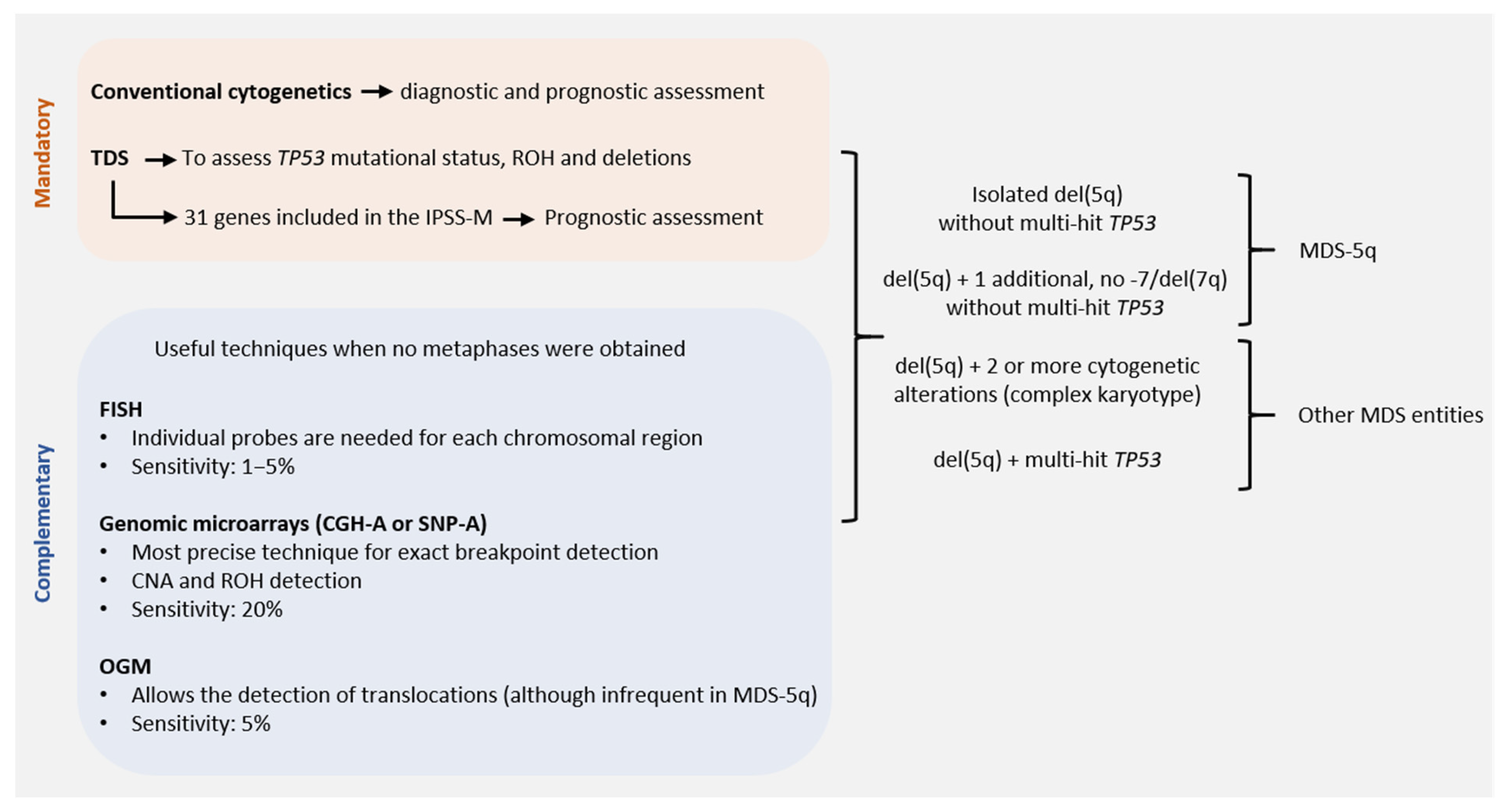
Conventional cytogenetics (CC) constitutes the gold standard for the genetic diagnosis and prognosis of MDS. However, fluorescence in situ hybridization (FISH) of 5q31 could be useful in cases without evidence of del(5q) by CC. When the presence of MDS-5q is suspected and/or if the cytogenetic study shows no metaphases or an aberrant karyotype with chromosome 5 is involved (no 5q deletion), it is recommended to perform FISH analysis [16,17]. Figure 2 shows the genetic studies available for the diagnosis and characterization of MDS-5q. During follow-up, genetics studies will be adapted to each patient, considering their comorbidities. A new BM aspiration, and the corresponding genetic study, will be performed on suspicion of disease evolution and no response to treatment. In the case of clonal evolution, the approach can be decided according to the general patient status.
The karyotype is a prognostic variable included in the International Prognostic Scoring System (IPSS) and the revised edition of the IPSS (IPSS-R) [18,19]. Del(5q) alone has always been considered a good prognostic variable and, in the IPSS-R, a concomitant cytogenetic alteration has been included. This change was based on a scoring system proposed by Schanz et al. based on an international data collection of 2902 patients [15]. Deletion 5q is a classical alteration detected in around 15–20% of MDS patients, with half being isolated, around 17% having an additional alteration, and 36% being part of a complex karyotype [3]. The prognostic impact of the accompanying abnormalities in del(5q) is difficult to determine because double abnormalities are highly variable. In 2011, Mallo et al. published an international collaborative study including a large series of del(5q) patients to determine the prognostic impact of adjunct prognostic abnormalities. The multivariate analysis showed that karyotype complexity was one of the main prognostic factors together with platelet count and BM blasts [14]. The good prognosis of del(5q) with one accompanying alteration was included in the MDS with the del(5q) category of the 2017 WHO classification, excluding cases harboring a chromosome 7 alteration [1].

3.1. Commonly Deleted Regions in Chromosome 5q

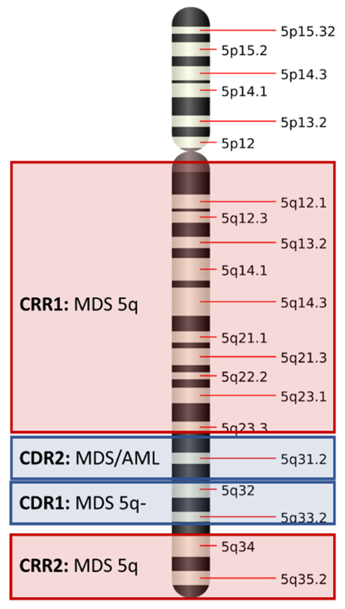
Two “commonly deleted regions” (CDR) were originally described by Boultwood and colleagues: a 1.5 Mb deletion encompassing 5q32–5q33, which was originally associated with the 5q- syndrome and better prognosis, and a more proximal CDR at 5q31. The latter was associated with other MDS subtypes and cases of acute myeloid leukemia (AML) cases, with complex karyotypes and a worse prognosis [20,21,22,23]. High-resolution techniques, such as genomic microarrays and optical genome mapping (OGM), can detect cryptic alterations accompanying the del(5q) and can help define the breakpoint. However, since high-density genomic microarrays work with DNA probes, this approach has become the most suitable technique to obtain precise del(5q) breakpoint genomic coordinates.
Most patients have large deletions that encompass both CDRs. This was corroborated by subsequent studies combining conventional cytogenetics and single nucleotide polymorphism arrays, with Mallo et al., describing a wider CDR that extended from q22.3 to q31.3. This region encompasses 14.6 Mb, while the median size of the total deletion detected in most cases is around 70 Mb [17].
Several studies have focused on the study of the 5q CDR, but in 2012, Jerez et al., published an article emphasizing the importance of the common retained region (CRR) [24]. Their work reinforces the idea that in the 5q- syndrome, the proximal and terminal regions are always retained. Thus, two CRRs were described: CRR1 for the proximal region (spanning 81.7 Mb and ending at band 5q14.2) and CRR2 for the distal region (5q34), with both being associated with disease subtypes. No CRR could be identified in other forms of MDS and AML with del(5q). As was previously described, patients with CRR had a lower number of genomic lesions and correlate with better prognosis. Additionally, this study supports the idea that genomic microarrays can add prognostic information to prognostic scoring systems as was reported by Arenillas et al., in 2013 [25]. Figure 3 shows the CDR and CRR identified for chromosome 5q.
It is widely known that additional techniques can help to describe the karyotype. Most of the complex cases carrying an apparent monosomy 5 have shown that, following studies with additional techniques such as FISH or genomic arrays, this apparent monosomy presents a partially retained 5q. As previously mentioned, genomic microarrays can help to accurately define the breakpoints [24,26].
Molecular studies revealed that haploinsufficiency of several genes (particularly RPS14, CSNK1A1, EGR1, miR-145, and miR-14a) located in 5q CDR contribute to the pathogenesis and hematological phenotype associated with MDS-5q [27,28,29]. For example, miR-145 and miR-14a are micro RNAs that have been found to be responsible for the negative regulation of effector molecules that regulate megakaryocytic differentiation. Thus, a deficiency of these micro RNAs is responsible for the thrombosis and megakaryocytic dysplasia (hypolobulated megakaryocytes) which characterize MDS-5q [30,31].

3.2. Karyotyping: Present and Future Directions

Point mutations have been described as a frequent event in MDS patients. In 2011, Bejar et al. described the clinical effects of these mutations and stated that the prognosis of these patients may be driven by the association of prognostic variables. Specific genes were found to be associated with specific risk groups such as TET2 in cases with a normal karyotype and TP53 in cases with a complex karyotype [32]. In 2022, the IPSS-M, a prognostic scoring system based on molecular data was published. This scoring system takes into account the mutational status of 31 genes; however, it still retains the karyotype as a prognostic parameter [9].
OGM has emerged as a promising non-sequencing-based technique for high resolution genome-wide structural variant profiling. It can simplify lab workflow by reducing multiple tests. Parallel studies with standard-of-care tests have been performed in hematological neoplasms and have shown high concordance [33]. A recent study published by Yang et al. showed that OGM results changed the comprehensive cytogenetic scoring system and the IPSS-R risk groups in 21% and 17%, respectively, of their MDS patient’s cohort with an improved prediction of prognosis. Although more studies especially focused on MDS-5q are needed, the combination of OGM and next-generation sequencing (NGS) seems to be a promising approach for the evaluation of prognosis [34].

4. Prognostic Impact of Somatic Mutations in MDS-5q

It has been described that one-third of MDS-5q patients present with no somatic alterations, while nearly half of patients (43%) can present with an isolated mutation [35,36]. The pattern of recurrently mutated genes is similar to other MDS subtypes, except for TP53 mutations that were found to be enriched in this subtype of patients [35,37,38]. In the subsequent section, the genes most frequently mutated in MDS-5q are described and Table 1 summarizes their biological and clinical associations and main characteristics and frequencies.
Based on data from Meggendorfer et al. [35], Malcovati et al. [39], Heuser et al. [40], and Mossner et al. [41].

4.1. SF3B1 Mutation

The SF3B1 gene encodes subunit 1 of the splicing factor 3b protein complex, which is a core component of the RNA splicing machinery. Mutations in SF3B1 have been reported in around 20% of MDS-5q cases and have been associated with a variable proportion of ring sideroblasts [5,6,39,42]. Evidence provided by several reports suggests that, in some cases of MDS-5q, the SF3B1 mutation might precede the cytogenetic alteration [41,43,44,45]. Despite the order of acquisition of such genetic events, cases with concomitant SF3B1 and del(5q) would still be classified within the category of MDS-5q in the WHO classification system, as well as in the ICC system [2,8].
Controversial data have been published regarding the prognosis of SF3B1 mutations in MDS-5q patients. On one hand, a study published by Meggendorfer et al., demonstrated a significantly shorter overall survival (OS) in patients harboring both alterations compared with MDS-5q patients without SF3B1 mutation [35]. On the other hand, no significant difference in OS was reported by Malcovati et al. when analyzing the same in their respective cohort [39].

4.2. DNMT3A, TET2 and ASXL1 Mutations

Mutations affecting the genes DNMT3A, TET2, and ASXL1 genes—commonly known as DTA mutations—are frequently found in clonal hematopoiesis of indeterminate potential (CHIP), which is a non-malignant condition associated with increased risk of progression to hematologic neoplasia compared with individuals without detectable mutations [46,47]. In line with this, in mutational hierarchy studies performed by Mossner et al., DTA mutations were found to be recurrent “founder” lesions in MDS patients, including the MDS-5q cases analyzed [36,41].
DNMT3A codifies for DNA methyltransferase 3 alpha, which is required for genome-wide de novo methylation and is essential for the establishment of DNA methylation patterns during development [48]. On the contrary, TET2 codifies for tet methylcytosine dioxygenase 2, which catalyzes the conversion of the modified genomic base 5-methylcytosine (5mC) into 5-hydroxymethylcytosine (5hmC) and plays a key role in active DNA demethylation. As mentioned previously, both genes are recurrently mutated in MDS. Specifically, in MDS-5q, DNMT3A mutations were found in roughly 18% of cases while TET2 mutations were described in nearly 12% of patients [35].
Some studies have reported that in MDS patients, DNMT3A mutations were associated with a higher risk of leukemia transformation and shorter OS, but no specific study describing either phenotypic or survival associations was exclusively performed in MDS-5q patients [49,50].
A report by Scharenberg et al. described that progression in patients with low- and intermediate-1-risk del(5q) MDS is predicted by mutations in a limited number of genes, among which TET2 is included. Specifically, 6/13 patients with evidence of disease progression presented mutations in the TET2 gene [51]. In MDS patient cohorts including all disease subtypes, TET2 mutations were found to be associated with shorter OS after hematopoietic stem cell transplantation and lower response rate to hypomethylating agents [9,52,53].
Located in chromosome 20q, additional sex combs 1 (ASXL1) codifies for a protein involved in transcriptional regulation. Mutations of mostly the frameshift type have been described in MDS patients in variable frequencies ranging from 14–24% in different cohorts [5,6,9,32,53,54]. Concretely in MDS-5q, they are less abundant and most studies describe frequencies of around 6% [35,53,54,55]. While it is a common event in early disease, Fernandez-Mercado et al. reported higher frequencies of this mutation of up to 25% among advanced cases of the disease, suggesting a role in disease progression in MDS-5q [36]. Similarly to DNMT3A and TET2, ASXL1 mutations were mostly studied among MDS patient cohorts, including all subtypes, finding an association with worse prognosis and a shorter OS, but no specific associations were mentioned between ASXL1 mutations and outcomes in MDS-5q cases have been described.

4.3. TP53 Mutations

The tumor-suppressor p53 gene (TP53) is located in chromosome band 17p13 and is essential for genome integrity. TP53 encodes for the p53 protein, which is a transcription factor involved in essential cell functions, such as DNA repair, cell cycle control, apoptosis, aging, and stemness [56,57].
TP53 gene mutations are detected in approximately 18% of MDS-5q [58,59]. It is the only mutation that was found to be significantly enriched in this MDS subtype compared with the other subtypes (18% vs. 6%) [35]. Data regarding the time of acquisition of this mutation are controversial. While it seems that there was a group of patients in which the mutation is already present in the early phases of the disease, there was another in which the TP53 mutation arises during disease evolution, especially after treatment with lenalidomide [43,51,59].
Mutations in the TP53 gene in MDS patients are associated with generally unfavorable outcomes, aggressive disease course, higher risk of transformation to AML, shorter overall survival (OS), and resistance to lenalidomide treatment [17,37,51,59].
Double or even triple hits in the TP53 gene locus were already reported in 2013 by Kulasekararaj et al. [37]. A more recent study published by Bernard et al., provides new insights regarding the importance of the TP53 allelic state (multi-hit). After studying 3324 patients, four main TP53 mutational profiles were identified: (1) monoallelic mutations; (2) multiple mutations; (3) mutation and concomitant deletion affecting 17p; and (4) mutation and concomitant loss of heterozygosity of the 17p region. They found that two-thirds of patients with a TP53 mutation present with multiple hits, while only one-third present with monoallelic mutations. Associations with high-risk presentation and poor outcomes were only specific to multi-hit patients, while surprisingly, monoallelic patients did not differ from TP53 wild-type patients in outcome and response to therapy. The authors described that the TP53 allelic state segregates patient outcomes across WHO subtypes, despite monoallelic TP53 being enriched by MDS-5q. Moreover, they found that patients with monoallelic TP53 mutations had longer survival compared with multi-hit patients [58].
In the 2017 edition, the WHO recommended assessing TP53 mutational status in MDS-5q to identify high-risk cases [1]. However, the upcoming edition of the WHO classification takes into consideration new insights regarding the allelic state of this gene to redefine a specific subtype of MDS associated with the presence of multiple alterations affecting the TP53 locus (Figure 2). This subtype is called MDS with biallelic TP53 inactivation (MDS-biTP53). However, the presence of a single TP53 mutation (unless it is multi-hit) does not per se exclude the diagnosis of MDS-5q [2]. Similarly, the ICC proposal takes into account the TP53 allelic state to define a new disease category called “myeloid neoplasms with mutated TP53”. In the case of MDS-5q, only single-hit TP53 mutations are admitted, otherwise, the diagnosis would change to the newly mentioned category [8].

4.4. CSNK1A1 Mutations

Located in the CDR 5q32, CSNK1A1 encodes for casein kinase 1A1 (CK1 α), a serine/threonine kinase that participates in many cellular processes, including growth and proliferation via the β catenin and Wnt signaling pathway, apoptosis, and response to DNA damage [60,61,62]. In 2015, a study by Kronke et al., identified CK1α as a lenalidomide target in myeloid cells and found that heterozygous deletion of CSNK1A1 in del(5q) MDS provides a therapeutic window for selective targeting of the malignant cells [63].
Missense mutations have been reported in exons 3 and 4 in 7–10% of MDS-5q patients [35,40,44,64,65]. Detected variant allele frequency values range from 3–78% and mimic a homozygous mutation status, which is consistent with the location of the CSNK1A1 gene and the CDR [35,40].
CSNK1A1 mutations were found to be associated with older age and some reports show a trend towards decreased response to lenalidomide, but no independent prognostic impact on OS has been described to date [40,60]. In a study performed by Meggendorfer et al., CSNK1A1 mutations were found to co-occur with SF3B1 mutations in 42% of the cases [35].

4.5. JAK2 Mutations

Janus kinase 2 (JAK2) encodes a non-receptor tyrosine kinase that plays a central role in cytokine and growth factor signaling. Somatic mutations in JAK2 constitute a major diagnostic criterion for myeloproliferative neoplasms (MPN) and are found in approximately 95% of polycythemia vera cases and 50% of essential thrombocythemia and primary myelofibrosis [2,9,66,67].
Mutations in this gene, specifically the V617F hotspot, were reported in approximately 6% of patients with MDS-5q and were found to correlate with higher platelet counts when compared with JAK2 wild-type patients [35,55,68]. Sangiorgio et al., performed a detailed microscopic analysis of BM aspirates of MDS-5q cases with concomitant JAK2 mutations and found greater reticulin fibrosis in mutated cases. Additionally, they found a combination of hypolobulated megakaryocytes (typically found in MDS-5q) and large forms with hyperlobulated nuclei, which are commonly seen in MPN [69].
Although the phenotypic characteristics have been described, no significant differences in OS or disease progression were found in such MDS-5q JAK2 mutated cases when compared with JAK2 wild-type cases [55,69].

5. Clonal Evolution

As mentioned previously, several authors have described the commonly mutated genes and concomitant copy number alterations in MDS-5q, but few studies have explored clonal evolution in this specific subtype of MDS.
The first systematic study providing molecular monitoring of long-term serial follow-up samples in a significant cohort of patients was by Mossner et al. [41]. As in most of the subsequent publications, such clonal evolution studies are based on bulk sequencing (exome or gene panel), in which clonal composition and evolutionary patterns are reconstructed based on variant allele frequency values of the detected mutations. The authors described that MDS “founder” lesions recurrently affected genes involved in the regulation of DNA methylation (e.g., TET2, DNMT3A), chromatin remodeling (e.g., ASXL1), or RNA splicing (e.g., SF3B1), and that del(5q) was acquired as a secondary lesion or constituted a minor independent clone in 62% of patients classified as MDS-5q. This is in contrast to previous studies proposing del(5q) as the initiating lesion in such patients [44]. In line with this, single-cell studies performed by our group demonstrated that in some MDS-5q cases, del(5q) can appear as the initiating lesion, while it can appear as a secondary hit in other cases [43].
As expected, the emergence and disappearance of specific clones in the BM are frequently correlated with changes in the clinical features in PB, such as hemoglobin and platelet levels. Moreover, it has been described that, in almost all cases, treatment with lenalidomide induced an effective reduction of cells carrying del(5q), however, it did not induce complete molecular remission of all clones carrying typical MDS mutations [41]. Furthermore, loss of response to lenalidomide is correlated with the gradual growth of a non-related clonal population already detectable at low levels before treatment or the expansion of a descendent from the original clone of the diagnosis [70].
In another longitudinal study, Scharenberg et al. described that 37% of their MDS-5q cohort progressed to either higher-risk MDS or transformed into AML in a median of 85 months after diagnosis. Interestingly, they found that all the cases harbored recurrent mutations in TP53, TET2, or RUNX1 in addition to del(5q) [51]. Thus, several patients showed an increased allele burden and gains of new mutations during the course of the disease and treatment. Particularly, the acquisition of TP53 mutations was relatively common in the progression of patients treated with lenalidomide, with some of them exhibiting more than one TP53 mutation.
In general, all the above-mentioned studies agreed that both linear and branched evolutionary patterns occur with and without disease-modifying treatments, and subclones that acquire additional mutations associated with treatment resistance or disease progression can be detected months before clinical changes become apparent [41,43,51,70].

6. Conclusions

Based on our understanding of MDS-5q, together with the changes in the inclusion criteria, the evaluation of prognosis evaluation and the management of the disease are clearly in line with the progress of molecular genetics, which at the same time are linked to the advances in technology and scientific discoveries.
With the arrival of the IPSS-M and the newly proposed classifications for MDS, NGS techniques are mandatory for correct disease classification and assessment of prognosis. However, the approaches to financing health care are extremely diverse and are country-specific, and therefore, there may still be situations in which NGS remains restricted to potentially guiding therapeutic decisions, such as treatment intensity or hematopoietic stem cell transplantation.
Although many advances have been achieved, especially in the last decade, unanswered questions remain. Techniques such as OGM and new single-cell techniques together with new clinical trials are just some future steps to better understanding this disease and ultimately improving patient care.

Author Contributions

P.A., M.M. and F.S. wrote, reviewed, and edited the manuscript. All authors have read and agreed to the published version of the manuscript.

Funding

This research received no external funding.

Institutional Review Board Statement

Not applicable.

Informed Consent Statement

Not applicable.

Data Availability Statement

Not applicable.

Conflicts of Interest

The authors declare no conflict of interest.

References

  1. Swerdlow, S.H.; Campo, E.; Harris, N.L.; Jaffe, E.S.; Pileri, S.A.; Stein, H.; Thiele, J. (Eds.) WHO Classification of Tumours of Hematopoietic and Lymphoid Tissues, 4th ed.; IARC: Lyon, France, 2017. [Google Scholar]
  2. Khoury, J.D.; Solary, E.; Abla, O.; Akkari, Y.; Alaggio, R.; Apperley, J.F.; Bejar, R.; Berti, E.; Busque, L.; Chan, J.K.C.; et al. The 5th Edition of the World Health Organization Classification of Haematolymphoid Tumours: Myeloid and Histiocytic/Dendritic Neoplasms. Leukemia 2022, 36, 1703–1719. [Google Scholar] [CrossRef] [PubMed]
  3. Haase, D. Cytogenetic Features in Myelodysplastic Syndromes. Ann. Hematol. 2008, 87, 515–526. [Google Scholar] [CrossRef] [PubMed] [Green Version]
  4. Haase, D.; Germing, U.; Schanz, J.; Pfeilstocker, M.; Nosslinger, T.; Hildebrandt, B.; Kundgen, A.; Lubbert, M.; Kunzmann, R.; Giagounidis, A.A.N.; et al. New Insights into the Prognostic Impact of the Karyotype in MDS and Correlation with Subtypes: Evidence from a Core Dataset of 2124 Patients. Blood 2007, 110, 4385–4395. [Google Scholar] [CrossRef]
  5. Papaemmanuil, E.; Gerstung, M.; Malcovati, L.; Tauro, S.; Gundem, G.; Van Loo, P.; Yoon, C.J.; Ellis, P.; Wedge, D.C.; Pellagatti, A.; et al. Clinical and Biological Implications of Driver Mutations in Myelodysplastic Syndromes. Blood 2013, 122, 3616–3627. [Google Scholar] [CrossRef] [PubMed] [Green Version]
  6. Haferlach, T.; Nagata, Y.; Grossmann, V.; Okuno, Y.; Bacher, U.; Nagae, G.; Schnittger, S.; Sanada, M.; Kon, A.; Alpermann, T.; et al. Landscape of Genetic Lesions in 944 Patients with Myelodysplastic Syndromes. Leukemia 2014, 28, 241–247. [Google Scholar] [CrossRef] [PubMed] [Green Version]
  7. Van Den Berghe, H.; Cassiman, J.-J.; David, G.; Fryns, J.-P.; Michaux, J.-L.; Sokal, G. Distinct Haematological Disorder with Deletion of Long Arm of No. 5 Chromosome. Nature 1974, 251, 437–438. [Google Scholar] [CrossRef] [PubMed]
  8. Arber, D.A.; Orazi, A.; Hasserjian, R.P.; Borowitz, M.J.; Calvo, K.R.; Kvasnicka, H.-M.; Wang, S.A.; Bagg, A.; Barbui, T.; Branford, S.; et al. International Consensus Classification of Myeloid Neoplasms and Acute Leukemias: Integrating Morphologic, Clinical, and Genomic Data. Blood 2022, 140, 1200–1228. [Google Scholar] [CrossRef]
  9. Bernard, E.; Tuechler, H.; Greenberg, P.L.; Hasserjian, R.P.; Arango Ossa, J.E.; Nannya, Y.; Devlin, S.M.; Creignou, M.; Pinel, P.; Monnier, L.; et al. Molecular International Prognostic Scoring System for Myelodysplastic Syndromes. NEJM Evid. 2022, 1. [Google Scholar] [CrossRef]
  10. Bennett, J.M.; Catovsky, D.; Daniel, M.T.; Flandrin, G.; Galton, D.A.; Gralnick, H.R.; Sultan, C. Proposals for the Classification of the Acute Leukaemias. French-American-British (FAB) Co-Operative Group. Br. J. Haematol. 1976, 33, 451–458. [Google Scholar] [CrossRef]
  11. Jaffe, E.S.; Harris, N.L.; Stein, H.; Vardiman, J.W. (Eds.) World Health Organization Classification of Tumours, Pathology and Genetics of Tumours of Haematopoietic and Lymphoid Tissues; IARC Press: Lyon, France, 2001. [Google Scholar]
  12. Swerdlow, S.H.; Campo, E.; Harris, N.L.; Jaffe, E.S.; Pileri, S.A.; Stein, H.; Thiele, J. (Eds.) WHO Classification of Tumours of Hematopoietic and Lymphoid Tissues; IARC: Lyon, France, 2008. [Google Scholar]
  13. Arber, D.A.; Orazi, A.; Hasserjian, R.; Thiele, J.; Borowitz, M.J.; Le Beau, M.M.; Bloomfield, C.D.; Cazzola, M.; Vardiman, J.W. The 2016 Revision to the World Health Organization Classification of Myeloid Neoplasms and Acute Leukemia. Blood 2016, 127, 2391–2405. [Google Scholar] [CrossRef]
  14. Mallo, M.; Cervera, J.; Schanz, J.; Such, E.; García-Manero, G.; Luño, E.; Steidl, C.; Espinet, B.; Vallespí, T.; Germing, U.; et al. Impact of Adjunct Cytogenetic Abnormalities for Prognostic Stratification in Patients with Myelodysplastic Syndrome and Deletion 5q. Leukemia 2011, 25, 110–120. [Google Scholar] [CrossRef] [PubMed]
  15. Schanz, J.; Tüchler, H.; Solé, F.; Mallo, M.; Luño, E.; Cervera, J.; Granada, I.; Hildebrandt, B.; Slovak, M.L.; Ohyashiki, K.; et al. New Comprehensive Cytogenetic Scoring System for Primary Myelodysplastic Syndromes (MDS) and Oligoblastic Acute Myeloid Leukemia After MDS Derived From an International Database Merge. JCO 2012, 30, 820–829. [Google Scholar] [CrossRef] [PubMed] [Green Version]
  16. Mallo, M.; Arenillas, L.; Espinet, B.; Salido, M.; Hernandez, J.M.; Lumbreras, E.; del Rey, M.; Arranz, E.; Ramiro, S.; Font, P.; et al. Fluorescence in Situ Hybridization Improves the Detection of 5q31 Deletion in Myelodysplastic Syndromes without Cytogenetic Evidence of 5q-. Haematologica 2008, 93, 1001–1008. [Google Scholar] [CrossRef] [PubMed] [Green Version]
  17. Mallo, M.; Del Rey, M.; Ibáñez, M.; Calasanz, M.J.; Arenillas, L.; Larráyoz, M.J.; Pedro, C.; Jerez, A.; Maciejewski, J.; Costa, D.; et al. Response to Lenalidomide in Myelodysplastic Syndromes with Del(5q): Influence of Cytogenetics and Mutations. Br. J. Haematol. 2013, 162, 74–86. [Google Scholar] [CrossRef] [PubMed]
  18. Greenberg, P.; Cox, C.; LeBeau, M.M.; Fenaux, P.; Morel, P.; Sanz, G.; Sanz, M.; Vallespi, T.; Hamblin, T.; Oscier, D.; et al. International Scoring System for Evaluating Prognosis in Myelodysplastic Syndromes. Blood 1997, 89, 2079–2088. [Google Scholar] [CrossRef]
  19. Greenberg, P.L.; Tuechler, H.; Schanz, J.; Sanz, G.; Garcia-Manero, G.; Sole, F.; Bennett, J.M.; Bowen, D.; Fenaux, P.; Dreyfus, F.; et al. Revised International Prognostic Scoring System for Myelodysplastic Syndromes. Blood 2012, 120, 2454–2465. [Google Scholar] [CrossRef] [Green Version]
  20. Boultwood, J.; Fidler, C.; Lewis, S.; Kelly, S.; Sheridan, H.; Littlewood, T.; Buckle, V.; Wainscoat, J. Molecular Mapping of Uncharacteristically Small 5q Deletions in Two Patients with the 5q- Syndrome: Delineation of the Critical Region on 5q and Identification of a 5q- Breakpoint. Genomics 1994, 19, 425–432. [Google Scholar] [CrossRef]
  21. Boultwood, J.; Fidler, C.; Strickson, A.J.; Watkins, F.; Gama, S.; Kearney, L.; Tosi, S.; Kasprzyk, A.; Cheng, J.-F.; Jaju, R.J.; et al. Narrowing and Genomic Annotation of the Commonly Deleted Region of the 5q- Syndrome. Blood 2002, 99, 4638–4641. [Google Scholar] [CrossRef]
  22. Jaju, R.; Boultwood, J.; Oliver, F.; Kostrzewa, M.; Fidler, C.; Parker, N.; McPherson, J.; Morris, S.; Müller, U.; Wainscoat, J.; et al. Molecular Cytogenetic Delineation of the Critical Deleted Region in the 5q- Syndrome. Genes Chromosom. Cancer 1998, 22, 251–256. [Google Scholar] [CrossRef]
  23. Nybakken, G.E.; Bagg, A. The Genetic Basis and Expanding Role of Molecular Analysis in the Diagnosis, Prognosis, and Therapeutic Design for Myelodysplastic Syndromes. J. Mol. Diagn. 2014, 16, 145–158. [Google Scholar] [CrossRef]
  24. Jerez, A.; Gondek, L.P.; Jankowska, A.M.; Makishima, H.; Przychodzen, B.; Tiu, R.V.; O’Keefe, C.L.; Mohamedali, A.M.; Batista, D.; Sekeres, M.A.; et al. Topography, Clinical, and Genomic Correlates of 5q Myeloid Malignancies Revisited. JCO 2012, 30, 1343–1349. [Google Scholar] [CrossRef] [PubMed] [Green Version]
  25. Arenillas, L.; Mallo, M.; Ramos, F.; Guinta, K.; Barragán, E.; Lumbreras, E.; Larráyoz, M.-J.; De Paz, R.; Tormo, M.; Abáigar, M.; et al. Single Nucleotide Polymorphism Array Karyotyping: A Diagnostic and Prognostic Tool in Myelodysplastic Syndromes with Unsuccessful Conventional Cytogenetic Testing: Snp Array In Myelodysplastic Syndromes. Genes Chromosom. Cancer 2013, 52, 1167–1177. [Google Scholar] [CrossRef] [PubMed]
  26. Galván, A.B.; Mallo, M.; Arenillas, L.; Salido, M.; Espinet, B.; Pedro, C.; Florensa, L.; Serrano, S.; Solé, F. Does Monosomy 5 Really Exist in Myelodysplastic Syndromes and Acute Myeloid Leukemia? Leuk. Res. 2010, 34, 1242–1245. [Google Scholar] [CrossRef]
  27. Ebert, B.L.; Galili, N.; Tamayo, P.; Bosco, J.; Mak, R.; Pretz, J.; Tanguturi, S.; Ladd-Acosta, C.; Stone, R.; Golub, T.R.; et al. An Erythroid Differentiation Signature Predicts Response to Lenalidomide in Myelodysplastic Syndrome. PLoS Med. 2008, 5, e35. [Google Scholar] [CrossRef]
  28. Pellagatti, A.; Hellström-Lindberg, E.; Giagounidis, A.; Perry, J.; Malcovati, L.; Della Porta, M.G.; Jädersten, M.; Killick, S.; Fidler, C.; Cazzola, M.; et al. Haploinsufficiency of RPS14 in 5q- Syndrome Is Associated with Deregulation of Ribosomal- and Translation-Related Genes. Br. J. Haematol. 2008, 142, 57–64. [Google Scholar] [CrossRef] [PubMed] [Green Version]
  29. Joslin, J.M.; Fernald, A.A.; Tennant, T.R.; Davis, E.M.; Kogan, S.C.; Anastasi, J.; Crispino, J.D.; Le Beau, M.M. Haploinsufficiency of EGR1, a Candidate Gene in the Del(5q), Leads to the Development of Myeloid Disorders. Blood 2007, 110, 719–726. [Google Scholar] [CrossRef] [Green Version]
  30. Kumar, M.S.; Narla, A.; Nonami, A.; Mullally, A.; Dimitrova, N.; Ball, B.; McAuley, J.R.; Poveromo, L.; Kutok, J.L.; Galili, N.; et al. Coordinate Loss of a MicroRNA and Protein-Coding Gene Cooperate in the Pathogenesis of 5q- Syndrome. Blood 2011, 118, 8. [Google Scholar] [CrossRef]
  31. Starczynowski, D.T.; Kuchenbauer, F.; Argiropoulos, B.; Sung, S.; Morin, R.; Muranyi, A.; Hirst, M.; Hogge, D.; Marra, M.; Wells, R.A.; et al. Identification of MiR-145 and MiR-146a as Mediators of the 5q– Syndrome Phenotype. Nat. Med. 2010, 16, 49–58. [Google Scholar] [CrossRef]
  32. Bejar, R.; Stevenson, K.; Abdel-Wahab, O.; Galili, N.; Nilsson, B.; Garcia-Manero, G.; Kantarjian, H.; Raza, A.; Levine, R.L.; Neuberg, D.; et al. Clinical Effect of Point Mutations in Myelodysplastic Syndromes. N. Engl. J. Med. 2011, 364, 2496–2506. [Google Scholar] [CrossRef] [Green Version]
  33. Smith, A.C.; Neveling, K.; Kanagal-Shamanna, R. Optical Genome Mapping for Structural Variation Analysis in Hematologic Malignancies. Am. J. Hematol. 2022, 97, 975–982. [Google Scholar] [CrossRef]
  34. Yang, H.; Garcia-Manero, G.; Sasaki, K.; Montalban-Bravo, G.; Tang, Z.; Wei, Y.; Kadia, T.; Chien, K.; Rush, D.; Nguyen, H.; et al. High-Resolution Structural Variant Profiling of Myelodysplastic Syndromes by Optical Genome Mapping Uncovers Cryptic Aberrations of Prognostic and Therapeutic Significance. Leukemia 2022, 36, 2306–2316. [Google Scholar] [CrossRef] [PubMed]
  35. Meggendorfer, M.; Haferlach, C.; Kern, W.; Haferlach, T. Molecular Analysis of Myelodysplastic Syndrome with Isolated Deletion of the Long Arm of Chromosome 5 Reveals a Specific Spectrum of Molecular Mutations with Prognostic Impact: A Study on 123 Patients and 27 Genes. Haematologica 2017, 102, 1502–1510. [Google Scholar] [CrossRef] [PubMed]
  36. Fernandez-Mercado, M.; Burns, A.; Pellagatti, A.; Giagounidis, A.; Germing, U.; Agirre, X.; Prosper, F.; Aul, C.; Killick, S.; Wainscoat, J.S.; et al. Targeted Re-Sequencing Analysis of 25 Genes Commonly Mutated in Myeloid Disorders in Del(5q) Myelodysplastic Syndromes. Haematologica 2013, 98, 1856–1864. [Google Scholar] [CrossRef] [PubMed] [Green Version]
  37. Kulasekararaj, A.G.; Smith, A.E.; Mian, S.A.; Mohamedali, A.M.; Krishnamurthy, P.; Lea, N.C.; Gäken, J.; Pennaneach, C.; Ireland, R.; Czepulkowski, B.; et al. TP53 Mutations in Myelodysplastic Syndrome Are Strongly Correlated with Aberrations of Chromosome 5, and Correlate with Adverse Prognosis. Br. J. Haematol. 2013, 160, 660–672. [Google Scholar] [CrossRef]
  38. Hosono, N.; Makishima, H.; Mahfouz, R.; Przychodzen, B.; Yoshida, K.; Jerez, A.; LaFramboise, T.; Polprasert, C.; Clemente, M.J.; Shiraishi, Y.; et al. Recurrent Genetic Defects on Chromosome 5q in Myeloid Neoplasms. Oncotarget 2017, 8, 6483–6495. [Google Scholar] [CrossRef] [Green Version]
  39. Malcovati, L.; Stevenson, K.; Papaemmanuil, E.; Neuberg, D.; Bejar, R.; Boultwood, J.; Bowen, D.T.; Campbell, P.J.; Ebert, B.L.; Fenaux, P.; et al. SF3B1-Mutant Myelodysplastic Syndrome as a Distinct Disease Subtype—A Proposal of the International Working Group for the Prognosis of Myelodysplastic Syndromes (IWG-PM). Blood 2020, 136, 157–170. [Google Scholar] [CrossRef]
  40. Heuser, M.; Meggendorfer, M.; Cruz, M.M.A.; Fabisch, J.; Klesse, S.; Köhler, L.; Göhring, G.; Ganster, C.; Shirneshan, K.; Gutermuth, A.; et al. Frequency and Prognostic Impact of Casein Kinase 1A1 Mutations in MDS Patients with Deletion of Chromosome 5q. Leukemia 2015, 29, 1942–1945. [Google Scholar] [CrossRef]
  41. Mossner, M.; Jann, J.-C.; Wittig, J.; Nolte, F.; Fey, S.; Nowak, V.; Obländer, J.; Pressler, J.; Palme, I.; Xanthopoulos, C.; et al. Mutational Hierarchies in Myelodysplastic Syndromes Dynamically Adapt and Evolve upon Therapy Response and Failure. Blood 2016, 128, 1246–1259. [Google Scholar] [CrossRef] [Green Version]
  42. Malcovati, L.; Karimi, M.; Papaemmanuil, E.; Ambaglio, I.; Jädersten, M.; Jansson, M.; Elena, C.; Gallì, A.; Walldin, G.; Della Porta, M.G.; et al. SF3B1 Mutation Identifies a Distinct Subset of Myelodysplastic Syndrome with Ring Sideroblasts. Blood 2015, 126, 233–241. [Google Scholar] [CrossRef] [Green Version]
  43. Acha, P.; Palomo, L.; Fuster-Tormo, F.; Xicoy, B.; Mallo, M.; Manzanares, A.; Grau, J.; Marcé, S.; Granada, I.; Rodríguez-Luaces, M.; et al. Analysis of Intratumoral Heterogeneity in Myelodysplastic Syndromes with Isolated Del(5q) Using a Single Cell Approach. Cancers 2021, 13, 841. [Google Scholar] [CrossRef]
  44. Woll, P.S.; Kjällquist, U.; Chowdhury, O.; Doolittle, H.; Wedge, D.C.; Thongjuea, S.; Erlandsson, R.; Ngara, M.; Anderson, K.; Deng, Q.; et al. Myelodysplastic Syndromes Are Propagated by Rare and Distinct Human Cancer Stem Cells In Vivo. Cancer Cell 2014, 25, 794–808. [Google Scholar] [CrossRef] [PubMed] [Green Version]
  45. Mian, S.A.; Rouault-Pierre, K.; Smith, A.E.; Seidl, T.; Pizzitola, I.; Kizilors, A.; Kulasekararaj, A.G.; Bonnet, D.; Mufti, G.J. SF3B1 Mutant MDS-Initiating Cells May Arise from the Haematopoietic Stem Cell Compartment. Nat. Commun. 2015, 6, 10004. [Google Scholar] [CrossRef] [PubMed] [Green Version]
  46. Steensma, D.P.; Bejar, R.; Jaiswal, S.; Lindsley, R.C.; Sekeres, M.A.; Hasserjian, R.P.; Ebert, B.L. Clonal Hematopoiesis of Indeterminate Potential and Its Distinction from Myelodysplastic Syndromes. Blood 2015, 126, 9–16. [Google Scholar] [CrossRef] [Green Version]
  47. Jaiswal, S.; Fontanillas, P.; Flannick, J.; Manning, A.; Grauman, P.V.; Mar, B.G.; Lindsley, R.C.; Mermel, C.H.; Burtt, N.; Chavez, A.; et al. Age-Related Clonal Hematopoiesis Associated with Adverse Outcomes. N. Engl. J. Med. 2014, 371, 2488–2498. [Google Scholar] [CrossRef] [PubMed] [Green Version]
  48. Chen, T.; Ueda, Y.; Xie, S.; Li, E. A Novel Dnmt3a Isoform Produced from an Alternative Promoter Localizes to Euchromatin and Its Expression Correlates with Activede Novo Methylation. J. Biol. Chem. 2002, 277, 38746–38754. [Google Scholar] [CrossRef] [PubMed] [Green Version]
  49. Lin, M.-E.; Hou, H.-A.; Tsai, C.-H.; Wu, S.-J.; Kuo, Y.-Y.; Tseng, M.-H.; Liu, M.-C.; Liu, C.-W.; Chou, W.-C.; Chen, C.-Y.; et al. Dynamics of DNMT3A Mutation and Prognostic Relevance in Patients with Primary Myelodysplastic Syndrome. Clin. Epigenet. 2018, 10, 42. [Google Scholar] [CrossRef] [PubMed] [Green Version]
  50. Thol, F.; Winschel, C.; Ludeking, A.; Yun, H.; Friesen, I.; Damm, F.; Wagner, K.; Krauter, J.; Heuser, M.; Ganser, A. Rare Occurrence of DNMT3A Mutations in Myelodysplastic Syndromes. Haematologica 2011, 96, 1870–1873. [Google Scholar] [CrossRef]
  51. Scharenberg, C.; Giai, V.; Pellagatti, A.; Saft, L.; Dimitriou, M.; Jansson, M.; Jädersten, M.; Grandien, A.; Douagi, I.; Neuberg, D.S.; et al. Progression in Patients with Low- and Intermediate-1-Risk Del(5q) Myelodysplastic Syndromes Is Predicted by a Limited Subset of Mutations. Haematologica 2017, 102, 498–508. [Google Scholar] [CrossRef] [Green Version]
  52. Itzykson, R.; Kosmider, O.; Cluzeau, T.; Mansat-De Mas, V.; Dreyfus, F.; Beyne-Rauzy, O.; Quesnel, B.; Vey, N.; Gelsi-Boyer, V.; Raynaud, S.; et al. Impact of TET2 Mutations on Response Rate to Azacitidine in Myelodysplastic Syndromes and Low Blast Count Acute Myeloid Leukemias. Leukemia 2011, 25, 1147–1152. [Google Scholar] [CrossRef] [Green Version]
  53. Bejar, R.; Stevenson, K.E.; Caughey, B.; Lindsley, R.C.; Mar, B.G.; Stojanov, P.; Getz, G.; Steensma, D.P.; Ritz, J.; Soiffer, R.; et al. Somatic Mutations Predict Poor Outcome in Patients with Myelodysplastic Syndrome After Hematopoietic Stem-Cell Transplantation. JCO 2014, 32, 2691–2698. [Google Scholar] [CrossRef]
  54. Thol, F.; Friesen, I.; Damm, F.; Yun, H.; Weissinger, E.M.; Krauter, J.; Wagner, K.; Chaturvedi, A.; Sharma, A.; Wichmann, M.; et al. Prognostic Significance of ASXL1 Mutations in Patients With Myelodysplastic Syndromes. JCO 2011, 29, 2499–2506. [Google Scholar] [CrossRef] [PubMed]
  55. Patnaik, M.M.; Lasho, T.L.; Finke, C.M.; Gangat, N.; Caramazza, D.; Holtan, S.G.; Pardanani, A.; Knudson, R.A.; Ketterling, R.P.; Chen, D.; et al. WHO-Defined ‘Myelodysplastic Syndrome with Isolated Del(5q)’ in 88 Consecutive Patients: Survival Data, Leukemic Transformation Rates and Prevalence of JAK2, MPL and IDH Mutations. Leukemia 2010, 24, 1283–1289. [Google Scholar] [CrossRef] [PubMed] [Green Version]
  56. Lu, W.-J.; Amatruda, J.F.; Abrams, J.M. P53 Ancestry: Gazing through an Evolutionary Lens. Nat. Rev. Cancer 2009, 9, 758–762. [Google Scholar] [CrossRef] [PubMed]
  57. Junttila, M.R.; Evan, G.I. P53—A Jack of All Trades but Master of None. Nat. Rev. Cancer 2009, 9, 821–829. [Google Scholar] [CrossRef]
  58. Bernard, E.; Nannya, Y.; Hasserjian, R.P.; Devlin, S.M.; Tuechler, H.; Medina-Martinez, J.S.; Yoshizato, T.; Shiozawa, Y.; Saiki, R.; Malcovati, L.; et al. Implications of TP53 Allelic State for Genome Stability, Clinical Presentation and Outcomes in Myelodysplastic Syndromes. Nat. Med. 2020, 26, 1549–1556. [Google Scholar] [CrossRef]
  59. Jädersten, M.; Saft, L.; Smith, A.; Kulasekararaj, A.; Pomplun, S.; Göhring, G.; Hedlund, A.; Hast, R.; Schlegelberger, B.; Porwit, A.; et al. TP53 Mutations in Low-Risk Myelodysplastic Syndromes with Del(5q) Predict Disease Progression. J. Clin. Oncol. 2011, 29, 1971–1979. [Google Scholar] [CrossRef]
  60. Smith, A.; Jiang, J.; Kulasekararaj, A.G.; Mian, S.; Mohamedali, A.; Gaken, J.; Ireland, R.; Czepulkowski, B.; Best, S.; Mufti, G.J. CSNK1A1 Mutations and Isolated Del(5q) Abnormality in Myelodysplastic Syndrome: A Retrospective Mutational Analysis. Lancet Haematol. 2015, 2, e212–e221. [Google Scholar] [CrossRef]
  61. Knippschild, U.; Gocht, A.; Wolff, S.; Huber, N.; Löhler, J.; Stöter, M. The Casein Kinase 1 Family: Participation in Multiple Cellular Processes in Eukaryotes. Cell Signal. 2005, 17, 675–689. [Google Scholar] [CrossRef]
  62. Schittek, B.; Sinnberg, T. Biological Functions of Casein Kinase 1 Isoforms and Putative Roles in Tumorigenesis. Mol. Cancer 2014, 13, 231. [Google Scholar] [CrossRef] [Green Version]
  63. Krönke, J.; Fink, E.C.; Hollenbach, P.W.; MacBeth, K.J.; Hurst, S.N.; Udeshi, N.D.; Chamberlain, P.P.; Mani, D.R.; Man, H.W.; Gandhi, A.K.; et al. Lenalidomide Induces Ubiquitination and Degradation of CK1α in Del(5q) MDS. Nature 2015, 523, 183–188. [Google Scholar] [CrossRef]
  64. Schneider, R.K.; Ademà, V.; Heckl, D.; Järås, M.; Mallo, M.; Lord, A.M.; Chu, L.P.; McConkey, M.E.; Kramann, R.; Mullally, A.; et al. Role of Casein Kinase 1A1 in the Biology and Targeted Therapy of Del(5q) MDS. Cancer Cell 2014, 26, 509–520. [Google Scholar] [CrossRef] [PubMed] [Green Version]
  65. Bello, E.; Pellagatti, A.; Shaw, J.; Mecucci, C.; Kušec, R.; Killick, S.; Giagounidis, A.; Raynaud, S.; Calasanz, M.J.; Fenaux, P.; et al. CSNK1A1 Mutations and Gene Expression Analysis in Myelodysplastic Syndromes with Del(5q). Br. J. Haematol. 2015, 171, 210–214. [Google Scholar] [CrossRef] [PubMed] [Green Version]
  66. Greenfield, G.; McMullin, M.F.; Mills, K. Molecular Pathogenesis of the Myeloproliferative Neoplasms. J. Hematol. Oncol. 2021, 14, 103. [Google Scholar] [CrossRef] [PubMed]
  67. Nangalia, J.; Green, A.R. Myeloproliferative Neoplasms: From Origins to Outcomes. Blood 2017, 130, 2475–2483. [Google Scholar] [CrossRef]
  68. Ingram, W.; Lea, N.C.; Cervera, J.; Germing, U.; Fenaux, P.; Cassinat, B.; Kiladjian, J.J.; Varkonyi, J.; Antunovic, P.; Westwood, N.B.; et al. The JAK2 V617F Mutation Identifies a Subgroup of MDS Patients with Isolated Deletion 5q and a Proliferative Bone Marrow. Leukemia 2006, 20, 1319–1321. [Google Scholar] [CrossRef] [Green Version]
  69. Sangiorgio, V.F.I.; Geyer, J.T.; Margolskee, E.; Al-Kawaaz, M.; Mathew, S.; Tam, W.; Orazi, A. Myeloid Neoplasms with Isolated Del(5q) and JAK2 V617F Mutation: A “Grey Zone” Combination of Myelodysplastic and Myeloproliferative Features? Haematologica 2020, 105, e276–e279. [Google Scholar] [CrossRef] [Green Version]
  70. da Silva-Coelho, P.; Kroeze, L.I.; Yoshida, K.; Koorenhof-Scheele, T.N.; Knops, R.; van de Locht, L.T.; de Graaf, A.O.; Massop, M.; Sandmann, S.; Dugas, M.; et al. Clonal Evolution in Myelodysplastic Syndromes. Nat. Commun. 2017, 8, 15099. [Google Scholar] [CrossRef]
Figure 1. Timeline showing the main discoveries involving MDS-5q and the changes in nomenclature and inclusion criteria. Abbreviations: CDR, commonly deleted region; chr, chromosome; ICC: International Consensus Classification; MN, myeloid neoplasms; NGS, next-generation sequencing.
Cancers 14 05531 g001
Figure 2. Genetic studies of MDS-5q according to the new diagnostic and prognostic guidelines: techniques available for correct diagnostic and prognostic assessment of MDS-5q according to the criteria of the next World Health Organization (WHO) classification, the proposal of the International Consensus Classification (ICC) and the Molecular International Prognosis Scoring System (IPSS-M). Abbreviations: CGH-A, comparative genomic hybridization; CNA, copy number alteration; FISH, fluorescence in situ hybridization; OGM, optical genome mapping; ROH, region of homozygosity; SNP-A, single nucleotide polymorphism array; TDS, targeted gene sequencing (assuming the use of probes that allow the detection of small CNA and ROH. Otherwise, SNP-A would be recommended to assess CNA and ROH in TP53 for accurate diagnostic and prognostic assessment).
Cancers 14 05531 g002
Figure 3. Commonly deleted regions (CDR) and commonly retained regions (CRR) in chromosome 5q.
Cancers 14 05531 g003
Table 1. Recurrently mutated genes in MDS-5q: clinical and biological correlations.
GenePathway/FunctionFrequencyClinical and Biological Correlations
SF3B1Splicing factor19–20%
  • Associated with RS
  • Controvert data regarding outcome of concomitant SF3B1 mutation and del(5q)
DNMT3ADNA methylation18%
  • Recurrent founder lesion (DTA mutations)
TP53Checkpoint/cell cycle18%
  • Aggressive disease course
  • Higher risk of transformation to AML
  • Shorter OS
  • Resistance to lenalidomide treatment
TET2DNA methylation12%
  • Recurrent founder lesion (DTA mutations)
CSNK1A1Proliferation, apoptosis, DNA damage response7–10%
  • Associated with older age
ASXL1Chromatin modification6%
  • Recurrent founder lesion (DTA mutations)
JAK2Tyrosine kinase6%
  • Associated with elevated platelet counts
Abbreviations: AML, acute myeloid leukemia; DTA, DNMT3A, TET2, and ASXL1; OS, overall survival; RS, ring sideroblasts.
Publisher’s Note: MDPI stays neutral with regard to jurisdictional claims in published maps and institutional affiliations.