Review

Bacterial Involvement in Progression and Metastasis of Adenocarcinoma of the Stomach

1
College of Osteopathic Medicine, Rocky Vista University, Ivins, UT 84738, USA
2
College of Osteopathic Medicine, Rocky Vista University, Parker, CO 80134, USA
3
Department of Biomedical Sciences, Rocky Vista University, Parker, CO 80134, USA
*
Author to whom correspondence should be addressed.
Cancers 2022, 14(19), 4886; https://doi.org/10.3390/cancers14194886
Submission received: 13 September 2022 / Revised: 26 September 2022 / Accepted: 3 October 2022 / Published: 6 October 2022
(This article belongs to the Special Issue Gastric Cancer Metastasis)

Abstract

:

Simple Summary

Infectious bacteria influence primary gastric carcinogenesis, organotropism, and metastatic progression by altering the microenvironment at the primary and secondary tumors. Key species include Helicobacter pylori (H. pylori) and Mycoplasma hyorhinis (M. hyorhinis). Inflammation caused by H. pylori virulence factors, such as CagA, VacA, and oipA, disrupt epithelial integrity, which allows the primary tumor to progress through the metastatic process. Evidence supports the activation of aquaporin-5 by CagA-positive H. pylori infection, promoting epithelial–mesenchymal transition via the extracellular signal-regulated kinase/mitogen-activated protein kinase (MEK/ERK) pathway, thus laying the foundation for metastatic disease. M. hyorhinis has also been implicated in gastric neoplasia via β-catenin stabilization and subsequent activation of the WNT-signaling pathway, promoting gastric cancer cell motility and inciting cancer progression.

Abstract

Gastric cancer metastasis is a process in which the tumor microenvironment may carry significant influence. Helicobacter pylori (H. pylori) infection is well-established as a contributor to gastric carcinoma. However, the role that these bacteria and others may play in gastric carcinoma metastasis is a current focus of study. A review of the literature was conducted to elucidate the process by which gastric adenocarcinoma metastasizes, including its ability to utilize both the lymphatic system and the venous system to disseminate. Studies that investigate the tumor microenvironment at both the primary and secondary sites were assessed in detail. H. pylori and Mycoplasma hyorhinis (M. hyorhinis) were found to be important drivers of the pathogenesis of gastric adenocarcinoma by modifying various steps in cell metastasis, including epithelial–mesenchymal transition, cell migration, and cell invasion. H. pylori is also a known driver of MALT lymphoma, which is often reversible simply with the eradication of infection. M. hyorhinis has been implicated in gastric neoplasia via β-catenin stabilization and subsequent activation of the WNT-signaling pathway, promoting gastric cancer cell motility and inciting cancer progression. Fusobacterium nucleatum (F. nucleatum) and its association with worse prognosis in diffuse-type gastric adenocarcinoma are also reviewed. Recognition of the roles that bacteria play within the metastatic cascade is vital in gastrointestinal adenocarcinoma treatment and potential reoccurrence. Further investigation is needed to establish potential treatment for metastatic gastric carcinoma by targeting the tumor microenvironment.

1. Introduction

This literature review is the second of a two-part in-depth literature review on bacterial involvement in gastrointestinal neoplasia metastasis. The first publication focused on bacterial involvement in colorectal carcinomas [1]. The current review examines the literature on gastric carcinoma and the bacterial drivers of its oncogenesis and metastasis.
Gastric adenocarcinoma was the fifth most common cancer and the fourth leading cause of cancer-related deaths worldwide in 2020 [2]. There were an estimated 1.1 million new cases and 770,000 gastric cancer-related deaths worldwide in 2020 [3]. Morgan et al. project that if the current rates remain stable, 1.8 million cases and 1.3 million deaths are expected to occur in 2040, which is 66% and 71% higher than that estimated in 2020, respectively [3]. It is known that greater than 90% of cancer-related deaths are caused by metastatic disease, which remains a significant clinical challenge in oncology [4]. The vast majority (90%) of non-cardia gastric neoplasms are attributable to Helicobacter pylori (H. pylori) infection [5].
While much has been written about the relationship between H. pylori infection and carcinogenesis, little has been written about H. pylori’s involvement in metastasis of gastric cancer. There is minimal research on the bacteria that may contribute to metastatic disease, and it is still unknown whether a tumor depends on the same microbial microenvironment before, during, or after hematogenous or lymphatic spread. While numerous associations between viruses, bacteria, parasites, and carcinogenesis have been made, it is a relatively new concept that specific microbial drivers might influence both primary tumors and their metastases. We selected gastric cancer as the entity of focus for this review because the interrelationship between H. pylori and gastric neoplasia is well established.
Lauren’s classification system for gastric adenocarcinoma is reviewed in Section 2 to orient the reader. Section 3 discusses the most common site of metastasis of gastric neoplasia, including a discussion on Krukenberg tumors and peritoneal seeding. We then review the literature to characterize gastric neoplasia tumor microenvironments (Section 4). In Section 5, we analyze the literature to detail the role of bacteria in gastric neoplasia metastasis, and the microenvironment of both primary and secondary sites of tumor proliferation. The literature on tumor microenvironment considerations in treatment and gastrectomy as the treatment for confirmed gastric CA is reviewed in Section 6. The central aim of this review is to add clarity and contribute to the understanding of the role of bacteria in the progression of neoplasia and metastatic disease and to uncover novel potential points of treatment augmentation.

2. Colorectal Neoplasia and Metastasis

The development of the Lauren classification system for gastric carcinomas in 1965 changed the approach to gastric cancer. Despite the varying causes of gastric adenocarcinoma, medical professionals were able to differentiate the tumors based on histology (general structure, cell structure, mucosal secretion, and mode of growth) into one of two types, intestinal type or diffuse type [6].
The intestinal type has distinct, large glandular lumina, which contain papillary folds. The cells of the intestinal type were characterized by their larger size, variability, and defined borders. Of special note during the study, there was little mucus secretion, and the growth of the tumor was clear and defined. It was understood that the intestinal-type carcinoma did not infiltrate past the defined border of the tumor.
Conversely, the diffuse type is described by clusters of cells that are scattered with small glandular lumina as its general structure. Often, there are prominent mucus droplets, giving rise to the descriptive name of “signet ring carcinoma.” The cell’s structure is remarkable for its fragile cytoplasm and uniformity amongst the cells. Mucus secretion was extensive in the diffuse-type gastric carcinomas and the mode of growth was wider than in the intestinal type [6]. The diffuse type is sometimes referred to as Linitis Plastica. These features provided a needed guideline that propelled the Lauren classification into medical practice and treatment decisions. In general, the intestinal-type carcinoma is more common in males and older individuals, whereas the diffuse type has a poorer prognosis overall [6]. A more recent study elaborated on the Lauren classification, with an emphasis on including a third type into the classification, with the three types being mesenchymal, proliferative, and metabolic [7]. The third type has not been widely accepted in current treatment protocols. The College of American Pathologists’ cancer protocol recommends using the world health organization classification of gastric adenocarcinoma, which builds upon Lauren’s criteria [8].

3. Metastasis Sites of Gastric Neoplasia

Gastric neoplasia includes adenomas, lymphomas, gastrointestinal stromal tumors (GIST), mucosa-associated lymphoid tissue (MALT) lymphoma, and carcinoid tumors. An interesting note about these cancers is their relative similarities in the secondary metastasis sites. After regional lymph nodes, gastric and colorectal cancers most commonly metastasize to the liver, non-regional lymph nodes, the lung, and peritoneum [9] (Figure 1). Unlike most gastrointestinal carcinomas, a key feature of gastric neoplasia is metastasis to the pleura and peritoneum [9]. Gastric adenomas, gastric lymphomas, gastric adenocarcinoma, GIST, and gastric carcinoids are significant in the discussion of gastrointestinal cancer. Riihimaki et al. found that gastric adenocarcinomas most commonly metastasize to the liver (53%), pleura/mediastinum (28%), bone (17%), and nervous system (11%) [10]. Gastric lymphomas were reported to spread to the liver, bone, and spleen [11]. In another study, GIST was found to metastasize most commonly to the liver (28%), and mesentery/omentum (30%) [12]. Lastly, gastric carcinoids were found to mostly metastasize to the liver [13].
An important distinction to establish is the difference between cardia gastric neoplasia and non-cardia gastric neoplasia, which are named based on their predominant anatomic location within the stomach. Cardia gastric CA is related to Barrett’s metaplasia and is thought to be a result of chronic inflammation and acid reflux. It commonly metastasizes to the lungs, bone, and nervous system. Non-cardia gastric cancer is the predominant type, is the prototype for Lauren’s criteria, and is the main topic of discussion in this review. Non-cardia gastric cancer metastasizes more commonly to the peritoneum [10]. One study showed a strong positive association between H. pylori infection and non-cardia gastric cancer, and a statistically significant negative association with cardia cancer [14]. These patterns of metastasis, which differ depending on the cancer type and location, hold significant value in the understanding of the tumor microenvironment and metastatic mechanism.
Furthermore, different metastasis patterns depend on the origin site and the specific type of carcinoma (Table 1). Various studies have been conducted to delve deeper into the role of the venous system in the metastatic cascade of gastric tumors. It is hypothesized that this spread occurs through circulating tumor cells (CTCs), which travel through hematogenous spread to distant sites of the body [4]. Li et al. illustrated the potential of applying CTCs to understand the severity and spread of advanced gastric cancer [15]. This study demonstrated that higher numbers of CTCs were found in patients with primary gastric cancer, compared to gastroesophageal cancer. Patients who exhibited lower CTC counts or converted to lower CTC counts throughout treatment had more favorable outcomes in terms of overall survival and progression-free survival. Patients with high initial CTC counts who had worse overall survival, despite not demonstrating progressive gastric cancer in terms of tumor diameter, indicate that CTCs are a potential biomarker that can be used to understand the severity of gastric cancer.
DNA fragments that arise from the primary tumor circulate in the blood and can be detected, isolated, and sequenced [16]. These fragments can provide insight into potential genetic mutations that the patient harbors, such as a TP53 mutation [17] and HER2 amplification [18]. Patients with increased circulating tumor DNA (ctDNA) levels in advanced-stage gastric cancer correlated with a lower five-year survival rate and worse prognosis [19].

3.1. Krukenberg Tumors and Diffuse-Type Gastric Adenocarcinoma

The Krukenberg tumor is a metastatic mucinous signet ring adenocarcinoma that often spreads to bilateral ovaries [20]. Studies have demonstrated that this tumor most commonly metastasizes from a gastric adenocarcinoma primary tumor (70%) [21]. However, some research has indicated that colorectal carcinoma may also be a common site of the primary tumor [22]. The proposed mechanism of metastasis is a large focus of current research. The three dominant theories on how the gastric tumor spread to the ovaries are either hematogenous, lymphatic, or peritoneal spread [23]. Lymphatic spread is the leading theory for how this process occurs, due to the extensive lymphatic network within the gastric mucosa and submucosa [21]. There also seems to be some association between the extent of lymph node involvement and ovarian metastasis, although the two are not directly correlated in mechanism [24].
Wang et al. found that during the metastasis process, specific biomarkers remain constant, such as HER2/neu, c-met, p53, and ki67, between the primary gastric tumor and the Krukenberg tumor [25]. This is a potential avenue for researchers to explore and understand how the metastasis process occurs.
No association has been found between bacteria within the primary tumor location and the Krukenberg tumor. Future research must be conducted to analyze if there is any connection that would associate the primary gastric tumor microenvironment to the secondary tumor within the ovarian microenvironment.

3.2. Epithelial-Mesenchymal Transition in Gastric Adenocarcinoma Metastasis

Adenocarcinoma metastasis involves the breakdown transmembrane glycoproteins that mediate intercellular adhesion and signaling, such as E-cadherins and beta-catenin, which are responsible for maintaining the connections between epithelial tumor cells in the primary tumor [26]. Diminished E-cadherin function underlines the pathogenesis of many epithelial tumors, including adenocarcinoma of the stomach [27]. Furthermore, loss of E-cadherin underlines advanced tumor stage and a poor prognosis [28]. The Wnt signaling pathway and cell–cell adhesion are both impacted by the disruption of the E-cadherin/beta-catenin complex. More specifically, cellular adhesions break down and the cells dissociated when E-cadherin is downregulated, inhibited, or eliminated [29,30].
Epithelial-mesenchymal transition (EMT) is the process by which epithelial cells are transformed into mesenchymal cells. EMT is thought to be the mechanism by which E-cadherin expression is silenced in some cancers [31,32,33]. It is a complex biological process that has been identified as a key component of carcinogenesis because EMT-derived tumor cells exhibit stem cell traits, proliferate quickly, and are extremely resistant to therapy [34]. EMT and metastasis are influenced by numerous kinase-mediated signaling pathways, some of which are triggered by bacterial infection. For instance, in the context of IL-10 deficiency, Enterococcus faecalis has been connected to the TGF-1/Smad signaling pathway in murine studies [35].

4. Tumor Microenvironments

The tumor microenvironment of GI cancers has been increasingly studied in recent literature. This microenvironment contains elements (fibroblasts, endothelial cells, pericytes, macrophages, stem cells, and invasive cells) that surround or are within the tumor to aid in survival mechanisms [36,37]. For example, the microenvironment has the ability to promote angiogenesis, invasion, and metastasis within the human body [38,39,40]. With an understanding of a tumor’s microenvironment, there is a strong possibility of predicting the mechanisms of cancer and potentially targeting the tumor microenvironment in future cancer treatments. Given the growing importance and understanding of the tumor microenvironment in cancer biology, cancer research and treatment have shifted from a cancer-centric model to a microenvironment-centric approach. However, the clinical effectiveness of therapeutic approaches that target the microenvironment of tumors, particularly the cells or pathways of the microenvironment itself, is not yet satisfactory [41].
Gastric neoplasia is often driven by chronic inflammation, which, in theory, changes the microenvironment of the tissue to support more inflammatory cells. This change in the tumor microenvironment promotes carcinogenesis. Tumor-associated macrophages have immunosuppressive effects that also contribute to the evolving tumor microenvironment of gastric cancers. In gastric cancer, Cadherin 11 was shown to be associated with the transformation of macrophages in the tumor microenvironment, which tilts the microenvironment towards an immune-based environment [42].
Autoimmune gastritis (AIG) is an autoimmune condition that specifically affects the fundus and body of the stomach and is still a field of evolving research, including exploring the degree that gut microbiota has an impact on its development and prognosis [43]. As the acid-secreting parietal cells are destroyed, the well-established sequelae are iron-deficiency anemia and gastric cancer [44]. This paper discusses the importance of gastric acidity in inhibiting detrimental microbiota growth. It is worth noting that fully identifying the microbial content of the stomach involves several hurdles to overcome, such as contamination from the oropharynx, inability to culture the vast majority of organisms, and normal variations in stomach acidity, amongst many other factors [45,46]. Although there have been studies published that look at the possible changes in microbial composition in AIG, these have been generally inconclusive, with any changes being attributed to the hypochlorhydria state allowing oral bacteria to migrate distally [47,48,49]. One study showed that patients with AIG had increased microbial burden and diversity, with particular increases in Streptococcus populations, which is suggestive of oral migration [50].

5. Bacterial Involvement in Gastric Carcinogenesis

Certain microbial species are associated with specific neoplastic patterns [51]. For example, H. pylori is a known cause of MALT lymphoma and gastric adenocarcinoma [52,53], while Streptococcus bovis is associated with colorectal cancer [54]. This review focuses on neoplasms of the stomach, which are among the most common significant causes of cancer morbidity and mortality in the world. It was previously shown that the incidence and mortality rates of gastric cancer have declined since the recognition of and ability to eradicate H. pylori infection. This trend evidently has reversed in recent years, with some research suggesting that the rates of stomach cancer may be increasing amongst younger age groups in the United States [55]. Three important bacterial species have been identified in the pathogenesis and metastasis of gastric adenocarcinoma, including H. pylori, M. hyorhinis, and F. nucleatum (Table 2). Although others are under investigation, the aforementioned three are certainly the most notable and the most dangerous and therefore will constitute the species of concern in this review.

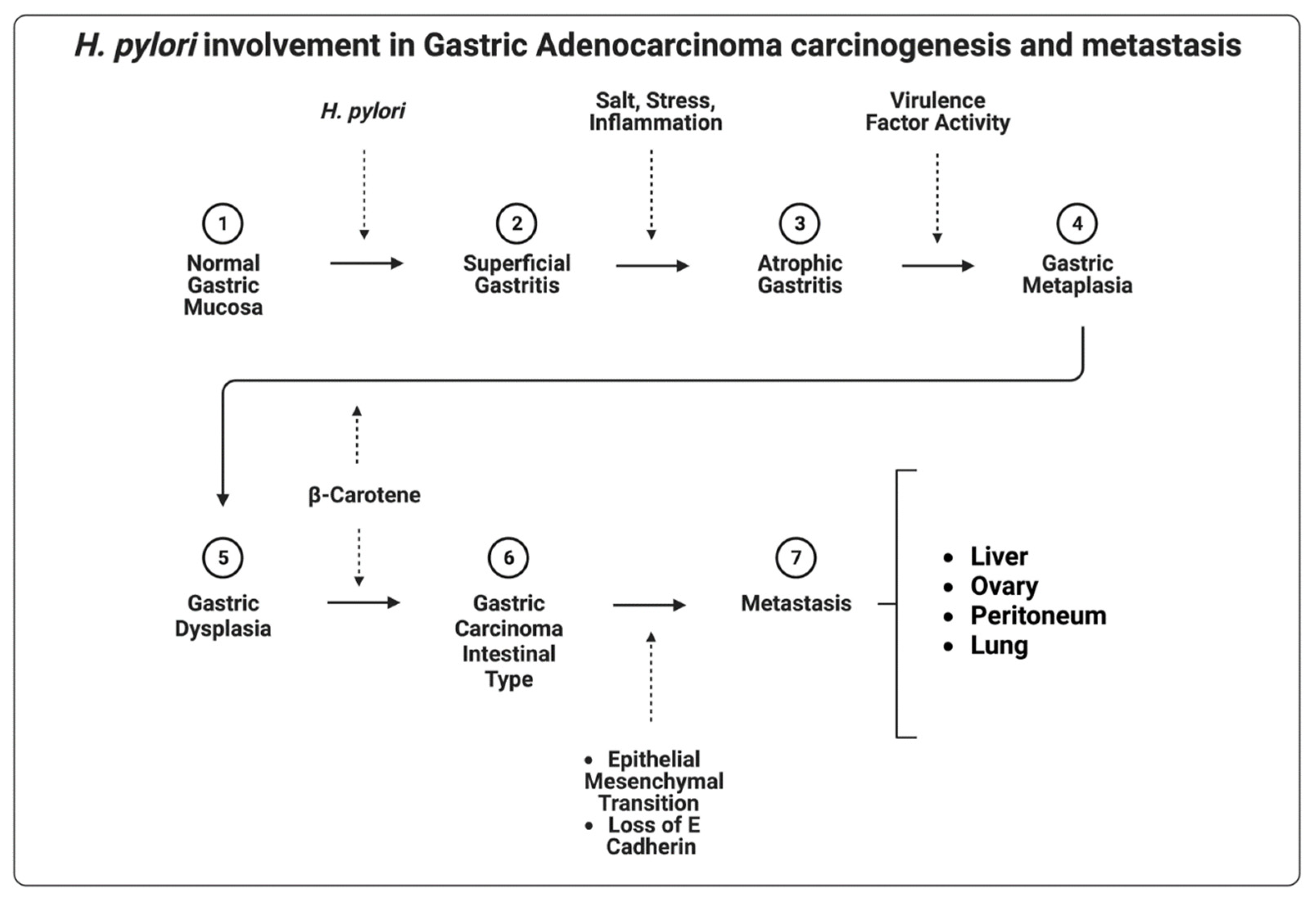
5.1. Helicobacter pylori

H. pylori are spiral-shaped, Gram-negative, urease-positive bacteria, with polar flagella that inhabit the inner lining of the gastric epithelium of humans [62]. The species is the first and only bacterium classified as a group A carcinogen by the International Agency for Research on Cancer [63]. If left untreated, H. pylori causes gastritis [64], peptic ulcer disease [65], MALT lymphoma [52], and gastric adenocarcinoma [53]. The proposed mechanism for the development of gastric adenocarcinoma in the setting of H. pylori infection involves increased gastric epithelial cell proliferation in the background of chronic inflammation (Figure 2) [66]. H. pylori are non-invasive, but infection with H. pylori creates an inflammatory environment that contains genotoxic agents, such as reactive oxygen species [67]. Histologically, H. pylori gastritis is often chronic active gastritis with a mix of mononuclear and neutrophilic leukocytes [68]. Additionally, the H. pylori genome contains genes directly implicated in oncogenesis. Cytotoxin-associated A (CagA) gene [69], vacuolating cytotoxin A (VacA) gene [70], and outer-inflammatory protein A (oipA) [71] gene are commonly cited as causes of serious disease [72]. CagA-expressing isolates of H. pylori colonize the gastric mucosa and induce proinflammatory cytokine secretion, atrophy, and subsequent intestinal metaplasia.
Over half of the world’s population carries H. pylori [73]; however, not all develop the serious associated sequelae. Why H. pylori infection does not cause ulcers in every infected person is unknown. The relationship between H. pylori virulence factors and host immunity is one element that determines the fraction of infected individuals who will develop serious illnesses [74]. Genetic polymorphisms that favor the expression of the proinflammatory cytokine tumor necrosis factor (TNF) [75] and interleukin-1 beta (IL-1β) [76], or decrease expression of interleukin-10 (IL-10) [77], are associated with increased development of pangastritis and sequelae. Virulence factors specific to the isolate are key determinants of outcome. For example, the CagA gene is present on 50% of isolates overall, but in 90% of H. pylori isolated in populations with an increased prevalence of gastric adenocarcinoma [78].
Other factors that determine the severity of disease progression in some individuals and not in others are ongoing areas of research. For example, H. pylori are detected in the gastroduodenal mucosa in the majority of patients with duodenal ulcers, but only a minority (10 to 15%) of H. pylori-infected patients develop peptic ulcer disease [79,80]. It is usually curative of peptic ulcer disease if successfully eradicated, and re-infection after eradication is rare [81]. It is likely that successful detection and eradication contribute to the low rates of disease progression among infected individuals.
Human gastric carcinogenesis is a multi-step and multifactorial process. This process begins as superficial gastritis and progresses into atrophic gastritis, metaplasia, dysplasia, and then gastric adenocarcinoma, in a manner first described by Corea et al. in 1988 [82,83]. Constituent Wnt/β-catenin pathway activation via upregulated expression of Wnt10a and Wnt10b by H. pylori virulence factors has been identified as a common mutation that drives this progression [84,85]. Evidence also supports the activation of aquaporin-5 by CagA-positive H. pylori infection, which promotes epithelial–mesenchymal transition via the extracellular signal-regulated kinase/mitogen-activated protein kinase (MEK/ERK) pathway, thus laying the foundation for metastatic disease [86].
H. pylori are also associated with extra nodal marginal zone B-cell lymphoma, also known as mucosa-associated lymphoid tissue (MALT) lymphoma [87]. MALT is not present in normal gastric mucosa, but can be induced in the setting of chronic gastritis. Induced gastric MALT is most commonly a result of H. pylori infection [56]. Three translocations linked to gastric MALT lymphoma lead to increased expression of intact MALT1 and BCL-10 proteins. NF-B, a transcription factor that promotes B-cell growth and survival, is constitutively activated as a result. BCL-10 and MALT-1 are necessary for the antigen-dependent activation of NF-B in healthy B and T cells. They collaborate in a signal transduction pathway downstream of lymphocyte antigen receptors, and their constitutive activation is pro-oncogenic [88]. H. pylori can evidently stimulate NF-κB activation through the MALT1/BCL-10 pathway in MALT lymphomas that lack inciting translocations [89]. This hypothesis is further supported by the fact that tumors that lack translocations are resolved with H. pylori eradication, but tumors with translocations that involve MALT1 or BCL-10 persist after eradication [90]. MALT lymphomas that accumulate inactivating mutations of tumor suppressor genes that encode for p53 or p16, for example, can become more aggressive tumors that are not amenable to H. pylori eradication [91]. Current research on H. pylori focuses on its epidemiology [2,73], improving testing [92], treatment [57], and a better understanding of its unique virulence factors [58], and its ability to propel progression [86]. Primary prevention through eradication of H. pylori and lifestyle modifications, including reducing salt intake, smoking, obesity, and alcohol, remains key in gastric CA control [3]. Additionally, eating foods that have been salted or smoked is associated with developing diffuse-type gastric adenocarcinoma. The biosynthesis of glycolipids in the cell wall of H. pylori is thought to be inhibited by glycans found in glandular mucus, and a high-salt diet (regular consumption of sodium beyond recommended daily value) is thought to increase superficial mucus cell mucus, while decreasing glandular mucus cell mucus [93].

5.2. Mycoplasma hyorhinis

Mycoplasma hyorhinis (M. hyorhinis) are Gram variable cell wall-deficient bacteria. This small Gram-negative pleomorphic coccobacilli is known to infect the respiratory tract of pigs. Numerous swine diseases have been linked to M. hyorhinis, with severe pathogenicity, high mortality rates, and ensuing financial losses. Antibiotic interventions to lessen these effects encourage the growth of drug-resistant M. hyorhinis strains which have the potential to infect human stomachs through pork consumption [94]. A previous report identified M. hyorhinis in 56% of gastric cancer [95].
Gastric and prostate neoplasms have been linked to M. hyorhinis dysbiosis via the NLRP3 inflammasome [96]. NLRP3 is a protein complex that regulates the maturation of pro-inflammatory cytokines, such as interleukin-1 (IL-1) and interleukin-18 (IL-18), and is also involved in tumorigenesis and metastasis. Mounting evidence suggests that M. hyorhinis infection results in pathology in human studies. Serology studies have confirmed M. hyorhinis in gastric carcinoma, colon carcinoma, and prostate and lung carcinoma biopsies [59,95].
A 2019 study by Liu et al. revealed M. hyorhinis involvement in gastric neoplasia via β-catenin stabilization and subsequent activation of the WNT-signaling pathway, promoting gastric cancer cell motility and inciting cancer progression [59]. They found that when glycogen synthase kinase 3 beta (GSK3β) and Wnt-receptor lipoprotein-receptor-related protein 6 (LRP6) did not interact, there was no increase in activating β-catenin stabilization, suggesting that this interaction has a carcinogenic effect. They also showed an interaction between LRP6 and p37, a mycoplasma membrane protein known to have carcinogenic effects [97]. In vitro studies have found p37 to promote cell motility, migration, and invasion through the activation of metalloproteinase-2 and epidermal growth factor receptor [60], veritably connecting M. hyorhinis infection and metastatic disease.

5.3. Fusobacterium nucleatum

Fusobacterium nucleatum (F. nucleatum) is an opportunistic, Gram-negative obligate anaerobic bacterium which is commonly located in the oral cavity of humans [98,99]. F. nucleatum is frequently detected in primary colorectal cancer (CRC) and its metastases, and has been linked to a worse prognosis in gastrointestinal cancers in general. Recently, studies have shown increased loads of F. nucleatum in gastric cancer tumor samples [94], although it is unclear if it is a causative agent. In one study, F. nucleatum positivity showed no association with chronic gastritis or preneoplastic conditions, such as intestinal metaplasia [61].
F. nucleatum is associated with worse prognosis in Lauren’s diffuse-type gastric cancer patients, but not in the intestinal type, in contrast to H. pylori [61]. F. nucleatum increases cell proliferation and tumor-promoting inflammation, while avoiding immune destruction, ultimately promoting a pro-inflammatory state and tumorigenic environment [100,101]. F. nucleatum is thought to stimulate cell proliferation via two primary signaling mechanisms, including (1) FadA binding to E-Cadherin to activate the WNT/B catenin pathway [102] and (2) interacting with the Toll-like receptor 4 (TLR-4) to activate P21-activated kinase 1 (PAK 1), a protein that phosphorylates the B-catenin pathway [103,104]. However, these patterns have only been established in CRC studies, and further studies are needed to explore the related mechanistic insights and potential therapeutic benefits of targeted antibiotic treatment in gastric cancer patients.

6. Considering the Microbiome in Gastrointestinal Cancer Treatment

Investigations into the complex interactions of microorganisms and tumor behavior are uncovering a variety of potential mechanisms by which infection/colonization contributes to more aggressive tumors. Factors such as increased acute and chronic inflammation, induction of various enzymes, stimulation of cytokines, changes in the microbiome, and even potential alterations in local oxygen tension may modulate the growth, invasiveness, and spread of neoplastic cells.
Tumor cells may degrade interstitial connective tissue and the basement membrane by secreting proteolytic enzymes or by inducing stromal cell proteolysis [105,106]. Ulceration or chronic inflammation in the setting of ulcerative colitis or bacterial infection could affect the process of basement membrane degradation, catalyzing this cascade step [65,107]. Matrix metalloproteinases (MMPs), amongst others, have been implicated in tumor cell invasion [108]. Both H. pylori and M. hyorhinis have been shown to induce metalloproteinase activity [60,109]. MMPs remodel the basement membrane and interstitial connective tissue to promote progressive invasion and metastasis [110]. They also promote metastasis by inducing factors related to malignant behavior. MMP-2 and MMP-9 have been linked to vascular endothelial growth factor (VEGF), a necessary compound for tumor vascularization [111].
It is important to note that the regions within the stomach have varying microflora compositions [112]. Eradication of H. pylori has been repeatedly shown to decrease the risk of neoplasia, especially when treated before pathologic site changes have occurred and even in those with prior gastric cancer history [113]. Prognosis is most accurately determined after thoroughly investigating the cells and microenvironment. Gastric cancers are often the culmination of lifestyle factors, such as smoking, diet, and Epstein–Barr virus infection [114], but that is not to say mutations are not important in tumorigenesis [115]. For example, a study found that gastric cancers often have a lower relative expression of NFKB2, which is part of a family of transcription factors [116].
However, bacterial consideration does not end there. A study showed detrimental gut bacteria such as Proteobacteria flourished after H. pylori eradication, which may warrant probiotic consideration in the treatment plan [117]. Bik et al. showed an overabundance of Propionibacterium acnes, Streptococcus anginosus, and Prevotella melaninogenica in the gastric tumoral microenvironment, which are associated with gastric cancer tumorigenesis through their specific mechanisms that increase local inflammation [118]. P. acnes releases short-chain fatty acids, which can cause lymphocytic gastritis. S. anginosus induces cytokine release via sulfur metabolism. P. melaninogenica contributes to excess gastric acidity in non-atrophic sites [119,120,121]. Identifying microbes that are similar to these and their pathways of pathogenicity is crucial for developing targeted treatment plans.

Microbiome after Gastrectomy

For early-stage tumors, gastrectomy is the most successful definitive treatment. A landmark study in 1982 recommended excision margins of at least six centimeters to properly excise serosa infiltrations and constituted the standard of treatment for nearly 30 years. However, increasing the amounts of gastric tissue removed leads to poorer patient quality of life and nutrition [122,123]. Many studies since then have tried to further define the most appropriate proximal resection margin (PRM) with conflicting results; a 2017 study recommended at least 2.1 cm of PRM [124]. In an attempt to provide a definitive answer, a 2020 retrospective analysis concluded that previously recommended PRMs may not be absolute, with distances as short as < 1 cm providing comparable rates of survival [125]. Furthermore, patients with larger-end PRMs may have partial gastrectomy that effectively provides the physiology of complete gastrectomy.
Many studies have analyzed the alteration in the microbiome post-gastrectomy. The Roux-en-Y gastrojejunal anastomosis (RYGJ) and Billroth II anastomosis (BII) are two types of partial gastrectomy that may be performed for neoplasm resection and are shown to alter gut microbiota. One study showed Ralstonia and Helicobacter predominating before and Streptococcus and Prevotella predominating after tumor resection [126]. Another study found an increased abundance of Anaerosinus, Butyrivibrio, Campylobacter, Clostridium, Coprococcus, Desulfovibrio, Oscillospira, Oxalobacter, Slackia, Sporobacter, Veillonella, and Victivallis after a BII or RYGJ, compared to controls without these surgeries [127]. Although the RYGB is commonly performed for weight loss, the evidence suggests that microbial alterations are more likely driven by direct changes to intestinal physiology, rather than weight loss, which can likely be also applied to BII [128].
Higher concentrations of dissolved oxygen in the hindgut after an RYGJ are thought to lead to an increase in facultative anaerobes, such as E. coli, K. pneumoniae, E. faecalis, and Streptococcus species [128]. Both the RYGJ and BII reduce the stomach luminal surface area, which in turn decreases acid secretions. This creates a less hostile environment for E. coli, promoting its colonization [129]. As a result of decreased acid secretion, the increased stomach pH permits oral flora, such as Streptococcus spp. and a few Veillonella spp., which are metabolically dependent on Streptococcus spp. in oral biofilms, to overcome the previously inhibitory gastric barrier [130]. As with PPIs and sleeve gastric bypass, which typically creates a permanent pH of 6.0, gastric pH levels that surpass 4.0 have significantly diminished antimicrobial effects [131,132]. There are also factors in addition to oxygen concentration and pH that likely affect microbial colonization.
We have gained insight by comparing alterations in the microbiome after partial gastrectomy for cancer and for morbid obesity. The majority of partial gastrectomies are performed to resect tumors. So, the question remains whether tumors found post-gastrectomy can truly be attributed to bacteria-induced inflammatory changes versus recurrence.
Furthermore, partial gastrectomy to treat morbid obesity yields different alterations. In one study, Yokenella regensburgei and Fusobacterium varium were found after bariatric surgery; these two, in particular, have already been associated with colonic inflammation, which can be theorized to occur in the stomach as well [133]. Evidence supports bariatric surgery being protective against obesity-related cancers, as defined by the International Agency for Research on Cancer (IARC), including esophageal adenocarcinoma, postmenopausal breast cancer, renal, colon, rectum, gastric cardia, liver, gallbladder, pancreas, ovary, corpus uteri, thyroid, multiple myeloma, and meningioma [134,135,136]. However, a study on delineated non-obesity-related carcinomas found that there was only a protective effect of bariatric surgery in females [135]; the sample size was vastly different between males and females, so this could be an area that warrants further research. It is important to note that early-onset gastric cancer is a distinct disease with worrisome trends and oncogenic features and unique clinical and genomic characteristics [137]; therefore, further study is needed to elaborate on bacterial involvement in this entity. The differences in microbiota may have a role in neoplasia protection post-gastrectomy.

7. Conclusions

Bacterial involvement in gastric neoplasia and metastasis is significant. Primary tumors are frequently curable with gastrectomy, when detected early. However, metastatic disease, which accounts for the majority of cancer-related deaths, remains a deadly clinical scenario. Growing evidence indicates that bacterial infection influences organotropism and metastatic progression by altering the microenvironment at the primary and secondary tumors, in addition to promoting carcinogenesis in primary gastric CA. Eradication of H. pylori can halt gastric MALT lymphoma progression and reverse early metaplasia. Inflammation and disruption of epithelial integrity, brought on by virulence factors such as CagA, VacA, and oipA, enable the primary tumor to go through the critical stages of the metastatic process. The evidence supports the activation of aquaporin-5 by CagA-positive H. pylori infection, which promotes epithelial–mesenchymal transition via the extracellular signal-regulated kinase/mitogen-activated protein kinase (MEK/ERK) pathway, thus laying the foundation for metastatic disease. M. hyorhinis has been implicated in gastric neoplasia via β-catenin stabilization and subsequent activation of the WNT-signaling pathway, promoting gastric cancer cell motility and inciting cancer progression. F. nucleatum is a driver of gastrointestinal cancers in general, and further investigation into its involvement in gastric cancer is needed. F. nucleatum is associated with worse prognosis in diffuse-type gastric adenocarcinoma. Recognition of the roles that bacteria play within the metastatic cascade is vital in gastrointestinal adenocarcinoma treatment and potential reoccurrence. At this time, further investigation is needed to establish potential treatment for metastatic gastric carcinoma by targeting the microbial contribution to the tumor microenvironment.

Author Contributions

Conceptualization, methodology and writing—original draft preparation: K.D.S., A.D.M., L.D.H. and G.M.F.; writing—editing: K.D.S., A.D.M., L.D.H. and G.M.F.; figures: K.D.S. and L.D.H.; supervision: J.M.S. All authors have read and agreed to the published version of the manuscript.

Funding

This research received no external funding.

Acknowledgments

The authors acknowledge the Department of Research and Scholarly Activity at Rocky Vista University for its support of this research.

Conflicts of Interest

The authors declare no conflict of interest.

References

  1. Seely, K.D.; Morgan, A.D.; Hagenstein, L.D.; Florey, G.M.; Small, J.M. Bacterial Involvement in Progression and Metastasis of Colorectal Neoplasia. Cancers 2022, 14, 1019. [Google Scholar] [CrossRef] [PubMed]
  2. Sung, H.; Ferlay, J.; Siegel, R.L.; Laversanne, M.; Soerjomataram, I.; Jemal, A.; Bray, F. Global Cancer Statistics 2020: GLOBOCAN Estimates of Incidence and Mortality Worldwide for 36 Cancers in 185 Countries. CA Cancer J. Clin. 2021, 71, 209–249. [Google Scholar] [CrossRef] [PubMed]
  3. Morgan, E.; Arnold, M.; Camargo, M.C.; Gini, A.; Kunzmann, A.T.; Matsuda, T.; Meheus, F.; Verhoeven, R.H.A.; Vignat, J.; Laversanne, M.; et al. The Current and Future Incidence and Mortality of Gastric Cancer in 185 Countries, 2020–40: A Population-Based Modelling Study. eClinicalMedicine 2022, 47, 101404. [Google Scholar] [CrossRef] [PubMed]
  4. Ganesh, K.; Massagué, J. Targeting Metastatic Cancer. Nat. Med. 2021, 27, 34–44. [Google Scholar] [CrossRef] [PubMed]
  5. Mukaisho, K.-I.; Nakayama, T.; Hagiwara, T.; Hattori, T.; Sugihara, H. Two Distinct Etiologies of Gastric Cardia Adenocarcinoma: Interactions among PH, Helicobacter Pylori, and Bile Acids. Front. Microbiol. 2015, 6, 412. [Google Scholar] [CrossRef] [PubMed] [Green Version]
  6. Lauren, P. The two histological main types of gastric carcinoma: Diffuse and so-called intestinal-type carcinoma. An attempt at a histo-clinical classification. Acta Pathol. Microbiol. Scand. 1965, 64, 31–49. [Google Scholar] [CrossRef]
  7. Turner, E.S.; Turner, J.R. Expanding the Lauren Classification: A New Gastric Cancer Subtype? Gastroenterology 2013, 145, 505–508. [Google Scholar] [CrossRef]
  8. Compton, C.; Sobin, L.H. Protocol for the Examination of Specimens Removed from Patients with Gastric Carcinoma: A Basis for Checklists. Members of the Cancer Committee, College of American Pathologists, and the Task Force for Protocols on the Examination of Specimens From Patients With Gastric Cancer. Arch. Pathol. Lab. Med. 1998, 122, 9–14. [Google Scholar]
  9. Budczies, J.; von Winterfeld, M.; Klauschen, F.; Bockmayr, M.; Lennerz, J.K.; Denkert, C.; Wolf, T.; Warth, A.; Dietel, M.; Anagnostopoulos, I.; et al. The Landscape of Metastatic Progression Patterns across Major Human Cancers. Oncotarget 2015, 6, 570–583. [Google Scholar] [CrossRef] [Green Version]
  10. Riihimäki, M.; Hemminki, A.; Sundquist, K.; Sundquist, J.; Hemminki, K. Metastatic Spread in Patients with Gastric Cancer. Oncotarget 2016, 7, 52307–52316. [Google Scholar] [CrossRef] [Green Version]
  11. Shi, Z.; Ding, H.; Shen, Q.W.; Lu, X.G.; Chen, J.Y.; Chen, X.; Tang, X. The Clinical Manifestation, Survival Outcome and Predictive Prognostic Factors of 137 Patients with Primary Gastrointestinal Lymphoma (PGIL): Strobe Compliant. Medicine 2018, 97, e9583. [Google Scholar] [CrossRef]
  12. Patnaik, S.; Jyotsnarani, Y.; Rammurti, S. Radiological Features of Metastatic Gastrointestinal Stromal Tumors. J. Clin. Imaging Sci. 2012, 2, 43. [Google Scholar] [CrossRef]
  13. Riihimäki, M.; Hemminki, A.; Sundquist, K.; Sundquist, J.; Hemminki, K. The Epidemiology of Metastases in Neuroendocrine Tumors. Int. J. Cancer 2016, 139, 2679–2686. [Google Scholar] [CrossRef]
  14. Hansen, S.; Melby, K.K.; Aase, S.; Jellum, E.; Vollset, S.E. Helicobacter Pylori Infection and Risk of Cardia Cancer and Non-Cardia Gastric Cancer. A Nested Case-Control Study. Scand. J. Gastroenterol. 1999, 34, 353–360. [Google Scholar] [CrossRef]
  15. Li, Y.; Gong, J.; Zhang, Q.; Lu, Z.; Gao, J.; Li, Y.; Cao, Y.; Shen, L. Dynamic Monitoring of Circulating Tumour Cells to Evaluate Therapeutic Efficacy in Advanced Gastric Cancer. Br. J. Cancer 2016, 114, 138–145. [Google Scholar] [CrossRef]
  16. Necula, L.; Matei, L.; Dragu, D.; Neagu, A.I.; Mambet, C.; Nedeianu, S.; Bleotu, C.; Diaconu, C.C.; Chivu-Economescu, M. Recent Advances in Gastric Cancer Early Diagnosis. World J. Gastroenterol. 2019, 25, 2029–2044. [Google Scholar] [CrossRef] [PubMed]
  17. Hamakawa, T.; Kukita, Y.; Kurokawa, Y.; Miyazaki, Y.; Takahashi, T.; Yamasaki, M.; Miyata, H.; Nakajima, K.; Taniguchi, K.; Takiguchi, S.; et al. Monitoring Gastric Cancer Progression with Circulating Tumour DNA. Br. J. Cancer 2015, 112, 352–356. [Google Scholar] [CrossRef] [PubMed]
  18. Zhang, C.; Chen, Z.; Chong, X.; Chen, Y.; Wang, Z.; Yu, R.; Sun, T.; Chen, X.; Shao, Y.; Zhang, X.; et al. Clinical Implications of Plasma CtDNA Features and Dynamics in Gastric Cancer Treated with HER2-targeted Therapies. Clin. Transl. Med. 2020, 10, e254. [Google Scholar] [CrossRef] [PubMed]
  19. Fang, W.-L.; Lan, Y.-T.; Huang, K.-H.; Liu, C.-A.; Hung, Y.-P.; Lin, C.-H.; Jhang, F.-Y.; Chang, S.-C.; Chen, M.-H.; Chao, Y.; et al. Clinical Significance of Circulating Plasma DNA in Gastric Cancer: Circulating Plasma DNA in Gastric Cancer. Int. J. Cancer 2016, 138, 2974–2983. [Google Scholar] [CrossRef] [PubMed] [Green Version]
  20. Lionetti, R.; DE Luca, M.; Raffone, A.; Travaglino, A.; Coppellotti, A.; Peltrini, R.; Bracale, U.; D’Ambra, M.; Insabato, L.; Zullo, F.; et al. Clinics and Pathology of Krukenberg Tumor: A Systematic Review and Meta-Analysis. Minerva Obstet. Gynecol. 2022, 74, 356–363. [Google Scholar] [CrossRef] [PubMed]
  21. Al-Agha, O.M.; Nicastri, A.D. An In-Depth Look at Krukenberg Tumor: An Overview. Arch. Pathol. Lab. Med. 2006, 130, 1725–1730. [Google Scholar] [CrossRef]
  22. Seow-En, I.; Hwarng, G.; Tan, G.H.C.; Ho, L.M.L.; Teo, M.C.C. Palliative Surgery for Krukenberg Tumors—12-Year Experience and Review of the Literature. World J. Clin. Oncol. 2018, 9, 13–19. [Google Scholar] [CrossRef] [Green Version]
  23. Aziz, M.; Killeen, R.B.; Kasi, A. Krukenberg Tumor. In StatPearls; StatPearls Publishing: Treasure Island, FL, USA, 2022. [Google Scholar]
  24. Agnes, A.; Biondi, A.; Ricci, R.; Gallotta, V.; D’Ugo, D.; Persiani, R. Krukenberg Tumors: Seed, Route and Soil. Surg. Oncol. 2017, 26, 438–445. [Google Scholar] [CrossRef] [PubMed]
  25. Wang, B.; Sun, K.; Zou, Y. Comparison of a Panel of Biomarkers Between Gastric Primary Cancer and the Paired Krukenberg Tumor. Appl. Immunohistochem. Mol. Morphol. 2017, 25, 639–644. [Google Scholar] [CrossRef] [PubMed]
  26. Belardi, B.; Son, S.; Felce, J.H.; Dustin, M.L.; Fletcher, D.A. Cell-Cell Interfaces as Specialized Compartments Directing Cell Function. Nat. Rev. Mol. Cell Biol. 2020, 21, 750–764. [Google Scholar] [CrossRef]
  27. Zhao, H.; Hu, H.; Chen, B.; Xu, W.; Zhao, J.; Huang, C.; Xing, Y.; Lv, H.; Nie, C.; Wang, J.; et al. Overview on the Role of E-Cadherin in Gastric Cancer: Dysregulation and Clinical Implications. Front. Mol. Biosci. 2021, 8, 689139. [Google Scholar] [CrossRef] [PubMed]
  28. Pećina-Šlaus, N. Tumor Suppressor Gene E-Cadherin and Its Role in Normal and Malignant Cells. Cancer Cell Int. 2003, 3, 17. [Google Scholar] [CrossRef] [PubMed] [Green Version]
  29. Tian, X.; Liu, Z.; Niu, B.; Zhang, J.; Tan, T.K.; Lee, S.R.; Zhao, Y.; Harris, D.C.H.; Zheng, G. E-Cadherin/β-Catenin Complex and the Epithelial Barrier. J. Biomed. Biotechnol. 2011, 2011, e567305. [Google Scholar] [CrossRef] [Green Version]
  30. Nguyen, V.H.L.; Hough, R.; Bernaudo, S.; Peng, C. Wnt/β-Catenin Signalling in Ovarian Cancer: Insights into Its Hyperactivation and Function in Tumorigenesis. J. Ovarian Res. 2019, 12, 122. [Google Scholar] [CrossRef] [PubMed] [Green Version]
  31. Babaei, G.; Aziz, S.G.-G.; Jaghi, N.Z.Z. EMT, Cancer Stem Cells and Autophagy; The Three Main Axes of Metastasis. Biomed. Pharmacother. 2021, 133, 110909. [Google Scholar] [CrossRef]
  32. Saitoh, M. Involvement of Partial EMT in Cancer Progression. J. Biochem. 2018, 164, 257–264. [Google Scholar] [CrossRef] [PubMed]
  33. Serrano-Gomez, S.J.; Maziveyi, M.; Alahari, S.K. Regulation of Epithelial-Mesenchymal Transition through Epigenetic and Post-Translational Modifications. Mol. Cancer 2016, 15, 18. [Google Scholar] [CrossRef] [PubMed] [Green Version]
  34. Yeung, K.T.; Yang, J. Epithelial–Mesenchymal Transition in Tumor Metastasis. Mol. Oncol. 2017, 11, 28–39. [Google Scholar] [CrossRef] [PubMed] [Green Version]
  35. Ruiz, P.A.; Shkoda, A.; Kim, S.C.; Sartor, R.B.; Haller, D. IL-10 Gene-Deficient Mice Lack TGF-β/Smad Signaling and Fail to Inhibit Proinflammatory Gene Expression in Intestinal Epithelial Cells after the Colonization with Colitogenic Enterococcus Faecalis. J. Immunol. 2005, 174, 2990–2999. [Google Scholar] [CrossRef] [PubMed] [Green Version]
  36. Hanahan, D.; Weinberg, R.A. Hallmarks of Cancer: The Next Generation. Cell 2011, 144, 646–674. [Google Scholar] [CrossRef] [Green Version]
  37. Multhoff, G.; Molls, M.; Radons, J. Chronic Inflammation in Cancer Development. Front. Immunol. 2012, 2, 98. [Google Scholar] [CrossRef] [Green Version]
  38. DeNardo, D.G.; Andreu, P.; Coussens, L.M. Interactions between Lymphocytes and Myeloid Cells Regulate Pro-versus Anti-Tumor Immunity. Cancer Metastasis Rev. 2010, 29, 309–316. [Google Scholar] [CrossRef] [Green Version]
  39. Grivennikov, S.I.; Greten, F.R.; Karin, M. Immunity, Inflammation, and Cancer. Cell 2010, 140, 883–899. [Google Scholar] [CrossRef] [Green Version]
  40. Qian, B.; Pollard, J.W. Macrophage Diversity Enhances Tumor Progression and Metastasis. Cell 2010, 141, 39–51. [Google Scholar] [CrossRef] [Green Version]
  41. Jin, M.-Z.; Jin, W.-L. The Updated Landscape of Tumor Microenvironment and Drug Repurposing. Signal Transduct. Target. Ther. 2020, 5, 166. [Google Scholar] [CrossRef]
  42. Feng, Z.; Li, X.; Ren, Z.; Feng, J.; He, X.; You, C. Prognostic and Predictive Value of Cadherin 11 for Patients with Gastric Cancer and Its Correlation with Tumor Microenvironment: Results from Microarray Analysis. BioMed Res. Int. 2020, 2020, 8107478. [Google Scholar] [CrossRef] [PubMed]
  43. Rugge, M.; Correa, P.; Dixon, M.F.; Fiocca, R.; Hattori, T.; Lechago, J.; Leandro, G.; Price, A.B.; Sipponen, P.; Solcia, E.; et al. Gastric Mucosal Atrophy: Interobserver Consistency Using New Criteria for Classification and Grading. Aliment. Pharmacol. Ther. 2002, 16, 1249–1259. [Google Scholar] [CrossRef]
  44. Cook, J.D.; Brown, G.M.; Valberg, L.S. The Effect of Achylia Gastrica on Iron Absorption*. J. Clin. Investig. 1964, 43, 1185–1191. [Google Scholar] [CrossRef]
  45. Engstrand, L.; Graham, D.Y. The Microbiome and Gastric Cancer. Dig. Dis. Sci. 2020, 65, 865–873. [Google Scholar] [CrossRef] [PubMed] [Green Version]
  46. Vartoukian, S.R.; Palmer, R.M.; Wade, W.G. Strategies for Culture of “unculturable” Bacteria. FEMS Microbiol. Lett. 2010, 309, 1–7. [Google Scholar] [CrossRef] [PubMed] [Green Version]
  47. Conti, L.; Annibale, B.; Lahner, E. Autoimmune Gastritis and Gastric Microbiota. Microorganisms 2020, 8, 1827. [Google Scholar] [CrossRef] [PubMed]
  48. Rajilic-Stojanovic, M.; Figueiredo, C.; Smet, A.; Hansen, R.; Kupcinskas, J.; Rokkas, T.; Andersen, L.; Machado, J.C.; Ianiro, G.; Gasbarrini, A.; et al. Systematic Review: Gastric Microbiota in Health and Disease. Aliment. Pharmacol. Ther. 2020, 51, 582–602. [Google Scholar] [CrossRef]
  49. Ferreira, R.M.; Pereira-Marques, J.; Pinto-Ribeiro, I.; Costa, J.L.; Carneiro, F.; Machado, J.C.; Figueiredo, C. Gastric Microbial Community Profiling Reveals a Dysbiotic Cancer-Associated Microbiota. Gut 2018, 67, 226–236. [Google Scholar] [CrossRef] [Green Version]
  50. Parsons, B.N.; Ijaz, U.Z.; D’Amore, R.; Burkitt, M.D.; Eccles, R.; Lenzi, L.; Duckworth, C.A.; Moore, A.R.; Tiszlavicz, L.; Varro, A.; et al. Comparison of the Human Gastric Microbiota in Hypochlorhydric States Arising as a Result of Helicobacter Pylori-Induced Atrophic Gastritis, Autoimmune Atrophic Gastritis and Proton Pump Inhibitor Use. PLoS Pathog. 2017, 13, e1006653. [Google Scholar] [CrossRef] [PubMed] [Green Version]
  51. Hansen, J.P.; Ali, W.M.; Sivadasan, R.; Rajeeve, K. Bacteria-Cancer Interface: Awaiting the Perfect Storm. Pathog. Basel Switz. 2021, 10, 1321. [Google Scholar] [CrossRef]
  52. Della Bella, C.; Soluri, M.F.; Puccio, S.; Benagiano, M.; Grassi, A.; Bitetti, J.; Cianchi, F.; Sblattero, D.; Peano, C.; D’Elios, M.M. The Helicobacter Pylori CagY Protein Drives Gastric Th1 and Th17 Inflammation and B Cell Proliferation in Gastric MALT Lymphoma. Int. J. Mol. Sci. 2021, 22, 9459. [Google Scholar] [CrossRef] [PubMed]
  53. Peng, C.; Li, N.-S.; Hu, Y.; Lu, N.-H. Impact Factors That Modulate Gastric Cancer Risk in Helicobacter Pylori-Infected Rodent Models. Helicobacter 2019, 24, e12580. [Google Scholar] [CrossRef] [PubMed]
  54. Mori, G.; Pasca, M.R. Gut Microbial Signatures in Sporadic and Hereditary Colorectal Cancer. Int. J. Mol. Sci. 2021, 22, 1312. [Google Scholar] [CrossRef] [PubMed]
  55. Anderson, W.F.; Rabkin, C.S.; Turner, N.; Fraumeni, J.F.; Rosenberg, P.S.; Camargo, M.C. The Changing Face of Noncardia Gastric Cancer Incidence Among US Non-Hispanic Whites. J. Natl. Cancer Inst. 2018, 110, 608–615. [Google Scholar] [CrossRef] [Green Version]
  56. Floch, P.; Mégraud, F.; Lehours, P. Helicobacter Pylori Strains and Gastric MALT Lymphoma. Toxins 2017, 9, 132. [Google Scholar] [CrossRef]
  57. Ford, A.C.; Yuan, Y.; Moayyedi, P. Helicobacter Pylori Eradication Therapy to Prevent Gastric Cancer: Systematic Review and Meta-Analysis. Gut 2020, 69, 2113–2121. [Google Scholar] [CrossRef]
  58. Baj, J.; Forma, A.; Sitarz, M.; Portincasa, P.; Garruti, G.; Krasowska, D.; Maciejewski, R. Helicobacter Pylori Virulence Factors—Mechanisms of Bacterial Pathogenicity in the Gastric Microenvironment. Cells 2021, 10, 27. [Google Scholar] [CrossRef]
  59. Liu, X.; Rong, Z.; Shou, C. Mycoplasma Hyorhinis Infection Promotes Gastric Cancer Cell Motility via β-Catenin Signaling. Cancer Med. 2019, 8, 5301–5312. [Google Scholar] [CrossRef] [PubMed] [Green Version]
  60. Gong, M.; Meng, L.; Jiang, B.; Zhang, J.; Yang, H.; Wu, J.; Shou, C. P37 from Mycoplasma Hyorhinis Promotes Cancer Cell Invasiveness and Metastasis through Activation of MMP-2 and Followed by Phosphorylation of EGFR. Mol. Cancer Ther. 2008, 7, 530–537. [Google Scholar] [CrossRef] [Green Version]
  61. Boehm, E.T.; Thon, C.; Kupcinskas, J.; Steponaitiene, R.; Skieceviciene, J.; Canbay, A.; Malfertheiner, P.; Link, A. Fusobacterium Nucleatum Is Associated with Worse Prognosis in Lauren’s Diffuse Type Gastric Cancer Patients. Sci. Rep. 2020, 10, 16240. [Google Scholar] [CrossRef] [PubMed]
  62. Cheok, Y.Y.; Lee, C.Y.Q.; Cheong, H.C.; Vadivelu, J.; Looi, C.Y.; Abdullah, S.; Wong, W.F. An Overview of Helicobacter Pylori Survival Tactics in the Hostile Human Stomach Environment. Microorganisms 2021, 9, 2502. [Google Scholar] [CrossRef]
  63. Schistosomes, Liver Flukes and Helicobacter Pylori. In IARC Monographs on the Evaluation of Carcinogenic Risks to Humans; IARC Working Group on the Evaluation of Carcinogenic Risks to Humans; International Agency for Research on Cancer: Lyon, France, 1994; Volume 61, pp. 1–241.
  64. Tang, L.; Tang, B.; Lei, Y.; Yang, M.; Wang, S.; Hu, S.; Xie, Z.; Liu, Y.; Vlodavsky, I.; Yang, S. Helicobacter Pylori-Induced Heparanase Promotes H. pylori Colonization and Gastritis. Front. Immunol. 2021, 12, 2352. [Google Scholar] [CrossRef]
  65. Narayanan, M.; Reddy, K.M.; Marsicano, E. Peptic Ulcer Disease and Helicobacter Pylori Infection. Mo. Med. 2018, 115, 219–224. [Google Scholar]
  66. Uotani, T.; Murakami, K.; Uchida, T.; Tanaka, S.; Nagashima, H.; Zeng, X.-L.; Akada, J.; Estes, M.K.; Graham, D.Y.; Yamaoka, Y. Changes of Tight Junction and Interleukin-8 Expression Using a Human Gastroid Monolayer Model of Helicobacter Pylori Infection. Helicobacter 2019, 24, e12583. [Google Scholar] [CrossRef]
  67. Jain, U.; Saxena, K.; Chauhan, N. Helicobacter Pylori Induced Reactive Oxygen Species: A New and Developing Platform for Detection. Helicobacter 2021, 26, e12796. [Google Scholar] [CrossRef] [PubMed]
  68. Fukuda, S.; Tanaka, M.; Soma, Y.; Shimoyama, T.; Mikami, T.; Crabtree, J.E.; Saito, H.; Munakata, A.; Yoshida, Y. Histological Analysis of Gastritis and Helicobacter pylori Infection in Patients with Early Gastric Cancer: A Case-Control Study. J. Gastroenterol. Hepatol. 2000, 15, 1370–1376. [Google Scholar] [CrossRef]
  69. Imai, S.; Ooki, T.; Murata-Kamiya, N.; Komura, D.; Tahmina, K.; Wu, W.; Takahashi-Kanemitsu, A.; Knight, C.T.; Kunita, A.; Suzuki, N.; et al. Helicobacter Pylori CagA Elicits BRCAness to Induce Genome Instability That May Underlie Bacterial Gastric Carcinogenesis. Cell Host Microbe 2021, 29, 941–958.e10. [Google Scholar] [CrossRef]
  70. Palframan, S.L.; Kwok, T.; Gabriel, K. Vacuolating Cytotoxin A (VacA), a Key Toxin for Helicobacter Pylori Pathogenesis. Front. Cell. Infect. Microbiol. 2012, 2, 92. [Google Scholar] [CrossRef] [Green Version]
  71. Farzi, N.; Yadegar, A.; Aghdaei, H.A.; Yamaoka, Y.; Zali, M.R. Genetic Diversity and Functional Analysis of OipA Gene in Association with Other Virulence Factors among Helicobacter Pylori Isolates from Iranian Patients with Different Gastric Diseases. Infect. Genet. Evol. J. Mol. Epidemiol. Evol. Genet. Infect. Dis. 2018, 60, 26–34. [Google Scholar] [CrossRef]
  72. Yamaoka, Y. Mechanisms of Disease: Helicobacter Pylori Virulence Factors. Nat. Rev. Gastroenterol. Hepatol. 2010, 7, 629–641. [Google Scholar] [CrossRef] [Green Version]
  73. Hooi, J.K.Y.; Lai, W.Y.; Ng, W.K.; Suen, M.M.Y.; Underwood, F.E.; Tanyingoh, D.; Malfertheiner, P.; Graham, D.Y.; Wong, V.W.S.; Wu, J.C.Y.; et al. Global Prevalence of Helicobacter Pylori Infection: Systematic Review and Meta-Analysis. Gastroenterology 2017, 153, 420–429. [Google Scholar] [CrossRef] [PubMed] [Green Version]
  74. Serrano, C.; Diaz, M.I.; Valdivia, A.; Godoy, A.; Peña, A.; Rollan, A.; Kirberg, A.; Hebel, E.; Fierro, J.; Klapp, G.; et al. Relationship between Helicobacter Pylori Virulence Factors and Regulatory Cytokines as Predictors of Clinical Outcome. Microbes Infect. 2007, 9, 428–434. [Google Scholar] [CrossRef]
  75. Yea, S.S.; Yang, Y.I.; Jang, W.H.; Lee, Y.J.; Bae, H.S.; Paik, K.H. Association between TNF-Alpha Promoter Polymorphism and Helicobacter Pylori CagA Subtype Infection. J. Clin. Pathol. 2001, 54, 703–706. [Google Scholar] [CrossRef] [PubMed]
  76. Figueiredo, C.; Machado, J.C.; Pharoah, P.; Seruca, R.; Sousa, S.; Carvalho, R.; Capelinha, A.F.; Quint, W.; Caldas, C.; van Doorn, L.-J.; et al. Helicobacter Pylori and Interleukin 1 Genotyping: An Opportunity to Identify High-Risk Individuals for Gastric Carcinoma. JNCI J. Natl. Cancer Inst. 2002, 94, 1680–1687. [Google Scholar] [CrossRef] [PubMed] [Green Version]
  77. Kim, J.; Cho, Y.A.; Choi, I.J.; Lee, Y.-S.; Kim, S.-Y.; Shin, A.; Cho, S.-J.; Kook, M.-C.; Nam, J.H.; Ryu, K.W.; et al. Effects of Interleukin-10 Polymorphisms, Helicobacter Pylori Infection, and Smoking on the Risk of Noncardia Gastric Cancer. PLoS ONE 2012, 7, e29643. [Google Scholar] [CrossRef] [Green Version]
  78. Takahashi-Kanemitsu, A.; Knight, C.T.; Hatakeyama, M. Molecular Anatomy and Pathogenic Actions of Helicobacter Pylori CagA That Underpin Gastric Carcinogenesis. Cell. Mol. Immunol. 2020, 17, 50–63. [Google Scholar] [CrossRef] [PubMed] [Green Version]
  79. Ramakrishnan, K.; Salinas, R.C. Peptic Ulcer Disease. Am. Fam. Physician 2007, 76, 1005–1012. [Google Scholar]
  80. NIH Consensus Conference. Helicobacter Pylori in Peptic Ulcer Disease. NIH Consensus Development Panel on Helicobacter Pylori in Peptic Ulcer Disease. JAMA 1994, 272, 65–69. [Google Scholar] [CrossRef]
  81. Xue, Y.; Zhou, L.-Y.; Lu, H.-P.; Liu, J.-Z. Recurrence of Helicobacter Pylori Infection: Incidence and Influential Factors. Chin. Med. J. 2019, 132, 765–771. [Google Scholar] [CrossRef]
  82. Correa, P. Chronic Gastritis: A Clinico-Pathological Classification. Am. J. Gastroenterol. 1988, 83, 504–509. [Google Scholar]
  83. Park, Y.H.; Kim, N. Review of Atrophic Gastritis and Intestinal Metaplasia as a Premalignant Lesion of Gastric Cancer. J. Cancer Prev. 2015, 20, 25–40. [Google Scholar] [CrossRef]
  84. Kirikoshi, H.; Sekihara, H.; Katoh, M. Up-Regulation of WNT10A by Tumor Necrosis Factor Alpha and Helicobacter Pylori in Gastric Cancer. Int. J. Oncol. 2001, 19, 533–536. [Google Scholar] [PubMed]
  85. Saitoh, T.; Kirikoshi, H.; Mine, T.; Katoh, M. Proto-Oncogene WNT10B Is up-Regulated by Tumor Necrosis Factor Alpha in Human Gastric Cancer Cell Line MKN45. Int. J. Oncol. 2001, 19, 1187–1192. [Google Scholar] [CrossRef]
  86. Li, N.; Xu, X.; Yang, H.; Wang, H.; Ouyang, Y.; Zhou, Y.; Peng, C.; Yuan, Z.; He, C.; Zeng, C.; et al. Activation of Aquaporin 5 by Carcinogenic Helicobacter Pylori Infection Promotes Epithelial-Mesenchymal Transition via the MEK/ERK Pathway. Helicobacter 2021, 26, e12842. [Google Scholar] [CrossRef] [PubMed]
  87. Biernat, M.M.; Wróbel, T. Bacterial Infection and Non-Hodgkin B-Cell Lymphoma: Interactions between Pathogen, Host and the Tumor Environment. Int. J. Mol. Sci. 2021, 22, 7372. [Google Scholar] [CrossRef] [PubMed]
  88. Hamoudi, R.A.; Appert, A.; Ye, H.; Ruskone-Fourmestraux, A.; Streubel, B.; Chott, A.; Raderer, M.; Gong, L.; Wlodarska, I.; De Wolf-Peeters, C.; et al. Differential Expression of NF-ΚB Target Genes in MALT Lymphoma with and without Chromosome Translocation: Insights into Molecular Mechanism. Leukemia 2010, 24, 1487–1497. [Google Scholar] [CrossRef]
  89. Pires, B.R.B.; Silva, R.C.M.C.; Ferreira, G.M.; Abdelhay, E. NF-KappaB: Two Sides of the Same Coin. Genes 2018, 9, 24. [Google Scholar] [CrossRef] [PubMed] [Green Version]
  90. Yeh, K.-H.; Kuo, S.-H.; Chen, L.-T.; Mao, T.-L.; Doong, S.-L.; Wu, M.-S.; Hsu, H.-C.; Tzeng, Y.-S.; Chen, C.-L.; Lin, J.-T.; et al. Nuclear Expression of BCL10 or Nuclear Factor Kappa B Helps Predict Helicobacter Pylori–Independent Status of Low-Grade Gastric Mucosa–Associated Lymphoid Tissue Lymphomas with or without t(11;18)(Q21;Q21). Blood 2005, 106, 1037–1041. [Google Scholar] [CrossRef] [Green Version]
  91. He, M.; Gao, L.; Zhang, S.; Tao, L.; Wang, J.; Yang, J.; Zhu, M. Prognostic Significance of MiR-34a and Its Target Proteins of FOXP1, P53, and BCL2 in Gastric MALT Lymphoma and DLBCL. Gastric Cancer 2014, 17, 431–441. [Google Scholar] [CrossRef] [Green Version]
  92. Best, L.M.; Takwoingi, Y.; Siddique, S.; Selladurai, A.; Gandhi, A.; Low, B.; Yaghoobi, M.; Gurusamy, K.S. Non-Invasive Diagnostic Tests for Helicobacter Pylori Infection. Cochrane Database Syst. Rev. 2018, 3, CD012080. [Google Scholar] [CrossRef] [Green Version]
  93. Kato, S.; Tsukamoto, T.; Mizoshita, T.; Tanaka, H.; Kumagai, T.; Ota, H.; Katsuyama, T.; Asaka, M.; Tatematsu, M. High Salt Diets Dose-Dependently Promote Gastric Chemical Carcinogenesis in Helicobacter Pylori-Infected Mongolian Gerbils Associated with a Shift in Mucin Production from Glandular to Surface Mucous Cells. Int. J. Cancer 2006, 119, 1558–1566. [Google Scholar] [CrossRef] [PubMed]
  94. Nascimento Araujo, C.D.; Amorim, A.T.; Barbosa, M.S.; Alexandre, J.C.P.L.; Campos, G.B.; Macedo, C.L.; Marques, L.M.; Timenetsky, J. Evaluating the Presence of Mycoplasma Hyorhinis, Fusobacterium Nucleatum, and Helicobacter Pylori in Biopsies of Patients with Gastric Cancer. Infect. Agent. Cancer 2021, 16, 70. [Google Scholar] [CrossRef] [PubMed]
  95. Huang, S.; Li, J.-Y.; Wu, J.; Meng, L.; Shou, C.-C. Mycoplasma Infections and Different Human Carcinomas. World J. Gastroenterol. 2001, 7, 266–269. [Google Scholar] [CrossRef] [PubMed]
  96. Xu, Y.; Li, H.; Chen, W.; Yao, X.; Xing, Y.; Wang, X.; Zhong, J.; Meng, G. Mycoplasma Hyorhinis Activates the NLRP3 Inflammasome and Promotes Migration and Invasion of Gastric Cancer Cells. PLoS ONE 2013, 8, e77955. [Google Scholar] [CrossRef] [PubMed]
  97. Ning, J.; Huang, S.; Wu, J.; Meng, L.; Shou, C. Protein P37 of Mycoplasma Hyorhinis Induces Secretion of TNF-α from Human Peripheral Blood Mononuclear Cells. Chin. Sci. Bull. 2003, 48, 658–662. [Google Scholar] [CrossRef]
  98. Loozen, G.; Ozcelik, O.; Boon, N.; De Mol, A.; Schoen, C.; Quirynen, M.; Teughels, W. Inter-Bacterial Correlations in Subgingival Biofilms: A Large-Scale Survey. J. Clin. Periodontol. 2014, 41, 1–10. [Google Scholar] [CrossRef]
  99. Brennan, C.A.; Garrett, W.S. Fusobacterium Nucleatum—Symbiont, Opportunist and Oncobacterium. Nat. Rev. Microbiol. 2019, 17, 156–166. [Google Scholar] [CrossRef]
  100. Abed, J.; Maalouf, N.; Manson, A.L.; Earl, A.M.; Parhi, L.; Emgård, J.E.M.; Klutstein, M.; Tayeb, S.; Almogy, G.; Atlan, K.A.; et al. Colon Cancer-Associated Fusobacterium Nucleatum May Originate From the Oral Cavity and Reach Colon Tumors via the Circulatory System. Front. Cell. Infect. Microbiol. 2020, 10, 400. [Google Scholar] [CrossRef]
  101. Wu, J.; Li, Q.; Fu, X. Fusobacterium Nucleatum Contributes to the Carcinogenesis of Colorectal Cancer by Inducing Inflammation and Suppressing Host Immunity. Transl. Oncol. 2019, 12, 846–851. [Google Scholar] [CrossRef]
  102. Rubinstein, M.R.; Wang, X.; Liu, W.; Hao, Y.; Cai, G.; Han, Y.W. Fusobacterium Nucleatum Promotes Colorectal Carcinogenesis by Modulating E-Cadherin/β-Catenin Signaling via Its FadA Adhesin. Cell Host Microbe 2013, 14, 195–206. [Google Scholar] [CrossRef] [PubMed] [Green Version]
  103. Chen, Y.; Peng, Y.; Yu, J.; Chen, T.; Wu, Y.; Shi, L.; Li, Q.; Wu, J.; Fu, X. Invasive Fusobacterium Nucleatum Activates Beta-Catenin Signaling in Colorectal Cancer via a TLR4/P-PAK1 Cascade. Oncotarget 2017, 8, 31802–31814. [Google Scholar] [CrossRef] [PubMed]
  104. Yu, T.; Guo, F.; Yu, Y.; Sun, T.; Ma, D.; Han, J.; Qian, Y.; Kryczek, I.; Sun, D.; Nagarsheth, N.; et al. Fusobacterium Nucleatum Promotes Chemoresistance to Colorectal Cancer by Modulating Autophagy. Cell 2017, 170, 548–563.e16. [Google Scholar] [CrossRef] [PubMed] [Green Version]
  105. Koikawa, K.; Ohuchida, K.; Ando, Y.; Kibe, S.; Nakayama, H.; Takesue, S.; Endo, S.; Abe, T.; Okumura, T.; Iwamoto, C.; et al. Basement Membrane Destruction by Pancreatic Stellate Cells Leads to Local Invasion in Pancreatic Ductal Adenocarcinoma. Cancer Lett. 2018, 425, 65–77. [Google Scholar] [CrossRef]
  106. Winkler, J.; Abisoye-Ogunniyan, A.; Metcalf, K.J.; Werb, Z. Concepts of Extracellular Matrix Remodelling in Tumour Progression and Metastasis. Nat. Commun. 2020, 11, 5120. [Google Scholar] [CrossRef]
  107. Cui, C.; Chakraborty, K.; Tang, X.A.; Zhou, G.; Schoenfelt, K.Q.; Becker, K.M.; Hoffman, A.; Chang, Y.-F.; Blank, A.; Reardon, C.A.; et al. Neutrophil Elastase Selectively Kills Cancer Cells and Attenuates Tumorigenesis. Cell 2021, 184, 3163–3177.e21. [Google Scholar] [CrossRef] [PubMed]
  108. Thakur, V.; Bedogni, B. The Membrane Tethered Matrix Metalloproteinase MT1-MMP at the Forefront of Melanoma Cell Invasion and Metastasis. Pharmacol. Res. 2016, 111, 17–22. [Google Scholar] [CrossRef] [PubMed]
  109. Mori, N.; Sato, H.; Hayashibara, T.; Senba, M.; Geleziunas, R.; Wada, A.; Hirayama, T.; Yamamoto, N. Helicobacter Pylori Induces Matrix Metalloproteinase-9 through Activation of Nuclear Factor ΚB. Gastroenterology 2003, 124, 983–992. [Google Scholar] [CrossRef]
  110. Lindholm, M.; Manon-Jensen, T.; Madsen, G.I.; Krag, A.; Karsdal, M.A.; Kjeldsen, J.; Mortensen, J.H. Extracellular Matrix Fragments of the Basement Membrane and the Interstitial Matrix Are Serological Markers of Intestinal Tissue Remodeling and Disease Activity in Dextran Sulfate Sodium Colitis. Dig. Dis. Sci. 2019, 64, 3134–3142. [Google Scholar] [CrossRef]
  111. Zheng, H.; Takahashi, H.; Murai, Y.; Cui, Z.; Nomoto, K.; Niwa, H.; Tsuneyama, K.; Takano, Y. Expressions of MMP-2, MMP-9 and VEGF Are Closely Linked to Growth, Invasion, Metastasis and Angiogenesis of Gastric Carcinoma. Anticancer Res. 2006, 26, 3579–3583. [Google Scholar]
  112. Liu, X.; Shao, L.; Liu, X.; Ji, F.; Mei, Y.; Cheng, Y.; Liu, F.; Yan, C.; Li, L.; Ling, Z. Alterations of Gastric Mucosal Microbiota across Different Stomach Microhabitats in a Cohort of 276 Patients with Gastric Cancer. EBioMedicine 2018, 40, 336–348. [Google Scholar] [CrossRef] [PubMed] [Green Version]
  113. Graham, D.Y. Helicobacter Pylori Update: Gastric Cancer, Reliable Therapy, and Possible Benefits. Gastroenterology 2015, 148, 719–731.e3. [Google Scholar] [CrossRef] [PubMed] [Green Version]
  114. Sun, K.; Jia, K.; Lv, H.; Wang, S.-Q.; Wu, Y.; Lei, H.; Chen, X. EBV-Positive Gastric Cancer: Current Knowledge and Future Perspectives. Front. Oncol. 2020, 10, 583463. [Google Scholar] [CrossRef] [PubMed]
  115. Machlowska, J.; Baj, J.; Sitarz, M.; Maciejewski, R.; Sitarz, R. Gastric Cancer: Epidemiology, Risk Factors, Classification, Genomic Characteristics and Treatment Strategies. Int. J. Mol. Sci. 2020, 21, E4012. [Google Scholar] [CrossRef] [PubMed]
  116. Marta, Ż.-N.; Agnieszka, W.; Jacek, P.; Jeleń, A.; Adrian, K.; Dagmara, S.-K.; Sałagacka-Kubiak, A.; Balcerczak, E. NFKB2 Gene Expression in Patients with Peptic Ulcer Diseases and Gastric Cancer. Mol. Biol. Rep. 2020, 47, 2015–2021. [Google Scholar] [CrossRef] [PubMed] [Green Version]
  117. Chen, L.; Xu, W.; Lee, A.; He, J.; Huang, B.; Zheng, W.; Su, T.; Lai, S.; Long, Y.; Chu, H.; et al. The Impact of Helicobacter Pylori Infection, Eradication Therapy and Probiotic Supplementation on Gut Microenvironment Homeostasis: An Open-Label, Randomized Clinical Trial. EBioMedicine 2018, 35, 87–96. [Google Scholar] [CrossRef]
  118. Bik, E.M.; Eckburg, P.B.; Gill, S.R.; Nelson, K.E.; Purdom, E.A.; Francois, F.; Perez-Perez, G.; Blaser, M.J.; Relman, D.A. Molecular Analysis of the Bacterial Microbiota in the Human Stomach. Proc. Natl. Acad. Sci. USA 2006, 103, 732–737. [Google Scholar] [CrossRef] [Green Version]
  119. Montalban-Arques, A.; Wurm, P.; Trajanoski, S.; Schauer, S.; Kienesberger, S.; Halwachs, B.; Högenauer, C.; Langner, C.; Gorkiewicz, G. Propionibacterium Acnes Overabundance and Natural Killer Group 2 Member D System Activation in Corpus-dominant Lymphocytic Gastritis. J. Pathol. 2016, 240, 425–436. [Google Scholar] [CrossRef] [Green Version]
  120. Coker, O.O.; Dai, Z.; Nie, Y.; Zhao, G.; Cao, L.; Nakatsu, G.; Wu, W.K.; Wong, S.H.; Chen, Z.; Sung, J.J.Y.; et al. Mucosal Microbiome Dysbiosis in Gastric Carcinogenesis. Gut 2018, 67, 1024–1032. [Google Scholar] [CrossRef]
  121. Dong, T.; Feng, Q.; Liu, F.; Chang, L.K.; Zhou, X.; Han, M.; Tian, X.; Zhong, N.; Liu, S. Alteration of Stomach Microbiota Compositions in the Progression of Gastritis Induces Nitric Oxide in Gastric Cell. Exp. Ther. Med. 2017, 13, 2793–2800. [Google Scholar] [CrossRef] [Green Version]
  122. Bozzetti, F.; Bonfanti, G.; Bufalino, R.; Menotti, V.; Persano, S.; Andreola, S.; Doci, R.; Gennari, L. Adequacy of Margins of Resection in Gastrectomy for Cancer. Ann. Surg. 1982, 196, 685–690. [Google Scholar] [CrossRef] [PubMed]
  123. Davies, J.; Johnston, D.; Sue-Ling, H.; Young, S.; May, J.; Griffith, J.; Miller, G.; Martin, I. Total or Subtotal Gastrectomy for Gastric Carcinoma? A Study of Quality of Life. World J. Surg. 1998, 22, 1048–1055. [Google Scholar] [CrossRef] [PubMed]
  124. Wang, J.; Liu, J.; Zhang, G.; Kong, D. Individualized Proximal Margin Correlates with Outcomes in Gastric Cancers with Radical Gastrectomy. Tumour Biol. J. Int. Soc. Oncodev. Biol. Med. 2017, 39, 1010428317711032. [Google Scholar] [CrossRef] [PubMed] [Green Version]
  125. Kim, A.; Kim, B.S.; Yook, J.H.; Kim, B.S. Optimal Proximal Resection Margin Distance for Gastrectomy in Advanced Gastric Cancer. World J. Gastroenterol. 2020, 26, 2232–2246. [Google Scholar] [CrossRef] [PubMed]
  126. Tseng, C.-H.; Lin, J.-T.; Ho, H.J.; Lai, Z.-L.; Wang, C.-B.; Tang, S.-L.; Wu, C.-Y. Gastric Microbiota and Predicted Gene Functions Are Altered after Subtotal Gastrectomy in Patients with Gastric Cancer. Sci. Rep. 2016, 6, 20701. [Google Scholar] [CrossRef] [PubMed] [Green Version]
  127. Lin, X.-H.; Huang, K.-H.; Chuang, W.-H.; Luo, J.-C.; Lin, C.-C.; Ting, P.-H.; Young, S.-H.; Fang, W.-L.; Hou, M.-C.; Lee, F.-Y. The Long Term Effect of Metabolic Profile and Microbiota Status in Early Gastric Cancer Patients after Subtotal Gastrectomy. PLoS ONE 2018, 13, e0206930. [Google Scholar] [CrossRef]
  128. Palleja, A.; Kashani, A.; Allin, K.H.; Nielsen, T.; Zhang, C.; Li, Y.; Brach, T.; Liang, S.; Feng, Q.; Jørgensen, N.B.; et al. Roux-En-Y Gastric Bypass Surgery of Morbidly Obese Patients Induces Swift and Persistent Changes of the Individual Gut Microbiota. Genome Med. 2016, 8, 67. [Google Scholar] [CrossRef] [Green Version]
  129. Duncan, S.H.; Louis, P.; Thomson, J.M.; Flint, H.J. The Role of PH in Determining the Species Composition of the Human Colonic Microbiota. Environ. Microbiol. 2009, 11, 2112–2122. [Google Scholar] [CrossRef]
  130. Mashima, I.; Nakazawa, F. Interaction between Streptococcus Spp. and Veillonella Tobetsuensis in the Early Stages of Oral Biofilm Formation. J. Bacteriol. 2015, 197, 2104–2111. [Google Scholar] [CrossRef] [Green Version]
  131. Martinsen, T.C.; Bergh, K.; Waldum, H.L. Gastric Juice: A Barrier against Infectious Diseases. Basic Clin. Pharmacol. Toxicol. 2005, 96, 94–102. [Google Scholar] [CrossRef]
  132. Carboni, M.; Guadagni, S.; Pistoia, M.A.; Amicucci, G.; Tuscano, D.; Negro, P.; Smith, P.L.; Walters, C.L. The Microflora of the Gastric Juice after Billroth I and Billroth II Partial Gastrectomy. Scand. J. Gastroenterol. 1986, 21, 461–470. [Google Scholar] [CrossRef] [PubMed]
  133. Palmisano, S.; Campisciano, G.; Silvestri, M.; Guerra, M.; Giuricin, M.; Casagranda, B.; Comar, M.; de Manzini, N. Changes in Gut Microbiota Composition after Bariatric Surgery: A New Balance to Decode. J. Gastrointest. Surg. Off. J. Soc. Surg. Aliment. Tract 2020, 24, 1736–1746. [Google Scholar] [CrossRef] [PubMed]
  134. Lauby-Secretan, B.; Scoccianti, C.; Loomis, D.; Grosse, Y.; Bianchini, F.; Straif, K. Body Fatness and Cancer—Viewpoint of the IARC Working Group. N. Engl. J. Med. 2016, 375, 794. [Google Scholar] [CrossRef] [PubMed] [Green Version]
  135. Schauer, D.P.; Feigelson, H.S.; Koebnick, C.; Caan, B.; Weinmann, S.; Leonard, A.C.; Powers, J.D.; Yenumula, P.R.; Arterburn, D.E. Bariatric Surgery and the Risk of Cancer in a Large Multisite Cohort. Ann. Surg. 2019, 269, 95–101. [Google Scholar] [CrossRef]
  136. Adams, T.D.; Hunt, S.C. Cancer and Obesity: Effect of Bariatric Surgery. World J. Surg. 2009, 33, 2028–2033. [Google Scholar] [CrossRef]
  137. Bergquist, J.R.; Leiting, J.L.; Habermann, E.B.; Cleary, S.P.; Kendrick, M.L.; Smoot, R.L.; Nagorney, D.M.; Truty, M.J.; Grotz, T.E. Early-Onset Gastric Cancer Is a Distinct Disease with Worrisome Trends and Oncogenic Features. Surgery 2019, 166, 547–555. [Google Scholar] [CrossRef]
Figure 1. The most common metastasis sites for gastric cancer include the liver, mesentery/omentum, spleen, and bone. Figure created using biorender.com.
Cancers 14 04886 g001
Figure 2. Gastric adenocarcinoma begins as superficial gastritis and can progress into atrophic gastritis, metaplasia, dysplasia, and gastric adenocarcinoma, followed by metastatic disease as the feared outcome. Superficial gastritis due to H. pylori is more likely to lead to intestinal type gastric adenocarcinoma.
Cancers 14 04886 g002
Table 1. Gastric neoplasia subtypes listed with their most common metastasis sites.
Cancer TypeMost Common SiteReference
AdenocarcinomaLiver, ovaries[10]
LymphomasSpleen, bone, liver[11]
Gastrointestinal stromal tumorsMesentery, omentum, ovaries[12]
CarcinoidsLiver[10]
Table 2. Overview of bacteria associated with gastric neoplasia.
BacteriaProposed PathogenesisReferences
H. pyloriIncreased cellular proliferation and signaling, loss of E-cadherin, β-catenin stabilization, and subsequent
activation of the WNT-signaling pathway via
virulence factors
[56,57,58]
M. hyorhinisP37 induction and activation of MMP-2, promotes gastric cancer cell motility via β-catenin stabilization and
subsequent activation of the WNT-signaling pathway
[59,60]
F. nucleatumAssociated with worse prognosis in Lauren’s diffuse-type gastric cancer patients[61]
Publisher’s Note: MDPI stays neutral with regard to jurisdictional claims in published maps and institutional affiliations.

