
Endoscopic Predictors of Neoplastic Lesions in Inflammatory Bowel Diseases Patients Undergoing Chromoendoscopy

 ,
, 
Abstract
:Simple Summary
Abstract
1. Introduction
2. Materials and Methods
2.1. Participants and Study Design
2.2. Endoscopic and Histopathological Assessment
2.3. Statistical Analysis
3. Results
3.1. Study Population
3.2. Endoscopic Findings and Histologic Assessment
3.3. Comparison between Patients with Dysplastic and Non-Dysplastic Lesions
3.4. Predictors of Dysplasia
4. Discussion
5. Conclusions
Author Contributions
Funding
Institutional Review Board Statement
Informed Consent Statement
Data Availability Statement
Conflicts of Interest
References
- Eaden, J.A.; Abrams, K.R.; Mayberry, J.F.; Eaden, J.A.; Abrams, K.R.; Mayberry, J.F. The risk of colorectal cancer in ulcerative colitis: A meta-analysis. Gut 2001, 48, 526–535. [Google Scholar] [CrossRef] [PubMed]
- Nuako, K.W.; Ahlquist, D.A.; Mahoney, D.W.; Schaid, D.J.; Siems, D.M.; Lindor, N.M. Familial predisposition for colorectal cancer in chronic ulcerative colitis: A case-control study. Gastroenterology 1998, 115, 1079–1083. [Google Scholar] [CrossRef]
- Rubin, C.E.; Haggitt, R.C.; Burmer, G.C.; Brentnall, T.A.; Stevens, A.C.; Levine, D.S.; Dean, P.J.; Kimmey, M.; Perera, D.R.; Rabinovitch, P.S. DNA aneuploidy in colonic biopsies predicts future development of dysplasia in ulcerative colitis. Gastroenterology 1992, 103, 1611–1620. [Google Scholar] [CrossRef]
- Itzkowitz, S.H.; Present, D.H.; Binder, V.; Boland, C.R.; Brentnall, T.A.; Chutkan, R.K.; Cogan, M.P.; Ekbom, A.; Ezaki, T.; Haggitt, R.C.; et al. Consensus conference: Colorectal cancer screening and surveillance in inflammatory bowel disease. Inflamm. Bowel Dis. 2005, 11, 314–321. [Google Scholar] [CrossRef]
- Kiesslich, R.; Fritsch, J.; Holtmann, M.; Koehler, H.H.; Stolte, M.; Kanzler, S.; Nafe, B.; Jung, M.; Galle, P.R.; Neurath, M.F. Methylene blue-aided chromoendoscopy for the detection of intraepithelial neoplasia and colon cancer in ulcerative colitis. Gastroenterology 2003, 124, 880–888. [Google Scholar] [CrossRef]
- Rutter, M.D.; Saunders, B.P.; Schofield, G.; Forbes, A.; Price, A.B.; Talbot, I.C. Pancolonic indigo carmine dye spraying for the detection of dysplasia in ulcerative colitis. Gut 2004, 53, 256–260. [Google Scholar] [CrossRef]
- Mooiweer, E.; Van Der Meulen-De Jong, A.E.; Ponsioen, C.Y.; Fidder, H.H.; Siersema, P.D.; Dekker, E.; Oldenburg, B. Chromoendoscopy for surveillance in inflammatory bowel disease does not increase neoplasia detection compared with conventional colonoscopy with random biopsies: Results from a large retrospective study. Am. J. Gastroenterol. 2015, 110, 1014–1021. [Google Scholar] [CrossRef]
- Laine, L.; Kaltenbach, T.; Barkun, A.; McQuaid, K.R.; Subramanian, V.; Soetikno, R. SCENIC international consensus statement on surveillance and management of dysplasia in inflammatory bowel disease. Gastrointest. Endosc. 2015, 81, 489–501.e26. [Google Scholar] [CrossRef]
- Annese, V.; Daperno, M.; Rutter, M.D.; Amiot, A.; Bossuyt, P.; East, J.; Ferrante, M.; Götz, M.; Katsanos, K.H.; Kießlich, R.; et al. European evidence based consensus for endoscopy in inflammatory bowel disease. J. Crohn’s Colitis 2013, 7, 982–1018. [Google Scholar] [CrossRef]
- Schroeder, K.W.; Tremaine, W.J.; Ilstrup, D.M. Coated Oral 5-Asa for Mildely To Moderately Active Ulcerative Colitis. A randomized study. N. Engl. J. Med. 1987, 317, 1625–1629. [Google Scholar] [CrossRef]
- Daperno, M.; D’Haens, G.; Van Assche, G.; Baert, F.; Bulois, P.; Maunoury, V.; Sostegni, R.; Rocca, R.; Pera, A.; Gevers, A.; et al. Development and validation of a new, simplified endoscopic activity score for Crohn’s disease: The SES-CD. Gastrointest. Endosc. 2004, 60, 505–512. [Google Scholar] [CrossRef]
- Satsangi, J.; Silverberg, M.S.; Vermeire, S.; Colombel, J.F. The Montreal classification of inflammatory bowel disease: Controversies, consensus, and implications. Gut 2006, 55, 749–753. [Google Scholar] [CrossRef]
- Lai, E.J.; Calderwood, A.H.; Doros, G.; Fix, O.K.; Jacobson, B.C. The Boston bowel preparation scale: A valid and reliable instrument for colonoscopy-oriented research. Gastrointest. Endosc. 2009, 69, 620–625. [Google Scholar] [CrossRef]
- Iacucci, M.; Cannatelli, R.; Tontini, G.E.; Panaccione, R.; Danese, S.; Fiorino, G.; Matsumoto, T.; Kochhar, G.S.; Shen, B.; Kiesslich, R.; et al. Improving the quality of surveillance colonoscopy in inflammatory bowel disease. Lancet Gastroenterol. Hepatol. 2019, 4, 971–983. [Google Scholar] [CrossRef]
- Moussata, D.; Allez, M.; Cazals-hatem, D.; Treton, X.; Laharie, D.; Reimund, J.; Bertheau, P.; Bourreille, A.; Lavergne-slove, A.; Brixi, H.; et al. Are random biopsies still useful for the detection of neoplasia in patients with IBD undergoing surveillance colonoscopy with chromoendoscopy? Gut 2017, 67, 616–624. [Google Scholar] [CrossRef]
- Alexandersson, B.; Hamad, Y.; Andreasson, A.; Rubio, C.A.; Ando, Y.; Tanaka, K.; Ichiya, T.; Rezaie, R.; Schmidt, P.T. High-Definition Chromoendoscopy Superior to High-Definition White-Light Endoscopy in Surveillance of Inflammatory Bowel Diseases in a Randomized Trial. Clin. Gastroenterol. Hepatol. 2020, 18, 2101–2107. [Google Scholar] [CrossRef]
- Yang, D.; Park, S.J.; Kim, H.; Park, Y.S.; Park, D., II. High-Definition Chromoendoscopy Versus High-Definition White Light Colonoscopy for Neoplasia Surveillance in Ulcerative Colitis: A Randomized Controlled Trial. Off. J. Am. Coll. Gastroenterol. 2019, 114, 1642–1648. [Google Scholar] [CrossRef]
- Iacucci, M.; Kaplan, G.G.; Panaccione, R.; Akinola, O.; Lethebe, B.C.; Lowerison, M.; Leung, Y.; Novak, K.L.; Seow, C.H.; Urbanski, S.; et al. A Randomized Trial Comparing High Defi nition Colonoscopy Alone With High Defi nition Dye Spraying and Electronic Virtual Chromoendoscopy for Detection of Colonic Neoplastic Lesions During IBD Surveillance Colonoscopy. Nat. Publ. Gr. 2017, 113, 225–234. [Google Scholar] [CrossRef]
- Kudo, S.; Rubio, C.A.; Teixeira, C.R.; Kashida, H.; Kogure, E. Pit pattern in colorectal neoplasia: Endoscopic magnifying view. Endoscopy 2001, 33, 367–373. [Google Scholar] [CrossRef]
- Carballal, S.; Maisterra, S.; López-Serrano, A.; Gimeno-García, A.Z.; Vera, M.I.; Marín-Garbriel, J.C.; DÍaz-Tasende, J.; Márquez, L.; Álvarez, M.A.; Hernández, L.; et al. Real-life chromoendoscopy for neoplasia detection and characterisation in long-standing IBD. Gut 2018, 67, 70–78. [Google Scholar] [CrossRef] [Green Version]
- Bisschops, R.; Bessissow, T.; Joseph, J.A.; Baert, F.; Ferrante, M.; Ballet, V.; Willekens, H.; Demedts, I.; Geboes, K.; De Hertogh, G.; et al. Chromoendoscopy versus narrow band imaging in UC: A prospective randomised controlled trial. Gut 2017, 67, 1087–1094. [Google Scholar] [CrossRef]
- Samuel, D.; Bizollon, T.; Trepo, C. Pitfalls of Pit Pattern Diagnosis in Ulcerative Colitis—Associated Dysplasia. Gastroenterology 2004, 126, 374–376. [Google Scholar] [CrossRef]


| Characteristics of Patients | |
|---|---|
| Age (years); median (range) | 53 (20–80) |
| Male, n (%) | 82 (56.6) |
| Disease duration Median (range) | 20 (2–62) |
| Age at disease onsetMedian (range) | 30 (3–64) |
| UC, n (%) | 125 (86.2) |
| E2 | 49 (39.2) |
| E3 | 76 (60.8) |
| CD, n (%) | 16 (11.1) |
| L2B | 9 (56.3) |
| L2B2 | 1 (6.2) |
| L3B1 | 5 (31.3) |
| L3B2 | 1 (6.2) |
| C-UNC, n (%) | 4 (2.7) |
| Risk factors for CRC, n (%) | |
| -High (CSP, Family history of CRC, Dysplasia in the last 5 years) | 30 (20.7) |
| -Medium | 39 (26.9) |
| -Low | 76 (52.4) |
| Previous therapy with steroids, ISS or biologics, n (%) | 62 (42.8) |
| Ongoing therapy with steroids, ISS or biologics, n (%) | 43 (29.7) |
| Smoking habit, n (%) | |
| Yes | 18 (12.4) |
| No | 90 (62) |
| Former | 37 (25.6) |
| Familiar history of IBD, n (%) | |
| Yes | 29 (20) |
| No | 116 (80) |
| Disease Activity | |
|---|---|
| Mayo UC score, (n) | 125 |
| 0, n (%) | 72 (57.6) |
| 1, n (%) | 20 (16) |
| 2, n (%) | 22 (17.6) |
| 3, n (%) | 11 (8.8) |
| SES-CD, (n) | 16 |
| Median, range | 1.5 (0–11) |
| Endoscopic remission in C-UNC, (n) | 4 |
| BBPS * | |
| Median, range | 9 (6–9) |
| Withdrawal Time (minutes) | |
| Median, range | 22 (8–70) |
| Dye | |
| Methylene Blue, n (%) | 131 (90.3) |
| Indigo carmine, n (%) | 14 (9.7) |
| Colonoscopies with Visible Lesions; | |
| n (%) | 79 (54.5) |
| Patients with Dysplastic Lesions (n = 28) | Patients without Dysplastic Lesions (n = 117) | p Value | |
|---|---|---|---|
| Age (years) | |||
| Median (range) | 62.5 (35–78) | 53 (20–80) | 0.0025 |
| Male n (%) | 17 (60.7) | 65 (55.6) | 0.67 |
| Disease duration (years) | |||
| Median (range) | 19 (11–44) | 20 (2–62) | 0.59 |
| Age at disease onset | |||
| Median (range) | 38 (5–62) | 27.5 (3–64) | 0.0012 |
| IBD type | |||
| UC | 22 (78.6) | 103 (88) | 0.22 |
| CD | 5 (17.9) | 11 (9.4) | 0.19 |
| C-UNC | 1 (3.5) | 3 (2.6) | 0.58 |
| UC extent, n (%) | |||
| E2 | 7 (31.8) | 42 (40.8) | 0.48 |
| E3 | 15 (68.2) | 61 (59.2) | 0.48 |
| Risk factors for CRC, n (%) | |||
| High (CSP, Familiar history of CRC, Dysplasia in the last 5 years) | 13 (46.4) | 17 (14.5) | 0.0005 |
| Medium | 3 (10.7) | 36 (30.8) | 0.03 |
| Low | 12 (42.9) | 64 (54.7) | 0.29 |
| Previous therapy with ISS or biologics, n (%) | 8 (28.6) | 54 (46.2) | 0.13 |
| Ongoing ISS therapy, n (%) | 6 (21.4) | 37 (31.6) | 0.36 |
| Smoker, n (%) | |||
| Current or former | 13 (46.4) | 42 (35.9) | 0.39 |
| Never | 15 (53.6) | 75 (64.1) | 0.39 |
| Familiar history of IBD, n (%) | 1 (3.5) | 27 (23) | 0.01 |
| Risk Factors | Univariate Analysis | Multivariate Analysis | ||
|---|---|---|---|---|
| OR (95% CI) | p Value | OR (95% CI) | p Value | |
| Pit Pattern (Kudo Classification) | ||||
| -No neoplastic lesions (Kudo I-II) | 0.92 (0.46 to 1.8) | 0.82 | - | |
| -Neoplastic lesions (Kudo III-IV) | 6.08 (2.77 to 13.4) | <0.0001 | 5.8 (2.3 to 14.6) | 0.0002 |
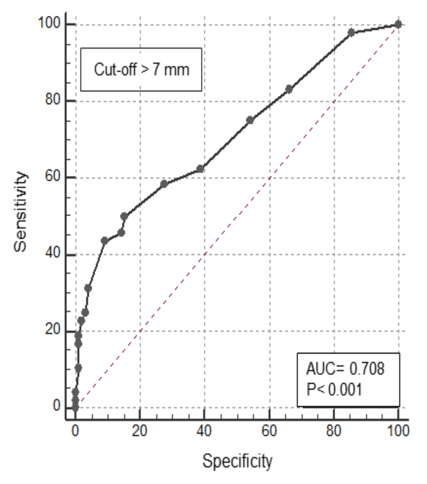
| Lesion’s size | 1.13 (1.14 to 1.21) | 0.0002 | 1.16 (1.06 to 1.3) | 0.0009 |
| Morphology (Parigi Classification) | ||||
| -Polypoid lesions (0-Is, 0-Isp, 0-Ip) | 2.93 (1.39 to 6.21) | 0.0037 | 7.4 (2.7 to 20.2) | 0.0001 |
| -No-polypoid lesions (0-IIa, 0-IIb-0-IIc, LST) | 0.34 (0.16 to 0.72) | 0.0047 | 0.13 (0.05 to 0.4) | 0.0001 |
| Lesion location | ||||
| -Cecum/right colon | 1.42 (0.68 to 2.94) | 0.35 | - | |
| -Transverse colon | 1.07 (0.46 to 2.47) | 0.86 | - | |
| -Left colon/sigmoid colon | 0.68 (0.25 to 1.81) | 0.44 | - | |
| -Rectum | 0.11 (0.01 to 0.92) | 0.04 | 0.19 (0.02 to 1.8) | 0.15 |
| Presence of mucosa inflammation around lesion | 4.55 (0.39 to 52.2) | 0.21 | - | |
| Lesion removal vs lesion biopsied | 1.29 (0.58 to 2.88) | 0.53 | - | |
| Sensitivity | Specificity | PPV | NPV | |
|---|---|---|---|---|
| Kudo III-IV | 79% | 80% | 58% | 92% |
| Lesion size > 7 mm | 46% | 79% | 53% | 75% |
| Polypoid Morphology | 67% | 61% | 41% | 81% |
| Kudo III-IV + lesion size > 7 mm | 67% | 90% | 87% | 72% |
| Kudo III-IV + Polypoid morphology | 87% | 85% | 75% | 91% |
| Polypoid morphology and lesion size > 7 mm | 71% | 95% | 83% | 91% |
| Kudo III-IV + polypoid lesions + lesion’s size > 7 mm | 90% | 100% | 100% | 96% |
Publisher’s Note: MDPI stays neutral with regard to jurisdictional claims in published maps and institutional affiliations. |
© 2022 by the authors. Licensee MDPI, Basel, Switzerland. This article is an open access article distributed under the terms and conditions of the Creative Commons Attribution (CC BY) license (https://creativecommons.org/licenses/by/4.0/).
Share and Cite
Lolli, E.; De Cristofaro, E.; Marafini, I.; Troncone, E.; Neri, B.; Zorzi, F.; Biancone, L.; Calabrese, E.; Monteleone, G. Endoscopic Predictors of Neoplastic Lesions in Inflammatory Bowel Diseases Patients Undergoing Chromoendoscopy. Cancers 2022, 14, 4426. https://doi.org/10.3390/cancers14184426
Lolli E, De Cristofaro E, Marafini I, Troncone E, Neri B, Zorzi F, Biancone L, Calabrese E, Monteleone G. Endoscopic Predictors of Neoplastic Lesions in Inflammatory Bowel Diseases Patients Undergoing Chromoendoscopy. Cancers. 2022; 14(18):4426. https://doi.org/10.3390/cancers14184426
Chicago/Turabian StyleLolli, Elisabetta, Elena De Cristofaro, Irene Marafini, Edoardo Troncone, Benedetto Neri, Francesca Zorzi, Livia Biancone, Emma Calabrese, and Giovanni Monteleone. 2022. "Endoscopic Predictors of Neoplastic Lesions in Inflammatory Bowel Diseases Patients Undergoing Chromoendoscopy" Cancers 14, no. 18: 4426. https://doi.org/10.3390/cancers14184426
APA StyleLolli, E., De Cristofaro, E., Marafini, I., Troncone, E., Neri, B., Zorzi, F., Biancone, L., Calabrese, E., & Monteleone, G. (2022). Endoscopic Predictors of Neoplastic Lesions in Inflammatory Bowel Diseases Patients Undergoing Chromoendoscopy. Cancers, 14(18), 4426. https://doi.org/10.3390/cancers14184426