Article

Risk of Diabetes Mellitus after Radiotherapy for Gastric Mucosa-Associated Lymphoid Tissue Lymphoma

1
Department of Radiation Oncology, Yonsei Cancer Center, Heavy Ion Therapy Research Institute, Yonsei University College of Medicine, Seoul 03722, Korea
2
Department of Radiation Oncology, Bundang CHA Medical Center, CHA University, Seongnam 13496, Korea
*
Author to whom correspondence should be addressed.
Cancers 2022, 14(17), 4110; https://doi.org/10.3390/cancers14174110
Submission received: 18 July 2022 / Revised: 19 August 2022 / Accepted: 23 August 2022 / Published: 25 August 2022
(This article belongs to the Special Issue Radiotherapy and New Biological Paradigms in Cancer Treatments)

Abstract

:

Simple Summary

In patients with gastric mucosa-associated lymphoid tissue lymphoma (GML), irradiation of the pancreas is inevitable owing to its anatomical proximity to the stomach. This is the first study to demonstrate the association between RT exposure and the risk of development of diabetes mellitus (DM) in patients with GML by measuring individual-based dose parameters of the pancreas. The findings of this study demonstrate the association between incidental pancreatic irradiation and DM. Obesity and sex were key risk factors for DM. Considering the risk of DM, the pancreas should no longer be neglected during RT planning, particularly in male patients and patients with obesity.

Abstract

The long-term effect of radiation on the pancreas in pediatric patients has been studied without individual radiation dosimetric data. This study investigated the effect of radiotherapy on the risk of developing diabetes mellitus (DM) in patients with gastric mucosa-associated lymphoid tissue lymphoma (GML), using individual radiation dosimetric analysis. Retrospective analysis reviewed the data of 225 patients without a history of DM receiving curative treatment for stage IE GML. Involved-site radiotherapy was delivered to the whole stomach in 83 patients. The pancreas was delineated in each patient’s computed tomography scan for dosimetric analysis. At a median follow-up of 49.0 months, the 5-year cumulative incidence of DM was 4.5%, 9.6%, and 1.6% in all patients, patients who received radiotherapy, and patients who did not receive radiotherapy, respectively (p = 0.009). Mean pancreatic dose (Dmean; p = 0.009), sex (p = 0.043), and body mass index (BMI; p = 0.008) were independently associated with DM. Using recursive partitioning analysis, patients were classified into low, intermediate, and high-risk groups, with 5-year DM incidence rates of 0.0%, 3.1%, and 15.6%, respectively (p < 0.001). Incidental irradiation of the pancreas can increase the risk of DM, which may be stratified according to patient sex and BMI.

1. Introduction

Gastric mucosa-associated lymphoid tissue lymphoma (GML) is an extranodal marginal zone lymphoma that arises in the stomach and exhibits excellent prognosis following treatment [1,2]. GML is related to Helicobacter pylori infection; hence, it can be treated through H. pylori eradication. In the absence of H. pylori infection or a lack of response after H. pylori eradication, involved-site radiotherapy (ISRT) targeting the entire stomach is the mainstay of treatment for GML. Despite the excellent prognosis, radiotherapy (RT)-related late toxicity has not been well described in these patients.
Diabetes mellitus (DM) can develop after RT as it can cause late parenchymal structural damage to the pancreas, resulting in a significant decrease in insulin secretion that is associated with glucose intolerance [3]. The Childhood Cancer Survivor Study showed that a dose of 10 Gy or more administered to the pancreas in pediatric patients significantly increased the risk of developing DM compared to that in patients irradiated with less than 10 Gy. Most patients were diagnosed with DM 20–30 years after RT [4]. However, few studies have analyzed the risk of developing DM when adult patients receive RT, and there is no suggestion of an RT dose constraint to the pancreas to prevent DM. Previous studies did not examine individual dose parameters but estimated the irradiated pancreatic dose using phantom-based models [4,5]. Thus, the actual dose delivered to the pancreas has not been evaluated accurately.
In patients with GML, irradiation of the pancreas is inevitable owing to its anatomical proximity to the stomach. In addition, cytotoxic chemotherapy is not a treatment option for localized GML, and thus it is an excellent model to describe the pure effect of RT on the pancreas. Only one study has reported on DM after RT in GML, but the number of patients analyzed was small, and dosimetric analysis using multiple dosimetric parameters was not performed [6]. In this study, we investigated the effect of RT on the development of DM with computed tomography (CT)-based individualized dosimetric analysis. Further, we defined risk groups according to radiation dose-volume parameters along with clinical factors.

2. Materials and Methods

2.1. Patient Selection

The study analyzed 267 patients who received ISRT and/or H. pylori eradication, with a curative aim, for histologically confirmed stage IE GML, between August 2007 and June 2020. All patients underwent fasting plasma glucose tests before and after treatment. Patients who were diagnosed with DM prior to receiving treatment (n = 32), those who underwent surgery and received chemotherapy (n = 3), or those who had double primary cancers (n = 7) were excluded from this study. Finally, 225 patients were retrospectively analyzed. This study was approved by the Severance Hospital institutional review board (No. 4–2021–1574), and the need for informed consent was waived due to the retrospective nature of the study.

2.2. Treatment of GML

Patients without H. pylori infection received upfront RT, although some received H. pylori eradication treatment prior to RT, at the physician’s discretion. Patients with H. pylori infection received RT if H. pylori eradication failed. All patients receiving RT underwent a planning CT on an empty stomach after fasting for at least 4 h. Respiratory movements were identified with four-dimensional CT (4D CT) [7]. Based on the International Lymphoma Radiation Oncology Group (ILROG) guideline, the internal target volume (ITV), clinical target volume (CTV), and planning target volume were defined as those including the range of movement of the entire stomach, a 1.0 cm margin from the ITV, and a 0.5 cm margin from the CTV, respectively [8].

2.3. Dosimetric Evaluation

The pancreas was contoured on each patient’s simulated CT image, using MIM software (MIM Software Inc., Cleveland, OH, USA), to measure the dosimetric parameters of the pancreas. The following dosimetric parameters were measured: the mean dose in gray irradiated to the whole pancreas (Dmean), the dose in gray that covered 0.03 cc of the whole pancreas (D0.03 cc), the minimum dose in gray irradiated to the whole pancreas (Dmin), and the percentage of pancreas volume receiving at least the indicated dose (VX Gy).

2.4. Outcome Assessment

DM was diagnosed if any of the diagnostic criteria were met on DM tests performed after treatment: fasting plasma glucose level ≥ 126 mg/dL, 2-h plasma glucose level ≥ 200 mg/dL during a 75 g oral glucose tolerance test, HbA1c level ≥ 6.5%, or random plasma glucose level ≥ 200 mg/dL with signs or symptoms of DM [9]. After treatment for GML, patients were followed up clinically every 3 months for the first year, every 6 months for two years, and once a year thereafter. Fasting plasma glucose level was measured at each follow up. The HbA1c levels were often evaluated for patients with plasma glucose levels ≥ 126 mg/dL. Patients with a plasma glucose level ≥ 126 mg/dL with a decreased plasma glucose level at the subsequent follow-up without treatment were not categorized as having a DM event.

2.5. Statistical Analysis

The baseline characteristics of patients treated with or without RT were compared using the Pearson χ2 test for categorical data and independent t-test for continuous data. Pearson’s correlation analysis and the Mann–Whitney U test were performed to analyze the correlation between RT-related factors and dosimetric parameters. The time to DM was defined as the interval from initiation of treatment to the date of first diagnosis of DM. The cumulative incidence of DM was estimated by competing risk analysis. Univariate and multivariate competing risk analyses were performed to ascertain the risk factors related to the development of DM. As the dosimetric variables could cross-correlate, only those dosimetric parameters with the lowest p-value in univariate competing risk analysis were included in the multivariate analysis. For the selected dosimetric parameter, maximally selected rank statistics were used to obtain the optimal cutoff value. Among clinical factors, variables with p < 0.05 in univariate competing risk analysis were included in the multivariate competing risk analysis. Recursive partitioning analysis (RPA) was also performed to stratify patients according to their risk of developing DM. All clinical and dosimetric variables were included for RPA. The cumulative incidence of DM according to the class stratified through RPA was compared using Gray’s test. Overall survival and cancer-specific survival for all patients were analyzed using the Kaplan–Meier curve.
Statistical significance was set at p-value of < 0.05. The cmprsk, maxstat, and rpart packages were used for the competing risks analyses, cutoff analysis, and RPA, respectively. All statistical analyses were performed using R (version 3.5.3; R Foundation for Statistical Computing, Vienna, Austria).

3. Results

Of the 225 patients (median age, 55 [range 31–86] years), 138 (61.3%) were female, 66 (29.3%) were obese (body mass index [BMI] ≥ 25.0 kg/m2) [10], and 22 (9.8%) had underlying hypertension. Overall, 102 (45.3%) patients were infected with H. pylori and 203 received H. pylori eradication treatment. Patients who received RT had significantly less H. pylori infection and eradication than those who did not (26.5% vs. 56.3% and 63.5% vs. 100.0%, respectively; both p < 0.001). In addition, although not statistically significant, there was a higher proportion of males in the group receiving RT than in the group that did not (47.0% vs. 33.8%, p = 0.050). However, the baseline median pancreas volume was similar between the two groups (64.9 cc vs. 65.8 cc, p = 0.409). The baseline characteristics of the patients are summarized in Table 1.
The ISRT was delivered to 83 (36.9%) patients at a median total dose of 30.0 Gy (range, 21.6–39.6 Gy) and median fractional dose of 1.5 Gy (range, 1.5–2.0 Gy). Of the 83 patients who received ISRT, 56 (67.5%) and 27 (32.5%) received three-dimensional conformal RT (3D-CRT) and intensity-modulated RT (IMRT), respectively.
Among patients receiving RT, the median values for dosimetric parameters to the pancreas were as follows: Dmean 27.3 Gy (range 16.6–35.9 Gy), D0.03 cc 31.3 Gy (range 23.9–40.4 Gy), Dmin 5.8 Gy (range 0.0–34.9 Gy), V5 Gy 100% (range 73.2–100.0%), V10 Gy 98.6% (range 62.9–100.0%), V15 Gy 96.4% (range 53.0–100.0%), V20 Gy 91.9% (range 47.3–100.0%) and, V25 Gy 85.5% (range 0.0–100.0%). The prescribed dose showed a significant correlation with Dmean (Pearson correlation coefficient 0.415, p < 0.001; Supplementary Figure S1). Dmean and V25 Gy were significantly lower in patients receiving IMRT than those receiving 3D-CRT (Table 2).
After a median follow-up of 49.0 months (range, 3.0–155.3), nine new patients were diagnosed with DM after the median 20.3 (range, 3.0–37.9) months of treatment. The medians were 20.3 and 20.6 months for patients who received or did not receive RT, respectively. Among the nine patients diagnosed with DM, seven had received RT while two had not. The 5-year cumulative incidence of DM was 4.5% (95% confidence interval [CI] 2.4–8.7%), 9.6% (95% CI 4.7–19.6%), and 1.6% (95% CI 0.4–6.3%) in all patients, patients who received RT, and patients who did not receive RT, respectively (p = 0.009; Figure 1).
Univariate competing risk analysis identified the dosimetric parameter most predictive of DM. The Dmean (hazard ratio [HR] 1.06, 95% CI 1.01–1.11, p = 0.015) exhibited the highest level of significance among the multiple parameters (Table 3). As a result of the maximally selected rank statistics, the optimal cutoff value of Dmean was 21.0 Gy. The Dmean ≥ 21.0 Gy was significantly associated with the risk of developing DM (HR 6.86, 95% CI 1.44–32.64, p = 0.016). Among clinical factors, sex (HR 0.18, 95% CI 0.04–0.84, p = 0.029) and BMI (HR 4.96, 95% CI 1.26–19.50, p = 0.022) were significantly associated with DM. Multivariate competing risk analysis demonstrated Dmean ≥ 21.0 Gy (HR 7.30, 95% CI 1.65–32.30, p = 0.009), BMI ≥ 25.0 kg/m2 (HR 5.76, 95% CI 1.56–21.17, p = 0.008), and male sex (HR 4.85, 95% CI 1.05–22.34, p = 0.043; Table 4) as significant risk factors for the development of DM.
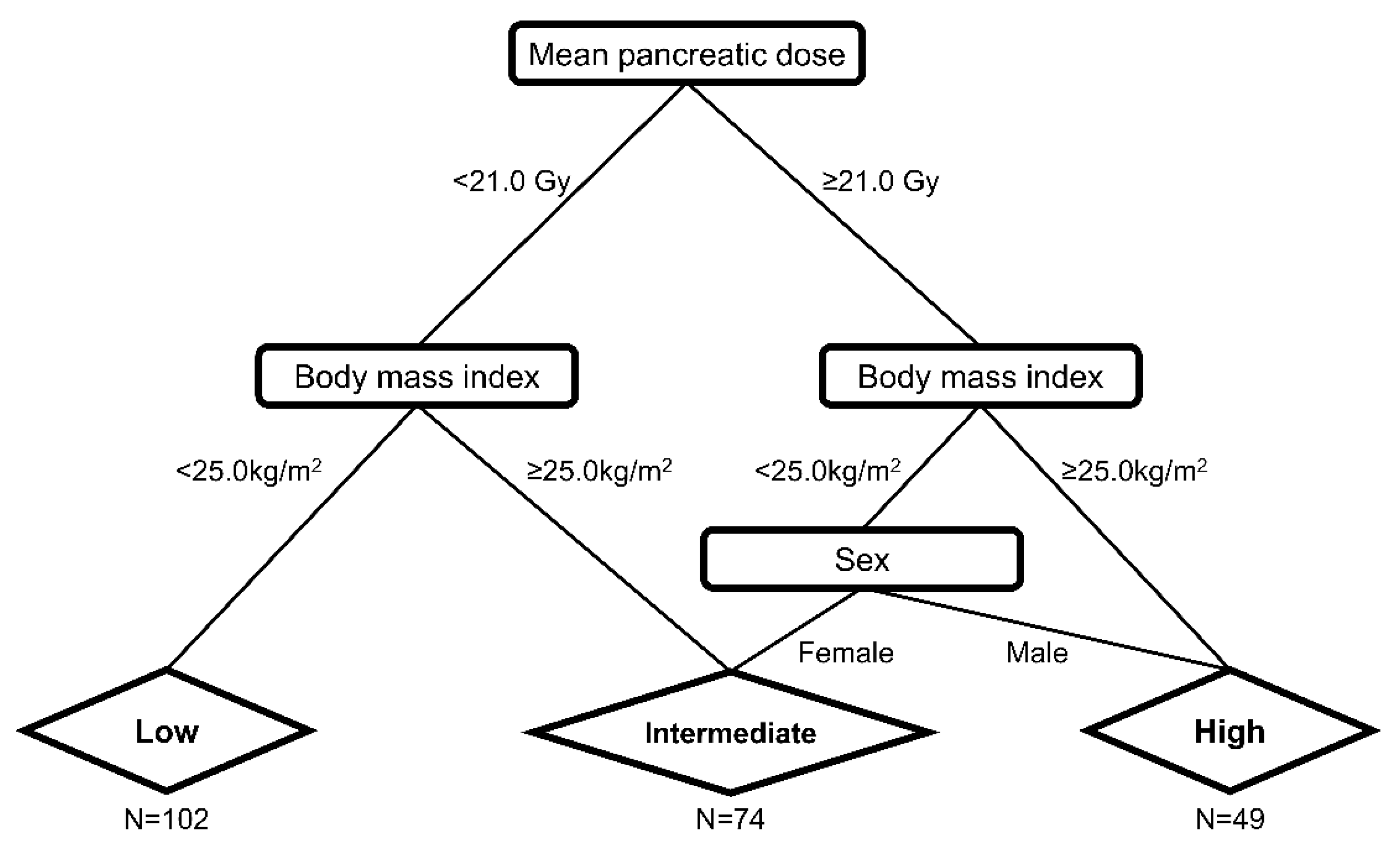
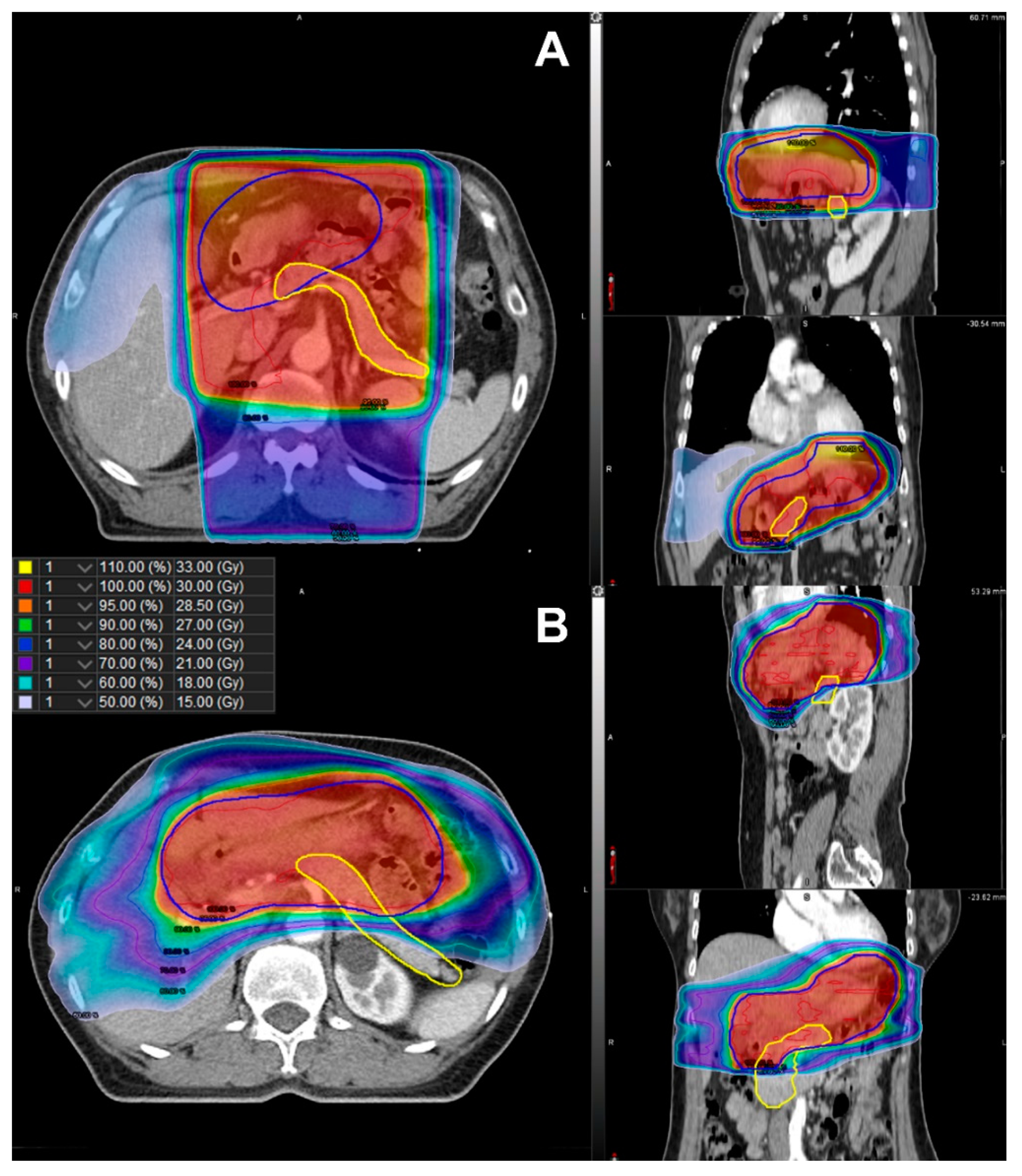
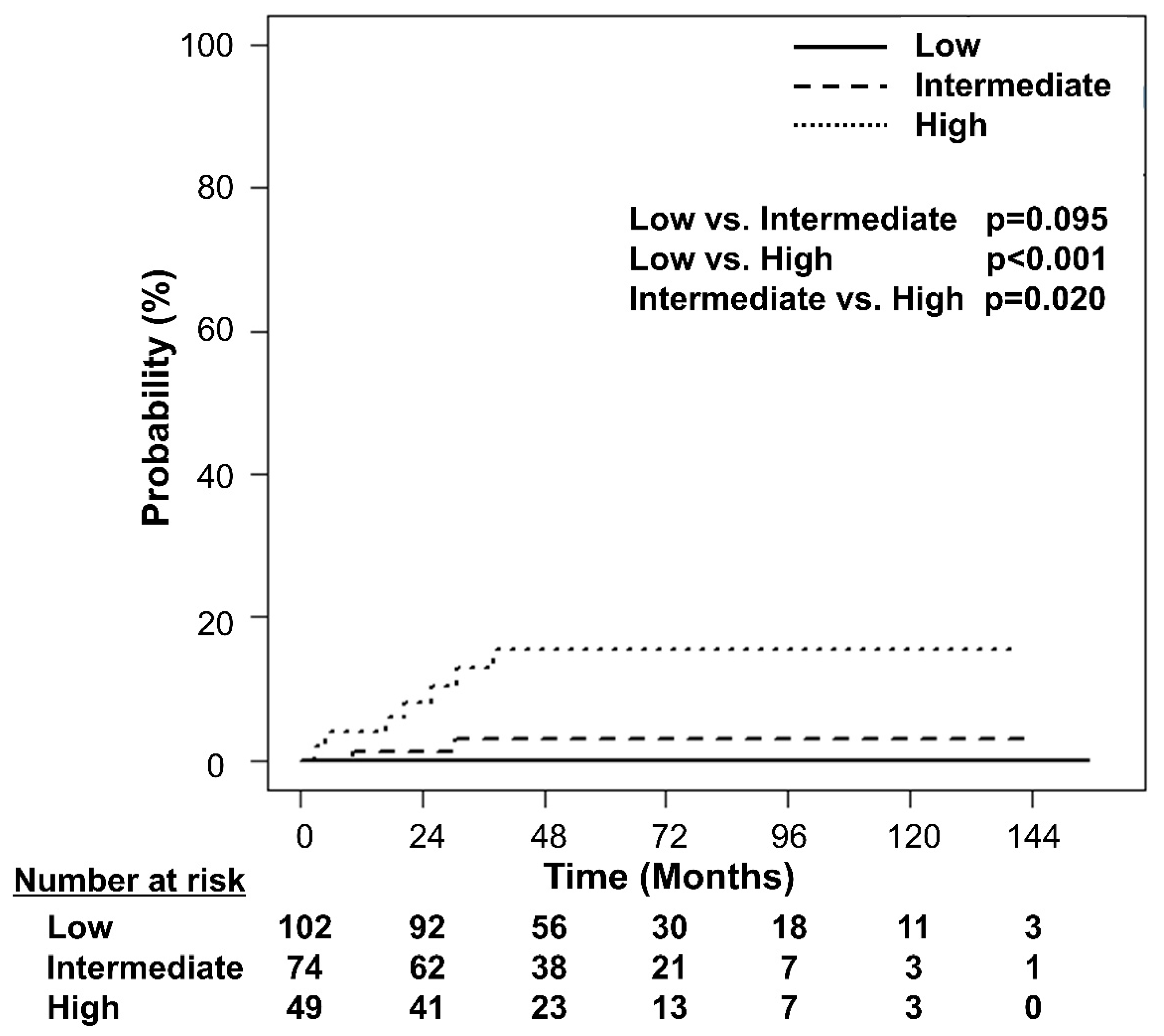
Further, RPA stratified the patients according to their risk of developing DM (Figure 2). Among the clinical factors and dosimetric parameters, patients were initially split between Dmean < 21.0 Gy and Dmean ≥ 21.0 Gy. Representative isodose distributions for a patient with Dmean ≥ 21.0 Gy using 3D-CRT and a patient with Dmean < 21.0 Gy using IMRT are presented in Figure 3. The split groups were further divided into BMI < 25.0 kg/m2 and BMI ≥ 25.0 kg/m2. The group with Dmean ≥ 21.0 Gy and BMI < 25.0 kg/m2 was once again stratified by sex. The patients with Dmean < 21.0 Gy and BMI < 25.0 kg/m2 were defined as the low-risk group (n = 102; Figure 2). Among the patients with Dmean ≥ 21.0 Gy, those with BMI ≥ 25.0 kg/m2 or male patients with BMI <25.0 kg/m2 were defined as high-risk (n = 49; Figure 2). Those patients who were not included in either the low- or high-risk groups were defined as the intermediate-risk group (n = 74; Figure 2). The 5-year cumulative incidence of DM was 0.0% (95% CI 0.0–0.0%), 3.1% (95% CI 0.8–12.3%), and 15.6% (95% CI 7.8–31.3%) in the low-, intermediate-, and high-risk groups, respectively (Figure 4, p < 0.001). The cumulative incidence of DM in the low- and intermediate-risk groups was significantly lower than that in the high-risk group (low vs. high, p < 0.001; intermediate vs. high, p = 0.020), while the difference between the low- and intermediate-risk groups was not statistically significant (p = 0.095).
The 5-year overall and cancer-specific survival rates were 98.9% (95% CI 97.3–100.0%) and 100.0% (95% CI 100.0–100.0), respectively (Supplementary Figure S2). Overall, two patients died during the follow-up period. One died of pancreatic cancer, and the other died of an unknown cause. In both patients, the GML was in complete remission.

4. Discussion

This is the first study to demonstrate the association between RT exposure and the risk of development of DM in patients with GML by measuring individual-based dose parameters of the pancreas. Patients with a mean dose ≥ 21.0 Gy irradiated to the pancreas had a 7.3-fold increased risk of developing DM, after adjusting for well-known clinical risk factors such as obesity and male sex [11,12]. Our finding implies the necessity of including the pancreas as an organ at risk and consider dosimetric constraints in the planning process. We also stratified the patients into three risk groups according to pancreatic RT dose, obesity, and sex. Patients with a Dmean ≥ 21.0 Gy with obesity or male sex had the highest risk of DM following treatment.
Radiation-induced DM was first reported in pediatric patients who underwent abdominal RT for Wilms’ tumor, with 8 of 121 patients developing DM [13]. Later, the Childhood Cancer Survivor Study that retrospectively analyzed cancer treatment-related DM in 8599 pediatric cancer survivors, reported that RT and chemotherapy were risk factors associated with the development of DM [14]. More recently, an analysis of 2520 survivors treated for solid cancers or lymphoma diagnosed during childhood provided the first proof of the dose-response relationship between the dose irradiated to the pancreas and the risk of developing DM [4]. However, DM development was confirmed only through a questionnaire, and the radiation dose was estimated based on phantom modeling rather than the actual dose irradiated. The recent Childhood Cancer Survivor Study demonstrated that BMI and pancreatic dose were significant risk factors for DM [5]. However, determination of the radiation dose was not based on individual CT-based calculations in this study either.
Among studies related to radiation-induced DM, relatively few have analyzed adult patients. A retrospective cohort study included adolescent and adult patients with Hodgkin’s lymphoma in whom treatment was initiated before the age of 51 years [15]. From the analysis, the risk of DM increased as the pancreatic dose increased, and there was a significant difference between a dose ≥ 36.0 Gy compared to a lower dose. At the same institution, a retrospective cohort analysis was performed on 2998 one-year survivors of testicular cancer diagnosed before the age of 50 years, with orchiectomy, and 161 patients were diagnosed with DM [16]. The risk of DM was 1.66 times higher in patients who received RT to the para-aortic lymph nodes, and the higher the prescribed dose, the higher the risk of DM. The aforementioned studies on Hodgkin lymphoma and testicular cancer had a major limitation: it was difficult to investigate the independent effect of RT due to the confounding effect of chemotherapy. However, in localized GML, cytotoxic chemotherapy is not routinely administered, and the direct effect of RT can be investigated.
The volume and location of the pancreas differ between patients. Therefore, it is essential to delineate the pancreas and calculate the actual dose irradiated to the pancreas for each patient. In previous studies on radiation toxicity, approximate estimation was used in absence of CT-based planning and accurate dosimetric data for each patient. Due to this particular limitation, it was impossible to ascertain the specific dose constraint for toxicity for a specific organ. However, recent emergence of individual dosimetry in the study of cardiac toxicity after RT in patients with lung or breast cancer, using individual-based dose parameters measured by contouring each patient’s heart, has allowed dosimetric parameters related to cardiac toxicity to be more accurately analyzed [17,18]. In line with this trend, we retrospectively contoured the pancreas for each patient and evaluated the dose-volume parameters for the contoured pancreas in each patient.
As a result of RPA and maximally selected rank statistics using individual dosimetry in our study, the cutoff value of Dmean was 21.0 Gy. The Dmean of the pancreas could be more easily controlled using IMRT than with 3D-CRT. In our study, we found that patients who received IMRT had a significantly lower Dmean compared to those who received 3D-CRT, although the pancreas was not considered an organ at risk during planning. However, sparing the pancreas could be challenging even with IMRT due to proximity of the pancreas to the stomach, and the target coverage or prescription dose may need to be reduced. Indeed, we found a significant correlation between the prescribed dose and the pancreas Dmean. Current guidelines recommend 30.0 Gy in 20 fractions to the whole stomach, and the recurrence rates of GML with this radiation dose are <10% [8,19,20]. However, the prescribed dose of 30.0 Gy is based on expert consensus, rather than level 1 evidence. Two phase III trials demonstrated excellent local control rates with 24.0 Gy in indolent lymphoma [21,22]. With respect to GML, several retrospective studies have demonstrated high complete response rates and disease control rates with a low prescribed total dose of 23.5–24.0 Gy [7,23]. Moreover, there are two single arm trials investigating the treatment outcome with a prescription dose of 20.0 Gy (NCT04097067) and an ultra low-dose of 4.0 Gy (NCT03680586) [24,25]. Considering the risk stratification results of our study and recent trends in the prescribed dose for lymphoma, considering a dose lower than 30.0 Gy using IMRT may significantly reduce the risk of DM while maintaining the therapeutic effect.
One of the limitations of this study is the limited number of patients compared to those in other radiation-induced DM studies. However, the strength of our study lies in the largest number of patients analyzed among studies investigating radiation-induced DM in GML and that individual-based dose parameters of the pancreas were measured and analyzed for each patient. Moreover, our results were not externally validated. Another limitation of this study is that the number of events of DM after treatment were limited. A possible reason could be that the diagnosis of DM may be underestimated owing to the retrospective nature of this study. Considering the fact that the incidence of DM continually increases beyond 5 years [15,16], the follow-up period of our study was relatively short. Further study with a larger cohort and longer follow-up is needed to confirm the findings of the present study.

5. Conclusions

In conclusion, the findings of this study demonstrate the association between incidental pancreatic irradiation and DM. Considering the risk of DM, the pancreas should no longer be neglected and should be considered as an organ at risk with dose constraints during RT planning, particularly in male patients and patients with obesity. However, the results are hypothesis generating and should be confirmed using robust studies in the future.

Supplementary Materials

The following supporting information can be downloaded at: https://www.mdpi.com/article/10.3390/cancers14174110/s1, Figure S1: A scatterplot and correlation analysis results for the prescribed dose and mean dose irradiated to the whole pancreas. Figure S2: Overall survival (OS) and cancer-specific survival (CSS) for all patients.

Author Contributions

K.H.K. conceived and designed the study. J.L. and K.H.K. performed the study. J.L., J.K. and K.H.K. analyzed the data. J.L., H.I.Y., J.C. and C.-O.S. contributed materials and analysis tools. J.L. and K.H.K. prepared all figures and tables and composed the main manuscript. All authors have read and agreed to the published version of the manuscript.

Funding

This study was supported by a new faculty research seed money grant of Yonsei University College of Medicine for 2022 (2022–32–0075) and National Research Foundation (NRF) grant (NRF-2021R1I1A1A01044024).

Institutional Review Board Statement

This study was approved by the Severance Hospital institutional review board (No. 4–2021–1574), and the requirement for informed consent was waived because of its retrospective nature.

Data Availability Statement

The raw data supporting the conclusions of this article will be made available by the authors, without undue reservation.

Conflicts of Interest

The authors declare that the research was conducted in the absence of any commercial or financial relationships that could be construed as a potential conflict of interest.

References

  1. Jaffe, E.S. The 2008 WHO classification of lymphomas: Implications for clinical practice and translational research. ASH Educ. Program Book 2009, 2009, 523–531. [Google Scholar] [CrossRef]
  2. Tsang, R.W.; Gospodarowicz, M.K.; Pintilie, M.; Wells, W.; Hodgson, D.C.; Sun, A.; Crump, M.; Patterson, B.J. Localized mucosa-associated lymphoid tissue lymphoma treated with radiation therapy has excellent clinical outcome. J. Clin. Oncol. 2003, 21, 4157–4164. [Google Scholar] [CrossRef] [PubMed]
  3. Du Toit, D.F.; Heydenrych, J.J.; Smit, B.; Zuurmond, T.; Louw, G.; Laker, L.; Els, D.; Weideman, A.; Wolfe-Coote, S.; Du Toit, L.B.; et al. The effect of ionizing radiation on the primate pancreas: An endocrine and morphologic study. J. Surg. Oncol. 1987, 34, 43–52. [Google Scholar] [CrossRef] [PubMed]
  4. De Vathaire, F.; El-Fayech, C.; Ben Ayed, F.F.; Haddy, N.; Guibout, C.; Winter, D.; Thomas-Teinturier, C.; Veres, C.; Jackson, A.; Pacquement, H.; et al. Radiation dose to the pancreas and risk of diabetes mellitus in childhood cancer survivors: A retrospective cohort study. Lancet Oncol. 2012, 13, 1002–1010. [Google Scholar] [CrossRef]
  5. Friedman, D.N.; Moskowitz, C.S.; Hilden, P.; Howell, R.M.; Weathers, R.E.; Smith, S.A.; Wolden, S.L.; Tonorezos, E.S.; Mostoufi-Moab, S.; Chow, E.J.; et al. Radiation Dose and Volume to the Pancreas and Subsequent Risk of Diabetes Mellitus: A Report from the Childhood Cancer Survivor Study. J. Natl. Cancer Inst. 2020, 112, 525–532. [Google Scholar] [CrossRef] [PubMed]
  6. Baek, J.Y.; Lim, D.H.; Oh, D.; Nam, H.; Kim, J.J.; Lee, J.H.; Min, B.-H.; Lee, H. Increased Risk of Diabetes after Definitive Radiotherapy in Patients with Indolent Gastroduodenal Lymphoma. Cancer Res. Treat. 2022, 54, 294–300. [Google Scholar] [CrossRef]
  7. Pinnix, C.C.; Gunther, J.R.; Milgrom, S.A.; Cruz-Chamorro, R.J.; Medeiros, L.J.; Khoury, J.D.; Amini, B.; Neelapu, S.; Lee, H.J.; Westin, J.; et al. Outcomes after Reduced-Dose Intensity Modulated Radiation Therapy for Gastric Mucosa-Associated Lymphoid Tissue (MALT) Lymphoma. Int. J. Radiat. Oncol. Biol. Phys. 2019, 104, 447–455. [Google Scholar] [CrossRef]
  8. Yahalom, J.; Illidge, T.; Specht, L.; Hoppe, R.T.; Li, Y.-X.; Tsang, R.; Wirth, A. Modern radiation therapy for extranodal lymphomas: Field and dose guidelines from the International Lymphoma Radiation Oncology Group. Int. J. Radiat. Oncol. Biol. Phys. 2015, 92, 11–31. [Google Scholar] [CrossRef]
  9. American Diabetes Association. Classification and diagnosis of diabetes: Standards of Medical Care in Diabetes—2021. Diabetes Care 2021, 44, S15–S33. [Google Scholar] [CrossRef]
  10. World Health Organization. Obesity: Preventing and managing the global epidemic. Report of a WHO consultation. World Health Organ. Tech. Rep. Ser. 2000, 894, 1–253. [Google Scholar]
  11. Lee, D.-C.; Park, I.; Jun, T.-W.; Nam, B.-H.; Cho, S.-I.; Blair, S.-N.; Kim, Y.-S. Physical activity and body mass index and their associations with the development of type 2 diabetes in korean men. Am. J. Epidemiol. 2012, 176, 43–51. [Google Scholar] [CrossRef] [PubMed] [Green Version]
  12. Oh, S.-H.; Ku, H.; Park, K.S. Prevalence and socioeconomic burden of diabetes mellitus in South Korean adults: A population-based study using administrative data. BMC Public Health 2021, 21, 548. [Google Scholar] [CrossRef]
  13. Teinturier, C.; Tournade, M.-F.; Caillat-Zucman, S.; Boitard, C.; Amoura, Z.; Bougneres, P.F.; Timsit, J. Diabetes mellitus after abdominal radiation therapy. Lancet 1995, 346, 633–634. [Google Scholar] [CrossRef]
  14. Meacham, L.R.; Sklar, C.A.; Li, S.; Liu, Q.; Gimpel, N.; Yasui, Y.; Whitton, J.A.; Stovall, M.; Robison, L.L.; Oeffinger, K.C. Diabetes mellitus in long-term survivors of childhood cancer. Increased risk associated with radiation therapy: A report for the childhood cancer survivor study. Arch. Intern. Med. 2009, 169, 1381–1388. [Google Scholar] [CrossRef] [PubMed]
  15. Van Nimwegen, F.A.; Schaapveld, M.; Janus, C.P.; Krol, A.D.; Raemaekers, J.M.; Kremer, L.C.; Stovall, M.; Aleman, B.M.; van Leeuwen, F.E. Risk of diabetes mellitus in long-term survivors of Hodgkin lymphoma. J. Clin. Oncol. 2014, 32, 3257–3263. [Google Scholar] [CrossRef]
  16. Groot, H.J.; Gietema, J.A.; Aleman, B.M.P.; Incrocci, L.; de Wit, R.; Witjes, J.A.; Groenewegen, G.; de Brouwer, P.; Meijer, O.W.M.; Hulshof, M.C.C.M.; et al. Risk of diabetes after para-aortic radiation for testicular cancer. Br. J. Cancer 2018, 119, 901–907. [Google Scholar] [CrossRef]
  17. Atkins, K.M.; Rawal, B.; Chaunzwa, T.L.; Lamba, N.; Bitterman, D.S.; Williams, C.L.; Kozono, D.E.; Baldini, E.H.; Chen, A.B.; Nguyen, P.L.; et al. Cardiac Radiation Dose, Cardiac Disease, and Mortality in Patients with Lung Cancer. J. Am. Coll. Cardiol. 2019, 73, 2976–2987. [Google Scholar] [CrossRef]
  18. Darby, S.C.; Ewertz, M.; McGale, P.; Bennet, A.M.; Blom-Goldman, U.; Brønnum, D.; Correa, C.; Cutter, D.; Gagliardi, G.; Gigante, B.; et al. Risk of ischemic heart disease in women after radiotherapy for breast cancer. N. Engl. J. Med. 2013, 368, 987–998. [Google Scholar] [CrossRef] [PubMed]
  19. Wirth, A.; Gospodarowicz, M.; Aleman, B.M.P.; Bressel, M.; Ng, A.; Chao, M.; Hoppe, R.T.; Thieblemont, C.; Tsang, R.; Moser, L.; et al. Long-term outcome for gastric marginal zone lymphoma treated with radiotherapy: A retrospective, multi-centre, International Extranodal Lymphoma Study Group study. Ann. Oncol. 2013, 24, 1344–1351. [Google Scholar] [CrossRef]
  20. Goda, J.S.; Gospodarowicz, M.; Pintilie, M.; Wells, W.; Hodgson, D.C.; Sun, A.; Crump, M.; Tsang, R.W. Long-term outcome in localized extranodal mucosa-associated lymphoid tissue lymphomas treated with radiotherapy. Cancer 2010, 116, 3815–3824. [Google Scholar] [CrossRef]
  21. Lowry, L.; Smith, P.; Qian, W.; Falk, S.; Benstead, K.; Illidge, T.; Linch, D.; Robinson, M.; Jack, A.; Hoskin, P. Reduced dose radiotherapy for local control in non-Hodgkin lymphoma: A randomized phase III trial. Radiother. Oncol. 2011, 100, 86–92. [Google Scholar] [CrossRef] [PubMed]
  22. Hoskin, P.J.; Kirkwood, A.A.; Popova, B.; Smith, P.; Robinson, M.; Gallop-Evans, E.; Coltart, S.; Illidge, T.; Madhavan, K.; Brammer, C.; et al. 4 Gy versus 24 Gy radiotherapy for patients with indolent lymphoma (FORT): A randomised phase 3 non-inferiority trial. Lancet Oncol. 2014, 15, 457–463. [Google Scholar] [CrossRef]
  23. Saifi, O.; Lester, S.C.; Rule, W.; Stish, B.J.; Stafford, S.; Pafundi, D.H.; Jiang, L.; Menke, D.; Moustafa, M.A.; Rosenthal, A.; et al. Comparable Efficacy of Reduced Dose Radiation Therapy for the Treatment of Early Stage Gastric Extranodal Marginal Zone Lymphoma of Mucosa-Associated Lymphoid Tissue. Adv. Radiat. Oncol. 2021, 6, 100714. [Google Scholar] [CrossRef] [PubMed]
  24. Rehn, S.; Reinartz, G.; Oertel, M.; Hering, D.; Pott, C.; Greve, B.; Eich, H. Phase II Trial to assess the Efficacy of Low Radiation Dose of 20 Gy for the Treatment of Marginal Zone Lymphoma or Follicular Lymphoma Stage I-II localized in the Stomach or the Duodenum. Strahlenther. Onkol. 2021, 197, S170–S171. [Google Scholar]
  25. US National Library of Medicine. ClinicalTrials.gov. Available online: https://clinicaltrials.gov/ct2/show/NCT03680586?cond=lymphoma+gastric&draw=2&rank=8 (accessed on 23 March 2021).
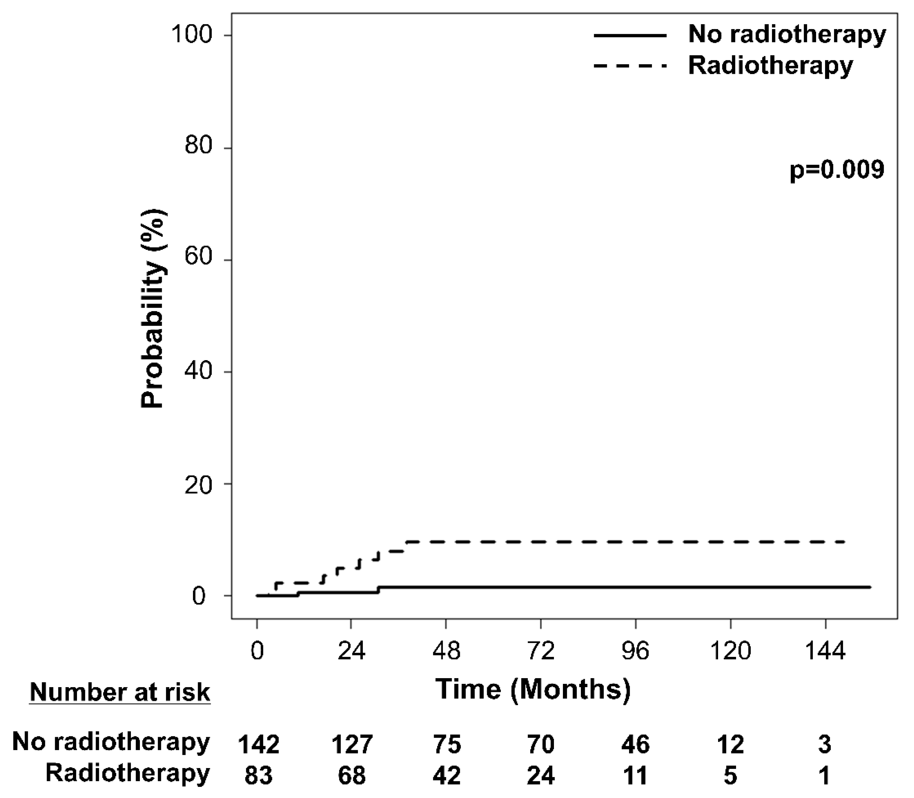
Figure 1. Cumulative incidence of the development of diabetes mellitus according to radiotherapy status.
Cancers 14 04110 g001
Figure 2. Diabetes mellitus risk prediction groups defined by recursive partitioning analysis in patients with gastric mucosa-associated lymphoid tissue lymphoma. The patients with Dmean < 21.0 Gy and BMI < 25.0 kg/m2 were defined as the low-risk group (n = 102). Among the patients with Dmean ≥ 21.0 Gy, those with BMI ≥ 25.0 kg/m2 or male patients with BMI < 25.0 kg/m2 were defined as having high risk (n = 49). The remaining patients not included in either the low- or high-risk groups were defined as being in the intermediate-risk group (n = 74). Dmean, mean dose in gray irradiated to the whole pancreas; BMI, body mass index.
Cancers 14 04110 g002
Figure 3. Comparison of isodose distribution of the planning target volume (blue line) and the pancreas (yellow line) in (A) three-dimensional conformal radiotherapy with Dmean ≥ 21.0 Gy and (B) intensity-modulated radiotherapy with Dmean < 21.0 Gy. Dmean, mean dose in gray irradiated to the whole pancreas.
Cancers 14 04110 g003
Figure 4. Cumulative incidence curves of the development of diabetes mellitus according to the risk groups defined by recursive partitioning analysis.
Cancers 14 04110 g004
Table 1. Baseline characteristics of all the patients and patients treated with or without radiotherapy.
CharacteristicsTotalRTNo RT
N = 225%N = 83%N = 142%p-Value
Age, years (median [range])55 (31–86)57 (34–86)55 (31–77)0.219
Sex 0.050
Female13861.34453.09466.2
Male8738.73947.04833.8
Body mass index 0.310
<25.0 kg/m215970.76274.79768.3
≥25.0 kg/m26629.32125.34531.7
Hypertension 0.180
No20390.27286.713192.3
Yes229.81113.3117.7
Helicobacter pylori infection <0.001
No12354.76173.56243.7
Yes10245.32226.58056.3
Helicobacter pylori eradication <0.001
No229.82226.500.0
Yes20390.26173.5142100.0
Pancreas volume, cc (median [range])65.7 (28.5–125.8)64.9 (28.5–114.3)65.8 (28.9–125.8)0.409
RT, radiotherapy.
Table 2. Dosimetric comparison between three-dimensional conformal radiotherapy and intensity-modulated radiotherapy.
3D-CRTIMRT
Median (Range)Median (Range)p-Value
Dmean (Gy)28.8 (16.6–35.9)26.1 (20.7–30.8)0.009
D0.03cc (Gy)31.2 (24.3–40.4)31.4 (23.9–32.8)0.463
Dmin (Gy)5.8 (0.0–34.9)5.6 (0.8–28.7)0.484
V5 Gy (%)100.0 (73.2–100.0)100.0 (82.8–100.0)0.802
V10 Gy (%)98.7 (62.9–100.0)98.5 (72.4–100.0)0.903
V15 Gy (%)96.3 (53.0–100.0)96.4 (65.8–100.0)0.497
V20 Gy (%)94.1 (47.3–100.0)88.2 (62.3–100.0)0.090
V25 Gy (%)90.9 (0.0–100.0)74.7 (0.0–100.0)<0.001
3D-CRT, three-dimensional conformal radiotherapy; IMRT, intensity-modulated radiotherapy; Dmean, mean dose in gray irradiated to the whole pancreas; D0.03 cc, the dose in gray that covers 0.03 cc of the whole pancreas; Dmin, minimum dose in gray irradiated to the whole pancreas; VX Gy, the pancreatic volume in percentage that is covered by a dose not <X Gy.
Table 3. Univariate competing risk analysis of dosimetric parameters of pancreas for predicting the development of diabetes mellitus.
Univariate Analysis
HR (95% CI)p-Value
Dmean1.06 (1.01–1.11)0.015
D0.03cc1.06 (1.01–1.10)0.016
Dmin1.06 (1.01–1.11)0.023
V5 Gy1.02 (1.00–1.04)0.021
V10 Gy1.02 (1.00–1.03)0.020
V15 Gy1.02 (1.00–1.03)0.018
V20 Gy1.02 (1.00–1.03)0.016
V25 Gy1.01 (1.00–1.03)0.049
All dosimetric parameters were treated as continuous variables. HR, hazard ratio; CI, confidence interval; Dmean, mean dose in gray irradiated to the whole pancreas; D0.03 cc, the dose in gray that covers 0.03 cc of the whole pancreas; Dmin, minimum dose in gray irradiated to the whole pancreas; VX Gy, the pancreatic volume in percentage that is covered by a dose not <X Gy.
Table 4. Univariate and multivariate competing risk analyses for predicting the development of diabetes mellitus.
Univariate AnalysisMultivariate Analysis
HR (95% CI)p-ValueHR (95% CI)p-Value
Age *1.05 (1.00–1.10)0.058
Sex
Female1 (Reference) 1 (Reference)
Male5.66 (1.20–26.76)0.029 4.85 (1.05–22.34)0.043
Body mass index
<25.0 kg/m21 (Reference) 1 (Reference)
≥25.0 kg/m24.96 (1.26–19.50)0.022 5.76 (1.56–21.17)0.008
Hypertension
No1 (Reference)
Yes2.74 (0.56–13.32)0.210
Helicobacter pylori infection
No1 (Reference)
Yes0.34 (0.07–1.63)0.180
Pancreas volume *1.03 (0.99–1.07)0.220
Dmean
<21.0 Gy1 (Reference) 1 (Reference)
≥21.0 Gy6.86 (1.44–32.64)0.016 7.30 (1.65-32.30)0.009
* Age and pancreas volume were treated as continuous variables. HR, hazard ratio; CI, confidence interval; Dmean, mean dose in gray irradiated to the whole pancreas.
Publisher’s Note: MDPI stays neutral with regard to jurisdictional claims in published maps and institutional affiliations.