Article

Tumor Infiltrating Lymphocytes in Multi-National Cohorts of Ductal Carcinoma In Situ (DCIS) of Breast

1
Department of Pathology and Laboratory Medicine, Emory University School of Medicine, Atlanta, GA 30322, USA
2
Winship Cancer Institute, Atlanta, GA 30322, USA
3
GE Global Research Center, Niskayuna, NY 12309, USA
4
Center for Computational Biology and Bioinformatics, Department of Biostatistics and Health Data Science, School of Medicine, Indiana University, Indianapolis, IN 46202, USA
5
Anatomical Pathology, Singapore General Hospital, Singapore 169856, Singapore
6
Division of Pathology, Singapore General Hospital, Singapore 169856, Singapore
7
Department of Laboratory Medicine and Pathology, Mayo Clinic Arizona, Phoenix, AZ 85054, USA
8
Department of Medical, Surgery and Health Sciences, University of Trieste, 34127 Trieste, Italy
9
Cancer and Haematology Centre, Department of Oncology, Oxford University, Oxford OX3 7LE, UK
*
Authors to whom correspondence should be addressed.
Cancers 2022, 14(16), 3916; https://doi.org/10.3390/cancers14163916
Submission received: 6 May 2022 / Revised: 8 August 2022 / Accepted: 9 August 2022 / Published: 13 August 2022
(This article belongs to the Collection Breast Cancer: From Pathophysiology to Prevention and Treatment)

Abstract

:

Simple Summary

Tumor-infiltrating lymphocytes (TILs) are prognostic in invasive breast cancer. However, their prognostic significance in ductal carcinoma in situ (DCIS) has been controversial. We used different scoring methods for TILs in multi-national cohorts from Asian and European women. Stromal TILs, touching TILs, circumferential TILs, and hotspots were quantified on H&E-stained slides and correlated with the development of second breast cancer events (BCE) and other clinico-pathological variables. Older women, hormone receptor positivity, and the presence of circumferential TILs were weakly associated with the absence of BCE at 5-year follow-up in all cohorts. In multivariable analysis, older women with circumferential TILs were less likely to develop BCEs (Wald test p = 0.01). Asian patients were younger with larger, higher grade, HR negative DCIS lesions, and higher TIL variables. The spatial arrangement of TILs may serve as a better prognostic indicator in DCIS cases than stromal TILs alone.

Abstract

Tumor-infiltrating lymphocytes (TILs) are prognostic in invasive breast cancer. However, their prognostic significance in ductal carcinoma in situ (DCIS) has been controversial. To investigate the prognostic role of TILs in DCIS outcome, we used different scoring methods for TILs in multi-national cohorts from Asian and European women. Self-described race was genetically confirmed using QC Infinium array combined with radmixture software. Stromal TILs, touching TILs, circumferential TILs, and hotspots were quantified on H&E-stained slides and correlated with the development of second breast cancer events (BCE) and other clinico-pathological variables. In univariate survival analysis, age older than 50 years, hormone receptor positivity and the presence of circumferential TILs were weakly associated with the absence of BCE at the 5-year follow-up in all cohorts (p < 0.03; p < 0.02; and p < 0.02, respectively, adjusted p = 0.11). In the multivariable analysis, circumferential TILs were an independent predictor of a better outcome (Wald test p = 0.01), whereas younger age was associated with BCE. Asian patients were younger with larger, higher grade, HR negative DCIS lesions, and higher TIL variables. The spatial arrangement of TILs may serve as a better prognostic indicator in DCIS cases than stromal TILs alone and may be added in guidelines for TILs evaluation in DCIS.

1. Introduction

Recent studies have clearly documented the role of immune cells (tumor infiltrating lymphocytes (TILs)) in human cancers, including breast cancer [1,2,3,4]. These studies show that increased numbers of lymphocytes are associated with an improved recurrence free survival (RFS) and overall survival (OS) in patients treated with standard of care chemotherapy with HER2+ and triple negative breast cancer. These associations have been predominantly observed with stromal TILs and not with intraepithelial TILs. Immune infiltration in tumors could be due to the presence of novel antigens generated by tumor associated mutations and/or could be due to the destruction of the stromal structures and local tissue damage. The study of invasive tumors is complicated due to factors such as (immune) response to invasion-related tissue damage. This can be potentially further modified by effects of prior neo-adjuvant therapy. Alternatively, the immune response in ductal carcinoma in situ (DCIS) is likely to be almost entirely due to a reaction to tumor neoantigens.
The immune response to an antigen is determined by a number of factors including the human leukocyte antigens (HLA). The Polyak group [5] has recently analyzed six cases of matched DCIS and invasive carcinomas and found MHC-1 associated differences in immune signaling. As HLA show linkage disequilibrium, ethnic differences could contribute to observed differences in the immune responses to early stage cancers. These have been reported in a very limited number of studies [6,7]. Ethnic/racial differences also contribute to a number of processes including the host’s response to infection, cytokine and chemokine mediated inflammation, and toll receptor signaling pathway [8,9]. Furthermore, ethnicity-based differences in outcomes have been documented in DCIS [10].
Ductal carcinoma in situ (DCIS) accounts for 20–25% of the cases of breast cancer [11]. DCIS is primarily treated by surgical excision. Patients with DCIS are at an increased risk of developing recurrent DCIS or invasive carcinoma. The incidence of ipsilateral disease recurrence is approximately 30% in patients treated with a lumpectomy. Clinical trials have documented that this risk for recurrent disease can be reduced by approximately 50% with the addition of radiation [12]. These data have resulted in mastectomies or surgical excisions plus radiation as the standard of care for the management of DCIS [12]. Tamoxifen is also offered for patients with ER+ DCIS [13].
Immune response in DCIS, in the form of tumor infiltrating lymphocytes (TILs), has been evaluated in a number of small and large single institutional studies [14,15,16,17,18,19,20,21,22]. These have shown a significant association between the presence of TILs and poor prognostic features of DCIS such as high grade, necrosis, absent ER expression, and positive HER2 status and dense TILs. In some studies, the presence of TILs is associated with a risk of recurrence [14,15,16]. More specifically, the assessment of stromal TILs, as detailed by the International Immuno-Oncology Biomarker Working Group, did not demonstrate an association with DCIS recurrence. Toss et al. [23], in a large single institutional study, analyzed the immune response to DCIS using a number of different methods. In addition to the classical stromal TILs, they also analyzed the distribution of the TILs and their relationship with the epithelial component. They categorized TILs as being circumferentially distributed around the ducts, whether they were “touching” the epithelial cells and whether there were hotspots; each of these findings were further subjectively assessed. Their analysis showed that touching TILs in DCIS was an independent prognostic variable in DCIS [23]. We adopted these methods to analyze a series of DCIS cases from four institutions and found that the presence of TILs was associated with incidences of recurrence [24]. Recent data indicates immunological differences in invasive cancer in Asians, but less is known about DCIS [25,26] The current study focuses on the assessment of TILs in a multi-national and multi-ethnic cohort to seek to understand the impact, if any, of TILs on the likelihood of development of invasive ipsilateral breast cancer events.

2. Materials and Methods

2.1. Clinical and Histological Data

After the appropriate institutional approvals, histologically confirmed cases of DCIS were obtained from the pathology databases of Oxford University (Oxford, UK), Singapore General Hospital, (SGH) Singapore, Trieste University Hospital (Trieste, Italy), and Mayo Clinic, Arizona. All cases had to have either a history of development of a second breast cancer event or a minimum of 5 years without any additional breast event. All cases were de-identified before analyses. A multi-institutional cohort of 266 cases, of which 70 had developed second breast cancer events, was generated; 10 patients were excluded as adequate information was not available. One hematoxylin and eosin (H&E) stained slide from each of the cases of DCIS from Mayo and Italian cohorts was reviewed. The analysis of Singapore cases was made on 2mm tissue microarray (TMA; 3 per case) sections. The analysis of the Oxford cases was performed on TMA sections.
The sections were assessed for the presence of tumor-infiltrating lymphocytes (TILs) by two observers (SB and YGP) and the discrepancies resolved by discussion. The observers were blinded to the clinical data. The amount and distribution of TILs were assessed using the previously described methodologies, including the estimation of stromal TILs, the estimation of TILs touching DCIS ducts, and the assessment of circumferential TILs and presence or absence of hotspots [23,24]. TILs associated with areas of necrosis, prior biopsy, and crush artefacts were excluded from the analysis. The clinical parameters of individual cohorts are detailed in Supplementary Table S1. Briefly, the stroma in the vicinity of DCIS was assessed for the percentage occupancy by TILs. Similar to previous publications [14], the percentage of stromal TILs was characterized as low (<=5%) or high (>5%). The distribution of TILs around the duct was further assessed as circumferential or not, based on whether there was circumferential or near-circumferential (>75% of circumference) cuffing of DCIS duct by lymphocytes or plasma cells [17]. The presence of a cluster of lymphoid cells (>50 cells) indicated the presence of a hotspot. TILs were categorized as touching if they were either touching or within one lymphocyte cell thickness from the DCIS duct basement membrane. The mean number of TILs per DCIS duct was calculated and divided into three categories: absent very scanty (≤5 touching TILs per duct), sparse (6–20 touching TILs per duct), and dense (>20 touching TILs per duct). For statistical analysis, absent, and sparse TILs were considered as absent, and dense touching TILs were classified as present. As per the International Immuno-Oncology Biomarker Working Group guidelines, cases of papillary DCIS were excluded from analysis.

2.2. Ancestry Analysis

Self-reported ethnicity, in addition to tissue samples for ancestry analysis, were available from Italian, Singapore, and Mayo cohorts. DNA was isolated from FFPE Samples using QIAGEN Allprep DNA/RNA FFPE Kit. After the DNA extraction, we used the Illumina FFPE QC Kit (real-time PCR assay) to evaluate the quality of DNA samples. Extracted FFPE samples that passed the QC test were processed for restoration using the Infinium HD FFPE Restore Kit. The protocol restored degraded FFPE DNA to an amplified state. The DNA was analyzed using the QC Infinium array assay using Roswell Park Genomic Shared Resource. The data was analyzed using an R implementation of the ADMIXTURE software [27] for individual ancestry inference, called ‘radmixture’. Specifically, ADMIXTURE is a software tool for maximum likelihood estimation of individual ancestries from multi-locus SNP genotype datasets. The 753 SNP markers that overlapped with 166,255 SNPs used in the K13 reference model were used for ancestry mapping.

2.3. Statistical Analysis

Associations of the patient age, DCIS size, nuclear grade, necrosis, estrogen and progesterone receptor status of touching TILs, stromal TILs, hotspots, and circumferential TILs were examined using Fisher’s exact tests with FDR = 0.1. The same approach was applied to evaluate if there was any difference in TIL scores in the individual cohort. Cox proportional hazard model [28] with FDR = 0.1 was used to evaluate the univariable associations of clinical outcome (second breast cancer event within 5 years) with clinical, histological, and TILs scoring. All p-values were adjusted using Benjamin-Hochberg procedure for multiple testing.
The stepwise variable selection procedure, the method of choice for identifying predictive variables in an unbiased manner, was used [29,30,31,32,33,34,35]. This heuristic procedure consists of a series of alternating forward selection and backward elimination steps, and ultimately selects a subset of variables to be included in the Cox-PH model. Stepwise variable selection procedure using My.stepwise.coxph function in R (with SLE = 0.25, and SLS = 0.25) was applied to obtain the best candidate model to associate with 5 years recurrence.
Since all patients from the Singapore cohort were Asian (albeit of Chinese, Malay, and Indian), while a high portion of Mayo/Italian cohort was non-Asian, we further sought to analyze cohort differences by studying the impact of ancestry. We compared clinical, histologic parameters, and TILs scoring in Asians among different cohorts to examine if there were any cohort differences.

3. Results

3.1. Analysis for Tumor-Infiltrating Lymphocytes (TILs) in Multi-Ethnic DCIS Cases

The study population whole sections were from 2 cohorts (Mayo, Arizona, and Trieste, Italy; n = 114) and TMA sections from 2 additional cohorts (Singapore and Oxford, UK; n = 142). Self-reported race was available in the Singapore cohort (63 Chinese, 1 Indian, 1 Malay, and 9 others); Mayo cohort (63 white, 1 Indian, and 1 Chinese), and Trieste cohort (all white). Approximately 24% of patients were below 50 years of age and 63% had DCIS size less than 20 mm. The grade of DCIS was high in 50% patients, intermediate in 37% patients, and low in 13% patients. Expression of hormone receptors (ER or PR) was noted in 81% of cases. A total of 27% of patients had been treated with a mastectomy and the rest were treated with a combination of lumpectomy, radiation, and hormone therapy. All patients had negative margins of resection, but the exact definition of margins varied amongst the cohorts and even within cohorts over the duration of the study. Follow-up data was available for all patients; this ranged from 6 months to 18 years. In total, 26.2% of patients had developed second breast cancer events.

3.1.1. Combined Analysis of All Cohorts

The analysis of the stromal TILs showed that 60.5% of all cases were scored as having greater than 5% stromal TILs, whereas 37.5% of all cases formed small hotspots (aggregates of >50 cells) (Table 1). In addition, 75 (29.3% of total) cases showed periductal lymphocytic infiltration. Touching TILs were categorized as absent/sparse (≤5 touching TILs per DCIS duct) in 83.6% cases, and dense (>20 touching TILs per DCIS duct) in 16.4 % of cases.

3.1.2. Analysis of TILs in Individual Cohorts

There was no difference in the distribution of stromal TILs in combined data when compared to individual cohorts (adjusted p = 0.421). Touching TILs were categorized as absent/sparse in 93.6 % of Italian, 88.1 % of Mayo, 78.3% of Oxford, and 78.1% of Singapore (adjusted p = 0.077) (Supplementary Table S2). Among the four cohorts, three cohorts represented very similar frequencies as the presence of circumferential TILs ranging between 20.9–29.8%, whereas the Singapore cohort had a higher percentage than the other cohorts (42.5%) with adjusted p = 0.044. The Singapore cohort had a higher frequency of stromal hotspots (54.8%) compared to the other cohorts (21.3% of Italian, 28.4% of Mayo, and 39.1% of Oxford) with adjusted p = 0.004.

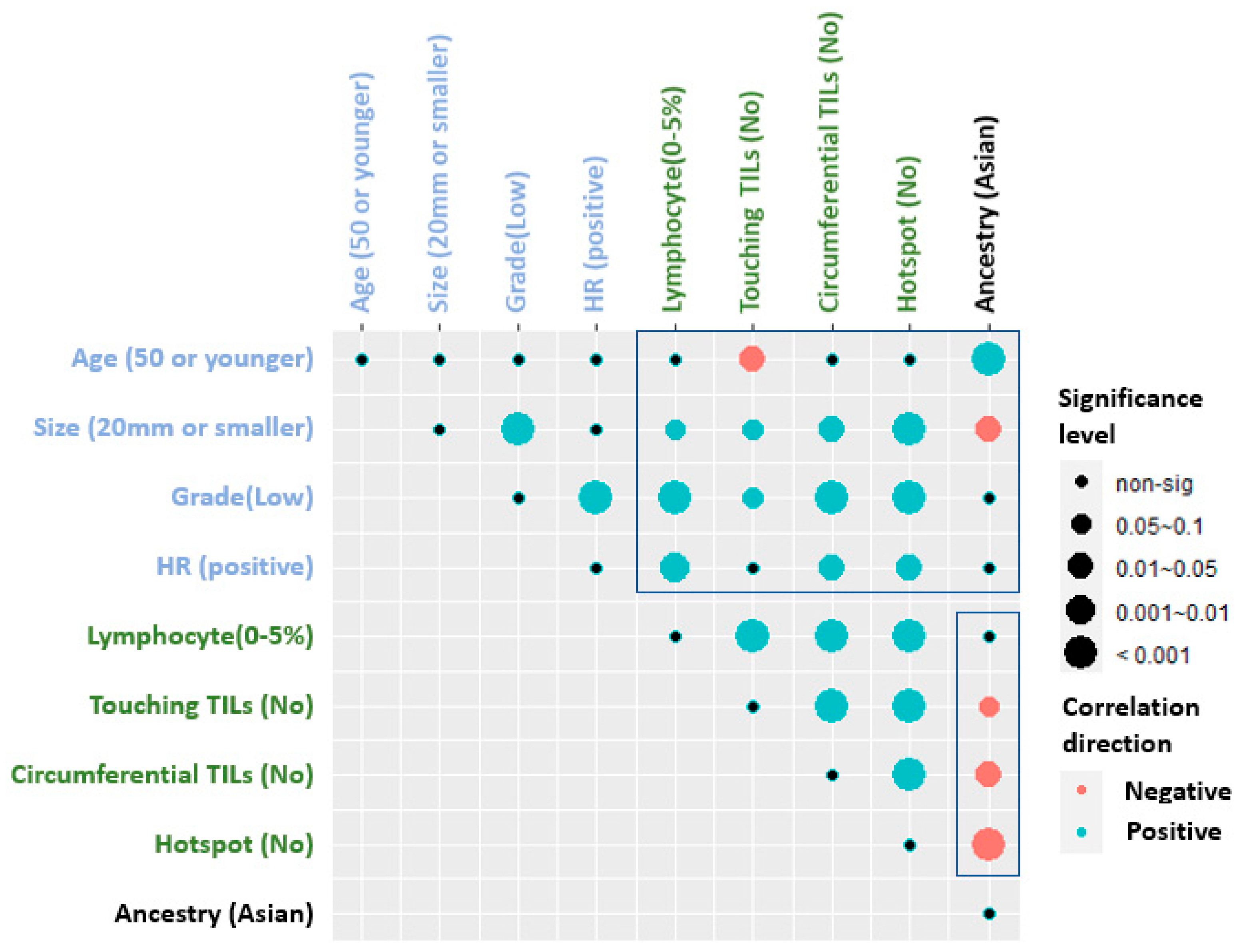
3.1.3. Correlation of TILs Assessments with Clinical and Histologic Parameters

Figure 1 shows the correlation plot of the assessments for all TILs scoring methods in relation to race/ethnicity and clinico-pathological parameters. Accordingly, Asian ethnicity (mostly Singapore) cohort compared to the three European ethnicity (Italian and Mayo) cohorts showed that Asian ethnicity tended to have higher scores with all TIL scoring methods in the following order for the presence of hotspots (adjusted p < 0.001), circumferential TILs (adjusted p = 0.064), touching TILs (adjusted p = 0.088), and stromal TILs was not different in Asians (p = 0.227). In addition, Asian patients were younger in age (adjusted p < 0.001) and had a larger tumor size (adjusted p = 0.022). Hormone receptor (HR) expression was not different in Asians. However, the presence of HR was positively correlated with stromal (adjusted p = 0.01), circumferential TILs (p = 0.015), and hotspots (p = 0.02) in overall population.

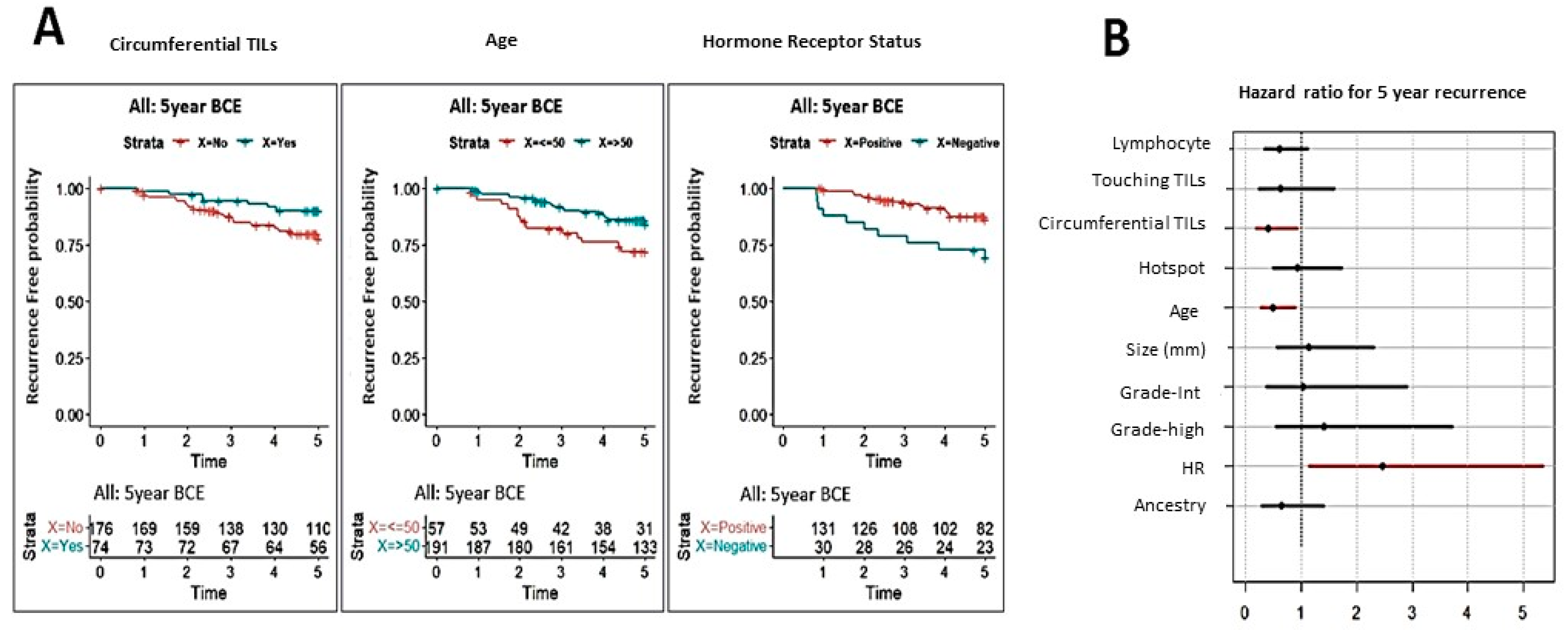
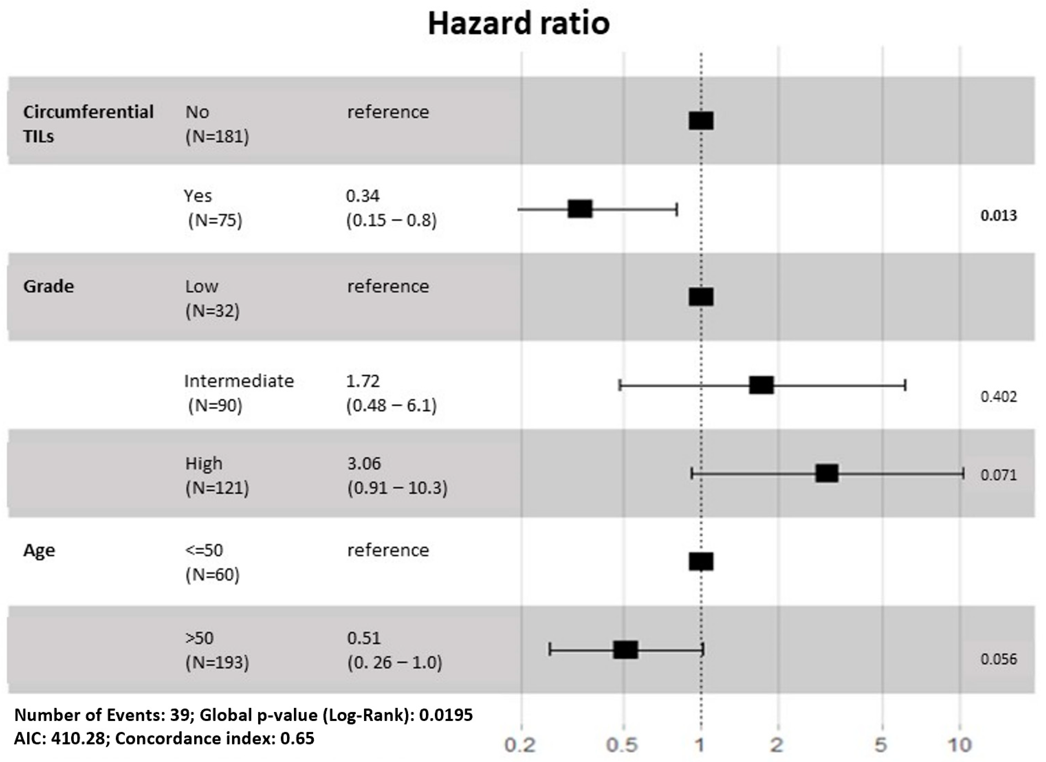
3.1.4. Correlation of TILs Assessments with Clinical Outcome

Cox proportional hazard models were developed to determine the association of nine variables (ancestry, age, size, grade, hormone receptor (HR), stromal lymphocyte, touching TILs, circumferential TILs, and hotspot) with outcomes at 5-year follow-up. As both shown in Figure 2A,B, three variables were weakly associated with 5-year second breast cancer events including age (p = 0.02; adjusted p = 0.11), circumferential TILs (p = 0.03; adjusted p = 0.11), and hormone receptor status (p = 0.02; adjusted p = 0.11). Survival analysis in individual cohorts by each of the TIL’s parameters is shown Supplemental Figures S1–S5.
In multivariable analysis, stepwise selection on Cox proportional hazard model were applied to 7 variables (age, size, grade, lymphocyte, touching TILS, circumferential TILs, hotspot) (Figure 3). The combination of three variables—age, grade, and circumferential TILs (Wald test p = 0.02) predicted the likelihood of a second breast cancer event.

3.1.5. Impact of Ethnicity

DNA for ethnicity determination was available for all but the Oxford cohorts. The K13 model was chosen for our analysis, as this provided more relevant information regarding the distribution of population in our cohorts. These analyses confirmed that the Singapore cases were mainly Southeast Asian origin (47%) with an admixture of Mediterranean (10%) and North European (21%). On the other hand, the Mayo cohort resulted in 47% North-European mixed with 29% Mediterranean and 9% West-Asian. In contrast, the Italian cohort comprised of North-European (35%), Mediterranean (28%), and 12% West-Asian.
As the largest ethnic group was Asian, we sought to further analyze the association of ancestry with TILs and recurrence. Asian ethnicity was associated with a younger age at diagnosis, a larger size, and a greater incidence of tumor infiltrating lymphocytes irrespective of the assessing methodology. Further exploratory analysis of Asians from Singapore cohort in comparison with Asians from other cohorts revealed no significant differences in incidence of variables or association with TILs or second breast cancer event(s) (Supplementary Figure S5A). In spite of this, Asians had numerically higher event rate (Supplementary Figure S5B).

4. Discussion

The association of tumor-infiltrating lymphocytes (TILs) with improved prognosis in invasive breast cancer, particularly HER2 and triple negative subtypes, led to an assumption that TILs assessment of H&E samples can provide a similar prognostic value in DCIS. However, the relationship between TILs and DCIS outcomes is not straight forward [20,21,22,36,37,38]. Many studies have used the International Working TILs Group guidelines [1] to evaluate the percentage of stromal TILs and reported little to no association with DCIS recurrence. One of the reasons for these discrepant results is due to the variability of the endpoints. These endpoints include any recurrence, ipsilateral recurrence, and development of invasive carcinoma. Furthermore, most studies are not multi-centric and instead are based on relatively small cohorts. More importantly, the event rates are relatively low in most studies, which hampers the stability of the statistical methods.
The standard of care for treatment of DCIS consists of mastectomy or lumpectomy plus radiation. The patient population in the current study had been treated with variable modalities; this is well reflected in our cohorts. All patients from Singapore had been treated with mastectomy, while other cohorts had been managed by a mix of mastectomy and lumpectomy plus radiation. In the combined analysis of the cohorts, differences in treatment did not emerge as an important confounding factor. The need for follow-up information undoubtedly led to a bias in the number of events observed. The Oxford cohort had a significantly higher event rate (39.1%) than the other cohorts. However, after the merging of the cohorts, we observed a recurrence of 26.4%. This higher incidence of events could lead to the identification of associations that were not observed in some of the prior studies.
Knopfelmacher et al. [39] had previously demonstrated significant association with circumferential TILs and high ODX DCIS scores in a study of 46 cases. Toss et al. [23] assessed the impact of seven TILs scoring methods using a training and validation cohort of DCIS with long-term follow-up data in a large single institutional study. Among these scoring methods, dense touching TILs have been shown as an independent prognostic variable in DCIS rather than stromal TILs, which have been used in most of the earlier studies. In a study of 198 cases with recurrence follow-up reported, a significant association between circumferential TILs and worse disease-free survival [40]. Recently, Komforti et al. [24] assessed the pattern of TILs in a series of 97 cases by three different methodologies [20,23,41]. Stromal TILs showed the lowest concordance rate, followed by touching TILs by all pathologists. On the other hand, the assessment of circumferential TILs had a greater concordance rate. This study showed significant associations of touching TILs and circumferential TILs with the intermediate/high ODX DCIS score, but not stromal TILs. In contrast to these studies, we found that circumferential TILs were associated with a good prognosis, but no significant association was found for other TILs variables, including touching TILs and stromal TILs. The differences in these compared to our study may be attributed to the distribution of other clinico-pathological parameters including age, tumor size, grade, or hormone receptor status [40].
A major impact of cohort size and location was observed in the analysis of the association of TILs with recurrence. Circumferential TILs were not significant in any of the independent cohorts. However, their presence was associated with a decreased risk of recurrence in the combined analysis (p = 0.03). The other TILs parameters were not significant in individual cohorts or in the combined analysis. Race/ethnicity can be another factor that may affect the DCIS outcome. Analyzing 18 NCI-SEER Registries, Lui et al. reported that black and Hispanic women had higher ipsilateral breast tumor (IBT) risk when compared to white women [10,42]. The risk for contralateral breast tumors (CBT) was also significantly high in black and Asian/Pacific Islander (PI) women compared with white women (p heterogeneity < 0.0001). While the black women older than 50 years and with comedo necrosis were more likely to have CBT, Asian/PI women younger than 50 years and with lack of comedo DCIS had more significantly associated with CBT [10]. These results were consistent with our study that Asian women were younger in age and had significantly higher recurrence rate than the European white cohort. The correlation with ethnicity with hormone receptor status was not analyzed due to large portion of missing data.
The current study is one of the larger studies to assess the impact of TILs in DCIS. In spite of this, there are several limitations. Part of the study was performed on TMAs as this was the only material available for analysis. However, Toss et al. [23] have previously documented a high degree of correlation between TILs observed in TMAs and in whole sections. Another limitation associated with retrospective multi-institutional studies is the variability in assessment of margin status and treatment regimens. Furthermore, the requirement of follow-up data could have led to a bias in selection of the patients for the study. This may have resulted in higher event rates observed in the study. The latter was a strength as well as a limitation of the study. The higher events rate could have made it possible to define associations not observed in prior studies, which had low event rates. Lastly, differences have been documented in risk of subsequent invasive carcinoma in Asian women of different geographic origin [43]. The current study had too few women from different Asian origins to tease out more information regarding this parameter.

5. Conclusions

In conclusion, our study and others provide promising results to include the different TILs scoring methods to the current guidelines for predicting the prognostic significance of TILs in DCIS.

Supplementary Materials

The following supporting information can be downloaded at: https://www.mdpi.com/article/10.3390/cancers14163916/s1. Table S1. Patient characteristics for each DCIS cohort. Table S2. Patient characteristics and association with TILs assessments for each DCIS cohort. Figure S1. Correlation analysis of tumor-infiltrating lymphocytes (TILs) with clinical outcome for each cohort. (A) The hazard ratio and 95% confidence intervals associated with TILs and 5 year recurrence, (B) Kaplan-Meier (KM) plots for 5 year recurrence-free survival for TILs for all cohorts, combined, (C) Kaplan-Meier (KM) plots for 5-year recurrence-free survival for TILs for each cohort. Figure S2. Correlation analysis of touching tumor-infiltrating lymphocytes (touching TILs) with clinical outcome for each cohort. (A) The hazard ratio and 95% confidence intervals associated with TILs and 5 year recurrence, (B) Kaplan-Meier (KM) plots for 5 year recurrence-free survival for TILs for all cohorts, combined, (C) Kaplan-Meier (KM) plots for 5-year recurrence-free survival for TILs for each cohort. Figure S3. Correlation analysis of circumferential tumor-infiltrating lymphocytes (TILs) with clinical outcome for each cohort. (A) The hazard ratio and 95% confidence intervals associated with TILs and 5 year recurrence, (B) Kaplan-Meier (KM) plots for 5 year recurrence-free survival for TILs for all cohorts, combined, (C) Kaplan-Meier (KM) plots for 5-year recurrence-free survival for TILs for each cohort. Figure S4. Correlation analysis of hotspot tumor-infiltrating lymphocytes (TILs) with clinical outcome for each cohort. (A) The hazard ratio and 95% confidence intervals associated with TILs and 5 year recurrence, (B) Kaplan-Meier (KM) plots for 5 year recurrence-free survival for TILs for all cohorts, combined, (C) Kaplan-Meier (KM) plots for 5-year recurrence-free survival for TILs for each cohort. Figure S5. Analysis of clinico-pathological variables, tumor-infiltrating lymphocytes (TILs) assessment and 5 year recurrence risk between Asian and European cohorts. (A) Correlation of clinico-pathological variables and tumor-infiltrating lymphocytes (TILs) and (B) KM plot of 5 year recurrence for the Asian and European cohorts.

Author Contributions

S.S.B., Y.G.-P., A.L.H., S.G. and F.G.: Concept, design, interpretation and execution of the entire project; S.C. (Sanghee Cho), X.L., and S.C. (Sha Cao): statistical and bioinformatics analyses and interpretation. Y.G.-P., S.C. (Sanghee Cho), P.H.T., S.G., A.A.T., I.T.O., D.G., F.Z., A.L.H., F.G. and S.S.B.: manuscript writing. All authors have read and agreed to the published version of the manuscript.

Funding

This study is supported by the National Cancer Institute of the National Institutes of Health under Award Number R01CA194600 to S Badve, and F Ginty. The content is solely the responsibility of the authors and does not necessarily represent the official views of the National Institutes of Health.

Institutional Review Board Statement

Ethical review and approval were waived by the Indiana University Institutional Review board due to the retrospective nature of the study. All samples were de-identified at respective institutions prior to reaching Indiana University for the TILs analysis. Clinical data was directly sent to the study statistician.

Informed Consent Statement

Patient consent was waived.

Data Availability Statement

The data presented in this study are available upon request.

Acknowledgments

We thank Prashant Singh and his team for his help with Ancestry Analysis at Genomics Shared Resource, Roswell Park Comprehensive Cancer Center. We thank Stephanie Jones and Clare Verrill at the Oxford Centre for Histopathology Research: https://www.nds.ox.ac.uk/ochre and Marian Taylor in Oxford University of Oncology for help in curating Oxford samples.

Conflicts of Interest

The authors declare no conflict of interest.

References

  1. Salgado, R.; Denkert, C.; Demaria, S.; Sirtaine, N.; Klauschen, F.; Pruneri, G.; Wienert, S.; Van den Eynden, G.; Baehner, F.L.; Penault-Llorca, F.; et al. The evaluation of tumor-infiltrating lymphocytes (TILs) in breast cancer: Recommendations by an International TILs Working Group 2014. Ann. Oncol. 2015, 26, 259–271. [Google Scholar] [CrossRef] [PubMed]
  2. Adams, S.; Goldstein, L.J.; Sparano, J.A.; Demaria, S.; Badve, S.S. Tumor infiltrating lymphocytes (TILs) improve prognosis in patients with triple negative breast cancer (TNBC). Oncoimmunology 2015, 4, e985930. [Google Scholar] [CrossRef] [PubMed]
  3. Adams, S.; Gray, R.J.; Demaria, S.; Goldstein, L.; Perez, E.A.; Shulman, L.N.; Martino, S.; Wang, M.; Jones, V.E.; Saphner, T.J.; et al. Prognostic value of tumor-infiltrating lymphocytes in triple-negative breast cancers from two phase III randomized adjuvant breast cancer trials: ECOG 2197 and ECOG 1199. J. Clin. Oncol. 2014, 32, 2959–2966. [Google Scholar] [CrossRef] [PubMed]
  4. Loi, S.; Drubay, D.; Adams, S.; Pruneri, G.; Francis, P.A.; Lacroix-Triki, M.; Joensuu, H.; Dieci, M.V.; Badve, S.; Demaria, S.; et al. Tumor-Infiltrating Lymphocytes and Prognosis: A Pooled Individual Patient Analysis of Early-Stage Triple-Negative Breast Cancers. J. Clin. Oncol. 2019, 37, 559–569. [Google Scholar] [CrossRef]
  5. Trinh, A.; Gil Del Alcazar, C.R.; Shukla, S.A.; Chin, K.; Chang, Y.H.; Thibault, G.; Eng, J.; Jovanovic, B.; Aldaz, C.M.; Park, S.Y.; et al. Genomic Alterations during the In Situ to Invasive Ductal Breast Carcinoma Transition Shaped by the Immune System. Mol. Cancer Res. 2020, 19, 623–635. [Google Scholar] [CrossRef]
  6. Pandey, J.P.; Namboodiri, A.M.; Kistner-Griffin, E. IgG and FcgammaR genotypes and humoral immunity to mucin 1 in prostate cancer. Hum. Immunol. 2013, 74, 1030–1033. [Google Scholar] [CrossRef]
  7. Yeyeodu, S.T.; Kidd, L.R.; Kimbro, K.S. Protective Innate Immune Variants in Racial/Ethnic Disparities of Breast and Prostate Cancer. Cancer Immunol. Res. 2019, 7, 1384–1389. [Google Scholar] [CrossRef]
  8. Zhou, Q.; Wang, C.; Wang, X.; Wu, X.; Zhu, Z.; Liu, B.; Su, L. Association between TLR4 (+896A/G and +1196C/T) polymorphisms and gastric cancer risk: An updated meta-analysis. PLoS ONE 2014, 9, e109605. [Google Scholar] [CrossRef]
  9. Hong, C.C.; Sucheston-Campbell, L.E.; Liu, S.; Hu, Q.; Yao, S.; Lunetta, K.L.; Haddad, S.A.; Ruiz-Narvaez, E.A.; Bensen, J.T.; Cheng, T.D.; et al. Genetic Variants in Immune-Related Pathways and Breast Cancer Risk in African American Women in the AMBER Consortium. Cancer Epidemiol. Biomark. Prev. 2018, 27, 321–330. [Google Scholar] [CrossRef]
  10. Liu, Y.; Colditz, G.A.; Gehlert, S.; Goodman, M. Racial disparities in risk of second breast tumors after ductal carcinoma in situ. Breast Cancer Res. Treat. 2014, 148, 163–173. [Google Scholar] [CrossRef]
  11. DeSantis, C.E.; Ma, J.; Gaudet, M.M.; Newman, L.A.; Miller, K.D.; Goding Sauer, A.; Jemal, A.; Siegel, R.L. Breast cancer statistics, 2019. CA Cancer J. Clin. 2019, 69, 438–451. [Google Scholar] [CrossRef] [PubMed]
  12. Early Breast Cancer Trialists’ Collaborative Group; Correa, C.; McGale, P.; Taylor, C.; Wang, Y.; Clarke, M.; Davies, C.; Peto, R.; Bijker, N.; Solin, L.; et al. Overview of the randomized trials of radiotherapy in ductal carcinoma in situ of the breast. J. Natl. Cancer Inst. Monogr. 2010, 2010, 162–177. [Google Scholar] [CrossRef] [PubMed]
  13. Cuzick, J.; Sestak, I.; Pinder, S.E.; Ellis, I.O.; Forsyth, S.; Bundred, N.J.; Forbes, J.F.; Bishop, H.; Fentiman, I.S.; George, W.D. Effect of tamoxifen and radiotherapy in women with locally excised ductal carcinoma in situ: Long-term results from the UK/ANZ DCIS trial. Lancet Oncol. 2011, 12, 21–29. [Google Scholar] [CrossRef]
  14. Miligy, I.; Mohan, P.; Gaber, A.; Aleskandarany, M.A.; Nolan, C.C.; Diez-Rodriguez, M.; Mukherjee, A.; Chapman, C.; Ellis, I.O.; Green, A.R.; et al. Prognostic significance of tumour infiltrating B lymphocytes in breast ductal carcinoma in situ. Histopathology 2017, 71, 258–268. [Google Scholar] [CrossRef]
  15. Farolfi, A.; Petracci, E.; Serra, L.; Ravaioli, A.; Bravaccini, S.; Ravaioli, S.; Tumedei, M.M.; Ulivi, P.; Canale, M.; Puccetti, M.; et al. Tumor-Infiltrating Lymphocytes (TILs) and Risk of a Second Breast Event After a Ductal Carcinoma in situ. Front. Oncol. 2020, 10, 1486. [Google Scholar] [CrossRef]
  16. Darvishian, F.; Ozerdem, U.; Adams, S.; Chun, J.; Pirraglia, E.; Kaplowitz, E.; Guth, A.; Axelrod, D.; Shapiro, R.; Price, A.; et al. Tumor-Infiltrating Lymphocytes in a Contemporary Cohort of Women with Ductal Carcinoma In Situ (DCIS). Ann. Surg. Oncol. 2019, 26, 3337–3343. [Google Scholar] [CrossRef]
  17. Beguinot, M.; Dauplat, M.M.; Kwiatkowski, F.; Lebouedec, G.; Tixier, L.; Pomel, C.; Penault-Llorca, F.; Radosevic-Robin, N. Analysis of tumour-infiltrating lymphocytes reveals two new biologically different subgroups of breast ductal carcinoma in situ. BMC Cancer 2018, 18, 129. [Google Scholar] [CrossRef]
  18. Agahozo, M.C.; van Bockstal, M.R.; Groenendijk, F.H.; van den Bosch, T.P.P.; Westenend, P.J.; van Deurzen, C.H.M. Ductal carcinoma in situ of the breast: Immune cell composition according to subtype. Mod. Pathol. 2020, 33, 196–205. [Google Scholar] [CrossRef]
  19. Gil Del Alcazar, C.R.; Huh, S.J.; Ekram, M.B.; Trinh, A.; Liu, L.L.; Beca, F.; Zi, X.; Kwak, M.; Bergholtz, H.; Su, Y.; et al. Immune Escape in Breast Cancer During In Situ to Invasive Carcinoma Transition. Cancer Discov. 2017, 7, 1098–1115. [Google Scholar] [CrossRef]
  20. Pruneri, G.; Lazzeroni, M.; Bagnardi, V.; Tiburzio, G.B.; Rotmensz, N.; DeCensi, A.; Guerrieri-Gonzaga, A.; Vingiani, A.; Curigliano, G.; Zurrida, S.; et al. The prevalence and clinical relevance of tumor-infiltrating lymphocytes (TILs) in ductal carcinoma in situ of the breast. Ann. Oncol. 2017, 28, 321–328. [Google Scholar] [CrossRef]
  21. Hendry, S.; Salgado, R.; Gevaert, T.; Russell, P.A.; John, T.; Thapa, B.; Christie, M.; van de Vijver, K.; Estrada, M.V.; Gonzalez-Ericsson, P.I.; et al. Assessing Tumor-infiltrating Lymphocytes in Solid Tumors: A Practical Review for Pathologists and Proposal for a Standardized Method From the International Immunooncology Biomarkers Working Group: Part 1: Assessing the Host Immune Response, TILs in Invasive Breast Carcinoma and Ductal Carcinoma In Situ, Metastatic Tumor Deposits and Areas for Further Research. Adv. Anat. Pathol. 2017, 24, 235–251. [Google Scholar] [CrossRef] [PubMed]
  22. Hendry, S.; Pang, J.B.; Byrne, D.J.; Lakhani, S.R.; Cummings, M.C.; Campbell, I.G.; Mann, G.B.; Gorringe, K.L.; Fox, S.B. Relationship of the Breast Ductal Carcinoma In Situ Immune Microenvironment with Clinicopathological and Genetic Features. Clin. Cancer Res. 2017, 23, 5210–5217. [Google Scholar] [CrossRef]
  23. Toss, M.S.; Miligy, I.; Al-Kawaz, A.; Alsleem, M.; Khout, H.; Rida, P.C.; Aneja, R.; Green, A.R.; Ellis, I.O.; Rakha, E.A. Prognostic significance of tumor-infiltrating lymphocytes in ductal carcinoma in situ of the breast. Mod. Pathol. 2018, 31, 1226–1236. [Google Scholar] [CrossRef]
  24. Komforti, M.; Badve, S.S.; Harmon, B.; Lo, Y.; Fineberg, S. Tumour-infiltrating lymphocytes in ductal carcinoma in situ (DCIS)-assessment with three different methodologies and correlation with Oncotype DX DCIS Score. Histopathology 2020, 77, 749–759. [Google Scholar] [CrossRef] [PubMed]
  25. Pan, J.W.; Zabidi, M.M.A.; Chong, B.K.; Meng, M.Y.; Ng, P.S.; Hasan, S.N.; Sandey, B.; Bahnu, S.; Rajadurai, P.; Yip, C.H.; et al. Germline APOBEC3B deletion increases somatic hypermutation in Asian breast cancer that is associated with Her2 subtype, PIK3CA mutations, and immune activation. Int. J. Cancer 2021, 148, 2489–2501. [Google Scholar] [CrossRef] [PubMed]
  26. Pan, J.W.; Zabidi, M.M.A.; Ng, P.S.; Meng, M.Y.; Hasan, S.N.; Sandey, B.; Sammut, S.J.; Yip, C.H.; Rajadurai, P.; Rueda, O.M.; et al. The molecular landscape of Asian breast cancers reveals clinically relevant population-specific differences. Nat. Commun. 2020, 11, 6433. [Google Scholar] [CrossRef]
  27. Alexander, D.H.; Novembre, J.; Lange, K. Fast model-based estimation of ancestry in unrelated individuals. Genome Res. 2009, 19, 1655–1664. [Google Scholar] [CrossRef]
  28. Grambsch, P.M.; Therneau, T.M. Proportional hazards tests and diagnostics based on weighted residuals. Biometrika 1994, 81, 515–526. [Google Scholar] [CrossRef]
  29. Armstrong, A.J.; Lin, P.; Higano, C.S.; Sternberg, C.N.; Sonpavde, G.; Tombal, B.; Templeton, A.J.; Fizazi, K.; Phung, D.; Wong, E.K.; et al. Development and validation of a prognostic model for overall survival in chemotherapy-naive men with metastatic castration-resistant prostate cancer. Ann. Oncol. 2018, 29, 2200–2207. [Google Scholar] [CrossRef]
  30. Zhang, C.; Zhang, Z.; Zhang, G.; Zhang, Z.; Luo, Y.; Wang, F.; Wang, S.; Che, Y.; Zeng, Q.; Sun, N.; et al. Clinical significance and inflammatory landscapes of a novel recurrence-associated immune signature in early-stage lung adenocarcinoma. Cancer Lett. 2020, 479, 31–41. [Google Scholar] [CrossRef]
  31. Doshi, R.P.; Chen, K.; Wang, F.; Schwartz, H.; Herzog, A.; Aseltine, R.H., Jr. Identifying risk factors for mortality among patients previously hospitalized for a suicide attempt. Sci. Rep. 2020, 10, 15223. [Google Scholar] [CrossRef] [PubMed]
  32. Roussel, E.; Peeters, E.; Vanthoor, J.; Bozzini, G.; Muneer, A.; Ayres, B.; Sri, D.; Watkin, N.; Bhattar, R.; Parnham, A.; et al. Predictors of local recurrence and its impact on survival after glansectomy for penile cancer: Time to challenge the dogma? BJU Int. 2021, 127, 606–613. [Google Scholar] [CrossRef] [PubMed]
  33. Chen, T.; Li, X.; Li, Y.; Xia, E.; Qin, Y.; Liang, S.; Xu, F.; Liang, D.; Zeng, C.; Liu, Z. Prediction and Risk Stratification of Kidney Outcomes in IgA Nephropathy. Am. J. Kidney Dis. 2019, 74, 300–309. [Google Scholar] [CrossRef] [PubMed]
  34. Hicks, C.W.; Canner, J.K.; Mathioudakis, N.; Lippincott, C.; Sherman, R.L.; Abularrage, C.J. Incidence and Risk Factors Associated With Ulcer Recurrence Among Patients With Diabetic Foot Ulcers Treated in a Multidisciplinary Setting. J. Surg. Res. 2020, 246, 243–250. [Google Scholar] [CrossRef]
  35. Miller, A. Subset Selection in Regression, 2nd ed.; Chapman and Hall: London, UK; CRC: Boca Raton, FL, USA, 2002. [Google Scholar]
  36. Thompson, E.; Taube, J.M.; Elwood, H.; Sharma, R.; Meeker, A.; Warzecha, H.N.; Argani, P.; Cimino-Mathews, A.; Emens, L.A. The immune microenvironment of breast ductal carcinoma in situ. Mod Pathol 2016, 29, 249–258. [Google Scholar] [CrossRef]
  37. Dieci, M.V.; Radosevic-Robin, N.; Fineberg, S.; van den Eynden, G.; Ternes, N.; Penault-Llorca, F.; Pruneri, G.; D’Alfonso, T.M.; Demaria, S.; Castaneda, C.; et al. Update on tumor-infiltrating lymphocytes (TILs) in breast cancer, including recommendations to assess TILs in residual disease after neoadjuvant therapy and in carcinoma in situ: A report of the International Immuno-Oncology Biomarker Working Group on Breast Cancer. Semin. Cancer Biol. 2018, 52, 16–25. [Google Scholar] [CrossRef]
  38. Campbell, M.J.; Baehner, F.; O’Meara, T.; Ojukwu, E.; Han, B.; Mukhtar, R.; Tandon, V.; Endicott, M.; Zhu, Z.; Wong, J.; et al. Characterizing the immune microenvironment in high-risk ductal carcinoma in situ of the breast. Breast Cancer Res. Treat. 2017, 161, 17–28. [Google Scholar] [CrossRef]
  39. Knopfelmacher, A.; Fox, J.; Lo, Y.; Shapiro, N.; Fineberg, S. Correlation of histopathologic features of ductal carcinoma in situ of the breast with the oncotype DX DCIS score. Mod. Pathol. 2015, 28, 1167–1173. [Google Scholar] [CrossRef]
  40. Thike, A.A.; Chen, X.; Koh, V.C.Y.; Binte Md Nasir, N.D.; Yeong, J.P.S.; Bay, B.H.; Tan, P.H. Higher densities of tumour-infiltrating lymphocytes and CD4(+) T cells predict recurrence and progression of ductal carcinoma in situ of the breast. Histopathology 2020, 76, 852–864. [Google Scholar] [CrossRef]
  41. Agahozo, M.C.; Hammerl, D.; Debets, R.; Kok, M.; van Deurzen, C.H.M. Tumor-infiltrating lymphocytes and ductal carcinoma in situ of the breast: Friends or foes? Mod. Pathol. 2018, 31, 1012–1025. [Google Scholar] [CrossRef]
  42. Liu, Y.; West, R.; Weber, J.D.; Colditz, G.A. Race and risk of subsequent aggressive breast cancer following ductal carcinoma in situ. Cancer 2019, 125, 3225–3233. [Google Scholar] [CrossRef] [PubMed]
  43. Taparra, K.; Fukui, J.; Killeen, J.; Sumida, K.; Loo, L.W.M.; Hernandez, B.Y. Racial and Ethnic Disparities in Rates of Invasive Second Breast Cancer Among Women With Ductal Carcinoma In Situ in Hawai’i. JAMA Netw. Open 2021, 4, e2128977. [Google Scholar] [CrossRef] [PubMed]
Figure 1. Correlation analysis of tumor-infiltrating lymphocytes (TILs) assessments with clinical and histologic parameters. The dependence of each pair was evaluated by Fishers exact test, and adjusted p (FDR) = 0.1 was applied to 36 pairs; p-value was adjusted using Bonferroni-Holm procedure; Blue and red circles represent positive and negative correlations, respectively; Significance levels were shown based on the size of the circles.
Cancers 14 03916 g001
Figure 2. Correlation analysis of tumor-infiltrating lymphocytes (TILs) assessment with clinical outcome; (A) Kaplan-Meier (KM) plots for 5-year recurrence-free survival according TILs assessments and clinical parameters (univariable analysis). The significant associations were shown in the figure; (B) A forest plot showing the hazard ratio and 95% confidence intervals associated with variables and the primary endpoint (5 year recurrence); Circles represent the hazard ratio and the horizontal bars extend from; abbreviations- Grade-Int, Grade intermediate; HR-Hormone receptor status.
Cancers 14 03916 g002
Figure 3. Forest plot of multivariable analysis of cohorts for tumor-infiltrating lymphocytes (TILs) assessments with clinical and histologic parameters. In total 3 variables were selected—age, grade, and circumferential TILs with significance (Wald test p = 0.01).
Cancers 14 03916 g003
Table 1. Patient characteristics and association with TILs assessments for DCIS cohorts.
Total (n = 256)
Age
N-Miss3
<=5060 (23.7%)
>50193 (76.3%)
Size (mm)
N-Miss50
<=20131 (63.6%)
>2075 (36.4%)
Grade
N-Miss13
Low32 (13.2%)
Intermediate90 (37.0%)
High121 (49.8%)
HR
N-Miss86
Positive137 (80.6%)
Negative33 (19.4%)
Recurrence
N189 (73.8%)
Y67 (26.2%)
Lymphocyte
0–5%101 (39.5%)
>5%155 (60.5%)
Touching TILs
0214 (83.6%)
>042 (16.4%)
Circumferential TILs
No181 (70.7%)
Yes75 (29.3%)
Hotspot
No160 (62.5%)
Dense96 (37.5%)
Publisher’s Note: MDPI stays neutral with regard to jurisdictional claims in published maps and institutional affiliations.

