
Lung Nodules Missed in Initial Staging of Breast Cancer Patients in PET/MRI—Clinically Relevant?

 , ,
, ,
Abstract
:Simple Summary
Abstract
1. Introduction
2. Materials and Methods
2.1. Patients
2.2. PET/MRI
- i.
- A transverse T2-w half Fourier acquisition single shot turbo spin echo (HASTE) sequence in breath-hold technique with a slice thickness of 7 mm (TE 97 ms; TR 1500 ms; turbo factor (TF) 194; FOV 400 mm; phase FOV 75%; acquisition matrix 320 × 240 mm; in plane resolution 1.3 × 1.3 mm; TA 0:47 min/bed position)
- ii.
- A fat-saturated post-contrast transverse 3-dimensional volumetric interpolated breath-hold examination (VIBE) sequence with a slice thickness of 3 mm (TE, 1.53 ms; TR, 3.64 ms; flip angle 9°; FOV 400 × 280 mm; phase FOV 75%; acquisition matrix 512 × 384, in-plane resolution 0.7 × 0.7 mm; TA 0:19 min/bed position)
- iii.
- A transversal diffusion-weighted (DW) echo-planar imaging (EPI) sequence in free breathing with a slice thickness of 5.0 mm (TR 7400 ms; TE 72 ms; b-values: 0, 500, and 1000 s/mm2, matrix size 160 × 90; FOV 400 mm × 315 mm, phase FOV, 75%; GRAPPA, acceleration factor 2; in-plane resolution 2.6 × 2.6 mm; TA 2:06 min/bed position.
2.3. Computed Tomography
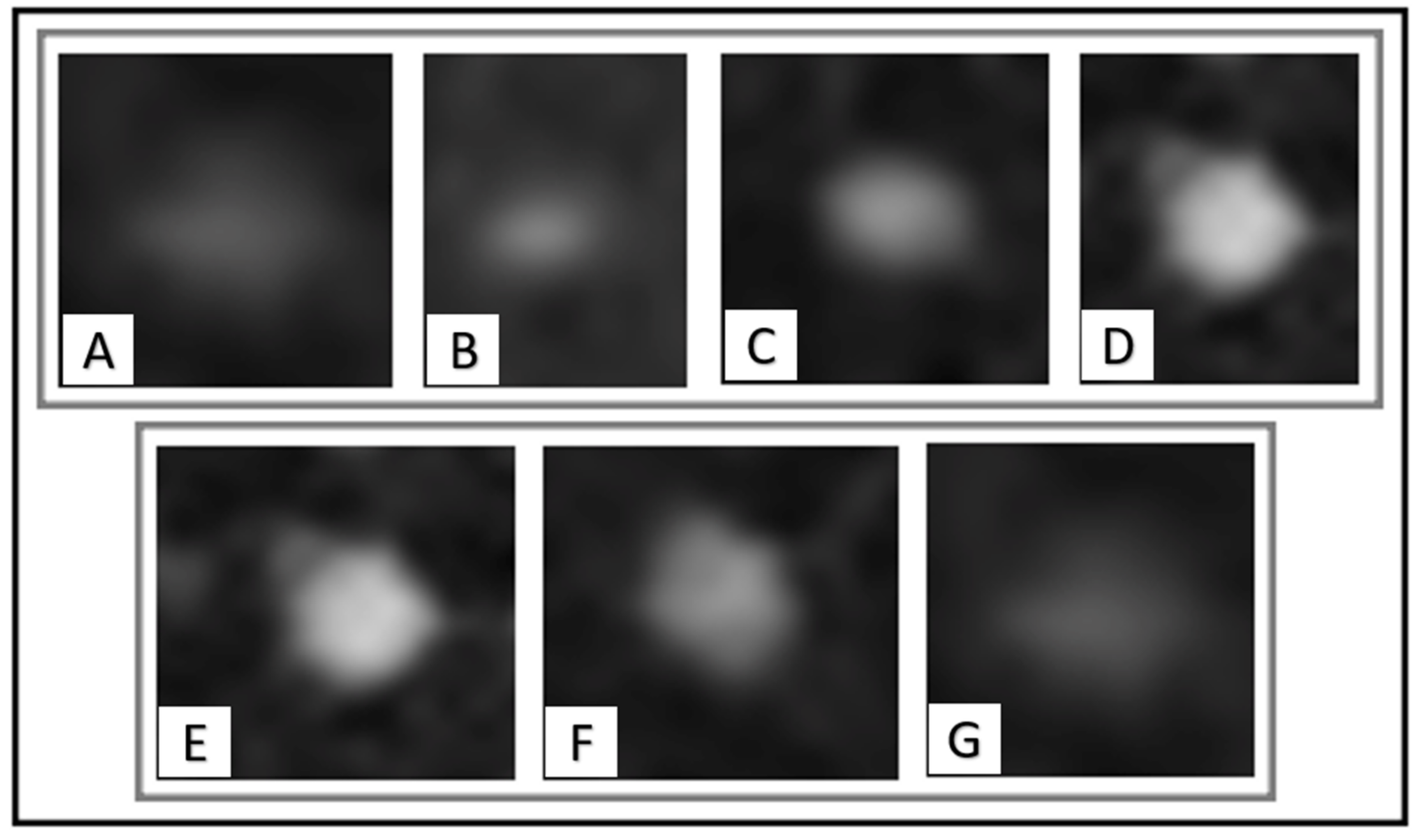
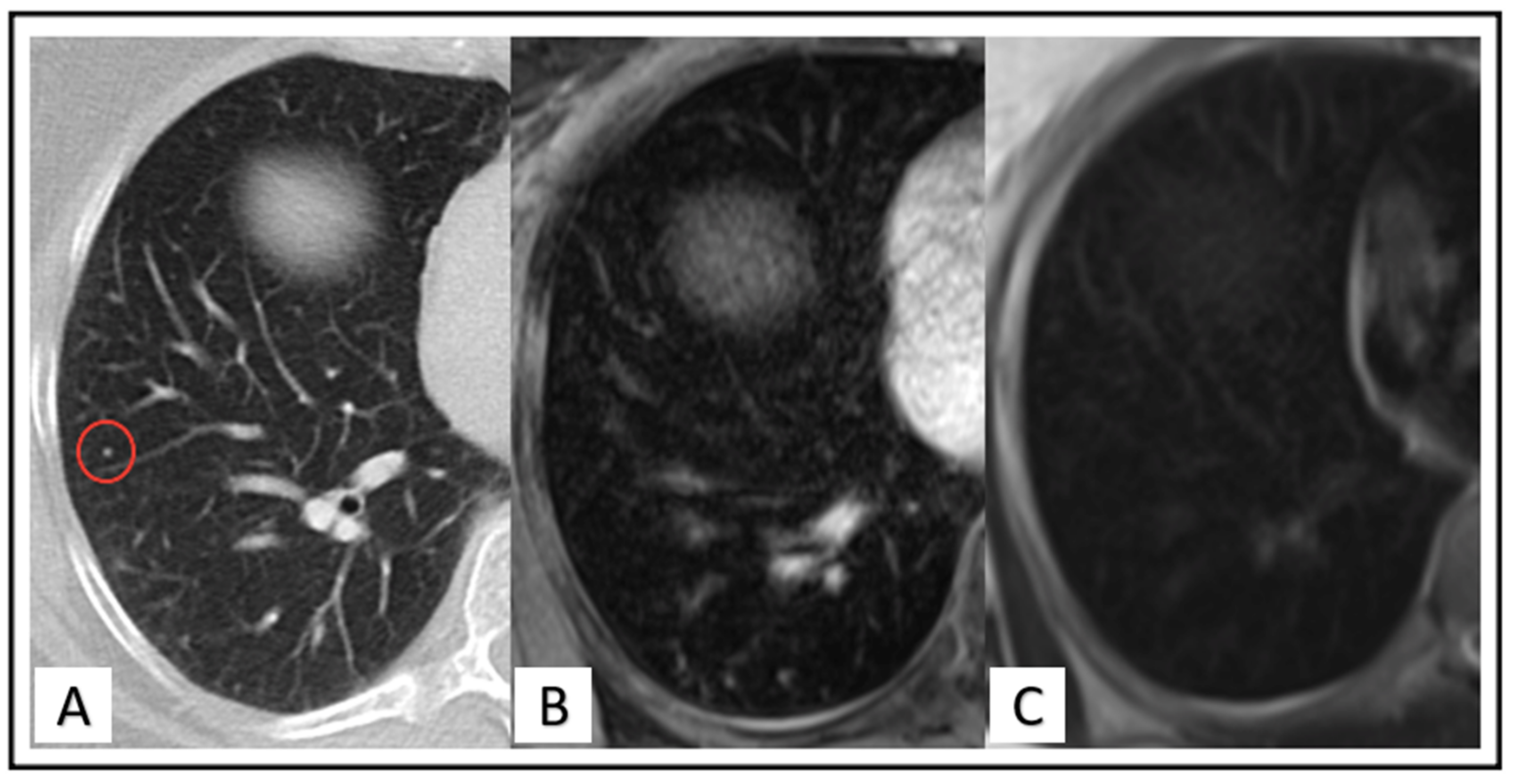
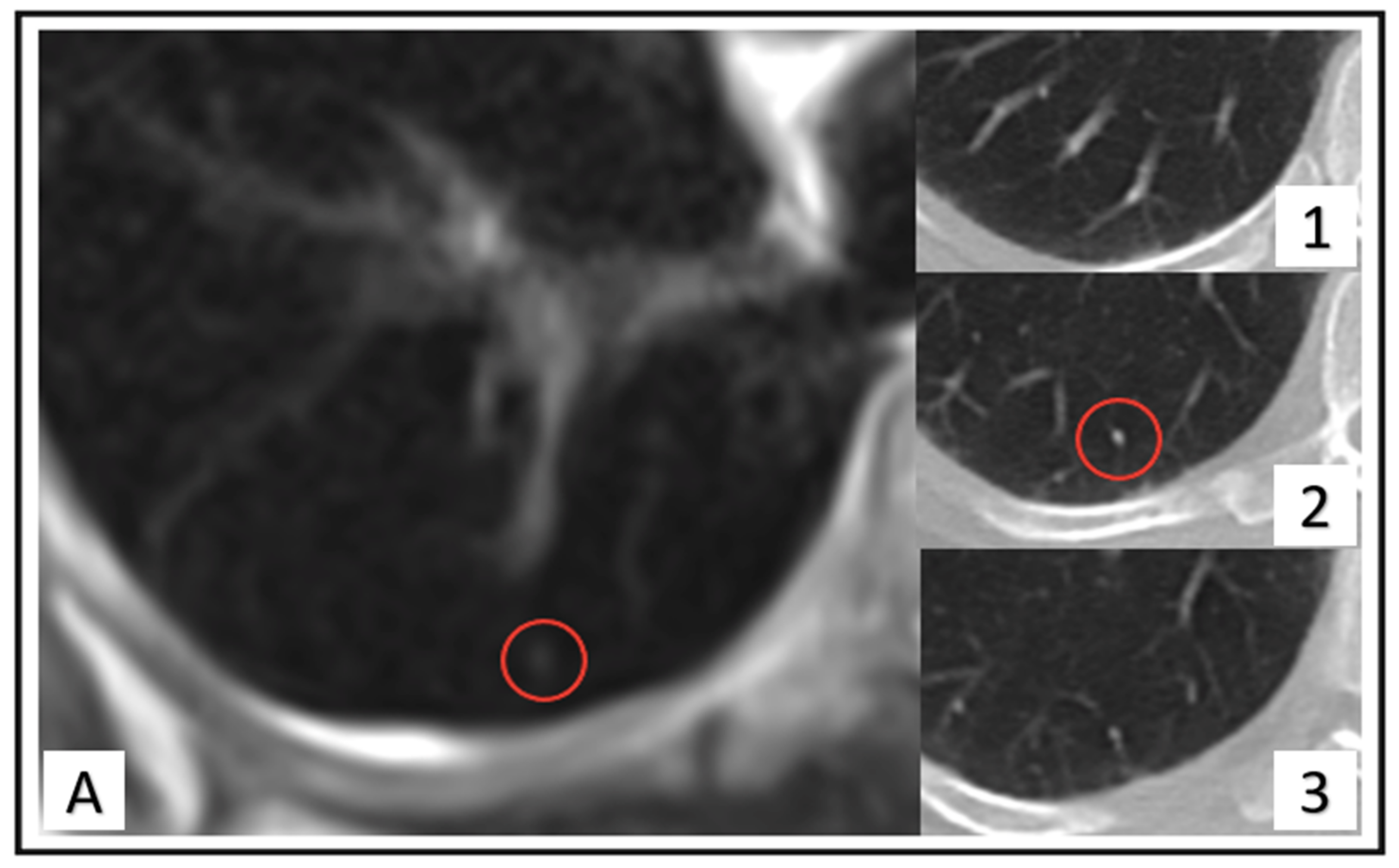
2.4. Image Analysis
2.5. Standard of Reference
2.6. Statistical Analysis
3. Results
3.1. Patient Based Analysis
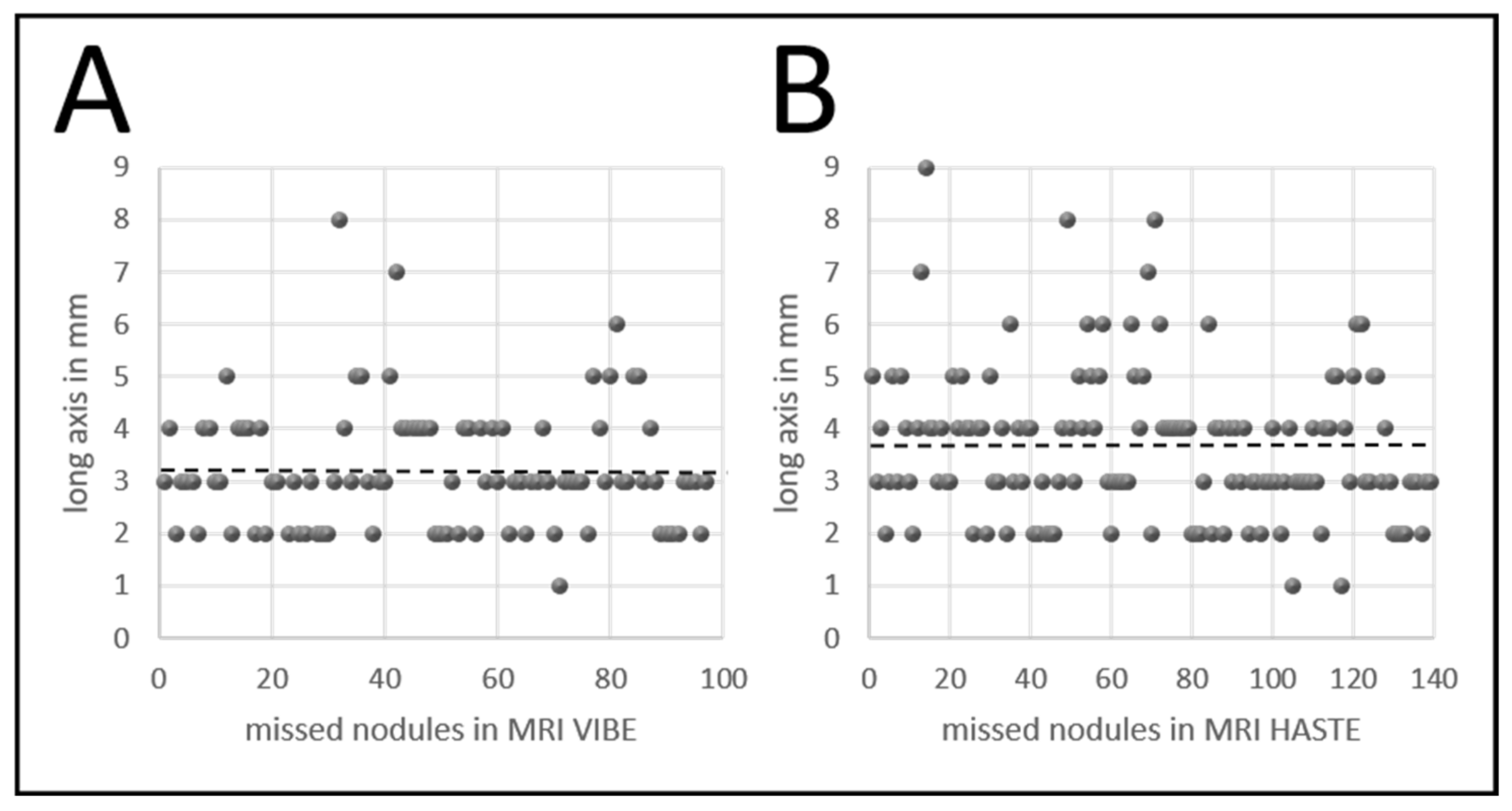
3.2. Lesion Based Analysis
3.3. False Findings and Image Quality
4. Discussion
5. Conclusions
Author Contributions
Funding
Institutional Review Board Statement
Informed Consent Statement
Data Availability Statement
Acknowledgments
Conflicts of Interest
References
- Bray, F.; Ferlay, J.; Soerjomataram, I.; Siegel, R.L.; Torre, L.A.; Jemal, A. Global cancer statistics 2018: GLOBOCAN estimates of incidence and mortality worldwide for 36 cancers in 185 countries. CA Cancer J. Clin. 2018, 68, 394–424. [Google Scholar] [CrossRef] [PubMed] [Green Version]
- Early Breast Cancer Trialists’ Collaborative Group. Trastuzumab for early-stage, HER2-positive breast cancer: A meta-analysis of 13,864 women in seven randomised trials. Lancet Oncol. 2021, 22, 1139–1150. [Google Scholar] [CrossRef]
- Gallagher, C.M.; More, K.; Masaquel, A.; Kamath, T.; Guérin, A.; Ionescu-Ittu, R.; Nitulescu, R.; Gauthier-Loiselle, M.; Sicignano, N.; Butts, E.; et al. Survival in patients with non-metastatic breast cancer treated with adjuvant trastuzumab in clinical practice. Springerplus 2016, 5, 395. [Google Scholar] [CrossRef] [Green Version]
- Leitlinienprogramm Onkologie (Deutsche Krebsgesellschaft DK, AWMF). S3-Leitlinie Früherkennung, Diagnose, Therapie und Nachsorge des Mammakarzinoms. Version 4.4. 2021. Available online: https://www.leitlinienprogramm-onkologie.de/fileadmin/user_upload/Downloads/Leitlinien/Mammakarzinom_4_0/Version_4.4/LL_Mammakarzinom_Kurzversion_4.3.pdf (accessed on 7 June 2022).
- Groheux, D.; Hindie, E. Breast cancer: Initial workup and staging with FDG PET/CT. Clin. Transl. Imaging 2021, 9, 221–231. [Google Scholar] [CrossRef] [PubMed]
- Ulaner, G.A.; Castillo, R.; Goldman, D.A.; Wills, J.; Riedl, C.; Pinker-Domenig, K.; Jochelson, M.S.; Gönen, M. 18F-FDG-PET/CT for systemic staging of newly diagnosed triple-negative breast cancer. Eur. J. Nucl. Med. Mol. Imaging 2016, 43, 1937–1944. [Google Scholar] [CrossRef] [Green Version]
- Ulaner, G.A.; Castillo, R.; Wills, J.; Gönen, M.; Goldman, D.A. 18F–FDG-PET/CT for systemic staging of patients with newly diagnosed ER-positive and HER2-positive breast cancer. Eur. J. Nucl. Med. Mol. Imaging 2017, 44, 1420–1427. [Google Scholar] [CrossRef]
- Bruckmann, N.M.; Kirchner, J.; Morawitz, J.; Umutlu, L.; Herrmann, K.; Bittner, A.-K.; Hoffmann, O.; Mohrmann, S.; Ingenwerth, M.; Schaarschmidt, B.M.; et al. Prospective comparison of CT and 18F-FDG PET/MRI in N and M staging of primary breast cancer patients: Initial results. PLoS ONE 2021, 16, e0260804. [Google Scholar] [CrossRef]
- Bruckmann, N.M.; Kirchner, J.; Umutlu, L.; Fendler, W.P.; Seifert, R.; Herrmann, K.; Bittner, A.-K.; Hoffmann, O.; Mohrmann, S.; Antke, C.; et al. Prospective comparison of the diagnostic accuracy of 18F-FDG PET/MRI, MRI, CT, and bone scintigraphy for the detection of bone metastases in the initial staging of primary breast cancer patients. Eur. Radiol. 2021, 31, 8714–8724. [Google Scholar] [CrossRef]
- Sawicki, L.M.; Grueneisen, J.; Schaarschmidt, B.M.; Buchbender, C.; Nagarajah, J.; Umutlu, L.; Antoch, G.; Kinner, S. Evaluation of 18F-FDG PET/MRI, 18F-FDG PET/CT, MRI, and CT in whole-body staging of recurrent breast cancer. Eur. J. Radiol. 2016, 85, 459–465. [Google Scholar] [CrossRef]
- Melsaether, A.N.; Raad, R.A.; Pujara, A.C.; Ponzo, F.D.; Pysarenko, K.M.; Jhaveri, K.; Babb, J.S.; Sigmund, E.E.; Kim, S.G.; Moy, L.A. Comparison of Whole-Body (18)F FDG PET/MR Imaging and Whole-Body (18)F FDG PET/CT in Terms of Lesion Detection and Radiation Dose in Patients with Breast Cancer. Radiology 2016, 281, 193–202. [Google Scholar] [CrossRef] [Green Version]
- Bruckmann, N.M.; Kirchner, J.; Morawitz, J.; Umutlu, L.; Fendler, W.P.; Herrmann, K.; Bittner, A.-K.; Hoffmann, O.; Fehm, T.; Lindemann, M.E.; et al. Free-breathing 3D Stack of Stars GRE (StarVIBE) sequence for detecting pulmonary nodules in 18F-FDG PET/MRI. EJNMMI Phys. 2022, 9, 11. [Google Scholar] [CrossRef] [PubMed]
- Wielpütz, M.O. MRI of Pulmonary Nodules: Closing the Gap on CT. Radiology 2022, 302, 707–708. [Google Scholar] [CrossRef] [PubMed]
- Rauscher, I.; Eiber, M.; Fürst, S.; Souvatzoglou, M.; Nekolla, S.G.; Ziegler, S.I.; Rummeny, E.J.; Schwaiger, M.; Beer, A.J. PET/MR Imaging in the Detection and Characterization of Pulmonary Lesions: Technical and Diagnostic Evaluation in Comparison to PET/CT. J. Nucl. Med. 2014, 55, 724–729. [Google Scholar] [CrossRef] [Green Version]
- Sawicki, L.M.; Grueneisen, J.; Buchbender, C.; Schaarschmidt, B.M.; Gomez, B.; Ruhlmann, V.; Umutlu, L.; Antoch, G.; Heusch, P. Evaluation of the Outcome of Lung Nodules Missed on 18F-FDG PET/MRI Compared with 18F-FDG PET/CT in Patients with Known Malignancies. J. Nucl. Med. 2016, 57, 15–20. [Google Scholar] [CrossRef] [PubMed] [Green Version]
- Benjamin, M.S.; Drucker, E.A.; McLoud, T.C.; Shepard, J.-A.O. Small Pulmonary Nodules: Detection at Chest CT and Outcome. Radiology 2003, 226, 489–493. [Google Scholar] [CrossRef] [PubMed]
- Bueno, J.; Landeras, L.; Chung, J.H. Updated Fleischner Society Guidelines for Managing Incidental Pulmonary Nodules: Common Questions and Challenging Scenarios. RadioGraphics 2018, 38, 1337–1350. [Google Scholar] [CrossRef] [Green Version]
- Association, W.M. World Medical Association Declaration of Helsinki: Ethical Principles for Medical Research Involving Human Subjects. JAMA 2013, 310, 2191–2194. [Google Scholar] [CrossRef] [Green Version]
- Cardoso, F.; Paluch-Shimon, S.; Senkus, E.; Curigliano, G.; Aapro, M.S.; André, F.; Barrios, C.H.; Bergh, J.; Bhattacharyya, G.S.; Biganzoli, L.; et al. 5th ESO-ESMO international consensus guidelines for advanced breast cancer (ABC 5). Ann. Oncol. 2020, 31, 1623–1649. [Google Scholar] [CrossRef]
- Cardoso, F.; Senkus, E.; Costa, A.; Papadopoulos, E.; Aapro, M.; André, F.; Harbeck, N.; Aguilar Lopez, B.; Barrios, C.H.; Bergh, J.; et al. 4th ESO-ESMO International Consensus Guidelines for Advanced Breast Cancer (ABC 4). Ann. Oncol. 2018, 29, 1634–1657. [Google Scholar] [CrossRef]
- Kirchner, J.; Grueneisen, J.; Martin, O.; Oehmigen, M.; Quick, H.H.; Bittner, A.-K.; Hoffmann, O.; Ingenwerth, M.; Catalano, O.A.; Heusch, P.; et al. Local and whole-body staging in patients with primary breast cancer: A comparison of one-step to two-step staging utilizing 18F-FDG-PET/MRI. Eur. J. Nucl. Med. Mol. Imaging 2018, 45, 2328–2337. [Google Scholar] [CrossRef]
- Rausch, I.; Quick, H.H.; Cal-Gonzalez, J.; Sattler, B.; Boellaard, R.; Beyer, T. Technical and instrumentational foundations of PET/MRI. Eur. J. Radiol. 2017, 94, A3–A13. [Google Scholar] [CrossRef] [PubMed]
- Ording, A.G.; Heide-Jørgensen, U.; Christiansen, C.F.; Nørgaard, M.; Acquavella, J.; Sørensen, H.T. Site of metastasis and breast cancer mortality: A Danish nationwide registry-based cohort study. Clin. Exp. Metastasis 2017, 34, 93–101. [Google Scholar] [CrossRef] [PubMed]
- Daglar, G.; Yuksek, Y.N.; Gozalan, U.; Tutuncu, T.; Kama, N.A. The significance of pulmonary nodule in breast cancer patients. Bratisl. Lek. Listy 2010, 111, 280–283. [Google Scholar] [PubMed]
- Li, F.; Armato, S.G.; Giger, M.L.; MacMahon, H. Clinical significance of noncalcified lung nodules in patients with breast cancer. Breast Cancer Res. Treat. 2016, 159, 265–271. [Google Scholar] [CrossRef]
- Sommer, G.; Tremper, J.; Koenigkam-Santos, M.; Delorme, S.; Becker, N.; Biederer, J.; Kauczor, H.-U.; Heussel, C.P.; Schlemmer, H.-P.; Puderbach, M. Lung nodule detection in a high-risk population: Comparison of magnetic resonance imaging and low-dose computed tomography. Eur. J. Radiol. 2014, 83, 600–605. [Google Scholar] [CrossRef]
- Biederer, J.; Hintze, C.; Fabel, M. MRI of pulmonary nodules: Technique and diagnostic value. Cancer Imaging 2008, 8, 125–130. [Google Scholar] [CrossRef]
- Evangelista, L.; Cuppari, L.; Burei, M.; Zorz, A.; Caumo, F. Head-to-head comparison between 18F-FDG PET/CT and PET/MRI in breast cancer. Clin. Transl. Imaging 2019, 7, 99–104. [Google Scholar] [CrossRef]
- Biondetti, P.; Vangel, M.G.; Lahoud, R.M.; Furtado, F.S.; Rosen, B.R.; Groshar, D.; Canamaque, L.G.; Umutlu, L.; Zhang, E.W.; Mahmood, U.; et al. PET/MRI assessment of lung nodules in primary abdominal malignancies: Sensitivity and outcome analysis. Eur. J. Nucl. Med. Mol. Imaging 2021, 48, 1976–1986. [Google Scholar] [CrossRef]
- Raad, R.A.; Friedman, K.P.; Heacock, L.; Ponzo, F.; Melsaether, A.; Chandarana, H. Outcome of small lung nodules missed on hybrid PET/MRI in patients with primary malignancy. J. Magn. Reson. Imaging 2016, 43, 504–511. [Google Scholar] [CrossRef]
- Chandarana, H.; Heacock, L.; Rakheja, R.; DeMello, L.R.; Bonavita, J.; Block, T.K.; Geppert, C.; Babb, J.S.; Friedman, K.P. Pulmonary Nodules in Patients with Primary Malignancy: Comparison of Hybrid PET/MR and PET/CT Imaging. Radiology 2013, 268, 874–881. [Google Scholar] [CrossRef]
- Müller, N.L.; Gamsu, G.; Webb, W.R. Pulmonary nodules: Detection using magnetic resonance and computed tomography. Radiology 1985, 155, 687–690. [Google Scholar] [CrossRef] [PubMed]
- Regier, M.; Kandel, S.; Kaul, M.G.; Hoffmann, B.; Ittrich, H.; Bansmann, P.M.; Kemper, J.; Nolte-Ernsting, C.; Heller, M.; Adam, G.; et al. Detection of small pulmonary nodules in high-field MR at 3 T: Evaluation of different pulse sequences using porcine lung explants. Eur. Radiol. 2007, 17, 1341–1351. [Google Scholar] [CrossRef] [PubMed]
- Schäfer, J.; Vollmar, J.; Schick, F.; Seemann, M.; Kamm, P.; Erdtmann, B.; Claussen, C. Detection of pulmonary nodules with breath-hold magnetic resonance imaging in comparison with computed tomography. Rofo 2005, 177, 41–49. [Google Scholar] [CrossRef] [PubMed]
- Kumar, S.; Rai, R.; Stemmer, A.; Josan, S.; Holloway, L.; Vinod, S.; Moses, D.; Liney, G. Feasibility of free breathing Lung MRI for Radiotherapy using non-Cartesian k-space acquisition schemes. Br. J. Radiol. 2017, 90, 20170037. [Google Scholar] [CrossRef] [PubMed] [Green Version]
- National Collaborating Centre for Cancer. National Institute for Health and Clinical Excellence: Guidance; The Diagnosis and Treatment of Lung Cancer (Update); National Collaborating Centre for Cancer (UK): Cardiff, UK, 2011. [Google Scholar]
- Colt, H.G.; Murgu, S.D.; Korst, R.J.; Slatore, C.G.; Unger, M.; Quadrelli, S. Follow-up and surveillance of the patient with lung cancer after curative-intent therapy: Diagnosis and management of lung cancer, 3rd ed: American College of Chest Physicians evidence-based clinical practice guidelines. Chest 2013, 143, e437S–e454S. [Google Scholar] [CrossRef] [Green Version]
- Koh, W.-J.; Greer, B.E.; Abu-Rustum, N.R.; Campos, S.M.; Cho, K.; Chon, H.S.; Chu, C.; Cohn, D.; Crispens, M.A.; Dizon, D.S.; et al. Vulvar Cancer, Version 1.2017, NCCN Clinical Practice Guidelines in Oncology. J. Natl. Compr. Cancer Netw. 2017, 15, 92–120. [Google Scholar] [CrossRef] [Green Version]


| (A) | Correspond to Initial CT | |||
| Missed Women with Lung Nodules Total | Benign Ratings | Follow-Up Needed | Sign of Malignancy | |
| VIBE | 46/84 | 32/84 (38%) | 14/84 (17%) | 0/84 |
| HASTE | 67/84 | 42/84 (50%) | 25/84 (30%) | 0/84 |
| VIBE and HASTE | 45/84 | 31/84 (37%) | 14/84 (17%) | 0/84 |
| (B) | Correspond to Follow-Up (MRI/CT) | |||
| Follow-Up Needed Women Total | Benign Ratings | Further Follow-Up Needed | Sign of Malignancy | |
| VIBE | 14/84 | 14/84 (17%) | 0/84 | 0/84 |
| HASTE | 25/84 | 24/84 (29%) | 0/84 | 1/84 (1%) |
| VIBE and HASTE | 14/84 | 14/84 (17%) | 0/84 | 0/84 |
| (A) | Correspond to Initial CT | |||
| Missed Nodules Total | Benign Ratings | Follow-Up Needed | Sign of Malignancy | |
| VIBE | 96/163 | 59/96 (62%) | 36/96 (38%) | 1/96 (1%) |
| HASTE | 138/163 | 74/138 (54%) | 62/138 (45%) | 2/138 (2%) |
| (B) | Correspond to Follow-Up (MRI/CT) | |||
| Follow-Up Needed Nodules | Benign Ratings | Further Follow-Up Needed | Sign of Malignancy | |
| VIBE | 36/96 | 92/96 (96%) | 0 | 4/96 (4%) |
| HASTE | 62/138 | 130/138 (94%) | 0 | 8/138 (6%) |
| CT Total | VIBE | HASTE | |
|---|---|---|---|
Localization (Quadrant):
| |||
| 40 | 23 (58%) | 34 (85%) | |
| 23 | 13 (57%) | 17 (74%) | |
| 52 | 30 (58%) | 44 (85%) | |
| 48 | 30 (63%) | 43 (90%) | |
Localization (lung tissue):
| |||
| 53 | 20 (38%) | 39 (74%) | |
| 55 | 33 (60%) | 47 (85%) | |
| 34 | 22 (65%) | 32 (94%) | |
| 21 | 21 (100%) | 20 (95%) | |
Contrast:
| |||
| 3 | 3 (100%) | 3 (100%) | |
| 27 | 12 (44%) | 24 (89%) | |
| 63 | 41 (65%) | 54 (86%) | |
| 70 | 40 (57%) | 57 (81%) | |
Density:
| |||
| 96 | 54 (56%) | 80 (83%) | |
| 47 | 29 (62%) | 40 (85%) | |
| 20 | 13 (65%) | 18 (90%) | |
Shape:
| |||
| 80 | 52 (65%) | 70 (88%) | |
| 54 | 27 (50%) | 43 (80%) | |
| 14 | 9 (64%) | 14 (100%) | |
| 1 | 1 (100%) | 1 (100%) | |
| 2 | 1 (50%) | 1 (50%) | |
| 12 | 6 (50%) | 9 (75%) |
| Initial Imaging | FU Imaging | |
|---|---|---|
| Chest CT | 4.0 ± 0.3 (CI: 3.9–4.0) | 4.0 ± 0.3 (CI: 3.9–4.0) |
| MRI VIBE | 3.5 ± 0.7 (CI: 3.4–3.6) | 3.4 ± 0.8 (CI: 3.3–3.6) |
| MRI HASTE | 3.6 ± 0.6 (CI: 3.5–3.7) | 3.4 ± 0.7 (CI: 3.3–3.6) |
Publisher’s Note: MDPI stays neutral with regard to jurisdictional claims in published maps and institutional affiliations. |
© 2022 by the authors. Licensee MDPI, Basel, Switzerland. This article is an open access article distributed under the terms and conditions of the Creative Commons Attribution (CC BY) license (https://creativecommons.org/licenses/by/4.0/).
Share and Cite
Jannusch, K.; Bruckmann, N.M.; Geuting, C.J.; Morawitz, J.; Dietzel, F.; Rischpler, C.; Herrmann, K.; Bittner, A.-K.; Hoffmann, O.; Mohrmann, S.; et al. Lung Nodules Missed in Initial Staging of Breast Cancer Patients in PET/MRI—Clinically Relevant? Cancers 2022, 14, 3454. https://doi.org/10.3390/cancers14143454
Jannusch K, Bruckmann NM, Geuting CJ, Morawitz J, Dietzel F, Rischpler C, Herrmann K, Bittner A-K, Hoffmann O, Mohrmann S, et al. Lung Nodules Missed in Initial Staging of Breast Cancer Patients in PET/MRI—Clinically Relevant? Cancers. 2022; 14(14):3454. https://doi.org/10.3390/cancers14143454
Chicago/Turabian StyleJannusch, Kai, Nils Martin Bruckmann, Charlotte Johanna Geuting, Janna Morawitz, Frederic Dietzel, Christoph Rischpler, Ken Herrmann, Ann-Kathrin Bittner, Oliver Hoffmann, Svjetlana Mohrmann, and et al. 2022. "Lung Nodules Missed in Initial Staging of Breast Cancer Patients in PET/MRI—Clinically Relevant?" Cancers 14, no. 14: 3454. https://doi.org/10.3390/cancers14143454
APA StyleJannusch, K., Bruckmann, N. M., Geuting, C. J., Morawitz, J., Dietzel, F., Rischpler, C., Herrmann, K., Bittner, A.-K., Hoffmann, O., Mohrmann, S., Quick, H. H., Umutlu, L., Antoch, G., & Kirchner, J. (2022). Lung Nodules Missed in Initial Staging of Breast Cancer Patients in PET/MRI—Clinically Relevant? Cancers, 14(14), 3454. https://doi.org/10.3390/cancers14143454