
mpMRI-US Fusion-Guided Targeted Cryotherapy in Patients with Primary Localized Prostate Cancer: A Prospective Analysis of Oncological and Functional Outcomes


Abstract
:Simple Summary
Abstract
1. Introduction
2. Materials and Methods
2.1. Study Design and Patient Enrollment
2.2. Patient Assessment and Measured Outcomes
2.3. Surgical Procedure
2.4. Statistics
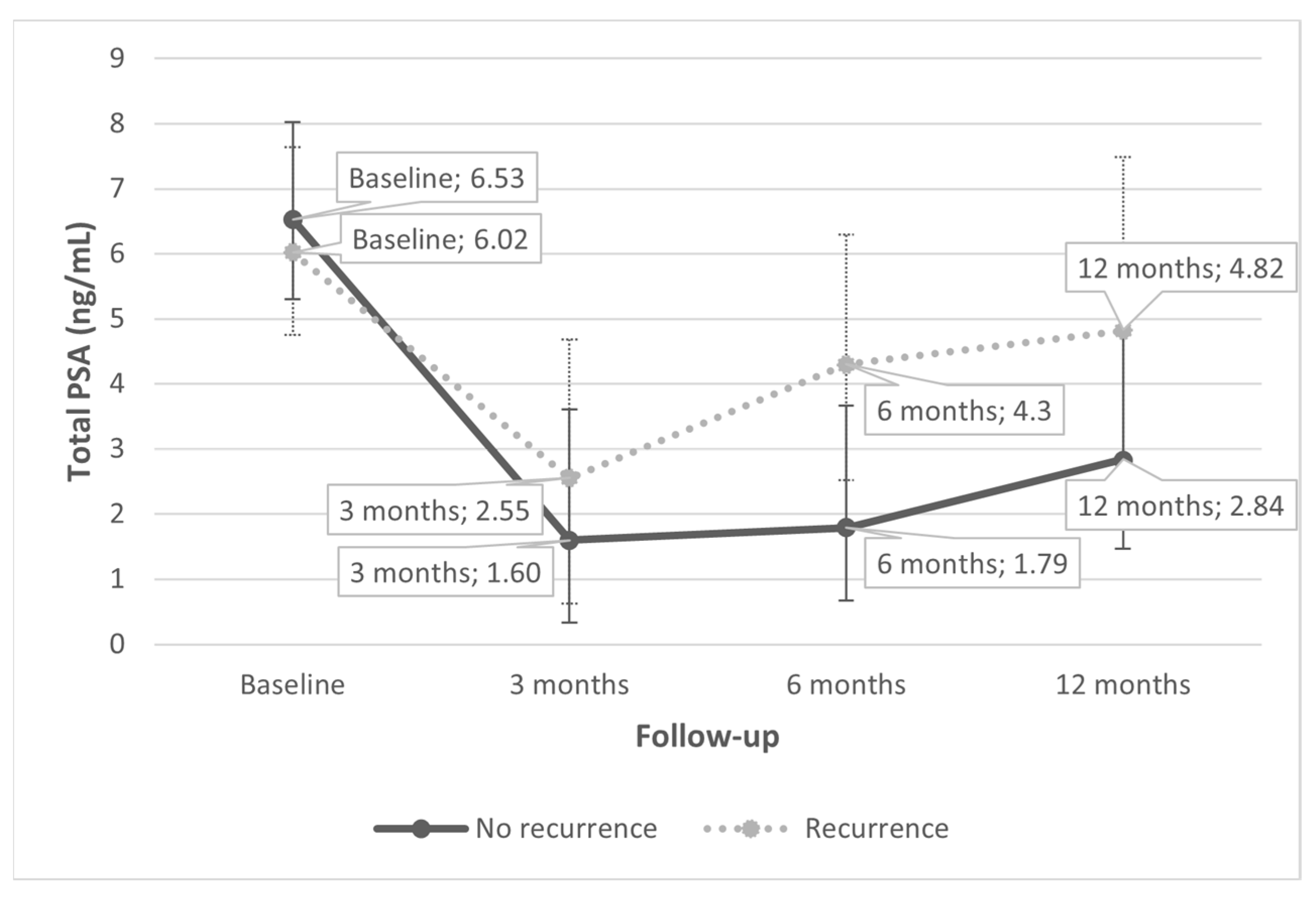
3. Results
4. Discussion
5. Conclusions
Author Contributions
Funding
Institutional Review Board Statement
Informed Consent Statement
Data Availability Statement
Conflicts of Interest
References
- Lebastchi, A.H.; George, A.K.; Polascik, T.J.; Coleman, J.; de la Rosette, J.; Turkbey, B.; Wood, B.J.; Gorin, M.A.; Sidana, A.; Ghai, S.; et al. Standardized Nomenclature and Surveillance Methodologies After Focal Therapy and Partial Gland Ablation for Localized Prostate Cancer: An International Multidisciplinary Consensus. Eur. Urol. 2020, 78, 371–378. [Google Scholar] [CrossRef] [PubMed]
- Eggener, S.E.; Scardino, P.T.; Carroll, P.R.; Zelefsky, M.J.; Sartor, O.; Hricak, H.; Wheeler, T.M.; Fine, S.W.; Trachtenberg, J.; Rubin, M.A.; et al. Focal Therapy for Localized Prostate Cancer: A Critical Appraisal of Rationale and Modalities. J. Urol. 2007, 178, 2260–2267. [Google Scholar] [CrossRef]
- Bates, A.S.; Ayers, J.; Kostakopoulos, N.; Lumsden, T.; Schoots, I.G.; Willemse, P.-P.M.; Yuan, Y.; van den Bergh, R.C.N.; Grummet, J.P.; van der Poel, H.G.; et al. A Systematic Review of Focal Ablative Therapy for Clinically Localised Prostate Cancer in Comparison with Standard Management Options: Limitations of the Available Evidence and Recommendations for Clinical Practice and Further Research. Eur. Urol. Oncol. 2021, 4, 405–423. [Google Scholar] [CrossRef] [PubMed]
- Van der Poel, H.G.; van den Bergh, R.C.N.; Briers, E.; Cornford, P.; Govorov, A.; Henry, A.M.; Lam, T.B.; Mason, M.D.; Rouvière, O.; De Santis, M.; et al. Focal Therapy in Primary Localised Prostate Cancer: The European Association of Urology Position in 2018. Eur. Urol. 2018, 74, 84–91. [Google Scholar] [CrossRef] [PubMed]
- Mottet, N.; Cornford, P.; van den Bergh, R.C.N.; Briers, E.; Expert Patient Advocate (European Prostate Cancer Coalition/Europa UOMO); De Santis, M.; Gillessen, S.; Grummet, J.; Henry, A.M.; van der Kwast, T.H.; et al. Guidelines on Prostate Cancer. In Proceedings of the EAU Annual Congress; Amsterdam, The Netherlands, 1–4 July 2022, EAU Guidelines Office: Arnhem, The Netherlands, 2022; ISBN 978-94-92671-16-5. [Google Scholar]
- Muller, B.G.; van den Bos, W.; Brausi, M.; Fütterer, J.J.; Ghai, S.; Pinto, P.A.; Popeneciu, I.V.; De Reijke, T.M.; Robertson, C.; De La Rosette, J.J.M.C.H.; et al. Follow-up modalities in focal therapy for prostate cancer: Results from a Delphi consensus project. World J. Urol. 2015, 33, 1503–1509. [Google Scholar] [CrossRef] [PubMed]
- Connor, M.J.; Gorin, M.A.; Ahmed, H.U.; Nigam, R. Focal therapy for localized prostate cancer in the era of routine multi-parametric MRI. Prostate Cancer Prostatic Dis. 2020, 23, 232–243. [Google Scholar] [CrossRef]
- Watts, K.L.; Frechette, L.; Muller, B.; Ilinksy, D.; Kovac, E.; Sankin, A.; Aboumohamed, A. Systematic review and meta-analysis comparing cognitive vs. image-guided fusion prostate biopsy for the detection of prostate cancer. Urol. Oncol. Semin. Orig. Investig. 2020, 38, 734.e19–734.e25. [Google Scholar] [CrossRef]
- Shoji, S.; Hiraiwa, S.; Hanada, I.; Kim, H.; Nitta, M.; Hasegawa, M.; Kawamura, Y.; Hashida, K.; Tajiri, T.; Miyajima, A. Current status and future prospective of focal therapy for localized prostate cancer: Development of multiparametric MRI, MRI-TRUS fusion image-guided biopsy, and treatment modalities. Int. J. Clin. Oncol. 2020, 25, 509–520. [Google Scholar] [CrossRef]
- Shah, T.T.; Peters, M.; Eldred-Evans, D.; Miah, S.; Yap, T.; Faure-Walker, N.A.; Hosking-Jervis, F.; Thomas, B.; Dudderidge, T.; Hindley, R.G.; et al. Early-Medium-Term Outcomes of Primary Focal Cryotherapy to Treat Nonmetastatic Clinically Significant Prostate Cancer from a Prospective Multicentre Registry. Eur. Urol. 2019, 76, 98–105. [Google Scholar] [CrossRef]
- Jung, J.H.; Risk, M.C.; Goldfarb, R.; Reddy, B.; Coles, B.; Dahm, P. Primary cryotherapy for localised or locally advanced prostate cancer. Cochrane Database Syst Rev. 2018, 5, CD005010. [Google Scholar] [CrossRef]
- Erinjeri, J.P.; Clark, T.W. Cryoablation: Mechanism of Action and Devices. J. Vasc. Interv. Radiol. 2010, 21, S187–S191. [Google Scholar] [CrossRef] [PubMed]
- World Medical Association. World Medical Association Declaration of Helsinki. Ethical principles for medical research in-volving human subjects. Bull. World Health Organ. 2001, 79, 373–374. [Google Scholar]
- Turkbey, B.; Rosenkrantz, A.B.; Haider, M.A.; Padhani, A.R.; Villeirs, G.; Macura, K.J.; Tempany, C.M.; Choyke, P.L.; Cornud, F.; Margolis, D.J.; et al. Prostate Imaging Reporting and Data System Version 2.1: 2019 Update of Prostate Imaging Reporting and Data System Version 2. Eur. Urol. 2019, 76, 340–351. [Google Scholar] [CrossRef]
- Ahmed, H.U. The Index Lesion and the Origin of Prostate Cancer. N. Engl. J. Med. 2009, 361, 1704–1706. [Google Scholar] [CrossRef] [PubMed]
- Oishi, M.; Gill, I.S.; Ashrafi, A.N.; Lin-Brande, M.; Nassiri, N.; Shin, T.; Bove, A.; Cacciamani, G.E.; Ukimura, O.; Bahn, D.K.; et al. Primary Whole-gland Cryoablation for Prostate Cancer: Biochemical Failure and Clinical Recurrence at 5.6 Years of Follow-up. Eur. Urol. 2019, 75, 208–214. [Google Scholar] [CrossRef] [PubMed]
- Barry, M.J.; Fowler, F.J.; O’Leary, M.P.; Bruskewitz, R.C.; Holtgrewe, H.L.; Mebust, W.K.; Cockett, A.T. The Measurement Committee of the American Urological Association the American Urological Association Symptom Index for Benign Prostatic Hyperplasia. J. Urol. 1992, 148, 1549–1557. [Google Scholar] [CrossRef]
- Avery, K.; Donovan, J.; Peters, T.J.; Shaw, C.; Gotoh, M.; Abrams, P. ICIQ: A brief and robust measure for evaluating the symptoms and impact of urinary incontinence. Neurourol. Urodyn. 2004, 23, 322–330. [Google Scholar] [CrossRef]
- Cappelleri, J.C.; Rosen, R.C.; Smith, M.D.; Mishra, A.; Osterloh, I.H. Diagnostic evaluation of the erectile function domain of the international index of erectile function. Urology 1999, 54, 346–351. [Google Scholar] [CrossRef]
- Holze, S.; Mende, M.; Healy, K.V.; Koehler, N.; Gansera, L.; Truss, M.C.; Rebmann, U.; Degener, S.; Stolzenburg, J.-U. Comparison of various continence definitions in a large group of patients undergoing radical prostatectomy: A multicentre, prospective study. BMC Urol. 2019, 19, 70. [Google Scholar] [CrossRef]
- Dindo, D.; Demartines, N.; Clavien, P.A. Classification of surgical complications: A new proposal with evaluation in a cohort of 6336 patients and results of a survey. Ann. Surg. 2004, 240, 205–213. [Google Scholar] [CrossRef]
- Ghasemi, A.; Zahediasl, S. Normality Tests for Statistical Analysis: A Guide for Non-Statisticians Asghar. Int. J. Endocrinol. Metab. 2012, 10, 486–489. [Google Scholar] [CrossRef] [PubMed] [Green Version]
- Hajian-Tilaki, K. Receiver Operating Characteristic (ROC) Curve Analysis for Medical Diagnostic Test Evaluation. Casp. J. Intern. Med. 2013, 4, 627–635. [Google Scholar]
- Lim, R.; Liong, M.L.; Lim, K.K.; Leong, W.S.; Yuen, K.H. The Minimum Clinically Important Difference of the International Consultation on Incontinence Questionnaires (ICIQ-UI SF and ICIQ-LUTSqol). Urology 2019, 133, 91–95. [Google Scholar] [CrossRef] [PubMed]
- Nickel, J.C.; Brock, G.B.; Herschorn, S.; Dickson, R.; Henneges, C.; Viktrup, L. Proportion of tadalafil-treated patients with clinically meaningful improvement in lower urinary tract symptoms associated with benign prostatic hyperplasia-integrated data from 1 499 study participants. BJU Int. 2015, 115, 815–821. [Google Scholar] [CrossRef] [PubMed] [Green Version]
- Rosen, R.C.; Allen, K.R.; Ni, X.; Araujo, A.B. Minimal clinically important differences in the erectile function domain of the international index of erectile function scale. Eur. Urol. 2011, 60, 1010–1016. [Google Scholar] [CrossRef] [PubMed]
- Bahn, D.; Abreu, A.L.D.C.; Gill, I.S.; Hung, A.J.; Silverman, P.; Gross, M.E.; Lieskovsky, G.; Ukimura, O. Focal Cryotherapy for Clinically Unilateral, Low-Intermediate Risk Prostate Cancer in 73 Men with a Median Follow-Up of 3.7 Years. Eur. Urol. 2012, 62, 55–63. [Google Scholar] [CrossRef]
- Valerio, M.; Shah, T.T.; Shah, P.; Mccartan, N.; Emberton, M.; Arya, M.; Ahmed, H.U. Magnetic resonance imaging-transrectal ultrasound fusion focal cryotherapy of the prostate: A prospective development study. Urol. Oncol. Semin. Orig. Investig. 2017, 35, 150.e1–150.e7. [Google Scholar] [CrossRef]
- Bergelson, I.; Saltman, A.; Hill, S.; Tobler, J.; Verma, S.; Sidana, A. Focal Cryoablation of the Prostate Using Multiparametric Magnetic Resonance Imaging–Ultrasound Fusion Technique. Videourology 2020, 34. [Google Scholar] [CrossRef]
- Bianco, F.; Lopez, A.; Pluchino, A.; Gheiler, E.; Kaufman, A.; Hu, J.; Gonzalez, P.; Egui-Benatuil, G.; Woodhouse, C. MP70-05 prostate MRI pirads validation for biopsies 1 year after MRI fusion target prostate partial gland ablation. J. Urol. 2020, 203, e1056–e1057. [Google Scholar] [CrossRef]
- Kongnyuy, M.; Lipsky, M.J.; Islam, S.; Robins, D.J.; Hager, S.; Halpern, D.M.; Kosinski, K.E.; Schiff, J.T.; Corcoran, A.T.; Wenske, S.; et al. Predictors of biochemical recurrence after primary focal cryosurgery (hemiablation) for localized prostate cancer: A multi-institutional analytic comparison of Phoenix and Stuttgart criteria. Urol. Oncol. Semin. Orig. Investig. 2017, 35, 530.e15–530.e19. [Google Scholar] [CrossRef]
- Blana, A.; Brown, S.C.W.; Chaussy, C.; Conti, G.N.; Eastham, J.A.; Ganzer, R.; Murat, F.J.; Pasticier, G.; Rebillard, X.; Rewcaste, J.C.; et al. High-intensity focused ultrasound for biochemical failure. BJU Int. 2009, 104, 1058–1062. [Google Scholar] [CrossRef] [PubMed]


| Subjects, n | 75 |
| Age, years Median (IQR) | 67 (62–72) |
| Prostate volume, cc Median (IQR) | 50 (35–70.3) |
| Total PSA, ng/mL Median (IQR) | 6.56 (5.1–8) |
| PSA-density, ng/mL/cc Median (IQR) | 0.12 (0.08–0.20) |
| IPSS, points Median (IQR) | 12 (7–16.8) |
| IIEF-EF, points Median (IQR) | 17 (7–22) |
| ICIQ-SF, points Median (IQR) | 0 (0–0) |
| Tumor stage, n (%) | T1c: 24 (32) T2a: 41 (54.7) T2b: 2 (2.7) T2c: 6 (8) NA: 2 (2.7) |
| Volume, cc Median (IQR) | 1.0 (0.54–2.47) |
| Size, mm Median (IQR) | 12 (9–16.5) |
| Prostate segment, n (%) | Base: 12 (16) Middle: 44 (58.7) Apex: 18 (24) NA: 1 (1.3) |
| Prostate lobe, n (%) | Right lobe: 37 (49.3) Left lobe: 33 (44) Both lobes: 5 (6.7) |
| Prostate zone, n (%) | Peripheral: 57 (76) Transition: 12 (16) Anterior fibromuscular stroma: 5 (6.7) Central zone: 0 (0) NA: 1 (1.3) |
| PI-RADS score, n (%) | 1: 0 (0) 2: 1 (1.3) 3: 11 (14.7) 4: 45 (60) 5: 17 (22.7) NA: 1 (1.3) |
| GS, n (%) | 6: 33 (44) 7 (3 + 4): 23 (30.7) 7 (4 + 3): 19 (25.3) |
| EAU Risk Group, n (%) | Low: 19 (25.3) Intermediate: 48 (64) High: 6 (8) NA: 2 (2.7) |
| Infield lesions | GS 6: 2 GS 7 (3 + 4): 6 GS 7 (4 + 3): 2 GS 8: 2 |
| Outfield lesions | GS 6: 7 GS 7 (3 + 4): 2 GS 7 (4 + 3): 5 GS 8: 2 |
| Baseline | 6 Months | 12 Months | p-Value * | |
|---|---|---|---|---|
| IPSS, points Median (IQR) | 67 patients 12 (7–16.8) | 67 patients 13 (9–16) | 60 patients 12 (8–15) | 0.176 |
| IIEF-EF, points Median (IQR) | 70 patients 17 (7–22) | 70 patients 15 (7–19) | 63 patients 16.5 (8–20) | <0.001 |
| ICIQ-SF, points Median (IQR, min-max) | 70 patients 0 (0–0, 0–4) | 67 patients 0 (0–0, 0–6) | 61 patients 0 (0–0, 0–5) | 0.689 |
Publisher’s Note: MDPI stays neutral with regard to jurisdictional claims in published maps and institutional affiliations. |
© 2022 by the authors. Licensee MDPI, Basel, Switzerland. This article is an open access article distributed under the terms and conditions of the Creative Commons Attribution (CC BY) license (https://creativecommons.org/licenses/by/4.0/).
Share and Cite
Fernández-Pascual, E.; Manfredi, C.; Martín, C.; Martínez-Ballesteros, C.; Balmori, C.; Lledó-García, E.; Quintana, L.M.; Curvo, R.; Carballido-Rodríguez, J.; Bianco, F.J., Jr.; et al. mpMRI-US Fusion-Guided Targeted Cryotherapy in Patients with Primary Localized Prostate Cancer: A Prospective Analysis of Oncological and Functional Outcomes. Cancers 2022, 14, 2988. https://doi.org/10.3390/cancers14122988
Fernández-Pascual E, Manfredi C, Martín C, Martínez-Ballesteros C, Balmori C, Lledó-García E, Quintana LM, Curvo R, Carballido-Rodríguez J, Bianco FJ Jr., et al. mpMRI-US Fusion-Guided Targeted Cryotherapy in Patients with Primary Localized Prostate Cancer: A Prospective Analysis of Oncological and Functional Outcomes. Cancers. 2022; 14(12):2988. https://doi.org/10.3390/cancers14122988
Chicago/Turabian StyleFernández-Pascual, Esaú, Celeste Manfredi, Cristina Martín, Claudio Martínez-Ballesteros, Carlos Balmori, Enrique Lledó-García, Luis Miguel Quintana, Raphael Curvo, Joaquín Carballido-Rodríguez, Fernando J. Bianco, Jr., and et al. 2022. "mpMRI-US Fusion-Guided Targeted Cryotherapy in Patients with Primary Localized Prostate Cancer: A Prospective Analysis of Oncological and Functional Outcomes" Cancers 14, no. 12: 2988. https://doi.org/10.3390/cancers14122988
APA StyleFernández-Pascual, E., Manfredi, C., Martín, C., Martínez-Ballesteros, C., Balmori, C., Lledó-García, E., Quintana, L. M., Curvo, R., Carballido-Rodríguez, J., Bianco, F. J., Jr., & Martínez-Salamanca, J. I. (2022). mpMRI-US Fusion-Guided Targeted Cryotherapy in Patients with Primary Localized Prostate Cancer: A Prospective Analysis of Oncological and Functional Outcomes. Cancers, 14(12), 2988. https://doi.org/10.3390/cancers14122988