
Analysis of Cross-Combinations of Feature Selection and Machine-Learning Classification Methods Based on [18F]F-FDG PET/CT Radiomic Features for Metabolic Response Prediction of Metastatic Breast Cancer Lesions

 ,
,  ,
,  ,
,  ,
,  and
and
Abstract
:Simple Summary
Abstract
1. Introduction
2. Materials and Methods
2.1. Patient Cohort
2.2. PET/CT Image Acquisition
2.3. ROI Delineation
2.4. Metabolic Parameters Extraction
2.5. Image Preprocessing
2.6. PET/CT Response Assessment
- Complete metabolic response (CMR): the disappearance of the metabolically active lesion;
- Partial metabolic response (PMR): more than 30% decrease in SULpeak;
- Progressive metabolic disease (PMD): more than 30% increase in SULpeak;
- Stable metabolic disease (SMD): does not meet the above criteria.
2.7. Radiomic Features Extraction
Texture Features
2.8. Univariate Statistical Analysis
2.9. Machine-Learning Model
2.9.1. Feature Selection
2.9.2. Classification Methods
2.9.3. Model Construction
- Data imputation by filling the empty data with a strategy of most frequent.
- Data splitting with a ratio (80:20) into X_train, X_test, y_train, and y_test, where X and y are the predictive features (clinical and radiomic features) and the target variable (responders or nonresponders), respectively. Only the training set was used to construct the models, and the test set for validation purposes.
- A synthetic minority oversampling technique (SMOTE) [30] was performed for oversampling the nonresponder to have the same number of instances as the responder in the training procedure.
- Data standardization: all variables are obligated to have a mean zero and standard deviation of one.
- The feature selection method, hierarchical clustering, was applied directly after the data-preprocessing methodology, to obtain a smaller number of features. However, ANOVA F-test, MI, PCA, IPA, Lasso, and Wilcoxon were initially coupled to each one of the seven ML classifier methods, and subsequently, an iterative process was implemented to find a subgroup of features with the best performance in terms of ACC and AUC. For them, curves of the number of features selected versus model performance were obtained, allowing for optimization of the final number of chosen features, i.e., to find the smaller number of features with only a small change in the model performance concerning the maximal (only changes < 0.05 were allowed if there were a significant reduction in the number of features). Additionally, we obtained a ranking of the features (i.e., the feature importance) for each cross-combination. For the feature selection method Lasso, a cross-validated estimation of the best alpha parameters was performed, using the mean squared error as cross-validation score, where higher values are better than lower values (Supplemental Figure S1).
- ML classifier hyperparameter tuning was also performed through cross-validation, and by using the class GridSearchCV of SciKit Learn. For GNB, the default hyperparameter setting was used.
- Finally, the 49 cross-combinations (each one with a specific subset of features, and an ML classifier with specific hyperparameters) were trained using the training cohort.
2.9.4. Model Performance Metrics and validation
3. Results
3.1. Clinical Characteristics
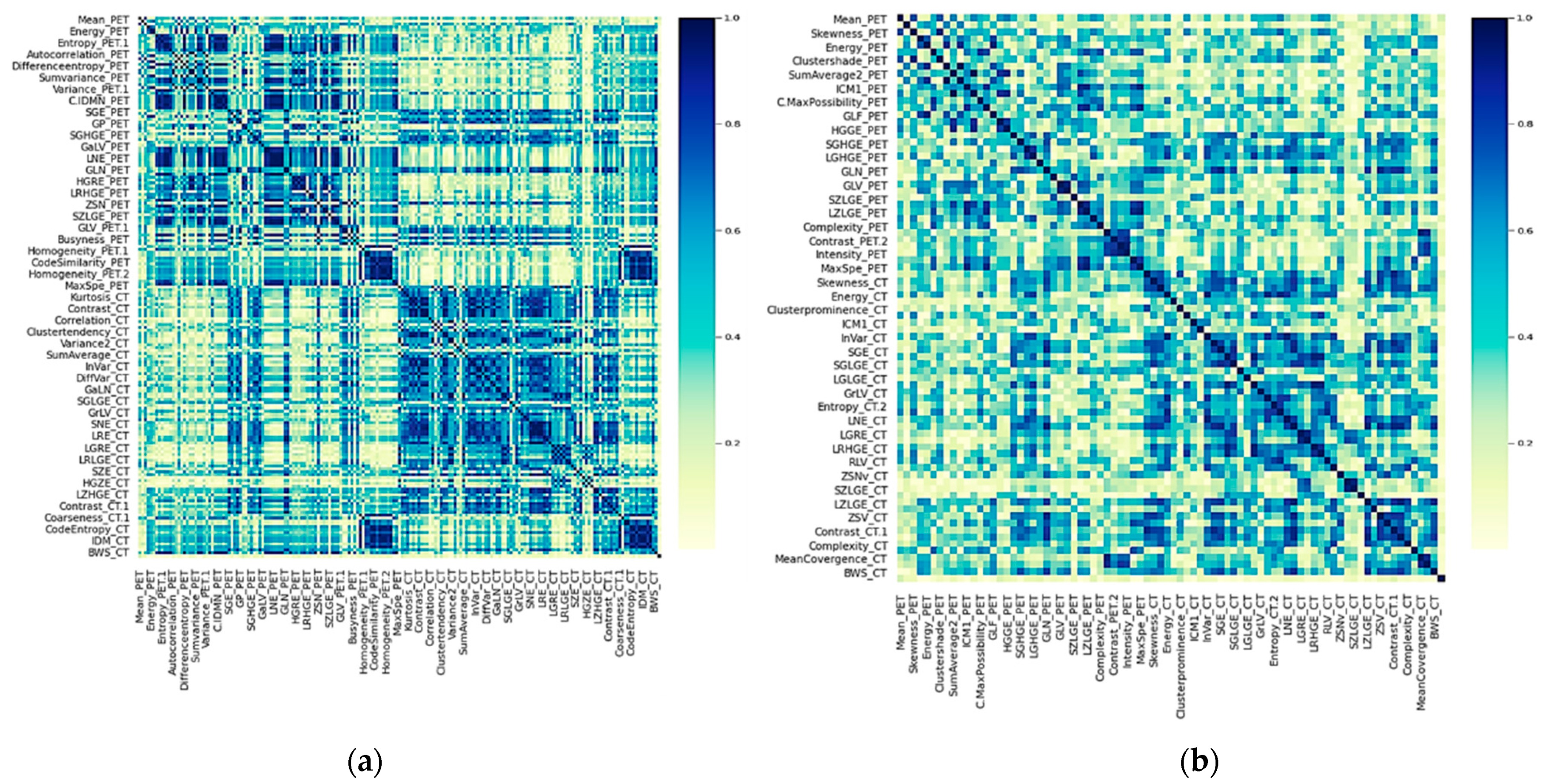
3.2. Feature Extraction and Correlation
3.3. Feature Reduction
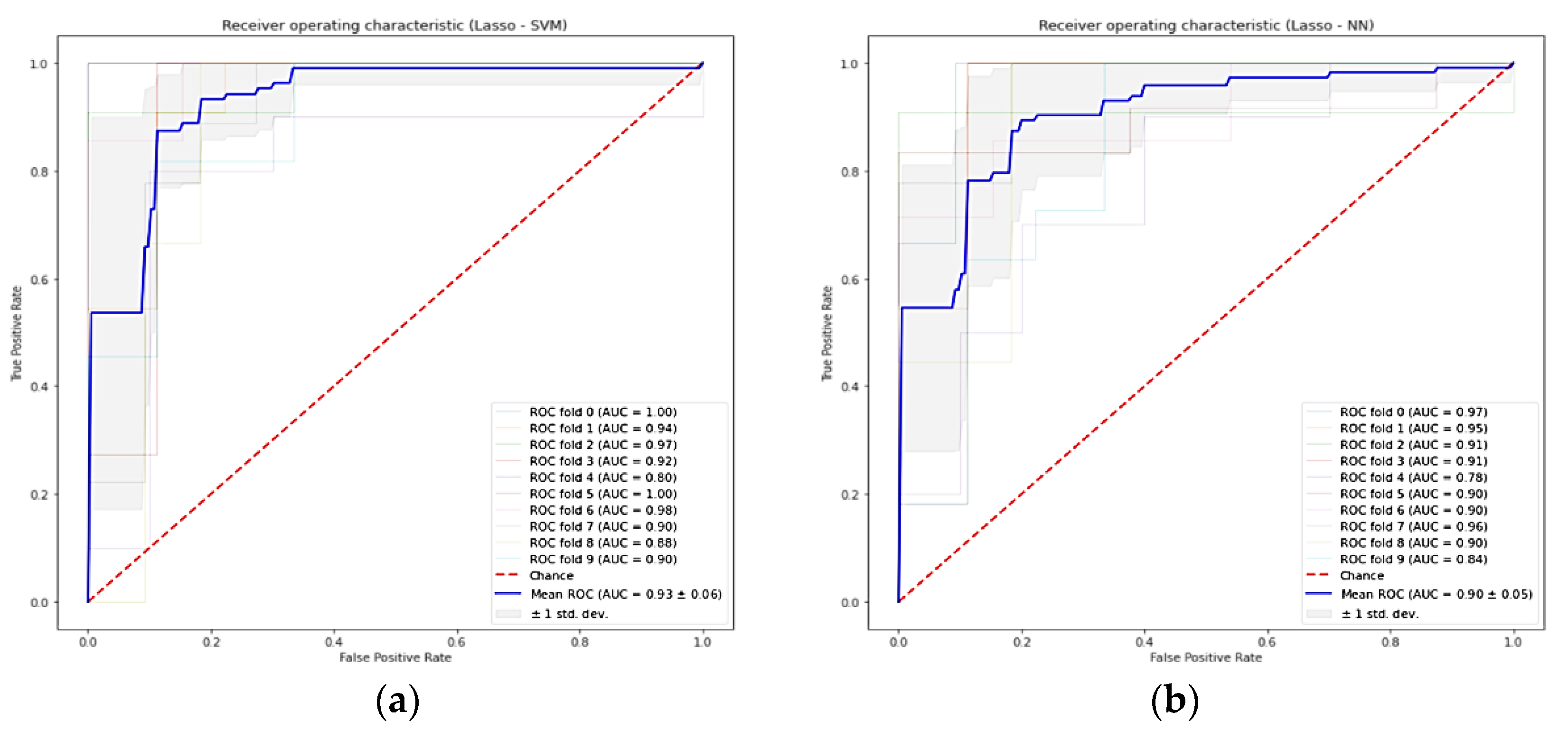
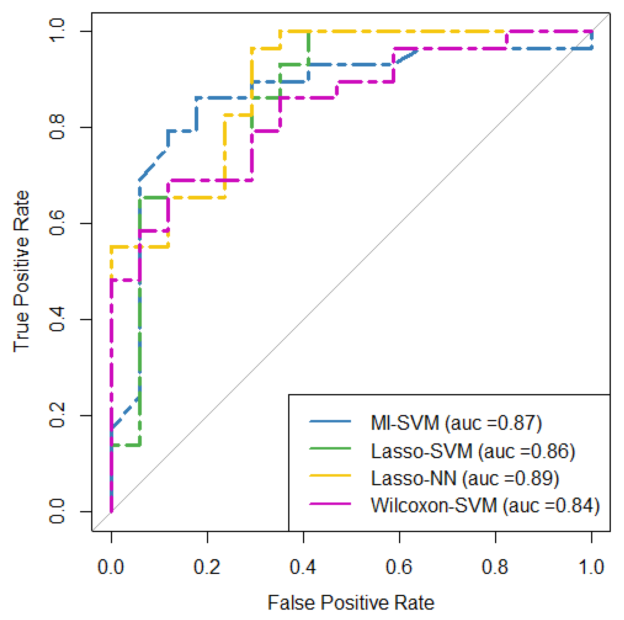
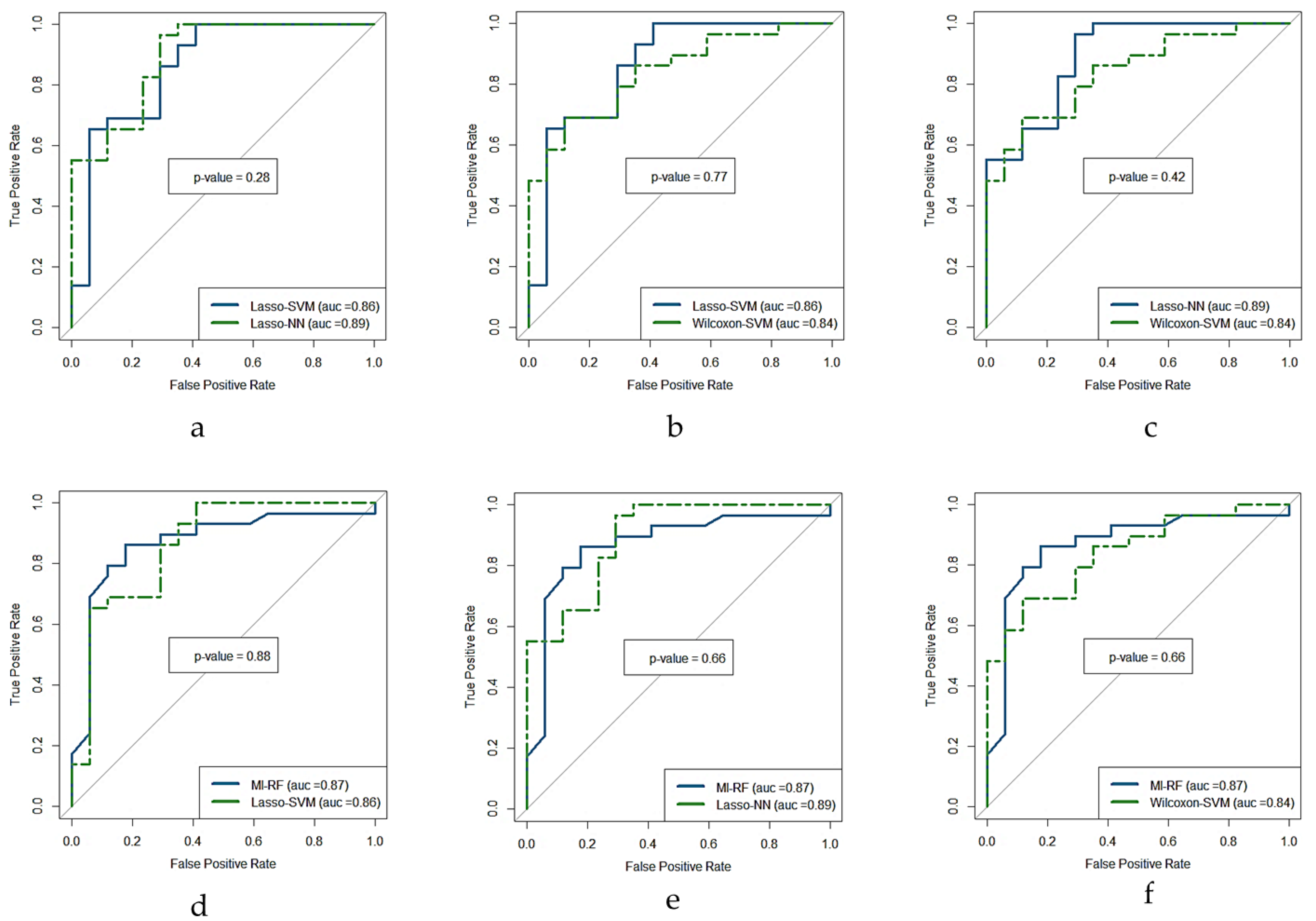
3.4. Cross-Validation
4. Discussion
5. Conclusions
Supplementary Materials
Author Contributions
Funding
Institutional Review Board Statement
Informed Consent Statement
Data Availability Statement
Acknowledgments
Conflicts of Interest
References
- Sung, H.; Ferlay, J.; Siegel, R.L.; Laversanne, M.; Soerjomataram, I.; Jemal, A.; Bray, F. Global Cancer Statistics 2020: GLOBOCAN Estimates of Incidence and Mortality Worldwide for 36 Cancers in 185 Countries. CA Cancer J. Clin. 2021, 71, 209–249. [Google Scholar] [CrossRef] [PubMed]
- Waks, A.G.; Winer, E.P. Breast Cancer Treatment: A Review. JAMA 2019, 321, 288–300. [Google Scholar] [CrossRef] [PubMed]
- Pinker, K.; Chin, J.; Melsaether, A.N.; Morris, E.A.; Moy, L. Precision Medicine and Radiogenomics in Breast Cancer: New Approaches toward Diagnosis and Treatment. Radiology 2018, 287, 732–747. [Google Scholar] [CrossRef] [PubMed]
- Ulaner, G.A. PET/CT for Patients with Breast Cancer: Where Is the Clinical Impact? Am. J. Roentgenol. 2019, 213, 254–265. [Google Scholar] [CrossRef] [PubMed]
- Groheux, D. Role of Fludeoxyglucose in Breast Cancer. PET Clin. 2018, 13, 395–414. [Google Scholar] [CrossRef]
- Zhang, F.; Xu, H.; Liu, J.; Xu, Y.; Chen, B.; Yang, Y.; Yan, N.; Song, S.; Lin, Y.; Xu, Y. 18F-FDG PET/CT for the Early Prediction of the Response Rate and Survival of Patients with Recurrent or Metastatic Breast Cancer. Oncol. Lett. 2018, 16, 4151–4158. [Google Scholar] [CrossRef] [Green Version]
- Humbert, O.; Chardin, D. Dissociated Response in Metastatic Cancer: An Atypical Pattern Brought Into the Spotlight With Immunotherapy. Front. Oncol. 2020, 10, 566297. [Google Scholar] [CrossRef]
- Huyge, V.; Garcia, C.; Alexiou, J.; Ameye, L.; Vanderlinden, B.; Lemort, M.; Bergmann, P.; Awada, A.; Body, J.-J.; Flamen, P. Heterogeneity of Metabolic Response to Systemic Therapy in Metastatic Breast Cancer Patients. Clin. Oncol. 2010, 22, 818–827. [Google Scholar] [CrossRef]
- Lambin, P.; Leijenaar, R.T.H.; Deist, T.M.; Peerlings, J.; de Jong, E.E.C.; van Timmeren, J.; Sanduleanu, S.; Larue, R.T.H.M.; Even, A.J.G.; Jochems, A.; et al. Radiomics: The Bridge between Medical Imaging and Personalized Medicine. Nat. Rev. Clin. Oncol. 2017, 14, 749–762. [Google Scholar] [CrossRef]
- Grossmann, P.; Stringfield, O.; El-Hachem, N.; Bui, M.M.; Rios Velazquez, E.; Parmar, C.; Leijenaar, R.T.; Haibe-Kains, B.; Lambin, P.; Gillies, R.J.; et al. Defining the Biological Basis of Radiomic Phenotypes in Lung Cancer. eLife 2017, 6, e23421. [Google Scholar] [CrossRef]
- Liu, Z.; Wang, S.; Dong, D.; Wei, J.; Fang, C.; Zhou, X.; Sun, K.; Li, L.; Li, B.; Wang, M.; et al. The Applications of Radiomics in Precision Diagnosis and Treatment of Oncology: Opportunities and Challenges. Theranostics 2019, 9, 1303–1322. [Google Scholar] [CrossRef] [PubMed]
- Song, J.; Yin, Y.; Wang, H.; Chang, Z.; Liu, Z.; Cui, L. A Review of Original Articles Published in the Emerging Field of Radiomics. Eur. J. Radiol. 2020, 127, 108991. [Google Scholar] [CrossRef] [PubMed]
- Ye, D.-M.; Wang, H.-T.; Yu, T. The Application of Radiomics in Breast MRI: A Review. Technol. Cancer Res. Treat. 2020, 19, 153303382091619. [Google Scholar] [CrossRef] [PubMed]
- Ha, S.; Park, S.; Bang, J.-I.; Kim, E.-K.; Lee, H.-Y. Metabolic Radiomics for Pretreatment 18F-FDG PET/CT to Characterize Locally Advanced Breast Cancer: Histopathologic Characteristics, Response to Neoadjuvant Chemotherapy, and Prognosis. Sci. Rep. 2017, 7, 1556. [Google Scholar] [CrossRef] [PubMed]
- Langs, G.; Röhrich, S.; Hofmanninger, J.; Prayer, F.; Pan, J.; Herold, C.; Prosch, H. Machine Learning: From Radiomics to Discovery and Routine. Radiologe 2018, 58, 1–6. [Google Scholar] [CrossRef] [PubMed] [Green Version]
- Mwangi, B.; Tian, T.S.; Soares, J.C. A Review of Feature Reduction Techniques in Neuroimaging. Neuroinformatics 2014, 12, 229–244. [Google Scholar] [CrossRef] [PubMed]
- Edge, S.B.; Compton, C.C. The American Joint Committee on Cancer: The 7th Edition of the AJCC Cancer Staging Manual and the Future of TNM. Ann. Surg. Oncol. 2010, 17, 1471–1474. [Google Scholar] [CrossRef]
- Papp, L.; Rausch, I.; Grahovac, M.; Hacker, M.; Beyer, T. Optimized Feature Extraction for Radiomics Analysis of 18F-FDG PET Imaging. J. Nucl. Med. 2019, 60, 864–872. [Google Scholar] [CrossRef] [Green Version]
- Ha, S.; Choi, H.; Paeng, J.C.; Cheon, G.J. Radiomics in Oncological PET/CT: A Methodological Overview. Nucl. Med. Mol. Imaging 2019, 53, 14–29. [Google Scholar] [CrossRef]
- Shafiq-ul-Hassan, M.; Zhang, G.G.; Latifi, K.; Ullah, G.; Hunt, D.C.; Balagurunathan, Y.; Abdalah, M.A.; Schabath, M.B.; Goldgof, D.G.; Mackin, D.; et al. Intrinsic Dependencies of CT Radiomic Features on Voxel Size and Number of Gray Levels. Med. Phys. 2017, 44, 1050–1062. [Google Scholar] [CrossRef]
- Zwanenburg, A.; Vallières, M.; Abdalah, M.A.; Aerts, H.J.W.L.; Andrearczyk, V.; Apte, A.; Ashrafinia, S.; Bakas, S.; Beukinga, R.J.; Boellaard, R.; et al. The Image Biomarker Standardization Initiative: Standardized Quantitative Radiomics for High-Throughput Image-Based Phenotyping. Radiology 2020, 295, 328–338. [Google Scholar] [CrossRef] [PubMed] [Green Version]
- O, J.H.; Lodge, M.A.; Wahl, R.L. Practical PERCIST: A Simplified Guide to PET Response Criteria in Solid Tumors 1.0. Radiology 2016, 280, 576–584. [Google Scholar] [CrossRef] [PubMed] [Green Version]
- Xu, H.; Lv, W.; Feng, H.; Du, D.; Yuan, Q.; Wang, Q.; Dai, Z.; Yang, W.; Feng, Q.; Ma, J.; et al. Subregional Radiomics Analysis of PET/CT Imaging with Intratumor Partitioning: Application to Prognosis for Nasopharyngeal Carcinoma. Mol. Imaging Biol. 2020, 22, 1414–1426. [Google Scholar] [CrossRef] [PubMed]
- Yin, P.; Mao, N.; Zhao, C.; Wu, J.; Sun, C.; Chen, L.; Hong, N. Comparison of Radiomics Machine-Learning Classifiers and Feature Selection for Differentiation of Sacral Chordoma and Sacral Giant Cell Tumour Based on 3D Computed Tomography Features. Eur. Radiol. 2019, 29, 1841–1847. [Google Scholar] [CrossRef]
- Du, D.; Feng, H.; Lv, W.; Ashrafinia, S.; Yuan, Q.; Wang, Q.; Yang, W.; Feng, Q.; Chen, W.; Rahmim, A.; et al. Machine Learning Methods for Optimal Radiomics-Based Differentiation Between Recurrence and Inflammation: Application to Nasopharyngeal Carcinoma Post-Therapy PET/CT Images. Mol. Imaging Biol. 2020, 22, 730–738. [Google Scholar] [CrossRef]
- Zhang, B.; He, X.; Ouyang, F.; Gu, D.; Dong, Y.; Zhang, L.; Mo, X.; Huang, W.; Tian, J.; Zhang, S. Radiomic Machine-Learning Classifiers for Prognostic Biomarkers of Advanced Nasopharyngeal Carcinoma. Cancer Lett. 2017, 403, 21–27. [Google Scholar] [CrossRef]
- Delzell, D.A.P.; Magnuson, S.; Peter, T.; Smith, M.; Smith, B.J. Machine Learning and Feature Selection Methods for Disease Classification With Application to Lung Cancer Screening Image Data. Front. Oncol. 2019, 9, 1393. [Google Scholar] [CrossRef] [Green Version]
- Parmar, C.; Grossmann, P.; Bussink, J.; Lambin, P.; Aerts, H.J.W.L. Machine Learning Methods for Quantitative Radiomic Biomarkers. Sci. Rep. 2015, 5, 13087. [Google Scholar] [CrossRef]
- Zwanenburg, A. Radiomics in Nuclear Medicine: Robustness, Reproducibility, Standardization, and How to Avoid Data Analysis Traps and Replication Crisis. Eur. J. Nucl. Med. Mol. Imaging 2019, 46, 2638–2655. [Google Scholar] [CrossRef]
- Chawla, N.V.; Bowyer, K.W.; Hall, L.O.; Kegelmeyer, W.P. SMOTE: Synthetic Minority Over-Sampling Technique. J. Air 2002, 16, 321–357. [Google Scholar] [CrossRef]
- Robin, X.; Turck, N.; Hainard, A.; Tiberti, N.; Lisacek, F.; Sanchez, J.-C.; Müller, M. PROC: An Open-Source Package for R and S+ to Analyze and Compare ROC Curves. BMC Bioinform. 2011, 12, 77. [Google Scholar] [CrossRef] [PubMed]
- Magometschnigg, H.; Pinker, K.; Helbich, T.; Brandstetter, A.; Rudas, M.; Nakuz, T.; Baltzer, P.; Wadsak, W.; Hacker, M.; Weber, M.; et al. PIK3CA Mutational Status Is Associated with High Glycolytic Activity in ER+/HER2− Early Invasive Breast Cancer: A Molecular Imaging Study Using [18F]FDG PET/CT. Mol. Imaging Biol. 2019, 21, 991–1002. [Google Scholar] [CrossRef] [PubMed] [Green Version]
- Hendlisz, A.; Deleporte, A.; Delaunoit, T.; Maréchal, R.; Peeters, M.; Holbrechts, S.; Van den Eynde, M.; Houbiers, G.; Filleul, B.; Van Laethem, J.-L.; et al. The Prognostic Significance of Metabolic Response Heterogeneity in Metastatic Colorectal Cancer. PLoS ONE 2015, 10, e0138341. [Google Scholar] [CrossRef]
- Hulikal, N.; Gajjala, S.R.; Kalawat, T.; Kadiyala, S.; Kottu, R. Predicting Response to Neoadjuvant Chemotherapy Using 18F FDG PET-CT in Patients with Locally Advanced Breast Cancer. Asian Pac. J. Cancer Prev. 2020, 21, 93–98. [Google Scholar] [CrossRef] [Green Version]
- Tian, F.; Shen, G.; Deng, Y.; Diao, W.; Jia, Z. The Accuracy of 18F-FDG PET/CT in Predicting the Pathological Response to Neoadjuvant Chemotherapy in Patients with Breast Cancer: A Meta-Analysis and Systematic Review. Eur. Radiol. 2017, 27, 4786–4796. [Google Scholar] [CrossRef]
- Azad, G.K.; Taylor, B.P.; Green, A.; Sandri, I.; Swampillai, A.; Harries, M.; Kristeleit, H.; Mansi, J.; Goh, V.; Cook, G.J.R. Prediction of Therapy Response in Bone-Predominant Metastatic Breast Cancer: Comparison of [18F] Fluorodeoxyglucose and [18F]-Fluoride PET/CT with Whole-Body MRI with Diffusion-Weighted Imaging. Eur. J. Nucl. Med. Mol. Imaging 2019, 46, 821–830. [Google Scholar] [CrossRef] [Green Version]
- Valdora, F.; Houssami, N.; Rossi, F.; Calabrese, M.; Tagliafico, A.S. Rapid Review: Radiomics and Breast Cancer. Breast Cancer Res. Treat. 2018, 169, 217–229. [Google Scholar] [CrossRef]
- Sollini, M.; Cozzi, L.; Ninatti, G.; Antunovic, L.; Cavinato, L.; Chiti, A.; Kirienko, M. PET/CT Radiomics in Breast Cancer: Mind the Step. Methods 2021, 188, 122–132. [Google Scholar] [CrossRef]
- Conti, A.; Duggento, A.; Indovina, I.; Guerrisi, M.; Toschi, N. Radiomics in Breast Cancer Classification and Prediction. Semin. Cancer Biol. 2021, 72, 238–250. [Google Scholar] [CrossRef]
- Antunovic, L.; De Sanctis, R.; Cozzi, L.; Kirienko, M.; Sagona, A.; Torrisi, R.; Tinterri, C.; Santoro, A.; Chiti, A.; Zelic, R.; et al. PET/CT Radiomics in Breast Cancer: Promising Tool for Prediction of Pathological Response to Neoadjuvant Chemotherapy. Eur. J. Nucl. Med. Mol. Imaging 2019, 46, 1468–1477. [Google Scholar] [CrossRef]
- Turashvili, G.; Brogi, E. Tumor Heterogeneity in Breast Cancer. Front. Med. 2017, 4, 227. [Google Scholar] [CrossRef] [PubMed] [Green Version]
- Whybra, P.; Parkinson, C.; Foley, K.; Staffurth, J.; Spezi, E. Assessing Radiomic Feature Robustness to Interpolation in 18F-FDG PET Imaging. Sci. Rep. 2019, 9, 9649. [Google Scholar] [CrossRef] [PubMed] [Green Version]
- Belli, M.L.; Mori, M.; Broggi, S.; Cattaneo, G.M.; Bettinardi, V.; Dell’Oca, I.; Fallanca, F.; Passoni, P.; Vanoli, E.G.; Calandrino, R.; et al. Quantifying the Robustness of [18F]FDG-PET/CT Radiomic Features with Respect to Tumor Delineation in Head and Neck and Pancreatic Cancer Patients. Phys. Med. 2018, 49, 105–111. [Google Scholar] [CrossRef] [PubMed]
- Leijenaar, R.T.H.; Carvalho, S.; Velazquez, E.R.; van Elmpt, W.J.C.; Parmar, C.; Hoekstra, O.S.; Hoekstra, C.J.; Boellaard, R.; Dekker, A.L.A.J.; Gillies, R.J.; et al. Stability of FDG-PET Radiomics Features: An Integrated Analysis of Test-Retest and Inter-Observer Variability. Acta Oncol. 2013, 52, 1391–1397. [Google Scholar] [CrossRef] [PubMed] [Green Version]
- Oliveira, C.; Amstutz, F.; Vuong, D.; Bogowicz, M.; Hüllner, M.; Foerster, R.; Basler, L.; Schröder, C.; Eboulet, E.I.; Pless, M.; et al. Preselection of Robust Radiomic Features Does Not Improve Outcome Modelling in Non-Small Cell Lung Cancer Based on Clinical Routine FDG-PET Imaging. EJNMMI Res. 2021, 11, 79. [Google Scholar] [CrossRef]


| Feature Selection Method | ML Classifier |
|---|---|
| AFT (ANOVA-F-test) | SVM (support vector machine) |
| MI (mutual information) | GNB (Gaussian naive Bayes) |
| PCA (principal component analysis) | RF (random forest) |
| ICA (independent component analysis) | LR (logistic regression) |
| Lasso (least absolute shrinkage and selection operator) | KNN (k-nearest neighborhood) |
| CL (clustering) | AdaBoost (adaptive boosting) |
| WT (Wilcoxon test) | NN (neural network) |
| Characteristic | Number | Percentage |
|---|---|---|
| Total patients | 48 (mean age 48.1 years) | 100 |
| Affected side | ||
| right | 26 | 54.2 |
| left | 22 | 45.8 |
| Histologic type | ||
| ductal | 42 | 87.5 |
| lobular | 5 | 10.4 |
| other | 1 | 2.1 |
| Tumor size a (pT) | ||
| T1a-b | 12 | 25 |
| T1c | 15 | 31.3 |
| T2 | 11 | 22.9 |
| T3 | 5 | 10.4 |
| Nodal affectation a (pN) | ||
| N0 | 14 | 29.2 |
| N1 | 22 | 45.8 |
| N2a-b | 4 | 8.3 |
| N3a | 2 | 4.2 |
| N3b | 1 | 2.1 |
| Mestatase a,b (M) | ||
| M0 | 20 | 39.6 |
| M1 | 1 | 2.1 |
| Mx | 22 | 43.8 |
| TNM clinical stage a | ||
| IA | 13 | 27.1 |
| IB | 0 | 0 |
| IIA | 16 | 33.3 |
| IIB | 4 | 8.3 |
| IIIA | 6 | 12.5 |
| IIIB | 0 | 0 |
| IIIC | 3 | 6.3 |
| IV | 1 | 2.1 |
| Estrogen receptor positivity | ||
| negative | 17 | 54.2 |
| low | 4 | 54.2 |
| moderate | 11 | 54.2 |
| strong | 16 | 54.2 |
| Progesterone receptor positivity | ||
| negative | 24 | 50 |
| low | 8 | 16.7 |
| moderate | 7 | 14.62 |
| strong | 9 | 18.8 |
| Her2 positivity | ||
| 0 | 33 | 68.8 |
| 1 | 15 | 31.3 |
| Histologic grade c | ||
| well | 1 | 2.1 |
| moderate | 20 | 41.7 |
| poor | 26 | 54.2 |
| Patient | Treatment | Metastatic Lesions |
|---|---|---|
| 1 | ChT | Liver (1) |
| 2 | ChT, Xgeva, Zoladex, and RT | Bone (1) |
| 3 | ChT and RT | Liver (1), Lung (3), LN (3) |
| 4 | ChT | Bone (1), LN (3) |
| 5 | Taxotere and Parjeta | Liver (1), LN (3) |
| 6 | Taxol and Herceptin | Breast (1), LN (2) |
| 7 | Taxotere, Herceptin, Perjeta, and Xgeva | Breast (1), Bone (3), Liver (3). LN (3) |
| 8 | Navelbine | Bone (1), LN (6) |
| 9 | Taxol | LN (4) |
| 10 | ChT | Bone (1), Liver (7) |
| 11 | Taxotere, Herceptin, and Perjeta | Liver (3), LN (5) |
| 12 | Paclitaxel and Bevacizumab | LN (7) |
| 13 | ChT | Bone (8), LN (4) |
| 14 | Navelbine | LN (9) |
| 15 | ChT | Bone (1), Liver (2). Pleura (8) |
| 16 | Aromasin, Afinitor, Xgeva, and RT | Bone (6), LN (3) |
| 17 | Xeloda, Avastin, Bortezomib, and RT | Bone (4), LN (4) |
| 18 | ChT and RT | LN (2) |
| 19 | Liver Meta Excision, Xgeva, and Zometa | Bone (7), Liver (3) |
| 20 | Taxotere, Herceptin, and Perjeta | Bone (14), Liver (2), LN (1) |
| 21 | Letrozol, changed to Fulvestrant | Bone (3), LN (2) |
| 22 | Paclitaxel | Bone (3), Liver (2) |
| 23 | Arimidex and Herceptin | LN (1) |
| 24 | Lipidox, lung meta excision | LN (1) |
| 25 | Vinorelbine and Trastuzumab | Liver (1) |
| 26 | Arimidex, lung Meta excision | Bone (1), LN (2) |
| 27 | ChT and Trastuzumab | LN (2) |
| 28 | ChT and RT | Bone (1), LN (2) |
| 29 | Avastin and Abraxane | Bone (1), Suprarenal (1) |
| 30 | ChT | Bone (3) |
| 31 | ChT | LN (1) |
| 32 | ChT and liver metastase excision | Liver (3) |
| 33 | Epirubicin und Docetaxel | Liver (1) |
| 34 | Xgeva and RT | Bone (2) |
| 35 | Xgeva and RT | Bone (1) |
| 36 | Xvega | Bone (6) |
| 37 | Zometa | Bone (4) |
| 38 | Zometa and RT | LN (1) |
| 39 | ChT | Bone (2) |
| 40 | ChT and RT | Bone (1), Liver (1). Lung (1). LN (1) |
| 41 | Radioembolization | Liver (1), Spleen (1) |
| 42 | Taxotere and Avastin | LN (2) |
| 43 | Taxotere and Avastin | Bone (6), LN (2) |
| 44 | Gemzar, Cisplatin, and Avastin | LN (3) |
| 45 | Taxol and Xgeva | Breast (1), Bone (6) |
| 46 | Trastuzumab and Xgeva | Bone (3), Spleen (4) |
| 47 | Xeloda and RT | Liver (2) |
| 48 | Methotrexate and Xgeva | Bone (1), LN (4) |
| Model | Number of Predictors | Ranked Predictors (Predictors on the Left Are of Greater Predictive Significance) |
|---|---|---|
| Lasso + SVM and Lasso + NN | 14 | BWS-PET, SUVmax, Skewness-CT, Kurtosis-PET, ΔER, PR, T, Her2neu-Metastasis, PR-Metastasis, Affectation-Side, ΔGrading, Her2neu-Primary, P53 |
| MI + RF | 13 | LGHGE-CT, PR, Energy-ET, Correlation-PET, Max-PET, ER, SUVpeak, ZSNv-CT, SRLGE-CT, Age at Diagnosis, BWS-PET, PR-Metastasis, P53 |
| Wilcoxon + SVM | 59 | PR, LGHGE-CT, Variance-T, GLN-CT, Correlation-PET, GLV-CT, GLV-PET, ER-Mestastasis, ZSNv-CT, BWS-PET … |
| Classifier | |||||||||
|---|---|---|---|---|---|---|---|---|---|
| Model | SVM | GNB | RF | LR | KNN | Ada Boost | NN | Mean FS | |
| Feature selection (FS) | AFT | 0.83 ± 0.06 | 0.78 ± 0.08 | 0.76 ± 0.08 | 0.74 ± 0.07 | 0.78 ± 0.12 | 0.80 ± 0.08 | 0.78 ± 0.08 | 0.78 ± 0.03 |
| MI | 0.80 ± 0.10 | 0.78 ± 0.10 | 0.80 ± 0.08 | 0.76 ± 0.08 | 0.86 ± 0.08 | 0.75 ± 0.06 | 0.78 ± 0.06 | 0.79 ± 0.04 | |
| PCA | 0.84 ± 0.08 | 0.79 ± 0.07 | 0.81 ± 0.07 | 0.71 ± 0.08 | 0.75 ± 0.11 | 0.68 ± 0.13 | 0.79 ± 0.07 | 0.77 ± 0.06 | |
| ICA | 0.88 ± 0.08 | 0.75 ± 0.05 | 0.75 ± 0.09 | 0.73 ± 0.04 | 0.73 ± 0.12 | 0.64 ± 0.09 | 0.74 ± 0.08 | 0.75 ± 0.07 | |
| Lasso | 0.93 ± 0.06 | 0.80 ± 0.10 | 0.92 ± 0.03 | 0.77 ± 0.08 | 0.92 ± 0.06 | 0.79 ± 0.13 | 0.90 ± 0.05 | 0.86 ± 0.07 | |
| CL | 0.80 ± 0.15 | 0.71 ± 0.08 | 0.86 ± 0.08 | 0.73 ± 0.10 | 0.77 ± 0.10 | 0.78 ± 0.10 | 0.75 ± 0.09 | 0.77 ± 0.05 | |
| WT | 0.84 ± 0.06 | 0.75 ± 0.08 | 0.76 ± 0.09 | 0.75 ± 0.09 | 0.82 ± 0.09 | 0.80 ± 0.09 | 0.79 ± 0.06 | 0.79 ± 0.04 | |
| Mean Classifier | 0.85 ± 0.05 | 0.77 ± 0.03 | 0.81 ± 0.06 | 0.74 ± 0.02 | 0.80 ± 0.07 | 0.75 ± 0.06 | 0.79 ± 0.05 | ||
| Classifier | |||||||||
|---|---|---|---|---|---|---|---|---|---|
| Model | SVM | GNB | RF | LR | KNN | Ada Boost | NN | Mean FS | |
| Feature selection (FS) | AFT | 0.78 | 0.70 | 0.77 | 0.76 | 0.80 | 0.72 | 0.82 | 0.76 ± 0.04 |
| MI | 0.79 | 0.79 | 0.87 | 0.69 | 0.78 | 0.74 | 0.81 | 0.78 ± 0.06 | |
| PCA | 0.80 | 0.69 | 0.81 | 0.68 | 0.65 | 0.71 | 0.7 | 0.72 ± 0.06 | |
| ICA | 0.83 | 0.66 | 0.74 | 0.63 | 0.72 | 0.71 | 0.76 | 0.72 ± 0.07 | |
| Lasso | 0.86 | 0.70 | 0.83 | 0.78 | 0.83 | 0.77 | 0.90 | 0.81 ± 0.07 | |
| CL | 0.81 | 0.78 | 0.77 | 0.79 | 0.81 | 0.65 | 0.80 | 0.77 ± 0.06 | |
| WT | 0.84 | 0.75 | 0.74 | 0.73 | 0.71 | 0.74 | 0.79 | 0.76 ± 0.04 | |
| Mean Classifier | 0.82 ± 0.03 | 0.72 ± 0.05 | 0.79 ± 0.05 | 0.72 ± 0.06 | 0.76 ± 0.07 | 0.72 ± 0.04 | 0.80 ± 0.06 | ||
| Classifier | |||||||||
|---|---|---|---|---|---|---|---|---|---|
| Model | SVM | GNB | RF | LR | KNN | Ada Boost | NN | Mean FS | |
| Feature selection (FS) | AFT | 0.72 | 0.72 | 0.67 | 0.70 | 0.70 | 0.70 | 0.70 | 0.70 ± 0.02 |
| MI | 0.70 | 0.63 | 0.85 | 0.67 | 0.72 | 0.74 | 0.72 | 0.72 ± 0.07 | |
| PCA | 0.74 | 0.63 | 0.74 | 0.70 | 0.63 | 0.70 | 0.63 | 0.68 ± 0.05 | |
| ICA | 0.72 | 0.57 | 0.61 | 0.61 | 0.72 | 0.72 | 0.74 | 0.67 ± 0.07 | |
| Lasso | 0.76 | 0.54 | 0.74 | 0.72 | 0.74 | 0.65 | 0.72 | 0.70 ± 0.08 | |
| CL | 0.72 | 0.74 | 0.67 | 0.72 | 0.72 | 0.65 | 0.74 | 0.71 ± 0.03 | |
| WT | 0.70 | 0.63 | 0.67 | 0.72 | 0.59 | 0.50 | 0.70 | 0.64 ± 0.08 | |
| Mean Classifier | 0.72 ± 0.02 | 0.64 ± 0.07 | 0.71 ± 0.08 | 0.69 ± 0.04 | 0.69 ± 0.06 | 0.67 ± 0.08 | 0.71 ± 0.04 | ||
Publisher’s Note: MDPI stays neutral with regard to jurisdictional claims in published maps and institutional affiliations. |
© 2022 by the authors. Licensee MDPI, Basel, Switzerland. This article is an open access article distributed under the terms and conditions of the Creative Commons Attribution (CC BY) license (https://creativecommons.org/licenses/by/4.0/).
Share and Cite
Gómez, O.V.; Herraiz, J.L.; Udías, J.M.; Haug, A.; Papp, L.; Cioni, D.; Neri, E. Analysis of Cross-Combinations of Feature Selection and Machine-Learning Classification Methods Based on [18F]F-FDG PET/CT Radiomic Features for Metabolic Response Prediction of Metastatic Breast Cancer Lesions. Cancers 2022, 14, 2922. https://doi.org/10.3390/cancers14122922
Gómez OV, Herraiz JL, Udías JM, Haug A, Papp L, Cioni D, Neri E. Analysis of Cross-Combinations of Feature Selection and Machine-Learning Classification Methods Based on [18F]F-FDG PET/CT Radiomic Features for Metabolic Response Prediction of Metastatic Breast Cancer Lesions. Cancers. 2022; 14(12):2922. https://doi.org/10.3390/cancers14122922
Chicago/Turabian StyleGómez, Ober Van, Joaquin L. Herraiz, José Manuel Udías, Alexander Haug, Laszlo Papp, Dania Cioni, and Emanuele Neri. 2022. "Analysis of Cross-Combinations of Feature Selection and Machine-Learning Classification Methods Based on [18F]F-FDG PET/CT Radiomic Features for Metabolic Response Prediction of Metastatic Breast Cancer Lesions" Cancers 14, no. 12: 2922. https://doi.org/10.3390/cancers14122922
APA StyleGómez, O. V., Herraiz, J. L., Udías, J. M., Haug, A., Papp, L., Cioni, D., & Neri, E. (2022). Analysis of Cross-Combinations of Feature Selection and Machine-Learning Classification Methods Based on [18F]F-FDG PET/CT Radiomic Features for Metabolic Response Prediction of Metastatic Breast Cancer Lesions. Cancers, 14(12), 2922. https://doi.org/10.3390/cancers14122922