Article

Tumor Signature Analysis Implicates Hereditary Cancer Genes in Endometrial Cancer Development

1
Department of Genetics and Computational Biology, QIMR Berghofer Medical Research Institute, Brisbane 4006, Australia
2
UQ Centre for Clinical Research, Faculty of Medicine, The University of Queensland, Brisbane 4029, Australia
3
Anatomical Pathology, Pathology Queensland, Brisbane 4029, Australia
4
Familial Cancer Service, Crown Princess Mary Cancer Centre, Westmead Hospital, Sydney 2145, Australia
5
Centre for Cancer Research, The Westmead Institute for Medical Research, Sydney Medical School, University of Sydney, Sydney 2145, Australia
*
Author to whom correspondence should be addressed.
These authors contributed equally to the work.
Cancers 2021, 13(8), 1762; https://doi.org/10.3390/cancers13081762
Submission received: 1 February 2021 / Revised: 22 March 2021 / Accepted: 29 March 2021 / Published: 7 April 2021

Abstract

:

Simple Summary

Women with a family history of cancer are at increased risk of cancer, including endometrial cancer (affecting the womb lining). In some of the women with such family history, the risk can be explained by deleterious changes in mismatch repair genes that cause Lynch syndrome. This study explored the role of other genes in risk of endometrial cancer, using several approaches. The number and type of changes in gene sequence information in women with endometrial cancer was compared to that from individuals in the general population. Gene sequence changes in endometrial cancer patients with a family history of cancer were also analyzed. Lastly, endometrial cancers from individuals with gene changes were examined for distinctive genomic patterns expected to be seen if a gene change was driving the cancer. This study has identified several additional genes for further exploration in relation to endometrial cancer risk and therapy.

Abstract

Risk of endometrial cancer (EC) is increased ~2-fold for women with a family history of cancer, partly due to inherited pathogenic variants in mismatch repair (MMR) genes. We explored the role of additional genes as explanation for familial EC presentation by investigating germline and EC tumor sequence data from The Cancer Genome Atlas (n = 539; 308 European ancestry), and germline data from 33 suspected familial European ancestry EC patients demonstrating immunohistochemistry-detected tumor MMR proficiency. Germline variants in MMR and 26 other known/candidate EC risk genes were annotated for pathogenicity in the two EC datasets, and also for European ancestry individuals from gnomAD as a population reference set (n = 59,095). Ancestry-matched case–control comparisons of germline variant frequency and/or sequence data from suspected familial EC cases highlighted ATM, PALB2, RAD51C, MUTYH and NBN as candidates for large-scale risk association studies. Tumor mutational signature analysis identified a microsatellite-high signature for all cases with a germline pathogenic MMR gene variant. Signature analysis also indicated that germline loss-of-function variants in homologous recombination (BRCA1, PALB2, RAD51C) or base excision (NTHL1, MUTYH) repair genes can contribute to EC development in some individuals with germline variants in these genes. These findings have implications for expanded therapeutic options for EC cases.

1. Introduction

Endometrial cancer (EC) is the most commonly diagnosed gynecological malignancy, with an increased prevalence rate in developed countries [1]. Modifiable factors such as obesity, lifestyle, and hormone levels are associated with increased risk of EC, and women with a family history of EC or other cancers, such as colorectal, are at ~2–3 fold increased risk of EC [2]. The genetic factors identified to date are either common low-risk cancer predisposition variants that act together to cause polygenic disease, or rare high-risk pathogenic variants in cancer syndrome genes generally present in patients with a monogenic disease phenotype [3].
The major known monogenic form of EC is Lynch syndrome, caused by germline pathogenic variants impacting the mismatch repair (MMR) genes MLH1, MSH2, MSH6, PMS2, as well as EPCAM deletions, which impact MSH2 expression. Lynch syndrome accounts for approximately 3–5% of EC at the population level and an increased proportion in cases with family history of colorectal, endometrial and other cancers [4]. The lifetime cumulative risk of EC for women with Lynch syndrome is 40–70%, depending on which MMR gene is disrupted [5]. EC is also a spectrum cancer of Cowden syndrome, caused by the inheritance of pathogenic PTEN variants. The cumulative risk of EC for women up to 60 years of age with Cowden syndrome is around 20% [6]. Studies to date suggest that PTEN pathogenic variants are very rarely detected in the general population, and mostly in the context of clinical features of Cowden syndrome [7].
Results from a recent study assessing risk associated with reported family history of endometrial and other cancers, after considering proband MMR proficiency and MMR germline test results, indicate that the genetic basis for a substantial fraction of familial EC patients with MMR deficient and MMR proficient tumors remains unexplained [8].
Several genes involved in other hereditary cancer syndromes have been either directly or indirectly implicated in hereditary EC, but with insufficient or conflicting support that germline DNA gene testing would provide clinically useful information for genetic counseling [4]. These include established hereditary cancer syndrome genes, such as POLE, POLD1, MUTYH, STK11, TP53, BRCA1 and BRCA2 [9,10,11,12,13,14,15,16,17,18,19,20,21]. Additionally, germline alterations in a number of other known or candidate cancer risk genes have been identified in EC patients from clinical or research studies, including homologous recombination (HR) DNA repair pathway genes (reviewed in [4]). However, because of the paucity of studies focusing on EC and limitations due to study design, there is uncertainty regarding EC risk associated with variants in these genes [4,22].
To explore which genes may influence the EC risk beyond the well-recognized MMR genes, we assessed the frequency of pathogenic variants in a total of 30 known or candidate EC risk genes in publicly available EC and population data. To assist with the interpretation of the EC driver status of pathogenic variants, we performed tumor mutational signature analysis. We also sequenced and analyzed the germline exomes or whole genomes of 33 EC cases with reported family history of endometrial and other cancer types with no evidence of tumor MMR deficiency.

2. Materials and Methods

2.1. Study Participants and Data Resources

EC cases unselected for family history were accessed from The Cancer Genome Atlas Uterine Corpus Endometrial Carcinoma study (TCGA-UCEC; n = 539). Germline and tumor whole exome sequencing data was used. To align with the most recent NIH genomic data sharing policy, TCGA IDs have been de-identified. For case–control variant frequency comparison, the analysis was limited to individuals of European ancestry (n = 308; Table S1). Ancestry was determined from SNP arrays and classified as European or Non-European [23]. Where SNP-determined ancestry was not available, cases were selected by self-reported race.
GnomAD r2.1.1 database was used as a control population (n = 15,708 genomes and n = 125,748 exomes). To overcome issues around population stratification for case–control comparison, we limited our analysis to individuals of European ancestry (gnomAD—Non-Finnish Europeans; n = 95,095).
Suspected familial EC cases were selected from the Australian National Endometrial Cancer Study (ANECS), a population-based study of epidemiological and genetic risk factors for EC. Details of the ANECS study design, including recruitment and data collection, are described in detail in previous publications [8,24,25]. Cases were selected for this study if they met all of the following criteria: the case provided detailed cancer report information in first, second and selected third degree relatives by structured questionnaire and follow-up interview [8]; the case (or for one individual—endometrial cancer affected sister) had previously demonstrated tumor MMR proficiency using immunohistochemistry [24,25]; the case had reported at least one affected relative with a cancer diagnosis (excluding skin cancer due to the significant role of environmental factors in Australia, and excluding EC after a breast cancer diagnosis due to possible confounding by tamoxifen exposure); and there was a germline DNA sample (extracted from whole blood) available for analysis. Germline sequencing was undertaken for 33 unrelated EC cases. The clinical features of the cohort are summarized in Table S2. Participants self-reported British/Irish heritage, and/or were confirmed to have European heritage based on genetic markers.

2.2. Sequencing for Suspected Familial EC Cases

Genomic DNA was extracted from blood using a salting out method. DNA samples from 6 cases were sequenced using whole exome sequencing and 27 samples were sequenced using whole genome sequencing. Exome libraries were prepared using the Nextera Rapid Capture Exome Kit (Illumina) and sequencing was performed on the NextSeq500 (Illumina) using 2 × 150 bp reads with an average read depth of 75× (Table S3). Whole genome sequencing was performed using HiSeq X Ten (Illumina) with an average read depth of 36× (Table S3). Tumor DNA of one ANECS EC patient (case 28) carrying a germline MUTYH variant was extracted from Formalin-Fixed Paraffin-Embedded (FFPE) tissue using Qiagen DNeasy Blood and Tissue kit (Qiagen, Hilden, Germany). Tumor DNA whole genome sequencing was performed using HiSeq X Ten (Illumina, San Diego, CA, USA) to an average read depth of 12×.

2.3. Sequence Analysis

TCGA-UCEC sequencing data were downloaded as aligned reads (BAM format) and converted to FASTQ format for processing.
Sequencing reads were trimmed using Cutadapt (version 1.9) [26] and aligned to the reference genome (GRCh37) with BWA-MEM (version 0.7.13) [27]. Duplicate aligned reads were marked with Picard (version 1.141) (http://picard.sourceforge.net accessed on 17 November 2015) and sorted using samtools (version 1.3) [28]. Somatic and germline variants were identified by a dual calling strategy using qSNP [29] and GATK Haplotype caller [30], as previously described [31]. For the FFPE tumor sample (case 28), single nucleotide variants (SNVs) were annotated to identify overlapping reads to prevent overcalling due to DNA fragmentation from formalin fixation. SNVs with at least 5 alternate bases after removal of overlapping reads and those absent in dbSNP were kept for signature analysis.
Germline variants were annotated using the Ensembl Variant Effect Predictor (VEP) [32], with population allele frequency based on the Exome Aggregation Consortium (ExAC-nonTCGA v3). The in silico predictions were annotated using VEP-plugins: REVEL [33] and MaxEntScan [34]. Variants were also annotated for variant pathogenicity as submitted to ClinVar [35], if present in this database.

2.4. Variant Prioritization

Analysis was focused on rare germline variants (minor allele frequency (MAF) of less than 1% in any population in the ExAC-nonTCGA) in 30 genes of interest [4], including the four MMR genes and EPCAM (Table S4). In this study, we excluded from analysis any variants in exons 9 and 11–15 of PMS2, due to homology with the PMS2L pseudogene in these regions [36]. For POLE and POLD1 genes, only missense variants were considered [37].
For the gnomAD and TCGA-UCEC dataset analysis, only pathogenic or likely pathogenic ClinVar variants or predicted truncating variants (termed as likely pathogenic in this study) were considered (Figure S1). The proportion of pathogenic/likely pathogenic carriers in TCGA and gnomAD datasets was calculated by dividing the number of observed pathogenic/likely pathogenic variants by the total number of individuals sequenced for that gene. For the gnomAD dataset, the number of individuals sequenced was calculated by halving the highest allele number for each gene.
For the familial EC dataset, variants present in three or more samples were excluded as common variants. The remaining variants were reviewed and included if they were: (i) predicted truncating variants (nonsense, frameshift indels, and splice donor or acceptor); (ii) predicted to be deleterious by in silico predictions using REVEL (cutoff of ≥ 0.5) or PROVEAN (cutoff of ≤ −2.5) [38]; (iii) predicted to disrupt native donor/acceptor site or create a de novo donor splice site (including synonymous) [34]; or (iv) annotated as pathogenic, likely pathogenic or uncertain significance (VUS), with supporting evidence provided, by multiple submitters in ClinVar database. All candidate variants identified in the familial EC samples were manually reviewed in the Integrated Genome Viewer (IGV) to eliminate any artefacts. Validation of the three prioritized variants was performed by Sanger sequencing.

2.5. Mutation Signature Analysis

At least 100 somatic SNVs per sample were required for signature analysis. SNV mutational signature analysis was performed using deconstructSigs and the COSMIC v2 signature catalogue with the minimum signature contribution set to 15% [39]. Default settings were used for the familial EC case 28 (whole-genome sequencing) and the exome settings for the TCGA-UCEC cohort. De novo signature analysis was previously performed using SigProfiler [40].
TCGA-UCEC data were assessed for tumor mutation burden (TMB), microsatellite instability (MSI) status, tumor enrichment of the germline variant in question and additional somatic variations in same gene for POLE and MMR genes. TMB was calculated as a number of all somatic mutations divided by the coverage (Mb) of capture kit used (hg18 Nimblegen v2—26.2 Mb, SureSelect All Exon—44 Mb, Nimblegen SeqCap EZ v2.0—36.5 Mb and Nimblegen SeqCap EZ v3.0—64 Mb). The level of MSI was assessed using MSIsensor (v0.2) on tumor-normal pairs [41]. The analysis was limited to the capture-covered regions. Samples with MSI scores ≥3.5 were classified as MSI-high. Germline variants were considered enriched in tumor if the percentage of sequence reads containing a variant was ≥60% in the tumor sample.
POLE somatic mutation status for TCGA-UCEC samples was determined by checking for somatic missense POLE mutations in exons 9–14. MMR gene somatic mutation status for TCGA-UCEC samples was assessed using the same approach as for the germline variants. MLH1 gene methylation and MSH2 gene deletion (copy number-based) information for TCGA-UCEC (Firehose legacy) study [42] was downloaded from cBioPortal [43,44]. MLH1 was classified as methylated if the beta-value was >0.3.

2.6. Code and Data Availability

Scripts used for TCGA and gnomAD data analysis are available on https://github.com/okon/EC_TCGA_vs_gnomAD. TCGA-UCEC data were downloaded from GDC data portal in October 2016. GnomAD variant files (r.2.1.1) were downloaded from the gnomAD portal in April 2019. ANECS sequencing data are available upon reasonable request and subject to ethics approval.

3. Results

3.1. Germline Variants in Data from Publicly Available EC Cases

We compared the frequency of germline variants between EC cases unselected for family history (TCGA-UCEC study) and the general population (gnomAD database) in a subset of 30 genes, previously highlighted as known or purported to be associated with risk of developing EC (Table S4) [4]. Pathogenic or likely pathogenic variants were selected based on ClinVar reports or predicted protein truncating effect, as outlined in Figure S1. We did not perform formal statistical comparisons because the EC cohort size (n = 308) was underpowered to detect significant differences for the expected rare observations, even for MMR genes.
A total of 19 distinct germline pathogenic or likely pathogenic variants were detected in 12 of 30 analyzed risk genes in 25 of 308 TCGA-UCEC cases (Table 1 and Table S5), similar to previous analyses [4,45]. The carrier frequency in the EC cases compared to the gnomAD population was more than double for three of the known MMR genes—MSH6 (1.3% vs. 0.23%), MSH2 (0.65% vs. 0.02%) and PMS2 (0.32% vs. 0.13%), as well as for the HR repair genes RAD51C (0.97% vs. 0.1%), PALB2 (0.32% vs. 0.14%) and NBN (0.32% vs. 0.15%). Pathogenic or likely pathogenic variants observed for other candidate EC risk genes occurred at less than 2-fold increased frequency or were found with a lower frequency in cases versus controls, namely: BRCA1 (0.32% vs. 0.24%), NTHL1 (0.65% vs. 0.45%), FAN1 (0.32% vs. 0.31%), SEC23B (0.32% vs. 0.33%), MUTYH (1.62% vs. 1.73%) and CHEK2 (0.97% vs. 1.86%).

3.2. Role of Germline Variants in Driving EC Development in TCGA-UCEC Cases

We explored the potential role of germline variants in known and candidate EC risk genes in cancer development by analyzing tumor sequencing data for evidence of tumor variant enrichment and presence of mutational signatures reflective of defective DNA repair pathways (e.g., HR pathway). We assessed 46 TCGA-UCEC cases, unselected by ancestry, with pathogenic or likely pathogenic germline variants (n = 31 distinct variants) in the 30 prioritized genes (Table S5).
Three of the eight cases with pathogenic or likely pathogenic germline variants in MMR genes had evidence of variant enrichment in tumor (one MSH2 and two MSH6 variants with >60% variant reads in the tumor sample; Figure 1). In three cases with MSH2 or MSH6 variants (one with germline variant enrichment in tumor), we detected a second somatic hit in the respective genes (Figure 1). While we did not observe tumor variant enrichment or second hits for the other three MMR-positive cases, all eight cases had high TMB (>10 Mut/Mb) indicative of MMR deficiency and MSI detected by MSIsensor. We also observed MMR-associated mutational signatures in all eight cases by de novo signature analysis (over 25% contribution; eight out of eight cases; Figure S2), and also by signature assignment to the 30 known COSMIC v2 signatures for two of the eight cases, further supporting the tumor driver role of MMR variants in these cases (Figure 1).
Nine cases with germline variants in HR-related genes PALB2, BRCA1, RAD51C, FAN1 and CHEK2 also showed evidence of enrichment of the germline variant in the tumor, while the other 12 cases with HR-related gene variants (seven FAN1, three CHEK2, one BRIP1, one NBN) did not (Figure 1). Using mutational signature assignment analysis, Signature 3—associated with HR deficiency, was detected in six of seven of tumors with BRCA1, PALB2 and RAD51C variants. We did not observe Signature 3 in the other cases with germline alterations in HR-related genes, suggesting that they were HR pathway proficient.
One of two cases that harbored germline inactivating NTHL1 variant (p.Gln90*) had evidence of tumor variant enrichment (Figure 1). This case showed high TMB and presence of Signature 30, characterized by the prevalence of C>T mutations and associated with deficiency in base excision repair expected due to NTHL1 inactivation [46]. However, this case also showed high MSI and MLH1 methylation. Finally, no cases with the germline pathogenic MUTYH variant (c.1187G>A, p.Gly396Asp) showed evidence of variant enrichment in the tumor nor presence of Signature 18, associated with MUTYH inactivation. Of note, while three cases with MUTYH variants had high TMB, we attributed it to MMR deficiency in the tumor due to MLH1 methylation or deletion of MSH2, supported by high MSI levels and MMR-deficient mutational signatures.

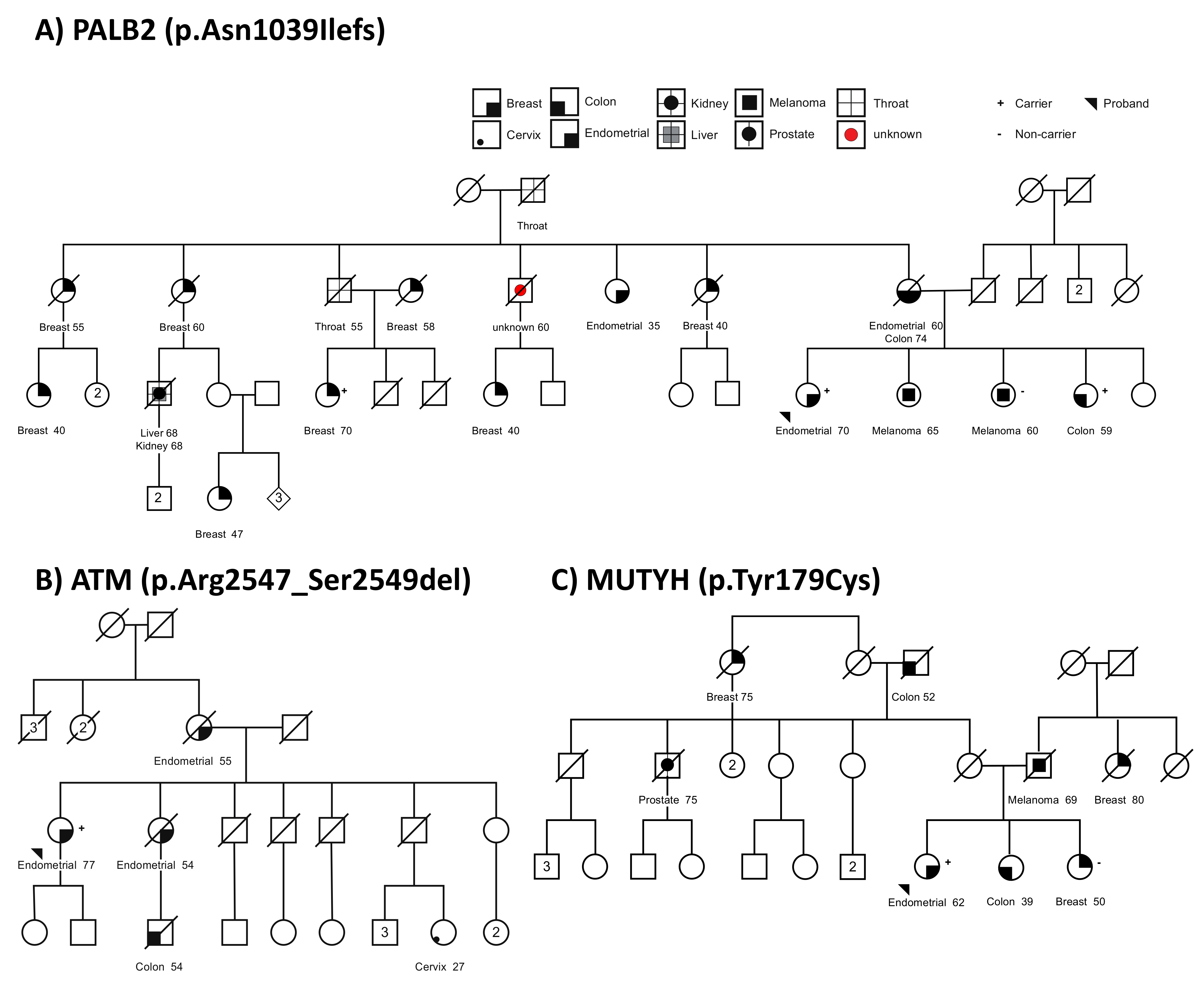
3.3. Germline Variants in Suspected Familial EC Cases

To further explore which genes may explain the etiology of familial EC beyond the well-recognized MMR genes, we sequenced the germline exomes or whole genomes of 33 familial EC cases with no evidence of tumor MMR deficiency, and reported family history of endometrial or other cancer types. The analysis was focused on the same 30 genes as in the sections above (Table S4). Out of the 33 cases, we identified three cases with candidate variants in the prioritized genes. These were a frameshift deletion in PALB2:c.3116delA (p.Asn1039Ilefs), an in-frame deletion in ATM:c.7638_7646del (p.Arg2547_Ser2549del) and a missense pathogenic variant MUTYH:c.536A>G (p.Tyr179Cys).
The patient (case 2) carrying the pathogenic PALB2 frameshift variant (c.3116delA, p.Asn1039Ilefs) was diagnosed with stage 1 endometrioid EC at age 70 years. She self-reported that 17 family members had been diagnosed with various types of cancer (Figure 2A), including two with EC—diagnosed at age 60 years (mother) and age 35 years (maternal aunt). Although DNA was not available from the EC-affected mother, the pedigree analysis indicates she is an obligate carrier; genotyping of three other relatives identified two carrying the PALB2 variant, specifically a sister with colon cancer and maternal cousin with breast cancer (Figure 2A).
The in-frame deletion ATM variant (c.7638_7646del, p.Arg2547_Ser2549del) was predicted to be deleterious by PROVEAN and was classified as pathogenic for Ataxia-telangiectasia syndrome by multiple ClinVar submitters. The carrier (case 1) of this variant was diagnosed with stage 1 endometrioid EC at age 77 years. Two of the family members were also diagnosed with EC: mother at age 55 years and sister at age 54 years (Figure 2B). Other family members were affected with colorectal cancer at age 54 years (nephew) and cervical cancer at age 27 years (niece). DNA from relatives was not available for testing.
The missense heterozygous MUTYH variant (c.536A>G, p.Tyr179Cys) was identified in a female affected with grade 2 endometrioid EC at age 62 years (case 28). This MUTYH variant is a known common pathogenic missense variant known to cause MUTYH-associated polyposis (MAP) in Western populations when detected in homozygous or compound heterozygous state [47]. The proband reported seven family members affected with various cancers (Figure 2C), including a father diagnosed with melanoma, three relatives with breast cancer (maternal great aunt, paternal aunt, sister), two relatives affected with colorectal cancer (maternal grandfather, sister), and a maternal uncle with prostate cancer. A DNA sample was only available for the female sibling with breast cancer and we identified her to be a non-carrier of the MUTYH variant. Although no MUTYH-related cancers were reported for the parents of the proband, her maternal grandfather and female sibling were both affected with colorectal cancer at relatively young age, age 52 years and 39 years, respectively.

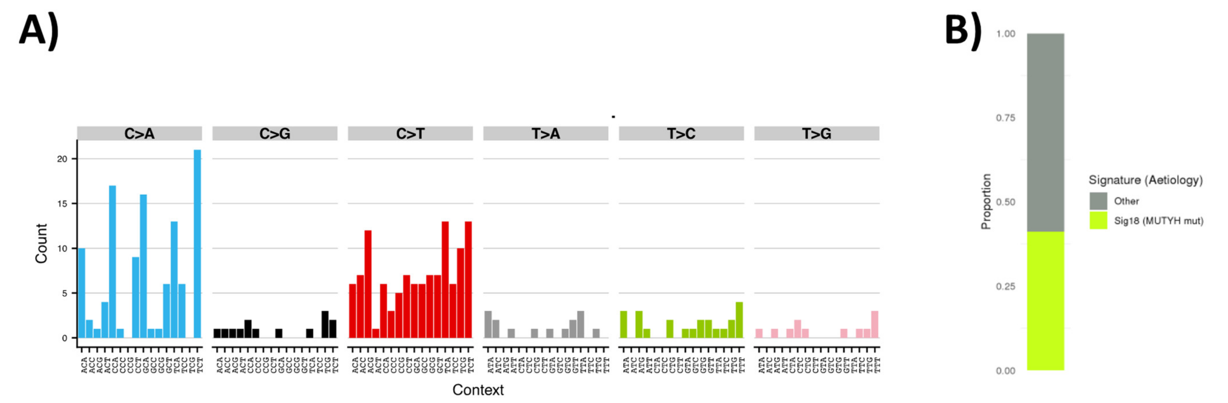
3.4. Tumor Sequencing to Assess Role of MUTYH Variant in a Suspected Familial EC Case

To explore the potential role of the germline heterozygous MUTYH variant in cancer development, we conducted tumor DNA sequencing of the heterozygous MUTYH variant carrier (case 28) to establish whether there was evidence of tumor variant enrichment and whether the MUTYH-associated mutational signature could be detected. We performed whole genome sequencing of an archival endometrial tumor block from the MUTYH carrier. The read depth was too low to accurately assess evidence of loss of heterozygosity at the MUTYH locus, although an increase in the percentage of variant reads from 43% in germline (16 of 37 reads) to 67% in the tumor (six of nine reads) was suggestive of tumor variant enrichment. The sequencing analysis also revealed a high proportion of C>T and C>A somatic mutations (Figure 3A). The pattern of C>T mutations is similar to COSMIC Signature 1, identified in many tumors and typically attributed to aging or deamination [48] and may be present due to formalin fixation. By performing signature assignment analysis we attributed 41% of all somatic single nucleotide variants to Signature 18 (Figure 3B), previously associated with inactivation of MUTYH in a series of familial colorectal cancer and adrenocortical carcinomas [49], indicating that the germline variant was driving the pattern of somatic mutations, and underlay development of EC in this individual.

4. Discussion

Based on the existing clinical management guidelines, a previous review suggested that only six genes currently have sufficient evidence of association with EC risk to be appropriate for hereditary EC diagnostic testing; these include the MMR genes (MLH1, MSH2, MSH6 and PMS2), EPCAM (deletions due to their effect on MSH2) and PTEN [4]. We explored the role of candidate EC risk genes [4] beyond the MMR and PTEN genes, by analyzing an EC sample set unselected for family history and a cohort of familial EC cases. We also performed tumor sequencing analysis to explore whether these genes are cancer drivers associated with somatic mutagenesis in endometrial tumors.
The findings suggest that variation in the following genes should be considered in future studies of EC risk: ATM, MUTYH, PALB2, RAD51C and NBN. PALB2 was highlighted by both case–control and suspected familial EC analysis. Tumor mutational signatures provided evidence that germline variation in BRCA1, PALB2, RAD51C, MUTYH and NTHL1 can be (but is not always) associated with tumor mutational signatures consistent with a functional role of these genes in endometrial tumor development.
ATM encodes for a cell cycle checkpoint kinase that initiates DNA damage response via error-free repair pathway, HR, for double-stranded DNA breaks [50]. The ATM variant identified in a suspected familial EC case was classified as pathogenic for the rare autosomal recessive ataxia-telangiectasia syndrome by multiple submitters in ClinVar. The syndrome manifests a variety of phenotypic characteristics, including high incidence of cancer. Pathogenic variants in ATM are associated with increased breast cancer risk. Monoallelic c.7271T>G carriers are at a significantly increased risk, a 60% cumulative risk by age 80 years, similar to penetrance conferred by pathogenic germline variants in BRCA2 [51]. Monoallelic carriers of other loss of function variants are reported to have a moderate increased risk of developing breast cancer (3-fold; 95% CI: 2.1–4.5) [52]. A number of ATM variants predicted to be deleterious to ATM protein function have been identified in EC cases, in unselected as well as a familial setting [7,53]. Another recent study [22] reporting results from germline panel testing of unselected EC cases identified ATM pathogenic variants as among the most common alterations observed (9/1170 cases), and estimated risk for ATM carriers to be OR 1.86 (p = 0.07) by comparison of case frequency to gnomAD non-Finnish European controls. Given that ATM loss of function variants are estimated to be associated with only a modest risk of breast cancer (OR 3.0, 95% CI 2.1–4.5) [52], larger well-designed studies will be required to determine if ATM variation confers a similar modest level of risk to EC.
PALB2 encodes for one of the key proteins involved in the HR DNA damage repair by recruiting BRCA2 to DNA breaks [54]. The PALB2 truncating variant identified in our familial cohort has been classified as a pathogenic variant for familial breast cancer by multiple submitters to ClinVar. PALB2 is emerging as a gene that confers a high risk of breast cancer, with data suggesting individuals with pathogenic variants in PALB2 have a high lifetime risk of around 32% [55]. PALB2 variants have also been associated with increased risk of ovarian and pancreatic cancers [56]. In our study, the EC patient carrying the PALB2 variant had a strong family history of various cancers, with carrier or obligate carrier status confirmed for relatives with breast, colon and EC. EC has been reported in relatives of breast cancer patients known to carry loss of function variants in PALB2 [57], but carrier status was not confirmed. PALB2 loss of function variants have also been detected in EC patients in several previous studies [45,57,58,59,60,61]. The results to date indicate that the role of PALB2 loss of function variants in conferring EC risk should be further explored.
Other HR pathway genes implicated in this study were BRCA1, NBN and RAD51C. Interestingly, while NBN and RAD51C had a more than 2-fold increased variant frequency in the EC sample set, BRCA1 did not. To date, the role of BRCA1 or BRCA2 in EC risk has been much debated, with numerous conflicting reports [4]. Overall findings indicate that increased EC risk for BRCA1/2 carriers has been associated with tamoxifen use for breast cancer prevention or treatment (since these genes confer high breast cancer risk) comparable to the risk observed in the general population [18,19]. There is also suggestive evidence that BRCA1 pathogenic variants may confer a modest risk EC increase in the absence of tamoxifen exposure, particularly for serous and serous-like subtype cancers [62,63]. Unfortunately, the patient cancer history or tamoxifen exposure was not well documented for the TCGA-UCEC cancer cohort used in this study, hence we were unable to assess the possible contribution of tamoxifen for BRCA1 or other genes that confer breast cancer risk. RAD51C has been recently shown to confer moderate risk for breast (relative risk (RR) = 1.99, 95% CI: 1.39–2.85) and high risk for ovarian cancers (RR = 7.55, 95% CI: 5.6–10.19) [64]; however, there have only been observational studies so far for EC [7,65]. Given the breast cancer risk, future studies on RAD51C and EC risk will need to account for tamoxifen exposure, same as for BRCA1/2 genes. The role of NBN in EC risk has largely been unexplored. It is notable that while certain NBN variants have previously been reported to increase breast cancer risk [66], the most recent evidence from a large-scale case–control analysis refutes (OR 0.90, 95% CI 0.67–1.20) an association of truncating NBN variants with breast cancer risk [67].
In addition to considering a role of the above-mentioned HR-related genes in EC risk, we also investigated their potential role in EC development by analyzing tumor mutational signatures. We observed HR-associated mutational signature (Signature 3) in most tumors with BRCA1, PALB2 and RAD51C pathogenic or likely pathogenic variants, but not in tumors with BRIP1, CHEK2, FAN1 or NBN variants. This is consistent with previous reports in breast cancer and cell line experiments where Signature 3 was only detected for BRCA1/2, PALB2 and RAD51C genes but not ATM or CHEK2 [68,69]. The presence of Signature 3 in cases with BRCA1, PALB2 and RAD51C variants, as well as tumor enrichment of these variants, suggest that these cancers are HR-deficient. Our observation is also supported by the report of tumor loss of heterozygosity in serous/serous-like EC with germline BRCA1 mutations (two of three cases) [62].
Other genes implicated in this study included DNA base excision repair genes, MUTYH and NTHL1. Signature 36 (COSMIC v3), similar to Signature 18 detected in this study (COSMIC v2), has been associated with inactivation of MUTYH in MAP colorectal cancer [70] and observed in 5% of pancreatic neuroendocrine tumors that bore heterozygous germline MUTYH variants and subsequent loss of the wildtype allele in the tumor [71]. Together these observations indicate that oxidative DNA damage due to MUTYH inactivation may contribute to cancer etiology in several organs. In our study, a MUTYH variant considered pathogenic for MAP was likely enriched in the tumor of a suspected familial EC case, which presented with a tumor mutational signature consistent with the driver status of the MUTYH variant. However, MUTYH pathogenic variants were not more common in the TCGA unselected EC cohort relative to the population reference group (1.62% vs. 1.73%), and there was no evidence for tumor enrichment or appropriate tumor mutational signature in the TCGA cases. The majority of MUTYH pathogenic variants identified were two well recognized common pathogenic variants identified in the Western population to cause MAP (c.536A>G (p.Tyr179Cys), as detected in the suspected familial EC case; and c.1187G>A (p.Gly368Asp)) [47]. MAP is an autosomal recessively inherited predisposition to adenomatous polyposis and colorectal cancer [72]. The cumulative colorectal cancer risk to age 70 years for biallelic carriers is reported to be 75% (95% CI: 41–97%) for males and 72% (95% CI: 41–97%) for females, and for monoallelic carriers it is estimated to be 7% (95% CI: 5–11%) for males and 6% (95% CI: 4–9%) for females [73]. Indeed, the case reported here had a family history of colorectal cancer (in two relatives, ages 39 and 52). However, the risk of extracolonic cancers for MUTYH monoallelic pathogenic variant carriers with a family history of colorectal cancer is still uncertain, current evidence derived from a single study estimated cumulative risk of EC age 70 to be 4% (95% CI: 2–8%) for monoallelic MUTYH carriers [11], with an updated analysis of the same cohort [74] reporting a modest 2-fold EC risk for carriers (95% C.I 1.1–3.9). These previous findings suggest that, MUTYH-associated risk of EC, if validated, is likely to be extremely modest.
We have identified several genes in this study as possible additional EC risk genes; however, these results should be considered preliminary, and require further exploration in the follow-up studies. Although this study has not provided conclusive evidence regarding the role of the aforementioned genes in EC risk, results could nevertheless be of relevance as secondary findings for the patient and their relatives. We have shown that at least some germline carriers had a tumor mutational signature supportive of the driver role of the respective gene in cancer development. Overall, 28% of carriers of HR-related gene variants had a presence of HR-deficiency associated tumor mutational signature, which increased to 86% of carriers when only well-recognized HR genes (BRCA1, PALB2 and RAD51C) were included [75]. This has implications for patient treatment decisions, since HR-deficient cancers are known to respond to PARP inhibitors [76]. Furthermore, cases with MMR-deficient or base excision repair-deficient (MUTYH or NTHL1-driven) tumors are likely to show hypermutated profiles, and thus would be good candidates for immunotherapy treatment, given the likely increase in neo-antigen production [77]. Our findings suggest potential value in secondary tumour profiling on identification of a germline gene alteration in EC patients, irrespective of a confirmed role of that gene in EC risk. Furthermore, somatic only changes would have the same implications for treatment decisions. It will thus be important to explore the overall proportion of EC cases with actionable tumor mutation profiles to determine the clinical value of unselected tumor mutational profiling.

5. Conclusions

We used genome sequencing and tumor mutational signature analysis to explore the role of purported EC risk genes in an EC sample set unselected for family history, and to identify candidate germline variants underlying the genetic cause of familial EC without MMR defects. Ancestry-matched case–control comparisons of germline variant frequency and/or sequence data from the suspected familial EC cases proposed several preliminary candidates for future risk association studies, with PALB2 highlighted by both approaches. Tumor analysis highlighted germline variation in HR-related repair genes, particularly BRCA1, PALB2 and RAD51C, to have a potential driver role in EC development based on the presence of mutational signature indicative of HR deficiency. For the heterozygous germline variants in other DNA damage repair genes, MUTYH and NTHL1, the mutational signature analysis indicates possible involvement in the etiology of EC, but only when there were indications of the germline variant being enriched in the tumor.
Inclusion of these highlighted genes in clinical testing panels for EC predisposition will require results from further large-scale studies, to assess the level of EC risk associated with loss of function variation in these genes. Such studies should preferably follow a population-based case–control design and consider the role of other genetic and environmental factors in disease penetrance, including previous exposure to tamoxifen. While we anticipate that genes outside of MMR pathway are unlikely to explain a large component of suspected familial EC, our results indicate that additional tumor signature analysis for individuals with a germline gene alteration has potential to impact therapeutic decisions.

Supplementary Materials

The following are available online at https://www.mdpi.com/article/10.3390/cancers13081762/s1, Figure S1: Variant filtering workflow used for germline pathogenic and likely pathogenic variants included in this study for TCGA-UCEC and gnomAD datasets, Figure S2: De novo mutational signature analysis in the TCGA-UCEC cases with pathogenic or likely pathogenic variants, Table S1: Subset of European TCGA-UCEC cases included in the variant frequency comparison with the gnomAD population of Non-Finnish Europeans, Table S2: Phenotypic characteristics of the familial endometrial cancer cases, Table S3: Summary of sequencing coverage and number of variants identified for each familial endometrial cancer case prior to filtering, Table S4: List of known and purported endometrial cancer risk genes, Table S5: List of pathogenic and likely pathogenic variants identified in the genes of interest in gnomAD (non-Finnish Europeans) and TCGA-UCEC cases.

Author Contributions

O.K. and J.S.: Designed the methods, carried out the analysis, interpreted the data and prepared the manuscript. A.B.S.: Conceived the study, designed the methods, and supervised the work. N.W.: Designed the methods, developed genomic analysis pipelines, supervised the work. T.A.O.: Designed the methods and supervised the work. F.N.: Carried out mutational analysis. A.E.M.R.: Reviewed and processed the tumor sample. S.R.L.: Reviewed the tumor sample and supervised the work. J.K.: Provided clinical input. J.V.P.: Managed the genomic data and developed the genomic pipelines for analysis. All authors read and approved the final manuscript.

Funding

J.S. is supported by a QIMR Berghofer PhD scholarship. N.W. is supported by a National Health and Medical Research Council (NHMRC) of Australia Senior Research Fellowship (APP1139071). A.B.S. was supported by NHMRC Funding (APP1061779, APP117524). T.A.O. was supported by NHMRC Funding (APP1111246, APP1173170). The Australian National Endometrial Cancer Study was supported by project grants from the NHMRC (Grant No. 339435); The Cancer Council Queensland (Grant No. 4196615); Cancer Council Tasmania (Grant No. 403031 and Grant No. 457636); the Cancer Australia Priority-driven Collaborative Cancer Research Scheme (#552468), Cancer Australia (Grant No. 1010859). The sequencing component of this study was funded by a “Rio Tinto Ride to Conquer Cancer & Weekend to End Women’s Cancer Research Grant” administered by QIMR Berghofer Medical Research Institute.

Institutional Review Board Statement

Accessing TCGA-UCEC genome data and undertaking reanalysis to detect variants in the tumour and matching normal was performed with approval from the QIMR Berghofer Human Research Ethics Committee (HREC/P2905). Sequencing and analysis of familial EC patients was undertaken with approval from QIMR Berghofer Ethics Committee (HREC/P1051).

Informed Consent Statement

All ANECS participants provided informed written consent for participation in EC-related research, with overall study approval by the QIMR Berghofer Medical Research Institute Human Research Ethics Committee (P853), encompassing approvals from participating hospitals and cancer registries.

Data Availability Statement

TCGA-UCEC data were downloaded from GDC data portal in October 2016. GnomAD variant files (r.2.1.1) were downloaded from the gnomAD portal in April 2019. ANECS sequencing data are available upon reasonable request and subject to ethics approval.

Acknowledgments

We thank the individuals who participated in ANECS, the institutes and clinicians who contributed to recruitment and data collection (see website: www.anecs.org.au for the full list of contributors). We thank Sullivan Nicolaides Pathology for providing the archival tumor tissue for whole genome sequencing, and Stephen Kazakoff, Michael Bowman, Katia Nones, Ann-Marie Patch for helpful advice and contributions to data cleaning and processing. We thank Kathy Tucker for critique regarding interpretation and representation of clinical relevance of findings. The results shown here are in part based upon data generated by the TCGA Research Network: https://www.cancer.gov/tcga. Information about TCGA can be found at http://cancergenome.nih.gov. The TCGA dataset used in this study was accessed through dbGaP accession number phs000178.v11.p8.

Conflicts of Interest

O.K. has consulted for XING Technologies. N.W. and J.V.P. are co-founders and Board members of genomiQa. The other authors declare that they have no competing interests. The funders had no role in the design of the study; in the collection, analyses, or interpretation of data; in the writing of the manuscript, or in the decision to publish the results.

References

  1. Zhang, S.; Gong, T.-T.; Liu, F.-H.; Jiang, Y.-T.; Sun, H.; Ma, X.-X.; Zhao, Y.-H.; Wu, Q.-J. Global, Regional, and National Burden of Endometrial Cancer, 1990–2017: Results from the Global Burden of Disease Study, 2017. Front. Oncol. 2019, 9, 1440. [Google Scholar] [CrossRef]
  2. Win, A.K.; Reece, J.C.; Ryan, S. Family History and Risk of Endometrial Cancer. Obstet. Gynecol. 2015, 125, 89–98. [Google Scholar] [CrossRef]
  3. O’Mara, T.A.; Glubb, D.M.; Kho, P.F.; Thompson, D.J.; Spurdle, A.B. Genome-Wide Association Studies of Endometrial Cancer: Latest Developments and Future Directions. Cancer Epidemiol. Biomark. Prev. 2019, 28, 1095–1102. [Google Scholar] [CrossRef]
  4. Spurdle, A.B.; Bowman, M.A.; Shamsani, J.; Kirk, J. Endometrial Cancer Gene Panels: Clinical Diagnostic vs Research Germline DNA testing. Mod. Pathol. 2017, 30, 1048–1068. [Google Scholar] [CrossRef]
  5. Randall, L.M.; Pothuri, B. The Genetic Prediction of Risk for Gynecologic Cancers. Gynecol. Oncol. 2016, 141, 10–16. [Google Scholar] [CrossRef]
  6. Nieuwenhuis, M.H.; Kets, C.M.; Murphy-Ryan, M.; Yntema, H.G.; Evans, D.G.; Colas, C.; Møller, P.; Hes, F.J.; Hodgson, S.V.; Olderode-Berends, M.J.W.; et al. Cancer Risk and Genotype–Phenotype Correlations in PTEN Hamartoma Tumor Syndrome. Fam. Cancer 2014, 13, 57–63. [Google Scholar] [CrossRef] [PubMed]
  7. Ring, K.L.; Bruegl, A.S.; Allen, B.A.; Elkin, E.P.; Singh, N.; Hartman, A.-R.; Daniels, M.S.; Broaddus, R.R. Germline Multi-Gene Hereditary Cancer Panel Testing in an Unselected Endometrial Cancer Cohort. Mod. Pathol. 2016, 29, 1381–1389. [Google Scholar] [CrossRef] [PubMed]
  8. Johnatty, S.E.; Tan, Y.Y.; Buchanan, D.D.; Bowman, M.; Walters, R.J.; Obermair, A.; Quinn, M.A.; Blomfield, P.B.; Brand, A.; Leung, Y.; et al. Family History of Cancer Predicts Endometrial Cancer Risk Independently of Lynch Syndrome: Implications for Genetic Counselling. Gynecol. Oncol. 2017, 147, 381–387. [Google Scholar] [CrossRef]
  9. Bellido, F.; Pineda, M.; Aiza, G.; Mas, R.M.V.; Navarro, M.; Puente, D.Á.; Pons, T.; González, S.; Iglesias, S.; Darder, E.; et al. POLE and POLD1 Mutations in 529 Kindred with Familial Colorectal Cancer and/or Polyposis: Review of Reported Cases and Recommendations for Genetic Testing and Surveillance. Genet. Med. 2016, 18, 325–332. [Google Scholar] [CrossRef] [PubMed] [Green Version]
  10. Hu, R.; Hilakivi-Clarke, L.; Clarke, R. Molecular Mechanisms of Tamoxifen-Associated Endometrial Cancer (Review). Oncol. Lett. 2015, 9, 1495–1501. [Google Scholar] [CrossRef] [PubMed] [Green Version]
  11. Win, A.K.; Cleary, S.P.; Dowty, J.G.; Baron, J.A.; Young, J.P.; Buchanan, D.D.; Southey, M.C.; Burnett, T.; Parfrey, P.S.; Green, R.C.; et al. Cancer Risks for Monoallelic MUTYH Mutation Carriers with a Family History of Colorectal Cancer. Int. J. Cancer 2010, 129, 2256–2262. [Google Scholar] [CrossRef] [PubMed] [Green Version]
  12. Vogt, S.; Jones, N.; Christian, D.; Engel, C.; Nielsen, M.; Kaufmann, A.; Steinke, V.; Vasen, H.F.; Propping, P.; Sampson, J.R.; et al. Expanded Extracolonic Tumor Spectrum in MUTYH-Associated Polyposis. Gastroenterology 2009, 137, 1976–1985.e10. [Google Scholar] [CrossRef]
  13. Giardiello, F.M.; Brensinger, J.D.; Tersmette, A.C.; Goodman, S.N.; Petersen, G.M.; Booker, S.V.; Cruz–Correa, M.; Offerhaus, J.A. Very High Risk of Cancer in Familial Peutz–Jeghers Syndrome. Gastroenterology 2000, 119, 1447–1453. [Google Scholar] [CrossRef] [PubMed] [Green Version]
  14. Pennington, K.P.; Walsh, T.; Lee, M.; Ms, C.P.; Novetsky, A.P.; Bs, K.J.A.; Thornton, A.; Garcia, R.; Mutch, D.; King, M.-C.; et al. BRCA1, TP53, andCHEK2 Germline Mutations in Uterine Serous Carcinoma. Cancer 2013, 119, 332–338. [Google Scholar] [CrossRef] [PubMed] [Green Version]
  15. Heitzer, E.; Lax, S.; Lafer, I.; Müller, S.M.; Pristauz, G.; Ulz, P.; Jahn, S.; Högenauer, C.; Petru, E.; Speicher, M.R.; et al. Multiplex Genetic Cancer Testing Identifies Pathogenic Mutations in TP53 and CDH1in a Patient with Bilateral Breast and Endometrial Adenocarcinoma. BMC Med. Genet. 2013, 14, 129. [Google Scholar] [CrossRef] [Green Version]
  16. Chao, A.; Lai, C.-H.; Lee, Y.-S.; Ueng, S.-H.; Lin, C.-Y.; Wang, T.-H. Molecular Characteristics of Endometrial Cancer Coexisting with Peritoneal Malignant Mesothelioma in LI-Fraumeni-like Syndrome. BMC Cancer 2015, 15, 8. [Google Scholar] [CrossRef] [PubMed] [Green Version]
  17. Hornreich, G.; Beller, U.; Lavie, O.; Renbaum, P.; Cohen, Y.; Levy-Lahad, E. Is Uterine Serous Papillary Carcinoma a BRCA1-Related Disease? Case Report and Review of the Literature. Gynecol. Oncol. 1999, 75, 300–304. [Google Scholar] [CrossRef]
  18. Duffy, D.L.; Antill, Y.C.; Stewart, C.J.; Young, J.P.; Spurdle, A.B. kConFab Report of Endometrial Cancer in Australian BRCA1 and BRCA2 Mutation-Positive Families. Twin Res. Hum. Genet. 2011, 14, 111–118. [Google Scholar] [CrossRef] [PubMed]
  19. Segev, Y.; Iqbal, J.; Lubinski, J.; Gronwald, J.; Lynch, H.T.; Moller, P.; Ghadirian, P.; Rosen, B.; Tung, N.; Kim-Sing, C.; et al. The Incidence of Endometrial Cancer in Women with BRCA1 and BRCA2 Mutations: An International Prospective Cohort Study. Gynecol. Oncol. 2013, 130, 127–131. [Google Scholar] [CrossRef] [PubMed]
  20. Bruchim, I.; Amichay, K.; Kidron, D.; Attias, Z.; Biron-Shental, T.; Drucker, L.; Friedman, E.; Werner, H.; Fishman, A. BRCA1/2 Germline Mutations in Jewish Patients with Uterine Serous Carcinoma. Int. J. Gynecol. Cancer 2010, 20, 1148–1153. [Google Scholar] [CrossRef]
  21. Lavie, O.; Ben-Arie, A.; Segev, Y.; Faro, J.; Barak, F.; Haya, N.; Auslender, R.; Gemer, O. Brca Germline Mutations in Women with Uterine Serous Carcinoma—Still a Debate. Int. J. Gynecol. Cancer 2010, 20, 1531–1534. [Google Scholar] [PubMed]
  22. Long, B.; Lilyquist, J.; Weaver, A.; Hu, C.; Gnanaolivu, R.; Lee, K.Y.; Hart, S.N.; Polley, E.C.; Bakkum-Gamez, J.N.; Couch, F.J.; et al. Cancer Susceptibility Gene Mutations in Type I and II Endometrial Cancer. Gynecol. Oncol. 2019, 152, 20–25. [Google Scholar] [CrossRef] [PubMed]
  23. Carvajal-Carmona, L.G.; O’Mara, T.A.; Painter, J.N.; Lose, F.A.; Dennis, J.; Michailidou, K.; Tyrer, J.P.; Ahmed, S.; Ferguson, K.; Healey, C.S.; et al. Candidate Locus Analysis of the TERT–CLPTM1L Cancer Risk Region on Chromosome 5p15 Identifies Multiple Independent Variants Associated with Endometrial Cancer Risk. Qual. Life Res. 2014, 134, 231–245. [Google Scholar] [CrossRef] [PubMed] [Green Version]
  24. Buchanan, D.D.; Tan, Y.Y.; Walsh, M.D.; Clendenning, M.; Metcalf, A.M.; Ferguson, K.; Arnold, S.T.; Thompson, B.A.; Lose, F.A.; Parsons, M.T.; et al. Tumor Mismatch Repair Immunohistochemistry and DNA MLH1 Methylation Testing of Patients with Endometrial Cancer Diagnosed at Age Younger Than 60 Years Optimizes Triage for Population-Level Germline Mismatch Repair Gene Mutation Testing. J. Clin. Oncol. 2014, 32, 90–100. [Google Scholar] [CrossRef] [PubMed] [Green Version]
  25. Tan, Y.Y.; McGaughran, J.; Ferguson, K.; Walsh, M.D.; Buchanan, D.D.; Young, J.P.; Webb, P.M.; Obermair, A.; Spurdle, A.B. On Behalf of the ANECS Group Improving Identification of Lynch Syndrome Patients: A Comparison of Research Data with Clinical Records. Int. J. Cancer 2013, 132, 2876–2883. [Google Scholar] [CrossRef]
  26. Martin, M. Cutadapt Removes Adapter Sequences from High-Throughput Sequencing Reads. EMBnet. J. 2011, 17, 10–12. [Google Scholar] [CrossRef]
  27. Li, H. Aligning Sequence Reads, Clone Sequences and Assembly Contigs with BWA-MEM. arXiv 2013, arXiv:1303.3997. [Google Scholar]
  28. Li, H.; Handsaker, B.; Wysoker, A.; Fennell, T.; Ruan, J.; Homer, N.; Marth, G.; Abecasis, G.; Durbin, R. 1000 Genome Project Data Processing Subgroup. The Sequence Alignment/Map format and SAMtools. Bioinformatics 2009, 25, 2078–2079. [Google Scholar] [CrossRef] [Green Version]
  29. Kassahn, K.S.; Holmes, O.; Nones, K.; Patch, A.-M.; Miller, D.K.; Christ, A.N.; Harliwong, I.; Bruxner, T.J.; Xu, Q.; Anderson, M.; et al. Somatic Point Mutation Calling in Low Cellularity Tumors. PLoS ONE 2013, 8, e74380. [Google Scholar] [CrossRef] [Green Version]
  30. McKenna, A.; Hanna, M.; Banks, E.; Sivachenko, A.; Cibulskis, K.; Kernytsky, A.; Garimella, K.; Altshuler, D.; Gabriel, S.B.; Daly, M.J.; et al. The Genome Analysis Toolkit: A MapReduce Framework for Analyzing Next-Generation DNA Sequencing Data. Genome Res. 2010, 20, 1297–1303. [Google Scholar] [CrossRef] [Green Version]
  31. Hayward, N.K.; Wilmott, J.S.; Waddell, N.; Johansson, P.A.; Field, M.A.; Nones, K.; Patch, A.-M.; Kakavand, H.; Alexandrov, L.B.; Burke, H.; et al. Whole-Genome Landscapes of Major Melanoma Subtypes. Nat. Cell Biol. 2017, 545, 175–180. [Google Scholar] [CrossRef]
  32. McLaren, W.; Gil, L.; Hunt, S.E.; Riat, H.S.; Ritchie, G.R.S.; Thormann, A.; Flicek, P.; Cunningham, F. The Ensembl Variant Effect Predictor. Genome Biol. 2016, 17, 1–14. [Google Scholar] [CrossRef] [Green Version]
  33. Ioannidis, N.M.; Rothstein, J.H.; Pejaver, V.; Middha, S.; McDonnell, S.K.; Baheti, S.; Musolf, A.; Li, Q.; Holzinger, E.; Karyadi, D.; et al. REVEL: An Ensemble Method for Predicting the Pathogenicity of Rare Missense Variants. Am. J. Hum. Genet. 2016, 99, 877–885. [Google Scholar] [CrossRef] [Green Version]
  34. Shamsani, J.; Kazakoff, S.H.; Armean, I.M.; McLaren, W.; Parsons, M.T.; Thompson, B.A.; O’Mara, T.A.E.; Hunt, S.; Waddell, N.; Spurdle, A.B. A Plugin for the Ensembl Variant Effect Predictor That Uses MaxEntScan to Predict Variant Spliceogenicity. Bioinformatics 2019, 35, 2315–2317. [Google Scholar] [CrossRef] [PubMed]
  35. Landrum, M.J.; Lee, J.M.; Riley, G.R.; Jang, W.; Rubinstein, W.S.; Church, D.M.; Maglott, D.R. ClinVar: Public Archive of Relationships among Sequence Variation and Human Phenotype. Nucleic Acids Res. 2014, 42, D980–D985. [Google Scholar] [CrossRef] [Green Version]
  36. Chadwick, R.B.; Meek, J.E.; Prior, T.W.; Peltomaki, P.; de la Chapelle, A. Polymorphisms in a Pseudogene Highly Homolo-Gous To PMS2. Hum. Mutat. 2000, 16, 530. [Google Scholar] [CrossRef]
  37. Mur, P.; Ms, S.G.-M.; Del Valle, J.; Ms, L.M.-P.; Vidal, A.; Pineda, M.; Cinnirella, G.; Ms, E.M.-R.; Pons, T.; López-Doriga, A.; et al. Role of POLE and POLD1 in familial cancer. Genet. Med. 2020, 22, 2089–2100. [Google Scholar] [CrossRef]
  38. Choi, Y.; Chan, A.P. PROVEAN Web Server: A Tool to Predict the Functional Effect of Amino Acid Substitutions and Indels. Bioinformatics 2015, 31, 2745–2747. [Google Scholar] [CrossRef] [Green Version]
  39. Rosenthal, R.; McGranahan, N.; Herrero, J.; Taylor, B.S.; Swanton, C. Deconstructsigs: Delineating Mutational Processes in Single Tumors Distinguishes DNA Repair Deficiencies and Patterns of Carcinoma Evolution. Genome Biol. 2016, 17, 31. [Google Scholar] [CrossRef] [Green Version]
  40. Bonazzi, V.F.; Kondrashova, O.; Smith, D.; Nones, K.; Sengal, A.T.; Ju, R.; Packer, L.M.; Koufariotis, L.T.; Kazakoff, S.H.; Davidson, A.L.; et al. Genomic analysis of patient-derived xenograft models reveals intra-tumor heterogeneity in endometrial cancer and can predict tumor growth inhibition with talazoparib. bioRxiv 2021. [Google Scholar] [CrossRef]
  41. Niu, B.; Ye, K.; Zhang, Q.; Lu, C.; Xie, M.; McLellan, M.D.; Wendl, M.C.; Ding, L. MSI Sensor: Microsatellite Instability Detection Using Paired Tumor-Normal Sequence Data. Bioinformatics 2014, 30, 1015–1016. [Google Scholar] [CrossRef] [Green Version]
  42. Levine, D.A.; Network, C.G.A.R. Integrated Genomic Characterization of Endometrial Carcinoma. Nature 2013, 497, 67–73. [Google Scholar] [CrossRef] [PubMed] [Green Version]
  43. Cerami, E.; Gao, J.; Dogrusoz, U.; Gross, B.E.; Sumer, S.O.; Aksoy, B.A.; Jacobsen, A.; Byrne, C.J.; Heuer, M.L.; Larsson, E.; et al. The cBio Cancer Genomics Portal: An Open Platform for Exploring Multidimensional Cancer Genomics Data. Cancer Discov. 2012, 2, 401–404. [Google Scholar] [CrossRef] [Green Version]
  44. Gao, J.; Aksoy, B.A.; Dogrusoz, U.; Dresdner, G.; Gross, B.; Sumer, S.O.; Sun, Y.; Jacobsen, A.; Sinha, R.; Larsson, E. Integrative Analysis of Complex Cancer Genomics and Clinical Profiles Using the cBioPortal. Sci. Signal. 2013, 6, pl1. [Google Scholar] [CrossRef] [PubMed] [Green Version]
  45. Huang, K.-L.; Mashl, R.J.; Wu, Y.; Ritter, D.I.; Wang, J.; Oh, C.; Paczkowska, M.; Reynolds, S.; Wyczalkowski, M.A.; Oak, N.; et al. Pathogenic Germline Variants in 10,389 Adult Cancers. Cell 2018, 173, 355–370.e14. [Google Scholar] [CrossRef] [Green Version]
  46. Grolleman, J.E.; De Voer, R.M.; Elsayed, F.A.; Nielsen, M.; Weren, R.D.A.; Palles, C.; Ligtenberg, M.J.L.; Vos, J.R.; Broeke, S.W.T.; De Miranda, N.F.C.C.; et al. Mutational Signature Analysis Reveals NTHL1 Deficiency to Cause a Multi-Tumor Phenotype Including a Predisposition to Colon and Breast Cancer. SSRN Electron. J. 2018, 35, 256–266. [Google Scholar] [CrossRef]
  47. Nielsen, M.; Van De Beld, M.C.J.-; Jones, N.; Vogt, S.; Tops, C.M.; Vasen, H.F.; Sampson, J.R.; Aretz, S.; Hes, F.J. Analysis of MUTYH Genotypes and Colorectal Phenotypes in Patients With MUTYH-Associated Polyposis. Gastroenterology 2009, 136, 471–476. [Google Scholar] [CrossRef]
  48. Alexandrov, L.B.; Nik-Zainal, S.; Wedge, D.C.; Aparicio, S.A.J.R.; Behjati, S.; Biankin, A.V.; Bignell, G.R.; Bolli, N.; Borg, A.; Børresen-Dale, A.-L.; et al. Signatures of Mutational Processes in Human Cancer. Nature 2013, 500, 415–421. [Google Scholar] [CrossRef] [PubMed] [Green Version]
  49. Pilati, C.; Shinde, J.; Alexandrov, L.B.; Assié, G.; André, T.; Hélias-Rodzewicz, Z.; Doucoudray, R.; Le Corre, D.; Zucman-Rossi, J.; Emile, J.-F.; et al. Mutational Signature Analysis Identifies MUTYH Deficiency in Colorectal Cancers and Adrenocortical Carcinomas. J. Pathol. 2017, 242, 10–15. [Google Scholar] [CrossRef] [Green Version]
  50. Maréchal, A.; Zou, L. DNA Damage Sensing by the ATM and ATR Kinases. Cold Spring Harb. Perspect. Biol. 2013, 5, a012716. [Google Scholar] [CrossRef] [PubMed]
  51. Goldgar, D.E.; Healey, S.; Dowty, J.G.; Da Silva, L.; Chen, X.; Spurdle, A.B.; Terry, M.B.; Daly, M.J.; Buys, S.M.; Southey, M.C.; et al. Rare Variants in the ATMgene and Risk of Breast Cancer. Breast Cancer Res. 2011, 13, R73. [Google Scholar] [CrossRef] [Green Version]
  52. Van Os, N.; Roeleveld, N.; Weemaes, C.; Jongmans, M.; Janssens, G.O.R.J.; Taylor, A.; Hoogerbrugge, N.; Willemsen, M. Health Risks for Ataxia-Telangiectasia Mutated Heterozygotes: A Systematic Review, Meta-Analysis and Evidence-Based Guideline. Clin. Genet. 2016, 90, 105–117. [Google Scholar] [CrossRef] [PubMed]
  53. Hu, C.; Hart, S.N.; Bamlet, W.R.; Moore, R.M.; Nandakumar, K.; Eckloff, B.W.; Lee, Y.K.; Petersen, G.M.; McWilliams, R.R.; Couch, F.J. Prevalence of Pathogenic Mutations in Cancer Predisposition Genes among Pancreatic Cancer Patients. Cancer Epidemiol. Biomark. Prev. 2016, 25, 207–211. [Google Scholar] [CrossRef] [PubMed] [Green Version]
  54. Xia, B.; Sheng, Q.; Nakanishi, K.; Ohashi, A.; Wu, J.; Christ, N.; Liu, X.; Jasin, M.; Couch, F.J.; Livingston, D.M. Control of BRCA2 Cellular and Clinical Functions by a Nuclear Partner, PALB2. Mol. Cell 2006, 22, 719–729. [Google Scholar] [CrossRef] [PubMed]
  55. Hu, C.; Hart, S.N.; Gnanaolivu, R.; Huang, H.; Lee, K.Y.; Na, J.; Gao, C.; Lilyquist, J.; Yadav, S.; Boddicker, N.J. A Popu-lation-Based Study of Genes Previously Implicated in Breast Cancer. N. Engl. J. Med. 2021, 384, 440–451. [Google Scholar] [CrossRef]
  56. Yang, X.; Leslie, G.; Doroszuk, A.; Schneider, S.; Allen, J.; Decker, B.; Dunning, A.M.; Redman, J.; Scarth, J.; Plaskocinska, I.; et al. Cancer Risks Associated with Germline PALB2 Pathogenic Variants: An International Study of 524 Families. J. Clin. Oncol. 2020, 38, 674–685. [Google Scholar] [CrossRef]
  57. Teo, Z.L.; Park, D.J.; Provenzano, E.; Chatfield, C.A.; Odefrey, F.A.; Nguyen-Dumont, T.; Dowty, J.G.; Hopper, J.L.; Winship, I.; Goldgar, D.E.; et al. Prevalence of PALB2 Mutations in Australasian Multiple-Case Breast Cancer Families. Breast Cancer Res. 2013, 15, R17. [Google Scholar] [CrossRef] [Green Version]
  58. Susswein, L.R.; Marshall, M.L.; Nusbaum, R.; Postula, K.J.V.; Weissman, S.M.; Yackowski, L.; Vaccari, E.M.; Bissonnette, J.; Booker, J.K.; Cremona, M.L.; et al. Pathogenic and Likely Pathogenic Variant Prevalence among the First 10,000 Patients Referred for Next-Generation Cancer Panel Testing. Genet. Med. 2016, 18, 823–832. [Google Scholar] [CrossRef] [Green Version]
  59. Fulk, K.; Milam, M.R.; Li, S.; Yussuf, A.; Black, M.H.; Chao, E.C.; LaDuca, H.; Stany, M.P. Women with Breast and Uterine Cancer Are More Likely to Harbor Germline Mutations Than Women with Breast or Uterine Cancer Alone: A Case for Expanded Gene Testing. Gynecol. Oncol. 2019, 152, 612–617. [Google Scholar] [CrossRef]
  60. Heeke, A.L.; Pishvaian, M.J.; Lynce, F.; Xiu, J.; Brody, J.R.; Chen, W.-J.; Baker, T.M.; Marshall, J.L.; Isaacs, C. Prevalence of Homologous Recombination–Related Gene Mutations Across Multiple Cancer Types. JCO Precis. Oncol. 2018, 2018, 1–13. [Google Scholar] [CrossRef]
  61. Chan, G.H.; Ong, P.Y.; Low, J.J.; Kong, H.L.; Ow, S.G.; Tan, D.S.; Lim, Y.W.; Lim, S.E.; Lee, S.-C. Clinical Genetic Testing Outcome with Multi-Gene Panel in Asian Patients with Multiple Primary Cancers. Oncotarget 2018, 9, 30649–30660. [Google Scholar] [CrossRef] [Green Version]
  62. Shu, C.A.; Pike, M.C.; Jotwani, A.R.; Friebel, T.M.; Soslow, R.A.; Levine, D.A.; Nathanson, K.L.; Konner, J.A.; Arnold, A.G.; Bogomolniy, F.; et al. Uterine Cancer After Risk-Reducing Salpingo-oophorectomy Without Hysterectomy in Women with BRCA Mutations. JAMA Oncol. 2016, 2, 1434–1440. [Google Scholar] [CrossRef] [Green Version]
  63. Thompson, D.; Easton, D.F. Cancer Incidence in BRCA1 Mutation Carriers. Obstet. Gynecol. Surv. 2003, 58, 27–28. [Google Scholar] [CrossRef] [Green Version]
  64. Yang, X.; Song, H.; Leslie, G.; Engel, C.; Hahnen, E.; Auber, B.; Horváth, J.; Kast, K.; Niederacher, D.; Turnbull, C.; et al. Ovarian and Breast Cancer Risks Associated with Pathogenic Variants in RAD51C and RAD51D. J. Natl. Cancer Inst. 2020, 112, 1242–1250. [Google Scholar] [CrossRef] [PubMed] [Green Version]
  65. Pelttari, L.; Shimelis, H.; Toiminen, H.; Kvist, A.; Törngren, T.; Borg, Å.; Blomqvist, C.; Bützow, R.; Couch, F.; Aittomäki, K. Gene-Panel Testing of Breast and Ovarian Cancer Patients Identifies a Recurrent RAD51C Duplication. Clin. Genet. 2018, 93, 595–602. [Google Scholar] [CrossRef] [PubMed] [Green Version]
  66. Gao, P.; Ma, N.; Li, M.; Tian, Q.-B.; Liu, D.-W. Functional Variants in NBS1 and Cancer Risk: Evidence from a Meta-Analysis of 60 Publications with 111 Individual Studies. Mutagenesis 2013, 28, 683–697. [Google Scholar] [CrossRef]
  67. Dorling, L.; Carvalho, S.; Allen, J.; González-Neira, A.; Luccarini, C.; Wahlström, C.; Pooley, K.A.; Parsons, M.T.; Fortuno, C.; Wang, Q. Breast Cancer Risk Genes-Association Analysis in More than 113,000 Women. N. Engl. J. Med. 2021, 384, 428–439. [Google Scholar]
  68. Polak, P.; Kim, J.; Braunstein, L.Z.; Karlic, R.; Haradhavala, N.J.; Tiao, G.; Rosebrock, D.; Livitz, D.; Kübler, K.; Mouw, K.W.; et al. A Mutational Signature Reveals Alterations Underlying Deficient Homologous Recombination Repair in Breast Cancer. Nat. Genet. 2017, 49, 1476–1486. [Google Scholar] [CrossRef]
  69. Póti, Á.; Gyergyák, H.; Németh, E.; Rusz, O.; Tóth, S.; Kovácsházi, C.; Chen, D.; Szikriszt, B.; Spisák, S.; Takeda, S. Correlation of Homologous Recombination Deficiency Induced Mutational Signatures with Sensitivity to Parp Inhibitors and Cytotoxic Agents. Genome Biol. 2019, 20, 240. [Google Scholar] [CrossRef]
  70. Viel, A.; Bruselles, A.; Meccia, E.; Fornasarig, M.; Quaia, M.; Canzonieri, V.; Policicchio, E.; Urso, E.D.; Agostini, M.; Genuardi, M. A Specific Mutational Signature Associated with DNA 8-Oxoguanine Persistence in MUTYH-Defective Colo-Rectal Cancer. EBioMedicine 2017, 20, 39–49. [Google Scholar] [CrossRef]
  71. Scarpa, A.; Chang, D.K.; Nones, K.; Corbo, V.; Patch, A.-M.; Bailey, P.; Lawlor, R.T.; Johns, A.L.; Miller, D.K.; Mafficini, A.; et al. Whole-Genome Landscape of Pancreatic Neuroendocrine Tumours. Nat. Cell Biol. 2017, 543, 65–71. [Google Scholar] [CrossRef] [PubMed]
  72. Balaguer, F.; Leoz, M.L.; Carballal, S.; Moreira, L.; Ocaña, T. The Genetic Basis of Familial Adenomatous Polyposis and Its Implications for Clinical Practice and Risk Management. Appl. Clin. Genet. 2015, 8, 95–107. [Google Scholar] [CrossRef] [PubMed] [Green Version]
  73. Win, A.K.; Dowty, J.G.; Cleary, S.P.; Kim, H.; Buchanan, D.D.; Young, J.P.; Clendenning, M.; Rosty, C.; MacInnis, R.J.; Giles, G.G.; et al. Risk of Colorectal Cancer for Carriers of Mutations in MUTYH, With and Without a Family History of Cancer. Gastroenterology 2014, 146, 1208–1211.e5. [Google Scholar] [CrossRef] [Green Version]
  74. Win, A.K.; Reece, J.C.; Dowty, J.G.; Buchanan, D.D.; Clendenning, M.; Rosty, C.; Southey, M.C.; Young, J.P.; Cleary, S.P.; Kim, H.; et al. Risk of Extracolonic Cancers for People with Biallelic and Monoallelic Mutations inMUTYH. Int. J. Cancer 2016, 139, 1557–1563. [Google Scholar] [CrossRef] [Green Version]
  75. Nguyen, L.; Martens, J.W.M.; Van Hoeck, A.; Cuppen, E. Pan-Cancer Landscape of Homologous Recombination Deficiency. Nat. Commun. 2020, 11, 5584. [Google Scholar] [CrossRef]
  76. D’Andrea, A.D. Mechanisms of PARP Inhibitor Sensitivity and Resistance. DNA Repair 2018, 71, 172–176. [Google Scholar] [CrossRef]
  77. Mouw, K.W.; Goldberg, M.S.; Konstantinopoulos, P.A.; D’Andrea, A.D. DNA Damage and Repair Biomarkers of Immu-Notherapy Response. Cancer Discov. 2017, 7, 675–693. [Google Scholar] [CrossRef] [Green Version]
Figure 1. Somatic mutational signature analysis of the germline variant carriers in the TCGA-UCEC cohort. Tumor mutation burden (TMB), microsatellite instability (MSI) scores and mutational signatures observed in the TCGA-UCEC cases with pathogenic or likely pathogenic variants in DNA damage repair genes associated with specific mutational signatures.
Cancers 13 01762 g001
Figure 2. Pedigrees of families of the endometrial cancer cases carrying candidate variants. (A) Family pedigree of PALB2 p.Asn1039Ilefs carrier. (B) Family pedigree of ATM p.Arg2547_Ser2549del carrier. (C) Family pedigree of the endometrial cancer case carrying candidate MUTYH p.Tyr179Cys. Squares symbolize males, circles symbolize females. Affected individuals are indicated by highlighted symbols, with cancer type and age at diagnosis noted below. Unaffected individuals are indicated by empty symbols. Endometrial cancer proband sequenced is indicated by black arrow below the symbol. Variant carriers are indicated by a (+) symbol and the non-carriers are indicated by a (−) symbol.
Cancers 13 01762 g002
Figure 3. Somatic mutational signature analysis of the MUTYH germline variant carrier (suspected familial endometrial cancer cohort). (A) A total of 287 somatic single nucleotide variants (SNVs) identified in the endometrial tumor and used in signature analysis, plotted as counts in a 96 trinucleotide context. (B) The proportion of mutations in the tumor sample which were assigned to Signature 18.
Cancers 13 01762 g003
Table 1. Overall frequency of pathogenic and likely pathogenic variants in 30 known and candidate endometrial cancer (EC) risk genes in an EC sample set (TCGA-UCEC study) and the general population (gnomAD).
GeneEndometrial Cancer CasesGeneral Population
(TCGA-UCEC)(gnomAD)
Number of CarriersNumber of Homozygote CarriersNumber of Total CasesCarrier Frequency (%)Number of CarriersNumber of Homozygote CarriersNumber of Total CasesCarrier Frequency (%)
MUTYH503081.621023359,0951.73
MSH6403081.3134059,0950.23
CHEK2303080.971099759,0931.86
RAD51C303080.9761059,0930.1
NTHL1203080.65268059,0900.45
MSH2203080.6511059,0920.02
SEC23B103080.32197059,0940.33
FAN1103080.32186059,0950.31
BRCA1103080.32140059,0950.24
NBN103080.3289059,0720.15
PALB2103080.3285059,0940.14
PMS2103080.3276059,0950.13
ATM0000284059,0880.48
BRCA20000182059,0790.31
BRIP10000123059,0900.21
FANCC0000104059,0950.18
RINT1000055059,0940.09
APC000050059,0900.08
MLH1000034059,0950.06
EPCAM000032059,0920.05
PTEN000027059,0950.05
SDHB000020059,0890.03
TP53000020059,0950.03
SDHC000014059,0930.02
SDHD00007059,0950.01
AKT100004059,0940.01
PIK3CA00003058,8390.01
STK1100002058,7530
POLD100000059,0920
POLE00000059,0950
Only cases with Non-Finnish European ethnicity were included. Genes highlighted in bold had a frequency of >2 times higher in TCGA-UCEC compared with gnomAD. Carrier frequency represents the sum of all (likely) pathogenic variants in that gene. Genes highlighted in bold had more than double variant carrier frequency in the EC cases compared to the gnomAD population.
Publisher’s Note: MDPI stays neutral with regard to jurisdictional claims in published maps and institutional affiliations.

