Simple Summary
Regarding the pandemic of obesity and chronic diseases associated to metabolic alterations that occur nowadays worldwide, here, we review the most recent studies related to bioactive compounds and diet derived ingredients with potential effects to augment the systemic energy expenditure. We specifically focus in two processes: the activation of thermogenesis in adipose tissue and the enhancement of the mitochondrial oxidative phosphorylation capacity in muscles. This may provide relevant information to develop diets and supplements to conduct nutritional intervention studies with the objective to ameliorate the metabolic and chronic inflammation in the course of obesity and related disorders.
Abstract
Obesity is associated to increased incidence and poorer prognosis in multiple cancers, contributing to up to 20% of cancer related deaths. These associations are mainly driven by metabolic and inflammatory changes in the adipose tissue during obesity, which disrupt the physiologic metabolic homeostasis. The association between obesity and hypercholesterolemia, hypertension, cardiovascular disease (CVD) and type 2 diabetes mellitus (T2DM) is well known. Importantly, the retrospective analysis of more than 1000 epidemiological studies have also shown the positive correlation between the excess of fatness with the risk of cancer. In addition, more important than weight, it is the dysfunctional adipose tissue the main driver of insulin resistance, metabolic syndrome and all cause of mortality and cancer deaths, which also explains why normal weight individuals may behave as “metabolically unhealthy obese” individuals. Adipocytes also have direct effects on tumor cells through paracrine signaling. Downregulation of adiponectin and upregulation of leptin in serum correlate with markers of chronic inflammation, and crown like structures (CLS) associated to the adipose tissue disfunction. Nevertheless, obesity is a preventable risk factor in cancer. Lifestyle interventions might contribute to reduce the adverse effects of obesity. Thus, Mediterranean diet interventional studies have been shown to reduce to circulation inflammatory factors, insulin sensitivity and cardiovascular function, with durable responses of up to 2 years in obese patients. Mediterranean diet supplemented with extra-virgin olive oil reduced the incidence of breast cancer compared with a control diet. Physical activity is another important lifestyle factor which may also contribute to reduced systemic biomarkers of metabolic syndrome associated to obesity. In this scenario, precision nutrition may provide complementary approaches to target the metabolic inflammation associated to “unhealthy obesity”. Herein, we first describe the different types of adipose tissue -thermogenic active brown adipose tissue (BAT) versus the energy storing white adipose tissue (WAT). We then move on precision nutrition based strategies, by mean of natural extracts derived from plants and/or diet derived ingredients, which may be useful to normalize the metabolic inflammation associated to “unhealthy obesity”. More specifically, we focus on two axis: (1) the activation of thermogenesis in BAT and browning of WAT; (2) and the potential of augmenting the oxidative capacity of muscles to dissipate energy. These strategies may be particularly relevant as complementary approaches to alleviate obesity associated effects on chronic inflammation, immunosuppression, angiogenesis and chemotherapy resistance in cancer. Finally, we summarize main studies where plant derived extracts, mainly, polyphenols and flavonoids, have been applied to increase the energy expenditure.
1. Introduction
According to the World-Health-Organization obesity and chronic metabolic diseases are augmenting alarmingly. The increase in high-fat-saturated diets and the sedentary lifestyle of western countries have become the main cause of obesity and type 2 diabetes which are becoming pandemic with many other metabolic disorders associated (Figure 1). During obesity, the adipose tissue (AT) becomes dysfunctional, promoting a pro-inflammatory and insulin-resistant environment that contributes to metabolic alterations of key metabolic organs, such as liver, pancreas, or muscle, implicated in the systemic energetic homeostasis. This imbalance contributes to the development of type 2 diabetes mellitus (T2DM) and a systemic proinflammatory state, which in turn may lead to metabolic alterations with multi-organ damage [1,2,3].
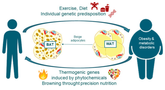
Figure 1.
Fat burning non-shivering thermogenesis and browning through precision nutrition as mechanisms to prevent and treat obesity associated disorders.
The most straightforward way to prevent obesity is to limit calorie intake or to increase the burning of calories through exercise and physical activity. However, majority of subjects fail in their attempts to accomplish these aims. Thus, at present, the only method to combat excessive obesity is through surgery, as many drugs have failed causing serious and deleterious side effects. Nonetheless, it is an invasive method not available for all patients which also requires long time of recovery. Therefore, it has gained importance to investigate in complementary approaches in the frame of Precision Nutrition to alleviate obesity and associated metabolic alterations (Figure 1). As AT contributes to the control of the systemic energetic balance, strategies to increase the thermogenic potential of AT to augment the energetic expenditure (EE) may be relevant in the control of metabolic alterations. In this regard, the crosstalk between adipose tissue with other metabolic organs such as skeletal muscle is another complementary strategy to be explored [4,5,6,7,8,9,10,11,12,13,14,15].
As indicated, the current obesity pandemic results from a physiological imbalance in which energy intake chronically exceeds EE. Approaches designed to increase EE have been demonstrated in animal models mainly oriented to stimulate the adaptive thermogenesis. At the cellular level, thermogenesis is mainly achieved by increasing the uncoupling of the oxidative phosphorylation from adenosine triphosphate (ATP) generation by the uncoupling protein 1 (UCP1), a tissue-specific protein highly expressed in mitochondria of brown adipose tissue (BAT). Physiological activation of BAT is mainly mediated through β-adrenergic receptors (AR) activation. This way, considerable efforts over the past 5 decades have been oriented to identify AR agonists capable of safely achieve a net negative energy balance while avoiding unwanted cardiovascular side effects. Recent discoveries of other BAT futile cycles based on creatine and succinate have provided additional targets. Cold and exercise are also physiological key players with potential to increase the EE, and developing strategies are being investigated with the aim to mimic these processes by mean of the use of bioactive compounds.
Future studies will address technical challenges such as how to accurately measure individual tissue thermogenesis in humans; how to safely activate BAT and thermogenesis in other metabolic organs, mainly white adipose tissue (WAT) and skeletal muscle; and how to sustain a negative energy balance over many years of treatment.
2. Methodology of Searching
The methodology used for the selection of the references included in this review was based on the searching algorithm presented below. For this, two different investigators searched in PubMed and Web of Science databases from 13 of April to 30 of October 2020, for studies that evaluated the principal targets that could be modulated by diet/bioactive compounds regarding thermogenesis in muscle and adipose tissue, and also, for studies that analyzed the effect of the five plants represented in this review regarding the modulation of thermogenesis in different studies.
Thus, we used our systematic criteria of searching for articles and reviews no older than 10 years that include in their title or in their abstract these following concepts:
[Mitochondrial thermogenesis OR white adipose tissue browning process OR brown adipocyte thermogenesis activation OR beige adipocyte thermogenesis OR Brown adipogenesis OR thermogenic obesity treatment OR cold-induced thermogenesis OR leptin thermogenesis OR energy expenditure and thermogenesis OR muscle non-shivering thermogenesis OR exercise contribution in energy expenditure and thermogenesis OR AMPK thermogenesis activation OR SIRT1 thermogenesis activation PGC1a thermogenesis activation AND/OR precision nutrition OR nutrigenomics OR nutrigenetics OR Resveratrol OR Pomegranate OR Silymarin OR Soy OR Ginkgo OR natural extracts.]
With those keywords, 1724 articles were found (once duplicates were excluded) on both websites. From this, we excluded 563 articles with no full-text assessed for eligibility. The abstract of the remaining 1171 articles where read, and finally 435 papers and reviews were included. 214 were included based on their relevant information related to thermogenesis, browning and energy expenditure.
We also searched the reference lists of these studies, to identify potentially eligible studies that were not included in the initial search. Disagreements in the searching procedure between the two independent investigators were resolved through scientific consensus.
3. Addressing Obesity through Precision Nutrition: Nutrigenetics and Nutrigenomics
It is noteworthy that the relationship between diet and health is a matter of great interest, and nutrition and diet derived ingredients can also be considered as tools to prevent and/or to treat different chronic diseases. It has been proved that general macronutrient recommendations do not affect individuals in the same way, as these recommendations do not take into consideration the genetic heterogenicity between individuals, such as genetic variants (single nucleotide polymorphisms or SNPs), nor the nutritional and metabolic status of individuals (such as dyslipidemia, insulin resistance, metabolic syndrome, cardiovascular disease, and cancer) that shape the final metabolic effects of diet derived ingredients. Therefore, the knowledge of interactions between genome and nutrients at the molecular level has led to the development of ”Nutritional Genomics”, which involves the sciences of nutrigenomics and nutrigenetics [16,17].
In a deeper meaning, nutrigenetics focuses on inherited or acquired mutations within genes and the interaction between bioactive compounds and ingredients from diet with the genetic susceptibilities to metabolic disorders. On the other hand, nutrigenomics includes several disciplines for the study of the dietary effects on genome stability regarding deoxyribonucleic acid (DNA) damage, epigenomic alterations, ribonucleic acid (RNA) expression, and protein expression, which will affect the individuals’ metabolic health. Thus, the key goal of nutritional genomics is the use of this information meaningfully to provide trustable personalized dietary recommendations for specific health and disorder prevention outcomes [16,18].
Precision nutrition is considered as a two-way route integrating by one hand the knowledge of the diversity in the genomes which also influences nutrient bioavailability and metabolism, and by the other hand, the knowledge of how nutrients may affect the expression of genes in critical metabolic pathways [19]. In addition, Precision nutrition considers additional factors related to lifestyle such as diet, exercise, alcohol consumption, as well as the nutritional and metabolic status of the individual. Based on this, the final objective of precision nutrition is to develop personalized nutrition interventions for health and disease prevention or treatment.
The development of multi-omics sciences and bioinformatics tools, together with the sequencing of the human genome, has contributed to the development of effective personalized nutritional interventions. Only the scientific knowledge of individuals´ genomic information combined with the molecular characterization of diet ingredients and the elucidation of their molecular mechanisms of action may provide effective personalized nutritional interventions [7,20,21].
Several studies have revealed genetic associations to the predisposition to obesity and/or type 2 diabetes and associated comorbidities [22]. For instance Alpha-Ketoglutarate Dependent Dioxygenase (FTO), Insulin Like Growth Factor 2 MRNA Binding Protein 2 (IGF2BP2), Brain Derived Neurotrophic Factor (BDNF), Melanocortin 4 Receptor (MC4R), and Apolipoprotein (APO-A) gene family, among others, have been shown to present SNPs associated with those disorders [23,24,25,26,27,28,29] The genetic susceptibility to obesity is also influenced by an obesogenic environment for its phenotypic expression (nutrigenetics) (Figure 1).
In this scenario, the identification of brown and beige adipocytes in adult humans has opened the possibility of using thermogenesis and browning of WAT as a strategy to prevent and treat obesity and related metabolic alterations diseases, which can compensate genetic polymorphisms and/or mutations in a personalized manner [30,31]. This way, it is possible to develop nutritional interventions by mean of the use of bioactive compounds and/or ingredients from diet to decrease fat storage -by mean of the induction of mitochondrial biogenesis, by increasing lipolysis coupled to fatty acid oxidation-, and to promote the brown adipogenesis in WAT to compensate the genetic and environmental factors implicated in the development of metabolic alterations.
4. Adipose Tissue Features and Fate: The Browning Process
In the last years it has been accepted that AT is not a passive organ, instead, it is a dynamic endocrine organ implicated in the control of the energy homeostasis and inflammation among other processes [32]. Therefore, AT as an endocrine organ releases a variety of adipokines (adipose-associated cytokines) which affect the functioning of different organs and tissues including the liver, pancreas, muscle, brain and immune system.
There are two types of AT differing in their mesenchymal progenitor origin, their functionality and their distribution within the body (Figure 2). The WAT constitutes the body’s main energy reserve, storing and mobilizing triglycerides depending on the requirement of the different peripheral tissues. WAT can be considered as an organ formed by a cellular component (preadipocytes, mature adipocytes, fibroblasts, immune infiltrated cells, and endothelial cells), and a non-cellular component (mainly stroma and matrix fibers). Mature white adipocytes synthesize and secrete adipokines to regulate the energy balance and the systemic glucose and lipid metabolisms [33,34]. This way, WAT senses the body’s energetic status and responds by storing fats in the form of triacylglycerides, or by releasing glycerol and fatty acids to be used as energy sources when required.
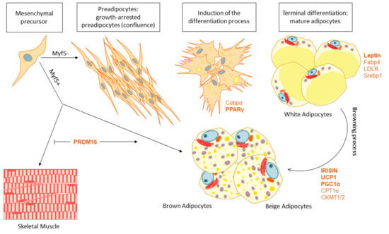
Figure 2.
Adipogenesis, the differentiation process from the mesenchymal precursors to BAT or WAT and the browning process, main genes implicated.
On the other hand, BAT is a thermogenic organ responsible for maintaining the thermal homeostasis through dissipating large amounts of chemical energy as heat. The existence of BAT in infants has been known for a long time, but the demonstration of metabolically active BAT in adults was only documented recently [35] The main characteristic of BAT is the high levels of mitochondria which provides high capacity to oxidize fatty acids and glucose [36]. This way, BAT may contribute to dissipate the excess of reductive power into heat in conditions of excess of glucose and fatty acids, preventing the appearance of insulin resistance and/or metabolic unhealthy obesity.
BAT has been shown to display a specific expression profile promoting lipolysis and mitochondrial uncoupling for energy dissipation (Figure 3) which contributes to alleviate the excessive metabolic stress in chronic diseases such as obesity, insulin resistance, cardiovascular disease (CVD) and cancer [37,38].
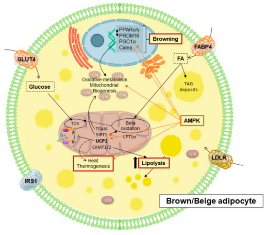
Figure 3.
Main mediators implicated of browning and thermogenesis in brown/beige adipocytes.
4.1. White and Brown Adipose Tissue Biogenesis
Adipogenesis is a process that starts during development and continues throughout life. Differentiating progenitors accumulate small lipid droplets in a process guided with the developing capillary networks. Eventually these progenitors mature to form terminal differentiated adipocytes [39].
Nevertheless, WAT and BAT are formed by different mesenchymal progenitors, with BAT cells coming from progenitors common to the skeletal muscle tissue which are positive for myogenic factor 5 (Myf5), whereas WAT cells come from progenitors lacking this factor. Hence, as we show in Figure 2, two different types of mesenchymal precursors give rise to two different types of AT that can be find in the body. Importantly, adipogenesis is controlled by distinct transcription factors and proteins to determine the fate of preadipocytes to WAT or BAT. For instance, PR domain containing 16 (PRDM16), Peroxisome Proliferator Activated Receptor Gamma Coactivator 1 Alpha (PGC1a), Carnitine Palmitoyltransferase 1A (CPT1a) and UCP1 are key factors promoting BAT adipogenesis and differentiation with increased mitochondrial biogenesis and fatty acid oxidation capacity. On the other hand, Peroxisome Proliferator Activated Receptor Gamma (PPARG), CCAAT Enhancer Binding Protein Alpha (C/EBPα), Sterol Regulatory Element Binding Transcription Factor 1 (SREBP1) and Fatty Acid Binding Protein 4 (FABP4) are implicated in WAT differentiation and lipid storage. Importantly, adipogenesis have been demonstrated to be modulated by external inductors such as cold exposure or nutritional agents [37,40,41].
Recently, it has been discovered that under specific circumstances, such as cold exposure, physical exercise or fasting, white adipocytes can reprogram their metabolism to behave as brown adipocytes in a process called browning. Similar to brown adipocytes, these “beige” adipocytes are able to dissipate energy in the form of heat, and therefore, may contribute to the energetic homeostasis together with BAT. Activating the browning process could be an alternative therapeutic strategy to increase EE counteracting the development of obesity and associated metabolic diseases [42,43].
4.2. Adipose Tissue Browning
Beige adipocytes, despite sharing some features of both brown and white adipocytes, are a unique type of adipocytes characterized by a distinct gene expression profile and adult origin compared to white or brown adipocytes [44,45]. They have multilocular lipid droplets, higher mitochondrial content with higher levels of UCP1 compared to white adipocytes, sharing a functional and phenotypic thermogenic capacity similar to that of brown adipocytes (Figure 3) [46]. Beige and brown adipocytes use uncoupling proteins (UCPs) from the inner membrane to dissipate energy as heat. All in all, brown and beige adipocytes contribute to the energetic homeostasis balancing storage and burning of fats and glucose. UCP1 is responsible for cold- and diet-induced thermogenesis and it is directly implicated in the phenotypic change of white adipocytes into beige adipocytes [43,44].
Together with UCPs, there are many different molecules implicated in the differentiation process to BAT and/or in the browning process of WAT (Figure 3). For instance, adenosine monophosphate activated protein kinase (AMPK) activation inhibits acetyl-CoA carboxylase diminishing fatty acid synthesis, and it also promotes the uptake of extracellular glucose and fatty acids for further oxidation at mitochondria [47,48]. Moreover, it also enhances mitochondrial function by directly phosphorylating and activating PGC1α [49], a master transcriptional regulator of genes involved in mitochondrial biogenesis and oxidative metabolism [50]. In addition, AMPK, through the activation of Sirtuin 1 (Sirt1), another important mitochondrial activator (Figure 3) [51], promotes the differentiation process into brown and beige, increasing the expression levels of UCP1.
The PPAR family includes a group of nuclear transcription factors regulating cellular differentiation, development, energy metabolism, and tumorigenesis [52]. There are three types of PPARs: PPARα, δ and γ. PPARγ is mainly expressed in WAT, internal organs and macrophages. In mature white adipocytes, it regulates the expression of genes involved in free fatty acid uptake and triglyceride synthesis. PPARγ also promotes browning due to the AMPK/Sirt1-dependent PPARγ deacetylation, which is required to recruit the BAT programmer coactivator PRDM16, leading to the selective induction of BAT genes like cell death-inducing DNA fragmentation factor-α-like effector A (CIDEA), while inhibiting typical WAT markers (Figure 3) [53,54]. PPARα also controls the transcription of this essential gene, which interacts with PGC1α to provide the machinery necessary for the differentiation of the beige adipocyte [7,55]. Reduction of AMPK signaling has been demonstrated to reduce PRDM16 activation, diminishing brown adipogenesis and promoting fibrogenesis (Figure 2) [53].
Another key factor implicated in the browning process is fibroblast Growth Factor 21 (FGF21), which is produced by BAT and beige cells, and is released into plasma where it acts as an autocrine/paracrine factor increasing the expression of UCP1 and other genes such as CIDEA to promote thermogenesis and EE [56,57]. Moreover, Irisin, a hormone produced by skeletal muscle in response to exercise, is also a potent inducer of WAT browning, highlighting the important crosstalk between these two metabolic organs (Figure 4) [58]. Therefore, targeting the expression of those genes through phytochemicals may provide a therapeutic potential for the treatment of obesity and its related disorders.
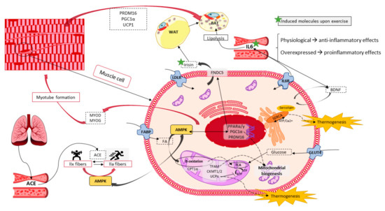
Figure 4.
Skeletal muscle potential in energy balance through thermogenesis, and its influence on the browning and fiber-switch processes. Genes and proteins implicated.
5. Thermogenesis within Adipose Tissue, Inductors and Their Implications in Obesity Disorders
As explained before, BAT and beige cells have the capacity to burn off nutrients upon specific external conditions and therefore to control the energy balance in the body. When mitochondrial electron chain is uncoupled from the ATP final production, the reductive power may be dissipated into heat, a process called thermogenesis [59]. Thus, thermogenesis is the ability to generate heat in the body due to metabolic reactions which, upon correct thermal homeostasis, allows mammals (like humans) to control their body temperature as a static value of approximately 36–37 °C.
Regarding this concept, there are two different types of thermogenesis, on the one hand, the involuntary activation of skeletal muscle movement which produces heat, a process called shivering; and, on the other hand, the activation of cell metabolism in BAT and skeletal muscle dissipating heat upon mitochondrial ATP uncoupling or upon a calcium flux through the sarcoplasmic reticulum in skeletal muscle fibers, a process called non-shivering thermogenesis [60]. In this context, thermogenesis can be induced by cold exposure and its consequent adrenergic stimulation [61], but also it can be induced by diet and/or by mean of thermogenic dietary supplements [7,8]. Thus, dissipating energy by enhancement thermogenesis via increasing BAT activity or mass within beige cells in WAT is one of the new approaches for the prevention and/or treatment of obesity related metabolic disorders [9,10].
It should be noted that activation of thermogenesis not only induces weight loss, but also has a positive impact on other metabolic alterations such as insulin and leptin resistance, hepatic steatosis, hyperlipidemia, hyperglycemia, hypercholesterolemia and hypertriglyceridemia, likely due to the ability of thermogenic adipocytes to uptake lipids and glucose from the circulation. Thus, it has been proposed, mostly based on in vitro and in vivo studies, that BAT may help to prevent or reverse excessive adiposity and to improve glucose and triglyceride clearance from blood. However, direct and convincing evidence from human studies is still lacked due to its relatively recent discovery [10,11].
5.1. Molecules and Processes Implicated in Mitochondrial Thermogenesis
In BAT and beige thermogenesis, the main operators are mitochondria. BAT has more and larger mitochondria than WAT, as well as higher expression of fatty acid oxidation-related enzymes. This fact gives BAT the appearance of being brownish in microscopic images because of the iron-containing heme cofactor in the mitochondrial enzyme cytochrome oxidase [62]. Thus, this suggests that BAT is a more prepared tissue for dissipating and wasting energy than for storing it. One of the key proteins here is UCP1 (Figure 3), which is located in the inner membrane and causes a proton leak across it, dissipating the electrochemical gradient into heat [59,63,64]. However, not everything regarding thermogenesis is UCP1 dependent, there are many other signaling pathways implicated in the activation of thermogenesis. Thus, the metabolic axis Sirt1/PGC1α/AMPK pathway has been described to promote the correct functioning of mitochondrial thermogenesis, implicating downstream effectors including mitochondrial transcription Factor A (TFAM) factor and CPT1α [37,50].
The in vivo treatment of mice born from obese parents with the AMPK activators metformin or 5-aminoimidazole-4-carboxamide ribonucleotide (AICAR) increases brown adipocyte progenitor cells and BAT weight, as well as WAT thermogenesis [53]. In addition, experimental reduction of AMPK in WAT, leads to reduced levels of PGC1α and mitochondrial markers, causing lipid and glucose accumulation and the promotion of liver steatosis and insulin resistance [48]. Hence, activating AMPK in adipocytes can increase browning and non-shivering thermogenesis, promoting the uptake and oxidation of glucose, fatty acids, and triglycerides, and potentially reducing the risks associated with obesity.
AMPK activation is affected by levels of reactive oxygen species (ROS) from mitochondria. Mitochondrial ROS (mtROS) influences the function of key thermogenic proteins in adipocytes. Succinate is the genuine source of thermogenic ROS in brown and beige adipocytes, being superoxide anion production proportional to UCP1-related uncoupled respiration. Upon a balanced diet, the limited ATP synthesis in BAT, due to the uncoupled respiration, maintains an elevated proton gradient and electron flux, which controls excessive mtROS accumulation. On the contrary, when there is an excess on the intake of nutrients, such as normally happens in obesity disorders, there is an enhanced mitochondrial redox pressure which may lead to excessive mtROS accumulation. Thus, when the functional redox threshold in mitochondria is overcome, mtROS disrupt the interaction between insulin receptor (IR) and insulin receptor substrate (IRS) causing insulin resistance and the impairment of adipogenesis, thermogenesis and adipocyte function [65,66,67]. Mitochondrial superoxide dismutase levels have been shown to protect against impaired thermogenesis, by reducing the levels of superoxide [65].
Thermogenesis is also depending on the mitochondrial fusion and fission balance. The remodeling of the mitochondrial structure is dynamic and sensitive to metabolic signals. The fusion process is regulated by nucleotide guanosine triphosphate hydrolase (GTPase) proteins such as mitofusin 1 and 2, and mitochondrial dynamin like GTPase (OPA1). Mitochondrial fusion allows the repolarization of membranes diminishing ROS, which is necessary for mitochondrial enhanced ATP production. On the contrary, mitochondrial fission is associated with increased mitochondrial ROS generation and the uncoupled respiration for BAT thermogenesis [62,68,69]. In obese individuals, it has been shown that the fusion-fission dynamics is disrupted, leading to reduced flexibility to switch between ATP production and mitochondrial uncoupling [36]. which is essential to balance changes in nutrient availability and metabolic demands.
Another important process in the regulation of mitochondrial dynamics and therefore, thermogenesis, is mitophagy, which selectively clears the excess of mitochondria through autophagy after damage. The principal proteins involved in this process are phosphatidylinositol-3,4,5-trisphosphate 3-phosphatase induced kinase 1 (PINK1) and p62. Mitophagy is induced by metabolic stress, including endoplasmic reticulum stress, oxidative stress, inflammation, and insulin resistance. It has been shown that the AT of obese and diabetic individuals display a disturbed mitophagy capacity [62,67], which has been associated to fat accumulation. On the other hand, in excess of mitophagy, the beige-to-white adipocyte transition is upregulated, which reduces the brown mass and therefore, the BAT characteristics upon the white ones [70,71]. The ambivalence of mitophagy in AT is necessary for the maintenance of the mitochondrial population and the tissue function [67].
5.2. Adrenergic Nervous System Activation of Thermogenesis Upon Cold Exposure
The brain controls many different energetic signals, including thermogenesis through the sympathetic nervous system (SNS). Its activation causes the release of norepinephrine (NE) which binds to β-3-adrenergic receptors (β3-AR) in adipocytes to activate thermogenesis [61]. Cold exposure causes the activation of specific channels located in thermoreceptor neurons innervating the surface of the body, thereby signaling for the release of NE (Figure 5). NE binds β3-AR activating adenylate cyclase, which also activates cAMP-dependent protein kinase A which promotes lipolysis and mitochondrial biogenesis through the expression of several genes including lipases, AMPK, UCP1 and PGC1α (Figure 5) [48,72].
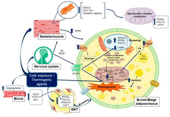
Figure 5.
Activation of BAT thermogenesis and WAT browning upon cold exposure: role of the SNS.
After cold exposition, the β3-AR stimulation augments the release of fatty acids and increases the expression of CPT1a, and UCP1 for further heat production. However, the classical BAT has a higher basal UCP1 expression levels and elevated uncoupled respiration compared to beige or WAT counterparts. Although, upon hormonal stimulation with β3-AR agonists, beige cells can also elevate their UCP1 levels to the ones seen in BAT. This results suggest that beige cells are programmed to switch their function between energy storage or heat production depending on different signals [57]. In this regard, it is well known that circulating hormones, such as triiodothyronine (T3), are implicated in BAT activation and WAT browning. T3 induces the local formation of BMP8b, a BAT factor that makes this tissue more sensitive for adrenergic signaling, and also enhances SNS signaling from the brain to the BAT [72,73].
In addition, cold exposure increases lipoprotein lipase and membrane proteins implicated in the lipid uptake of plasma triglycerides or free fatty acids in BAT. Cold exposure also increases the up-regulation and translocation of glucose transporters, glucose transporter (GLUT) 1 and 4, facilitating the uptake of glucose from plasma (Figure 5). All in all, this axis provides protection against hypertriglyceridemia, hyperglycemia and insulin resistance, which are common disorders associated to obesity [10]. Additionally, cold exposure produces an increase in the production of vascular endothelial growth factor (VEGF) (Figure 5) which enhances angiogenesis and provides a venue for heat dissipation. Moreover, it has been reported that the increase in VEGF causes an increase in UCP1 expression in WAT and the expansion and activation of BAT [74].
FGF21 is also induced by cold and adrenergic activation through a cAMP-dependent mechanism (Figure 5). It is expressed mostly in the liver, adipocytes and skeletal muscle, correlating positively with the increase in EE and thermogenesis activation, and therefore, promoting browning within the AT [56,75].
Moreover, another SNS regulated gene upon cold exposure is leptin (LEP), a hormone predominantly secreted from subcutaneous AT. This adipokine has multiple functions especially implicated in the control of appetite, fat storage, and glucose homeostasis. Nonetheless, although cold reduces reversibly its expression and increases browning and thermogenesis (Figure 5), it appears to exist a balance between leptin production and thermogenesis. Leptin has been shown to increase lipolysis and browning too, although much less than upon SNS activation, and many of the hypothalamic neurons involved in the regulation of thermogenesis are also leptin sensitive [76,77]. Therefore, taking all together, it appears that leptin has a double role in promoting browning and lipolysis, but after cold exposure the decreased in leptin expression is compensated by other thermogenic drivers indicated before.
6. Skeletal Muscle Potential in Energy Expenditure and Heat Production
6.1. Skeletal Muscle Features and Functions
Muscles are important metabolic organs and the main source of amino acids, storing up to 75% of the total proteins within the body. Amino acids are released when needed elsewhere in the organism. Besides, the muscle has the ability of storing glucose in the form of glycogen, being the principal source of energy for the rapid initiation of contraction even when glucose is not readily available from the diet [78].
Moreover, muscles are composed of two main types of fibers, with marked differences in their metabolic profile in regard to the speed and manner in which they metabolize glucose. The names ”fast” and ”slow” fibers, indicate the type of glucose metabolism occurring within them. The fast ones use the anaerobic metabolism of glycolysis for the quick ATP generation upon rapid contraction, and therefore they fatigue sooner. On the contrary, slow fibers are aerobic and high oxidative allowing them to be high resistant to fatigue. This second type of fibers preferentially use fatty acids as substrates for ATP production [79]. However, a remarkable feature within the skeletal muscle is the ability to a fast-shift in substrate usage between fat and glucose, depending on the needs of other organs. Therefore, the muscle is turned into a sensor of the global energy state of the body, with the ability to balance the use of different nutrients for energy dissipation or for their storage for the further use by other tissues [80].
However, when the lipid storage capacity in AT is overcome, free fatty acids are released and accumulated by other organs such as muscle and liver. Lipotoxicity in muscle impairs the contractility capacity and function of skeletal muscle, a condition named sarcopenia. This condition is increased in obese subjects as consequence of the excess of lipids excess, being worst in fast fibers and being aggravated in aging [81]. Thus, bearing in mind the important crosstalk between these the AT and muscle, the activation of the thermogenic signaling between these two organs may contribute to control the excess of lipids.
6.2. Thermogenesis within Skeletal Muscle
Skeletal muscle metabolism can also be activated by cold. As shown in Figure 2, BAT and skeletal muscle are derived from a common progenitor (Myf5+). Besides, it is well known that skeletal muscle is a metabolic organ which produces heat upon contraction in the process of shivering thermogenesis, thus, it is not surprising that AT and muscle might also share the capacity to accomplish the non-shivering one [60], as proton leak is also present in skeletal muscle through another uncoupling protein, the UCP3. However, while UCP3 can uncouple oxidative phosphorylation and dissipate energy as heat, this effect is secondary to its primary role which is the control of mitochondrial ROS and fatty acid oxidation (Figure 4) [10,82]. UCP3 lowers mitochondrial membrane potential and protects muscle cells against an overload of fatty acids, reducing the stress caused by excessive ROS production. In this context, SIRT1 acts as a major repressor of the UCP3 gene expression in response to glucocorticoids, which are activators of SIRT1. This requires its deacetylase activity and results in histone deacetylation in the UCP3 promoter [83], therefore controlling its levels.
Although skeletal muscle is prepared for mitochondrial non-shivering thermogenesis, UCP3 is not totally in charge of it. An alternative mechanism involving Ca2+ changes within the sarcoplasmic reticulum exists, which does not interfere with the mitochondrial ATP synthesis necessary for slow and fast contraction. This mechanism mainly occurs in oxidative slow fibers, through the ion pump sarcoplasmic reticulum calcium ATPase (SERCA). In this mechanism, sarcolipin, a small peptide which senses calcium levels, has been shown to activate SERCA after an influx of Ca2+ (Figure 4). Hence, sarcolipin promotes SERCA dependent uncoupling causing a futile cycling in the flux of Ca2+ through the reticulum, and promoting the dissipation of energy in the form of heat [5,84,85]. Here, with the rise of cytosolic Ca2+, sarcolipin promotes Ca+2 to concentrate at the cytoplasmic side of the membrane, instead of at the luminal side, and hence, promoting the hydrolysis of ATP producing heat. In addition, the adenosine diphosphate (ADP) produced is phosphorylated again by the oxidative phosphorylation at mitochondria which also contributes to the non-shivering thermogenesis in the muscle.
This mechanism also implicates the increased expression of PPARγ and PGC1α in muscle, which can also be induced by cold exposure (Figure 5), increasing the mitochondrial biogenesis necessary for the increased thermogenic capacity within the fibers [4,86]. Taking all of this into account, heat will be generated by SERCA during both shivering and non-shivering thermogenesis, when Ca2+ pumping is coupled to myofibril contraction and when it is uncoupled.
6.3. Exercise Performance as a Molecular Inductor of Thermogenesis and Browning
Skeletal muscle -an organ that provides us with physical force- also changes upon exercise training which also provides multiple benefits for human health maintenance and improvement. Therefore, it is not surprising that exercise endurance capacity interferes in the thermogenesic and oxidative capacity within the fibers. Exercise increases sarcolipin to produce a shift in the oxidative capacity of the fibers and hence, enhances thermogenesis. Moreover, exercise can also change the mitochondrial fate upon aging or disease by augmenting its function, oxidative capacity, mitochondrial DNA content and biomass, as well as increasing the expression of several mitochondrial biogenesis related transcription factors such as TFAM and PGC1α [87,88]. Additionally, AMPK and SIRT1 are also activated upon exercise, increasing the systemic insulin sensitivity by their role in the increase of mitochondrial biogenesis, oxidative phosphorylation and density of fibers in the skeletal muscle (Figure 4) [49,89]. Thus, the increase of the non-shivering thermogenesis in skeletal muscle is another strategy for preventing metabolic diseases.
Besides that, exercise has been shown to induce markers of BAT in WAT, augmenting the number of beige adipocytes. Skeletal muscle, which can also be considered as an endocrine organ, stablishes a communication network with other organs, such as AT. This crosstalk is mediated by several myokines, released from muscles, such as irisin, implicated in the initiation of the thermogenic and browning program in AT [10,67,90]. Irisin is proteolytically cleaved from fibronectin Type III Domain Containing 5 (FNDC5) by a PGC1α dependent mechanism and then it is released into the blood system producing a browning effect in WAT [32,58]. Another important myokine is BDNF, a factor produced by the nervous system (Figure 5) and by muscles, (Figure 4) which is able to induce lipolysis in AT, and neurogenesis in the brain. BDNF expression within the skeletal muscle is enhanced by irisin and by interleukin 6 (IL6) upon exercise (Figure 4), being the latter an important cytokine which also may act as a myokine to induce browning and lipolysis in WAT ([32,91,92].
Hence, increasing exercise, the release of browning myokines, and the skeletal muscle non-shivering thermogenesis, may be contribute to restore the metabolic homeostasis within obesity or other metabolic disorders [93,94].
7. Results, Discussion and Conclusions
7.1. Phytochemicals as Thermogenic and Anti-Adipogenic Agents
As shown before, non-shivering thermogenesis is a potential complementary approach to prevent and/or treat obesity-related disorders [14,95]. Indeed, in the last years, there have been reported several plant-derived bioactive compounds with the capacity to activate and to augment thermogenesis. Therefore, the upregulation of several signaling pathways including UCP1, AMPK, PGC1α, Sirt1, PRDM16 and/or PPARs have been shown to increase the WAT browning [96,97,98,99,100,101,102,103,104,105].
Here, we review the evidence upon this field of some of some of the most studied natural extracts in the activation of thermogenesis.
7.1.1. Pomegranate
Most health benefits of pomegranate are associated to the presence of ellagitannins, mainly punicalagin and ellagic acid [106]. Preclinical models and intervention studies with humans have shown that pomegranate, or its bioactive compound punicalagin, are able to reduce the negative effects upon high fat diet (HFD) in mice, reducing cholesterol, triglycerides and glucose in plasma, as well as the weight gain in obese individuals [107,108]. Moreover, punicalagin has the capacity of promoting mitochondrial function in vitro and in vivo, by mean of the activation of the AMPK pathway upregulating the mitochondrial biogenesis and ameliorating the oxidative stress and inflammation after a HFD [109,110]. Pomegranate extract and juice are also effective on increasing the vascular endothelial nitric oxide synthase and plasma nitric oxide levels, which in turn, increase the response to acetylcholine in vitro, which has been proposed to be beneficial against metabolic syndrome [106].
Moreover, pomegranate also contents potent antioxidants, capable of reducing lipid peroxidation, and acting as hypotensive agents to reduce blood pressure [111]. Moreover, there are several intervention studies in humans that report benefits related to the improvement of insulin sensitive or reducing weight and fat mass gain [112,113].
Furthermore, it is known that obesity is associated with immune dysfunction and a state of chronic inflammation, features that are reduced by exercise. In this regard, it has been reported that pomegranate extract in combination with exercise improves immune function in HFD-fed rats, restoring immunomodulatory factors in serum, inhibiting inflammation and decreasing oxidative stress, compared to the extract or the exercise alone [114]. Besides, it has been demonstrated that Pomegranate juice slightly reduces muscle damage markers, fatigue, and the recuperation time in elite athletes, which provides additional benefits against skeletal muscle damage and cachexia [115,116].
Punicalagin and urolithin A exert anti-adipogenic properties associated to a reduction in triglyceride accumulation and to the expression levels of adiponectin, PPARγ, GLUT4, and FABP4 in early steps of 3T3-L1 adipocytes differentiation [117]. Moreover, urolithin A increases the EE in mice by enhancing thermogenesis and browning in BAT and WAT, by mean of the elevation of triiodothyronine levels [118].
7.1.2. Ginkgo Biloba
Ginkgo Biloba extract, a mixture of polyphenols with antioxidant properties, might be efficient in the prevention and treatment of obesity associated disorders such as insulin resistance and adipocyte hypertrophy as indicated by a variety of experimental models of endocrine dysfunctions [119]. In this regard, it have been reported several in vivo preclinical studies where Ginkgo Biloba extract significantly reduced food intake, body weight gain and adiposity, reducing the epididymal adipocyte volume, while protecting against hyperglycemia and dyslipidemia in diet-induced obesity in rats [120,121,122]. Moreover, this extract increased AMPK and adiponectin signaling pathways [123].
The anti-adipogenic effects of extracts derived from Ginkgo Biloba have been demonstrated [124]. For instance, bilobalide, a sesquiterpene compound from Ginkgo Biloba leaves [125], or Ginkgetin, a Biflavone from Ginkgo Biloba leaves [126], blocked the differentiation of preadipocytes into adipocytes, reducing PPARγ and C/EBPα expression in 3T3-L1 adipocytes during adipogenesis. In addition, they also increased lipolysis by activating AMPK signaling pathway and the expression of CPT1α, which contributed to reduce hypertrophy of WAT in HFD mice. Thus, suggesting a potentially anti-obesogenic effect in longer-term therapies.
Another bioactive extract derived from the seed coat of Ginkgo Biloba have also been investigated. Although this part is rarely use and it is typically discarded, ginkgo vinegar from the seed coat has been reported to suppress the expression of C/EBPδ and PPARγ, key proteins in adipogenesis, and to inhibit lipid accumulation in 3T3-L1 cells that were induced to become adipocytes, hence, inhibiting adipocyte differentiation [127].
7.1.3. Milk Thistle
Silymarin, which belongs to the flavonolignan group, is the main bioactive constituent found in Milk Thistle (Silybum marianum). Silymarin has been shown to exhibit antioxidant, plasma lipid-lowering effects, antihypertensive, antidiabetic, antiatherosclerotic, anti-obesity, and hepatoprotective effects [128,129].
The beneficial effects have been related to the increase of genes implicated in the promotion of BAT (Sirt-1, PPARα, PGC-1α, and UCPs) and to the decrease on the expression of genes related to WAT differentiation (PPARγ, FABP4, FASN, SREBP1c, C/EBPα) [130,131]. In addition, silymarin has been shown to diminish lipid accumulation and early adipogenesis via the regulation of cell cycle and AMPK signaling pathways in vitro [132]. Therefore, by mean of the induction of thermogenesis and the promotion of brown remodeling in adipocytes, silymarin reduced fatty acid accumulation and adipocyte size. Besides that, in a recent preclinical assay in obese mice, Silymarin reversed the AT inflammation and adipocyte hypertrophy, stopping weight gain without changes in food intake. In addition, it reversed liver disorders restoring insulin sensitivity, and glucose and lipid homeostasis [133].
In conclusion, silymarin and silybin, its major active constituent, have important roles in the treatment and prevention of obesity through several mechanisms including the suppression of the expression of adipogenesis-related genes, and the reduction of lipid mass but augmenting the functional capacity of adipocytes [129].
7.1.4. Soy
Because of the lower frequency of obesity and diabetes II diseases in Asian countries, attention has been turned toward their diet, which consists in high amounts of soy and soy-based products. Their principal bioactive components are isoflavones, such as genistein and daidzein, which are similar in structure to endogenous estrogens [134]. They interact with estrogen receptors, which results in the reduction of intracellular lipids. Therefore, although various studies have focused on the phytoestrogenic function of isoflavones, their potential to increase WAT browning and non-shivering thermogenesis has been investigated [135,136].
Besides protein may also be particularly effective in preventing diet-induced-obesity. protein has been reported to prevent fat mass gain under HFD, inducing browning in WAT and lipolysis and thermogenesis in BAT, by mean of the increase of UCP1 expression and the leptin sensitivity in the hypothalamus [137,138] found that diets rich in Isoflavones also increased triiodothyronine levels and UCP1 mRNA levels in the BAT of rats, although the core body temperature decreased. Isoflavones have demonstrated to enhance mitochondrial biogenesis in AT through the SIRT1/PGC1α pathway [137].
Soy ssoflavones are known to exert lipolytic and anti-adipogenic effects in WAT, alone or in combination with other bioactive compounds. For instance, they have an additive, not synergic, anti-adipogenic effect in combination with green tea extract and grape resveratrol, reducing the expression of PPARγ, C/EBPα and FABP4 among others, in in vitro studies with 3T3-L1 and human adipocytes [138]. Daidzein alone also suppressed adipogenesis in 3T3-L1 preadipocytes [139]. Another study also showed that daidzein and genistein inhibited adipogenesis in human adipocytes reducing the expression of WAT markers, PPARγ, SREBP-1, FASN, C/EBPα, although by mean of different mechanisms of action, as daidzein inhibited adipogenesis through the stimulation of lipolysis, and genistein inhibited the glycerol-3-phosphate dehydrogenase activity acting via the activation of estrogen receptors [140,141]. In contrast to this results, [142] showed in vitro that Genistein and Daidzein functioned as PPARγ agonists.
Genistein has been shown to induce BAT adipogenesis in 3T3-L1 preadipocytes when treated at initial points of the differentiation process. In another study, genistein induced the expression of BAT specific markers (UCP1, SIRT1 and PGC1α), reduced WAT markers (FASN, FABP4), and increased the mitochondrial proton leak and oxygen consumption, promoting features of beige adipocytes [97]. Genistein also increased the body temperature and plasma levels of triiodothyronine of obese mice [143].
In addition, genistein exerted a dose-dependent effect on adipocyte differentiation and function being able to control adiposity [144]. Genistein and daidzein increased PGC-1β gene expression and augmented the EE energy in a preclinical model of HFD induced obesity [145]. Besides that, daidzein reduced weight gain and fat content in liver, which was associated to the increase of fatty acids oxidation and to the enhanced expression of UCP1 in BAT [146,147].
Usually, obesity also involves the ectopic accumulation of lipids in the skeletal muscle. Genistein enhanced fatty acid oxidation in muscle, by mean of AMPK dependent mechanism, and increased the expression of PGC1α and PPARδ, through a mechanism that involved cAMP in a leptin receptor-independent manner [148]. Daidzein enhanced TFAM expression through the SIRT1/PGC1a pathway to promote mitochondrial biogenesis in muscle [149]. In addition, Soy Isoflavones have been shown to protect against muscle atrophy through a SIRT1 dependent mechanism [150], augmenting the diameter and number of mitotubes, and increasing the expression of insulin growth factor and myosin heavy chain in C2C12 muscle cells [151]. They also increased soleus muscle mass, but only in female mice, a feature that could be related to its similarity in structure with estrogens [152].
7.1.5. Resveratrol
Resveratrol is a non-flavonoid polyphenol present in grapes and other food vegetables. It is an antioxidant and has anti-inflammatory effects that can improve mitochondrial biogenesis and fat browning. Resveratrol induced thermogenesis in BAT, reduced fat accumulation in WAT, and increased lipolysis in liver and muscle, and improved aerobic respiratory capacity in muscle cells in vitro. [153,154]. It is one of the most studied bioactive compounds regarding plant-based food products, thus, many studies have reported its effects in several fields such as aging, obesity, immunology and cancer [155]. However, there is still a lack of satisfactory results in human studies regarding its effects in the activation of thermogenesis and body weight management [156,157].
In this context, resveratrol supplementation increased the oxidative capacity and mtDNA content, and reduced lipogenesis and insulin resistance markers in vitro [158,159]. Many studies have shown in vivo that resveratrol supplementation reduced body fat, as well as increased thermogenesis by activating BAT activity and mitochondrial function. Resveratrol is a natural activator of sirtuin, and it has been demonstrated to promote the expression of lipolytic and thermogenic genes like UCP1 and PRDM16, to augment mitochondrial biogenesis by mean of the increase expression of PGC1α, SIRT1, TFAM in BAT [160,161], and to increase UCP3 and TFAM expression in muscle of obese animals [162]. In non-obese mice, resveratrol improved insulin resistance and mitochondrial function in muscles, suggesting benefits in the prevention of metabolic disorders [163,164]. Resveratrol also stimulated mitochondrial fusion (increasing mitofusin-2 expression, which enhanced the mitochondrial mass within cells, and thus, improved the oxidative respiration and thermogenic capacity [165]. Moreover, in a model of metabolic syndrome in rats, the supplementation of Resveratrol in combination with quercetin improved UCP2 expression in WAT, raising the levels of oleic and linoleic fatty acids to activate PPARα [166].
In the same line, pterostilbene, a dimethyl ether derivative from Resveratrol, had similar effects regarding BAT thermogenesis and WAT browning by mean of SIRT1 dependent activation of AMPK to increase PGC1α, CPT1α and UCP1 expression levels [167,168]. In addition, AMPK activation induced WAT browning in in vivo models of obesity [159,169]. Resveratrol promoted lipolysis in SGBS human and 3T3L1 murine adipocytes in vitro, as well as in white AT from mice, by mean of the increased expression of adipose triglyceride lipase (ATGL) levels [170] which reduced fat content and body weight after an obesogenic diet.
Furthermore, [171] showed that the supplementation of mice mothers during pregnancy and lactation increased browning and thermogenesis in WAT of mice after weaning. Nevertheless, recently it has been reported that this browning in WAT tissues upon the supplementation with resveratrol is sex-dependent, as it is significative more notorious in the males´ primary neonatal adipocytes than in the females’ ones [172]. In line with this, supplementation with resveratrol in early steps of life positively affected the browning process of adulthood WAT in males when exposed to HFD [173].
As commented above, another important protein in thermogenesis is irisin, which increases the expression of browning markers. In this context, it has also been reported in primary subcutaneous adipocytes from humans that FNDC5, the peptide that releases irisin after proteolysis, is highly increased after resveratrol supplementation [174]. However, the in vitro treatment of C2C12 derived myotubes with resveratrol did not increased FNDC5 expression [175].
Resveratrol decreased adipogenesis in 3T3-L1 preadipocytes in a dose-dependent manner [176,177], and in human visceral derived preadipocytes [99]. These effects were mediated through AMPK-SIRT1 pathway which decreased the expression of PPARγ, C/EBPα, SREBP1c and fatty acid synthase (FAS), and inhibited insulin signaling, mitochondrial biogenesis, and lipogenesis in preadipocytes [178,179]. In vivo resveratrol inhibited the visceral adipogenesis and inflammation in HFD-fed mice [180]. Resveratrol also decreased lipogenesic markers in mice treated with high protein diets [181]. Additionally, resveratrol improved the plasmatic levels of lipids and glucose in mice treated with standard diets [182].
Besides its actions in AT, Resveratrol has benefits in muscle tissues promoting increased oxidative capacity. For instance, in a human study, resveratrol supplementation increased the running time and consumption of oxygen in muscle fibers, augmenting markers of oxidative phosphorylation and mitochondrial biogenesis, such as PGC1α activity and SIRT1, as well as key regulators of energy and metabolic homeostasis [183]. Resveratrol improved the mitochondrial function and biogenesis, through the SIRT1/PGC-1 pathway, in an in vitro model of endothelial cells and in the aortas of type 2 diabetic mice [184]. Additionally, in humans, a low dose of resveratrol supplementation activated the SIRT1/PGC1α pathway in skeletal muscle, improving mitochondrial function, although no changes in BAT were observed [185]. This could be attributed to interindividual differences in the metabolism of resveratrol, and/or due to differences in the BAT content between humans and animals.
Moreover, resveratrol also increased muscle aerobic capacity, and reduced fatigue [186]. It is known that exercise training improves the endurance capacity of muscles by increasing both mitochondrial number and function. Resveratrol supplementation has been suggested to enhance the physical performance due to its effect to augment the capacity for fatty acid oxidation [187]. Resveratrol supplementation in humans has been shown to improve muscle glycogen content, insulin sensitivity, and to reduce muscle hypertrophy and muscle fatigue in combination with exercise [188], and in elderly humans [189], suggesting an anabolic role in exercise-induced adaptations. Although [190] did not find the same effect in this regard in aging mice.
Based on these findings, the use of polyphenols like resveratrol to enhance UCP1-dependent and independent thermogenesis in BAT, and the enhanced capacity to augment the oxidative capacity of muscles are promising strategies to alleviate the metabolic stress associated to chronic diseases such as obesity.
Table 1 summarizes main studies describing the antiobesity and thermogenic effects of pomegranate, silymarin, ginkgo and resveratrol extracts and their molecular mechanism of action.

Table 1.
Antiobesity and thermogenic effects of pomegranate, silymarin, ginkgo and resveratrol extracts and their potential molecular mechanism of action.
7.2. Relevance of Research on Bioactive Compounds to Augment Energy Expenditure
Obesity and its associated metabolic disorders are currently a serious problem in the world population. Numerous treatment alternatives to combat different aspects of metabolic alterations related to obesity are currently being studied. Thus, in this review, we propose to increase EE by promoting a correct balance of nutrients in a personalized way, throughout the induction of adaptive thermogenesis in adipose tissue and muscle by phytochemicals. Bioactive compounds from natural sources or diet derived ingredients can ameliorate metabolic and oxidative stress by targeting relevant pathways including activation of AMPK/PGC1a/SIRT1, involved in mitochondrial biogenesis and activation of thermogenesis (which increases aerobic capacity in muscle and browning in adipose tissue); the induction mitochondrial uncoupling through the upregulation of UCP1, the reduction of pro-inflammatory markers such as IL6 and TNFa, among others features. Therefore, bioactive compounds can provide complementary approaches in the treatment of obesity and associated metabolic alterations including insulin resistance, hyperglycemia, hypercholesterolemia, hypertension, dyslipidemias, low grade of chronic inflammation and even cancer. However, few studies have evaluated the potential of bioactive compounds to promote energy expenditure in clinical trials in humans. Thus, this revision may provide relevant information to develop diets and supplements to conduct nutritional intervention studies with the objective to ameliorate the metabolic and chronic inflammation in the course of obesity and related disorders.
7.3. Concluding Remarks
In conclusion, as many processes implicated in obesity, type 2 diabetes, insulin resistance, and metabolic syndrome are related to AT dysfunction and skeletal muscle atrophy, it is highly important to know the molecular pathways implicated in their processes. Thus, with this knowledge, a nutrigenomic personalized strategy could be accomplished if those pathways are clear, and if we know how different nutrients could affect them. Therefore, as natural bioactive compounds are considered an excellent alternative strategy for developing safe and cost-effective anti-obesity agents, we should make rapid and substantial progress in the discovery of new inducers of thermogenic natural phytochemicals for precision nutrition strategies in the upcoming years. Thus, future studies are needed for clarifying important processes related with those disorders in a genetic nutrition context.
Funding
This work was supported by the Plan Nacional I + D + i PID2019-110183RB-C21; the Regional Government of Community of Madrid P2018/BAA-4343-ALIBIRD2020-CM; Ramon Areces Foundation; EU Structural Funds, and COST Action CA17118. Sonia Wagner and Marina Reguero are predoctoral fellows in the industrial predoctoral program of the Community of Madrid (IND2018/BIO-10097 and IND2017/BIO-7826, respectively).
Institutional Review Board Statement
Not applicable.
Informed Consent Statement
Not applicable.
Data Availability Statement
Not applicable.
Conflicts of Interest
The authors declare no conflict of interest.
Abbreviations
| AICAR | 5-aminoimidazole-4-carboxamide ribonucleotide |
| ADP | Adenosine diphosphate |
| ATP | Adenosine triphosphate |
| AMPK | Adenosine monophosphate -activated protein kinase |
| FTO | Alpha-Ketoglutarate Dependent Dioxygenase |
| APO-A | Apolipoprotein A |
| AT | Adipose Tissue |
| b3-AR | β-3-adrenergic receptors |
| b3-AR | β-3-adrenergic receptors |
| BDNF | Brain Derived Neurotrophic Factor |
| BAT | Brown Adipose Tissue |
| CVD | Cardiovascular disease |
| CPT1a | Carnitine Palmitoyltransferase 1A |
| C/EBPα | CCAAT-enhancer-binding protein α |
| CIDEA | Cell death-inducing DNA fragmentation factor-α-like effector A |
| DNA | Deoxyribonucleic acid |
| EE | Energy expenditure |
| FABP4 | Fatty Acid Binding Protein 4 |
| FAS | Fatty acid synthase |
| FTO | Fat mass and obesity-associated protein |
| FGF21 | Fibroblast growth factor-21 |
| FNDC5 | Fibronectin Type III Domain Containing 5 |
| GLUT | Glucose transporter |
| HDL | High density lipoprotein |
| HFD | High Fat Diet |
| IL6 | Interleukin 6 |
| IGF2BP2 | Insulin-like growth factor 2 mRNA-binding protein 2 |
| IR | Insulin Receptor |
| IRS | Insulin Receptor Substrate |
| LEP | Leptin |
| LDL | Low density lipoprotein |
| MC4R | Melanocortin 4 receptor |
| Myf5 | Myogenic factor 5 |
| OPA1 | Mitochondrial dynamin like GTPase |
| mtROS | Mitochondrial reactive oxygen species |
| TFAM | Mitochondrial Transcription Factor A |
| TNFa | Necrosis tumoral factor a |
| NE | Norepinephrine |
| GTPase | Nucleotide guanosine triphosphate hydrolase |
| OPA1 | Optic atrophy 1 |
| PPARs | Peroxisome Proliferator-Activated Receptors |
| PGC1α | Peroxisome proliferator-activated receptor gamma—coactivator 1a |
| PINK1 | Phosphatidylinositol-3,4,5-trisphosphate 3-phosphatase -induced kinase 1 |
| PRDM16 | PR domain containing 16 |
| RNA | Ribonucleic acid |
| ROS | Reactive oxygen species |
| SNS | Sympathetic Nervous System |
| SNP | Single Nucleotide Polymorphisms |
| SERCA | Sarco/endoplasmic reticulum calcium ATPase |
| Sirt1 | Sirtuin 1 |
| SREBP1c | Sterol regulatory element-binding transcription factor 1 |
| T3 | Triiodothyronine |
| T2DM | Type 2 diabetes mellitus |
| UCPs | Uncoupling proteins |
| VEGF | Vascular endothelial growth factor |
| WAT | White Adipose Tissue |
References
- Chait, A.; den Hartigh, L.J. Adipose Tissue Distribution, Inflammation and Its Metabolic Consequences, Including Diabetes and Cardiovascular Disease. Front. Cardiovasc. Med. 2020, 7, 22. [Google Scholar] [CrossRef]
- Pérez, L.M.; Pareja-Galeano, H.; Sanchis-Gomar, F.; Emanuele, E.; Lucia, A.; Gálvez, B.G. ‘Adipaging’: Ageing and Obesity Share Biological Hallmarks Related to a Dysfunctional Adipose Tissue: Adipaging. J. Physiol. 2016, 594, 3187–3207. [Google Scholar] [CrossRef] [PubMed]
- Smith, R.L.; Soeters, M.R.; Wüst, R.C.I.; Houtkooper, R.H. Metabolic Flexibility as an Adaptation to Energy Resources and Requirements in Health and Disease. Endocr. Rev. 2018, 39, 489–517. [Google Scholar] [CrossRef] [PubMed]
- Nowack, J.; Giroud, S.; Arnold, W.; Ruf, T. Muscle Non-Shivering Thermogenesis and Its Role in the Evolution of Endothermy. Front. Physiol. 2017, 8, 889. [Google Scholar] [CrossRef]
- Bal, N.C.; Periasamy, M. Uncoupling of Sarcoendoplasmic Reticulum Calcium ATPase Pump Activity by Sarcolipin as the Basis for Muscle Non-Shivering Thermogenesis. Phil. Trans. R. Soc. B 2020, 375, 20190135. [Google Scholar] [CrossRef]
- Rupasinghe, H.P.V.; Sekhon-Loodu, S.; Mantso, T.; Panayiotidis, M.I. Phytochemicals in Regulating Fatty Acid β-Oxidation: Potential Underlying Mechanisms and Their Involvement in Obesity and Weight Loss. Pharmacol. Ther. 2016, 165, 153–163. [Google Scholar] [CrossRef]
- Zhang, X.; Li, X.; Fang, H.; Guo, F.; Li, F.; Chen, A.; Huang, S. Flavonoids as Inducers of White Adipose Tissue Browning and Thermogenesis: Signalling Pathways and Molecular Triggers. Nutr. Metab. 2019, 16, 47. [Google Scholar] [CrossRef] [PubMed]
- Li, H.; Qi, J.; Li, L. Phytochemicals as Potential Candidates to Combat Obesity via Adipose Non-Shivering Thermogenesis. Pharmacol. Res. 2019, 147, 104393. [Google Scholar] [CrossRef]
- Trayhurn, P. Origins and Early Development of the Concept That Brown Adipose Tissue Thermogenesis Is Linked to Energy Balance and Obesity. Biochimie 2017, 134, 62–70. [Google Scholar] [CrossRef]
- Palmer, B.F.; Clegg, D.J. Non-Shivering Thermogenesis as a Mechanism to Facilitate Sustainable Weight Loss: Non-Shivering Thermogenesis and Weight Loss. Obes. Rev. 2017, 18, 819–831. [Google Scholar] [CrossRef]
- Song, D.; Cheng, L.; Zhang, X.; Wu, Z.; Zheng, X. The Modulatory Effect and the Mechanism of Flavonoids on Obesity. J. Food Biochem. 2019, 43. [Google Scholar] [CrossRef]
- Konstantinidi, M.; Koutelidakis, A.E. Functional Foods and Bioactive Compounds: A Review of Its Possible Role on Weight Management and Obesity’s Metabolic Consequences. Medicines 2019, 6, 94. [Google Scholar] [CrossRef] [PubMed]
- Wood dos Santos, T.; Cristina Pereira, Q.; Teixeira, L.; Gambero, A.; Villena, J.A.; Lima Ribeiro, M. Effects of Polyphenols on Thermogenesis and Mitochondrial Biogenesis. IJMS 2018, 19, 2757. [Google Scholar] [CrossRef]
- Kang, H.; Lee, S.; Otieno, D.; Ha, K. Flavonoids, Potential Bioactive Compounds, and Non-Shivering Thermogenesis. Nutrients 2018, 10, 1168. [Google Scholar] [CrossRef]
- Azhar, Y.; Parmar, A.; Miller, C.N.; Samuels, J.S.; Rayalam, S. Phytochemicals as Novel Agents for the Induction of Browning in White Adipose Tissue. Nutr. Metab. (Lond.) 2016, 13, 89. [Google Scholar] [CrossRef]
- Fenech, M.; El-Sohemy, A.; Cahill, L.; Ferguson, L.R.; French, T.-A.C.; Tai, E.S.; Milner, J.; Koh, W.-P.; Xie, L.; Zucker, M.; et al. Nutrigenetics and Nutrigenomics: Viewpoints on the Current Status and Applications in Nutrition Research and Practice. J. Nutr. Nutr. 2011, 4, 69–89. [Google Scholar] [CrossRef] [PubMed]
- Peña-Romero, A.C.; Navas-Carrillo, D.; Marín, F.; Orenes-Piñero, E. The Future of Nutrition: Nutrigenomics and Nutrigenetics in Obesity and Cardiovascular Diseases. Crit. Rev. Food Sci. Nutr. 2018, 58, 3030–3041. [Google Scholar] [CrossRef]
- Braconi, D.; Bernardini, G.; Millucci, L.; Santucci, A. Foodomics for Human Health: Current Status and Perspectives. Expert Rev. Proteom. 2018, 15, 153–164. [Google Scholar] [CrossRef]
- Bush, C.L.; Blumberg, J.B.; El-Sohemy, A.; Minich, D.M.; Ordovás, J.M.; Reed, D.G.; Behm, V.A.Y. Toward the Definition of Personalized Nutrition: A Proposal by The American Nutrition Association. J. Am. Coll. Nutr. 2020, 39, 5–15. [Google Scholar] [CrossRef] [PubMed]
- Ferguson, L.R. Nutrigenomics Approaches to Functional Foods. J. Am. Diet. Assoc. 2009, 109, 452–458. [Google Scholar] [CrossRef]
- Sun, Y.-S.; Qu, W. Dietary Apigenin Promotes Lipid Catabolism, Thermogenesis, and Browning in Adipose Tissues of HFD-Fed Mice. Food Chem. Toxicol. 2019, 133, 110780. [Google Scholar] [CrossRef]
- Goni, L.; Cuervo, M.; Milagro, F.I.; Martínez, J.A. Future Perspectives of Personalized Weight Loss Interventions Based on Nutrigenetic, Epigenetic, and Metagenomic Data. J. Nutr. 2015, 146, 905S–912S. [Google Scholar] [CrossRef]
- Xi, B.; AGEN-T2D Consortium; Takeuchi, F.; Chandak, G.R.; Kato, N.; Pan, H.W.; Zhou, D.H.; Pan, H.Y.; Mi, J. Common Polymorphism near the MC4R Gene Is Associated with Type 2 Diabetes: Data from a Meta-Analysis of 123,373 Individuals. Diabetologia 2012, 55, 2660–2666. [Google Scholar] [CrossRef]
- Graff, M.; Scott, R.A.; Justice, A.E.; Young, K.L.; Feitosa, M.F.; Barata, L.; Winkler, T.W.; Chu, A.Y.; Mahajan, A.; Hadley, D.; et al. Genome-Wide Physical Activity Interactions in Adiposity ― A Meta-Analysis of 200,452 Adults. PLoS Genet 2017, 13, e1006528. [Google Scholar] [CrossRef]
- Yu, K.; Li, L.; Zhang, L.; Guo, L.; Wang, C. Association between MC4R Rs17782313 Genotype and Obesity: A Meta-Analysis. Gene 2020, 733, 144372. [Google Scholar] [CrossRef]
- Akbarian, S.-A.; Salehi-Abargouei, A.; Pourmasoumi, M.; Kelishadi, R.; Nikpour, P.; Heidari-Beni, M. Association of Brain-Derived Neurotrophic Factor Gene Polymorphisms with Body Mass Index: A Systematic Review and Meta-Analysis. Adv. Med. Sci. 2018, 63, 43–56. [Google Scholar] [CrossRef]
- Jia, H.; Yu, L.; Jiang, Z.; Ji, Q. Association Between IGF2BP2 Rs4402960 Polymorphism and Risk of Type 2 Diabetes Mellitus: A Meta-Analysis. Arch. Med. Res. 2011, 42, 361–367. [Google Scholar] [CrossRef]
- Qi, Q.; Kilpeläinen, T.O.; Downer, M.K.; Tanaka, T.; Smith, C.E.; Sluijs, I.; Sonestedt, E.; Chu, A.Y.; Renström, F.; Lin, X.; et al. FTO Genetic Variants, Dietary Intake and Body Mass Index: Insights from 177 330 Individuals. Hum. Mol. Genet. 2014, 23, 6961–6972. [Google Scholar] [CrossRef] [PubMed]
- Domínguez-Reyes, T.; Astudillo-López, C.C.; Salgado-Goytia, L.; Muñoz-Valle, J.F.; Salgado-Bernabé, A.B.; Guzmán-Guzmán, I.P.; Castro-Alarcón, N.; Moreno-Godínez, M.E.; Parra-Rojas, I. Interaction of Dietary Fat Intake with APOA2, APOA5 and LEPR Polymorphisms and Its Relationship with Obesity and Dyslipidemia in Young Subjects. Lipids Health Dis. 2015, 14, 106. [Google Scholar] [CrossRef] [PubMed]
- De Toro-Martín, J.; Arsenault, B.; Després, J.-P.; Vohl, M.-C. Precision Nutrition: A Review of Personalized Nutritional Approaches for the Prevention and Management of Metabolic Syndrome. Nutrients 2017, 9, 913. [Google Scholar] [CrossRef]
- Silvester, A.J.; Aseer, K.R.; Yun, J.W. Dietary Polyphenols and Their Roles in Fat Browning. J. Nutr. Biochem. 2019, 64, 1–12. [Google Scholar] [CrossRef] [PubMed]
- Rodríguez, A.; Catalán, V.; Ramírez, B.; Unamuno, X.; Portincasa, P.; Gómez-Ambrosi, J.; Frühbeck, G.; Becerril, S. Impact of Adipokines and Myokines on Fat Browning. J. Physiol. Biochem. 2020, 76, 227–240. [Google Scholar] [CrossRef] [PubMed]
- Rosen, E.D.; Spiegelman, B.M. What We Talk About When We Talk About Fat. Cell 2014, 156, 20–44. [Google Scholar] [CrossRef] [PubMed]
- Scherer, P.E. Adipose Tissue: From Lipid Storage Compartment to Endocrine Organ. Diabetes 2006, 55, 1537–1545. [Google Scholar] [CrossRef] [PubMed]
- Van Marken Lichtenbelt, W.D.; Vanhommerig, J.W.; Smulders, N.M.; Drossaerts, J.M.A.F.L.; Kemerink, G.J.; Bouvy, N.D.; Schrauwen, P.; Teule, G.J.J. Cold-Activated Brown Adipose Tissue in Healthy Men. N. Engl. J. Med. 2009, 360, 1500–1508. [Google Scholar] [CrossRef]
- Wikstrom, J.D.; Mahdaviani, K.; Liesa, M.; Sereda, S.B.; Si, Y.; Las, G.; Twig, G.; Petrovic, N.; Zingaretti, C.; Graham, A.; et al. Hormone-Induced Mitochondrial Fission Is Utilized by Brown Adipocytes as an Amplification Pathway for Energy Expenditure. EMBO J. 2014. [Google Scholar] [CrossRef] [PubMed]
- Townsend, K.L.; Tseng, Y.-H. Brown Fat Fuel Utilization and Thermogenesis. Trends Endocrinol. Metab. 2014, 25, 168–177. [Google Scholar] [CrossRef]
- Tapia, P.; Fernández-Galilea, M.; Robledo, F.; Mardones, P.; Galgani, J.E.; Cortés, V.A. Biology and Pathological Implications of Brown Adipose Tissue: Promises and Caveats for the Control of Obesity and Its Associated Complications: Brown Adipose Tissue in Health and Disease. Biol. Rev. 2018, 93, 1145–1164. [Google Scholar] [CrossRef] [PubMed]
- Poulos, S.P.; Hausman, D.B.; Hausman, G.J. The Development and Endocrine Functions of Adipose Tissue. Mol. Cell. Endocrinol. 2010, 323, 20–34. [Google Scholar] [CrossRef]
- Peirce, V.; Carobbio, S.; Vidal-Puig, A. The Different Shades of Fat. Nature 2014, 510, 76–83. [Google Scholar] [CrossRef]
- Lee, Y.-H.; Mottillo, E.P.; Granneman, J.G. Adipose Tissue Plasticity from WAT to BAT and in Between. Biochim. Et Biophys. Acta (Bba) Mol. Basis Dis. 2014, 1842, 358–369. [Google Scholar] [CrossRef] [PubMed]
- Lizcano, F. The Beige Adipocyte as a Therapy for Metabolic Diseases. IJMS 2019, 20, 5058. [Google Scholar] [CrossRef] [PubMed]
- Harms, M.; Seale, P. Brown and Beige Fat: Development, Function and Therapeutic Potential. Nat. Med. 2013, 19, 1252–1263. [Google Scholar] [CrossRef] [PubMed]
- Wu, J.; Cohen, P.; Spiegelman, B.M. Adaptive Thermogenesis in Adipocytes: Is Beige the New Brown? Genes Dev. 2013, 27, 234–250. [Google Scholar] [CrossRef]
- Castro, É.; Silva, T.E.O.; Festuccia, W.T. Critical Review of Beige Adipocyte Thermogenic Activation and Contribution to Whole-Body Energy Expenditure. Horm. Mol. Biol. Clin. Investig. 2017, 31. [Google Scholar] [CrossRef]
- Ikeda, K.; Maretich, P.; Kajimura, S. The Common and Distinct FeatuRes. of Brown and Beige Adipocytes. Trends Endocrinol. Metab. 2018, 29, 191–200. [Google Scholar] [CrossRef] [PubMed]
- Van Dam, A.D.; Kooijman, S.; Schilperoort, M.; Rensen, P.C.N.; Boon, M.R. Regulation of Brown Fat by AMP-Activated Protein Kinase. Trends Mol. Med. 2015, 21, 571–579. [Google Scholar] [CrossRef]
- Desjardins, E.M.; Steinberg, G.R. Emerging Role of AMPK in Brown and Beige Adipose Tissue (BAT): Implications for Obesity, Insulin Resistance, and Type 2 Diabetes. Curr. Diab. Rep. 2018, 18, 80. [Google Scholar] [CrossRef]
- Jager, S.; Handschin, C.; St.-Pierre, J.; Spiegelman, B.M. AMP-Activated Protein Kinase (AMPK) Action in Skeletal Muscle via Direct Phosphorylation of PGC-1. Proc. Natl. Acad. Sci. USA 2007, 104, 12017–12022. [Google Scholar] [CrossRef] [PubMed]
- Fernandez-Marcos, P.J.; Auwerx, J. Regulation of PGC-1α, a Nodal Regulator of Mitochondrial Biogenesis. Am. J. Clin. Nutr. 2011, 93, 884S–890S. [Google Scholar] [CrossRef]
- Cantó, C.; Gerhart-Hines, Z.; Feige, J.N.; Lagouge, M.; Noriega, L.; Milne, J.C.; Elliott, P.J.; Puigserver, P.; Auwerx, J. AMPK Regulates Energy Expenditure by Modulating NAD+ Metabolism and SIRT1 Activity. Nature 2009, 458, 1056–1060. [Google Scholar] [CrossRef]
- Rosen, E.D.; Spiegelman, B.M. PPARγ: A Nuclear Regulator of Metabolism, Differentiation, and Cell Growth. J. Biol. Chem. 2001, 276, 37731–37734. [Google Scholar] [CrossRef] [PubMed]
- Yang, Q.; Liang, X.; Sun, X.; Zhang, L.; Fu, X.; Rogers, C.J.; Berim, A.; Zhang, S.; Wang, S.; Wang, B.; et al. AMPK/α-Ketoglutarate Axis Dynamically Mediates DNA Demethylation in the Prdm16 Promoter and Brown Adipogenesis. Cell Metab. 2016, 24, 542–554. [Google Scholar] [CrossRef]
- Tontonoz, P.; Spiegelman, B.M. Fat and Beyond: The Diverse Biology of PPARγ. Annu. Rev. Biochem. 2008, 77, 289–312. [Google Scholar] [CrossRef]
- Hondares, E.; Rosell, M.; Díaz-Delfín, J.; Olmos, Y.; Monsalve, M.; Iglesias, R.; Villarroya, F.; Giralt, M. Peroxisome Proliferator-Activated Receptor α (PPARα) Induces PPARγ Coactivator 1α (PGC-1α) Gene Expression and Contributes to Thermogenic Activation of Brown Fat: INVOLVEMENT OF PRDM16. J. Biol. Chem. 2011, 286, 43112–43122. [Google Scholar] [CrossRef]
- Fisher, F.M.; Kleiner, S.; Douris, N.; Fox, E.C.; Mepani, R.J.; Verdeguer, F.; Wu, J.; Kharitonenkov, A.; Flier, J.S.; Maratos-Flier, E.; et al. FGF21 Regulates PGC-1 and Browning of White Adipose Tissues in Adaptive Thermogenesis. Genes Dev. 2012, 26, 271–281. [Google Scholar] [CrossRef]
- Wu, J.; Boström, P.; Sparks, L.M.; Ye, L.; Choi, J.H.; Giang, A.-H.; Khandekar, M.; Virtanen, K.A.; Nuutila, P.; Schaart, G.; et al. Beige Adipocytes Are a Distinct Type of Thermogenic Fat Cell in Mouse and Human. Cell 2012, 150, 366–376. [Google Scholar] [CrossRef]
- Boström, P.; Wu, J.; Jedrychowski, M.P.; Korde, A.; Ye, L.; Lo, J.C.; Rasbach, K.A.; Boström, E.A.; Choi, J.H.; Long, J.Z.; et al. A PGC1-α-Dependent Myokine That Drives Brown-Fat-like Development of White Fat and Thermogenesis. Nature 2012, 481, 463–468. [Google Scholar] [CrossRef]
- Chouchani, E.T.; Kazak, L.; Spiegelman, B.M. New Advances in Adaptive Thermogenesis: UCP1 and Beyond. Cell Metab. 2019, 29, 27–37. [Google Scholar] [CrossRef]
- Blondin, D.P.; Haman, F. Shivering and nonshivering thermogenesis in skeletal muscles. In Handbook of Clinical Neurology; Elsevier: Amsterdam, The Netherlands, 2018; Volume 156, pp. 153–173. ISBN 978-0-444-63912-7. [Google Scholar]
- Contreras, C.; Nogueiras, R.; Diéguez, C.; Medina-Gómez, G.; López, M. Hypothalamus and Thermogenesis: Heating the BAT, Browning the WAT. Mol. Cell. Endocrinol. 2016, 438, 107–115. [Google Scholar] [CrossRef]
- Wai, T.; Langer, T. Mitochondrial Dynamics and Metabolic Regulation. Trends Endocrinol. Metab. 2016, 27, 105–117. [Google Scholar] [CrossRef]
- Bouillaud, F.; Alves-Guerra, M.-C.; Ricquier, D. UCPs, at the Interface between Bioenergetics and Metabolism. Biochim. Et Biophys. Acta (Bba) Mol. Cell Res. 2016, 1863, 2443–2456. [Google Scholar] [CrossRef]
- Nicholls, D.G. The Hunt for the Molecular Mechanism of Brown Fat Thermogenesis. Biochimie 2017, 134, 9–18. [Google Scholar] [CrossRef] [PubMed]
- Lettieri Barbato, D.; Tatulli, G.; Vegliante, R.; Cannata, S.M.; Bernardini, S.; Ciriolo, M.R.; Aquilano, K. Dietary Fat Overload Reprograms Brown Fat Mitochondria. Front. Physiol. 2015, 6. [Google Scholar] [CrossRef]
- Anderson, E.J.; Lustig, M.E.; Boyle, K.E.; Woodlief, T.L.; Kane, D.A.; Lin, C.-T.; Price, J.W.; Kang, L.; Rabinovitch, P.S.; Szeto, H.H.; et al. Mitochondrial H2O2 Emission and Cellular Redox State Link Excess Fat Intake to Insulin Resistance in Both Rodents and Humans. J. Clin. Investig. 2009, 119, 573–581. [Google Scholar] [CrossRef]
- Lee, J.H.; Park, A.; Oh, K.J.; Lee, S.C.; Kim, W.K.; Bae, K.H. Lee; Park; Oh; Lee; Kim; Bae The Role of Adipose Tissue Mitochondria: Regulation of Mitochondrial Function for the Treatment of Metabolic Diseases. IJMS 2019, 20, 4924. [Google Scholar] [CrossRef]
- Westermann, B. Mitochondrial Fusion and Fission in Cell Life and Death. Nat. Rev. Mol. Cell Biol. 2010, 11, 872–884. [Google Scholar] [CrossRef] [PubMed]
- Liu, Y.J.; McIntyre, R.L.; Janssens, G.E.; Houtkooper, R.H. Mitochondrial Fission and Fusion: A Dynamic Role in Aging and Potential Target for Age-Related Disease. Mech. Ageing Dev. 2020, 186, 111212. [Google Scholar] [CrossRef] [PubMed]
- Wrighton, K.H. Mitophagy Turns Beige Adipocytes White. Nat. Rev. Mol. Cell Biol. 2016, 17, 607. [Google Scholar] [CrossRef]
- Altshuler-Keylin, S.; Shinoda, K.; Hasegawa, Y.; Ikeda, K.; Hong, H.; Kang, Q.; Yang, Y.; Perera, R.M.; Debnath, J.; Kajimura, S. Beige Adipocyte Maintenance Is Regulated by Autophagy-Induced Mitochondrial Clearance. Cell Metab. 2016, 24, 402–419. [Google Scholar] [CrossRef]
- Contreras, C.; Nogueiras, R.; Diéguez, C.; Rahmouni, K.; López, M. Traveling from the Hypothalamus to the Adipose Tissue: The Thermogenic Pathway. Redox Biol. 2017, 12, 854–863. [Google Scholar] [CrossRef]
- Whittle, A.J.; Carobbio, S.; Martins, L.; Slawik, M.; Hondares, E.; Vázquez, M.J.; Morgan, D.; Csikasz, R.I.; Gallego, R.; Rodriguez-Cuenca, S.; et al. BMP8B Increases Brown Adipose Tissue Thermogenesis through Both Central and Peripheral Actions. Cell 2012, 149, 871–885. [Google Scholar] [CrossRef]
- Sun, K.; Kusminski, C.M.; Luby-Phelps, K.; Spurgin, S.B.; An, Y.A.; Wang, Q.A.; Holland, W.L.; Scherer, P.E. Brown Adipose Tissue Derived VEGF-A Modulates Cold Tolerance and Energy Expenditure. Mol. Metab. 2014, 3, 474–483. [Google Scholar] [CrossRef] [PubMed]
- Hondares, E.; Iglesias, R.; Giralt, A.; Gonzalez, F.J.; Giralt, M.; Mampel, T.; Villarroya, F. Thermogenic Activation Induces FGF21 Expression and Release in Brown Adipose Tissue. J. Biol. Chem. 2011, 286, 12983–12990. [Google Scholar] [CrossRef] [PubMed]
- Seoane-Collazo, P.; Martínez-Sánchez, N.; Milbank, E.; Contreras, C. Incendiary Leptin. Nutrients 2020, 12, 472. [Google Scholar] [CrossRef]
- Caron, A.; Lee, S.; Elmquist, J.K.; Gautron, L. Leptin and Brain–Adipose Crosstalks. Nat. Rev. Neurosci. 2018, 19, 153–165. [Google Scholar] [CrossRef]
- Argilés, J.M.; Campos, N.; Lopez-Pedrosa, J.M.; Rueda, R.; Rodriguez-Mañas, L. Skeletal Muscle Regulates Metabolism via Interorgan Crosstalk: Roles in Health and Disease. J. Am. Med. Dir. Assoc. 2016, 17, 789–796. [Google Scholar] [CrossRef]
- Bottinelli, R.; Reggiani, C. Human Skeletal Muscle Fibres: Molecular and Functional Diversity. Prog. Biophys. Mol. Biol. 2000, 73, 195–262. [Google Scholar] [CrossRef]
- Westerblad, H.; Bruton, J.D.; Katz, A. Skeletal Muscle: Energy Metabolism, Fiber Types, Fatigue and Adaptability. Exp. Cell Res. 2010, 316, 3093–3099. [Google Scholar] [CrossRef] [PubMed]
- Carter, C.S.; Justice, J.N.; Thompson, L. Lipotoxicity, Aging, and Muscle Contractility: Does Fiber Type Matter? GeroScience 2019, 41, 297–308. [Google Scholar] [CrossRef]
- Bal, N.C.; Singh, S.; Reis, F.C.G.; Maurya, S.K.; Pani, S.; Rowland, L.A.; Periasamy, M. Both Brown Adipose Tissue and Skeletal Muscle Thermogenesis Processes Are Activated during Mild to Severe Cold Adaptation in Mice. J. Biol. Chem. 2017, 292, 16616–16625. [Google Scholar] [CrossRef]
- Amat, R.; Solanes, G.; Giralt, M.; Villarroya, F. SIRT1 Is Involved in Glucocorticoid-Mediated Control of Uncoupling Protein-3 Gene Transcription. J. Biol. Chem. 2007, 282, 34066–34076. [Google Scholar] [CrossRef]
- Mall, S.; Broadbridge, R.; Harrison, S.L.; Gore, M.G.; Lee, A.G.; East, J.M. The Presence of Sarcolipin Results in Increased Heat Production by Ca 2+ -ATPase. J. Biol. Chem. 2006, 281, 36597–36602. [Google Scholar] [CrossRef]
- Bal, N.C.; Maurya, S.K.; Sopariwala, D.H.; Sahoo, S.K.; Gupta, S.C.; Shaikh, S.A.; Pant, M.; Rowland, L.A.; Bombardier, E.; Goonasekera, S.A.; et al. Sarcolipin Is a Newly Identified Regulator of Muscle-Based Thermogenesis in Mammals. Nat. Med. 2012, 18, 1575–1579. [Google Scholar] [CrossRef]
- Maurya, S.K.; Herrera, J.L.; Sahoo, S.K.; Reis, F.C.G.; Vega, R.B.; Kelly, D.P.; Periasamy, M. Sarcolipin Signaling Promotes Mitochondrial Biogenesis and Oxidative Metabolism in Skeletal Muscle. Cell Rep. 2018, 24, 2919–2931. [Google Scholar] [CrossRef] [PubMed]
- Koltai, E.; Hart, N.; Taylor, A.W.; Goto, S.; Ngo, J.K.; Davies, K.J.A.; Radak, Z. Age-Associated Declines in Mitochondrial Biogenesis and Protein Quality Control Factors Are Minimized by Exercise Training. Am. J. Physiol. Regul. Integr. Comp. Physiol. 2012, 303, R127–R134. [Google Scholar] [CrossRef] [PubMed]
- Kang, C.; Chung, E.; Diffee, G.; Ji, L.L. Exercise Training Attenuates Aging-Associated Mitochondrial Dysfunction in Rat Skeletal Muscle: Role of PGC-1α. Exp. Gerontol. 2013, 48, 1343–1350. [Google Scholar] [CrossRef]
- Rodgers, J.T.; Lerin, C.; Haas, W.; Gygi, S.P.; Spiegelman, B.M.; Puigserver, P. Nutrient Control of Glucose Homeostasis through a Complex of PGC-1α and SIRT1. Nature 2005, 434, 113–118. [Google Scholar] [CrossRef]
- Iizuka, K.; Machida, T.; Hirafuji, M. Skeletal Muscle Is an Endocrine Organ. J. Pharm. Sci. 2014, 125, 125–131. [Google Scholar] [CrossRef]
- Pedersen, B.K.; Febbraio, M.A. Muscles, Exercise and Obesity: Skeletal Muscle as a Secretory Organ. Nat. Rev. Endocrinol. 2012, 8, 457–465. [Google Scholar] [CrossRef]
- Schnyder, S.; Handschin, C. Skeletal Muscle as an Endocrine Organ: PGC-1α, Myokines and Exercise. Bone 2015, 80, 115–125. [Google Scholar] [CrossRef] [PubMed]
- Maurya, S.K.; Bal, N.C.; Sopariwala, D.H.; Pant, M.; Rowland, L.A.; Shaikh, S.A.; Periasamy, M. Sarcolipin Is a Key Determinant of the Basal Metabolic Rate, and Its Overexpression Enhances Energy Expenditure and Resistance against Diet-Induced Obesity. J. Biol. Chem. 2015, 290, 10840–10849. [Google Scholar] [CrossRef]
- Maurya, S.K.; Periasamy, M. Sarcolipin Is a Novel Regulator of Muscle Metabolism and Obesity. Pharmacol. Res. 2015, 102, 270–275. [Google Scholar] [CrossRef] [PubMed]
- Bonet, M.L.; Mercader, J.; Palou, A. A Nutritional Perspective on UCP1-Dependent Thermogenesis. Biochimie 2017, 134, 99–117. [Google Scholar] [CrossRef]
- Zhang, X.; Zhang, Q.-X.; Wang, X.; Zhang, L.; Qu, W.; Bao, B.; Liu, C.-A.; Liu, J. Dietary Luteolin Activates Browning and Thermogenesis in Mice through an AMPK/PGC1α Pathway-Mediated Mechanism. Int. J. Obes. 2016, 40, 1841–1849. [Google Scholar] [CrossRef]
- Aziz, S.A.; Wakeling, L.A.; Miwa, S.; Alberdi, G.; Hesketh, J.E.; Ford, D. Metabolic Programming of a Beige Adipocyte Phenotype by Genistein. Mol. Nutr. Food Res. 2017, 61, 1600574. [Google Scholar] [CrossRef] [PubMed]
- Wang, J.; Zhang, L.; Dong, L.; Hu, X.; Feng, F.; Chen, F. 6-Gingerol, a Functional Polyphenol of Ginger, Promotes Browning through an AMPK-Dependent Pathway in 3T3-L1 Adipocytes. J. Agric. Food Chem. 2019, 67, 14056–14065. [Google Scholar] [CrossRef]
- Sun, W.; Yu, S.; Han, H.; Yuan, Q.; Chen, J.; Xu, G. Resveratrol Inhibits Human Visceral Preadipocyte Proliferation and Differentiation In Vitro. Lipids 2019, 54, 679–686. [Google Scholar] [CrossRef]
- Grossini, E.; Farruggio, S.; Raina, G.; Mary, D.; Deiro, G.; Gentilli, S. Effects of Genistein on Differentiation and Viability of Human Visceral Adipocytes. Nutrients 2018, 10, 978. [Google Scholar] [CrossRef]
- Neyrinck, A.M.; Bindels, L.B.; Geurts, L.; Van Hul, M.; Cani, P.D.; Delzenne, N.M. A Polyphenolic Extract from Green Tea Leaves Activates Fat Browning in High-Fat-Diet-Induced Obese Mice. J. Nutr. Biochem. 2017, 49, 15–21. [Google Scholar] [CrossRef] [PubMed]
- Varela, C.E.; Rodriguez, A.; Romero-Valdovinos, M.; Mendoza-Lorenzo, P.; Mansour, C.; Ceballos, G.; Villarreal, F.; Ramirez-Sanchez, I. Browning Effects of (-)-Epicatechin on Adipocytes and White Adipose Tissue. Eur. J. Pharmacol. 2017, 811, 48–59. [Google Scholar] [CrossRef] [PubMed]
- Han, Y.; Wu, J.-Z.; Shen, J.; Chen, L.; He, T.; Jin, M.; Liu, H. Pentamethylquercetin Induces Adipose Browning and Exerts Beneficial Effects in 3T3-L1 Adipocytes and High-Fat Diet-Fed Mice. Sci. Rep. 2017, 7, 1123. [Google Scholar] [CrossRef]
- Lee, S.G.; Parks, J.S.; Kang, H.W. Quercetin, a Functional Compound of Onion Peel, Remodels White Adipocytes to Brown-like Adipocytes. J. Nutr. Biochem. 2017, 42, 62–71. [Google Scholar] [CrossRef] [PubMed]
- Arias, N.; Picó, C.; Teresa Macarulla, M.; Oliver, P.; Miranda, J.; Palou, A.; Portillo, M.P. A Combination of Resveratrol and Quercetin Induces Browning in White Adipose Tissue of Rats Fed an Obesogenic Diet: Polyphenol Combination and Brite Cell Induction. Obesity 2017, 25, 111–121. [Google Scholar] [CrossRef]
- Al-Muammar, M.N.; Khan, F. Obesity: The Preventive Role of the Pomegranate (Punica Granatum). Nutrition 2012, 28, 595–604. [Google Scholar] [CrossRef] [PubMed]
- Lei, F.; Zhang, X.N.; Wang, W.; Xing, D.M.; Xie, W.D.; Su, H.; Du, L.J. Evidence of Anti-Obesity Effects of the Pomegranate Leaf Extract in High-Fat Diet Induced Obese Mice. Int. J. Obes. 2007, 31, 1023–1029. [Google Scholar] [CrossRef] [PubMed]
- Cerd, B.; Llorach, R.; Cern, J.J.; Espn, J.C.; Toms-Barbern, F.A. Evaluation of the Bioavailability and Metabolism in the Rat of Punicalagin, an Antioxidant Polyphenol from Pomegranate Juice. Eur. J. Nutr. 2003, 42, 18–28. [Google Scholar] [CrossRef] [PubMed]
- Zou, X.; Yan, C.; Shi, Y.; Cao, K.; Xu, J.; Wang, X.; Chen, C.; Luo, C.; Li, Y.; Gao, J.; et al. Mitochondrial Dysfunction in Obesity-Associated Nonalcoholic Fatty Liver Disease: The Protective Effects of Pomegranate with Its Active Component Punicalagin. Antioxid. Redox Signal. 2014, 21, 1557–1570. [Google Scholar] [CrossRef]
- Cao, K.; Xu, J.; Pu, W.; Dong, Z.; Sun, L.; Zang, W.; Gao, F.; Zhang, Y.; Feng, Z.; Liu, J. Punicalagin, an Active Component in Pomegranate, Ameliorates Cardiac Mitochondrial Impairment in Obese Rats via AMPK Activation. Sci. Rep. 2015, 5, 14014. [Google Scholar] [CrossRef]
- Aviram, M.; Rosenblat, M.; Gaitini, D.; Nitecki, S.; Hoffman, A.; Dornfeld, L.; Volkova, N.; Presser, D.; Attias, J.; Liker, H.; et al. Pomegranate Juice Consumption for 3 Years by Patients with Carotid Artery Stenosis Reduces Common Carotid Intima-Media Thickness, Blood Pressure and LDL Oxidation. Clin. Nutr. 2004, 23, 423–433. [Google Scholar] [CrossRef]
- Mirmiran, P.; Fazeli, M.R.; Asghari, G.; Shafiee, A.; Azizi, F. Effect of Pomegranate Seed Oil on Hyperlipidaemic Subjects: A Double-Blind Placebo-Controlled Clinical Trial. Br. J. Nutr. 2010, 104, 402–406. [Google Scholar] [CrossRef]
- Vroegrijk, I.O.C.M.; van Diepen, J.A.; van den Berg, S.; Westbroek, I.; Keizer, H.; Gambelli, L.; Hontecillas, R.; Bassaganya-Riera, J.; Zondag, G.C.M.; Romijn, J.A.; et al. Pomegranate Seed Oil, a Rich Source of Punicic Acid, Prevents Diet-Induced Obesity and Insulin Resistance in Mice. Food Chem. Toxicol. 2011, 49, 1426–1430. [Google Scholar] [CrossRef] [PubMed]
- Zhao, F.; Pang, W.; Zhang, Z.; Zhao, J.; Wang, X.; Liu, Y.; Wang, X.; Feng, Z.; Zhang, Y.; Sun, W.; et al. Pomegranate Extract and Exercise Provide Additive Benefits on Improvement of Immune Function by Inhibiting Inflammation and Oxidative Stress in High-Fat-Diet-Induced Obesity in Rats. J. Nutr. Biochem. 2016, 32, 20–28. [Google Scholar] [CrossRef]
- Ammar, A.; Turki, M.; Chtourou, H.; Hammouda, O.; Trabelsi, K.; Kallel, C.; Abdelkarim, O.; Hoekelmann, A.; Bouaziz, M.; Ayadi, F.; et al. Pomegranate Supplementation Accelerates Recovery of Muscle Damage and Soreness and Inflammatory Markers after a Weightlifting Training Session. PLoS ONE 2016, 11, e0160305. [Google Scholar] [CrossRef]
- Ammar, A.; MounaTurki; Trabelsi, K.; Bragazzi, N.L.; Boukhris, O.; Bouaziz, M.; Ayadi, F.; El Abed, K.; Driss, T.; Souissi, N.; et al. Effects of Natural Polyphenol-Rich Pomegranate Juice on the Acute and Delayed Response of Homocysteine and Steroidal Hormones Following Weightlifting Exercises: A Double-Blind, Placebo-Controlled Trial. J. Int. Soc. Sports Nutr. 2020, 17, 15. [Google Scholar] [CrossRef]
- Les, F.; Arbonés-Mainar, J.M.; Valero, M.S.; López, V. Pomegranate Polyphenols and Urolithin A Inhibit α-Glucosidase, Dipeptidyl Peptidase-4, Lipase, Triglyceride Accumulation and Adipogenesis Related Genes in 3T3-L1 Adipocyte-like Cells. J. Ethnopharmacol. 2018, 220, 67–74. [Google Scholar] [CrossRef] [PubMed]
- Xia, B.; Shi, X.C.; Xie, B.C.; Zhu, M.Q.; Chen, Y.; Chu, X.Y.; Cai, G.H.; Liu, M.; Yang, S.Z.; Mitchell, G.A.; et al. Urolithin A Exerts Antiobesity Effects through Enhancing Adipose Tissue Thermogenesis in Mice. PLoS Biol. 2020, 18, e3000688. [Google Scholar] [CrossRef] [PubMed]
- Eisvand, F.; Razavi, B.M.; Hosseinzadeh, H. The Effects of Ginkgo Biloba on Metabolic Syndrome: A Review. Phytother. Res. 2020, 34, 1798–1811. [Google Scholar] [CrossRef]
- Banin, R.M.; Hirata, B.K.S.; Andrade, I.S.; Zemdegs, J.C.S.; Clemente, A.P.G.; Dornellas, A.P.S.; Boldarine, V.T.; Estadella, D.; Albuquerque, K.T.; Oyama, L.M.; et al. Beneficial Effects of Ginkgo Biloba Extract on Insulin Signaling Cascade, Dyslipidemia, and Body Adiposity of Diet-Induced Obese Rats. Braz. J. Med. Biol. Res. 2014, 47, 780–788. [Google Scholar] [CrossRef]
- Hirata, B.K.S.; Banin, R.M.; Dornellas, A.P.S.; de Andrade, I.S.; Zemdegs, J.C.S.; Caperuto, L.C.; Oyama, L.M.; Ribeiro, E.B.; Telles, M.M. Ginkgo Biloba Extract Improves Insulin Signaling and Attenuates Inflammation in Retroperitoneal Adipose Tissue Depot of Obese Rats. Mediat. Inflamm. 2015, 2015, 1–9. [Google Scholar] [CrossRef] [PubMed]
- Hirata, B.K.S.; Cruz, M.M.; de Sá, R.D.C.C.; Farias, T.S.M.; Machado, M.M.F.; Bueno, A.A.; Alonso-Vale, M.I.C.; Telles, M.M. Potential Anti-Obesogenic Effects of Ginkgo Biloba Observed in Epididymal White Adipose Tissue of Obese Rats. Front. Endocrinol. 2019, 10, 284. [Google Scholar] [CrossRef]
- Liu, G.; Grifman, M.; Macdonald, J.; Moller, P.; Wong-Staal, F.; Li, Q.-X. Isoginkgetin Enhances Adiponectin Secretion from Differentiated Adiposarcoma Cells via a Novel Pathway Involving AMP-Activated Protein Kinase. J. Endocrinol. 2007, 194, 569–578. [Google Scholar] [CrossRef]
- Gautam, J.; Kushwaha, P.; Swarnkar, G.; Khedgikar, V.; Nagar, G.K.; Singh, D.; Singh, V.; Jain, M.; Barthwal, M.; Trivedi, R. EGb 761 Promotes Osteoblastogenesis, Lowers Bone Marrow Adipogenesis and Atherosclerotic Plaque Formation. Phytomedicine 2012, 19, 1134–1142. [Google Scholar] [CrossRef]
- Bu, S.; Yuan, C.Y.; Xue, Q.; Chen, Y.; Cao, F. Bilobalide Suppresses Adipogenesis in 3T3-L1 Adipocytes via the AMPK Signaling Pathway. Molecules 2019, 24, 3503. [Google Scholar] [CrossRef] [PubMed]
- Cho, Y.-L.; Park, J.-G.; Kang, H.J.; Kim, W.; Cho, M.J.; Jang, J.-H.; Kwon, M.-G.; Kim, S.; Lee, S.-H.; Lee, J.; et al. Ginkgetin, a Biflavone from Ginkgo Biloba Leaves, Prevents Adipogenesis through STAT5-Mediated PPARγ and C/EBPα Regulation. Pharmacol. Res. 2019, 139, 325–336. [Google Scholar] [CrossRef]
- Hosoda, S.; Kawazoe, Y.; Shiba, T.; Numazawa, S.; Manabe, A. Anti-Obesity Effect of Ginkgo Vinegar, a Fermented Product of Ginkgo Seed Coat, in Mice Fed a High-Fat Diet and 3T3-L1 Preadipocyte Cells. Nutrients 2020, 12, 230. [Google Scholar] [CrossRef]
- Guo, Y.; Wang, S.; Wang, Y.; Zhu, T. Silymarin Improved Diet-Induced Liver Damage and Insulin Resistance by Decreasing Inflammation in Mice. Pharm. Biol. 2016, 54, 2995–3000. [Google Scholar] [CrossRef]
- Tajmohammadi, A.; Razavi, B.M.; Hosseinzadeh, H. Silybum Marianum (Milk Thistle) and Its Main Constituent, Silymarin, as a Potential Therapeutic Plant in Metabolic Syndrome: A Review: Silybum Marianum and Metabolic Syndrome. Phytother. Res. 2018, 32, 1933–1949. [Google Scholar] [CrossRef]
- Barbagallo, I.; Vanella, L.; Cambria, M.T.; Tibullo, D.; Godos, J.; Guarnaccia, L.; Zappalà, A.; Galvano, F.; Li Volti, G. Silibinin Regulates Lipid Metabolism and Differentiation in Functional Human Adipocytes. Front. Pharmacol. 2016, 6. [Google Scholar] [CrossRef]
- Park Silibinin Attenuates Adipogenesis in 3T3-L1 Preadipocytes through a Potential Upregulation of the Insig Pathway. Int. J. Mol. Med. 2009, 23. [CrossRef]
- Suh, H.J.; Cho, S.Y.; Kim, E.Y.; Choi, H.-S. Blockade of Lipid Accumulation by Silibinin in Adipocytes and Zebrafish. Chem. -Biol. Interact. 2015, 227, 53–62. [Google Scholar] [CrossRef]
- Alsaggar, M.; Bdour, S.; Ababneh, Q.; El-Elimat, T.; Qinna, N.; Alzoubi, K.H. Silibinin Attenuates Adipose Tissue Inflammation and Reverses Obesity and Its Complications in Diet-Induced Obesity Model in Mice. BMC Pharm. Toxicol 2020, 21, 8. [Google Scholar] [CrossRef] [PubMed]
- Ørgaard, A.; Jensen, L. The Effects of Soy Isoflavones on Obesity. Exp. Biol. Med. (Maywood) 2008, 233, 1066–1080. [Google Scholar] [CrossRef]
- Wang, S.; Wang, Y.; Pan, M.-H.; Ho, C.-T. Anti-Obesity Molecular Mechanism of Soy Isoflavones: Weaving the Way to New Therapeutic Routes. Food Funct. 2017, 8, 3831–3846. [Google Scholar] [CrossRef] [PubMed]
- Huang, C.; Pang, D.; Luo, Q.; Chen, X.; Gao, Q.; Shi, L.; Liu, W.; Zou, Y.; Li, L.; Chen, Z. Soy Isoflavones Regulate Lipid Metabolism through an AKT/MTORC1 Pathway in Diet-Induced Obesity (DIO) Male Rats. Molecules 2016, 21, 586. [Google Scholar] [CrossRef]
- Rasbach, K.A.; Schnellmann, R.G. Isoflavones Promote Mitochondrial Biogenesis. J. Pharm. Exp. 2008, 325, 536–543. [Google Scholar] [CrossRef] [PubMed]
- Ahmed, B.; Liu, S.; Si, H. Antiadipogenic Effects and Mechanisms of Combinations of Genistein, Epigallocatechin-3-Gallate, and/or Resveratrol in Preadipocytes. J. Med. Food 2017, 20, 162–170. [Google Scholar] [CrossRef]
- Seo, S.G.; Yang, H.; Shin, S.H.; Min, S.; Kim, Y.A.; Yu, J.G.; Lee, D.E.; Chung, M.-Y.; Heo, Y.-S.; Kwon, J.Y.; et al. A Metabolite of Daidzein, 6,7,4’-Trihydroxyisoflavone, Suppresses Adipogenesis in 3T3-L1 Preadipocytes via ATP-Competitive Inhibition of PI3K. Mol. Nutr. Food Res. 2013, 57, 1446–1455. [Google Scholar] [CrossRef]
- Kim, M.-H.; Park, J.-S.; Seo, M.-S.; Jung, J.-W.; Lee, Y.-S.; Kang, K.-S. Genistein and Daidzein Repress Adipogenic Differentiation of Human Adipose Tissue-Derived Mesenchymal Stem Cells via Wnt/β-Catenin Signalling or Lipolysis: Anti-Adipogenic Effect of Isoflavones. Cell Prolif. 2010, 43, 594–605. [Google Scholar] [CrossRef] [PubMed]
- Park, H.J.; Della-Fera, M.A.; Hausman, D.B.; Rayalam, S.; Ambati, S.; Baile, C.A. Genistein Inhibits Differentiation of Primary Human Adipocytes. J. Nutr. Biochem. 2009, 20, 140–148. [Google Scholar] [CrossRef]
- Hall, J.M.; Powell, H.R.; Rajic, L.; Korach, K.S. The Role of Dietary Phytoestrogens and the Nuclear Receptor PPARγ in Adipogenesis: An in Vitro Study. Environ. Health Perspect. 2019, 127, 037007. [Google Scholar] [CrossRef] [PubMed]
- Rockwood, S.; Broderick, T.L.; Al-Nakkash, L. Feeding Obese Diabetic Mice a Genistein Diet Induces Thermogenic and Metabolic Change. J. Med. Food 2018, 21, 332–339. [Google Scholar] [CrossRef]
- Dang, Z.C. Dose-Dependent Effects of Soy Phyto-Oestrogen Genistein on Adipocytes: Mechanisms of Action. Obes. Rev. 2009, 10, 342–349. [Google Scholar] [CrossRef]
- Uchitomi, R.; Nakai, S.; Matsuda, R.; Onishi, T.; Miura, S.; Hatazawa, Y.; Kamei, Y. Genistein, Daidzein, and Resveratrols Stimulate PGC-1β-Mediated Gene Expression. Biochem. Biophys. Rep. 2019, 17, 51–55. [Google Scholar] [CrossRef] [PubMed]
- Crespillo, A.; Alonso, M.; Vida, M.; Pavón, F.; Serrano, A.; Rivera, P.; Romero-Zerbo, Y.; Fernández-Llebrez, P.; Martínez, A.; Pérez-Valero, V.; et al. Reduction of Body Weight, Liver Steatosis and Expression of Stearoyl-CoA Desaturase 1 by the Isoflavone Daidzein in Diet-Induced Obesity: Pharmacological Effect of Daidzein in Obesity. Br. J. Pharmacol. 2011, 164, 1899–1915. [Google Scholar] [CrossRef]
- Takahashi, Y.; Ide, T. Effects of Soy Protein and Isoflavone on Hepatic Fatty Acid Synthesis and Oxidation and MRNA Expression of Uncoupling Proteins and Peroxisome Proliferator-Activated Receptor γ in Adipose Tissues of Rats. J. Nutr. Biochem. 2008, 19, 682–693. [Google Scholar] [CrossRef] [PubMed]
- Palacios-González, B.; Zarain-Herzberg, A.; Flores-Galicia, I.; Noriega, L.G.; Alemán-Escondrillas, G.; Zariñan, T.; Ulloa-Aguirre, A.; Torres, N.; Tovar, A.R. Genistein Stimulates Fatty Acid Oxidation in a Leptin Receptor-Independent Manner through the JAK2-Mediated Phosphorylation and Activation of AMPK in Skeletal Muscle. Biochim. Et Biophys. Acta (Bba) Mol. Cell Biol. Lipids 2014, 1841, 132–140. [Google Scholar] [CrossRef]
- Yoshino, M.; Naka, A.; Sakamoto, Y.; Shibasaki, A.; Toh, M.; Tsukamoto, S.; Kondo, K.; Iida, K. Dietary Isoflavone Daidzein Promotes Tfam Expression That Increases Mitochondrial Biogenesis in C2C12 Muscle Cells. J. Nutr. Biochem. 2015, 26, 1193–1199. [Google Scholar] [CrossRef]
- Hirasaka, K.; Maeda, T.; Ikeda, C.; Haruna, M.; Kohno, S.; Abe, T.; Ochi, A.; Mukai, R.; Oarada, M.; Eshima-Kondo, S.; et al. Isoflavones Derived from Soy Beans Prevent MuRF1-Mediated Muscle Atrophy in C2C12 Myotubes through SIRT1 Activation. J. Nutr. Sci. Vitam. 2013, 59, 317–324. [Google Scholar] [CrossRef]
- Zheng, W.; Hemker, M.; Xie, M.; Soukup, S.; Diel, P. Anabolic Activity of a Soy Extract and Three Major Isoflavones in C2C12 Myotubes. Planta Med. 2018, 84, 1022–1029. [Google Scholar] [CrossRef]
- Ogawa, M.; Kitano, T.; Kawata, N.; Sugihira, T.; Kitakaze, T.; Harada, N.; Yamaji, R. Daidzein Down-Regulates Ubiquitin-Specific Protease 19 Expression through Estrogen Receptor β and Increases Skeletal Muscle Mass in Young Female Mice. J. Nutr. Biochem. 2017, 49, 63–70. [Google Scholar] [CrossRef] [PubMed]
- Fernández-Quintela, A.; Milton-Laskibar, I.; González, M.; Portillo, M.P. Antiobesity Effects of Resveratrol: Which Tissues Are Involved?: Antiobesity Effects of Resveratrol. Ann. N. Y. Acad. Sci. 2017, 1403, 118–131. [Google Scholar] [CrossRef]
- Milton-Laskíbar, I.; Gómez-Zorita, S.; Arias, N.; Romo-Miguel, N.; González, M.; Fernández-Quintela, A.; Portillo, M.P. Effects of Resveratrol and Its Derivative Pterostilbene on Brown Adipose Tissue Thermogenic Activation and on White Adipose Tissue Browning Process. J. Physiol. Biochem. 2020, 76, 269–278. [Google Scholar] [CrossRef] [PubMed]
- Pan, M.-H.; Wu, J.-C.; Ho, C.-T.; Lai, C.-S. Antiobesity Molecular Mechanisms of Action: Resveratrol and Pterostilbene: Antiobesity Molecular Mechanisms of Action. BioFactors 2018, 44, 50–60. [Google Scholar] [CrossRef]
- Christenson, J.; Whitby, S.J.; Mellor, D.; Thomas, J.; McKune, A.; Roach, P.D.; Naumovski, N. The Effects of Resveratrol Supplementation in Overweight and Obese Humans: A Systematic Review of Randomized Trials. Metab. Syndr. Relat. Disord. 2016, 14, 323–333. [Google Scholar] [CrossRef]
- De la Lastra, C.A.; Villegas, I. Resveratrol as an Antioxidant and Pro-Oxidant Agent: Mechanisms and Clinical Implications. Biochem. Soc. Trans. 2007, 35, 1156–1160. [Google Scholar] [CrossRef] [PubMed]
- Mercader, J.; Palou, A.; Bonet, M.L. Resveratrol Enhances Fatty Acid Oxidation Capacity and Reduces Resistin and Retinol-Binding Protein 4 Expression in White Adipocytes. J. Nutr. Biochem. 2011, 22, 828–834. [Google Scholar] [CrossRef]
- Wang, S.; Liang, X.; Yang, Q.; Fu, X.; Rogers, C.J.; Zhu, M.; Rodgers, B.D.; Jiang, Q.; Dodson, M.V.; Du, M. Resveratrol Induces Brown-like Adipocyte Formation in White Fat through Activation of AMP-Activated Protein Kinase (AMPK) Α1. Int. J. Obes. 2015, 39, 967–976. [Google Scholar] [CrossRef]
- Milton-Laskibar, I.; Aguirre, L.; Etxeberria, U.; Milagro, F.; Martínez, J.; Portillo, M. Do the Effects of Resveratrol on Thermogenic and Oxidative Capacities in IBAT and Skeletal Muscle Depend on Feeding Conditions? Nutrients 2018, 10, 1446. [Google Scholar] [CrossRef] [PubMed]
- Pan, M.-H.; Koh, Y.-C.; Lee, T.-L.; Wang, B.; Chen, W.-K.; Nagabhushanam, K.; Ho, C.-T. Resveratrol and Oxyresveratrol Activate Thermogenesis via Different Transcriptional Coactivators in High-Fat Diet-Induced Obese Mice. J. Agric. Food Chem. 2019, 67, 13605–13616. [Google Scholar] [CrossRef] [PubMed]
- Alberdi, G.; Rodríguez, V.M.; Miranda, J.; Macarulla, M.T.; Churruca, I.; Portillo, M.P. Thermogenesis Is Involved in the Body-Fat Lowering Effects of Resveratrol in Rats. Food Chem. 2013, 141, 1530–1535. [Google Scholar] [CrossRef]
- Andrade, J.M.O.; Frade, A.C.M.; Guimarães, J.B.; Freitas, K.M.; Lopes, M.T.P.; Guimarães, A.L.S.; de Paula, A.M.B.; Coimbra, C.C.; Santos, S.H.S. Resveratrol Increases Brown Adipose Tissue Thermogenesis Markers by Increasing SIRT1 and Energy Expenditure and Decreasing Fat Accumulation in Adipose Tissue of Mice Fed a Standard Diet. Eur. J. Nutr. 2014, 53, 1503–1510. [Google Scholar] [CrossRef]
- Ku, C.R.; Cho, Y.H.; Hong, Z.-Y.; Lee, H.; Lee, S.J.; Hong, S.; Lee, E.J. The Effects of High Fat Diet and Resveratrol on Mitochondrial Activity of Brown Adipocytes. Endocrinol. Metab. 2016, 31, 328. [Google Scholar] [CrossRef] [PubMed]
- Robb, E.L.; Moradi, F.; Maddalena, L.A.; Valente, A.J.F.; Fonseca, J.; Stuart, J.A. Resveratrol Stimulates Mitochondrial Fusion by a Mechanism Requiring Mitofusin-2. Biochem. Biophys. Res. Commun. 2017, 485, 249–254. [Google Scholar] [CrossRef]
- Castrejón-Tellez, V.; Rodríguez-Pérez, J.; Pérez-Torres, I.; Pérez-Hernández, N.; Cruz-Lagunas, A.; Guarner-Lans, V.; Vargas-Alarcón, G.; Rubio-Ruiz, M. The Effect of Resveratrol and Quercetin Treatment on PPAR Mediated Uncoupling Protein (UCP-) 1, 2, and 3 Expression in Visceral White Adipose Tissue from Metabolic Syndrome Rats. IJMS 2016, 17, 1069. [Google Scholar] [CrossRef]
- Aguirre, L.; Milton-Laskibar, I.; Hijona, E.; Bujanda, L.; Rimando, A.M.; Portillo, M.P. Effects of Pterostilbene in Brown Adipose Tissue from Obese Rats. J. Physiol. Biochem. 2016, 73, 457–464. [Google Scholar] [CrossRef] [PubMed]
- Price, N.L.; Gomes, A.P.; Ling, A.J.Y.; Duarte, F.V.; Martin-Montalvo, A.; North, B.J.; Agarwal, B.; Ye, L.; Ramadori, G.; Teodoro, J.S.; et al. SIRT1 Is Required for AMPK Activation and the Beneficial Effects of Resveratrol on Mitochondrial Function. Cell Metab. 2012, 15, 675–690. [Google Scholar] [CrossRef]
- Wang, S.; Liang, X.; Yang, Q.; Fu, X.; Zhu, M.; Rodgers, B.D.; Jiang, Q.; Dodson, M.V.; Du, M. Resveratrol Enhances Brown Adipocyte Formation and Function by Activating AMP-Activated Protein Kinase (AMPK) Α1 in Mice Fed High-Fat Diet. Mol. Nutr. Food Res. 2017, 61, 1600746. [Google Scholar] [CrossRef] [PubMed]
- Lasa, A.; Schweiger, M.; Kotzbeck, P.; Churruca, I.; Simón, E.; Zechner, R.; Portillo, M. del P. Resveratrol Regulates Lipolysis via Adipose Triglyceride Lipase. J. Nutr. Biochem. 2012, 23, 379–384. [Google Scholar] [CrossRef]
- Zou, T.; Chen, D.; Yang, Q.; Wang, B.; Zhu, M.-J.; Nathanielsz, P.W.; Du, M. Resveratrol Supplementation of High-Fat Diet-Fed Pregnant Mice Promotes Brown and Beige Adipocyte Development and Prevents Obesity in Male Offspring: Maternal Resveratrol Promotes Beige Adipogenesis in Offspring. J. Physiol. 2017, 595, 1547–1562. [Google Scholar] [CrossRef]
- Asnani-Kishnani, M.; Rodríguez, A.M.; Serrano, A.; Palou, A.; Bonet, M.L.; Ribot, J. Neonatal Resveratrol and Nicotinamide Riboside Supplementations Sex-Dependently Affect Beige Transcriptional Programming of Preadipocytes in Mouse Adipose Tissue. Front. Physiol. 2019, 10, 83. [Google Scholar] [CrossRef] [PubMed]
- Serrano, A.; Asnani-Kishnani, M.; Rodríguez, A.M.; Palou, A.; Ribot, J.; Bonet, M.L. Programming of the Beige Phenotype in White Adipose Tissue of Adult Mice by Mild Resveratrol and Nicotinamide Riboside Supplementations in Early Postnatal Life. Mol. Nutr. Food Res. 2018, 62, 1800463. [Google Scholar] [CrossRef]
- Andrade, J.M.O.; Barcala-Jorge, A.S.; Batista-Jorge, G.C.; Paraíso, A.F.; de Freitas, K.M.; de Farias Lelis, D.; Guimarães, A.L.S.; de Paula, A.M.B.; Santos, S.H.S. Effect of Resveratrol on Expression of Genes Involved Thermogenesis in Mice and Humans. Biomed. Pharmacother. 2019, 112, 108634. [Google Scholar] [CrossRef]
- Abedi-Taleb, E.; Vahabi, Z.; Sekhavati-Moghadam, E.; Khedmat, L.; Jazayeri, S.; Saboor-Yaraghi, A.A. Upregulation of FNDC5 Gene Expression in C2C12 Cells after Single and Combined Treatments of Resveratrol and ATRA. Lipids Health Dis. 2019, 18, 181. [Google Scholar] [CrossRef] [PubMed]
- Rayalam, S.; Yang, J.-Y.; Ambati, S.; Della-Fera, M.A.; Baile, C.A. Resveratrol Induces Apoptosis and Inhibits Adipogenesis in 3T3-L1 Adipocytes: RESVERATROL EFFECTS ON 3T3-L1 ADIPOCYTES. Phytother. Res. 2008, 22, 1367–1371. [Google Scholar] [CrossRef]
- Aranaz, P.; Navarro-Herrera, D.; Zabala, M.; Miguéliz, I.; Romo-Hualde, A.; López-Yoldi, M.; Martínez, J.; Vizmanos, J.; Milagro, F.; González-Navarro, C. Phenolic Compounds Inhibit 3T3-L1 Adipogenesis Depending on the Stage of Differentiation and Their Binding Affinity to PPARγ. Molecules 2019, 24, 1045. [Google Scholar] [CrossRef]
- Li, S.; Bouzar, C.; Cottet-Rousselle, C.; Zagotta, I.; Lamarche, F.; Wabitsch, M.; Tokarska-Schlattner, M.; Fischer-Posovszky, P.; Schlattner, U.; Rousseau, D. Resveratrol Inhibits Lipogenesis of 3T3-L1 and SGBS Cells by Inhibition of Insulin Signaling and Mitochondrial Mass Increase. Biochim. Et Biophys. Acta (Bba) Bioenerg. 2016, 1857, 643–652. [Google Scholar] [CrossRef]
- Liu, X.; Zhao, H.; Jin, Q.; You, W.; Cheng, H.; Liu, Y.; Song, E.; Liu, G.; Tan, X.; Zhang, X.; et al. Resveratrol Induces Apoptosis and Inhibits Adipogenesis by Stimulating the SIRT1-AMPKα-FOXO1 Signalling Pathway in Bovine Intramuscular Adipocytes. Mol. Cell Biochem. 2018, 439, 213–223. [Google Scholar] [CrossRef] [PubMed]
- Kim, S.; Jin, Y.; Choi, Y.; Park, T. Resveratrol Exerts Anti-Obesity Effects via Mechanisms Involving down-Regulation of Adipogenic and Inflammatory Processes in Mice. Biochem. Pharmacol. 2011, 81, 1343–1351. [Google Scholar] [CrossRef]
- Mendes, K.L.; de Pinho, L.; Andrade, J.M.O.; Paraíso, A.F.; Lula, J.F.; Macedo, S.M.; Feltenberger, J.D.; Guimarães, A.L.S.; de Paula, A.M.B.; Santos, S.H.S. Distinct Metabolic Effects of Resveratrol on Lipogenesis Markers in Mice Adipose Tissue Treated with High-Polyunsaturated Fat and High-Protein Diets. Life Sci. 2016, 153, 66–73. [Google Scholar] [CrossRef]
- De Almeida Pinheiro, T.; de Almeida Pinheiro, T.; Feltenberger, J.D.; Andrade, J.M.O.; Neves Ferreira, E.C.; De Farias Lelis, D.; Guimaraes, A.L.S.; de Paula, A.M.B.; Caldeira, A.P.; Sousa Santos, S.H. Effects of Resveratrol and ACE Inhibitor Enalapril on Glucose and Lipid Profiles in Mice. PPL 2017, 24. [Google Scholar] [CrossRef] [PubMed]
- Lagouge, M.; Argmann, C.; Gerhart-Hines, Z.; Meziane, H.; Lerin, C.; Daussin, F.; Messadeq, N.; Milne, J.; Lambert, P.; Elliott, P.; et al. Resveratrol Improves Mitochondrial Function and Protects against Metabolic Disease by Activating SIRT1 and PGC-1α. Cell 2006, 127, 1109–1122. [Google Scholar] [CrossRef]
- Csiszar, A.; Labinskyy, N.; Pinto, J.T.; Ballabh, P.; Zhang, H.; Losonczy, G.; Pearson, K.; de Cabo, R.; Pacher, P.; Zhang, C.; et al. Resveratrol Induces Mitochondrial Biogenesis in Endothelial Cells. Am. J. Physiol. Heart Circ. Physiol. 2009, 297, H13–H20. [Google Scholar] [CrossRef]
- De Ligt, M.; Bruls, Y.M.H.; Hansen, J.; Habets, M.-F.; Havekes, B.; Nascimento, E.B.M.; Moonen-Kornips, E.; Schaart, G.; Schrauwen-Hinderling, V.B.; van Marken Lichtenbelt, W.; et al. Resveratrol Improves Ex Vivo Mitochondrial Function but Does Not Affect Insulin Sensitivity or Brown Adipose Tissue in First Degree Relatives of Patients with Type 2 Diabetes. Mol. Metab. 2018, 12, 39–47. [Google Scholar] [CrossRef]
- Wu, R.-E.; Huang, W.-C.; Liao, C.-C.; Chang, Y.-K.; Kan, N.-W.; Huang, C.-C. Resveratrol Protects against Physical Fatigue and Improves Exercise Performance in Mice. Molecules 2013, 18, 4689–4702. [Google Scholar] [CrossRef]
- Dolinsky, V.W.; Jones, K.E.; Sidhu, R.S.; Haykowsky, M.; Czubryt, M.P.; Gordon, T.; Dyck, J.R.B. Improvements in Skeletal Muscle Strength and Cardiac Function Induced by Resveratrol during Exercise Training Contribute to Enhanced Exercise Performance in Rats: Resveratrol Enhances Exercise Performance. J. Physiol. 2012, 590, 2783–2799. [Google Scholar] [CrossRef]
- Kan, N.-W.; Lee, M.-C.; Tung, Y.-T.; Chiu, C.-C.; Huang, C.-C.; Huang, W.-C. The Synergistic Effects of Resveratrol Combined with Resistant Training on Exercise Performance and Physiological Adaption. Nutrients 2018, 10, 1360. [Google Scholar] [CrossRef] [PubMed]
- Alway, S.E.; McCrory, J.L.; Kearcher, K.; Vickers, A.; Frear, B.; Gilleland, D.L.; Bonner, D.E.; Thomas, J.M.; Donley, D.A.; Lively, M.W.; et al. Resveratrol Enhances Exercise-Induced Cellular and Functional Adaptations of Skeletal Muscle in Older Men and Women. J. Gerontol. Ser. A 2017, 72, 1595–1606. [Google Scholar] [CrossRef]
- Ringholm, S.; Olesen, J.; Pedersen, J.T.; Brandt, C.T.; Halling, J.F.; Hellsten, Y.; Prats, C.; Pilegaard, H. Effect of Lifelong Resveratrol Supplementation and Exercise Training on Skeletal Muscle Oxidative Capacity in Aging Mice; Impact of PGC-1α. Exp. Gerontol. 2013, 48, 1311–1318. [Google Scholar] [CrossRef] [PubMed]
- Priyanka, A.; Sindhu, G.; Shyni, G.; Preetha Rani, M.; Nisha, V.; Raghu, K. Bilobalide Abates Inflammation, Insulin Resistance and Secretion of Angiogenic Factors Induced by Hypoxia in 3T3-L1 Adipocytes by Controlling NF-ΚB and JNK Activation. Int. Immunopharmacol. 2017, 42, 209–217. [Google Scholar] [CrossRef] [PubMed]
- Abdel-Zaher, A.O.; Farghaly, H.S.M.; El-Refaiy, A.E.M.; Abd-Eldayem, A.M. Protective Effect of the Standardized Extract of Ginkgo Biloba (EGb761) against Hypertension with Hypercholesterolemia-Induced Renal Injury in Rats: Insights in the Underlying Mechanisms. Biomed. Pharmacother. 2017, 95, 944–955. [Google Scholar] [CrossRef]
- Sayin, F.; Buyukbas, S.; Basarali, M.; Alp, H.; Toy, H.; Ugurcu, V. Effects of Silybum Marianum Extract on High-Fat Diet Induced Metabolic Disorders in Rats. Pol. J. Food Nutr. Sci. 2016, 66, 43–49. [Google Scholar] [CrossRef]
- Poruba, M.; Kazdová, L.; Oliyarnyk, O.; Malinská, H.; Matusková, Z.; Tozzi di Angelo, I.; Skop, V.; Vecera, R. Improvement Bioavailability of Silymarin Ameliorates Severe Dyslipidemia Associated with Metabolic Syndrome. Xenobiotica 2015, 45, 751–756. [Google Scholar] [CrossRef]
- Yao, J.; Zhi, M.; Minhu, C. Effect of Silybin on High-Fat-Induced Fatty Liver in Rats. Braz. J. Med. Biol. Res. 2011, 44, 652–659. [Google Scholar] [CrossRef] [PubMed]
- Yao, J.; Zhi, M.; Gao, X.; Hu, P.; Li, C.; Yang, X. Effect and the Probable Mechanisms of Silibinin in Regulating Insulin Resistance in the Liver of Rats with Non-Alcoholic Fatty Liver. Braz. J. Med. Biol. Res. 2013, 46, 270–277. [Google Scholar] [CrossRef] [PubMed]
- Han, S.; Komatsu, Y.; Murayama, A.; Steffensen, K.R.; Nakagawa, Y.; Nakajima, Y.; Suzuki, M.; Oie, S.; Parini, P.; Vedin, L.-L.; et al. Estrogen Receptor Ligands Ameliorate Fatty Liver through a Nonclassical Estrogen Receptor/Liver X Receptor Pathway in Mice: Hepatology, Vol. XX, No. X, 2013 Han et Al. Hepatology 2014, 59, 1791–1802. [Google Scholar] [CrossRef]
- Heim, M.; Frank, O.; Kampmann, G.; Sochocky, N.; Pennimpede, T.; Fuchs, P.; Hunziker, W.; Weber, P.; Martin, I.; Bendik, I. The Phytoestrogen Genistein Enhances Osteogenesis and Represses Adipogenic Differentiation of Human Primary Bone Marrow Stromal Cells. Endocrinology 2004, 145, 848–859. [Google Scholar] [CrossRef]
- Szkudelska, K.; Nogowski, L.; Szkudelski, T. Genistein Affects Lipogenesis and Lipolysis in Isolated Rat Adipocytes. J. Steroid Biochem. Mol. Biol. 2000, 75, 265–271. [Google Scholar] [CrossRef]
- Hwang, J.-T.; Park, I.-J.; Shin, J.-I.; Lee, Y.K.; Lee, S.K.; Baik, H.W.; Ha, J.; Park, O.J. Genistein, EGCG, and Capsaicin Inhibit Adipocyte Differentiation Process via Activating AMP-Activated Protein Kinase. Biochem. Biophys. Res. Commun. 2005, 338, 694–699. [Google Scholar] [CrossRef]
- Kim, H.-K.; Nelson-Dooley, C.; Della-Fera, M.A.; Yang, J.-Y.; Zhang, W.; Duan, J.; Hartzell, D.L.; Hamrick, M.W.; Baile, C.A. Genistein Decreases Food Intake, Body Weight, and Fat Pad Weight and Causes Adipose Tissue Apoptosis in Ovariectomized Female Mice. J. Nutr. 2006, 136, 409–414. [Google Scholar] [CrossRef]
- Cederroth, C.R.; Vinciguerra, M.; Gjinovci, A.; Kuhne, F.; Klein, M.; Cederroth, M.; Caille, D.; Suter, M.; Neumann, D.; James, R.W.; et al. Dietary Phytoestrogens Activate AMP-Activated Protein Kinase With Improvement in Lipid and Glucose Metabolism. Diabetes 2008, 57, 1176–1185. [Google Scholar] [CrossRef] [PubMed]
- Torre-Villalvazo, I.; Tovar, A.R.; Ramos-Barragán, V.E.; Cerbón-Cervantes, M.A.; Torres, N. Soy Protein Ameliorates Metabolic Abnormalities in Liver and Adipose Tissue of Rats Fed a High Fat Diet. J. Nutr. 2008, 138, 462–468. [Google Scholar] [CrossRef]
- Lephart, E.D.; Porter, J.P.; Lund, T.D.; Bu, L.; Setchell, K.D.; Ramoz, G.; Crowley, W.R. Dietary Isoflavones Alter Regulatory Behaviors, Metabolic Hormones and Neuroendocrine Function in Long-Evans Male Rats. Nutr. Metab. (Lond) 2004, 1, 16. [Google Scholar] [CrossRef]
- Christie, D.R.; Grant, J.; Darnell, B.E.; Chapman, V.R.; Gastaldelli, A.; Sites, C.K. Metabolic Effects of Soy Supplementation in Postmenopausal Caucasian and African American Women: A Randomized, Placebo-Controlled Trial. Am. J. Obstet. Gynecol. 2010, 203, 153.e1–153.e9. [Google Scholar] [CrossRef] [PubMed]
- Hsu, C.-L.; Lin, Y.-J.; Ho, C.-T.; Yen, G.-C. Inhibitory Effects of Garcinol and Pterostilbene on Cell Proliferation and Adipogenesis in 3T3-L1 Cells. Food Funct. 2012, 3, 49–57. [Google Scholar] [CrossRef]
- Seo, Y.-J.; Kim, K.-J.; Koh, E.-J.; Choi, J.; Lee, B.-Y. Anti-Adipogenesis Mechanism of Pterostilbene through the Activation of Heme Oxygenase-1 in 3T3-L1 Cells. Phytomedicine 2017, 33, 7–13. [Google Scholar] [CrossRef]
- Gomez-Zorita, S.; Belles, C.; Briot, A.; Fernández-Quintela, A.; Portillo, M.P.; Carpéné, C. Pterostilbene Inhibits Lipogenic Activity Similar to Resveratrol or Caffeine but Differently Modulates Lipolysis in Adipocytes: Pterostilbene Direct Effects on Human Adipocytes. Phytother. Res. 2017, 31, 1273–1282. [Google Scholar] [CrossRef]
- Chang, C.-C.; Lin, K.-Y.; Peng, K.-Y.; Day, Y.-J.; Hung, L.-M. Resveratrol Exerts Anti-Obesity Effects in High-Fat Diet Obese Mice and Displays Differential Dosage Effects on Cytotoxicity, Differentiation, and Lipolysis in 3T3-L1 Cells. Endocr. J. 2016, 63, 169–178. [Google Scholar] [CrossRef] [PubMed]
- Aguirre-Portolés, C.; Fernández, L.; Ramírez de Molina, A. Precision Nutrition for Targeting Lipid Metabolism in Colorectal Cancer. Nutrients 2017, 9, 1076. [Google Scholar] [CrossRef]
- Konings, E.; Timmers, S.; Boekschoten, M.V.; Goossens, G.H.; Jocken, J.W.; Afman, L.A.; Müller, M.; Schrauwen, P.; Mariman, E.C.; Blaak, E.E. The Effects of 30 Days Resveratrol Supplementation on Adipose Tissue Morphology and Gene Expression Patterns in Obese Men. Int. J. Obes. 2014, 38, 470–473. [Google Scholar] [CrossRef]
- Goh, K.P.; Lee, H.Y.; Lau, D.P.; Supaat, W.; Chan, Y.H.; Koh, A.F.Y. Effects of Resveratrol in Patients with Type 2 Diabetes Mellitus on Skeletal Muscle SIRT1 Expression and Energy Expenditure. Int. J. Sport Nutr. Exerc. Metab. 2014, 24, 2–13. [Google Scholar] [CrossRef] [PubMed]


Publisher’s Note: MDPI stays neutral with regard to jurisdictional claims in published maps and institutional affiliations. |
© 2021 by the authors. Licensee MDPI, Basel, Switzerland. This article is an open access article distributed under the terms and conditions of the Creative Commons Attribution (CC BY) license (http://creativecommons.org/licenses/by/4.0/).