Article

Integrated Clinical Genotype-Phenotype Characteristics of Blastic Plasmacytoid Dendritic Cell Neoplasm

1
Department of Hematopathology, The University of Texas, MD Anderson Cancer Center, Houston, TX 77030, USA
2
Department of Leukemia, The University of Texas, MD Anderson Cancer Center, Houston, TX 77030, USA
3
Department of Stem Cell Transplantation, The University of Texas, MD Anderson Cancer Center, Houston, TX 77030, USA
*
Authors to whom correspondence should be addressed.
Cancers 2021, 13(23), 5888; https://doi.org/10.3390/cancers13235888
Submission received: 14 October 2021 / Revised: 17 November 2021 / Accepted: 18 November 2021 / Published: 23 November 2021
(This article belongs to the Special Issue Innovations in Cancer Diagnostic Evaluation and Biomarker Detection)

Abstract

:

Simple Summary

Mutation and protein-level profiling has expanded our understanding of the pathogenesis of blastic plasmacytoid dendritic cell neoplasm (BPDCN) and offered insights into potential therapeutic vulnerabilities. However, the prognostic impact of gene mutations in this disease remains controversial, and studies integrating the mutational landscape with cytogenetic and immunophenotypic characteristics in a real-world clinical context remain limited. We provide an overview of genotypic and phenotypic characteristics of a large, single-institution cohort of BPDCN patients. Besides gene mutations in DNA methylation and histone modification, BPDCN cells often harbor mutations affecting signal transduction, RNA splicing components, and transcription factors, particularly ETV6 and IKZF1. Older age, multiple mutations, and mutations in the DNA methylation pathway are poor prognostic factors.

Abstract

Blastic plasmacytoid dendritic cell neoplasm (BPDCN) is a rare, aggressive neoplasm derived from plasmacytoid dendritic cells. While advances in understanding the pathophysiology of the disease have been made, integrated systematic analyses of the spectrum of immunophenotypic and molecular alterations in real-world clinical cases remain limited. We performed mutation profiling of 50 BPDCN cases and assessed our findings in the context of disease immunophenotype, cytogenetics, and clinical characteristics. Patients included 42 men and 8 women, with a median age of 68 years (range, 14–84) at diagnosis. Forty-two (84%) patients had at least one mutation, and 23 (46%) patients had ≥3 mutations. The most common mutations involved TET2 and ASXL1, detected in 28 (56%) and 23 (46%) patients, respectively. Co-existing TET2 and ASXL1 mutations were present in 17 (34%) patients. Other recurrent mutations included ZRSR2 (16%), ETV6 (13%), DNMT3A (10%), NRAS (10%), IKZF1 (9%), SRSF2 (9%), IDH2 (8%), JAK2 (6%), KRAS (4%), NOTCH1 (4%), and TP53 (4%). We also identified mutations that have not been reported previously, including ETNK1, HNRNPK, HRAS, KDM6A, RAD21, SF3A1, and SH2B3. All patients received chemotherapy, and 20 patients additionally received stem cell transplantation. With a median follow-up of 10.5 months (range, 1–71), 21 patients achieved complete remission, 4 had persistent disease, and 24 died. Patients younger than 65 years had longer overall survival compared to those who were ≥65 years (p = 0.0022). Patients who had ≥3 mutations or mutations in the DNA methylation pathway genes had shorter overall survival (p = 0.0119 and p = 0.0126, respectively). Stem cell transplantation significantly prolonged overall survival regardless of mutation status. In conclusion, the majority of patients with BPDCN have somatic mutations involving epigenetic regulators and RNA splicing factors, in addition to ETV6 and IKZF1, which are also frequently mutated. Older age, multiple mutations, and mutations in the DNA methylation pathway are poor prognostic factors.

1. Introduction

Blastic plasmacytoid dendritic cell neoplasm (BPDCN) is a rare, aggressive hematologic malignancy derived from precursors of plasmacytoid dendritic cells. Most patients are older than 60 years at diagnosis, and men are affected more often than women. Patients usually present with widespread disease involving multiple anatomic sites, primarily the skin, bone marrow, peripheral blood, and lymph nodes. The diagnosis of BPDCN is based on a combination of morphologic features and characteristic immunophenotype. BPDCN cells are usually positive for CD4, CD56, CD123, TCL-1, and TCF-4 and negative for lineage-specific markers for B-cells, T-cells, and myeloid cells [1]. Despite initial response to chemotherapy, most patients develop early relapse with median overall survival of about one year [2].
Molecular profiling using next-generation sequencing (NGS) techniques has revealed recurrent gene mutations in BPDCN, expanding our understanding of its pathogenesis and providing novel therapeutic targets. Two genes that play a role in epigenetic regulation, TET2 and ASXL1, have been identified as the most frequently mutated genes in BPDCN, and such mutations have been associated with worse clinical outcome in some studies [3]. However, the prognostic impact of gene mutations in this disease remains controversial, and studies integrating the mutational landscape with cytogenetic and immunophenotypic characteristics in a clinical, real-world context remain limited. In this study, we provide an integrated overview of the genotypic and phenotypic characteristics of a large, single-institution cohort of BPDCN patients.

2. Materials and Methods

2.1. Study Group

We identified all patients diagnosed with BPDCN seen at the University of Texas MD Anderson Cancer Center (MDACC) from 1 January 2000 to 31 December 2020. Among a total of 115 patients seen during this period, 50 were included in this study group on the basis of available mutation profiling data. All patients fulfilled the diagnostic criteria of BPDCN as specified in the World Health Organization classification [4]. Clinical and laboratory data were obtained by review of electronic medical records. This study was approved by the Institutional Review Board of MDACC and conducted in accordance with the Declaration of Helsinki.

2.2. Immunophenotypic Analysis

Immunophenotypic analysis using multicolor flow cytometry was performed on bone marrow (BM) aspirate specimens collected in EDTA anticoagulant tubes and processed within 12 hours of collection using a standard lyse/wash technique (PharmLyseTM, BD Biosciences, San Diego, CA, USA). For each analysis, a minimum of 200,000 events were acquired on FACSCanto II instruments (BD Biosciences). As described previously, the panel of monoclonal antibodies used included reagents specific for CD2, CD3 (surface and cytoplasmic), CD4, CD5, CD7, CD10, CD13, CD14, CD15, CD19, CD20, CD22, CD25, CD33, CD34, CD38, CD43, CD45, CD52, CD56, CD64, CD117, CD123, CD303, myeloperoxidase, HLA-DR, and TdT (BD Biosciences) [5]. An isotype control was used for each antibody.
Immunohistochemical studies were performed using formalin-fixed, paraffin-embedded tissue specimens using the avidin–biotin–peroxidase complex method on automated Leica Bond stainers (Leica Biosystems, Buffalo Grove, IL, USA). The antibodies utilized included TCL1, TCF4/CD123 multiplex stain, MYC, p53, and Ki-67 as described previously [6,7,8].

2.3. Conventional Cytogenetic and FISH Analysis

Conventional cytogenetic analysis was performed on metaphase cells prepared from bone marrow aspirates cultured for 24–48 hours without mitogens, using standard techniques. Giemsa-banded metaphases were analyzed, and the results were reported using the International System for Human Cytogenetic Nomenclature, 2016 (ISCN, 2016). FISH analysis was performed using probes specific for CDKN2A (p16), ETV6, MYB, MYC, or TP53 on a subset of the cases with indications.

2.4. Next-Generation Sequencing

Genomic DNA extracted from BM was amplified and subjected to mutation analysis using a panel of 28 (n = 12 cases) or 81 (n = 32 cases) genes (commonly mutated in hematopoietic neoplasms) by NGS on an Illumina MiSeq platform (Illumina Inc., San Diego, CA, USA), as described previously [9].

2.5. Statistical Analysis

Statistical analysis was performed using GraphPad Prism 8 (GraphPad Software, San Diego, CA, USA). Overall survival was calculated from the date of initial diagnosis to the date of death or last follow-up. Survival was analyzed using the Kaplan-Meier method and was compared using the log-rank test. A value of p < 0.05 was considered statistically significant.

3. Results

3.1. Clinical Findings

Patients included 42 men and 8 women, with a median age of 68 years (range, 14–84 years) at diagnosis. All patients had bone marrow involvement, ranging from 5% to 95% (median, 30%). BPDCN involved skin and lymph nodes in 43 (86%) and 12 (24%) patients, respectively. Eight of 22 (36%) patients evaluated had tumor cells detected in cerebrospinal fluid. Five patients had another myeloid neoplasm subsequent to the diagnosis of BPDCN, including myelodysplastic syndrome (n = 3) and chronic myelomonocytic leukemia (n = 2). One patient from the latter group subsequently developed acute myelomonocytic leukemia.
Upon presentation at our hospital, 20 (40%) patients had leukopenia, and 5 (10%) had leukocytosis (median white blood cell count, 4.9 k/μL; range, 1.4–80.0 k/μL; reference range, 4–11 k/μL). Anemia was detected in 37 (74%) patients (median hemoglobin, 12.8 g/dL; range, 6.3–17.1 g/dL; reference ranges, 14–18 g/dL for men and 12–16 g/dL for women). Twenty-four (48%) patients had thrombocytopenia (median platelet count, 150 k/μL; range, 13–365 k/μL; reference range, 140–440 k/μL). Twenty-two (44%) patients had elevated serum lactate dehydrogenase (LDH) (median, 213 IU/L; range, 131–1800 IU/L; reference range, 135–225 IU/L), and 13 of 15 (87%) patients had elevated serum β2-microglobulin (β2M) level (median, 3.2 mg/L; range, 1.5–5.5 mg/L; reference range, 0.6–2.0 mg/L).

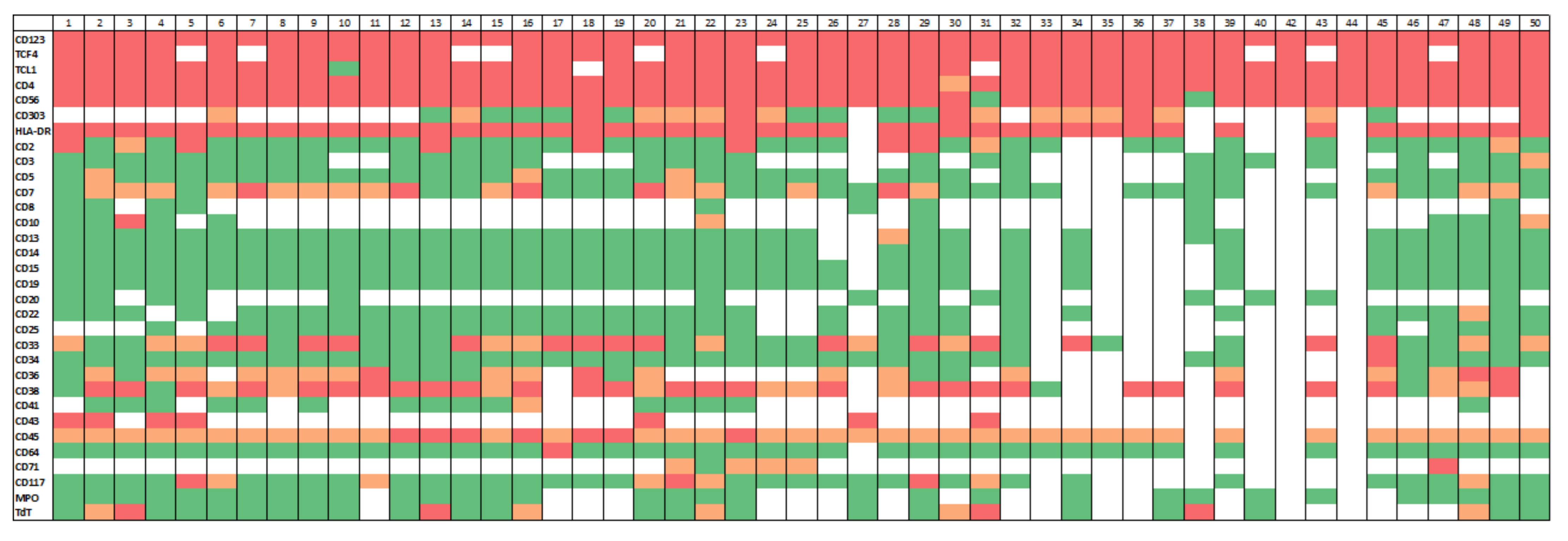
3.2. Immunophenotypic Findings

The immunophenotypic characteristic of the study group are summarized in Figure 1.
Multiparameter flow cytometry immunophenotypic analysis demonstrated that the neoplastic cells were positive for CD4, CD45 (dim), CD123, and HLA-DR in all cases. Other markers expressed in subsets of patients included CD56 (47/49, 96%), CD38 (36/40, 90%), CD71 (4/5, 80%), CD36 (20/29, 69%), CD303 (16/26, 62%), CD33 (24/41, 59%), CD7 (21/44, 48%), TdT (8/28, 29%), CD117 (10/40, 25%), CD2 (9/42, 21%), CD10 (2/10, 20%), CD5 (3/38, 8%), CD41 (1/16, 6%), CD34 (2/39, 5%), cytoplasmic CD3 (1/22, 5%), CD13 (1/37, 3%), and CD64 (1/44, 2%). All cases assessed were negative for surface CD3 (n = 23), CD14 (n = 37), CD15 (n = 38), CD19 (n = 38), CD22 (n = 33), CD25 (n = 29), and myeloperoxidase (n = 28).
Immunophenotyping by immunohistochemistry further showed that BPDCN cells were positive for TCF4/CD123 (40/40, 100%), TCL1 (46/47, 98%), and CD43 (10/11, 91%) and negative for CD8 (n = 9) and CD20 (n = 18). The median Ki-67 proliferation index was 50% (range, 15% to 100%). Among the 27 patients evaluated for p53 expression, 1 patient (#28) expressed a mutant p53 pattern, and the finding correlated with the detection of TP53c.743G>A p.R248Q. Among the 24 patients evaluated by MYC immunohistochemistry, none showed protein overexpression.

3.3. Cytogenetic Findings

Conventional cytogenetic analysis was performed on 36 patients. Nineteen (53%) patients had diploid karyotype. Sixteen (44%) patients had a complex karyotype (≥3 abnormalities). One (3%) patient had two abnormalities (#12, -Y and +12). The most common aberrations were -13/del13q (n = 8), -5/del5q (n = 6), -7/del7q (n = 5), 12/del12p (n = 5), -11/del11q (n = 4), and -21 (n = 4) (Table 1).
FISH analysis was performed on a subset of cases. ETV6 deletion and rearrangement were detected in three and two of seven cases assessed, respectively. MYC deletion and rearrangement were detected in one and one of six cases assessed, respectively. TP53 deletion was detected in all five cases assessed. CDKN2A (p16) deletion was detected in one case assessed. One case showed a signal pattern suggestive of MYB gene rearrangement.

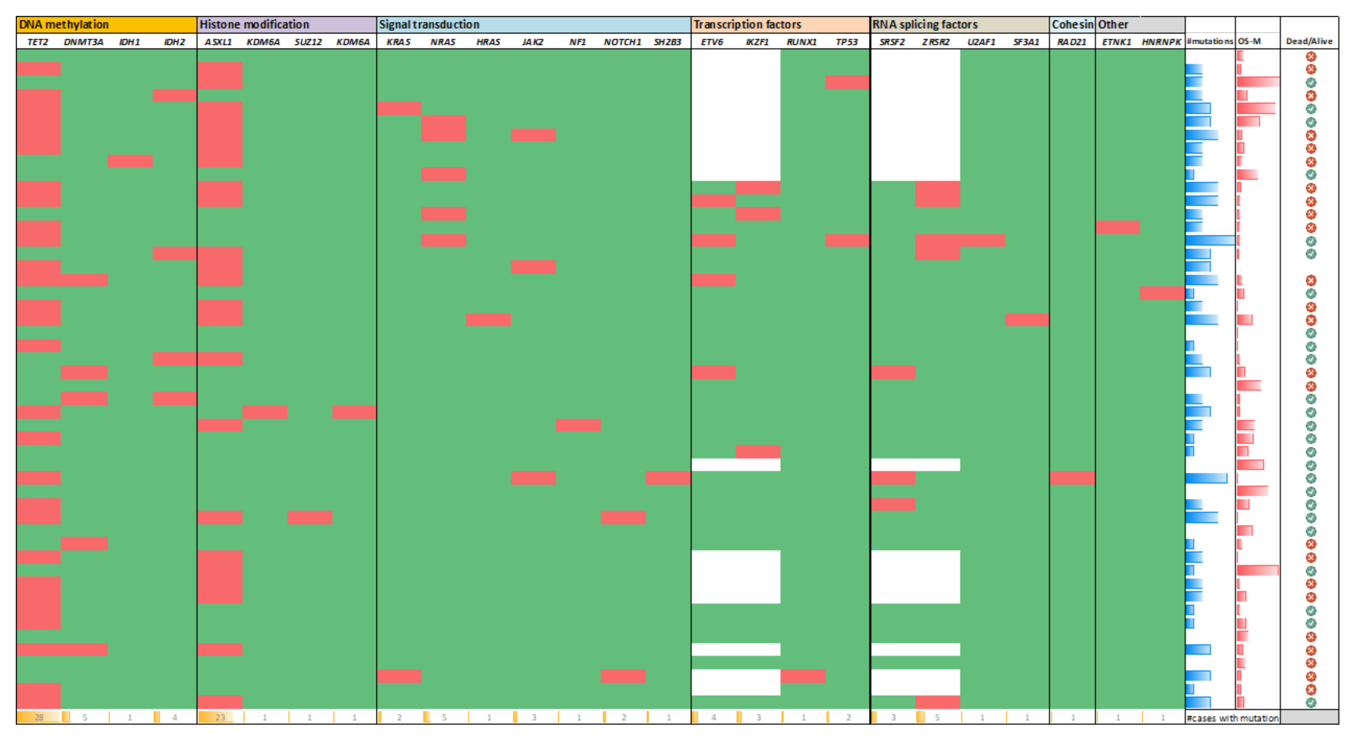
3.4. Gene Mutation Results

Mutations were detected in 42 (84%) patients: nine had one mutation, ten had two mutations, eight had three mutations, nine had four mutations, three had five mutations, two had six mutations, and one patient had eight mutations. In eight patients, no mutations were detected by 28-gene (n = 2) or 81-gene (n = 6) mutation profiling panels.
The most common mutations occurred in TET2 and ASXL1 genes, seen in 28 (56%) and 23 (46%) patients, respectively, and 17 (34%) patients had both TET2 and ASXL1 mutations. Mutations in ASXL1 included 16 frameshift mutations and 7 nonsense mutations, with a median variant allele frequency (VAF) of 17.9% (range, 1.8% to 43.6%). Of note, ASXL1 c.1934dupG p.E646fs was recurrently detected in 11 patients and ASXL1 c.1990_1922del p.E635fs in 3 patients; other ASXL1 mutations were non-recurrent. Mutations in TET2 included 19 frameshift mutations, 16 missense mutations, 12 nonsense mutations, and 1 splice mutations, with a median VAF of 27% (range, 1.2% to 48.3%). The majority of TET2 mutations were non-recurrent, except TET2 c.4075C>T p.R1359C, which was detected in three patients. Half of the patients showed more than one TET2 mutation, e.g., ten patients had two TET2 clones, three patients had three TET2 clones, and one patient had five TET2 clones (Figure 2).
Other recurrently mutated genes included ZRSR2 (5/32, 16%, median VAF 68%), ETV6 (4/32, 13%, median VAF 17%), DNMT3A (5/50, 10%, median VAF 21.8%), NRAS (5/10, 10%, median VAF 21.4%), IKZF1 (3/32, 9%, median VAF 46.3%), SRSF2 (3/32, 9%, median VAF 14.3%), IDH2 (4/50, 8%, median VAF 38%), JAK2 (3/50, 6%, median VAF 14.3%), KRAS (2/50, 4%, median VAF 11%), NOTCH1 (2/50, 4%, median VAF 8%), and TP53 (2/50, 4%, median VAF 43.3%) (Figure 2).
Non-recurrent mutations included ETNK1, HNRNPK, HRAS, IDH1, KDM6A, NF1, RAD21, RUNX1, SF3A1, SH2B3, U2AF1, and SUZ12, seen in one patient each (Figure 2).
Sequential analysis performed on two patients showed TET2 and ASXL1 mutations initially, with one acquiring IKZF1 and ZRSR2 mutations five months later and the other acquiring DNMT3A and ETV6 mutations two months later. In the first case, the patient had a normal diploid karyotype initially but subsequently had a complex karyotype, 46,XY,t(6;17)(q23;q22),der(7)add(7)(q11.2)t(?;21)(?;q11.2),add(21)(p12)[cp4]/47,XY,+mar[1]/46,XY[15].

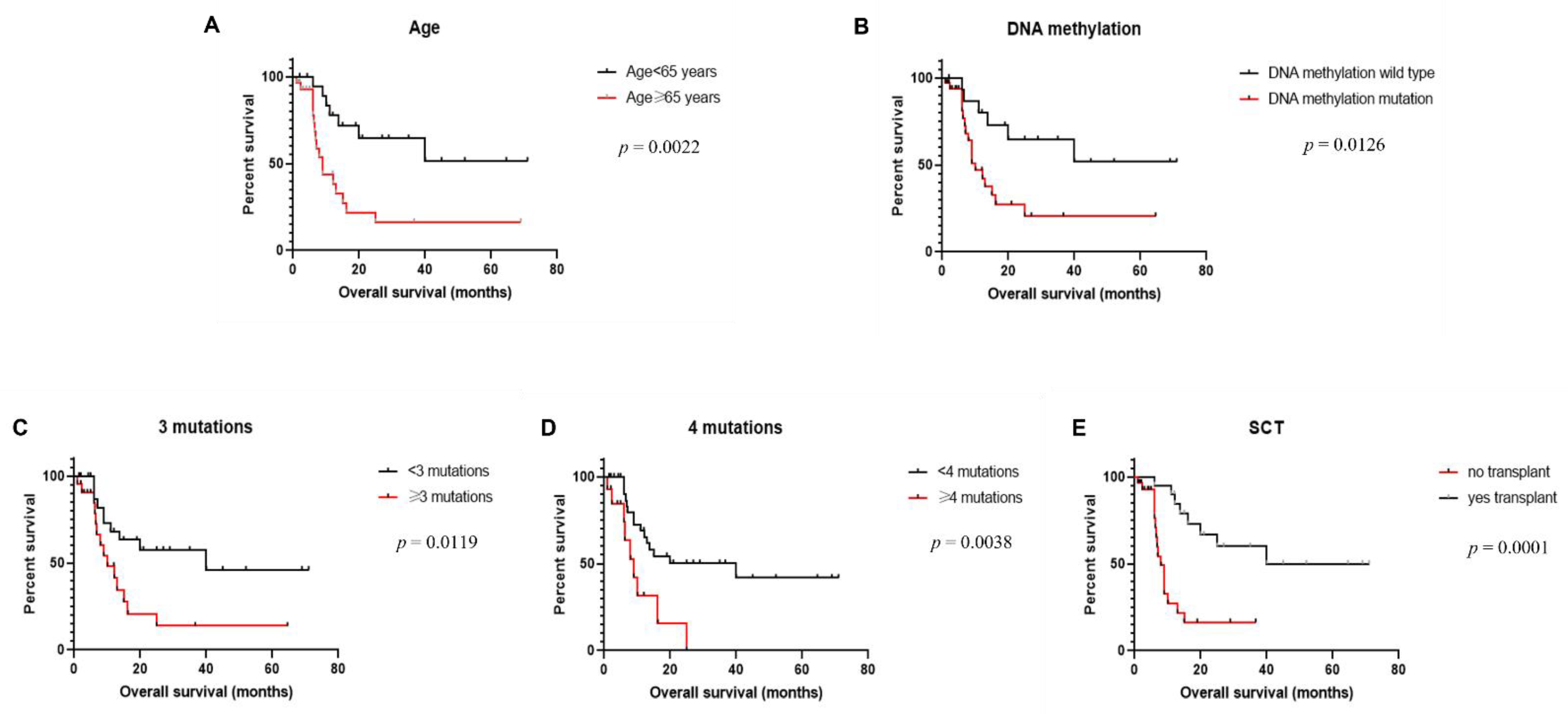
3.5. Clinical Outcome

All patients received chemotherapy, 22 patients received tagraxofusp-based regimens and 18 patients received HCVAD-based regimens. Among all 50 patients, 36 received multi-agent chemotherapy, and 13 received tagraxofusp as a single agent. Twenty patients additionally received hematopoietic stem cell transplantation, 15 allogeneic and 5 autologous. With a median follow-up of 10.5 months (range, 1–71 months), 21 patients achieved complete remission, 4 patients had persistent disease, and 24 patients died. One patient was lost to follow-up.
We performed univariate analysis to evaluate the association between clinical, laboratory, cytogenetic, and mutation data and overall survival (Table 2). Age older than 65 years (p = 0.0022) was associated with shorter overall survival (Figure 3A). We did not observe any significant difference between overall survival and mutation status of each individual gene assessed, including TET2, ASXL1, DNMT3A, NRAS, and ZRSR2. No difference in survival was detected among patients with no mutation or 1–2 mutations. We further grouped genes that were mutated in our study based on their function, and we analyzed the correlation between survival and mutation status based on functional pathways. We identified significantly reduced overall survival among patients with gene mutations involved in the DNA methylation pathway, including TET2, DNMT3A, IDH1, and IDH2, as compared to patients with a wild-type genotype for these genes (p = 0.0126) (Figure 3B). No statistically significant difference was detected between overall survival and gene mutations involved in histone modification (ASXL1, KDM6A, and SUZ12), signal transduction pathway (KRAS, NRAS, HRAS, JAK2, NF1, NOTCH1, and SH2B3), transcription factors (ETV6, TP53, IKZF1, and RUNX1), or RNA splicing factors (SF3A1, SRSF2, ZRSR2, and U2AF1). Moreover, we found that patients with three or more mutations had significantly shorter overall survival than those with two or fewer mutations (p = 0.0019), and the significance was more pronounced when we used four or more mutations as the cutoff (p = 0.0038) (Figure 3C,D). Finally, patients who received hematopoietic stem cell transplantation had significantly longer overall survival compared to those without transplantation (p = 0.0001) (Figure 3E), and such a difference was not dependent on mutation status. There was no significant association between overall survival and other parameters assessed, including sex, white blood cell count, hemoglobin, platelet count, level of LDH, level of β2M, and presence of cytogenetic aberrations.

4. Discussion

Collectively, gene mutations in BPDCN affect several functional classes of genes, mainly DNA methylation, histone modification, signal transduction, transcription factors, cell-cycle regulation, and splicing factors [10,11,12,13,14]. However, the reported frequency of each mutation has varied widely, and the impact of these mutations on prognosis has remained incompletely understood due to the rarity of this disease. In this study, we assessed the mutation profile and its clinical impact in 50 BPDCN cases evaluated by NGS using targeted panels that sequence genes commonly mutated in hematologic neoplasms. We detected mutations in the interrogated genes in 84% of the cases, and 66% of patients had more than one mutation. Genes mutated in our study included those affecting DNA methylation (TET2, DNMT3A, IDH1, and IDH2), histone modification (ASXL1, KDM6A, and SUZ12), signal transduction (NRAS, KRAS, HRAS, JAK2, NF1, NOTCH1, SH2B3), transcription factors (ETV6, TP53, IKZF1, RUNX1), RNA splicing factors (SF3A1, SRSF2, ZRSR2, U2AF1), and cohesin complex component (RAD21). In keeping with previous studies, our results showed that TET2 and ASXL1 were the most frequently mutated genes, seen in 56% and 46% of cases, respectively. All ASXL1 mutations were frameshift or nonsense, and most were located within or upstream of the catalytic domain, which may cause disruption of protein function [15]. Similarly, TET2 mutations were scattered along all exons, and most (69%) were frameshift or nonsense with a potential to affect the function of the protein [16]. In two cases for which we had sequential samples, both had TET2 and ASXL1 initially, and they subsequently acquired additional mutations that included IKZF1 and ZRSR2 in one and DNMT3A and ETV6 in another. These findings suggest that TET2 and ASXL1 mutations may represent early events in the pathogenesis of BPDCN, whereas the other mutations are acquired secondarily due to genomic instability and clonal evolution resulting from epigenetic dysregulation. In addition, we identified a few mutations that have not been reported previously in BPDCN, including ETNK1, HNRNPK, HRAS, KDM6A, RAD21, SF3A1, and SH2B3, or have only been described in single case reports, including NF1, NOTCH1, and SUZ12 [12,17,18]. Similar to ASXL1 and TET2 mutations, most of these mutations, such as ZRSR2 (3/5), DNMT3A (3/7), IKZF1 (2/3), ETV6 (2/4), NOTCH1 (1/2), KDM6A (1/1), and SH2B3 (1/1) were also frameshift, premature stop, or splice site mutation, with probable loss of function of the encoded proteins. By contrast, TP53 mutations are uncommon in BPDCN even though copy loss at the TP53 locus is not infrequent. Surprisingly, genes that are most frequently mutated in AML, in particular FLT3 and NPM1, as well as some mutated genes reported previously, such as EZH2, KIT, and SF3B1, were not mutated in our study group [11,13].
ASXL1 encodes a polycomb repressive complex protein with a role in chromatin regulation. ASXL1 interacts with polycomb complex proteins and other transcription activators or repressors to regulate transcription and translation. Recurrent ASXL1 mutations have been reported in a variety of myeloid neoplasms and have been linked to an aggressive disease course, blast transformation, and poor clinical outcome [19,20,21]. TET2 plays important roles in stem cell self-renewal, lineage commitment, and terminal differentiation of monocytes [19,20,21]. Loss-of-function mutations of TET2 are common in both myeloid and lymphoid neoplasms and are associated with DNA hypermethylation and abnormal gene expression in hematopoietic cells [22]. Its role in prognosis has been controversial. While TET2 mutations have been associated with blast transformation in myeloproliferative neoplasms and unfavorable prognosis in acute myeloid leukemia [23,24,25], other studies failed to identify a strong association between TET2 mutations and overall survival [20,26]. We did not detect any correlation between overall survival and mutation status of each individual gene assessed. However, upon grouping of the genes based on their functions, patients with gene mutations in the DNA methylation pathway had significantly reduced overall survival. There was no difference between survival and mutations affecting other functional classes, including histone modification, signal transduction, transcription factors, and RNA splicing. Further analysis demonstrated that patients who carried three or more mutations had significantly shorter overall survival compared to those with two or fewer mutations, and patients with four or more mutations had even inferior clinical outcome. Therefore, we observed a likely synergistic effect of mutations, either based on the function pathway affected or based on the number of mutations, on the clinical outcome, which may be explained by the more complex genetic landscape and more implicated signaling pathways in patients with multiple mutations.
Our results show that genes encoding RNA splicing components are frequently mutated in BPDCN, with a collective frequency of 23%. Renosi et al. [27] performed NGS targeting 68 genes on 13 cases of BPDCN and detected ZRSR2 mutations in 4 (31%) cases and SRSF2 mutations in 2 (15%) cases. Summerer et al. [28] analyzed 1367 mutations in 1210 genes in 21 BPDCN cases and found mutations in SRSF2 in 7 (33%), SF3B1 in 2 (10%), U2AF1 in 2 (10%), and ZRSR2 in 2 (10%) cases. Loss-of-function mutations in ZRSR2 have been shown to impair plasmacytoid dendritic cell activation and apoptosis after inflammatory stimuli, thus may impair immunity and predispose to leukemic transformation [29]. The presence of frequent mutations of RNA splicing factor may result in impairment of RNA splicing and defective assembly of gene transcripts, which could contribute, at least in part, to the disruption of signaling pathways involved in plasmacytoid dendritic cell maturation and thus BPDCN tumorigenesis.
Another recently discovered recurrent mutation in BPDCN is IKZF1, a member of the IKAROS gene family. IKZF1 encodes a transcription factor that is essential for B-cell development and regulates the differentiation and function of plasmacytoid dendritic cells [30]. IKZF1 deficiency has been correlated with a reduced number of plasmacytoid dendritic cells and decreased production of IFN-alpha, TNF, and IL-12 in a mouse model [30]. Menezes et al. [11] first reported IKAROS family gene mutation (IKZF1, IKZF2, and IKZF3) in 5 of 25 (20%) of BPDCN cases. Torres et al. [31] performed whole-genome sequencing and RNA sequencing, which revealed that IKZF1 was focally inactivated via recurrent structural alterations. IKZF1 deficiency leads to up-regulation of cellular processes responsible for cell–cell and cell–extracellular matrix interaction and may play a central role in the pathobiology of BPDCN.
Patients with BPDCN carry heterogeneous karyotypic aberrations. It has been reported that approximately 60% of patients have a complex karyotype, with imbalanced chromosomal losses being most common, including -5/del(5q31) (NR3C1), del(6q25) (SYNE1), -7/del(7q), del(7p12.2) (IKZF1), del(9p21) (CDKN2A, CDKN2B), del(11q), del(12p13) (CDKN1B, ETV6), del(13q13-q14) (RB1), del(15q), and del(17p13) (TP53) [27,32]. These changes may lead to loss of tumor suppressor genes and contribute to tumorigenesis. It has been reported that biallelic loss of 9p21.3 or mono-allelic loss of 5q31 conferred poor clinical outcome [33,34]. However, most other cytogenetic aberrations carry no prognostic value. The most common aberrations were similar to those reported, including -13/del(13q), -5/del(5q), -7/del(7q), -12/del(12p), and -11/del(11q). We identified a high rate of 12p13/ETV6 deletions/rearrangements (5/7, 71%) in BPDCN. Genomic aberrations involving 12p13 locus containing ETV6 have been reported as the most common findings in BPDCN and may represent early clonal events [35]. Similar to other published data, 8q24/MYC abnormalities were detected in one-third of the patients, and one patient had t(6;8)(p21;q24). Rearrangements involving the MYC locus at 8q24 may lead to MYC protein overexpression and correlate with inferior clinical outcome [36]. An extra copy of the MYB gene or MYB rearrangement at 6q23.3 was detected in one patient who was a 78-year-old man, in contrast to prior studies that showed that MYB rearrangement was more common in children and young adults [37]. The rearrangement has been shown to induce MYB activation by truncating the negative regulator domain [37].
A variety of chemotherapeutic regimens designed for myeloid and lymphoid neoplasms have been used to treat patients with BPDCN. However, most patients experience an aggressive clinical course with poor outcomes despite intensive chemotherapy. Identification of recurrent gene mutations and rearrangements may aid in the development of novel therapeutic approaches. Sapienza et al. [14] performed whole-exome sequencing and RNA sequencing on 14 BPDCN cases, identified deregulation of the epigenetic program as a genetic hallmark of BPDCN, and suggested a novel therapeutic approach based on the combination of two hypomethylating agents, 5′-azicytidine and decitabine. Stem cell transplantation has been shown to result in durable remissions [38,39]. Our studies also showed that stem cell transplantation significantly prolonged the overall survival of patients with BPDCN.

5. Conclusions

In summary, we show that besides gene mutations in DNA methylation and histone modification, mutations affecting signal transduction, RNA splicing factors, and transcription factors, particularly ETV6 and IKZF1, are frequently present in BPDCN. We identified that older age, mutation in genes involved in DNA methylation, and presence of multiple (≥3) mutations were poor prognostic markers in this disease.

Author Contributions

Conceptualization and manuscript writing, C.C.Y. and J.D.K.; methodology, C.C.Y., O.A. and J.D.K.; data generation and data curation, C.C.Y., N.P., M.J.Y., S.L., J.X., W.W., Z.T., O.A., K.N.B., M.H.Q., M.K. and J.D.K.; review and edits, C.C.Y., N.P., M.J.Y., S.L., J.X., W.W., Z.T., O.A., K.N.B., M.H.Q., M.K. and J.D.K. All authors have read and agreed to the published version of the manuscript.

Funding

This research received no external funding.

Institutional Review Board Statement

The study was conducted according to the guidelines of the Declaration of Helsinki and approved by the Institutional Review Board of the University of Texas MD Anderson Cancer Center. The Ethics Code is PA11-0704.

Informed Consent Statement

The Institutional Review Board has waived the requirement for informed consent for this retrospective study.

Data Availability Statement

The data presented in this study are available upon request from the corresponding authors.

Conflicts of Interest

The authors declare no conflict of interest.

References

  1. Khoury, J.D. Blastic Plasmacytoid Dendritic Cell Neoplasm. Curr. Hematol. Malig. Rep. 2018, 13, 477–483. [Google Scholar] [CrossRef] [PubMed]
  2. Wilson, N.R.; Konopleva, M.; Khoury, J.D.; Pemmaraju, N. Novel Therapeutic Approaches in Blastic Plasmacytoid Dendritic Cell Neoplasm (BPDCN): Era of Targeted Therapy. Clin. Lymphoma Myeloma Leuk. 2021. [Google Scholar] [CrossRef] [PubMed]
  3. Pemmaraju, N.; Kantarjian, H.M.; Cortes, J.E.; Duvic, M.; Khoury, J.D.; Patel, K.; Daver, N.; O’Brien, S.; Pierce, S.; Garcia-Manero, G.; et al. Blastic Plasmacytoid Dendritic Cell Neoplasm (BPDCN): A Large Single-Center Experience: Analysis of Clinical and Molecular Characteristics and Patient Outcomes. Blood 2015, 126, 3746. [Google Scholar] [CrossRef]
  4. Facchetti, F.; Petrella, T.; Pileri, S.A. Blastic plasmacytoid dendritic cell neoplasm. In WHO Classification of Tumours of Haematopoietic and Lymphoid Tissues; Swerdlow, S.H., Campo, E., Harris, N.L., Jaffe, E.S., Pileri, S.A., Stein, H., Thiele, J., Eds.; IARC: Lyon, France, 2017; pp. 174–177. [Google Scholar]
  5. Wang, W.; Khoury, J.D.; Miranda, R.N.; Jorgensen, J.L.; Xu, J.; Loghavi, S.; Li, S.; Pemmaraju, N.; Nguyen, T.; Medeiros, L.J.; et al. Immunophenotypic characterization of reactive and neoplastic plasmacytoid dendritic cells permits establishment of a 10-color flow cytometric panel for initial workup and residual disease evaluation of blastic plasmacytoid dendritic cell neoplasm. Haematologica 2021, 106, 1047–1055. [Google Scholar] [CrossRef] [Green Version]
  6. Sukswai, N.; Aung, P.P.; Yin, C.C.; Li, S.; Wang, W.; Wang, S.A.; Ortega, V.; Lyapichev, K.; Nagarajan, P.; Alfattal, R.; et al. Dual Expression of TCF4 and CD123 Is Highly Sensitive and Specific For Blastic Plasmacytoid Dendritic Cell Neoplasm. Am. J. Surg. Pathol. 2019, 43, 1429–1437. [Google Scholar] [CrossRef]
  7. Wang, X.J.; Medeiros, L.J.; Bueso-Ramos, C.E.; Tang, G.; Wang, S.; Oki, Y.; Desai, P.; Khoury, J.D.; Miranda, R.N.; Tang, Z.; et al. P53 expression correlates with poorer survival and augments the negative prognostic effect of MYC rearrangement, expression or concurrent MYC/BCL2 expression in diffuse large B-cell lymphoma. Mod. Pathol. 2017, 30, 194–203. [Google Scholar] [CrossRef] [Green Version]
  8. Loghavi, S.; Al-Ibraheemi, A.; Zuo, Z.; Garcia-Manero, G.; Yabe, M.; Wang, S.A.; Kantarjian, H.M.; Yin, C.C.; Miranda, R.N.; Luthra, R.; et al. TP53 overexpression is an independent adverse prognostic factor in de novo myelodysplastic syndromes with fibrosis. Br. J. Haematol. 2015, 171, 91–99. [Google Scholar] [CrossRef] [Green Version]
  9. Wu, L.; Xia, M.; Sun, X.; Han, X.; Zu, Y.; Jabbour, E.J.; You, M.J.; Lin, P.; Li, S.; Xu, J.; et al. High levels of immunoglobulin expression predict shorter overall survival in patients with acute myeloid leukemia. Eur. J. Haematol. 2020, 105, 449–459. [Google Scholar] [CrossRef]
  10. Jardin, F.; Ruminy, P.; Parmentier, F.; Troussard, X.; Vaida, I.; Stamatoullas, A.; Lepretre, S.; Penther, D.; Duval, A.B.; Picquenot, J.M.; et al. TET2 and TP53 mutations are frequently observed in blastic plasmacytoid dendritic cell neoplasm. Br. J. Haematol. 2011, 153, 413–416. [Google Scholar] [CrossRef] [PubMed]
  11. Menezes, J.; Acquadro, F.; Wiseman, M.; Gomez-Lopez, G.; Salgado, R.N.; Talavera-Casanas, J.G.; Buno, I.; Cervera, J.V.; Montes-Moreno, S.; Hernandez-Rivas, J.M.; et al. Exome sequencing reveals novel and recurrent mutations with clinical impact in blastic plasmacytoid dendritic cell neoplasm. Leukemia 2014, 28, 823–829. [Google Scholar] [CrossRef] [PubMed]
  12. Sapienza, M.R.; Fuligni, F.; Agostinelli, C.; Tripodo, C.; Righi, S.; Laginestra, M.A.; Pileri, A., Jr.; Mancini, M.; Rossi, M.; Ricci, F.; et al. Molecular profiling of blastic plasmacytoid dendritic cell neoplasm reveals a unique pattern and suggests selective sensitivity to NF-kB pathway inhibition. Leukemia 2014, 28, 1606–1616. [Google Scholar] [CrossRef] [PubMed] [Green Version]
  13. Stenzinger, A.; Endris, V.; Pfarr, N.; Andrulis, M.; Johrens, K.; Klauschen, F.; Siebolts, U.; Wolf, T.; Koch, P.S.; Schulz, M.; et al. Targeted ultra-deep sequencing reveals recurrent and mutually exclusive mutations of cancer genes in blastic plasmacytoid dendritic cell neoplasm. Oncotarget 2014, 5, 6404–6413. [Google Scholar] [CrossRef] [PubMed] [Green Version]
  14. Sapienza, M.R.; Abate, F.; Melle, F.; Orecchioni, S.; Fuligni, F.; Etebari, M.; Tabanelli, V.; Laginestra, M.A.; Pileri, A.; Motta, G.; et al. Blastic plasmacytoid dendritic cell neoplasm: Genomics mark epigenetic dysregulation as a primary therapeutic target. Haematologica 2019, 104, 729–737. [Google Scholar] [CrossRef] [PubMed]
  15. Yannakou, C.K.; Jones, K.; McBean, M.; Thompson, E.R.; Ryland, G.L.; Doig, K.; Markham, J.; Westerman, D.; Blombery, P. ASXL1 c.1934dup;p.Gly646Trpfs*12-a true somatic alteration requiring a new approach. Blood Cancer J. 2017, 7, 656. [Google Scholar] [CrossRef] [PubMed]
  16. Delhommeau, F.; Dupont, S.; Della Valle, V.; James, C.; Trannoy, S.; Masse, A.; Kosmider, O.; Le Couedic, J.P.; Robert, F.; Alberdi, A.; et al. Mutation in TET2 in myeloid cancers. N. Engl. Med. 2009, 360, 2289–2301. [Google Scholar] [CrossRef] [PubMed]
  17. Chamoun, K.; Loghavi, S.; Pemmaraju, N.; Konopleva, M.; Kroll, M.; Nguyen-Cao, M.; Hornbaker, M.; DiNardo, C.D.; Kadia, T.; Jorgensen, J.; et al. Early detection of transformation to BPDCN in a patient with MDS. Exp. Hematol. Oncol. 2018, 7, 26. [Google Scholar] [CrossRef] [PubMed]
  18. Szczepaniak, A.; Machnicki, M.; Gniot, M.; Pepek, M.; Rydzanicz, M.; Ploski, R.; Kazmierczak, M.; Stoklosa, T.; Lewandowski, K. Germline missense NF1 mutation in an elderly patient with a blastic plasmacytoid dendritic cell neoplasm. Int. J. Hematol. 2019, 110, 102–106. [Google Scholar] [CrossRef] [PubMed]
  19. Abdel-Wahab, O.; Adli, M.; LaFave, L.M.; Gao, J.; Hricik, T.; Shih, A.H.; Pandey, S.; Patel, J.P.; Chung, Y.R.; Koche, R.; et al. ASXL1 mutations promote myeloid transformation through loss of PRC2-mediated gene repression. Cancer Cell 2012, 22, 180–193. [Google Scholar] [CrossRef] [PubMed] [Green Version]
  20. Bejar, R.; Stevenson, K.; Abdel-Wahab, O.; Galili, N.; Nilsson, B.; Garcia-Manero, G.; Kantarjian, H.; Raza, A.; Levine, R.L.; Neuberg, D.; et al. Clinical effect of point mutations in myelodysplastic syndromes. N. Engl. J. Med. 2011, 364, 2496–2506. [Google Scholar] [CrossRef] [PubMed] [Green Version]
  21. Itzykson, R.; Kosmider, O.; Renneville, A.; Gelsi-Boyer, V.; Meggendorfer, M.; Morabito, M.; Berthon, C.; Ades, L.; Fenaux, P.; Beyne-Rauzy, O.; et al. Prognostic score including gene mutations in chronic myelomonocytic leukemia. J. Clin. Oncol. 2013, 31, 2428–2436. [Google Scholar] [CrossRef]
  22. Rasmussen, K.D.; Jia, G.; Johansen, J.V.; Pedersen, M.T.; Rapin, N.; Bagger, F.O.; Porse, B.T.; Bernard, O.A.; Christensen, J.; Helin, K. Loss of TET2 in hematopoietic cells leads to DNA hypermethylation of active enhancers and induction of leukemogenesis. Genes Dev. 2015, 29, 910–922. [Google Scholar] [CrossRef] [PubMed] [Green Version]
  23. Beer, P.A.; Delhommeau, F.; LeCouedic, J.P.; Dawson, M.A.; Chen, E.; Bareford, D.; Kusec, R.; McMullin, M.F.; Harrison, C.N.; Vannucchi, A.M.; et al. Two routes to leukemic transformation after a JAK2 mutation-positive myeloproliferative neoplasm. Blood 2010, 115, 2891–2900. [Google Scholar] [CrossRef] [Green Version]
  24. Chou, W.C.; Chou, S.C.; Liu, C.Y.; Chen, C.Y.; Hou, H.A.; Kuo, Y.Y.; Lee, M.C.; Ko, B.S.; Tang, J.L.; Yao, M.; et al. TET2 mutation is an unfavorable prognostic factor in acute myeloid leukemia patients with intermediate-risk cytogenetics. Blood 2011, 118, 3803–3810. [Google Scholar] [CrossRef] [PubMed] [Green Version]
  25. Schaub, F.X.; Looser, R.; Li, S.; Hao-Shen, H.; Lehmann, T.; Tichelli, A.; Skoda, R.C. Clonal analysis of TET2 and JAK2 mutations suggests that TET2 can be a late event in the progression of myeloproliferative neoplasms. Blood 2010, 115, 2003–2007. [Google Scholar] [CrossRef] [PubMed] [Green Version]
  26. Nibourel, O.; Kosmider, O.; Cheok, M.; Boissel, N.; Renneville, A.; Philippe, N.; Dombret, H.; Dreyfus, F.; Quesnel, B.; Geffroy, S.; et al. Incidence and prognostic value of TET2 alterations in de novo acute myeloid leukemia achieving complete remission. Blood 2010, 116, 1132–1135. [Google Scholar] [CrossRef]
  27. Renosi, F.; Roggy, A.; Giguelay, A.; Soret, L.; Viailly, P.J.; Cheok, M.; Biichle, S.; Angelot-Delettre, F.; Asnafi, V.; Macintyre, E.; et al. Transcriptomic and genomic heterogeneity in blastic plasmacytoid dendritic cell neoplasms: From ontogeny to oncogenesis. Blood Adv. 2021, 5, 1540–1551. [Google Scholar] [CrossRef]
  28. Summerer, I.; Walter, W.; Meggendorfer, M.; Kern, W.; Haferlach, T.; Haferlach, C.; Stengel, A. Comprehensive analysis of the genetic landscape of 21 cases with blastic plasmacytoid dendritic cell neoplasm by whole genome and whole transcriptome sequencing. Leuk. Lymphoma 2021, 62, 2543–2546. [Google Scholar] [CrossRef] [PubMed]
  29. Togami, K.; Chung, S.S.; Madan, V.; Booth, C.A.G.; Kenyon, C.M.; Cabal-Hierro, L.; Taylor, J.; Kim, S.S.; Griffin, G.K.; Ghandi, M.; et al. Sex-biased ZRSR2 mutations in myeloid malignancies impair plasmacytoid dendritic cell activation and apoptosis. Cancer Discov. 2021. [Google Scholar] [CrossRef]
  30. Cytlak, U.; Resteu, A.; Bogaert, D.; Kuehn, H.S.; Altmann, T.; Gennery, A.; Jackson, G.; Kumanovics, A.; Voelkerding, K.V.; Prader, S.; et al. Ikaros family zinc finger 1 regulates dendritic cell development and function in humans. Nat. Commun. 2018, 9, 1239. [Google Scholar] [CrossRef] [PubMed]
  31. Bastidas Torres, A.N.; Cats, D.; Mei, H.; Fanoni, D.; Gliozzo, J.; Corti, L.; Paulli, M.; Vermeer, M.H.; Willemze, R.; Berti, E.; et al. Whole-genome analysis uncovers recurrent IKZF1 inactivation and aberrant cell adhesion in blastic plasmacytoid dendritic cell neoplasm. Genes Chromosomes Cancer 2020, 59, 295–308. [Google Scholar] [CrossRef] [Green Version]
  32. Leroux, D.; Mugneret, F.; Callanan, M.; Radford-Weiss, I.; Dastugue, N.; Feuillard, J.; Le Mee, F.; Plessis, G.; Talmant, P.; Gachard, N.; et al. CD4(+), CD56(+) DC2 acute leukemia is characterized by recurrent clonal chromosomal changes affecting 6 major targets: A study of 21 cases by the Groupe Francais de Cytogenetique Hematologique. Blood 2002, 99, 4154–4159. [Google Scholar] [CrossRef]
  33. Emadali, A.; Hoghoughi, N.; Duley, S.; Hajmirza, A.; Verhoeyen, E.; Cosset, F.L.; Bertrand, P.; Roumier, C.; Roggy, A.; Suchaud-Martin, C.; et al. Haploinsufficiency for NR3C1, the gene encoding the glucocorticoid receptor, in blastic plasmacytoid dendritic cell neoplasms. Blood 2016, 127, 3040–3053. [Google Scholar] [CrossRef] [Green Version]
  34. Lucioni, M.; Novara, F.; Fiandrino, G.; Riboni, R.; Fanoni, D.; Arra, M.; Venegoni, L.; Nicola, M.; Dallera, E.; Arcaini, L.; et al. Twenty-one cases of blastic plasmacytoid dendritic cell neoplasm: Focus on biallelic locus 9p21.3 deletion. Blood 2011, 118, 4591–4594. [Google Scholar] [CrossRef] [PubMed] [Green Version]
  35. Tang, Z.Y.; Li, Y.; Wang, W.; Yin, C.C.; Tang, G.L.; Aung, P.P.; Hu, S.M.; Lu, X.Y.; Toruner, G.A.; Medeiros, L.J.; et al. Genomic aberrations involving 12p/ETV6 are highly prevalent in blastic plasmacytoid dendritic cell neoplasms and might represent early clonal events. Leuk. Res. 2018, 73, 86–94. [Google Scholar] [CrossRef] [PubMed]
  36. Sakamoto, K.; Katayama, R.; Asaka, R.; Sakata, S.; Baba, S.; Nakasone, H.; Koike, S.; Tsuyama, N.; Dobashi, A.; Sasaki, M.; et al. Recurrent 8q24 rearrangement in blastic plasmacytoid dendritic cell neoplasm: Association with immunoblastoid cytomorphology, MYC expression, and drug response. Leukemia 2018, 32, 2590–2603. [Google Scholar] [CrossRef] [PubMed]
  37. Suzuki, K.; Suzuki, Y.; Hama, A.; Muramatsu, H.; Nakatochi, M.; Gunji, M.; Ichikawa, D.; Hamada, M.; Taniguchi, R.; Kataoka, S.; et al. Recurrent MYB rearrangement in blastic plasmacytoid dendritic cell neoplasm. Leukemia 2017, 31, 1629–1633. [Google Scholar] [CrossRef] [PubMed]
  38. Kharfan-Dabaja, M.A.; Al Malki, M.M.; Deotare, U.; Raj, R.V.; El-Jurdi, N.; Majhail, N.; Cherry, M.A.; Bashir, Q.; Darrah, J.; Nishihori, T.; et al. Haematopoietic cell transplantation for blastic plasmacytoid dendritic cell neoplasm: A North American multicentre collaborative study. Br. J. Haematol. 2017, 179, 781–789. [Google Scholar] [CrossRef]
  39. Bashir, Q.; Milton, D.R.; Popat, U.R.; Kebriaei, P.; Hosing, C.; Khouri, I.F.; Rezvani, K.; Nieto, Y.; Oran, B.; Srour, S.A.; et al. Allogeneic hematopoietic cell transplantation for patients with blastic plasmacytoid dendritic cell neoplasm (BPDCN). Bone Marrow Transplant. 2021, 1–6. [Google Scholar] [CrossRef]
Figure 1. Immunophenotypic landscape of blastic plasmacytoid dendritic cell neoplasm.
Cancers 13 05888 g001
Figure 2. Somatic mutation landscape of blastic plasmacytoid dendritic cell neoplasm.
Cancers 13 05888 g002
Figure 3. Overall survival of patients with blastic plasmacytoid dendritic cell neoplasm. (A) Comparison between patients ≥65 years and <65 years of age. (B) Comparison between patients with and without mutations affecting DNA methylation pathway. (C) Comparison between patients with <3 vs. ≥3 mutations. (D) Comparison between patients with <4 vs. ≥4 mutations. (E) Comparison between patients who did and those who did not receive hematopoietic stem cell transplantation (SCT).
Cancers 13 05888 g003
Table 1. Karyotypes of cases of blastic plasmacytoid dendritic cell neoplasm.
CaseKaryotype
1.46,XY[19]/47,XY,+1[1]
2.46,XY,t(1;6)(p21;p36.3),del(5)(q13q33),der(7)t(1;7)(q12;p22),del(11)(q13q23),del(12)(p11.2p13),add(15)(q15)[10]
3.42,XY,-5,-12,-15,add(17)(p11.2),add(17)(p13),-18,add(20)(q13.3)[3]/43,idem,+mar[8]/42,idem,-13,+mar[2]/46,XY[4]
4.46,XY,+1,add(1)(p13),der(1)dup(1)(q21q32)add(1)(q42),-2,-4,+11,add(12)(p11.2), -13,-21,+2mar[8]/48,XY,+1,der(1)dup(1)(q21q32)add(1)(q42),-2,-4,+11,add(12)(p11.2),-13,+3mar[1]/46,XY[11]
5.46,XY[20]
6.46,XY[20]
7.46,XY,t(2;7)(p23;p15),add(9)(p13),del(9)(p21)[20]
8.46,XX[20]
9.46, XY[20]
10.45,XY,der(6)del(6)(q13q22)t(6;9)(q25;q34),-7,der(9)del(9)(p21)t(6;9),der(9;15)(q10;q10),-13,-14,+2~3mar[cp6]/46,XY[14]
11.46,XY,t(6;17)(q23;q22),der(7)add(7)(q11.2)t(?;21)(?;q11.2),add(21)(p12)[cp4]/47,XY,+mar[1]/46,XY[15]
12.46,X,-Y,+12[3]/46,XY[17]
13.44~45,XX,del(5)(q33q35),del(6)(q13q23),der(7;9)(q10;q10),-13[cp12]/46,XX[8]
14.44,XX,add(1)(p36.1),del(9)(p21),add(12)(p12),-13,add(17)(p12),-21,-22,add(22)(q12),+mar[5]/46,XX[15]
16.48,XY,t(6;8)(p12;q24.2),add(7)(p11.2),del(13)(q12q22),+16,add(20)(q11.2),+21[10]/46,XY[10]
17.46, XY[20]
18.46,XY[20]
19.86~87,XXYY,-5,add(6)(q21)x2,-11,add(11)(q23),-12,i(12)(q10),add(19)(p13.3),+3mar[cp3]/46,XY[17]
20.46,XY[20]
21.44~47,XY,add(3)(p13),add(5)(p15.1),del(5)(q13q33),-8,add(8)(q13),del(9)(p21),del(9)(q13q22), del(10)(q24),del(11)(q13q23),del(12)(p12),- 13,del(13)(q12q22),+del(17)(p11.2),add(20)(q13.3),add(21)(p13),+mar[cp11]/46,XY[9]
22.42,XY,-6,-7,-8,add(9)(q13),der(11)add(11)(p15)add(11)(q24),der(13;15)(q10;q10),add(17)(p11.2),-22,+2mar[4]/46,XY[16]
23.46~48,XX,del(6)(q13q23),+der(7)add(7)(p13)del(7)(q11.2q22)add(7)(q32)x2,der(7)add(7)(p13)del(7)(q11.2q22), add(7)(q32)x2,-8,add(9)(p24),add(12)(p13),-14,del(16)(q23),+21,+22,+mar[cp19]/46,XX[1]
24.46,XY,del(12)(p11.2p13)[1]/46,XY[19]
25.46,XY,del(3)(p11.2)[1]/46,XY[19]
26.47,XY,+Y[1]/46,XY[19]
27.46,XY,inv(9)(p12q13)[20]
28.45,XY,?del(6)(q24),-7,?i(7)(q10),+?12,-14,15,+mar[3]/46,XY[12]
29.46,XY,del(12)(q14)[1]/46,XY[19]
30.46,XX[20]
31.46,XX[20]
32.46,XY[20]
33.46,XY[19]
46.46,XY,t(3;9)(q25;q34)[1]/46,XY[19]
47.45,XY,i(7)(q10),i(14)(q10),-21[2]/46,XY[18]
48.46,XY[4]
49.81~82,XXYY,+1,+1,add(1)(p22),del(1)(p31),del(1)(q21),add(2)(p25),add(2)(q37),-3,-4,-5,-5,-6,-9,-10,-12,-13,-15,+16,+16,-17,-18,-20,-21,+2~4mar[cp2]/46,XY[18]
Table 2. Univariate analysis of clinical, laboratory, and molecular genetic data to predict overall survival.
Featuresp ValueHR Ratio95% CI of HR
Age (<65 years vs. ≥65 years)0.0022 *0.2870.1273–0.6473
Sex (men vs. women)0.84441.1120.3941–3.136
WBC (low vs. normal vs. high)0.0561NANA
Hb (low vs. normal)0.60071.2930.5159–3.243
Platelet (low vs. normal)0.41.4030.6285–3.133
LDH (high vs. normal)0.60431.240.5522–2.784
β2M (high vs. normal)0.76330.80770.2143–3.044
Karyotype (abnormal vs. diploid)0.09690.5160.2094–1.272
Mutation (yes vs. no)0.29020.57240.2296–1.427
ASXL1 (mut vs. wt)0.26180.63820.2845–1.432
DNMT3A (mut vs. wt)0.10980.44030.1016–1.909
NRAS (mut vs. wt)0.80361.1980.3122–4.595
TET2 (mut vs. wt)0.07430.48780.2182–1.090
ZRSR2 (mut vs. wt)0.33740.49770.0692–3.581
DNA methylation (mut vs. wt)0.0126*0.33990.1527–0.7567
Histone modification (mut vs. wt)0.26970.64290.2867–1.442
Signal transduction (mut vs. wt)0.44351.5050.5825–3.886
Transcription factors (mut vs. wt)0.22260.56270.1843–1.718
RNA splicing factors (mut vs. wt)0.54140.71590.2080–2.464
#mutation (<3 vs. ≥3)0.0119 *0.37660.1624–0.8737
#mutation (<4 vs. ≥4)0.0038 *0.32460.1099–0.9589
Transplantation (yes vs. no)0.0001 *0.23780.1010–0.5597
β2M, β2-microglobulin; Hb, hemoglobin; LDH, lactate dehydrogenase; mut, mutated; NA, not available; WBC, white blood cell count; wt, wild type, * Denotes p Values that are statistically significant.
Publisher’s Note: MDPI stays neutral with regard to jurisdictional claims in published maps and institutional affiliations.

