Review

A Perspective on the Role of Microbiome for Colorectal Cancer Treatment

Microbiome Research Centre, St George and Sutherland Clinical School, The University of New South Wales, Sydney 2052, Australia
*
Author to whom correspondence should be addressed.
These authors contributed equally to the authorship of this manuscript and should be considered equal first authors.
Cancers 2021, 13(18), 4623; https://doi.org/10.3390/cancers13184623
Submission received: 9 August 2021 / Revised: 13 September 2021 / Accepted: 13 September 2021 / Published: 15 September 2021
(This article belongs to the Section Cancer Therapy)

Abstract

:

Simple Summary

Colorectal cancer is the third most diagnosed cancer worldwide and contributes significantly to global mortality and morbidity. The gut microbiome, composed of the trillions of microbes endemic to the human gastrointestinal tract, has been shown to be implicated in colorectal cancer oncogenesis; however, the roles of microbiota and dysbiosis in CRC treatment remain poorly understood. This review sought to characterize this relationship and in doing so, identify how these interactions may inform future treatments in the form of synbiotics designed to alter the host microbiota to achieve optimized treatment outcomes.

Abstract

In healthy hosts, trillions of microbes colonise the gut and oral cavity in a well-balanced state, maintaining a mutually beneficial relationship. Loss of this balance, termed dysbiosis, is strongly implicated in the pathogenesis of colorectal cancer (CRC). However, the roles of microbiota and dysbiosis in CRC treatment remain poorly understood. Recent studies suggest that the gut microbiota has the ability to affect the host response to chemotherapeutic agents by enhancing drug efficacy, promoting chemoresistance and mediating chemotherapy-induced toxicity and side effects via a variety of mechanisms. Several other studies have also proposed manipulation of the microbiota to optimise CRC treatment. In this review, we summarise the current advancement of knowledge on how microbiota and CRC treatments interact with each other and how this interaction may shed some light on the development of personalised microbiota manipulations that improve CRC treatment outcomes.

1. Introduction

1.1. Epidemiology

Colorectal cancer (CRC) is a major public health problem, being the third most commonly diagnosed malignancy and the second most common cause of cancer-related death worldwide in 2020 [1,2,3]. CRC is an aetiologically heterogenous disease that arises through three major pathways–the adenoma-carcinoma sequence, the serrated and the inflammatory pathways and is also associated with certain genetic factors [4]. However, most CRC cases are sporadic in nature, emerging from a complex interplay between modifiable environmental risk factors attributable to westernisation [4,5]. As such, the global burden is expected to further increase as a result of the growth and ageing of the population and the adoption of westernized behaviours and lifestyles [6]. A topical area in CRC research has emerged from recent studies demonstrating a state of imbalance or dysbiosis in CRC patients, implicating the gut microbiota in colorectal carcinogenesis [7,8]. There is wide geographical variation with over two-thirds of its incidence and approximately 60% of total deaths occurring in countries with a high or very high human development index [1]. The lifetime risk of developing CRC in many regions is roughly 5%, with deaths occurring in just under half (45%) of those diagnosed despite treatment [9]. There is a poor prognosis for patients with metastatic CRC with a median 5-year survival rate of 12.5% in the USA, underlining the necessity of developing more effective treatments [10].

1.2. The Microbiome

The human body houses a vast and highly integrated microbial community of bacteria, fungi, viruses, archaea and parasites, collectively termed the microbiota. The bacteria are predominately represented by the phyla Firmicutes, Bacteroidetes and Actinobacteria [11]. The gastrointestinal tract (GIT) contains over 100 trillion microorganisms and is the site of principal communication between host cells, the immune system and the microbiota [11,12,13]. The gut microbiota has co-evolved with the host and plays an active role in regulating metabolism and immunity, and maintaining homeostasis and nutritional absorption [14,15]. The proposed causative role of the microbiota in carcinogenesis is through dysbiosis-related inflammation and biosynthesis of carcinogens, with shifts in the microbiome commencing as early as in precancerous adenomas [15,16,17]. Next-generation sequencing technologies have been used to study the microbiome composition [18]. Analysis of the microbiome entails sample collection and processing, next-generation sequencing and a bioinformatics analysis [19]. Many bodily samples can be collected and studied, such as saliva, faeces, tissue biopsies, intestinal fluids, buccal swabs, skin and the vaginal swab [19]. Nucleic acids of these samples can be sequenced by gene amplicon sequencing and whole-genome shotgun metagenomics sequencing [20]. Gene amplicon sequencing is used to identify microbial composition by sequencing the hypervariable regions of a target gene that are conserved among taxa in a particular kingdom of microorganisms. For example, the hypervariable region of 16S rRNA can be amplified and sequenced to determine the bacterial composition in the given samples. [20] This technique is mostly employed on DNA samples that have a high host to microbial DNA ratio. These samples include tissue biopsies and skin. Conversely, unlike 16S rRNA sequencing, metagenomics uses genome-wide shotgun sequencing, targeting the whole genome to provide a superior taxonomic resolution [20]. This technique is mostly employed on DNA samples that have a low host to microbial DNA ratio. These samples include faeces, intestinal fluid, buccal and vaginal swabs.

1.3. Treatment for CRC

Despite being the most effective, surgical interventions are unable to completely cure CRC, and thus post-operative adjuvant chemotherapy has emerged as an essential therapeutic option to prevent recurrence and metastasis [21]. CRC is commonly treated with cytotoxic agents that interfere with DNA replication such as 5-fluorouracil (5-FU), capecitabine and oxaliplatin, as well as regimens such as FOLFOX (5-FU and oxaliplatin), FOLFIRI (5-FU and leucovorin and irinotecan) and XELOX (oxaliplatin and capecitabine) [21,22]. Most forms of chemotherapy target tissues that have a high rate of cell turnover and division, thereby affecting other normally dividing cells with a similar division rate. This may cause severe side effects such as gastrointestinal mucositis requiring dose reduction or deferral of treatment, resulting in poorer prognosis [23].
Immunotherapy is another mode of systemic cancer therapy that drives a “tumour-specific” immunity by establishing a durable population of highly active T cells that can target and lyse tumour cells [24,25]. The efficacy of current immunotherapies vastly depends on the tumour category of the cancer. CRC can be classed as being mismatch-repair-deficient or high microsatellite instability (dmmR-mSI-H) or more commonly, as mismatch-repair-proficient or microsatellite instability-low (pmmR-mSI-H) [10]. Current immunotherapy utilises monoclonal antibodies targeting programmed cell death 1 (PD1) such as pembrolizumab and nivolumab, which have demonstrated improved survival only in metastatic dmmR-mSI-H CRC [10]. However, targeted immunotherapies functioning beyond PD1 blockade with greater tumour efficacy are ongoing.
Recent studies have investigated the relationships between the microbiome and the outcome of cancer therapy such as efficacy and toxicity. Understanding the relationship between the microbiome changes before and after CRC treatment will hold the key to reducing toxicity and improving efficacy of the treatments. This review will attempt to address these aspects.

2. Effect of Microbiome on Chemotherapy Efficacy and Toxicity

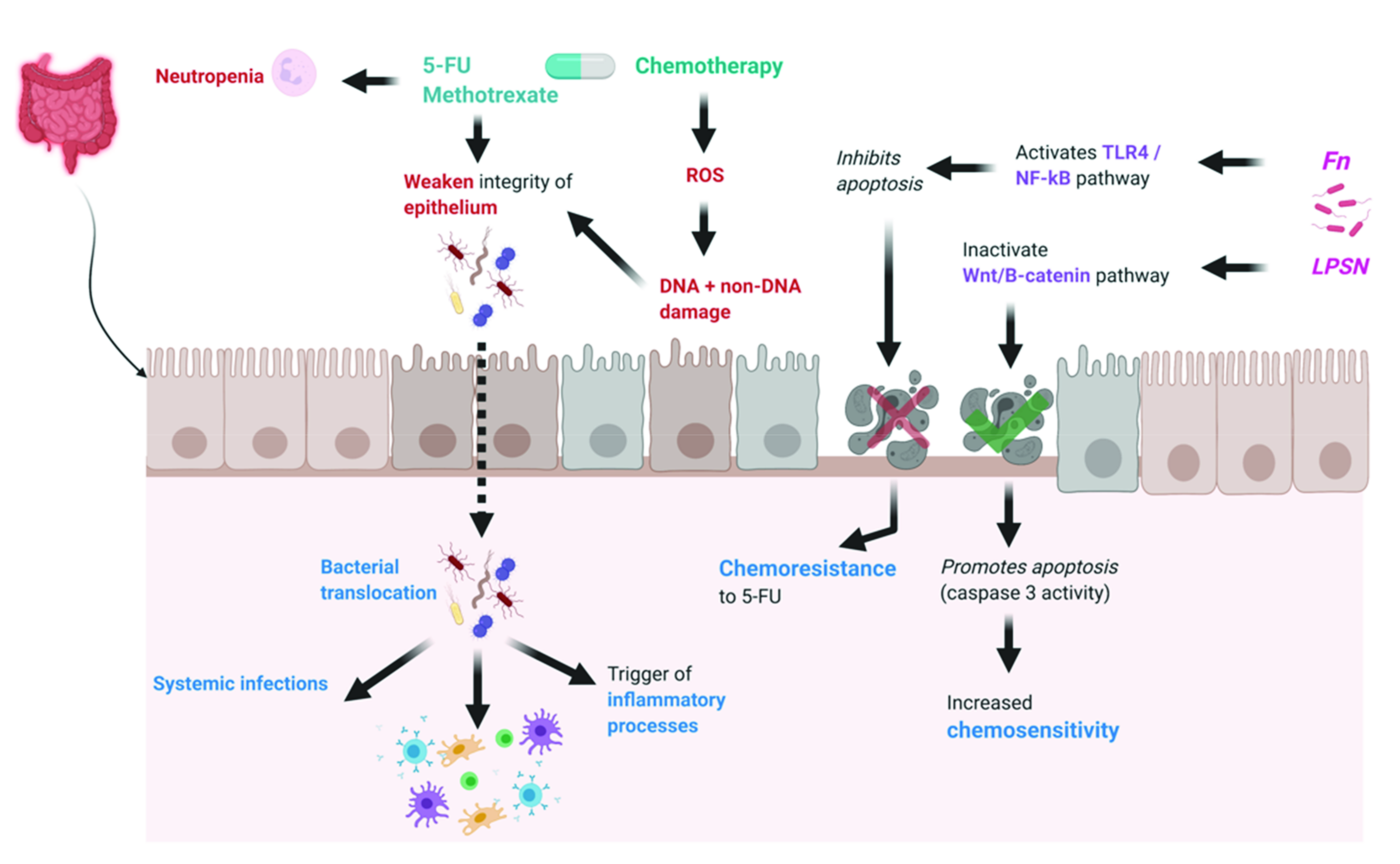
2.1. Effects of Microbiome on Chemotherapy Efficacy

The gut microbiota has the ability to affect the host response to chemotherapeutic agents by facilitating drug efficacy, promoting chemoresistance and/or mediating chemotherapeutic toxicity and side effects [26,27]. The translocation, immunomodulation, metabolism, enzymatic degradation and reduced diversity and ecological variation (TIMER) mechanistic framework (Figure 1) has been proposed as a model to explain the variety of mechanisms that allow the gut microbiota to modulate chemotherapy agents [27]. These mechanisms are important in understanding chemotherapy success and failure in a variety of tumours and specifically in colorectal cancer (CRC).
Two elegant studies using high-throughput sequencing in a C. elegans model [28,29] highlighted the importance of host–microbe interactions in promoting the anti-tumour efficacy of fluoropyrimidine type chemotherapy agents—those that are standard as a first line treatment against CRC. Fluoropyrimidines such as 5-fluorouracil (5-FU) are thought to exert their anticancer effects by inhibiting thymidylate synthase, thus impeding nucleotide biosynthesis and hence cell division [28]. Interestingly, a study by García-González et al. [29] suggested that 5-FU and 5-fluoro-2′-deoxyuridine (FUDR) act by affecting ribonucleotide rather than DNA metabolism, a process that is dependent on active bacterial metabolism. They showed, within their model, that Escherichia Coli (E. coli) use an inherent pathway to convert 5-FU and FUDR into fluorouridine monophosphate (FUMP), an analogue of uridine monophosphate (UMP) that has been shown to block de novo pyrimidine synthesis. Scott et al. [28] echoed this in their C. elegans model, showing that inhibition of bacterial ribonucleotide metabolism significantly antagonised drug efficacy. Additionally, their results imply that the diversity of the nematode’s microbiome is critical to the host response to fluoropyrimidines, with pharmacodynamics of the drug varying as much as 40-fold with different bacterial strains and up to 256-fold with disruption of bacterial metabolism. This symbiosis between the host microbiome and fluoropyrimidine efficacy is further supported in a mouse model reported by Yuan et al. [30]. In their study, they compared the anti-CRC efficacy of 5-FU in mice treated with a cocktail of antibiotics of vancomycin, ampicillin, neomycin and metronidazole with those without these treatments. Their results showed that after 35 days, the tumour volume was significantly lower in the mice that were not treated with antibiotics compared to those that were. They concluded that antibiotic administration caused the disruption of the gut microbiota and thus reduced 5-FU efficacy, implying that microbiome dysbiosis is unfavourable to chemotherapeutic outcomes. This was echoed in Wang et al.’s mouse model, which demonstrated that microbiota dysbiosis induced by dysfunctional TGF-ß signalling was more likely to develop CRC resistant to 5-FU [31]. Other bacteria endemic in the microbiome such as Lactobacillus plantarum supernatant (LPSN) may play a role in increasing chemosensitivity in 5-FU CRC resistant cells (Figure 1). This mechanism by which LPSN is able to improve chemosensitivity is multifaceted and may involve inhibiting expression of particular biomarkers on cancer stem cells, selectively inactivating the Wnt/B-catenin signalling pathway and promoting cell death and apoptosis by inducing caspase 3 activity [32,33].
The efficacy of anticancer therapy is proposed to depend on microbiota-mediated innate and adaptive immune responses [34,35]. In a study by Lehouritis et al. [36], 30 drugs were tested in vitro in the presence of non-pathogenic E. coli or Listeria welshimeri. While 10 of the drugs were found to be inhibited by one or both species, the efficacy of six of the drugs was enhanced. In another experimental study, the anti-tumour effect of cyclophosphamide was attenuated when the microbiota was altered [35]. Sterilisation by broad spectrum antibiotics or vancomycin to specifically targeted gram-positive bacteria resulted in reduction of cyclophosphamide’s ability to induce an inflammatory response. Cyclophosphamide induces CD4+ T cells in the spleen to become IL-17 producers, and the gut microbiota is vital for this process. Antibiotic-treated and germfree mice experienced reduced anti-tumour effects in response to cyclophosphamide treatment [35]. Similarly, Iida et al. [34] treated mice with an antibiotic cocktail of vancomycin, imipenem, and neomycin in drinking water for 3 weeks before inoculating with MC38 colon tumour cells. The mice subsequently underwent immunotherapy and chemotherapy with either oxaliplatin or cisplatin. Mice treated with antibiotics or germfree mice had an attenuated response to therapy. In these mice, tumour-infiltrating myeloid-derived cells produced reduced levels of several cytokines (including TNF α ) as well as reactive oxygen species (ROS) after treatment. Hence, an intact commensal microbiota may modulate myeloid-derived cell functions in the tumour microenvironment and is crucial for the early cytotoxic response to cancer immunotherapy and chemotherapy [34]. Two commensals in particular, E. hirae and B. intestinihominis, have been shown to stimulate specific-memory Th1 cell immune responses, leading to a longer progression-free survival in advanced lung and ovarian cancer patients treated with cyclophosphamide [37]. Moreover, it has been shown that colon cancer prognosis is in a large way dictated by the abundance and functional response of tumour infiltrating lymphocytes (TIL), including follicular T helper cells, in addition to the efficacy of chemotherapy-induced immune responses [38,39,40]. Roberti et al. demonstrated the importance of the ileal microbiota in dictating tolerogenic versus immunogenic ileal intestinal epithelial cell death and the accumulation of follicular T helper cells in both patients and mice with colorectal cancer [41]. They found that the immunogenicity of oxaliplatin therapy is enhanced when ileal intestinal epithelial cells are colonised with immunogenic commensals, particularly Bacteroides fragilis (B. Fragilis) and Erysipelotrichaceae. In the presence of these bacteria, chemotherapy-induced apoptotic ileal crypt cells produced interleukin-1R1 and interleukin-12, which in turn elicited a PD-1+ follicular T helper cell response, thus improving chemotherapeutic efficacy. Their findings not only show the importance of immunogenic ileal apoptosis in the prognosis of chemotherapy-treated CRC, but they also outline the vital role the microbiota play in dictating this response, further emphasising the influence of the microbiota in modifying the efficacy of anticancer therapy [41].
Contrasting the perceived symbiosis between an intact microbiome and chemotherapy efficacy, a number of studies have suggested that the host microbiota might serve to increase the chemoresistance of 5-FU. While Fusobacterium Nucleatum (Fn) has been implicated in the pathogenesis of CRC, Zhang et al. [42] show that Fn might play a role in CRC chemoresistance to 5-FU through activation of the TLR4/NF-kB pathway causing upregulation of BIRC3, an inhibitor of apoptosis (IAP) protein on CRC cells. They showed that Fn abundance was correlated with chemoresistance and identified high amounts of Fn as an independent risk factor for recurrence in advanced CRC patients. Yu et al. [43] also demonstrated a mechanistic role for Fn in promoting resistance to 5-FU and oxaliplatin chemotherapy regimens. They showed that Fn activates TLR4 and MYD88 immune signalling to inhibit the expression of microRNA (miRNA)-18a and miRNA-4802. This in turn suppresses the autophagy pathway and the apoptosis of the colon cancer cells induced by 5-FU and oxaliplatin, leading to chemoresistance (Figure 1).
Irinotecan (CPT-11) is a commonly used chemotherapeutic agent that often results in gastrointestinal side effects as a result of its unique metabolism. Bacterial β-glucuronidases in the colon are crucial to the conversion of CPT-11 to its active agent SN-38, which is responsible for anti-tumour activity as well as dose-limiting toxicity [32]. SN-38 undergoes hepatic glucuronidation and is secreted into bile as the inactive glucuronide SN-38G [44]. Bacterial β-glucuronidases in the colon deconjugate SN-38G, exposing the intestinal epithelia to SN-38, thus causing gut toxicity as well as allowing bacterial organisms to translocate, causing systemic infection and sepsis [45,46,47]. Antibiotic prophylaxis not only reduces SN-38 concentration, but also diarrhoea in animal and human models [47,48].
Increasingly, immunotherapy is becoming a major treatment modality for a subset of CRC subtypes. In particular, immune checkpoint inhibitors (ICIs) such as anti-PD1 and anti-CTLA-4 treatments have shown some efficacy in dMMR-MSI-H CRC [10]. The microbiome may be able to modulate the host response to immunotherapy to increase its antineoplastic activity. Specific bacteria within the microbiota have been shown to correlate positively with immunotherapeutic response, and mouse models have demonstrated enhanced immunotherapy efficacy with particular bacterial microbiota strains [34,49,50]. Currently, these findings have not been replicated in human studies, and therefore the importance of these bacterial strains in optimising clinical outcomes is still unknown.
The studies above demonstrate the diverse impact gut microbiota can have across a spectrum of antineoplastic treatment in CRC. The studies above have highlighted potential avenues to harness the microbiome by creating a microenvironment that is beneficial to increasing treatment efficacy; however, the challenge lies in replicating these results in human trials and documenting improved outcomes for these patients. Understanding the role of the gut microbiota in influencing the host response to anti-tumour treatments, including chemotherapy and new immunotherapeutic approaches, will be essential in developing personalised treatments to achieve optimal efficacy and tumour clearance. Using this knowledge as a basis to investigate the synergistic role of faecal microbiota transplantation or symbiotics with anticancer therapy is an exciting next step in combatting CRC resistance and mortality.

2.2. Effects of Microbiome on Chemotherapy Toxicity

The cytotoxic effects of chemotherapy give rise to a wide range of toxicities, including the inflammatory condition of mucositis. Current drugs or other approaches to counteract chemotherapy-induced adverse effects are often incompletely effective, frequently do not address potential longer-term sequelae or may even induce other side-effects that only add to patient discomfort [51]. Chemotherapy drugs such as 5-FU and methotrexate are highly toxic for intestinal epithelial cells lining the gut mucosa and other cells with high proliferation rates [52]. These drugs cause neutropenia and weaken the integrity of the epithelial barrier, facilitating bacterial translocation across the gut mucosa, allowing for systemic infections and triggering of inflammatory processes [52,53]. Such processes are associated with mucositis, which affects up to 80% of patients depending on treatment regimen [54]. These toxicities increase the risk of infections and interfere with the provision of optimal cancer treatment [55].
Chemotherapy-induced gastrointestinal toxicity (CIGT) covers a constellation of cancer treatment-related adverse events [54]. CIGT is characterised by painful, ulcerative lesions throughout the gastrointestinal tract that specifically affect the non-keratinized mucosa [55]. CIGT is associated with an increased risk of other adverse events such as infection and diarrhoea [54]. Though it is a self-limiting condition, debilitating symptoms including clinically significant gastrointestinal bleeding and pain as well as complications requiring increased hospital stays and parenteral nutrition translate to reductions in antineoplastic therapies and hence reduced survival [54,56].
Mucositis is one of the most common and significant toxicities of chemotherapy, with an incidence of 15% among those receiving low-risk treatments and between 60–100% of those treated with high-dose CT, radiotherapy and bone marrow transplantation [57]. However, its pathophysiology is multifaceted and thought to be associated with dysbiosis in the host. Mucosal ulcerations are suggested to develop in five stages [23,58]. The first stage begins within seconds of exposure to chemotherapy or radiation and is characterised by the generation of ROS, direct DNA and non-DNA damage and activation of the immune response [57]. This activates the inflammasome and pyroptosis, followed by proinflammatory transcription factors such as nuclear factor- kB (NF-kB) [59,60]. NF-kB-mediated gene expression causes the release of pro-inflammatory cytokines, including tumour necrosis factor-α (TNF-α), interleukin (IL)-1β, and IL-6, which further amplify damage to mucosal cells and breakdown of connective tissue [57,59,60]. These stages are asymptomatic as they occur in a continuous loop, amplifying the effects and causing progressive tissue injury, and thus mucositis becomes clinically evident during the development of ulceration and inflammation [59]. It is in these stages that the integrity of epithelial tight junctions is suggested to be compromised, resulting in increased intestinal permeability, allowing for translocation of bacteria and thus altered gut microbial composition and an increased risk of bacteraemia and sepsis (Figure 1) [59,61].
Studies have identified that microorganisms occupy niches that are essential for the development of mucositis [52]. It is thought that the intestinal microbiota exerts a dual role in the development of mucositis either through the production of beneficial metabolites such as butyrate to reduce inflammation or via the deconjugation of SN-38 glucuronide by β -glucuronidase-producing bacteria such as Enterobacteria that propagate intestinal damage [62]. Specific shifts in microbial composition have been observed in the following clinical and pre-clinical studies.
Germ-free (GF) pre-clinical studies have hypothesised that the microbiome is heavily implicated in mucositis development, with GF mice being more resistant to 5-FU-induced mucositis [62,63]. Stringer and Al-Dasooqi [64] reported a marked change in the intestinal microbiome in patients with chemotherapy-induced gastrointestinal mucositis, particularly a decrease in beneficial commensals such as Bifidobacterium spp., Lactobacillus spp., Bacteroides spp. and Enterococcus spp. This disease state was also associated with a shift towards pathobionts such as E. Coli and Staphylococcus spp. The finding of Enterobacteriaceae enrichment following chemotherapy mimics the findings of a preclinical IBD study, suggesting inflammation occurs in these patients [65]. Similarly, a longitudinal study of subjects undergoing 5-FU or doxorubicin-based chemotherapy by Hong et al. [55] found an association between chemotherapy-induced oral mucositis and dysbiosis. The shifts to mucositis-associated bacteriome, as measured in saliva and buccal mucosa samples, entailed a depletion of symbiotic bacteria from the genera Streptococcus, Actinomyces, Gemella, Granulicatella and Veillonella as well as an enrichment of Gram-negative bacteria such as Fusobacterium nucleatum and Prevotella oris. These shifts were attributed to the inflammation-associated oral dysbiosis rather than the direct antibacterial effects of 5-FU via an unknown mechanism, highlighting a potential avenue for future research.
Chemotherapy may also induce changes that confer long term toxicity to the patient that extend beyond local mucositis. Chemotherapeutic DNA damage within the intestinal microbiota is likely to activate the bacterial SOS response system, a global response to DNA damage in which the cell cycle is arrested and DNA repair occurs via a multi-step process. The SOS response system has been shown to increase bacterial mutagenesis via low-fidelity DNA polymerase repair, resulting in de novo antimicrobial resistance [61]. Mechanistically, Thi et al. [66] showed that eight different antibiotics induced mutagenesis in E. coli with trimethoprim, individually or in combination with sulfamethoxazole, producing the highest level of mutagenicity. They postulated that the reduction in the thymidine nucleotide pool may work synergistically with SOS response activation to increase bacterial mutagenesis. Chemotherapeutic agents such as 5-FU and capecitabin, both commonly used in CRC treatment, block thymidylate synthase, thus altering thymidine nucleotide levels in a similar fashion to trimethoprim/sulfamethoxazole, thus increasing the possibility of bacterial mutagenic change resulting in de novo antimicrobial resistance.
It is likely that any chemotherapeutic agent that causes DNA damage may activate the bacterial SOS response system and increase bacterial mutagenicity. However, it is still unknown which agents potentiate the greatest mutagenic effect resulting in clinically significant antimicrobial resistance [61]. The studies above suggest that agents blocking thymidylate synthase may confer the greatest risk of creating de novo antimicrobial resistance, and thus future research should initially focus on chemotherapeutic agents that have this property, such as 5-FU and capecitabin.

2.3. Effects of Microbiome on Immunotherapy Efficacy

The gut microbiota has been heavily implicated in the functioning of the immune system, as demonstrated by a multitude of GF pre-clinical studies. GF mice have been demonstrated to have poor immune functioning secondary to factors such as fewer and smaller goblet cells and Peyer’s patches, lack of lymphoid follicles in the lamina propria, reduced CD4+ T cells, plasma cells, and decreased IgA production, which can be reversed following colonisation with commensal bacteria [67]. As a result, studies have explored the role of the microbiota and its influence on immunotherapy efficacy.
Vetizou et al. [68] showed that the efficacy of the monoclonal antibody ipilimumab against anti-CTLA-4 relied on the presence of Bacteroides species—specifically B. thetaiotaomicron or B. fragilis in patients with melanoma. A novel study utilising 18 “bulk” RNA-seq datasets (n = 2269) and four single-cell RNA-seq datasets to generate a “Signature associated with FOLFIRI resistant and Microenvironment” (SFM) illustrated that SFM subtypes were associated with differing outcomes, including gut microbiome composition, and this impacted treatment response in colorectal cancer [69]. Specifically, SFM-C (a subtype of SFM based on SFM signature discriminating between the tumour microenvironment and drug sensitivity) increased abundance of Bacteroides thetaiotaomicron, Fn, and B. fragilis, and was shown to be responsive to immunotherapy. Conversely, SMF-F was enriched with Corynebacterium aurimucosum and Pseudomonas putida and was not responsive to immunotherapy [69]. It was hypothesised that the tumour microenvironment (TME) of SFM-C was enriched with the MSI phenotype and so the immune suppression could be blocked by immune inhibitors, whilst the TME of SFM-F was enriched with a phenotype that could also cause immunosuppression but could not be reversed by immune inhibitors. These findings are significant as they demonstrate an avenue for microbiota modification to improve treatment response; however, further research on how this directly applies to CRC is warranted given the relative novelty of immunotherapy in this patient cohort.

2.4. Effects of Microbiome on Immunotherapy Toxicity

Reported adverse outcomes following immunotherapy treatment include a lack of response, immune-related adverse events such as diarrhoea or colitis and acceleration of tumour progression [70,71]. It is thought that these unfavourable outcomes are due to dysbiosis.
A study analysing faecal samples of 26 patients at baseline and prior to ipilimumab treatment demonstrated that a baseline microbiota with enriched Faecalibacterium genus and other Firmicutes had better response, demonstrated by a longer progression-free survival than those with a baseline enrichment of Bacteroides in patients with melanoma [72]. Importantly, such generalisations may not hold at lower levels of taxonomy, as Streptococcus (taxa within Firmicutes) is actually associated with poor antitumour effect [73]. Studies have demonstrated that enrichment of Bacteroides is associated with less frequent occurrence of ICI toxicity such as colitis when treating melanoma [72,74,75]. A recent review identified several studies that highlighted the correlation, in patients with melanoma, between the Ruminococcaceae family of the Firmicutes phylum with therapeutic efficiency and treatment-linking toxicity of ICI [73].
Although there is a paucity of studies exploring the link between immunotherapy in CRC and the microbiome, current research has demonstrated a relationship between the microbiome and immunotherapy toxicity and efficacy in other cancers, in particular melanoma. This paucity highlights the need for a more systematic approach in analysis of specific disease processes studied and the sample types used.

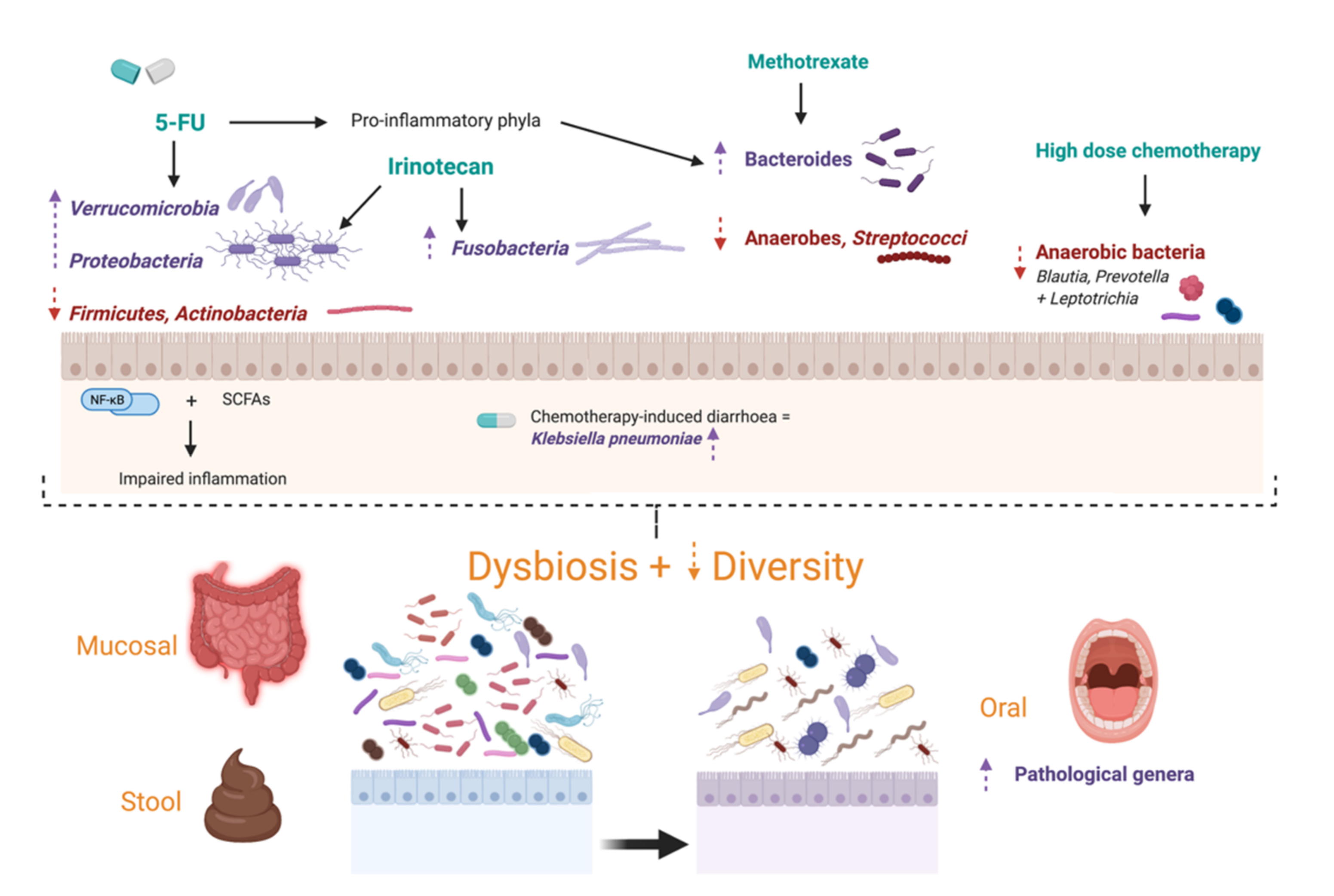
3. Effects of Antineoplastic Treatment on the Microbiome

Despite the improved efficacy and survival with modern treatments, both the adverse effects and sequelae of chemotherapy represent a major cause for concern amongst patients and clinicians [51]. A salient concept to consider is that pathological disease states or, conversely, medical therapies may promote dysbiosis, thereby influencing clinical outcomes [Figure 2] [27].
Chemotherapy causes a disturbance in microbial community structure and is associated with a reduction in microbiome diversity as well as a decrease in the richness and abundance of operational taxonomic units [76,77]. A pre-clinical study revealed an enrichment of predominantly Gram-negative bacteria following administration of 5-FU in rats [78]. This was validated in more recent clinical studies showing an enrichment, in faecal samples, of Proteobacteria, reduction of Firmicutes, Actinobacteria and taxa that impair inflammation through modulating the NF-kB pathway and producing short-chain fatty acids [77,79]. Similarly, Galloway-Peña et al. [80] unveiled statistically significant increases in Lactobacillus with significant decreases in primarily anaerobic genera including Blautia, Prevotella, and Leptotrichia in buccal and faecal samples. A decrease in anaerobic bacteria was induced in both adult and paediatric patients undergoing conditioning and high-dose chemotherapy regimens, respectively [77,81].

3.1. Specific Chemotherapy Regimens

Various human and animal studies have illustrated that certain chemotherapy regimens result in dysbiosis. A recent study demonstrated a similar shift away from beneficial bacteria such as Actinobacteria and towards phyla with pro-inflammatory traits such as Bacteroidetes and Verrucomicrobia upon the administration of 5-FU in mice [52]. Such results are also seen with the administration of methotrexate in a rat model, which induced an absolute and relative decrease in anaerobes (13-fold) and Streptococci (296-fold) as well as a relative increase in Bacteroides [77]. Importantly, these changes were most prominent at the peak of mucositis severity clinically, and reduced bacterial presence was related to the presence of diarrhoea. Enrichment of inflammatory bacteria such as Bacteroides following chemotherapy has been shown in human faecal samples as well [81,82]. Chemotherapy-induced diarrhoea was recently found to be significantly associated with Klebsiella pneumoniae enrichment in patients with resected stage III CRC undergoing the CapeOX regimen [83]. A recent study demonstrated statistically significant differences in gut microbial abundance before and after chemotherapy with both XELOX and FOLFIRI regimens-including differences in the abundance of Peptostreoptococcus, Clostridiales, and Prevotella as well as altered gut fungi [21].
Various studies have reported statistically significant alterations in microbial composition following oxaliplatin administration. A study of 40 male BALB/c mice demonstrated increased abundance of Gram-negative bacteria in the gut following oxaliplatin administration, specifically a reduction in Parabacteroides and Prevotella1 species and an increase in Prevotella2 and Odoribacter in tissue samples [84]. These genera are from the Bacteroides phylum, which stems from the Bacteroidetes family—commensals to the GI tract but also opportunistic pathogens when the intestinal barrier is disrupted. Another tissue analysis of 40 Kunming female mice similarly demonstrated an increased abundance of Bacteroidetes and reduced abundance of Prevotella following oxaliplatin [85]. Interestingly, faecal samples from both mice and humans after taking probiotics had a lower abundance of Bacteroides and a higher abundance of Prevotella [85].
Furthermore, a pre-clinical study in Sprague-Dawley rats showed that irinotecan had a greater impact than 5-FU and oxaliplatin on the composition of faecal microbiota, but both chemotherapeutic drugs induced microbial and metabolic changes, activating inflammatory processes [86]. In particular, irinotecan was associated with an increased relative abundance of Fusobacteria and Proteobacteria. The latter has also been enriched in human studies [Table 1] [76,82]. There does not appear to be a specific bacterial community that is consistently altered following chemotherapy, most likely due to a lack of homogeneity of patient groups and sample types analysed, as well as a lack of standardization in laboratory protocols and computation methods. Nevertheless, various clinical and pre-clinical studies have demonstrated a shift away from the normal microbiota, emphasising that despite the heterogeneity in microbial shifts, chemotherapy does influence the gut microenvironment to cause dysbiosis. Future research with standardized protocols should be pursued to qualify and quantify this change accurately with a view to harnessing it to improve treatment outcomes and reduce adverse effects (Table 1 and Table 2).

3.2. Oral Dysbiosis

The alterations of the oral microbiota in the context of chemotherapy are not well established. A systematic review by Napeñas et al. [89] and prospective study by de Mendonca et al. [90] demonstrated shifts in the oral microbiota towards pathological genera such as Streptococcus viridans, Neisseria spp. and Candida spp. during cancer therapy. These findings support the hypothesis of oral dysbiosis in the setting of chemotherapy [55]. Alterations in the oral bacteriome were detected in mucosal samples but of greater magnitude in salivary communities, and though they correlated with mucositis severity, they were universal in the mouth—constant in healthy, erythematous and ulcerated sites. These studies also highlighted the paucity of current, longitudinal, well-controlled studies using highly sensitive high-throughput sequencing to characterise the oral micro-environment throughout chemotherapy. Disruption of the indigenous microbial community with growth of pathobionts and reduction of beneficial commensals would impair the ability of the mucosa to remain intact during an antineoplastic challenge [55]. The plausibility of this hypothesis was suggested by Perales-Puchalt et al. [91] in a murine model of intestinal mucositis in the context of the antineoplastic agent cisplatin. 16S rRNA sequencing analysis of faecal DNA confirmed that cisplatin induced measurable dysbiosis. This dysbiosis was characterised by significant increases in bacteria of the Bacteroidaceae and Erysipelotrichaceae families, as well as in Bacteroides uniformis. In contrast, cisplatin caused a significant decrease in Ruminococcus gnavus, a trans-sialidase-expressing bacterial strain that acquires nutritional competitive advantage by degrading mucins. Furthermore, gavage of faecal pellets overturned cisplatin-induced increases in Bacteroidaceae and Erysipelotrichaceae family bacteria.
In a similar way, these studies highlight chemotherapy-induced oral microbial dysbiosis but are unable to clarify the significance of these shifts on treatment efficacy. Further research that seeks to address these questions is needed to optimize patient outcomes.

3.3. Effects of Immunotherapy on the Microbiome

ICIs, specifically ipilimumab (anti-CTLA-4), tremelimumab (anti-CTLA-4) and nivolumab (anti-PD-1), have revolutionised cancer therapy [73]. Immunotherapy in CRC shows promise in improving patient outcomes, in particular nivolumab and pembrolizumab with metastatic dmmR-mSI-H CRC; however, the evidence to support its widespread use remains preliminary at this stage [10]. Nevertheless, it is prudent to characterise the effect immunotherapy has on the gut microbiome so that patient outcomes can be optimised when these agents become more commonly used.
Pre-clinical studies have demonstrated that immunotherapies cause a shift in the microbiota composition. The ground-breaking study by Vetizou et al. [68] analysing stool samples in recolonised GF and antibiotic-treated mice demonstrated that the microbiome composition following ipilimumab administration had enrichment of Clostridiales and reduced abundance of Bacteroidales and Burkholderiales. Furthermore, whilst the Bacteroides species was decreased in faeces, Bacteroides thetaiotaomicron and Bacteroides uniformis were enriched in mucosal samples from the small intestine, suggesting that the microbiome shifts following ICI treatment vary depending on samples studied. Analysis of stool samples following nivolumab treatment in patients with non-small-cell lung cancer (NSCLC) demonstrated enrichment of Rikenellaceae, Prevotella, Streptococcus, Lactobacillus, Bacteroides plebeius, Oscillospira and Enterobacteriaceae compared to healthy controls [92]. Another study of 11 NSCLC patients demonstrated a positive correlation between increased Granulicatella abundance and improved treatment response to nivolumab [93]. It also demonstrated higher abundance of commensals such as Akkermansia muciniphila, Rikenellaceae, Bacteroides, Peptostreptococcaceae, Mogibacteriaceae and Clostridiaceae in the controls than those in the patients receiving nivolumab. However, future studies with greater sample sizes are required to reproduce statistically significant results.

4. Effects of Synbiotics

Probiotics are defined by the World Health Organisation as “live microorganisms which when administered in adequate amounts confer a health benefit on the host” [94]. As a novel approach to augment cancer therapy, they are thought to improve the diversity profile of the intestinal microbiota and reduce the extent of chronic inflammation and production of carcinogenic material in dysbiosis [2,95,96]. Prebiotics are non-digestible food constituents that selectively alter the growth of certain host-beneficial bacteria [97]. The combination of probiotics and prebiotics is called synbiotics. Both prebiotics and probiotics have demonstrated ability to alter the commensal microbiota toward a beneficial composition and perhaps be used advantageously in patients with CRC [98]. Additionally, emerging evidence has shown their effects on the efficacy and toxicity of chemotherapy and immunotherapy.

4.1. Probiotics

Despite promising results being reported in some pre-clinical models, overall results for probiotics are largely inconsistent. For example, S. thermophiles TH-4 leads to attenuation of intestinal damage in non-tumour bearing rats treated with methotrexate and 5-FU [99,100]. However, a subsequent study in tumour-bearing rats treated with methotrexate was unable to demonstrate any beneficial effect [101]. Similarly, probiotic factors derived from E. coli Nissle 1917 and Lactobacillus fermentum (L. fermentum) BR11 partially protected the intestine from 5-FU-induced mucositis, and treatment with Lactobacillus acidophilus (L. acidophilus) improved the inflammatory and functional aspects of 5-FU-induced intestinal mucositis [102,103]. Additionally, oral probiotic Lactobacillus rhamnosus (L. rhamnosus) and Bifidobacterium infantis (B. infantis) prevented FOLFOX treatment-induced intestinal mucositis in a CRC-bearing mouse model and CRC-bearing rat model, respectively, thereby avoiding dose reduction caused by intestinal toxicity [2,104,105]. L. rhamnosus may also confer advantages in augmenting the anti-tumour response produced by immunotherapy. Owens et al. [106] showed that administration of L. rhamnosus GG (LGG) decreased tumour burden via increasing the CD8 T-cell response in a murine CRC model consistent with literature regarding immunotherapy for melanoma, suggesting that an absence of lactobacilli correlates with a poorer response to immunotherapy [107].
In clinical studies, the use of Yakult (Bifidobacterium breve strain, 109 living bacteria) in children undergoing chemotherapy resulted in reduction in incidence of fever and prevented some modifications in gut microbiota such as an increase in Enterobacteriaceae [108]. In a single blinded RCT, Ishikawa et al. [109] investigated the effect of whether the probiotic Lactibacillus casei (L. casei) had any role in preventing tumour recurrence in a Japanese cohort of patients free from CRC having had 2 or more tumours removed in the past. They showed that L. casei reduced the atypia of tumours that recurred, resulting in reduced severity, but were unable to demonstrate a reduction in total new colorectal tumours developing. A larger study, which enlisted adults undergoing 5-FU regimens with L. rhamnosus supplementation, found a decreased incidence of Grade 3 or 4 diarrhoea and the need for chemotherapy dose reduction [110].
Promisingly, Benito et al. [111] demonstrated that microencapsulated probiotics Bifidobacterium bifidum (B. bifidum) and Lactobacillus gasseri (L. gasseri) reduced intestinal lesions and faecal occult blood loss in murine colorectal cancer (APCMin/+ mice), likely through inhibiting the Wnt/ β -catenin signalling pathway. Furthermore, an even greater protective effect was conferred with the co-administration of the probiotics with the flavonoid quercetin [111]. Bifidobacterium breve (B. breve) is another probiotic of the Bifidobacterium spp. that has been shown to confer anti-tumour properties. Yoon et al.’s murine model isolated two specific B. breve species that were able to improve anti-tumour immunity when used in combination with anti-cancer therapeutics such as oxaliplatin and PD-1 blockade as measured through increased CD4+/Treg, CD8+/Treg and effector CD8+/Treg as well as increased intra-tumour cytokine expression [112]. Whilst these two trials show encouraging results, they need to be further tested in clinical trials to adequately assess the efficacy of probiotic use in augmenting the response to chemo- and immunotherapeutics in patient populations.
It is important to note that despite the generally accepted notion of probiotics being a safe food adjunct, with many probiotic products being granted a ‘generally regarded as safe’ status, probiotic bacteria can translocate from the gastrointestinal tract and result in clinically significant disease [113]. In healthy subjects, probiotic bacteria generally do not result in severe disease even when they do translocate. However, studies looking at immunocompromised populations suggest otherwise, with some reports of septic complications due to probiotics [113]. Additionally, there are a number of other challenges when conducting probiotic associated research. Most notably, humans exhibit individual, region and strain specific mucosal colonisation patterns resulting in an individualised impact on microbiota in response to probiotic administration [114]. Moreover, stool sample microbiome analysis only partially correlates with human gut mucosal microbiome, making predicting the individual response to probiotics especially challenging [114]. This notion challenges the application of a universal probiotic that can be applied to all patients with CRC and rather advocates for the research and development of personalised probiotic approaches to achieve optimal patient outcomes [114].

4.2. Prebiotics

Administration of certain prebiotics demonstrates beneficial shifts in the microbiome. For example, fructo-oligosaccharide administration has been shown to increase Bifidobacteria spp. in both rat studies and human studies [115,116]. In addition, fructo-oligosaccharide is also linked to increased mucin production [116]. Butyrate, as discussed previously, has been identified as a potential antineoplastic agent in the colon [117]. It plays an essential role in mucosal regeneration and in the inhibition of pro-inflammatory cytokine production [118,119]. Glutamine, which is known to be an effective gut protectant during stressful conditions, reduces the incidence and severity of late-onset diarrhoea following CPT-11 treatment in rats [120,121]. Water-soluble polysaccharide extracted from the sporoderm-removed spores of Ganoderma Lucidum (GLP) was shown to reduce inflammation-induced tumorigenesis and microbiota dysbiosis in an azoxymethane/dextran sulfate sodium (AOM/DSS) mouse model through a variety of mechanisms including modulation of endotoxaemia induced by the TLR4/MyD88/NF-kB pathway, strengthening colonic epithelial integrity and goblet cell function and increasing short-chain fatty acid production amongst others [122]. Guo et al. [122] therefore postulate that GLP might be an effective prebiotic treatment to use to ameliorate AOM/DSS induced tumorigenesis in CRC.
When looking specifically at improving chemotherapy outcomes, however, prebiotics have thus far not shown benefit. In an experimental study of mucositis induced by 5-FU in rats, L. fermentum reduced jejunal inflammation with no additional benefit added by the prebiotic fructo-oligosaccharide [123]. Furthermore, dietary fibre intervention has been shown to alter the composition of GI microbiota, specifically increasing the number of Bifidobacterium and Lactobacillus spp. as well as increasing faecal butyrate concentration in humans, thought to be beneficial in increasing the suppressing neoplastic activity of CRC [124]. However, despite prebiotic fibre intervention having a tumour-suppressive effect in a CRC gnotobiotic mouse model, clinical trials have not been able to demonstrate similar tumour-suppressive activity with dietary fibre intervention [125,126,127,128].
Synbiotics have been shown to be able to augment the microbiome; however, the clinical utility of this in CRC treatment is yet to be established. It is possible that prebiotics and probiotics will become a novel adjunct to anti-cancer treatment in the future; however, for now, it is prudent for further research to be conducted, especially in human studies, to qualify and quantify their treatment benefit.

5. Conclusions

Substantial evidence shows that the gut microbiota influences the efficacy of chemotherapy and severity of toxicity and facilitates chemotherapy resistance. Several studies have demonstrated a direct relationship between an intact microbiome, immune functioning and chemotherapy efficacy. Contrastingly, others have suggested that certain microbes, such as Fn, may increase chemoresistance to 5-FU and that dysbiosis has a negative correlation with chemotherapeutic outcomes. Additionally, the TIMER framework that was previously proposed by Alexander et al. [27] can been used to understand pharmacomicrobiomics in the context of CRC. On the other hand, chemotherapy has been reported to induce a dysbiosis in both humans and animals, with microbial shifts extending beyond colonic mucosa and involving the oral microenvironment. This is characterised by a shift away from eubiosis and towards inflammatory phyla such as Bacteroidetes. Ultimately, whilst there is growing evidence in mouse models, there is a major discrepancy and lack of evidence within clinical trials that support the use of prebiotics and probiotics to improve chemotherapy and immunotherapy outcomes.
Though findings in current literature are promising, a greater understanding of the exact relationship between the gut microbiota, host response and outcomes of anti-cancer treatment is warranted to ensure an individualised, more effective approach in the treatment of CRC and a reduction of associated toxicities.

Author Contributions

Conceptualization, E.E.-O. and H.C.H.Y.; Writing, review and editing, all authors; Figures and table preparation, S.K. and J.E. All authors have read and agreed to the published version of the manuscript.

Funding

H.C.H.Y. and E.E.-O. are funded by grants from the Australian Federal Government to the St George and Sutherland Medical Research Foundation.

Conflicts of Interest

The authors declare no conflict of interest.

References

  1. Arnold, M.; Sierra, M.S.; Laversanne, M.; Soerjomataram, I.; Jemal, A.; Bray, F. Global patterns and trends in colorectal cancer incidence and mortality. Gut 2017, 66, 683. [Google Scholar] [CrossRef] [Green Version]
  2. Sánchez-Alcoholado, L.; Ramos-Molina, B.; Otero, A.; Laborda-Illanes, A.; Ordóñez, R.; Medina, J.A.; Gómez-Millán, J.; Queipo-Ortuño, M.I. The Role of the Gut Microbiome in Colorectal Cancer Development and Therapy Response. Cancers 2020, 12, 1406. [Google Scholar] [CrossRef]
  3. Sung, H.; Ferlay, J.; Siegel, R.L.; Laversanne, M.; Soerjomataram, I.; Jemal, A.; Bray, F. Global Cancer Statistics 2020: GLOBOCAN Estimates of Incidence and Mortality Worldwide for 36 Cancers in 185 Countries. CA A Cancer J. Clin. 2021, 71, 209–249. [Google Scholar] [CrossRef]
  4. Keum, N.; Giovannucci, E. Global burden of colorectal cancer: Emerging trends, risk factors and prevention strategies. Nat. Rev. Gastroenterol. Hepatol. 2019, 16, 713–732. [Google Scholar] [CrossRef]
  5. Visnovska, T.; Biggs, P.J.; Schmeier, S.; Frizelle, F.A.; Purcell, R.V. Metagenomics and transcriptomics data from human colorectal cancer. Sci. Data 2019, 6, 116. [Google Scholar] [CrossRef]
  6. Favoriti, P.; Carbone, G.; Greco, M.; Pirozzi, F.; Pirozzi, R.E.; Corcione, F. Worldwide burden of colorectal cancer: A review. Updates Surg. 2016, 68, 7–11. [Google Scholar] [CrossRef]
  7. Ternes, D.; Karta, J.; Tsenkova, M.; Wilmes, P.; Haan, S.; Letellier, E. Microbiome in Colorectal Cancer: How to Get from Meta-omics to Mechanism? Trends Microbiol. 2020, 28, 401–423. [Google Scholar] [CrossRef]
  8. Ahmed, I.; Umar, S. Microbiome and Colorectal Cancer. Curr. Colorectal Cancer Rep. 2018, 14, 217–225. [Google Scholar] [CrossRef]
  9. Schreuders, E.H.; Ruco, A.; Rabeneck, L.; Schoen, R.E.; Sung, J.J.Y.; Young, G.P.; Kuipers, E.J. Colorectal cancer screening: A global overview of existing programmes. Gut 2015, 64, 1637. [Google Scholar] [CrossRef]
  10. Ganesh, K.; Stadler, Z.K.; Cercek, A.; Mendelsohn, R.B.; Shia, J.; Segal, N.H.; Diaz, L.A., Jr. Immunotherapy in colorectal cancer: Rationale, challenges and potential. Nat. Rev. Gastroenterol. Hepatol. 2019, 16, 361–375. [Google Scholar] [CrossRef]
  11. Takiishi, T.; Fenero, C.I.M.; Câmara, N.O.S. Intestinal barrier and gut microbiota: Shaping our immune responses throughout life. Tissue Barriers 2017, 5, e1373208. [Google Scholar] [CrossRef]
  12. Gagnière, J.; Raisch, J.; Veziant, J.; Barnich, N.; Bonnet, R.; Buc, E.; Bringer, M.A.; Pezet, D.; Bonnet, M. Gut microbiota imbalance and colorectal cancer. World J. Gastroenterol. 2016, 22, 501–518. [Google Scholar] [CrossRef]
  13. Nakatsu, G.; Li, X.; Zhou, H.; Sheng, J.; Wong, S.H.; Wu, W.K.K.; Ng, S.C.; Tsoi, H.; Dong, Y.; Zhang, N.; et al. Gut mucosal microbiome across stages of colorectal carcinogenesis. Nat. Commun. 2015, 6, 8727. [Google Scholar] [CrossRef]
  14. Brennan, C.A.; Garrett, W.S. Gut Microbiota, Inflammation, and Colorectal Cancer. Ann. Rev. Microbiol. 2016, 70, 395–411. [Google Scholar] [CrossRef] [Green Version]
  15. Saus, E.; Iraola-Guzmán, S.; Willis, J.R.; Brunet-Vega, A.; Gabaldón, T. Microbiome and colorectal cancer: Roles in carcinogenesis and clinical potential. Mol. Asp. Med. 2019, 69, 93–106. [Google Scholar] [CrossRef]
  16. Mori, G.; Rampelli, S.; Orena, B.S.; Rengucci, C.; De Maio, G.; Barbieri, G.; Passardi, A.; Casadei Gardini, A.; Frassineti, G.L.; Gaiarsa, S.; et al. Shifts of Faecal Microbiota During Sporadic Colorectal Carcinogenesis. Sci. Rep. 2018, 8, 10329. [Google Scholar] [CrossRef]
  17. Rezasoltani, S.; Asadzadeh Aghdaei, H.; Dabiri, H.; Akhavan Sepahi, A.; Modarressi, M.H.; Nazemalhosseini Mojarad, E. The association between fecal microbiota and different types of colorectal polyp as precursors of colorectal cancer. Microb. Pathog. 2018, 124, 244–249. [Google Scholar] [CrossRef]
  18. Tang, Q.; Jin, G.; Wang, G.; Liu, T.; Liu, X.; Wang, B.; Cao, H. Current Sampling Methods for Gut Microbiota: A Call for More Precise Devices. Front. Cell. Infect. Microbiol. 2020, 10, 151. [Google Scholar] [CrossRef]
  19. Kumar, R.; Eipers, P.; Little, R.B.; Crowley, M.; Crossman, D.K.; Lefkowitz, E.J.; Morrow, C.D. Getting Started with Microbiome Analysis: Sample Acquisition to Bioinformatics. Curr. Protoc. Hum. Genet. 2014, 82, 18.8.1–18.8.29. [Google Scholar] [CrossRef] [Green Version]
  20. Bharti, R.; Grimm, D.G. Current challenges and best-practice protocols for microbiome analysis. Brief. Bioinform. 2021, 22, 178–193. [Google Scholar] [CrossRef] [Green Version]
  21. Shuwen, H.; Xi, Y.; Yuefen, P.; Jiamin, X.; Quan, Q.; Haihong, L.; Yizhen, J.; Wei, W. Effects of postoperative adjuvant chemotherapy and palliative chemotherapy on the gut microbiome in colorectal cancer. Microb. Pathog. 2020, 149, 104343. [Google Scholar] [CrossRef]
  22. Gaines, S.; Shao, C.; Hyman, N.; Alverdy, J.C. Gut microbiome influences on anastomotic leak and recurrence rates following colorectal cancer surgery. Br. J. Surg. 2018, 105, e131–e141. [Google Scholar] [CrossRef] [Green Version]
  23. Sonis, S.T.; Elting, L.S.; Keefe, D.; Peterson, D.E.; Schubert, M.; Hauer-Jensen, M.; Bekele, B.N.; Raber-Durlacher, J.; Donnelly, J.P.; Rubenstein, E.B. Perspectives on cancer therapy-induced mucosal injury: Pathogenesis, measurement, epidemiology, and consequences for patients. Cancer 2004, 100, 31. [Google Scholar]
  24. Besse, B.; Charrier, M.; Lapierre, V.; Dansin, E.; Lantz, O.; Planchard, D.; Le Chevalier, T.; Livartoski, A.; Barlesi, F.; Laplanche, A.; et al. Dendritic cell-derived exosomes as maintenance immunotherapy after first line chemotherapy in NSCLC. Oncoimmunology 2016, 5, e1071008. [Google Scholar] [CrossRef] [Green Version]
  25. Emens, L.A.; Middleton, G. The interplay of immunotherapy and chemotherapy: Harnessing potential synergies. Cancer Immunol. Res. 2015, 3, 436–443. [Google Scholar] [CrossRef] [Green Version]
  26. Roy, S.; Trinchieri, G. Microbiota: A key orchestrator of cancer therapy. Nat. Rev. Cancer 2017, 17, 271–285. [Google Scholar] [CrossRef]
  27. Alexander, J.L.; Wilson, I.D.; Teare, J.; Marchesi, J.R.; Nicholson, J.K.; Kinross, J.M. Gut microbiota modulation of chemotherapy efficacy and toxicity. Nat. Rev. Gastroenterol. Hepatol. 2017, 14, 356. [Google Scholar] [CrossRef]
  28. Scott, T.A.; Quintaneiro, L.M.; Norvaisas, P.; Lui, P.P.; Wilson, M.P.; Leung, K.Y.; Herrera-Dominguez, L.; Sudiwala, S.; Pessia, A.; Clayton, P.T.; et al. Host-Microbe Co-metabolism Dictates Cancer Drug Efficacy in C. elegans. Cell 2017, 169, 442–456. [Google Scholar] [CrossRef] [Green Version]
  29. Garcia-Gonzalez, A.P.; Ritter, A.D.; Shrestha, S.; Andersen, E.C.; Yilmaz, L.S.; Walhout, A.J.M. Bacterial Metabolism Affects the C. elegans Response to Cancer Chemotherapeutics. Cell 2017, 169, 431–441. [Google Scholar] [CrossRef] [Green Version]
  30. Yuan, L.; Zhang, S.; Li, H.; Yang, F.; Mushtaq, N.; Ullah, S.; Shi, Y.; An, C.; Xu, J. The influence of gut microbiota dysbiosis to the efficacy of 5-Fluorouracil treatment on colorectal cancer. Biomed. Pharmacother. Biomed. Pharmacother. 2018, 108, 184–193. [Google Scholar] [CrossRef]
  31. Wang, Z.; Hopson, L.M.; Singleton, S.S.; Yang, X.; Jogunoori, W.; Mazumder, R.; Obias, V.; Lin, P.; Nguyen, B.N.; Yao, M.; et al. Mice with Dysfunctional TGF-β Signaling Develop Altered Intestinal Microbiome and Colorectal Cancer Resistant to 5FU. Biochim. Biophys. Acta Mol. Basis Dis. 2021, 1867, 166179. [Google Scholar] [CrossRef]
  32. Meng, C.; Bai, C.; Brown, T.D.; Hood, L.E.; Tian, Q. Human Gut Microbiota and Gastrointestinal Cancer. Genom. Proteom. Bioinform. 2018, 16, 33–49. [Google Scholar] [CrossRef]
  33. An, J.; Ha, E.M. Combination Therapy of Lactobacillus plantarum Supernatant and 5-Fluouracil Increases Chemosensitivity in Colorectal Cancer Cells. J. Microbiol. Biotechnol. 2016, 26, 1490–1503. [Google Scholar] [CrossRef]
  34. Iida, N.; Dzustev, A.; Smith, L.; Bouladoux, N.; Weingarten, R.A.; Molina, D.A.; Salcedo, R.; Back, T.; Cramer, S.; Dai, R.M.; et al. Commensal bacteria control cancer response to therapy by modulating the tumor microenvironment. Science 2013, 342, 4. [Google Scholar]
  35. Viaud, S.; Saccheri, F.; Mignot, F.; Yamazaki, T.; Daillere, R.; Hannani, D.; Enot, D.P.; Pfirschke, C.; Engblom, C.; Pittet, M.J.; et al. The intestinal microbiota modulates the anticancer immune effects of cyclophosphamide. Science 2013, 342, 6. [Google Scholar]
  36. Lehouritis, P.; Cummins, J.; Stanton, M.; Murphy, C.T.; McCarthy, F.O.; Reid, G.; Urbaniak, C.; Byrne, W.L.; Tangney, M. Local bacteria affect the efficacy of chemotherapeutic drugs. Sci. Rep. 2015, 5, 14554. [Google Scholar]
  37. Daillere, R.; Vetizou, M.; Waldschmitt, N.; Yamazaki, T.; Isnard, C.; Poirier-Colame, V.; Duong, C.P.M.; Flament, C.; Lepage, P.; Roberti, M.P.; et al. Enterococcus hirae and Barnesiella intestinihominis facilitate cyclophosphamide-induced therapeutic immunomodulatory effects. Immunity 2016, 45, 931–943. [Google Scholar]
  38. Pagès, F.; Mlecnik, B.; Marliot, F.; Bindea, G.; Ou, F.S.; Bifulco, C.; Lugli, A.; Zlobec, I.; Rau, T.T.; Berger, M.D.; et al. International validation of the consensus Immunoscore for the classification of colon cancer: A prognostic and accuracy study. Lancet 2018, 391, 2128–2139. [Google Scholar] [CrossRef]
  39. Bindea, G.; Mlecnik, B.; Tosolini, M.; Kirilovsky, A.; Waldner, M.; Obenauf, A.C.; Angell, H.; Fredriksen, T.; Lafontaine, L.; Berger, A.; et al. Spatiotemporal dynamics of intratumoral immune cells reveal the immune landscape in human cancer. Immunity 2013, 39, 782–795. [Google Scholar] [CrossRef] [Green Version]
  40. Ogino, S.; Nosho, K.; Irahara, N.; Meyerhardt, J.A.; Baba, Y.; Shima, K.; Glickman, J.N.; Ferrone, C.R.; Mino-Kenudson, M.; Tanaka, N.; et al. Lymphocytic reaction to colorectal cancer is associated with longer survival, independent of lymph node count, microsatellite instability, and CpG island methylator phenotype. Clin. Cancer Res. 2009, 15, 6412–6420. [Google Scholar] [CrossRef] [Green Version]
  41. Roberti, M.P.; Yonekura, S.; Duong, C.P.M.; Picard, M.; Ferrere, G.; Tidjani Alou, M.; Rauber, C.; Iebba, V.; Lehmann, C.H.K.; Amon, L.; et al. Chemotherapy-induced ileal crypt apoptosis and the ileal microbiome shape immunosurveillance and prognosis of proximal colon cancer. Nat. Med. 2020, 26, 919–931. [Google Scholar] [CrossRef]
  42. Zhang, S.; Yang, Y.; Weng, W.; Guo, B.; Cai, G.; Ma, Y.; Cai, S. Fusobacterium nucleatum promotes chemoresistance to 5-fluorouracil by upregulation of BIRC3 expression in colorectal cancer. J. Exp. Clin. Cancer Res. CR 2019, 38, 14. [Google Scholar] [CrossRef]
  43. Yu, T.; Guo, F.; Yu, Y.; Sun, T.; Ma, D.; Han, J.; Qian, Y.; Kryczek, I.; Sun, D.; Nagarsheth, N.; et al. Fusobacterium nucleatum Promotes Chemoresistance to Colorectal Cancer by Modulating Autophagy. Cell 2017, 170, 548–563. [Google Scholar] [CrossRef] [Green Version]
  44. Gupta, E.; Lestingi, T.M.; Mick, R.; Ramirez, J.; Vokes, E.E.; Ratain, M.J. Metabolic fate of irinotecan in humans: Correlation of glucuronidation with diarrhea. Cancer Res. 1994, 54, 3. [Google Scholar]
  45. Nakao, T.; Kurita, N.; Komatsu, M.; Yoshikawa, K.; Iwata, T.; Utusnomiya, T.; Shimada, M. Irinotecan injures tight junction and causes bacterial translocation in rat. J. Surg. Res. 2012, 173, 341–347. [Google Scholar]
  46. Sparreboom, A.; de Jonge, M.; de Bruijn, P.; Brouwer, E.; Nooter, K.; Loos, W.J.; van Alphen, R.J.; Mathijssen, R.H.; Stoter, G.; Verweij, J. Irinotecan (CPT-11) metabolism and disposition in cancer patients. Clin. Cancer Res. 1998, 4, 2747–2754. [Google Scholar]
  47. Takasuna, K.; Hagiwara, T.; Hirohashi, M.; Kato, M.; Nomura, M.; Nagai, E.; Yokoi, T.; Kamataki, T. Involvement of beta-glucuronidase in intestinal microflora in the intestinal toxicity of the antitumor camptothecin derivative irinotecan hydrochloride (CPT-11) in rats. Cancer Res. 1996, 56, 3752–3757. [Google Scholar]
  48. Takasuna, K.; Hagiwara, T.; Hirohashi, M.; Kato, M.; Nomura, M.; Nagai, E.; Yokoi, T.; Kamataki, T. Inhibition of intestinal microflora beta-glucuronidase modifies the distribution of the active metabolite of the antitumor agent, irinotecan hydrochloride (CPT-11) in rats. Cancer Chemother. Pharmacol. 1998, 42, 280–286. [Google Scholar]
  49. Tanoue, T.; Morita, S.; Plichta, D.R.; Skelly, A.N.; Suda, W.; Sugiura, Y.; Narushima, S.; Vlamakis, H.; Motoo, I.; Sugita, K.; et al. A defined commensal consortium elicits CD8 T cells and anti-cancer immunity. Nature 2019, 565, 600–605. [Google Scholar] [CrossRef]
  50. Wong, S.H.; Yu, J. Gut microbiota in colorectal cancer: Mechanisms of action and clinical applications. Nat. Rev. Gastroenterol. Hepatol. 2019, 16, 690–704. [Google Scholar] [CrossRef]
  51. Nurgali, K.; Jagoe, R.T.; Abalo, R. Editorial: Adverse Effects of Cancer Chemotherapy: Anything New to Improve Tolerance and Reduce Sequelae? Front Pharmacol. 2018, 9, 245. [Google Scholar] [CrossRef]
  52. Carvalho, R.; Vaz, A.; Pereira, F.L.; Dorella, F.; Aguiar, E.; Chatel, J.-M.; Bermudez, L.; Langella, P.; Fernandes, G.; Figueiredo, H.; et al. Gut microbiome modulation during treatment of mucositis with the dairy bacterium Lactococcus lactis and recombinant strain secreting human antimicrobial PAP. Sci. Rep. 2018, 8, 15072. [Google Scholar] [CrossRef]
  53. Karin, M.; Jobin, C.; Balkwill, F. Chemotherapy, immunity and microbiota—A new triumvirate? Nat. Med. 2014, 20, 126–127. [Google Scholar] [CrossRef]
  54. Secombe, K.R.; Coller, J.K.; Gibson, R.J.; Wardill, H.R.; Bowen, J.M. The bidirectional interaction of the gut microbiome and the innate immune system: Implications for chemotherapy-induced gastrointestinal toxicity. Int. J. Cancer 2019, 144, 2365–2376. [Google Scholar] [CrossRef]
  55. Hong, B.-Y.; Sobue, T.; Choquette, L.; Dupuy, A.K.; Thompson, A.; Burleson, J.A.; Salner, A.L.; Schauer, P.K.; Joshi, P.; Fox, E.; et al. Chemotherapy-induced oral mucositis is associated with detrimental bacterial dysbiosis. Microbiome 2019, 7, 66. [Google Scholar] [CrossRef]
  56. Thorpe, D.W.; Stringer, A.M.; Gibson, R.J. Chemotherapy-induced mucositis: The role of the gastrointestinal microbiome and toll-like receptors. Exp. Biol. Med. 2013, 238, 1–6. [Google Scholar] [CrossRef]
  57. Cinausero, M.; Aprile, G.; Ermacora, P.; Basile, D.; Vitale, M.G.; Fanotto, V.; Parisi, G.; Calvetti, L.; Sonis, S.T. New Frontiers in the Pathobiology and Treatment of Cancer Regimen-Related Mucosal Injury. Front. Pharmacol. 2017, 8, 1428. [Google Scholar] [CrossRef] [Green Version]
  58. Hou, J.; Zheng, H.; Li, P.; Liu, H.; Zhou, H.; Yang, X. Distinct shifts in the oral microbiota are associated with the progression and aggravation of mucositis during radiotherapy. Radiother. Oncol. 2018, 129, 44–51. [Google Scholar] [CrossRef]
  59. Vasconcelos, R.M.; Sanfilippo, N.; Paster, B.J.; Kerr, A.R.; Li, Y.; Ramalho, L.; Queiroz, E.L.; Smith, B.; Sonis, S.T.; Corby, P.M. Host-Microbiome Cross-talk in Oral Mucositis. J. Dent. Res. 2016, 95, 725–733. [Google Scholar] [CrossRef] [Green Version]
  60. Wang, Y.; Gao, W.; Shi, X.; Ding, J.; Liu, W.; He, H.; Wang, K.; Shao, F. Chemotherapy drugs induce pyroptosis through caspase-3 cleavage of a Gasdermin. Nature 2017, 547, 99–103. [Google Scholar] [CrossRef]
  61. Kanarek, N.; Grivennikov, S.I.; Leshets, M.; Lasry, A.; Alkalay, I.; Horwitz, E.; Shaul, Y.D.; Stachler, M.; Voronov, E.; Apte, R.N.; et al. Critical role for IL-1β in DNA damage-induced mucositis. Proc. Natl. Acad. Sci. USA 2014, 111, E702–E711. [Google Scholar] [CrossRef] [Green Version]
  62. Papanicolas, L.E.; Gordon, D.L.; Wesselingh, S.L.; Rogers, G.B. Not Just Antibiotics: Is Cancer Chemotherapy Driving Antimicrobial Resistance? Trends Microbiol. 2018, 26, 393–400. [Google Scholar] [CrossRef]
  63. Pedroso, S.H.S.P.; Vieira, A.T.; Bastos, R.W.; Oliveira, J.S.; Cartelle, C.T.; Arantes, R.M.E.; Soares, P.M.G.; Generoso, S.V.; Cardoso, V.N.; Teixeira, M.M.; et al. Evaluation of mucositis induced by irinotecan after microbial colonization in germ-free mice. Microbiology 2015, 161, 1950–1960. [Google Scholar] [CrossRef]
  64. Brandi, G.; Dabard, J.; Raibaud, P.; Di Battista, M.; Bridonneau, C.; Pisi, A.M.; Morselli Labate, A.M.; Pantaleo, M.A.; De Vivo, A.; Biasco, G. Intestinal microflora and digestive toxicity of irinotecan in mice. Clin. Cancer Res. 2006, 12, 1299. [Google Scholar] [CrossRef] [Green Version]
  65. Stringer, A.M.; Al-Dasooqi, N. Biomarkers of chemotherapy-induced diarrhoea: A clinical study of intestinal microbiome alterations, inflammation and circulating matrix metalloproteinases. Supportive Care Cancer 2013, 21, 1843–1852. [Google Scholar]
  66. Rooks, M.G.; Veiga, P.; Wardwell-Scott, L.H.; Tickle, T.; Segata, N.; Michaud, M.; Gallini, C.A.; Beal, C.; van Hylckama-Vlieg, J.E.; Ballal, S.A.; et al. Gut microbiome composition and function in experimental colitis during active disease and treatment-induced remission. ISME J. 2014, 8, 1403–1417. [Google Scholar] [CrossRef]
  67. Thi, T.D.; Lopez, E.; Rodriguez-Rojas, A.; Rodriguez-Beltran, J.; Couce, A.; Guelfo, J.R.; Castaneda-Garcia, A.; Blazquez, J. Effect of recA inactivation on mutagenesis of Escherichia coli exposed to sublethal concentrations of antimicrobials. J. Antimicrob. Chemother. 2011, 66, 531–538. [Google Scholar] [CrossRef] [Green Version]
  68. Fessler, J.; Matson, V.; Gajewski, T.F. Exploring the emerging role of the microbiome in cancer immunotherapy. J. Immunother. Cancer 2019, 7, 108. [Google Scholar] [CrossRef]
  69. Vétizou, M.; Pitt, J.M.; Daillère, R.; Lepage, P.; Waldschmitt, N.; Flament, C.; Rusakiewicz, S.; Routy, B.; Roberti, M.P.; Duong, C.P.; et al. Anticancer immunotherapy by CTLA-4 blockade relies on the gut microbiota. Science 2015, 350, 1079–1084. [Google Scholar] [CrossRef] [Green Version]
  70. Zhu, X.; Tian, X.; Ji, L.; Zhang, X.; Cao, Y.; Shen, C.; Hu, Y.; Wong, J.W.H.; Fang, J.-Y.; Hong, J.; et al. A tumor microenvironment-specific gene expression signature predicts chemotherapy resistance in colorectal cancer patients. NPJ Precis. Oncol. 2021, 5, 7. [Google Scholar] [CrossRef]
  71. Larkin, J.; Chiarion-Sileni, V.; Gonzalez, R.; Grob, J.J.; Cowey, C.L.; Lao, C.D.; Schadendorf, D.; Dummer, R.; Smylie, M.; Rutkowski, P.; et al. Combined Nivolumab and Ipilimumab or Monotherapy in Untreated Melanoma. N. Engl. J. Med. 2015, 373, 23–34. [Google Scholar] [CrossRef] [Green Version]
  72. Saâda-Bouzid, E.; Defaucheux, C.; Karabajakian, A.; Coloma, V.P.; Servois, V.; Paoletti, X.; Even, C.; Fayette, J.; Guigay, J.; Loirat, D.; et al. Hyperprogression during anti-PD-1/PD-L1 therapy in patients with recurrent and/or metastatic head and neck squamous cell carcinoma. Ann. Oncol. 2017, 28, 1605–1611. [Google Scholar] [CrossRef]
  73. Chaput, N.; Lepage, P.; Coutzac, C.; Soularue, E.; Le Roux, K.; Monot, C.; Boselli, L.; Routier, E.; Cassard, L.; Collins, M.; et al. Baseline gut microbiota predicts clinical response and colitis in metastatic melanoma patients treated with ipilimumab. Ann. Oncol. 2017, 28, 1368–1379. [Google Scholar] [CrossRef]
  74. Sun, J.-Y.; Yin, T.-L.; Zhou, J.; Xu, J.; Lu, X.-J. Gut microbiome and cancer immunotherapy. J. Cell. Physiol. 2020, 235, 4082–4088. [Google Scholar] [CrossRef]
  75. Dubin, K.; Callahan, M.K.; Ren, B.; Khanin, R.; Viale, A.; Ling, L.; No, D.; Gobourne, A.; Littmann, E.; Huttenhower, C.; et al. Intestinal microbiome analyses identify melanoma patients at risk for checkpoint-blockade-induced colitis. Nat. Commun. 2016, 7, 10391. [Google Scholar] [CrossRef] [Green Version]
  76. Frankel, A.E.; Coughlin, L.A.; Kim, J.; Froehlich, T.W.; Xie, Y.; Frenkel, E.P.; Koh, A.Y. Metagenomic Shotgun Sequencing and Unbiased Metabolomic Profiling Identify Specific Human Gut Microbiota and Metabolites Associated with Immune Checkpoint Therapy Efficacy in Melanoma Patients. Neoplasia 2017, 19, 848–855. [Google Scholar] [CrossRef]
  77. Montassier, E.; Gastinne, T.; Vangay, P.; Al-Ghalith, G.A.; Bruley des Varannes, S.; Massart, S. Chemotherapy-driven dysbiosis in the intestinal microbiome. Aliment. Pharmacol. Ther. 2015, 42. [Google Scholar] [CrossRef]
  78. Fijlstra, M.; Ferdous, M.; Koning, A.M.; Rings, E.H.H.M.; Harmsen, H.J.M.; Tissing, W.J.E. Substantial decreases in the number and diversity of microbiota during chemotherapy-induced gastrointestinal mucositis in a rat model. Supportive Care Cancer 2015, 23, 10. [Google Scholar]
  79. Von Bultzingslowen, I.; Adlerberth, I.; Wold, A.E.; Dahlen, G.; Jontell, M. Oral and intestinal microflora in 5-fluorouracil treated rats, translocation to cervical and mesenteric lymph nodes and effects of probiotic bacteria. Oral Microbiol. Immunol. 2003, 18, 278–284. [Google Scholar]
  80. van Vliet, M.J.; Harmsen, H.J.; de Bont, E.S.; Tissing, W.J. The role of intestinal microbiota in the development and severity of chemotherapy-induced mucositis. PLoS Pathog. 2010, 6, e1000879. [Google Scholar] [CrossRef] [Green Version]
  81. Galloway-Peña, J.R.; Smith, D.P.; Sahasrabhojane, P.; Ajami, N.J.; Wadsworth, W.D.; Daver, N.G.; Chemaly, R.F.; Marsh, L.; Ghantoji, S.S.; Pemmaraju, N.; et al. The role of the gastrointestinal microbiome in infectious complications during induction chemotherapy for acute myeloid leukemia. Cancer 2016, 122, 2186–2196. [Google Scholar] [CrossRef] [Green Version]
  82. Zwielehner, J.; Lassl, C.; Hippe, B.; Pointner, A.; Switzeny, O.J.; Remely, M.; Kitzweger, E.; Ruckser, R.; Haslberger, A.G. Changes in human fecal microbiota due to chemotherapy analyzed by TaqMan-PCR, 454 sequencing and PCR-DGGE fingerprinting. PLoS ONE 2011, 6, e28654. [Google Scholar] [CrossRef] [Green Version]
  83. Vanlancker, E.; Vanhoecke, B.; Stringer, A.; Van de Wiele, T. 5-Fluorouracil and irinotecan (SN-38) have limited impact on colon microbial functionality and composition in vitro. PeerJ 2017, 5, e4017. [Google Scholar] [CrossRef]
  84. Fei, Z.; Lijuan, Y.; Xi, Y.; Wei, W.; Jing, Z.; Miao, D.; Shuwen, H. Gut microbiome associated with chemotherapy-induced diarrhea from the CapeOX regimen as adjuvant chemotherapy in resected stage III colorectal cancer. Gut Pathog. 2019, 11, 18. [Google Scholar] [CrossRef]
  85. Stojanovska, V.; McQuade, R.M.; Fraser, S.; Prakash, M.; Gondalia, S.; Stavely, R.; Palombo, E.; Apostolopoulos, V.; Sakkal, S.; Nurgali, K. Oxaliplatin-induced changes in microbiota, TLR4+ cells and enhanced HMGB1 expression in the murine colon. PLoS ONE 2018, 13, e0198359. [Google Scholar] [CrossRef]
  86. Yuan, W.; Xiao, X.; Yu, X.; Xie, F.; Feng, P.; Malik, K.; Wu, J.; Ye, Z.; Zhang, P.; Li, X. Probiotic Therapy (BIO-THREE) Mitigates Intestinal Microbial Imbalance and Intestinal Damage Caused by Oxaliplatin. Probiotics Antimicrob. Proteins 2021. [Google Scholar] [CrossRef]
  87. Forsgard, R.A.; Marrachelli, V.G.; Korpela, K.; Frias, R.; Collado, M.C.; Korpela, R.; Monleon, D.; Spillmann, T.; Osterlund, P. Chemotherapy-induced gastrointestinal toxicity is associated with changes in serum and urine metabolome and fecal microbiota in male Sprague-Dawley rats. Cancer Chemother. Pharmacol. 2017, 80, 317–332. [Google Scholar] [CrossRef] [Green Version]
  88. Stringer, A.M.; Gibson, R.J.; Logan, R.; Bowen, J.; Yeoh, A.; Keefe, D.M. Faecal microflora and beta-glucuronidase expression are altered in an irinotecan-induced diarrhea model in rats. Cancer Biol. Ther. 2008, 7, 1919–1925. [Google Scholar]
  89. Lin, X.B.; Dieleman, L.A.; Ketabi, A.; Bibova, I.; Sawyer, M.B.; Xue, H.; Field, C.J.; Baracos, V.E.; Gänzle, M.G. Irinotecan (CPT-11) Chemotherapy Alters Intestinal Microbiota in Tumour Bearing Rats. PLoS ONE 2012, 7, e39764. [Google Scholar] [CrossRef]
  90. Napeñas, J.J.; Brennan, M.T.; Bahrani-Mougeot, F.K.; Fox, P.C.; Lockhart, P.B. Relationship between mucositis and changes in oral microflora during cancer chemotherapy. Oral Surg. Oral Med. Oral Pathol. Oral Radiol. Endodontol. 2007, 103, 48–59. [Google Scholar] [CrossRef]
  91. de Mendonca, R.M.; de Araujo, M.; Levy, C.E.; Morari, J.; Silva, R.A.; Yunes, J.A.; Brandalise, S.R. Prospective evaluation of HSV, Candida spp., and oral bacteria on the severity of oral mucositis in pediatric acute lymphoblastic leukemia. Supportive Care Cancer Off. J. Multinatl. Assoc. Supportive Care Cancer 2012, 20, 1101–1107. [Google Scholar] [CrossRef]
  92. Perales-Puchalt, A.; Perez-Sanz, J.; Payne, K.K.; Svoronos, N.; Allegrezza, M.J.; Chaurio, R.A.; Anadon, C.; Calmette, J.; Biswas, S.; Mine, J.A.; et al. Frontline Science: Microbiota reconstitution restores intestinal integrity after cisplatin therapy. J. Leukoc. Biol. 2018, 103, 799–805. [Google Scholar] [CrossRef]
  93. Botticelli, A.; Vernocchi, P.; Marini, F.; Quagliariello, A.; Cerbelli, B.; Reddel, S.; Del Chierico, F.; Di Pietro, F.; Giusti, R.; Tomassini, A.; et al. Gut metabolomics profiling of non-small cell lung cancer (NSCLC) patients under immunotherapy treatment. J. Transl. Med. 2020, 18, 49. [Google Scholar] [CrossRef]
  94. Vernocchi, P.; Gili, T.; Conte, F.; Del Chierico, F.; Conta, G.; Miccheli, A.; Botticelli, A.; Paci, P.; Caldarelli, G.; Nuti, M.; et al. Network Analysis of Gut Microbiome and Metabolome to Discover Microbiota-Linked Biomarkers in Patients Affected by Non-Small Cell Lung Cancer. Int. J. Mol. Sci. 2020, 21, 8730. [Google Scholar] [CrossRef]
  95. FAO; WHO. Evaluation of health and nutritional properties of powder milk and live lactic acid bacteria. Prevention 2001, 5, 1–10. [Google Scholar]
  96. Fuller, R. Probiotics in human medicine. Gut 1991, 32, 439–442. [Google Scholar] [CrossRef] [Green Version]
  97. Liu, J.; Liu, C.; Yue, J. Radiotherapy and the gut microbiome: Facts and fiction. Radiat. Oncol. 2021, 16, 9. [Google Scholar] [CrossRef]
  98. Tsai, Y.L.; Lin, T.L.; Chang, C.J.; Wu, T.R.; Lai, W.F.; Lu, C.C.; Lai, H.C. Probiotics, prebiotics and amelioration of diseases. J. Biomed. Sci. 2019, 26, 3. [Google Scholar] [CrossRef]
  99. Dikeocha, I.J.; Al-Kabsi, A.M.; Eid, E.E.M.; Hussin, S.; Alshawsh, M.A. Probiotics supplementation in patients with colorectal cancer: A systematic review of randomized controlled trials. Nutr. Rev. 2021. [Google Scholar] [CrossRef]
  100. Whitford, E.J.; Cummins, A.; Butler, R.N.; Prisciandaro, L.D.; Fauser, J.K.; Yazbeck, R.; Lawrence, A.; Cheah, K.Y.; Wright, T.H.; Lymn, K.A.; et al. Effects of Streptococcus thermophilus TH-4 on intestinal mucositis induced by the chemotherapeutic agent, 5-Fluorouracil (5-FU). Cancer Biol. Ther. 2009, 8, 505–511. [Google Scholar]
  101. Tooley, K.; Howarth, G.S.; Lymn, K.; Lawrence, A.; Butler, R. Oral ingestion of Streptococcus thermophilus diminishes severity of small intestinal mucositis in methotrexate treated rats. Cancer Biol. Ther. 2006, 5, 593–600. [Google Scholar]
  102. Tooley, K.; Howarth, G.; Lymn, K.; Lawrence, A.; Butler, R. Oral ingestion of Streptococcus thermophilus does not affect mucositis severity or tumor progression in the tumor-bearing rat. Cancer Biol. Ther. 2011, 12, 131–138. [Google Scholar]
  103. Justino, P.F.; Melo, L.; Nogueira, A.F.; Morais, C.M.; Mendes, W.O.; Franco, A.X.; Souza, E.P.; Ribeiro, R.A.; Souza, M.H.L.P.; Soares, P.M.G. Regulatory role of Lactobacillus acidophilus on inflammation and gastric dysmotility in intestinal mucositis induced by 5-fluorouracil in mice. Cancer Chemother. Pharmacol. 2015, 75, 559–567. [Google Scholar]
  104. Prisciandaro, L.D.; Geier, M.S.; Butler, R.N.; Cummins, A.G.; Howarth, G.S. Probiotic factors partially improve parameters of 5-fluorouracil-induced intestinal mucositis in rats. Cancer Biol. Ther. 2011, 11, 671–677. [Google Scholar]
  105. Chang, C.W.; Liu, C.Y.; Lee, H.C.; Huang, Y.H.; Li, L.H.; Chiau, J.C.; Wang, T.E.; Chu, C.H.; Shih, S.C.; Tsai, T.H.; et al. Lactobacillus casei Variety rhamnosus Probiotic Preventively Attenuates 5-Fluorouracil/Oxaliplatin-Induced Intestinal Injury in a Syngeneic Colorectal Cancer Model. Front Microbiol. 2018, 9, 983. [Google Scholar] [CrossRef]
  106. Mi, H.; Dong, Y.; Zhang, B.; Wang, H.; Peter, C.C.K.; Gao, P.; Fu, H.; Gao, Y. Bifidobacterium Infantis Ameliorates Chemotherapy-Induced Intestinal Mucositis Via Regulating T Cell Immunity in Colorectal Cancer Rats. Cell. Physiol. Biochem. Int. J. Exp. Cell. Physiol. Biochem. Pharmacol. 2017, 42, 2330–2341. [Google Scholar] [CrossRef] [Green Version]
  107. Owens, J.A.; Saeedi, B.J.; Naudin, C.R.; Hunter-Chang, S.; Barbian, M.E.; Eboka, R.U.; Askew, L.; Darby, T.M.; Robinson, B.S.; Jones, R.M. Lactobacillus rhamnosus GG Orchestrates an Anti-tumor Immune Response. Cell. Mol. Gastroenterol. Hepatol. 2021, 21, 115–116. [Google Scholar] [CrossRef]
  108. Gopalakrishnan, V.; Spencer, C.N.; Nezi, L.; Reuben, A.; Andrews, M.C.; Karpinets, T.V.; Prieto, P.A.; Vicente, D.; Hoffman, K.; Wei, S.C.; et al. Gut microbiome modulates response to anti-PD-1 immunotherapy in melanoma patients. Science 2018, 359, 97–103. [Google Scholar] [CrossRef] [Green Version]
  109. Wada, M.; Nagata, S.; Saito, M.; Shimizu, T.; Yamashiro, Y.; Matsuki, T.; Asahara, T.; Nomoto, K. Effects of the enteral administration of Bifidobacterium breve on patients undergoing chemotherapy for pediatric malignancies. Supportive Care Cancer 2010, 18, 751–759. [Google Scholar]
  110. Ishikawa, H.; Akedo, I.; Otani, T.; Suzuki, T.; Nakamura, T.; Takeyama, I.; Ishiguro, S.; Miyaoka, E.; Sobue, T.; Kakizoe, T. Randomized trial of dietary fiber andLactobacillus casei administration for prevention of colorectal tumors. Int. J. Cancer 2005, 116, 762–767. [Google Scholar] [CrossRef]
  111. Osterlund, P.; Ruotsalainen, T.; Korpela, R.; Saxelin, M.; Ollus, A.; Valta, P.; Kouri, M.; Elomaa, I.; Joensuu, H. Lactobacillus supplementation for diarrhoea related to chemotherapy of colorectal cancer: A randomised study. Br. J. Cancer 2007, 97, 1028–1034. [Google Scholar]
  112. Benito, I.; Encío, I.J.; Milagro, F.I.; Alfaro, M.; Martínez-Peñuela, A.; Barajas, M.; Marzo, F. Microencapsulated Bifidobacterium bifidum and Lactobacillus gasseri in Combination with Quercetin Inhibit Colorectal Cancer Development in Apc(Min/+) Mice. Int. J. Mol. Sci. 2021, 22, 4906. [Google Scholar] [CrossRef]
  113. Yoon, Y.; Kim, G.; Jeon, B.-N.; Fang, S.; Park, H. Bifidobacterium Strain-Specific Enhances the Efficacy of Cancer Therapeutics in Tumor-Bearing Mice. Cancers 2021, 13, 957. [Google Scholar] [CrossRef]
  114. Liong, M.-T. Safety of probiotics: Translocation and infection. Nutr. Rev. 2008, 66, 192–202. [Google Scholar]
  115. Zmora, N.; Zilberman-Schapira, G.; Suez, J.; Mor, U.; Dori-Bachash, M.; Bashiardes, S.; Kotler, E.; Zur, M.; Regev-Lehavi, D.; Brik, R.B.-Z.; et al. Personalized Gut Mucosal Colonization Resistance to Empiric Probiotics Is Associated with Unique Host and Microbiome Features. Cell 2018, 174, 1388–1405. [Google Scholar] [CrossRef] [Green Version]
  116. Lara-Villoslada, F.; de Haro, O.; Camuesco, D.; Comalada, M.; Velasco, J.; Zarzuelo, A.; Xaus, J.; Galvez, J. Short-chain fructooligosaccharides, in spite of being fermented in the upper part of the large intestine, have anti-inflammatory activity in the TNBS model of colitis. Eur. J. Nutr. 2006, 45, 418–425. [Google Scholar]
  117. Ten Bruggencate, S.J.; Bovee-Oudenhoven, I.M.; Lettink-Wissink, M.L.; Katan, M.B.; van der Meer, R. Dietary fructooligosaccharides affect intestinal barrier function in healthy men. J. Nutri. 2006, 136, 70–74. [Google Scholar]
  118. Scharlau, D.; Borowicki, A.; Habermann, N.; Hofmann, T.; Klenow, S.; Miene, C.; Munjal, U.; Stein, K.; Glei, M. Mechanisms of primary cancer prevention by butyrate and other products formed during gut flora-mediated fermentation of dietary fibre. Mutat. Res. 2009, 628, 39–53. [Google Scholar]
  119. Looijer-van Langen, M.; Dieleman, L. Prebiotics in chronic intestinal inflammation. Inflamm. Bowel Dis. 2009, 15, 454–462. [Google Scholar]
  120. Sauer, J.; Richter, K.K.; Pool-Zobel, B.L. Products formed during fermentation of the prebiotic inulin with human gut flora enhance expression of biotransformation genes in human primary colon cells. Br. J. Nutri. 2007, 97, 928–937. [Google Scholar]
  121. Manhart, N.; Vierlinger, K.; Spittler, A.; Bergmeister, H.; Sautner, T.; Roth, E. Oral feeding with glutamine prevents lymphocyte and glutathione depletion of Peyer’s patches in endotoxemic mice. Ann. Surg. 2001, 234, 92–97. [Google Scholar]
  122. Decker-Baumann, C.; Buhl, K.; Frohmuller, S.; von Herbay, A.; Dueck, M.; Schlag, P.M. Reduction of chemotherapy-induced side effects by parenteral glutamine supplementation in patients with metastatic colorectal cancer. Eur. J. Cancer 1999, 35, 202–207. [Google Scholar]
  123. Guo, C.; Guo, D.; Fang, L.; Sang, T.; Wu, J.; Guo, C.; Wang, Y.; Wang, Y.; Chen, C.; Chen, J.; et al. Ganoderma lucidum polysaccharide modulates gut microbiota and immune cell function to inhibit inflammation and tumorigenesis in colon. Carbohydr. Polym. 2021, 267, 118231. [Google Scholar] [CrossRef]
  124. Smith, C.; Geier, M.S.; Yazbeck, R.; Torres, D.; Butler, R.; Howarth, G.S. Lactobacillus fermentum BR11 and fructo-oligosaccharide partially reduce jejunal inflammation in a model of intestinal mucositis in rats. Nutr. Cancer 2008, 60, 757–767. [Google Scholar]
  125. So, D.; Whelan, K.; Rossi, M.; Morrison, M.; Holtmann, G.; Kelly, J.T.; Shanahan, E.R.; Staudacher, H.M.; Campbell, K.L. Dietary fiber intervention on gut microbiota composition in healthy adults: A systematic review and meta-analysis. Am. J. Clin. Nutr. 2018, 107, 965–983. [Google Scholar] [CrossRef] [Green Version]
  126. Alberts, D.S.; Martínez, M.E.; Roe, D.J.; Guillén-Rodríguez, J.M.; Marshall, J.R.; Van Leeuwen, J.B.; Reid, M.E.; Ritenbaugh, C.; Vargas, P.A.; Bhattacharyya, A.B.; et al. Lack of Effect of a High-Fiber Cereal Supplement on the Recurrence of Colorectal Adenomas. N. Engl. J. Med. 2000, 342, 1156–1162. [Google Scholar] [CrossRef]
  127. Schatzkin, A.; Lanza, E.; Corle, D.; Lance, P.; Iber, F.; Caan, B.; Shike, M.; Weissfeld, J.; Burt, R.; Cooper, M.R.; et al. Lack of Effect of a Low-Fat, High-Fiber Diet on the Recurrence of Colorectal Adenomas. N. Engl. J. Med. 2000, 342, 1149–1155. [Google Scholar] [CrossRef]
  128. Lanza, E.; Yu, B.; Murphy, G.; Albert, P.S.; Caan, B.; Marshall, J.R.; Lance, P.; Paskett, E.D.; Weissfeld, J.; Slattery, M.; et al. The Polyp Prevention Trial Continued Follow-up Study: No Effect of a Low-Fat, High-Fiber, High-Fruit, and -Vegetable Diet on Adenoma Recurrence Eight Years after Randomization. Cancer Epidemiol. Biomark. Prev. 2007, 16, 1745–1752. [Google Scholar] [CrossRef] [Green Version]
Figure 1. The effect of microbiome on chemotherapy efficacy and toxicity. The gut microbiota can affect the host response to chemotherapeutic agents by facilitating drug efficacy, promoting chemoresistance and/or mediating chemotherapeutic toxicity and side effects. On the other hand, the efficacy of anticancer therapy is dependent on microbiota-mediated innate and adaptive immune responses. As a whole, chemotherapy induces ROS-mediated DNA and non-DNA damage, resulting in bacterial translocation across the intestinal epithelium. This in turn induces an inflammatory response and can provoke systemic infections. Abbreviations: 5-FU, 5-fluorouracil; DNA, deoxyribonucleic acid; Fn, fusobacterium nucleatum; LPSN, lactobacillus plantarum supernatant; ROS, reactive oxygen species. The bolded arrows represent the effects of the microbiome and chemotherapy, and the broken arrow represents bacterial translocation.
Cancers 13 04623 g001
Figure 2. The impact of chemotherapy and immunotherapy on the microbiome. Dysbiosis with a reduction in diversity and operational taxonomic units is evidenced following different treatment regimes. This dysbiosis occur in colon mucosa, stool and oral cavity of the treated patients. The oral dysbiosis includes the enrichment of pathological genera such as Neisseria spp. The gut mucosal and stool dysbiosis include a specific shift towards pro-inflammatory bacteria with a reduction of anaerobic bacteria. Dotted arrows represent the shift in microbiome and bold arrows represent the effect of chemotherapy on the microbiome composition.
Cancers 13 04623 g002
Table 1. Stool and Mucosal Dysbiosis following Chemotherapy in Humans.
AuthorStudy Subjects (n)Specimen TypesMethodMicrobiota Changes
Montassier, Gastinne, Vangay, Al-Ghalith, Bruley des Varannes and Massart [76]Non-Hodgkin’s Lymphoma (28)Stool16S rRNA↓ Firmicutes
  Actinobacteria
Proteobacteria
Galloway-Peña, Smith, Sahasrabhojane, Ajami, Wadsworth, Daver, Chemaly, Marsh, Ghantoji, Pemmaraju, Garcia-Manero, Rezvani, Alousi, Wargo, Shpall, Futreal, Guindani, Petrosino, Kontoyiannis and Shelburne [80]Acute myelogenous leukaemia patients (34)Buccal specimens
Stool
16S rRNA (V4) Oral and stool α -diversity (with carbapenem)
 Lactobacillus (oral and buccal)
Anaerobes e.g., Blautia, Prevotella, Leptotrichia
Zwielehner, Lassl, Hippe, Pointner, Switzeny, Remely, Kitzweger, Ruckser and Haslberger [81]Chemotherapy patients (17)Stool16S rRNA Diversity of Clostridium clusters IV and XIVa
 Bacteroides
Bifidobacterium
Fei, Lijuan, Xi, Wei, Jing, Miao and Shuwen [83]Resected stage III CRC patients undergoing CapeOX regimen (17)Stool16S rRNA Microbial richness and diversity
 Klebsiella pneumoniae
Vanlancker, Vanhoecke, Stringer and Van de Wiele [82]In vitro mucosal stimulator treating stool and mucosa with 5-FU and SN-38 (active metabolite of irinotecan) (6 human donors)Stool
Mucosal
16S rRNA (V3–4)   Proteobacteria (Escherichia/Shigella)
  Bacteroidetes (Bacteroides)
  Firmicutes (Clostridium cluster XIVa, Veillonella)
 Bacteroides
 Escherichia/Shigella
Abbreviations: ↑, increased; ↓, decreased.
Table 2. Stool and Mucosal Dysbiosis following Chemotherapy in Animals.
AuthorStudy Subjects (n)Specimen TypesMethodMicrobiota Changes
Fijlstra et al. [77]Rats treated with methotrexateStoolFISH Anaerobes
Streptococci
 Bacteroides
Forsgard, Marrachelli, Korpela, Frias, Collado, Korpela, Monleon, Spillmann and Osterlund [86]Sprague-Dawley rats injected with 5-FU, oxaliplatin or irinotecan (48)Stool16S rRNAIrinotecan:
Fusobacteria
Proteobacteria
5-FU and Oxaliplatin caused minor shifts
Stringer et al. [87]Irinotecan-treated rats (81)StoolDNA extraction + PCR  E. coli
 Staphylococcus spp.
 Clostridium spp.
 Lactobacillus spp.
 Bifidobacterium spp.
 Bacteroides spp.
Lin et al. [88]Tumour-bearing rats receiving irinotecan +/− oral glutamine bolus (6)
Rats receiving two cycles of irinotecan followed by 5-FU (6)
StoolDNA extraction + PCR  Clostridium cluster XI
 Enterobacteriaceae
Glutamate caused:
 Clostridium cluster VI
 Bacteroides
Carvalho, Vaz, Pereira, Dorella, Aguiar, Chatel, Bermudez, Langella, Fernandes, Figueiredo, Goes-Neto and Azevedo [52]5-FU treated mice (72)Stool16s rRNA (V4)  Bacteroidetes
 Firmicutes
 Proteobacteria
Abbreviations: ↑, increased; ↓, decreased.
Publisher’s Note: MDPI stays neutral with regard to jurisdictional claims in published maps and institutional affiliations.

