Next Article in Journal
Claisened Hexafluoro Inhibits Metastatic Spreading of Amoeboid Melanoma Cells
Previous Article in Journal
Multi-Layer Nanofibrous PCL Scaffold-Based Colon Cancer Cell Cultures to Mimic Hypoxic Tumor Microenvironment for Bioassay
 
 
cancers-logo
Article Menu

Article Menu

Article

Cemiplimab for Locally Advanced and Metastatic Cutaneous Squamous-Cell Carcinomas: Real-Life Experience from the French CAREPI Study Group

by
Candice Hober
1,†,‡,
Lisa Fredeau
2,†,‡,
Anne Pham-Ledard
3,‡,
Marouane Boubaya
2,‡,
Florian Herms
4,‡,
Philippe Celerier
5,‡,
François Aubin
6,‡,
Nathalie Beneton
7,‡,
Monica Dinulescu
8,‡,
Arnaud Jannic
9,‡,
Nicolas Meyer
10,11,‡,
Anne-Bénédicte Duval-Modeste
12,‡,
Laure Cesaire
13,‡,
Ève-Marie Neidhardt
14,‡,
Élodie Archier
15,‡,
Brigitte Dréno
16,17,18,‡,
Candice Lesage
19,‡,
Clémence Berthin
20,‡,
Nora Kramkimel
21,‡,
Florent Grange
22,23,‡,
Julie de Quatrebarbes
24,‡,
Pierre-Emmanuel Stoebner
25,26,‡,
Nicolas Poulalhon
27,‡,
Jean-Philippe Arnault
28,‡,
Safia Abed
29,‡,
Bertille Bonniaud
30,‡,
Sophie Darras
31,‡,
Valentine Heidelberger
32,‡,
Suzanne Devaux
33,‡,
Marie Moncourier
34,‡,
Laurent Misery
35,‡,
Sandrine Mansard
36,‡,
Maxime Etienne
37,‡,
Florence Brunet-Possenti
38,‡,
Caroline Jacobzone
39,‡,
Romain Lesbazeilles
40,41,‡,§,
François Skowron
23,‡,‖,
Julia Sanchez
22,‡,¶,
Stéphanie Catala
42,‡,
Mahtab Samimi
43,44,‡,
Youssef Tazi
45,‡,
Dominique Spaeth
46,‡,
Caroline Gaudy-Marqueste
47,‡,
Olivier Collard
48,‡,
Raoul Triller
49,‡,
Marc Pracht
50,‡,
Marc Dumas
51,‡,
Lucie Peuvrel
52,‡,
Pierre Combe
53,‡,
Olivier Lauche
54,‡,
Pierre Guillet
55,‡,
Yves Reguerre
56,‡,
Ingrid Kupfer-Bessaguet
41,‡,
David Solub
57,‡,
Amélie Schoeffler
58,‡,
Christophe Bedane
30,59,‡,
Gaëlle Quéreux
16,17,18,‡,
Sophie Dalac
30,‡,
Laurent Mortier
1,60,‡ and
Ève Maubec
2,61,62,*,‡
add Show full author list remove Hide full author list
1
Centre Hospitalier Universitaire (CHU) de Lille, 59037 Lille, France
2
Hôpital Avicenne, Assistance Publique–Hôpitaux de Paris (APHP), 93000 Bobigny, France
3
CHU de Bordeaux and University of Bordeaux, 33000 Bordeaux, France
4
Hôpital Saint-Louis, APHP, 75010 Paris, France
5
CH Saint-Louis de la Rochelle, 17000 La Rochelle, France
6
Université de Bourgogne–Franche-Comté and CHU de Besançon, 25000 Besançon, France
7
CH du Mans, 72037 Le Mans, France
8
Hôpital Pontchaillou, 35000 Rennes, France
9
Hôpital Henri-Mondor, APHP, 94000 Créteil, France
10
Institut Universitaire du Cancer de Toulouse, 31100 Toulouse, France
11
CHU de Toulouse, 31300 Toulouse, France
12
Hôpital Charles-Nicolle, 76038 Rouen, France
13
Hôpital Côte-de-Nacre, 14000 Caen, France
14
Centre Léon-Bérard, 69008 Lyon, France
15
Hôpital Saint-Joseph, 13008 Marseille, France
16
CHU de Nantes and Université de Nantes, 44000 Nantes, France
17
Centre d’Investigation Clinique 1413, Institut National de la Santé et de la Recherche Médicale (INSERM), CHU de Nantes, 44000 Nantes, France
18
Centre de Recherche en Cancérologie et Immunologie Nantes Angers (CRCINA), Institut National de la Santé et de la Recherche Médicale (INSERM), 44007 Nantes, France
19
CHU de Montpellier, 34295 Montpellier, France
20
CHU d’Angers, 49100 Angers, France
21
APHP, Hôpital Cochin, 75014 Paris, France
22
CHU de Reims, 51092 Reims, France
23
CH de Valence, 26000 Valence, France
24
CH Annecy Genevois, 74370 Annecy, France
25
CHU de Nîmes, 30900 Nîmes, France
26
UMR CNRS 5247, Université Montpellier I, 34090 Montpellier, France
27
Hôpital Lyon Sud–Hospices Civils de Lyon, 69310 Lyon, France
28
CHU Amiens-Picardie, 80000 Amiens, France
29
Hôpital d’Instruction des Armées Sainte-Anne, 83000 Toulon, France
30
CHU F.-Mitterrand Dijon-Bourgogne, 21000 Dijon, France
31
CH de Boulogne-sur-Mer, 62200 Boulogne-sur-Mer, France
32
CH Robert-Ballanger, 93600 Aulnay-sous-Bois, France
33
CH Côte Basque, 64109 Bayonne, France
34
CHU de Grenoble-Alpes, 38700 Grenoble, France
35
CHU de Brest and University of Bretagne Occidentale, 29200 Brest, France
36
CHU de Clermont-Ferrand, 63100 Clermont-Ferrand, France
37
CH de Cornouaille, CH Intercommunal de Quimper, 29000 Quimper, France
38
APHP, Hôpital Bichat, 75018 Paris, France
39
Hôpital du Scorff, 56322 Lorient, France
40
CHU de Poitiers, 86021 Poitiers, France
41
CH de Niort, 79000 Niort, France
42
Clinique Saint-Pierre, 66000 Perpignan, France
43
CH Régional Universitaire Trousseau de Tours, 37170 Chambray les Tours, France
44
ISP1282 UMR INRA-Université de Tours, 37000 Tours, France
45
Clinique Sainte-Anne, 67000 Strasbourg, France
46
Centre d’Oncologie de Gentilly, 54000 Nancy, France
47
University of Aix—Marseille and CHU de la Timone, 13005 Marseille, France
48
Institut de Cancérologie de la Loire–Lucien-Neuwirth (ICLN), 42270 Saint-Priest-en-Jarez, France
49
Institut Franco-Britannique, 92300 Levallois-Perret, France
50
Groupe Hospitalier de St-Malo, 35400 St-Malo, France
51
CH René-Dubos, 95300 Pontoise, France
52
Institut de Cancérologie de l’Ouest, 44800 Saint-Herblain, France
53
Pôle Santé Léonard-de-Vinci, 37170 Chambray-les-Tours, France
54
Clinique Clémentville, 34070 Montpellier, France
55
Hôpital Privé Toulon Hyères Saint-Jean, 83100 Toulon, France
56
CHU de Saint-Denis, 97400 Saint-Denis de la Réunion, France
57
Hôpital Louis Pasteur, 28630 Le Coudray, France
58
CH Régional Metz-Thionville, 57100 Metz, France
59
CHU de Limoges, 87000 Limoges, France
60
INSERM U 1189, University of Lille, 59037 Lille, France
61
Campus de Bobigny—Université Sorbonne Paris Nord, 93017 Bobigny, France
62
UMR 1124, Campus Saint Germain des Prés, 75006 Paris, France
*
Author to whom correspondence should be addressed.
These authors contributed equally to this study.
These authors belong to French Cutaneous Squamous Cell Carcinoma Study Group.
§
Present address: CH Niort, 79000 Niort, France.
Present address: Hôpitaux Drôme Nord, 26100 Romans-sur-Isère, France.
Present address: CH Saint-Quentin, 02321 Saint-Quentin, France.
Cancers 2021, 13(14), 3547; https://doi.org/10.3390/cancers13143547
Submission received: 6 June 2021 / Revised: 6 July 2021 / Accepted: 7 July 2021 / Published: 15 July 2021
(This article belongs to the Section Cancer Therapy)

Abstract

:

Simple Summary

Prognosis of advanced cutaneous squamous-cell carcinoma (CSCC) is poor. Recent clinical trials have shown that immunotherapy achieves significantly improved survival of patients with advanced CSCCs. However, few real-world data are available on treatment patterns and clinical outcomes of patients with advanced CSCCs receiving anti-programmed cell-death protein-1 (PD-1). To approach this issue, we conducted a retrospective study on 245 patients with advanced CSCCs from 58 centers who had been enrolled in an early-access program; 240 received cemiplimab. Our objectives were to evaluate, in the real-life setting, best overall response rate, progression-free survival, overall survival and safety. Results demonstrated cemiplimab efficacy in patients with advanced CSCCs, regardless of immune status. Patients with good Eastern Cooperative Oncology Group performance status benefited more from cemiplimab. The safety profile was acceptable.

Abstract

Although cemiplimab has been approved for locally advanced (la) and metastatic (m) cutaneous squamous-cell carcinomas (CSCCs), its real-life value has not yet been demonstrated. An early-access program enrolled patients with la/mCSCCs to receive cemiplimab. Endpoints were best overall response rate (BOR), progression-free survival (PFS), overall survival (OS), duration of response (DOR) and safety. The 245 patients (mean age 77 years, 73% male, 49% prior systemic treatment, 24% immunocompromised, 27% Eastern Cooperative Oncology Group performance status (PS) ≥ 2) had laCSCCs (35%) or mCSCCs (65%). For the 240 recipients of ≥1 infusion(s), the BOR was 50.4% (complete, 21%; partial, 29%). With median follow-up at 12.6 months, median PFS was 7.9 months, and median OS and DOR were not reached. One-year OS was 73% versus 36%, respectively, for patients with PS < 2 versus ≥ 2. Multivariate analysis retained PS ≥ 2 as being associated during the first 6 months with PFS and OS. Head-and-neck location was associated with longer PFS. Immune status had no impact. Severe treatment-related adverse events occurred in 9% of the patients, including one death from toxic epidermal necrolysis. Cemiplimab real-life safety and efficacy support its use for la/mCSCCs. Patients with PS ≥ 2 benefited less from cemiplimab, but it might represent an option for immunocompromised patients.

1. Introduction

Cutaneous squamous-cell carcinoma (CSCC) is the second most common skin cancer after basal-cell carcinoma [1]. In Europe, the reported age-standardized CSCC incidence ranges from 15 to 77 per 100,000 individuals per year, predominantly occurring in males [2,3]. The incidence is constantly increasing, probably because of early CSCC resection, population aging and changing UV-exposure habits [4].The CSCC risk is heightened for immunocompromised patients, being about 100-times higher after organ transplantation [5,6,7,8,9], and for those CSCC oncogenic human papillomavirus-positive, or with chronic dermatitis, exposure to arsenic or ionizing radiation, or genodermatosis (e.g., dystrophic epidermolysis bullosa, xeroderma pigmentosum, albinism and Muir–Torre syndrome) [10,11,12,13,14,15]. At an early stage, CSCC prognosis is excellent, with 90% 10-year survival [16]. However, ~5% of the patients experience local recurrences, ~4% of them develop regional disease and outcomes are fatal for ~2% [16,17,18,19,20,21,22]. According to American data [23], the CSCC mortality rate is of the same order of magnitude as that of melanoma. The 5-year overall survival (OS) rate of patients with resectable, regional CSCCs was 50–60% [18,19]. The prognosis becomes more uncertain for locally advanced or metastatic disease, with either regional or distant metastases.
Since 2018, anti-programed cell-death protein-1 (PD-1) monoclonal antibodies have emerged as first-line treatments for the management of unresectable, locally advanced or metastatic CSCCs. Cemiplimab was the first immunotherapy approved by the Food and Drug Administration and the European Medicines Agency [24], followed by pembrolizumab, in the United States, for patients who are not candidates for curative radiotherapy or surgery [25]. Immunotherapies have demonstrated anti-tumor activity with response rates exceeding 40% and acceptable safety profiles [24,25,26,27].
In France, an early-access program made cemiplimab available to patients with locally advanced or metastatic CSCCs during the time between completion of enrollment in cemiplimab clinical trials and its regulatory approval. This retrospective, multicenter CAREPI trial aimed to evaluate cemiplimab efficacy and safety in the real-life setting of those early-access patients. Our results confirmed cemiplimab efficacy in real life and identified clinical characteristics of those patients associated with progression-free survival (PFS) and OS.

2. Materials and Methods

Patients eligible for the early-access program (August 2018 to October 2019) were adults with locally advanced or metastatic CSCCs not amenable to surgery. Exclusion criteria were active autoimmune diseases or infections, uncontrolled brain metastases, pregnancy or breastfeeding. Patients received intravenous cemiplimab infusions (3 mg/kg every 2 weeks) until death from any cause, unacceptable toxicity, or patient’s or physician’s decision. Investigators were asked to complete a standardized case-report form for each patient included in the early-access program.
This retrospective study was approved by the local Avicenne Hospital Ethics Committee (CLEA-2019-75). The national database has been declared to the French data-protection agency (CNIL approval number 2215607). In compliance with French law, consent regarding non-opposition to collect and use the data was obtained from each patient.
The primary endpoint was the best overall response rate (BOR); secondary endpoints included PFS, OS, duration of response (DOR) and safety. Standard-of-care tumor assessments were carried out at the treating facility without central review. Adverse events (AEs) were graded according to the Common Terminology Criteria for Adverse Events version 5. Efficacy and safety were assessed for all patients who received at least one cemiplimab infusion.
Patient characteristics are expressed as numbers (percentages) for discrete variables, and mean ± standard deviation or median (range) for continuous variables. Data cutoff was 19 June 2020. Median follow-up was estimated using the Kaplan–Meier reverse method. OS and PFS were defined, respectively, as the times from the first cemiplimab dose to death from any cause and until disease progression or death from any cause, whichever occurred first. DOR was defined as the time from BOR to first documentation of disease progression. OS, PFS, duration of cemiplimab treatment, and DOR were censored at the date of last information update, estimated using the Kaplan–Meier method and expressed as median (95% confidence intervals (CIs)). Prognostic factors associated with PFS and OS were identified with log-rank tests. A multivariate Cox proportional hazards regression model with a step function was used because Eastern Cooperative Oncology Group performance status (PS) violated the proportional hazards assumption. PS was determined twice (< or ≥6 months). The cumulative incidence of relapses was estimated according to type of response using competing-risk analyses and were compared with Gray’s test. All tests were two-sided, with significance set at p < 0.05. Analyses were computed with R statistical software V.4.0.3 (R Foundation for Statistical Computing, Vienna, Austria).

3. Results

3.1. Patients

All information concerning 245 patients, from 58 French centers, was collected. Five patients died before the first infusion and were not analyzed for efficacy and safety. Baseline (pre-cemiplimab) patient characteristics are reported for the 245 intent-to-treat patients in Table 1. Their mean age was 77 years, 73% were male, 27% had PS ≥ 2 and 24% were immunocompromised. Among the 59 immunocompromised, 64% had blood disorders, including 34% with chronic lymphocytic leukemia. Among the intent-to-treat population, CSCCs were 35% localized, 39% regional disease and 26% had distant metastases; 11% had chronic dermatitis and 3% had cutaneous ulcers. Two-thirds of CSCCs were located on the head and neck. Histopathological examination revealed 23% were poorly differentiated and 11% exhibited perineural invasion.
Regarding previous treatments (see Table S1), 60% of intent-to-treat patients had received radiotherapy and 79% had undergone surgical excision. Moreover, about half had received systemic treatment, which was most frequently (38%) anti-epidermal growth factor receptor (EGFR) plus chemotherapy. Three-quarters received one line of systemic therapy before cemiplimab.
Cemiplimab administration lasted a median of 5.5 (95% CI 4.6–8.8) months, for a median of 10 (1–40) infusions for each per-protocol patient, with 29% (95% CI 23–36) of the patients still being treated beyond 12 months.

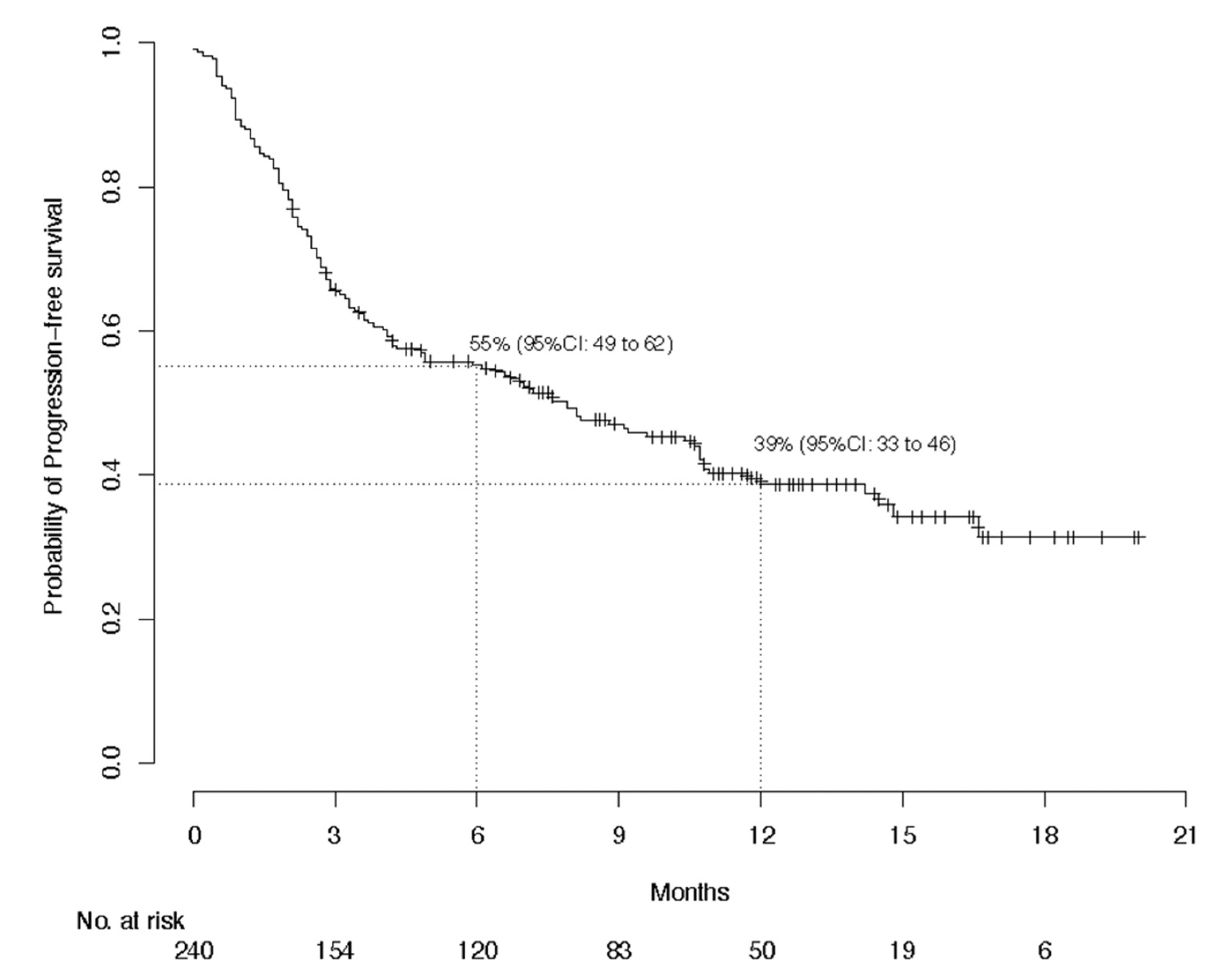
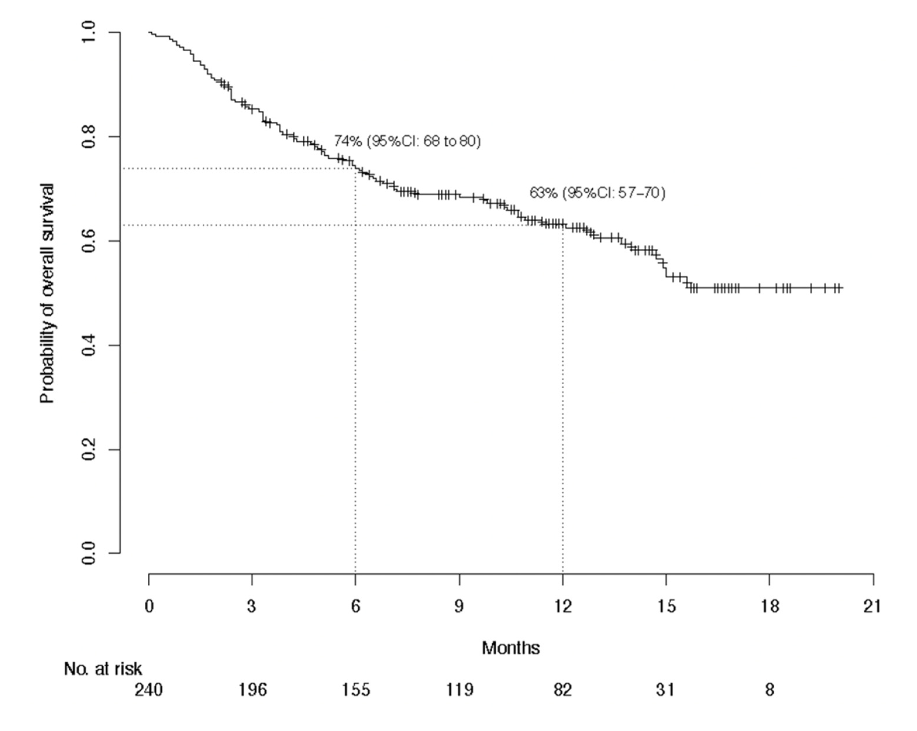
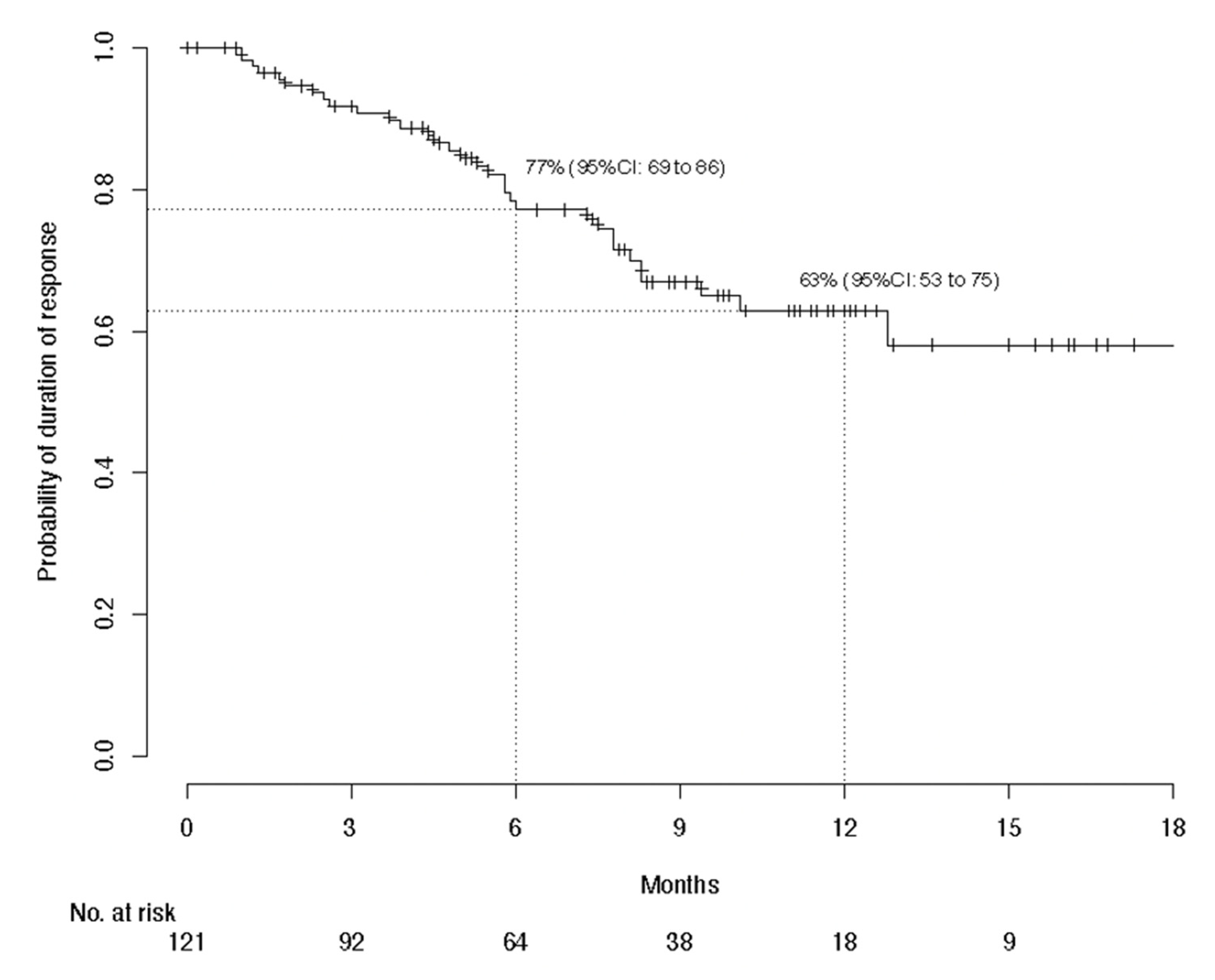
3.2. Efficacy Evaluation

Responses of the 240 assessable patients are detailed in Table 2: 21% complete responses and 29% partial responses, for a BOR of 50% (95% CI 44–57). Only 64% of responses were confirmed. The BORs did not differ according to immunocompromised versus immunocompetent status (50% versus 51%, respectively), with prior systemic treatment versus without (51% versus 50%, respectively; p = 0.9), or according to local, regional or distant disease (48%, 56% or 46%, respectively; p = 0.41). However, the BORs were lower for patients with PS ≥ 2 versus <2 (37% versus 56%, respectively; p = 0.01). Patients with chronic dermatitis tended to have poorer responses than those without (32% versus 52%, respectively; p = 0.06). The disease-control rate was 59.6% (95% CI 53.1–65.8).
The median time to complete response was 5.9 (range 1.7–13.6) months. Complete responders’ median treatment duration was 11.3 months (range 13–516 days), versus 7.5 months (range 43–595 days) for partial responders. The reasons for cemiplimab discontinuation were not fully available for these patients. Among the 51 complete responders, only three (6%) progressed during follow-up: two progressed on cemiplimab after 318 or 471 days of treatment and one progressed 3 months after stopping cemiplimab, which had been administered for 241 days (see Figure S1). A median of 61 days of follow-up were available for 27 (53%) complete responders after cemiplimab discontinuation: only one of them relapsed. At 1 year, relapses were significantly more frequent for partial responders (53%) than complete responders (9%) (p = 0.007).
With global median follow-up at 12.6 months, median PFS lasted 7.9 (95% CI, 4.9–10.7) months and 1-year PFS was 38.7%; median global OS was not reached and the 1-year OS was 63.1%; and median global DOR was not reached and the 1-year DOR rate was 62.9% (Figure 1, Figure 2 and Figure 3). The 1-year PFS and OS rates did not differ according to immune status or previous systemic treatment status (p > 0.21). However, their durations were significantly shorter for patients with PS ≥ 2 versus PS < 2, with respective estimated percentages (95% CI) of 25.1% (15.0–41.8%) and 43.5% (36.3–52.3%) (p < 0.0001) for PFS, and 36% (25–52%) and 73% (66–81%) (p < 0.0001) for OS. The highly significant impact of PS ≥ 2 on PFS and OS was confirmed during the first 6 months, after adjustment for age, sex, chronic dermatitis, primary CSCC site and disease stage (Table 3). After 6 months, PS was no longer associated with PFS or OS. Primary head-and-neck CSCC was also associated with a better PFS.

3.3. Adverse Events

One-third of the patients experienced treatment-related AEs (TRAEs; Table 4), with the most common being (in decreasing order): fatigue, arthralgias/myalgias, hepatic disorders, diarrhea and pruritus. They led to treatment discontinuation for 16 (7%) patients. Twenty-two patients experienced at least one grade-3 or higher TRAE, as detailed in Table 5. They were mostly hepatic disorders and fatigue, but also renal impairment, arthralgias/myalgias, and two kidney-transplant rejections. The death of one patient from toxic epidermal necrolysis (Lyell’s syndrome) was attributed to cemiplimab. A median of 6 (range 0–70) weeks separated cemiplimab onset and the first AE. The response rates for patients with TRAEs (54.7%) and those without (47.3%) did not differ significantly (p = 0.45).

4. Discussion

This retrospective study on 240 CSCC patients confirmed cemiplimab efficacy in the real-life setting as a curative treatment for unresectable, locally advanced or metastatic disease. Patients in this series share characteristics with the 193 patients enrolled in the phase II trial evaluating cemiplimab that led to its approval [24,26,28,29]: predominantly men, older age, 29% poorly differentiated tumors [26], and mostly head-and-neck primary locations. Unlike those study participants, our population included 24% immunocompromised patients, with 16% having blood disorders (i.e., chronic lymphocytic leukemia and other hemopathies), and 27% with PS ≥ 2. Notably, in our series, 49% had received systemic treatment before starting cemiplimab, versus 34% in the phase II trial [28], 3% had a genodermatosis and 11% had an underlying chronic dermatitis, most frequently chronic wounds. The BOR herein was 50%, including 21% complete responses, which is of the same order of magnitude as in other trials evaluating anti-PD-1 [24,25,26,27,28,29].
Our results suggest that immunocompromised patients, including those with blood disorders, respond and survive as well as immunocompetent patients, meaning they apparently benefit from anti-PD-1, despite usually being excluded from trials. However, management of these patients, particularly transplant recipients, must be extremely attentive so as to avoid rejection, as highlighted by the two kidney-transplant rejections observed herein; nonetheless, they should be included in trials. Indeed, anti-PD-1 increased the risk of graft rejection and, when rejection occurred, mortality was recently estimated at 36%, with a high risk for liver-transplant recipients [30]. An ongoing trial is evaluating the safety and efficacy of cemiplimab with everolimus/sirolimus plus prednisone or without as treatment for advanced CSCCs in kidney transplantees (NCT04339062).
Our findings also support that systemic treatment-naïve patients responded as well as pretreated patients. They also showed that frail patients with poor PS responded less well. However, because more than one-third of them responded to cemiplimab, anti-PD-1 should remain the first-line systemic treatment of choice. It is now critical to identify factors predictive of response in these frail patients. Our observations indicate that patients with underlying chronic dermatitis might respond less well to cemiplimab than patients without, but that outcome remains to be confirmed by a larger study.
Remarkably, our complete responders rarely relapsed (6%), even after stopping cemiplimab. Notably, although disease progression after an objective response was observed in 21% of responders, the risk of relapse was markedly higher for partial responders than complete responders, as previously reported for melanoma patients [31,32]. Determining responders’ factors predictive of relapse and optimal treatment duration for partial responders would contribute greatly to improving their management.
Although direct comparison is impossible, for our entire population, 1-year PFS (38.7%) and OS (63.1%) were substantially lower than in Migden et al.’s phase II study [24]. Indeed, their patients’ 1-year PFS and OS ranged between 47% and 58%, and 76% and 93%, respectively, according to the different patient subgroups [28,33]. One factor that could explain this difference would be our overestimation of the response rate, attributable to either the high frequency of unconfirmed responses or the lack of independent central review. Indeed, in Migden et al.’s phase II study [26], the response rate was overestimated by investigators (53%) compared to blinded independent central reviewers (44%), as recently demonstrated by the analysis of 20 trials that had central and investigators’ BOR assessments available [34]. However, our series’ BOR was very close to the investigators’ estimated response rate in Migden et al.’s trial [26].
Another hypothesis might be that our lower-than-expected PFS and OS might reflect our patients’ characteristics, i.e., 27% with PS ≥ 2, whose 1-year OS at 36% was significantly shorter, as reported for lung cancer [35], whereas that OS rate for patients with PS < 2 reached the lower threshold of the OS estimated in the cemiplimab phase II CSCC trial [28,33]. Our best model for OS included only PS, while our best model for PFS included PS and primary head-and-neck. Although it is difficult to attribute a protective effect to head-and-neck CSCCs, it can be hypothesized that the tumor mutational burden would be increased in CSCCs located at that site, a chronically sun-exposed area, compared to other cutaneous areas not chronically sun-exposed. Because high tumor mutational burden predicts prolonged survival in patients receiving anti PD-1 [36], such a higher burden might help explain the association between longer PFS and head-and-neck site retained by our multivariate analysis. Further molecular studies are needed to confirm this hypothesis. Notably, PFS and OS did not differ according to CSCC stage, prior systemic treatment status or immune status, thereby suggesting that it would be of interest to enroll immunocompromised patients in trials evaluating anti-PD-1.
The cemiplimab-safety profile for our series was comparable with that in other studies on PD-1–blocking agents to treat CSCC [24,26,37]. Most AEs were manageable, except for 16 (7% of the patients) that necessitated cemiplimab discontinuation. One cemiplimab-related death from toxic epidermal necrolysis occurred. About 20 Stevens–Johnson syndrome/toxic epidermal necrolysis cases have been reported with other inhibitors of PD-1 or its ligand [38,39,40,41,42,43,44,45,46,47,48,49,50,51,52,53,54,55]. Toxic epidermal necrolysis is responsible for high mortality [56]. According to the American Society of Clinical Oncology guidelines, cyclosporine or intravenous immunoglobulins combined with corticosteroids should be initiated when toxic epidermal necrolysis is diagnosed [57]. Indeed, with the increasing use of immune-checkpoint inhibitors, physicians should be aware of this very rare AE. Twenty-two (9%) of our patients developed severe grade-3 or -4 TRAEs, a rate consistent with previous studies on PD-1–blocking agents [24,25,26,50]. One early-onset cemiplimab-induced grade-4 drug reaction with eosinophilia and systemic symptoms with a favorable outcome occurred in a 76-year-old woman. Considering these frail patients, the safety profile seems acceptable.
Limitations of this study are its retrospective design, the lack of central assessment of disease response, the too short follow-up that precluded accurate determinations of OS, DOR, and the long-term outcomes of responders after stopping anti-PD-1. Indeed, longer follow-up would be helpful. Moreover, PFS results may not be very accurate because assessments were made according to standard of care and may have been performed at different timepoints.

5. Conclusions

The results of this retrospective study confirm cemiplimab’s strong anti-tumor activity and manageable safety, meaning it should be offered to patients with unresectable, locally advanced or metastatic CSCCs. Our analysis of the characteristics of CSCC patients who received cemiplimab in the real-life setting demonstrated the poor prognosis associated with PS ≥ 2. The association between head-and-neck involvement and longer PFS requires additional molecular prognostic studies to determine whether or not that site has a protective effect on PFS for patients with locally advanced or metastatic disease. Moreover, the results of this analysis indicate that cemiplimab might be beneficial for immunocompromised patients.

Supplementary Materials

The following are available online at https://www.mdpi.com/article/10.3390/cancers13143547/s1, Figure S1. The responses for each cemiplimab-recipient and the times at which they and other events occurred are reported. Table S1. Therapies given to the 245 intent-to-treat patients before cemiplimab.

Author Contributions

Conceptualization, S.D. (Sophie Dalac), L.M. (Laurent Mortier) and È.M.; formal analysis, M.B. and È.M.; investigation, C.H., L.F. and È.M.; methodology, M.B.; project administration, È.M.; resources, C.H., L.F., A.P.-L., F.H., P.C. (Philippe Celerier), F.A., N.B., M.D. (Monica Dinulescu), A.J., N.M., A.-B.D.-M., L.C., È.-M.N., É.A., B.D., C.L., C.B. (Clémence Berthin), N.K., F.G., J.d.Q., P.-E.S., N.P., J.-P.A., S.A., B.B., S.D. (Sophie Darras), V.H., S.D. (Suzanne Devaux), M.M., L.M. (Laurent Misery), S.M., M.E., F.B.-P., C.J., R.L., F.S., J.S., S.C., M.S., Y.T., D.S. (Dominique Spaeth), C.G.-M., O.C., R.T., M.P., M.D. (Marc Dumas), L.P., P.C. (Pierre Combe), O.L., P.G., Y.R., I.K.-B., D.S. (David Solub), A.S., C.B. (Christophe Bedane) and G.Q.; software, M.B.; supervision, È.M.; visualization, C.H., L.F. and È.M.; writing—review and editing, C.H., L.F., A.P.-L., M.B., F.H., P.C. (Philippe Celerier), F.A., N.B., M.D. (Monica Dinulescu), A.J., N.M., A.-B.D.-M., L.C., È.-M.N., É.A., B.D., C.L., C.B. (Clémence Berthin), N.K., F.G., J.d.Q., P.-E.S., N.P., J.-P.A., S.A., B.B., S.D. (Sophie Darras), V.H., S.D. (Suzanne Devaux), M.M., L.M. (Laurent Misery), S.M., M.E., F.B.-P., C.J., R.L., F.S., J.S., S.C., M.S., Y.T., D.S. (Dominique Spaeth), C.G.-M., O.C., R.T., M.P., M.D. (Marc Dumas), L.P., P.C. (Pierre Combe), O.L., P.G., Y.R., I.K.-B., D.S. (David Solub), A.S., C.B. (Christophe Bedane), G.Q., S.D. (Sophie Dalac), L.M. (Laurent Mortier) and È.M. All authors have read and agreed to the published version of the manuscript.

Funding

This research received no external funding.

Institutional Review Board Statement

The study was conducted in accordance with the guidelines of the Declaration of Helsinki, and approved by the Ethics Committee of AVICENNE HOSPITAL (protocol code CLEA-2019-75; date of approval 27 September 2019). The national database has been declared to the French data-protection agency (CNIL approval number 2215607).

Informed Consent Statement

In compliance with French law, consent regarding non-opposition to collect and use the data was obtained from each patient.

Data Availability Statement

Relevant data supporting the findings of this study are available within the article and Supplementary Materials and are available from the authors upon reasonable request.

Acknowledgments

The authors would like to thank Elisa Funck-Brentano from Hôpital Ambroise Paré, AP-HP, Boulogne, who included 3 cases in this study, for her contribution to this study. They would like to thank all members of the Groupe Français de Cancérologie and of the French Cutaneous Squamous Cell Carcinoma Study Group (CAREPI). They would like to thank other physicians involved in the study and the patients who participated in this early access program. Editorial assistance was provided by Janet Jacobson. The authors thank Margot Denis for technical assistance.

Conflicts of Interest

M. Samimi received fees from Janssen Cilag for speaking at an educational meeting and has received reimbursement for travel and accommodation expenses for attending national and international medical congresses from Abbvie, Amgen SAS, Bristol Myers Squibb, Celgene SAS, Galderma International, Lilly France SAS, MSD France. N. Meyer: Investigator and/or consultant and/or speaker and/or research grants from BMS, MSD, Roche, Novartis, Pierre Fabre, Merck, Sanofi, Sun Pharma. J.-P. Arnault: Novartis (boards), speaker (BMS and MSD). E. Maubec: Investigator and/or consultant and/or research grants from BMS, MSD, Novartis, Pierre Fabre, Sanofi. F. Herms reports receiving fees for consulting, advisory boards and travel accommodations for attending congresses from Sanofi et SUN Pharma. S. Mansard reports participation on boards and having received support for travel accommodations for attending congresses from Sanofi, Novartis, Pierre Fabre, MSD, BMS. G. Quereux reports receiving fees for consulting, advisory boards and being an investigator from Sanofi, Novartis, Pierre Fabre, MSD, Roche and BMS.

References

  1. Rogers, H.W.; Weinstock, M.A.; Feldman, S.R.; Coldiron, B.M. Incidence estimate of nonmelanoma skin cancer (keratinocyte carcinomas) in the U.S. population, 2012. JAMA Dermatol. 2015, 151, 1081–1086. [Google Scholar] [CrossRef] [PubMed]
  2. Stratigos, A.J.; Garbe, C.; Dessinioti, C.; Lebbe, C.; Bataille, V.; Bastholt, L.; Dréno, B.; Fargnoli, M.C.; Forsea, A.M.; Frenard, C.; et al. European interdisciplinary guideline on invasive squamous cell carcinoma of the skin: Part 1. epidemiology, diagnostics and prevention. Eur. J. Cancer 2020, 128, 60–82. [Google Scholar] [CrossRef] [PubMed] [Green Version]
  3. Maubec, E. Update of the management of cutaneous squamous-cell carcinoma. Acta Dermatol. Venereol. 2020, 100, adv00143. [Google Scholar] [CrossRef] [PubMed]
  4. Christensen, G.B.; Ingvar, C.; Hartman, L.W.; Olsson, H.; Nielsen, K. Sunbed use increases cutaneous squamous cell carcinoma risk in women: A large-scale, prospective study in Sweden. Acta Derm. Venereol. 2019, 99, 878–883. [Google Scholar] [CrossRef] [PubMed] [Green Version]
  5. Jensen, P.; Hansen, S.; Møller, B.; Leivestad, T.; Pfeffer, P.; Geiran, O.; Fauchald, P.; Simonsen, S. Skin cancer in kidney and heart transplant recipients and different long-term immunosuppressive therapy regimens. J. Am. Acad. Dermatol. 1999, 40, 177–186. [Google Scholar] [CrossRef]
  6. Garrett, G.L.; Blanc, P.D.; Boscardin, J.; Lloyd, A.A.; Ahmed, R.L.; Anthony, T.; Bibee, K.; Breithaupt, A.; Cannon, J.; Chen, A.; et al. Incidence of and risk factors for skin cancer in organ transplant recipients in the United States. JAMA Dermatol. 2017, 153, 296–303. [Google Scholar] [CrossRef] [Green Version]
  7. Rizvi, S.M.H.; Aagnes, B.; Holdaas, H.; Gude, E.; Boberg, K.M.; Bjørtuft, Ø.; Helsing, P.; Leivestad, T.; Møller, B.; Gjersvik, P. Long-term change in the risk of skin cancer after organ transplantation: A population-based nationwide cohort study. JAMA Dermatol. 2017, 153, 1270–1277. [Google Scholar] [CrossRef] [Green Version]
  8. Lindelöf, B.; Sigurgeirsson, B.; Gäbel, H.; Stern, R.S. Incidence of skin cancer in 5356 patients following organ transplantation. Br. J. Dermatol. 2000, 143, 513–519. [Google Scholar]
  9. Grulich, A.E.; van Leeuwen, M.T.; Falster, M.O.; Vajdic, C.M. Incidence of cancers in people with HIV/AIDS compared with immunosuppressed transplant recipients: A meta-analysis. Lancet 2007, 370, 59–67. [Google Scholar] [CrossRef]
  10. Faust, H.; Andersson, K.; Luostarinen, T.; Gislefoss, R.E.; Dillner, J. Cutaneous human papillomaviruses and squamous cell carcinoma of the skin: Nested case-control study. Cancer Epidemiol. Biomark. Prev. 2016, 25, 721–724. [Google Scholar] [CrossRef] [Green Version]
  11. Torchia, D.; Massi, D.; Caproni, M.; Fabbri, P. Multiple cutaneous precanceroses and carcinomas from combined iatrogenic/professional exposure to arsenic. Int. J. Dermatol. 2008, 47, 592–593. [Google Scholar] [CrossRef] [PubMed]
  12. Reed, W.B.; College, J., Jr.; Francis, M.J.O.; Zachariae, H.; Mohs, F.; Sher, M.A.; Sneddon, I.B. Epidermolysis bullosa dystrophica with epidermal neoplasms. Arch. Dermatol. 1974, 110, 894–902. [Google Scholar] [CrossRef] [PubMed]
  13. Anderson, D.E. An inherited form of large bowel cancer: Muir’s syndrome. Cancer 1980, 45, 1103–1107. [Google Scholar] [CrossRef]
  14. King, R.A.; Creel, D.; Cervenka, J.; Okoro, A.N.; Witkop, C.J. Albinism in Nigeria with delineation of new recessive oculocutaneous type. Clin. Genet. 1980, 17, 259–270. [Google Scholar] [CrossRef]
  15. Kraemer, K.H.; Lee, M.M.; Scotto, J. Xeroderma pigmentosum. Cutaneous, ocular, and neurologic abnormalities in 830 published cases. Arch. Dermatol. 1987, 123, 241–250. [Google Scholar] [CrossRef]
  16. Karia, P.S.; Jambusaria-Pahlajani, A.; Harrington, D.P.; Murphy, G.F.; Qureshi, A.A.; Schmults, C.D. Evaluation of American Joint Committee on Cancer, International Union Against Cancer, and Brigham and Women’s Hospital tumor staging for cutaneous squamous cell carcinoma. J. Clin. Oncol. 2014, 32, 327–334. [Google Scholar] [CrossRef] [Green Version]
  17. Leibovitch, I.; Huilgol, S.C.; Selva, D.; Hill, D.; Richards, S.; Paver, R. Cutaneous squamous cell carcinoma treated with Mohs micrographic surgery in Australia, I. Experience over 10 years. J. Am. Acad. Dermatol. 2005, 53, 253–260. [Google Scholar] [CrossRef] [PubMed]
  18. Varra, V.; Woody, N.M.; Reddy, C.; Joshi, N.P.; Geiger, J.; Adelstein, D.J.; Burkey, B.B.; Scharpf, J.; Prendes, B.; Lamarre, E.D.; et al. Suboptimal outcomes in cutaneous squamous cell cancer of the head and neck with nodal metastases. Anticancer Res. 2018, 38, 5825–5830. [Google Scholar] [CrossRef]
  19. Veness, M.J.; Morgan, G.J.; Palme, C.E.; Gebski, V. Surgery and adjuvant radiotherapy in patients with cutaneous head and neck squamous cell carcinoma metastatic to lymph nodes: Combined treatment should be considered best practice. Laryngoscope 2005, 115, 870–875. [Google Scholar] [CrossRef]
  20. Veness, M.J.; Palme, C.E.; Morgan, G.J. High-risk cutaneous squamous cell carcinoma of the head and neck: Results from 266 treated patients with metastatic lymph node disease. Cancer 2006, 106, 2389–2396. [Google Scholar] [CrossRef] [Green Version]
  21. Schmults, C.D.; Karia, P.S.; Carter, J.B.; Han, J.; Qureshi, A.A. Factors predictive of recurrence and death from cutaneous squamous cell carcinoma: A 10-year, single-institution cohort study. JAMA Dermatol. 2013, 149, 541–547. [Google Scholar] [CrossRef] [Green Version]
  22. Osterlind, A.; Hjalgrim, H.; Kulinsky, B.; Frentz, G. Skin cancer as a cause of death in Denmark. Br. J. Dermatol. 1991, 125, 580–582. [Google Scholar] [CrossRef]
  23. Karia, P.S.; Han, J.; Schmults, D. Cutaneous squamous cell carcinoma: Estimated incidence of disease, nodal metastasis, and deaths from disease in the United States, 2012. J. Am. Acad. Dermatol. 2013, 68, 957–966. [Google Scholar] [CrossRef]
  24. Migden, M.R.; Rischin, D.; Schmults, C.D.; Guminski, A.; Hauschild, A.; Lewis, K.D.; Chung, C.H.; Hernandez-Aya, L.; Lim, A.M.; Chang, A.L.S.; et al. PD-1 blockade with cemiplimab in advanced cutaneous squamous-cell carcinoma. N. Engl. J. Med. 2018, 379, 341–351. [Google Scholar] [CrossRef] [Green Version]
  25. Grob, J.J.; Gonzalez, R.; Basset-Seguin, N.; Vornicova, O.; Schachter, J.; Joshi, A.; Meyer, N.; Grange, F.; Piulats, J.M.; Bauman, J.R.; et al. Pembrolizumab monotherapy for recurrent or metastatic cutaneous squamous cell carcinoma: A single-arm phase II trial (KEYNOTE-629). J. Clin. Oncol. 2020, 38, 2916–2925. [Google Scholar] [CrossRef]
  26. Migden, M.R.; Khushalani, N.I.; Chang, A.L.S.; Lewis, K.D.; Schmults, C.D.; Hernandez-Aya, L.; Meier, F.; Schadendorf, D.; Guminski, A.; Hauschild, A.; et al. Cemiplimab in locally advanced cutaneous squamous cell carcinoma: Results from an open-label, phase 2, single-arm trial. Lancet Oncol. 2020, 21, 294–305. [Google Scholar] [CrossRef]
  27. Maubec, E.; Boubaya, M.; Petrow, P.; Beylot-Barry, M.; Basset-Seguin, N.; Deschamps, L.; Grob, J.J.; Dréno, B.; Scheer-Senyarich, I.; Bloch-Queyrat, C.; et al. Phase II study of pembrolizumab as first-line, single-drug therapy for patients with unresectable cutaneous squamous cell carcinomas. J. Clin. Oncol. 2020, 38, 3051–3061. [Google Scholar] [CrossRef]
  28. Rischin, D.; Khushalani, N.I.; Schmults, C.D.; Guminski, A.D.; Chang, A.L.; Lewis, K.D.; Lim, A.M.; Hernandez-Aya, L.F.; Hughes, B.G.M.; Schadendorf, D.; et al. Phase II study of cemiplimab in patients (pts) with advanced cutaneous squamous cell carcinoma (CSCC): Longer follow-up. J. Clin. Oncol. 2020, 38, 10018. [Google Scholar] [CrossRef]
  29. Rischin, D.; Migden, M.R.; Lim, A.M.; Schmults, C.D.; Khushalani, N.I.; Hughes, B.G.M.; Schadendorf, D.; Dunn, L.A.; Hernandez-Aya, L.; Chang, A.L.S.; et al. Phase 2 study of cemiplimab in patients with metastatic cutaneous squamous cell carcinoma: Primary analysis of fixed-dosing, long-term outcome of weight-based dosing. J. Immunother. Cancer 2020, 8, e000775. [Google Scholar] [CrossRef]
  30. Nguyen, L.S.; Ortuno, S.; Lebrun-Vignes, B.; Johnson, D.B.; Moslehi, J.J.; Hertig, A.; Salem, J.E. Transplant rejections associated with immune checkpoint inhibitors: A pharmacovigilance study and systematic literature review. Eur. J. Cancer 2021, 148, 36–47. [Google Scholar] [CrossRef] [PubMed]
  31. Robert, C.; Ribas, A.; Schachter, J.; Arance, A.; Grob, J.J.; Mortier, L.; Daud, A.; Carlino, M.S.; McNeil, C.M.; Lotem, M.; et al. Pembrolizumab versus ipilimumab in advanced melanoma (KEYNOTE-006): Post-hoc 5-year results from an open-label, multicentre, randomised, controlled, phase 3 study. Lancet Oncol. 2019, 20, 1239–1251. [Google Scholar] [CrossRef]
  32. Jansen, Y.J.L.; Rozeman, E.A.; Mason, R.; Goldinger, S.M.; Geukes Foppen, M.H.; Hoejberg, L.; Schmidt, H.; van Thienen, J.V.; Haanen, J.B.A.G.; Tiainen, L.; et al. Discontinuation of anti-PD-1 antibody therapy in the absence of disease progression or treatment limiting toxicity: Clinical outcomes in advanced melanoma. Ann. Oncol. 2019, 30, 1154–1161. [Google Scholar] [CrossRef] [PubMed] [Green Version]
  33. Libtayo 350 mg Concentrate Solution for Infusion—Summary of Product Characteristics (SmPC)—(emc) [Internet]. Available online: https://www.medicines.org.uk/emc/product/10438 (accessed on 31 March 2021).
  34. Dello Russo, C.; Cappoli, N.; Navarra, P. A comparison between the assessments of progression-free survival by local investigators versus blinded independent central reviews in phase III oncology trials. Eur. J. Clin. Pharmacol. 2020, 76, 1083–1092. [Google Scholar] [CrossRef]
  35. Matsubara, T.; Seto, T.; Takamori, S.; Fujishita, T.; Toyozawa, R.; Ito, K.; Yamaguchi, M.; Okamoto, T. Anti-PD-1 monotherapy for advanced NSCLC patients with older age or those with poor performance status. Oncol. Targets Ther. 2021, 14, 1961–1968. [Google Scholar] [CrossRef]
  36. Samstein, R.M.; Lee, C.H.; Shoushtari, A.N.; Hellmann, M.D.; Shen, R.; Janjigian, Y.Y.; Barron, D.A.; Zehir, A.; Jordan, E.J.; Omuro, A.; et al. Tumor mutational load predicts survival after immunotherapy across multiple cancer types. Nat. Genet. 2019, 51, 202–206. [Google Scholar] [CrossRef]
  37. Wang, Y.; Zhou, S.; Yang, F.; Qi, X.; Wang, X.; Guan, X.; Shen, C.; Duma, N.; Vera Aguilera, J.; Chintakuntlawar, A.; et al. Treatment-related adverse events of PD-1 and PD-L1 inhibitors in clinical trials: A systematic review and meta-analysis. JAMA Oncol. 2019, 5, 1008–1019. [Google Scholar] [CrossRef]
  38. Nayar, N.; Briscoe, K.; Penas, P.F. Toxic epidermal necrolysis-like reaction with severe satellite cell necrosis associated with nivolumab in a patient with ipilimumab refractory metastatic melanoma. J. Immunother. 2016, 39, 149–152. [Google Scholar] [CrossRef]
  39. Saw, S.; Lee, H.Y.; Ng, Q.S. Pembrolizumab-induced Stevens-Johnson syndrome in non-melanoma patients. Eur. J. Cancer 2017, 81, 237–239. [Google Scholar] [CrossRef]
  40. Demirtas, S.; El Aridi, L.; Acquitter, M.; Fleuret, C.; Plantin, P. Toxic epidermal necrolysis due to anti-PD1 treatment with fatal outcome. Ann. Dermatol. Venereol. 2017, 144, 65–66. [Google Scholar] [CrossRef]
  41. Vivar, K.L.; Deschaine, M.; Messina, J. Epidermal programmed cell death-ligand 1 expression in TEN associated with nivolumab therapy. J. Cutan. Pathol. 2017, 44, 381–384. [Google Scholar] [CrossRef] [PubMed]
  42. Rouyer, L.; Bursztejn, A.C.; Charbit, L.; Schmutz, J.L.; Moawad, S. Stevens-Johnson syndrome associated with radiation recall dermatitis in a patient treated with nivolumab. Eur. J. Dermatol. 2018, 28, 380–381. [Google Scholar] [CrossRef] [PubMed]
  43. Shah, K.M.; Rancour, E.A.; Al-Omari, A.; Rahnama-Moghadam, S. Striking enhancement at the site of radiation for nivolumab-induced Stevens-Johnson syndrome. Dermatol. Online J. 2018, 24. [Google Scholar] [CrossRef]
  44. Haratake, N.; Tagawa, T.; Hirai, F.; Toyokawa, G.; Miyazaki, R.; Maehara, Y. Stevens-Johnson syndrome induced by pembrolizumab in a lung cancer patient. J. Thorac. Oncol. 2018, 13, 1798–1799. [Google Scholar] [CrossRef] [Green Version]
  45. Chirasuthat, P.; Chayavichitsilp, P. Atezolizumab-induced Stevens-Johnson syndrome in a patient with non-small cell lung carcinoma. Case Rep. Dermatol. 2018, 10, 198–202. [Google Scholar] [CrossRef]
  46. Griffin, L.L.; Cove-Smith, L.; Alachkar, H.; Radford, J.A.; Brooke, R.; Linton, K.M. Toxic epidermal necrolysis (TEN) associated with the use of nivolumab (PD-1 inhibitor) for lymphoma. JAAD Case Rep. 2018, 4, 229–231. [Google Scholar] [CrossRef] [Green Version]
  47. Salati, M.; Pifferi, M.; Baldessari, C.; Bertolini, F.; Tomasello, C.; Cascinu, S.; Barbieri, F. Stevens-Johnson syndrome during nivolumab treatment of NSCLC. Ann. Oncol. 2018, 29, 283–284. [Google Scholar] [CrossRef]
  48. Hwang, A.; Iskandar, A.; Dasanu, C.A. Stevens-Johnson syndrome manifesting late in the course of pembrolizumab therapy. J. Oncol. Pharm. Pract. 2019, 25, 1520–1522. [Google Scholar] [CrossRef]
  49. Dasanu, C.A. Late-onset Stevens-Johnson syndrome due to nivolumab use for hepatocellular carcinoma. J. Oncol. Pharm. Pract. 2019, 25, 2052–2055. [Google Scholar] [CrossRef]
  50. Cohen, E.E.W.; Soulières, D.; Le Tourneau, C.; Dinis, J.; Licitra, L.; Ahn, M.J.; Soria, A.; Machiels, J.P.; Mach, N.; Mehra, R.; et al. Pembrolizumab versus methotrexate, docetaxel, or cetuximab for recurrent or metastatic head-and-neck squamous cell carcinoma (KEYNOTE-040): A randomised, open-label, phase 3 study. Lancet 2019, 393, 156–167. [Google Scholar] [CrossRef]
  51. Cai, Z.R.; Lecours, J.; Adam, J.P. Toxic epidermal necrolysis associated with pembrolizumab. J. Oncol. Pharm. Pract. 2020, 26, 1259–1265. [Google Scholar] [CrossRef] [PubMed]
  52. Keerty, D.; Koverzhenko, V.; Belinc, D.; LaPorta, K.; Haynes, E. Immune-mediated toxic epidermal necrolysis. Cureus 2020, 12, e9587. [Google Scholar] [CrossRef]
  53. Cassaday, R.D.; Garcia, K.A.; Fromm, J.R. Phase 2 study of pembrolizumab for measurable residual disease in adults with acute lymphoblastic leukemia. Blood Adv. 2020, 4, 3239–3245. [Google Scholar] [CrossRef]
  54. Riano, I.; Cristancho, C.; Treadwell, T. Stevens-Johnson syndrome-like reaction after exposure to pembrolizumab and recombinant zoster vaccine in a patient with metastatic lung cancer. J. Investig. Med. High Impact Case Rep. 2020, 8. [Google Scholar] [CrossRef] [PubMed] [Green Version]
  55. Maloney, N.J.; Ravi, V.; Cheng, K.; Bach, D.Q.; Worswick, S. Stevens-Johnson syndrome and toxic epidermal necrolysis-like reactions to checkpoint inhibitors: A systematic review. Int. J. Dermatol. 2020, 59, e183–e188. [Google Scholar] [CrossRef]
  56. Dodiuk-Gad, R.P.; Chung, W.H.; Valeyrie-Allanore, L.; Shear, N.H. Stevens-Johnson syndrome and toxic epidermal necrolysis: An update. Am. J. Clin. Dermatol. 2015, 16, 475–493. [Google Scholar] [CrossRef]
  57. Brahmer, J.R.; Lacchetti, C.; Schneider, B.J.; Atkins, M.B.; Brassil, K.J.; Caterino, J.M.; Chau, I.; Ernstoff, M.S.; Gardner, J.M.; Ginex, P.; et al. Management of immune-related adverse events in patients treated with immune checkpoint inhibitor therapy: American Society of Clinical Oncology Clinical Practice Guideline. J. Clin. Oncol. 2018, 36, 1714–1768. [Google Scholar] [CrossRef]
Figure 1. Kaplan–Meier estimations of the 6-month and 1-year probabilities of progression-free survival for the per-protocol population (n = 240).
Cancers 13 03547 g001
Figure 2. Kaplan–Meier estimations of the 6-month and 1-year probabilities of overall survival for the per-protocol population (n = 240).
Cancers 13 03547 g002
Figure 3. Kaplan–Meier estimations of the 6-month and 1-year probabilities of duration of response after cemiplimab treatment for the per-protocol population (n = 240).
Cancers 13 03547 g003
Table 1. Baseline characteristics of all 245 intent-to-treat CSCC patients.
CharacteristicValue
Age, years77.1 ± 13.3
Male sex178 (73)
ECOG performance status
060 (25)
1118 (48)
≥266 (27)
Unknown1 (0.4)
Immunocompromised59 (24)
Human immunodeficiency virus-positive8 (3)
Organ transplant7 (3)
Chronic lymphocytic leukemia20 (8)
Other blood disorders a18 (7)
Immunosuppressive drugs6 (3)
Genodermatosis8 (3)
Inherited epidermolysis bullosa2 (0.8)
Muir–Torre syndrome2 (0.8)
Xeroderma pigmentosum1 (0.4)
Ichthyosis2 (0.8)
Epidermodysplasia verruciformis1 (0.4)
Chronic dermatitis28 (11)
Burns4 (1.6)
Scars2 (0.8)
Lichen planus2 (0.8)
Chronic wounds9 (4)
Warts/condylomas4 (1.6)
Arsenic keratosis2 (0.8)
Radiodermatitis3 (1.2)
Others b2 (0.8)
≥3 primary CSCCs80 (33)
Primary CSCC site
Head-and-neck c164 (70)
Trunk9 (4)
Anorectal and/or genital12 (5)
Arm or leg58 (24)
Unknown3 (1.2)
Histopathological characteristics
Poor differentiation57 (23)
Perineural invasion26 (11)
Both9 (4)
None69 (28)
Unknown84 (34)
CSCC stage
Localized85 (35)
Regional95 (39)
Distant metastases64 (26)
Unknown1 (0.4)
Results are expressed as mean ± standard deviation or number (%). ECOG, Eastern Cooperative Oncology Group; CSCC, cutaneous squamous-cell carcinoma. a Other blood disorders included: polycythemia vera, four; Waldenström’s macroglobulinemia, three; two each: mantle-cell lymphoma or myelodysplastic syndrome; one each: large B-cell lymphoma, cutaneous T-cell lymphoma, essential thrombocythemia, multiple myeloma associated with amyloid light-chain amyloidosis, IgM monoclonal gammopathy, thrombopenia of unspecified cause or idiopathic CD4 lymphocytopenia. b Carcinomas due to phototherapy or erosive pustular dermatosis of the scalp. c Including two CSCCs located on the lips.
Table 2. Best overall responses (n = 240) as assessed by investigators.
Outcomen (%)
Complete response51 (21)
Confirmed36 (15)
Unconfirmed15 (6)
Partial response70 (29)
Confirmed41 (17)
Unconfirmed29 (12)
Stable disease22 (9)
Progressive disease84 (35)
Not assessable13 (5)
Best overall response rate, n (% [95% CI])121 (50.4 [43.9–56.9])
Confirmed77 (32)
Unconfirmed44 (18)
Best overall disease control rate, n (% [95% CI])143 (59.6 [53.1–65.8])
Results are expressed as number (%), unless stated otherwise.
Table 3. Factors associated with progression-free survival or overall survival in univariate and multivariate analyses.
FactorUnivariateMultivariate
HR (95% CI)pHR (95% CI)p
Progression-free survival
Age1.00 (0.98–1.01)0.621.00 (0.98–1.01)0.63
Male sex0.79 (0.55–1.15)0.220.91 (0.61–1.37)0.66
Immunocompromised1.03 (0.7–1.51)0.891.15 (0.76–1.76)0.5
ECOG PS ≥ 2
≤6 months2.3 (1.53–3.44)<0.00012.33 (1.52–3.55)0.0001
6 months0.88 (0.31–2.51)0.810.85 (0.3–2.46)0.77
Chronic dermatitis1.67 (1.02–2.71)0.041.07 (0.61–1.87)0.8
Primary head-or-neck CSCC0.58 (0.41–0.81)0.00020.52 (0.34–0.79)0.0025
Localized disease1.16 (0.82–1.64)0.410.72 (0.49–1.05)0.09
Previous systemic treatment0.88 (0.62–1.23)0.441.03 (0.71–1.50)0.88
Overall survival
Age1.00 (0.99–1.02)0.810.99 (0.98–1.01)0.46
Male sex0.9 (0.56–1.44)0.661.01 (0.61–1.67)0.97
Immunocompromised0.82 (0.49–1.35)0.430.91 (0.53–1.56)0.72
ECOG PS ≥ 2
≤6 months4.39 (2.62–7.33)<0.00014.56 (2.64–7.85)0.0001
>6 months1.61 (0.61–4.27)0.341.69 (0.63–4.52)0.3
Chronic dermatitis0.98 (0.49–1.95)0.950.7 (0.32–1.51)0.36
Primary head-or-neck CSCC0.76 (0.49–1.18)0.220.67 (0.4–1.13)0.13
Localized disease1.02 (0.66–1.58)0.940.74 (0.45–1.2)0.22
Previous systemic treatment0.76 (0.5–1.17)0.211.09 (0.68–1.76)0.72
ECOG PS, Eastern Cooperative Oncology Group performance status; CSCC, cutaneous squamous-cell carcinoma.
Table 4. Each cemiplimab-related adverse event occurred in at least two of the 240 treated patients.
Adverse EventAny GradeGrade ≥ 3
Any75 (31)22 (9)
Led to cemiplimab discontinuation16 (7)12 (5)
Fatigue21 (9)4 (2)
Arthralgias/myalgias17 (7)2 (1)
Cholestasis/cytolysis/hepatitis10 (4)5 (2)
Diarrhea7 (3)0
Pruritus6 (3)0
Rash5 (2)0
Hypothyroidism5 (2)0
Renal failure5 (2)3 (1)
Hyperthyroidism4 (2)0
Lymphopenia3 (1)0
Decreased appetite3 (1)1 (0.4)
Peripheral neuropathy3 (1)0
Anemia2 (1)0
Neutropenia2 (1)0
Myocarditis2 (1)1 (0.4)
Corticotropic insufficiency2 (1)0
Colitis2 (1)2 (1)
Vomiting2 (1)1 (0.4)
Loss of weight2 (1)0
Balance disorder2 (1)0
Transplant rejection2 (1)2 (1)
Results are expressed as number (%).
Table 5. Serious cemiplimab-related adverse events in the 240 treated patients.
Adverse EventSeverity Grade
AnyGrade 3Grade 4Grade 5
Cholestasis/cytolysis/hepatitis5 (2)320
Fatigue4 (2)400
Renal impairment3 (1)210
Arthralgias/myalgias2 (1)200
Colitis2 (1)200
Transplant rejection2 (1)110
Decreased appetite1 (0.4)100
Myocarditis1 (0.4)100
Vomiting1 (0.4)100
Acute pancreatitis1 (0.4)100
Interstitial lung disease1 (0.4)100
Drug reaction with eosinophilia and systemic symptoms1 (0.4)010
Toxic epidermal necrolysis1 (0.4)001
Results are expressed as number (%) or number.
Publisher’s Note: MDPI stays neutral with regard to jurisdictional claims in published maps and institutional affiliations.

