Simple Summary
Metabolic reprogramming is required for both malignant transformation and tumor development, including invasion and metastasis. Melatonin (5-methoxy-N-acetyltryptamine) is a methoxyindole that is synthesized in the pineal gland. Importantly, melatonin has anticancer effects by stimulating apoptosis, regulation of survival signaling, suppression of metastasis and angiogenesis and regulation of epigenetic modifications that contribute to malignant transformation. Furthermore, melatonin affects steps associated with the Warburg phenotype and suppresses the switch from oxidative phosphorylation to aerobic glycolysis through the regulation of critical enzymes and glucose transporters. Melatonin is involved in regulation of p53 and HIF-1, directly participate in signaling cascades that modulate aerobic glycolysis, gluconeogenesis, the tricarboxylic acid cycle and the pentose phosphate pathway. A significant impact of melatonin in the modulation of metabolic cascades represent a unique opportunity to inhibit pathways metabolic reprogramming.
Abstract
Metabolic reprogramming characterized by alterations in nutrient uptake and critical molecular pathways associated with cancer cell metabolism represents a fundamental process of malignant transformation. Melatonin (N-acetyl-5-methoxytryptamine) is a hormone secreted by the pineal gland. Melatonin primarily regulates circadian rhythms but also exerts anti-inflammatory, anti-depressant, antioxidant and anti-tumor activities. Concerning cancer metabolism, melatonin displays significant anticancer effects via the regulation of key components of aerobic glycolysis, gluconeogenesis, the pentose phosphate pathway (PPP) and lipid metabolism. Melatonin treatment affects glucose transporter (GLUT) expression, glucose-6-phosphate dehydrogenase (G6PDH) activity, lactate production and other metabolic contributors. Moreover, melatonin modulates critical players in cancer development, such as HIF-1 and p53. Taken together, melatonin has notable anti-cancer effects at malignancy initiation, progression and metastasing. Further investigations of melatonin impacts relevant for cancer metabolism are expected to create innovative approaches supportive for the effective prevention and targeted therapy of cancers.
1. Introduction
Tumor cell metabolism is characteristically different from that of healthy cells [1]. The ability of cancer cells to modify their metabolism and adapt to nutrient-deprived environments to salvage nutrients and thus build biomass and accelerate proliferation is a well-known feature of malignant transformation [2]. Metabolic reprogramming of cancer cells is a hallmark of tumor development. Metabolic changes in tumor cells are driven by oncogenic mutations, hypoxic conditions, altered molecular signals that upregulate anabolic processes and the inhibition of catabolic cascades [1,3]. Changes in specific pathways, including glycolysis, gluconeogenesis, glutaminolysis, the pentose phosphate pathway (PPP), mitochondrial biogenesis and lipid metabolism, contribute to tumor development, invasion and metastasis [4]. Melatonin (C13H16N2O2; PubChem CID: 896; Available from: https://pubchem.ncbi.nlm.nih.gov/compound/Melatonin; (cited 20 April 2021)), a hormone secreted by the pineal gland, contributes to the regulation of circadian rhythms. Since its discovery (more than 60 years ago), melatonin has been extensively investigated in preclinical and clinical research [5]. Clinically, melatonin is used to manage sleep disorders, jetlag, depressive symptoms and anxiety [6,7]. Importantly, melatonin is a strong antioxidant and can protect organisms from carcinogenesis and neurodegeneration [7].
Moreover, melatonin has oncostatic effects by stimulating apoptosis, regulation of survival signaling, suppression of metastasis and angiogenesis and on the epigenetic machinery contributing to the malignant transformation demonstrated in vitro and in vivo [8,9,10,11]. Significantly, melatonin can attenuate the metabolic reprogramming of cancer cells [12,13]. Indeed, melatonin exerts a wide range of different effects, and its functional chemical groups play a key role in the induced oncostatic properties. This is illustrated by the chemical background, where melatonin belongs to the group of acetamides that is acetamide in which one of the hydrogens joined to the nitrogen atom is substituted by a 2-(5-methoxy-1H-indol-3-yl)ethyl group [14]. Acetamides have previously been reported to exhibit anticancer activities [15]. While experimental research has suggested a broad spectrum of melatonin’s anticancer abilities, the hormone’s impact on cancer metabolism requires further investigation. Understanding the processes behind melatonin’s effects on tumor metabolism can support the introduction of new therapeutic strategies to improve quality of life and prolong the overall survival of cancer patients in the context of preventive, predictive and personalized medicine.
1.1. Aim of the Study
This comprehensive review evaluates the effects of melatonin on cancer metabolism. Metabolic reprogramming of cancer cells promotes accelerated proliferation, acquisition of an invasive phenotype, metastasis and chemo/radio resistance development. The core of this manuscript focuses on melatonin’s role in the regulation of metabolic pathways in vitro and in vivo. Based on positive results from preclinical research, we emphasize a need to implement melatonin in the clinical sphere to attenuate metabolic transformations in tumor cells.
1.2. Source of the Data
Relevant data were collected from the biomedical literature using “melatonin“ and “cancer“ or “cancer metabolism“ or “aerobic glycolysis“ or “Warburg effect“ or “gluconeogenesis“ or “pentose phosphate pathway“ or “lipid metabolism“ or other associated terms as keywords or medical subject heading (MeSH) terms for searches in the PubMed database (https://pubmed.ncbi.nlm.nih.gov/ (data collected from January to April 2021)). We have focused on the recent publications from the years 2016–2021.
2. Structural and Functional Aspects of Melatonin
Melatonin, or 5-methoxy-N-acetyltryptamine, is a methoxyindole discovered in 1958 that is synthesized in the pineal gland (Figure 1) [16]. Melatonin is synthesized from tryptophan and secreted during the night (dark) phase of the day. Its secretion and synthesis are inhibited during the light phase of the day [7]. Tryptophan, an essential amino acid for melatonin synthesis, is hydroxylated into 5-hydroxytryptophan by tryptophan hydroxylase (TPH). 5-hydroxytryptophan is then converted to serotonin by 5-hydroxytryptophan decarboxylase [17]. Arylalkylamine N-acetyltransferase (AANAT) acetylates serotonin to form acetyl-serotonin and serves as a rate-limiting enzyme that regulates the rhythmic synthesis of melatonin [18,19]. Acetyl-serotonin is then converted to melatonin by acetylserotonin-O-methyltransferase (ASMT). Importantly, AANAT activity depends on cyclic AMP (cAMP) production. Light deficiency leads to norepinephrine release from sympathetic nerve fibers, resulting in cAMP synthesis [20]. Synthesized melatonin is released from the pineal gland into circulation [21]. Even though it is mainly produced in the pineal gland, melatonin is also produced elsewhere. Melatonin production occurs in various other tissues; however, these processes occur independently of circadian rhythms, and the synthesized melatonin is not released into circulation. Therefore, melatonin exists in two pools with different functions [7,22]. Melatonin regulates circadian rhythms, and the suprachiasmatic nucleus (SCN) regulates its circadian release. The SCN receives photic information about the environmental day/night cycle via the retinohypothalamic tract (RHT); melatonin biosynthesis occurs in the absence of light. Concurrently, melatonin controls SCN activity via feedback to its receptors (MT1 and MT2) in the SCN [23,24]. Dysregulation of melatonin-related pathways leads to sleep disorders and various health problems [25]. Moreover, melatonin exerts antioxidant and anti-inflammatory effects [26]. Its antioxidant role is associated with the neutralization of reactive nitrogen (RNS) and oxygen (ROS) species that affect the normal function of cells. Free radical accumulation due to disturbed oxidant-antioxidant machinery results in numerous pathological conditions [27,28]. Recent evidence suggests that melatonin treatment increases superoxide dismutase activity (SOD) and other antioxidant enzymes [29]. Moreover, melatonin stimulates an immune response through its receptors [30]. The immunoregulatory effects of melatonin are mediated by the stimulation of cytokines and acceleration of the T helper immune response. Melatonin promotes the production of interleukins (IL)-1, -6 and -12 by monocytes [31]. Moreover, melatonin supports antigen presentation by macrophages to T cells, resulting in cytotoxic T cell activation and proliferation [32]. Additionally, melatonin contributes to blood pressure regulation and autonomic control of cardiovascular function and has protective roles in various cardiovascular diseases [33]. Several studies reveal that melatonin inhibits carcinogenesis through various mechanisms [8,34,35,36,37]. Melatonin’s anticancer effects include pro-apoptotic [38], antiproliferative [39] and anti-angiogenic activities [10,40,41]. Moreover, melatonin exerts a tumor-suppressive capacity through the modulation of free radical scavenger action and immunoregulation via the activation of anticancer immune cells and the attenuation of T-regulatory cells (Tregs) and cancer-associated fibroblasts (CAF) [35,42].
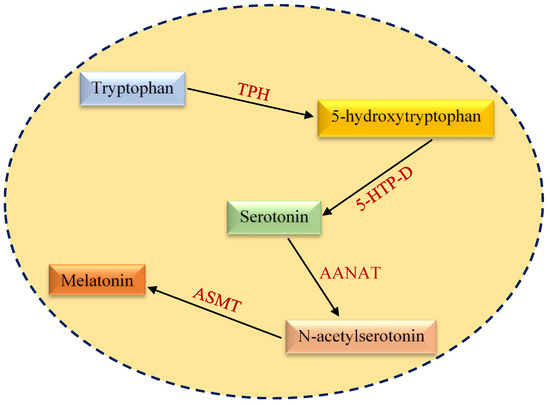
Figure 1.
An overview of melatonin biosynthesis pathway. TPH, tryptophan hydroxylase; 5-HTP-D, 5-hydroxytryptophan decarboxylase; AANAT, arylalkylamine N-acetyltransferase; ASMT, acetylserotonin O-methyltransferase.
2.1. Aberrations in Cancer Metabolism
Cancer cells are characterized by metabolic transformation, migration and uncontrolled proliferation [43]. Bioenergetic changes in tumors include the acceleration of glycolysis, elevation of glutaminolytic flux, enhancement of mitochondrial biogenesis, stimulation of the PPP and biosynthesis of macromolecules [4]. Alterations in glucose metabolism are essential features of cancer transformation; altogether, they constitute a shift from oxidative phosphorylation (OXPHOS) to glycolysis, even under normoxic conditions. This phenomenon is known as aerobic glycolysis or the Warburg effect [44,45]. Even though it is less energy efficient than OXPHOS (producing only 2 ATP molecules per glucose), aerobic glycolysis enables faster ATP production. Moreover, the metabolic switch from OXPHOS to aerobic glycolysis leads to the production of many intermediates, which are funneled into metabolic cascades for the generation of nucleotides, amino acids, NADPH and lipids [46]. Cellular glucose uptake is regulated by the functional glucose transporter (GLUT) family [47]. Increases in GLUT-mediated glucose uptake are characteristic of various tumor types, including breast cancer [48], prostate cancer [49], oral squamous cell carcinoma [50] and esophageal cancer [51]. Additionally, the transition from OXPHOS to the Warburg phenotype is promoted by the elevated expression of key glycolytic enzymes, such as hexokinase 2 (HK2), phosphofructokinase-1 (PFK1), lactate dehydrogenase A (LDHA) and pyruvate kinase type M2 (PKM2), which are associated with neoplastic transformation [52,53,54,55]. Changes in molecular cascades such as the phosphoinositide 3-kinase/Akt/mammalian target of rapamycin (PI3K/Akt/mTOR) pathway, upregulation of hypoxia-inducible factor 1 (HIF-1) and c-MYC, insufficient p53-mediated control and epigenetic mechanisms all contribute to the deregulation of glycolytic enzymes [56,57].
Gluconeogenesis is responsible for generating glucose from non-carbohydrate carbon precursors such as pyruvate, lactate, propionate and glycerol [58,59]. Seven enzymes are shared between glycolysis and gluconeogenesis. Furthermore, there are four enzymes unique to gluconeogenesis: pyruvate carboxylase, which converts pyruvate to oxaloacetate (OAA); phosphoenolpyruvate carboxykinase (PCK), which catalyzes the conversion of OAA to phosphoenolpyruvate (PEP); fructose-1,6-bisphosphatase (FBPase), which converts fructose 1,6-bisphosphate (F1,6P) to fructose 6-phosphate (F6P); and glucose-6-phosphatase (G6Pase), which hydrolyzes glucose-6-phosphate [58]. In cancer cells, gluconeogenesis generates intermediate metabolites necessary for biomolecule synthesis, especially during glucose deprivation [58]. Moreover, the key enzymes of gluconeogenesis (PCK, FBPase, G6Pase) affect cell survival, signaling and proliferation, as well as cancer stem cell (CSC) phenotypes [60]. Several studies revealed that cytoplasmatic PCK1 and mitochondrial PCK2 contribute to cancer growth [61,62,63]. Additionally, FBP1 deficiency was documented in lung [64], breast [65] and renal cancers [66].
The tricarboxylic acid (TCA) cycle is a central hub of oxidative metabolism, synthesis of macromolecules and redox balance [67]. The TCA cycle is a series of enzyme-catalyzed biochemical reactions [68]. Deficiencies in succinate dehydrogenase (SDH), fumarate hydratase (FH) and isocitrate dehydrogenase (IDH) due to mutations (inherited or acquired) result in metabolic changes [69]. The accumulation of succinate and fumarate due to defects in SDH and FH leads to the inhibition of prolyl hydroxylase enzymes (PHD), stabilization of HIF-1α and subsequent acceleration of glycolysis in cancer cells [70]. In contrast, the accumulation of α-ketoglutarate results in the destabilization of HIF-1α [71]. Moreover, alterations of c-MYC, P53 or RAS may modulate the TCA cycle [72].
The PPP is an important cascade in glucose metabolism [73]. The PPP is responsible for the generation of the reduced form of nicotinamide adenine dinucleotide phosphate (NADPH) and the production of ribose 5-phosphate (R5P) that is necessary for nucleotide synthesis [74]. Glucose-6-phosphate (G6P) is a major precursor that enters the PPP from glycolysis. The PPP consists of an oxidative and a non-oxidative phase [75]. Glucose-6-phosphate dehydrogenase (G6PDH), 6-phosphogluconolactonase (6PGL) and 6-phosphogluconate dehydrogenase (6PGDH) are crucial for the synthesis of ribulose-5 phosphate and NADPH through the oxidative phase [76]. The non-oxidative phase is characterized by a series of non-oxidative reactions leading to the synthesis of five-carbon sugars that serve as precursors for nucleotide biosynthesis or glycolytic intermediates (e.g., F6P and glyceraldehyde-3-phosphate (G3P)) [77,78]. PPP deregulation is frequently observed during cancer development to fulfill the increased R5P and NADPH requirements of rapidly dividing cancer cells [75]. G6PDH is upregulated in renal cell [79], breast [80], gastric [81] and colon cancers [82]. Similarly, the expression levels of other PPP enzymes (6PGL, 6PGDH) change during cancer development [73,83,84]. Molecular analyses of PPP regulation revealed several mechanisms; deregulation of these mechanisms lead to a cancerous phenotype. Alterations in p53 activity increased glucose uptake in tumor cells through the upregulation of GLUT1 and GLUT4; this led to elevated G6P levels for the PPP and glycolysis [85,86]. Moreover, alterations in mTOR [87], nuclear factor erythroid 2-related factor 2 (Nrf2) [88] and KRAS [89] were associated with modulation of the PPP.
Metabolic reprogramming is common to many cancer types and is suggested to play a significant role in developing therapeutic resistance [90]. Moreover, monitoring for areas of high glucose uptake is utilized for cancer diagnosis and treatment. It is, therefore, logical that key elements of the enzymatic cascades involved in aerobic glycolysis are considered potential targets for anticancer therapies [91]. Beyond glucose metabolism, the metabolism of amino acids and lipids is altered significantly by cancer development and progression; thus, there are many possibilities for interference with pathologically modified pathways [92,93]. Nonetheless, only a few anticancer agents modulating metabolism are clinically available [94].
2.2. Links between Mitochondrial Dysfunction, Melatonin and Cancer
Recent evidence revealed differences in tumor metabolisms during the daytime and nighttime. During the day, cancer cells manifest the Warburg phenotype associated with an elevated level of cytosolic glycolysis. On the other hand, at night tumors exhibit decreased aerobic glycolysis and metabolic reprogramming leading to OXPHOS, and thus cancer cells use mitochondrial oxidation of glucose to ATP generation [95]. Acquired data showed that metabolic reprogramming of cancer cells to healthy phenotype correlated with rising of circulating melatonin during the night. Importantly, tumor-bearing animals exposed to light during the night exhibited inhibition of nocturnal melatonin, resulting in increased glucose uptake and lactate secretion [96]. As was discussed previously, glucose is transported into cells via glucose transporters. In healthy cells, glucose is converted to pyruvate that enters to mitochondria, and subsequently, pyruvate is transformed to acetyl CoA via pyruvate dehydrogenase complex (PDC) [97]. Acetyl CoA plays an essential role in the delivery of the acetyl group to the TCA. Cancer cells are characterized by disruption of acetyl CoA synthesis due to upregulation of pyruvate dehydrogenase kinase (PDK), directly inhibiting PDC. An anticancer drug such as DCA can inhibit PDK [98]. Similar to DCA, blood circulating melatonin secreted by pineal glands is presumably able to inhibit PDK activity, reactivates PDC and thus reverses the Warburg effect [99]. Prediction of melatonin’s role in PDK inhibition is associated with the ability of melatonin to enter mitochondria. Based on recent studies, it has been observed that peripherally injected melatonin accumulates in mitochondria [100].
It is generally known that mitochondria of healthy cells produce melatonin. Intramitochondrial melatonin could suppresses PDK activity and blocks metabolic reprogramming leading to aerobic glycolysis. The assumed deprivation of melatonin in cancer cells, particularly during the day, is associated with the interrupted biogenesis of melatonin in mitochondria. Acetyl CoA plays an important role in melatonin synthesis. Acetyl CoA is the necessary substrate for the rate-limiting enzyme AANAT that contributes to the melatonin synthetic pathway [18,101]. An insufficient amount of acetyl CoA in cancer cell mitochondria disrupt melatonin synthesis, so these cells cannot produce their own melatonin. An elevated level of circulating melatonin by the pineal gland during the night would suppress PDK activity and allowing tumor cells metabolic switch from aerobic glycolysis to mitochondrial OXPHOS [102].
Only more in-depth analyzes of mechanisms by which melatonin affects mitochondrial oxidative phosphorylation can bring novel therapeutic strategies for effective inhibition of metabolic reprogramming of cancer cells.
2.3. Melatonin Regulating Cancer Metabolism In Vitro
As mentioned, bioenergetic alterations are required for malignant transformation and tumor progression. New molecular insights beyond metabolic reprogramming introduce new opportunities to target essential steps of cancer-associated energetic processes. Melatonin affects steps associated with the Warburg phenotype and suppresses the switch from OXPHOS to aerobic glycolysis [12].
Impressive effects of melatonin on glucose metabolism were observed in two prostate cancer (androgen-sensitive LNCaP and insensitive PC-3) cell lines using a 13C stable isotope. Acquired data revealed that melatonin reduced glucose uptake in prostate cancer cells. Moreover, melatonin in the culture medium significantly reduced the ATP/AMP ratio and lactate 13C-labeling in both androgen-sensitive and androgen insensitive cancer cells and downregulated the TCA in LNCaP cells. Furthermore, lactate dehydrogenase (LDH) activity was reduced in LNCaP cells incubated in a melatonin-containing medium. G6PDH is another enzyme of the PPP. Indeed, PPP activity (measured by an increment of G6PDH) was significantly reduced in androgen-sensitive LNCaP cells cultured in a melatonin-containing glucose medium [103]. Antitumoral effects of melatonin were also documented in Ewing sarcoma (TC-71, A-673 and A-4573) and chondrosarcoma (sw-1353) cells. Ewing sarcoma cells exhibit metabolically reprogrammed phenotypes, as observed through elevated LDH activity, increased glucose uptake and activated HIF-1α. On the other hand, chondrosarcoma cells do not exhibit the Warburg phenotype. Decreased glucose uptake was observed in all three Ewing sarcoma cell lines cultured with melatonin, but no changes were observed in sw-1353 cells. Similarly, LDH activity was reduced in TC-71, A-673 and A-4573 cells, while no changes in lactate level or LDH activity were documented in sw-1353 chondrosarcoma cells. Interestingly, melatonin exerts regulatory effects on HIF-1α, as evidenced by the accumulation of inactive (hydroxylated) HIF-1α in TC-71, A-673 and A-4573 cells following melatonin treatment [104]. Recent evidence suggested a potent antineoplastic effect of melatonin by regulating cisplatin resistance and glucose metabolism mediated by Hippo signaling in hepatocellular carcinoma (HCC) HepG2 and Hep3B cells. The Hippo signaling pathway regulates tissue growth, and its deregulation is associated with tumorigenesis [105]. Moreover, the Yes-associated protein (YAP) is a downstream effector of the Hippo; its ectopic expression induces oncogenic transformation [106,107]. Melatonin treatment suppressed cancer metabolism in HCC HepG2 and Hep3B cells by downregulating GLUT3 transporters and consequently inhibiting glucose uptake and ATP production. Concerning the apoptotic cascade, melatonin downregulated Bcl-2 in both HCC cell lines. Lower mRNA and protein levels of YAP were further identified as consequences of melatonin intervention. On the other hand, higher levels of YAP were detected in HepG2 and Hep3B cells without melatonin treatment; these partially reversed the melatonin-supported suppression of proliferation, metabolic reprogramming and apoptosis mediated by cisplatin [108]. Furthermore, recent evidence showed the regulatory effects of melatonin on nickel-induced metabolic changes. Nickel induces carcinogenesis and promotes the Warburg phenotype through the stabilization of HIF-1α due to ROS generation. As described above, melatonin directly increases cellular ROS scavenging [109]. Thus, melatonin attenuated the nickel-mediated metabolic switch from OXPHOS to aerobic glycolysis in a normal bronchial epithelium (BEAS-2B) cell line. Further analysis revealed that melatonin suppressed molecular components of aerobic glycolysis, such as miR-210, iron-sulfur cluster assembly scaffold protein (ISCU1/2) and HIF-1α [110]. Moreover, FMS-like tyrosine kinase 3 (FLT3) internal tandem duplication (ITD) is the most common genetic alteration observed in patients with acute myeloid leukemia [111]. Recent evidence points to a significant role of FLT3-ITD in cancer cell proliferation, survival and metabolic reprogramming. Puente-Moncada et al. [112] evaluated differences in proliferation, apoptosis and glucose metabolism between AML cell lines with (MV-4-11 and MOLM-13) and without FLT3-ITD mutations (OCI-AML3 and U-937) after melatonin treatment. Melatonin induced apoptosis in MV-4-11 and MOLM-13 cells, but only suppressed the proliferation of OCI-AML3 and U-937 cells. In addition, melatonin inhibited tumor growth and prolonged overall survival in FT3-ID AML xenografts. Analysis of metabolic changes showed that melatonin decreased glucose uptake, LDH activity and lactate generation, and HIF-1α activation [112]. Last but not least, melatonin affected cancer metabolism in Cal-27 and SCC-9 head and neck squamous cell carcinoma (HNCC) cells by regulating mitochondrial function and structure. Melatonin stimulated OXPHOS and suppressed cytosolic aerobic glycolysis leading to ROS production, mitophagy, apoptosis and reduction of cell proliferation [113].
2.4. Impact of Melatonin on the Metabolic Reprogramming In Vivo
Up to now, several studies have investigated the impact of melatonin on metabolic changes in animal models of carcinogenesis. Recent evidence revealed that melatonin is transported into cells by glucose transporters. This process reduced glucose uptake in prostate cancer cells and inhibited glucose-induced tumor growth and proliferation in a model of transgenic adenocarcinoma of the mouse prostate (TRAMP) [114]. Moreover, Dauchy et al. [115] evaluated the connections between light intensity, duration, spectral quality, melatonin and cancer metabolism. The authors tested the hypothesis that blue light (white light through blue-tinted cages) during the day amplifies nocturnal melatonin and result in the suppression of metabolism and growth in prostate cancer xenografts. Interestingly, acquired data suggested that the Warburg effect (lactate production and glucose uptake) and tumor growth were inhibited in PC3 xenografts in blue cages compared to clear cages. Based on these results, daytime blue light exposure can affect the circadian reorganization of metabolic and hormonal processes, thereby inhibiting cancer metabolism, growth and proliferation [115]. Additionally, nighttime light exposure disrupted the circadian organization in a breast cancer MCF-7 xenograft model. Dim light at night inhibited melatonin release from the pineal gland. The deregulation of the circadian clock affected breast cancer progression by enhancing aerobic glycolysis, proliferation and lipid signaling [96]. Moreover, melatonin affected glucose metabolism and doxorubicin resistance in MCF-7 xenografts in nude rats. Dim light exposure at night disrupted circadian stimulation by melatonin in the tested animals. Melatonin intervention affected metabolic reprogramming by decreasing glucose uptake and lactate release. Furthermore, melatonin re-established the doxorubicin sensitivity of breast cancer cells [116]. Similarly, dim light exposure suppressed melatonin release in female athymic inbred nude rats. Experimental findings showed that the disruption of nocturnal melatonin release promoted tamoxifen resistance and stimulated metabolic reprogramming associated with tumor growth and proliferation [117]. In addition, melatonin affected metabolic pathways in 7,12-dimethylbenz(a)anthracene (DMBA)-induced ovarian carcinogenesis in vivo by downregulating key proteins that regulate HIF-1 signaling, energy generation and the production of metabolites essential for tumor growth and proliferation [118]. Likewise, in a study focusing on leiomyosarcoma, melatonin suppressed the Warburg phenotype in SK-LMS-1 xenografts. Indeed, melatonin intervention inhibited glucose uptake and lactate production in vivo [119].
In conclusion, melatonin suppresses the metabolic reprogramming of cancer cells by modulating various signaling cascades, including those related to glucose transporters and key glycolytic enzymes. Table 1 summarizes recent experimental in vitro and in vivo studies investigating the regulatory effects of melatonin on metabolic changes associated with cancer development.

Table 1.
Melatonin targeting cancer metabolism in preclinical research.
2.5. Melatonin’s Impact on the “Critical Players” of Metabolic Reprogramming
As discussed earlier, several critical players contribute to the metabolic reprogramming of cancer cells. Among them, p53 and HIF-1 directly participate in signaling cascades that modulate aerobic glycolysis, gluconeogenesis, the TCA cycle and the PPP [120,121,122,123,124]. Several experimental studies analyzed melatonin’s impact on these critical regulators. Melatonin promoted apoptosis and cell cycle arrest in HCC HepG2 cells by activating caspase-3, -8 and -9, BAX, poly (ADP-ribose) polymerase (PARP) proteolysis and cytochrome C release. Notably, p53 was upregulated in the tested HepG2 cell line [125]. Furthermore, melatonin significantly upregulated p53 and caspase-3 and -9, and downregulated Bcl-2 at the mRNA level, in Ehrlich ascites carcinoma cells inoculated into BALB/c mice; this inhibited tumor growth, proliferation and neovascularization [126]. Interestingly, melatonin also exerts anticancer abilities in combination with phytochemicals. Naturally occurring phytochemicals have numerous beneficial effects on human health [127,128,129]. They exhibit anticancer efficacy through the regulation of molecular cascades associated with cancer initiation, promotion and progression, including those for angiogenesis [130,131,132,133], hypoxia [134], metabolism [44], metastasis [135], apoptosis [136,137] and epigenetic machinery [138,139,140]. Zhang et al. [141] evaluated the combined anticancer effects of melatonin and epigallocatechin-3 gallate (EGCG) in the HCC HepG2 cell line. Melatonin suppressed p21 and thereby sensitized HepG2 cells to EGCG toxicity. Indeed, lower expression of p21 is correlated with the melatonin-induced downregulation of p53 in HepG2 cells. Experimental data indicated that EGCG and melatonin are together more effective against cancer cells, as melatonin both reduces EGCG hepatotoxicity and increases EGCG’s anticancer capacity [141]. Nowadays, excessive intake of phytochemicals is connected to side effects such as hemolytic anemia and hepatotoxicity [142]. However, melatonin exerts protective effects against phytochemical-induced side effects, as demonstrated through its attenuation of high-dose EGCG-induced hepatotoxicity in vivo [143]. Moreover, activation of the PI3k/Akt/Mouse double minute 2 homolog (MDM2) pathway induces metabolic reprogramming due to increased p53 degradation [144]. Nevertheless, melatonin blocked the Akt/MDM2 pathway and subsequently upregulated p53, leading to the inhibition of proliferation and induction of apoptosis in gastric cancer SGC-7901 cells [145]. Similarly, melatonin suppressed MDM2 gene expression and blocked its nuclear transport in breast cancer MCF-7 cells. On the other hand, melatonin significantly increased p53 acetylation to protect it from MDM2-dependent degradation, increasing p53 activity in the same MCF-7 cell line [146].
Moreover, HIF-1 plays an essential role in metabolic reprogramming by regulating enzymes associated with the Warburg phenotype. As the adaptive response to hypoxia, HIF-1 represents a promising target in anticancer therapy [122,134]. Indeed, melatonin can affect cancer metabolism through the regulation of HIF-1. Melatonin downregulated HIF-1α and vascular endothelial growth factor (VEGF) in human pancreatic cancer (PANC-1), cervical cancer (HeLa) and lung adenocarcinoma (A549) cell lines under hypoxic conditions mimicked by cobalt chloride [147]. An analogous phenomenon was documented in human umbilical vein endothelial cells (HUVECs) under hypoxic and normoxic conditions, as melatonin inhibited the HIF-1/ROS/VEGF cascade [41]. Furthermore, in mice inoculated with BALB/c-derived renal adenocarcinoma cells (RENCA), melatonin suppressed tumor growth and neovascularization by reducing HIF-1 activity [148]. Moreover, the invasive properties of the HCC HepG2 cell line were disrupted by melatonin, which suppressed proliferation and neovascularization by downregulating VEGF and HIF-1α [149]. In addition, downregulation of HIF-1α was observed in prostate cancer DU145, PC-3 and LNCaP cell lines after melatonin treatment. Indeed, the downregulation of HIF-1α was mediated by the dephosphorylation of p70S6K and RPS6, which regulate HIF-1α expression at the translational level [150]. Finally, melatonin antagonized hypoxia-mediated migration and invasion by suppressing HIF-1α, matrix metalloproteinase 2 (MMP2) and VEGF in glioblastoma U251 and U87 cells [151].
In conclusion, melatonin can potently suppress the Warburg phenotype by modulating p53 and HIF-1. An overview of melatonin’s anticancer properties is provided in Table 2. Figure 2 provides a complex overview of melatonin´s impact on cancer metabolism, as described in this review.
Table 2.
Melatonin’s impact on critical regulators related to cancer metabolism.
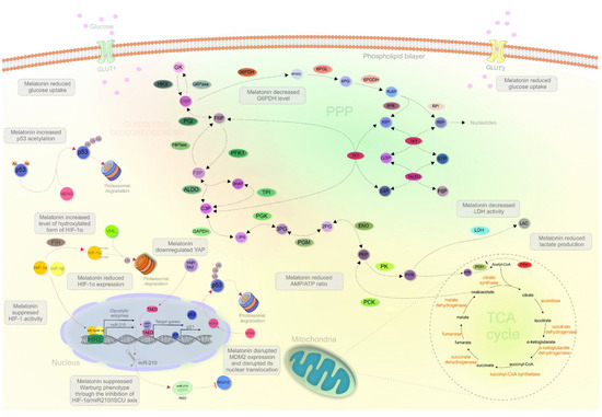
Figure 2.
Melatonin targeting cancer metabolism. Abbreviations: GK, glucose; HKII, hexokinase II; G6P, glucose 6-phosphate; F6P, fructose 6-phosphate; PGI, phosphoglucose isomerase; PFK1, phosphofructokinase 1; FBP, fructose-1,6-bisphosphate; DHAP, dihydroxyacetone phosphate; G3P, glyceraldehyde 3-phosphate; 1,3PG, 1,3-bisphosphoglycerate; 3PG, 3-phosphoglycerate; 2PG, 2-phosphoglycerate; PEP, phosphoenolpyruvate; PYR, pyruvate; LAC, lactate; ALDO, aldolase; TPI, triosephosphate isomerase; GAPDH, glyceraldehyde 3-phosphate dehydrogenase; PGK, phosphoglycerate kinase; PGM, phosphoglycerate mutase; ENO, enolase; PK, pyruvate kinase; LDH, lactate dehydrogenase; PDH, pyruvate dehydrogenase; PDK1, pyruvate dehydrogenase kinase 1; PCK, phosphoenolpyruvate carboxykinase; FBPase, fructose-1,6-bisphosphatase; G6Pase, glucose-6-phosphatase; G6PDH, glucose-6-phosphate dehydrogenase; 6PGDL, 6-phosphogluconolactone; 6PG, 6-phosphogluconate; Ru5P, ribulose 5-phosphate; X5P, xylulose 5-phosphate; R5P, ribose 5-phosphate; S7P, sedoheptulose 7-phosphate; E4P, erythrose 4-phosphate; TKT, transketolase; TALDO, transaldolase; RPE, ribulose-5-phosphate 3-epimerase; RPI, ribose-5-phosphate isomerase; 6PGDH, 6-phosphogluconate dehydrogenase; 6PGL, 6-phosphogluconolactonase; GLUT1/3, glucose transporter 1/3; MDM2, mouse double minute 2; Ac, acetylation; Ub, ubiquitination; HIF-1α/1β, hypoxia-inducible factor 1α/1β; VHL, von Hippel–Lindau; FIH, factor inhibiting HIF; YAP/TAZ, yes-associated protein/WW domain–containing transcription regulator 1; HRE, HRE, hypoxia-response elements; TAED, transcriptional enhancer factor TEF-1; RISC, RNA-induced silencing complex; ISCU 1/2, iron-sulfur cluster assembly scaffold protein ½.
3. Expert Recommendations in the Framework of Predictive, Preventive and Personalized (3P) Medicine
Melatonin demonstrates protective effects in sleep disturbances and sleep-related disorders, depression, mitochondrial dysfunction, chronic inflammation as well as their interrelationship highly relevant for carcinogenesis and other related pathologies such as stroke [6,25,45,152,153,154,155,156,157,158]. Based on current experimental research, melatonin is proposed to be used as the pharmacological agents with a multi-functional capacity such as to modulate mitochondrial functions in cancer, among others [159]. Current preclinical evidence suggests that melatonin can modulate different molecular cascades directly connected to the suppression of cancer development and progression. Antitumor efficacy of melatonin mediated by inhibition of metastatic potential was demonstrated in different in vitro model systems [160,161,162]. Even though a number of experimental studies confirmed anticancer abilities of melatonin, massive clinical testing is still absent. The imminent need to identify and investigate novel therapeutic approaches in cancer research determines melatonin as a promising agent targeted on cancer combined with conventional therapies. Further investigation on melatonin’s role in cancer initiation and progression can improve its therapeutical potential in the clinical sphere [102,163,164,165].
The above discussed preclinical research provides valuable insights into the effects of melatonin on tumor metabolism and the Warburg phenotype, an essential step for unrestricted tumor cell proliferation and cancer progression. Melatonin regulates the critical components associated with cancer metabolism, such as GLUTs, LDH, G6PDH, HIF-1 or p53. [96,103,104,108,110,112,114,115,116,117,118,119]. Following notable preclinical achievements in the field, we emphasize the necessity to investigate melatonin effects focused on the clinical needs [166,167,168,169].
To this end, population screening focused on individuals in sub-optimal health conditions prior to the clinical onset of the pathologies followed by cost-effective targeted treatment is considered the optimal approach in the framework of 3P medicine as a concept of medicine of the 21st century [170,171,172].
4. Conclusions
In conclusion, comprehensive knowledge of melatonin’s capacity to regulate tumor metabolism are expected to strongly contribute to the identification of innovative approaches to an improved cancer management. The above-discussed results of preclinical research provide valuable knowledge about the effect of melatonin on tumor metabolism and the Warburg phenotype, an essential step for unrestricted tumor cell proliferation and cancer progression. Despite significant results from preclinical research, we emphasize the need to further investigate the effect of melatonin on cancer processes through the regulation of the Warburg phenotype within the scope of other more complex modalities of cancer research, including clinical investigation.
Author Contributions
Conceptualization: M.S. (Marek Samec), P.K., D.B., M.S. (Mehdi Shakibaei) and O.G.; writing—original draft preparation, M.S. (Marek Samec), A.L., L.K., K.Z., E.V., M.Š. and A.B.; writing—review and editing, S.M.S., V.L., M.K., M.P., K.B. and K.K.; supervision, P.K., S.T.S.H. and D.B.; O.G. provided expertise in Predictive, Preventive and Personalised medicine (3PM/PPPM). All authors have read and agreed to the published version of the manuscript.
Funding
The present study was supported by the Scientific Grant Agency of the Ministry of Education, Science, Research and Sport of the Slovak Republic (Bratislava, Slovak Republic; grant no. VEGA 1/0136/19), and LISPER project (grant Nr. 313011V446) in bilateral agreement with the European Association for Predictive, Preventive and Personalised Medicine. S.M.S. and E.V. were supported by the NPRP11S-1214-170101 grant (June 2019–current) awarded to D.B. by the Qatar National Research Fund (QNRF), Doha, Qatar.
Institutional Review Board Statement
Not applicable.
Informed Consent Statement
Not applicable.
Data Availability Statement
Data is contained within the article.
Conflicts of Interest
The authors declare no conflict of interest. The funders had no role in the design of the study; in the collection, analyses, or interpretation of data; in the writing of the manuscript, or in the decision to publish the results.
References
- Allison, K.E.; Coomber, B.L.; Bridle, B.W. Metabolic reprogramming in the tumour microenvironment: A hallmark shared by cancer cells and t lymphocytes. Immunology 2017, 152, 175–184. [Google Scholar] [CrossRef] [PubMed]
- Sun, X.; Wang, M.; Wang, M.; Yu, X.; Guo, J.; Sun, T.; Li, X.; Yao, L.; Dong, H.; Xu, Y. Metabolic reprogramming in triple-negative breast cancer. Front. Oncol. 2020, 10, 428. [Google Scholar] [CrossRef]
- Giannattasio, S.; Mirisola, M.G.; Mazzoni, C. Editorial: Cell stress, metabolic reprogramming, and cancer. Front. Oncol. 2018, 8, 236. [Google Scholar] [CrossRef] [PubMed]
- Phan, L.M.; Yeung, S.-C.J.; Lee, M.-H. Cancer metabolic reprogramming: Importance, main features, and potentials for precise targeted anti-cancer therapies. Cancer Biol. Med. 2014, 11, 1–19. [Google Scholar] [CrossRef] [PubMed]
- Mayo, J.C.; Cernuda, R.; Quiros, I.; Rodriguez, P.; Garcia, J.I.; Hevia, D.; Sainz, R.M. Understanding the role of melatonin in cancer metabolism. Melatonin Res. 2019, 2, 76–104. [Google Scholar] [CrossRef]
- Hansen, M.V.; Andersen, L.T.; Madsen, M.T.; Hageman, I.; Rasmussen, L.S.; Bokmand, S.; Rosenberg, J.; Gögenur, I. Effect of melatonin on depressive symptoms and anxiety in patients undergoing breast cancer surgery: A randomized, double-blind, placebo-controlled trial. Breast Cancer Res. Treat. 2014, 145, 683–695. [Google Scholar] [CrossRef]
- Kostoglou-Athanassiou, I. Therapeutic applications of melatonin. Ther. Adv. Endocrinol. Metab. 2013, 4, 13–24. [Google Scholar] [CrossRef]
- Li, Y.; Li, S.; Zhou, Y.; Meng, X.; Zhang, J.-J.; Xu, D.-P.; Li, H.-B. Melatonin for the prevention and treatment of cancer. Oncotarget 2017, 8, 39896–39921. [Google Scholar] [CrossRef] [PubMed]
- Mociková-Kalická, K.; Bojková, B.; Adámeková, E.; Mníchová-Chamilová, M.; Kubatka, P.; Ahlersová, E.; Ahlers, I. Preventive effect of indomethacin and melatonin on 7,12-dimethybenz/a/anthracene-induced mammary carcinogenesis in female sprague-dawley rats. A preliminary report. Folia Biol. 2001, 47, 75–79. [Google Scholar]
- Orendáš, P.; Kubatka, P.; Bojková, B.; Kassayová, M.; Kajo, K.; Výbohová, D.; Kružliak, P.; Péč, M.; Adamkov, M.; Kapinová, A.; et al. Melatonin potentiates the anti-tumour effect of pravastatin in rat mammary gland carcinoma model. Int. J. Exp. Pathol. 2014, 95, 401–410. [Google Scholar] [CrossRef]
- Bojková, B.; Kubatka, P.; Qaradakhi, T.; Zulli, A.; Kajo, K. Melatonin may increase anticancer potential of pleiotropic drugs. Int. J. Mol. Sci. 2018, 19, 3910. [Google Scholar] [CrossRef] [PubMed]
- Reiter, R.J.; Sharma, R.; Rosales-Corral, S. Anti-warburg effect of melatonin: A proposed mechanism to explain its inhibition of multiple diseases. Int. J. Mol. Sci. 2021, 22, 764. [Google Scholar] [CrossRef] [PubMed]
- Kubatka, P.; Zubor, P.; Busselberg, D.; Kwon, T.K.; Adamek, M.; Petrovic, D.; Opatrilova, R.; Gazdikova, K.; Caprnda, M.; Rodrigo, L.; et al. Melatonin and breast cancer: Evidences from preclinical and human studies. Crit. Rev. Oncol. Hematol. 2018, 122, 133–143. [Google Scholar] [CrossRef] [PubMed]
- Zhao, Y.; Ren, J.; Hillier, J.; Jones, M.; Lu, W.; Jones, E.Y. Structural characterization of melatonin as an inhibitor of the wnt deacylase notum. J. Pineal. Res. 2020, 68, e12630. [Google Scholar] [CrossRef]
- Rani, P.; Pal, D.; Hegde, R.R.; Hashim, S.R. Acetamides: Chemotherapeutic agents for inflammation-associated cancers. J. Chemother. 2016, 28, 255–265. [Google Scholar] [CrossRef]
- Tordjman, S.; Chokron, S.; Delorme, R.; Charrier, A.; Bellissant, E.; Jaafari, N.; Fougerou, C. Melatonin: Pharmacology, functions and therapeutic benefits. Curr. Neuropharmacol. 2017, 15, 434–443. [Google Scholar] [CrossRef]
- Zhao, D.; Yu, Y.; Shen, Y.; Liu, Q.; Zhao, Z.; Sharma, R.; Reiter, R.J. Melatonin synthesis and function: Evolutionary history in animals and plants. Front. Endocrinol. 2019, 10, 249. [Google Scholar] [CrossRef]
- Saha, S.; Singh, K.M.; Gupta, B.B.P. Melatonin synthesis and clock gene regulation in the pineal organ of teleost fish compared to mammals: Similarities and differences. Gen. Comp. Endocrinol. 2019, 279, 27–34. [Google Scholar] [CrossRef]
- Benyassi, A.; Schwartz, C.; Coon, S.L.; Klein, D.C.; Falcón, J. Melatonin synthesis: Arylalkylamine N-acetyltransferases in trout retina and pineal organ are different. Neuroreport 2000, 11, 255–258. [Google Scholar] [CrossRef]
- Low, M.J. Neuroendocrinology. In Williams Textbook of Endocrinology, 13th ed.; Melmed, S., Polonsky, K.S., Larsen, P.R., Kronenberg, H.M., Eds.; Elsevier: Philadelphia, PA, USA, 2016; pp. 109–175. ISBN 978-0-323-29738-7. [Google Scholar]
- Cook, J.S.; Sauder, C.L.; Ray, C.A. Melatonin differentially affects vascular blood flow in humans. Am. J. Physiol. Heart Circ. Physiol. 2011, 300, H670–H674. [Google Scholar] [CrossRef]
- Reiter, R.J. Circadian and non-circadian melatonin: Influences on glucose metabolism in cancer cells. J. Curr. Sci. Technol. 2020, 10, 85–98. [Google Scholar] [CrossRef]
- Hardeland, R. Chronobiology of melatonin beyond the feedback to the suprachiasmatic nucleus—Consequences to melatonin dysfunction. Int. J. Mol. Sci. 2013, 14, 5817–5841. [Google Scholar] [CrossRef]
- Dubocovich, M.L. Melatonin receptors: Role on sleep and circadian rhythm regulation. Sleep Med. 2007, 8 (Suppl. S3), 34–42. [Google Scholar] [CrossRef]
- Poza, J.J.; Pujol, M.; Ortega-Albás, J.J.; Romero, O. Melatonin in sleep disorders. Neurología 2020. [Google Scholar] [CrossRef]
- Ashrafizadeh, M.; Najafi, M.; Kavyiani, N.; Mohammadinejad, R.; Farkhondeh, T.; Samarghandian, S. Anti-inflammatory activity of melatonin: A focus on the role of NLRP3 inflammasome. Inflammation 2021. [Google Scholar] [CrossRef]
- Karaaslan, C.; Suzen, S. Antioxidant properties of melatonin and its potential action in diseases. Curr. Top Med. Chem. 2015, 15, 894–903. [Google Scholar] [CrossRef] [PubMed]
- Ahmadi, Z.; Ashrafizadeh, M. Melatonin as a potential modulator of Nrf2. Fundam. Clin. Pharmacol. 2020, 34, 11–19. [Google Scholar] [CrossRef] [PubMed]
- Abadi, S.H.M.H.; Shirazi, A.; Alizadeh, A.M.; Changizi, V.; Najafi, M.; Khalighfard, S.; Nosrati, H. The effect of melatonin on superoxide dismutase and glutathione peroxidase activity, and malondialdehyde levels in the targeted and the non-targeted lung and heart tissues after irradiation in xenograft mice colon cancer. Curr. Mol. Pharmacol. 2018, 11, 326–335. [Google Scholar] [CrossRef] [PubMed]
- Pohanka, M. Impact of melatonin on immunity: A review. Cent. Eur. J. Med. 2013, 8, 369–376. [Google Scholar] [CrossRef]
- Maestroni, G.J.M. Melatonin and the immune system therapeutic potential in cancer, viral diseases, and immunodeficiency states. In The Pineal Gland and Cancer: Neuroimmunoendocrine Mechanisms in Malignancy; Bartsch, C., Bartsch, H., Blask, D.E., Cardinali, D.P., Hrushesky, W.J.M., Mecke, D., Eds.; Springer: Berlin/Heidelberg, Germany, 2001; pp. 384–394. ISBN 978-3-642-59512-7. [Google Scholar]
- Miller, S.C.; Pandi, P.S.R.; Esquifino, A.I.; Cardinali, D.P.; Maestroni, G.J.M. The role of melatonin in immuno-enhancement: Potential application in cancer. Int. J. Exp. Pathol. 2006, 87, 81–87. [Google Scholar] [CrossRef]
- Campos, L.A.; Bueno, C.; Barcelos, I.P.; Halpern, B.; Brito, L.C.; Amaral, F.G.; Baltatu, O.C.; Cipolla-Neto, J. Melatonin therapy improves cardiac autonomic modulation in pinealectomized patients. Front. Endocrinol. 2020, 11, 239. [Google Scholar] [CrossRef] [PubMed]
- Wang, Y.; Wang, P.; Zheng, X.; Du, X. Therapeutic strategies of melatonin in cancer patients: A systematic review and meta-analysis. OncoTargets Ther. 2018, 11, 7895–7908. [Google Scholar] [CrossRef] [PubMed]
- Di Bella, G.; Mascia, F.; Gualano, L.; Di Bella, L. Melatonin anticancer effects: Review. Int. J. Mol. Sci. 2013, 14, 2410–2430. [Google Scholar] [CrossRef]
- Wang, T.; Liu, B.; Guan, Y.; Gong, M.; Zhang, W.; Pan, J.; Liu, Y.; Liang, R.; Yuan, Y.; Ye, L. Melatonin inhibits the proliferation of breast cancer cells induced by bisphenol a via targeting estrogen receptor-related pathways. Thorac. Cancer 2018, 9, 368–375. [Google Scholar] [CrossRef]
- Hill, S.M.; Frasch, T.; Xiang, S.; Yuan, L.; Duplessis, T.; Mao, L. Molecular mechanisms of melatonin anticancer effects. Integr. Cancer Ther. 2009, 8, 337–346. [Google Scholar] [CrossRef]
- Rodriguez, C.; Martín, V.; Herrera, F.; García-Santos, G.; Rodriguez-Blanco, J.; Casado-Zapico, S.; Sánchez-Sánchez, A.M.; Suárez, S.; Puente-Moncada, N.; Anítua, M.J.; et al. Mechanisms involved in the pro-apoptotic effect of melatonin in cancer cells. Int. J. Mol. Sci. 2013, 14, 6597–6613. [Google Scholar] [CrossRef] [PubMed]
- Moretti, R.M.; Marelli, M.M.; Maggi, R.; Dondi, D.; Motta, M.; Limonta, P. Antiproliferative action of melatonin on human prostate cancer LNCaP cells. Oncol. Rep. 2000, 7, 347–351. [Google Scholar] [CrossRef] [PubMed]
- Gatti, G.; Lucini, V.; Dugnani, S.; Calastretti, A.; Spadoni, G.; Bedini, A.; Rivara, S.; Mor, M.; Canti, G.; Scaglione, F.; et al. Antiproliferative and pro-apoptotic activity of melatonin analogues on melanoma and breast cancer cells. Oncotarget 2017, 8, 68338–68353. [Google Scholar] [CrossRef]
- Cheng, J.; Yang, H.-L.; Gu, C.-J.; Liu, Y.-K.; Shao, J.; Zhu, R.; He, Y.-Y.; Zhu, X.-Y.; Li, M.-Q. Melatonin restricts the viability and angiogenesis of vascular endothelial cells by suppressing HIF-1α/ROS/VEGF. Int. J. Mol. Med. 2019, 43, 945–955. [Google Scholar] [CrossRef]
- Mortezaee, K.; Potes, Y.; Mirtavoos-Mahyari, H.; Motevaseli, E.; Shabeeb, D.; Musa, A.E.; Najafi, M.; Farhood, B. Boosting immune system against cancer by melatonin: A mechanistic viewpoint. Life Sci. 2019, 238, 116960. [Google Scholar] [CrossRef]
- Tang, Z.; Xu, Z.; Zhu, X.; Zhang, J. New insights into molecules and pathways of cancer metabolism and therapeutic implications. Cancer Commun. 2021, 41, 16–36. [Google Scholar] [CrossRef] [PubMed]
- Samec, M.; Liskova, A.; Koklesova, L.; Samuel, S.M.; Zhai, K.; Buhrmann, C.; Varghese, E.; Abotaleb, M.; Qaradakhi, T.; Zulli, A.; et al. Flavonoids against the warburg phenotype—Concepts of predictive, preventive and personalised medicine to cut the gordian knot of cancer cell metabolism. EPMA J. 2020, 11, 377–398. [Google Scholar] [CrossRef]
- Koklesova, L.; Samec, M.; Liskova, A.; Zhai, K.; Büsselberg, D.; Giordano, F.A.; Kubatka, P.; Golunitschaja, O. Mitochondrial impairments in aetiopathology of multifactorial diseases: Common origin but individual outcomes in context of 3P medicine. EPMA J. 2021, 1–14. [Google Scholar] [CrossRef]
- Yu, L.; Chen, X.; Wang, L.; Chen, S. The sweet trap in tumors: Aerobic glycolysis and potential targets for therapy. Oncotarget 2016, 7, 38908–38926. [Google Scholar] [CrossRef] [PubMed]
- Kuo, M.-H.; Chang, W.-W.; Yeh, B.-W.; Chu, Y.-S.; Lee, Y.-C.; Lee, H.-T. Glucose transporter 3 is essential for the survival of breast cancer cells in the brain. Cells 2019, 8, 1568. [Google Scholar] [CrossRef] [PubMed]
- Hussein, Y.R.; Bandyopadhyay, S.; Semaan, A.; Ahmed, Q.; Albashiti, B.; Jazaerly, T.; Nahleh, Z.; Ali-Fehmi, R. Glut-1 expression correlates with basal-like breast cancer. Transl. Oncol. 2011, 4, 321–327. [Google Scholar] [CrossRef] [PubMed]
- Gonzalez-Menendez, P.; Hevia, D.; Alonso-Arias, R.; Alvarez-Artime, A.; Rodriguez-Garcia, A.; Kinet, S.; Gonzalez-Pola, I.; Taylor, N.; Mayo, J.C.; Sainz, R.M. GLUT1 protects prostate cancer cells from glucose deprivation-induced oxidative stress. Redox Biol. 2018, 17, 112–127. [Google Scholar] [CrossRef] [PubMed]
- Ayala, F.R.R.; Rocha, R.M.; Carvalho, K.C.; Carvalho, A.L.; da Cunha, I.W.; Lourenço, S.V.; Soares, F.A. Glut1 and Glut3 as potential prognostic markers for oral squamous cell carcinoma. Molecules 2010, 15, 2374–2387. [Google Scholar] [CrossRef] [PubMed]
- Chiba, I.; Ogawa, K.; Morioka, T.; Shimoji, H.; Sunagawa, N.; Iraha, S.; Nishimaki, T.; Yoshimi, N.; Murayama, S. Clinical significance of GLUT-1 expression in patients with esophageal cancer treated with concurrent chemoradiotherapy. Oncol. Lett. 2011, 2, 21–28. [Google Scholar] [CrossRef]
- Feng, J.; Li, J.; Wu, L.; Yu, Q.; Ji, J.; Wu, J.; Dai, W.; Guo, C. Emerging roles and the regulation of aerobic glycolysis in hepatocellular carcinoma. J. Exp. Clin. Cancer Res. 2020, 39, 126. [Google Scholar] [CrossRef] [PubMed]
- Feng, Y.; Xiong, Y.; Qiao, T.; Li, X.; Jia, L.; Han, Y. Lactate dehydrogenase A: A key player in carcinogenesis and potential target in cancer therapy. Cancer Med. 2018, 7, 6124–6136. [Google Scholar] [CrossRef] [PubMed]
- Hsu, M.-C.; Hung, W.-C. Pyruvate kinase M2 fuels multiple aspects of cancer cells: From Cellular metabolism, transcriptional regulation to extracellular signaling. Mol. Cancer 2018, 17, 35. [Google Scholar] [CrossRef]
- Yi, W.; Clark, P.M.; Mason, D.E.; Keenan, M.C.; Hill, C.; Goddard, W.A.; Peters, E.C.; Driggers, E.M.; Hsieh-Wilson, L.C. PFK1 glycosylation is a key regulator of cancer cell growth and central metabolic pathways. Science 2012, 337, 975–980. [Google Scholar] [CrossRef] [PubMed]
- Yeung, S.J.; Pan, J.; Lee, M.-H. Roles of P53, Myc and HIF-1 in regulating glycolysis—The seventh hallmark of cancer. Cell. Mol. Life Sci. 2008, 65, 3981. [Google Scholar] [CrossRef] [PubMed]
- Wang, Y.-P.; Lei, Q.-Y. Metabolic recoding of epigenetics in cancer. Cancer Commun. 2018, 38, 25. [Google Scholar] [CrossRef] [PubMed]
- Grasmann, G.; Smolle, E.; Olschewski, H.; Leithner, K. Gluconeogenesis in cancer cells—Repurposing of a starvation-induced metabolic pathway? Biochim. Biophys. Acta Rev. Cancer 2019, 1872, 24–36. [Google Scholar] [CrossRef]
- Engelking, L.R. Gluconeogenesis. In Textbook of Veterinary Physiological Chemistry, 3rd ed.; Engelking, L.R., Ed.; Academic Press: Boston, MA, USA, 2015; pp. 225–230. ISBN 978-0-12-391909-0. [Google Scholar]
- Wang, Z.; Dong, C. Gluconeogenesis in cancer: Function and regulation of PEPCK, FBPase, and G6Pase. Trends Cancer 2019, 5, 30–45. [Google Scholar] [CrossRef] [PubMed]
- Shi, L.; An, S.; Liu, Y.; Liu, J.; Wang, F. PCK1 regulates glycolysis and tumor progression in clear cell renal cell carcinoma through LDHA. OncoTargets Ther. 2020, 13, 2613–2627. [Google Scholar] [CrossRef] [PubMed]
- Yamaguchi, N.; Weinberg, E.M.; Nguyen, A.; Liberti, M.V.; Goodarzi, H.; Janjigian, Y.Y.; Paty, P.B.; Saltz, L.B.; Kingham, T.P.; Loo, J.M.; et al. PCK1 and DHODH drive colorectal cancer liver metastatic colonization and hypoxic growth by promoting nucleotide synthesis. Elife 2019, 8, e52135. [Google Scholar] [CrossRef]
- Zhao, J.; Li, J.; Fan, T.W.M.; Hou, S.X. Glycolytic reprogramming through PCK2 regulates tumor initiation of prostate cancer cells. Oncotarget 2017, 8, 83602–83618. [Google Scholar] [CrossRef] [PubMed]
- Liu, G.-M.; Zhang, Y.-M. Targeting FBPase is an emerging novel approach for cancer therapy. Cancer Cell Int. 2018, 18, 36. [Google Scholar] [CrossRef] [PubMed]
- Dong, C.; Yuan, T.; Wu, Y.; Wang, Y.; Fan, T.W.M.; Miriyala, S.; Lin, Y.; Yao, J.; Shi, J.; Kang, T.; et al. Loss of FBP1 by snail-mediated repression provides metabolic advantages in basal-like breast cancer. Cancer Cell 2013, 23, 316–331. [Google Scholar] [CrossRef] [PubMed]
- Li, B.; Qiu, B.; Lee, D.S.M.; Walton, Z.E.; Ochocki, J.D.; Mathew, L.K.; Mancuso, A.; Gade, T.P.F.; Keith, B.; Nissim, I.; et al. Fructose-1,6-bisphosphatase opposes renal carcinoma progression. Nature 2014, 513, 251–255. [Google Scholar] [CrossRef] [PubMed]
- Filipp, F.V.; Scott, D.A.; Ronai, Z.A.; Osterman, A.L.; Smith, J.W. Reverse TCA cycle flux through isocitrate dehydrogenases 1 and 2 is required for lipogenesis in hypoxic melanoma cells. Pigment Cell Melanoma Res. 2012, 25, 375–383. [Google Scholar] [CrossRef] [PubMed]
- Haddad, A.; Mohiuddin, S.S. Biochemistry, citric acid cycle. In StatPearls; StatPearls Publishing: Treasure Island, FL, USA, 2020. [Google Scholar]
- Cardaci, S.; Ciriolo, M.R. TCA Cycle Defects and Cancer: When Metabolism Tunes Redox State. Available online: https://www.hindawi.com/journals/ijcb/2012/161837/ (accessed on 6 February 2021).
- King, A.; Selak, M.A.; Gottlieb, E. Succinate dehydrogenase and fumarate hydratase: Linking mitochondrial dysfunction and cancer. Oncogene 2006, 25, 4675–4682. [Google Scholar] [CrossRef] [PubMed]
- Tennant, D.A.; Frezza, C.; MacKenzie, E.D.; Nguyen, Q.D.; Zheng, L.; Selak, M.A.; Roberts, D.L.; Dive, C.; Watson, D.G.; Aboagye, E.O.; et al. Reactivating HIF prolyl hydroxylases under hypoxia results in metabolic catastrophe and cell death. Oncogene 2009, 28, 4009–4021. [Google Scholar] [CrossRef]
- Anderson, N.M.; Mucka, P.; Kern, J.G.; Feng, H. The emerging role and targetability of the TCA cycle in cancer metabolism. Protein Cell 2018, 9, 216–237. [Google Scholar] [CrossRef]
- Choi, J.; Kim, E.-S.; Koo, J.S. Expression of Pentose Phosphate Pathway-Related Proteins in Breast Cancer. Available online: https://www.hindawi.com/journals/dm/2018/9369358/ (accessed on 7 February 2021).
- Cossu, V.; Bonanomi, M.; Bauckneht, M.; Ravera, S.; Righi, N.; Miceli, A.; Morbelli, S.; Orengo, A.M.; Piccioli, P.; Bruno, S.; et al. Two high-rate pentose-phosphate pathways in cancer cells. Sci. Rep. 2020, 10, 22111. [Google Scholar] [CrossRef] [PubMed]
- Ge, T.; Yang, J.; Zhou, S.; Wang, Y.; Li, Y.; Tong, X. The role of the pentose phosphate pathway in diabetes and cancer. Front. Endocrinol. 2020, 11, 365. [Google Scholar] [CrossRef]
- Stine, Z.E.; Altman, B.J.; Hsieh, A.L.; Gouw, A.M.; Dang, C.V. Deregulation of the cellular energetics of cancer cells. In Pathobiology of Human Disease; McManus, L.M., Mitchell, R.N., Eds.; Academic Press: San Diego, CA, USA, 2014; pp. 444–455. ISBN 978-0-12-386457-4. [Google Scholar]
- Jin, L.; Zhou, Y. Crucial role of the pentose phosphate pathway in malignant tumors. Oncol. Lett. 2019, 17, 4213–4221. [Google Scholar] [CrossRef] [PubMed]
- Berg, J.M.; Tymoczko, J.L.; Stryer, L. 20.3 the pentose phosphate pathway generates NADPH and synthesizes five-carbon sugars. In Biochemistry, 5th ed.; NCBI: Bethesda, MD, USA, 2002. [Google Scholar]
- Zhang, Q.; Yi, X.; Yang, Z.; Han, Q.; Di, X.; Chen, F.; Wang, Y.; Yi, Z.; Kuang, Y.; Zhu, Y. Overexpression of G6PD represents a potential prognostic factor in clear cell renal cell carcinoma. J. Cancer 2017, 8, 665–673. [Google Scholar] [CrossRef]
- Pu, H.; Zhang, Q.; Zhao, C.; Shi, L.; Wang, Y.; Wang, J.; Zhang, M. Overexpression of G6PD is associated with high risks of recurrent metastasis and poor progression-free survival in primary breast carcinoma. World J. Surg. Oncol. 2015, 13, 323. [Google Scholar] [CrossRef]
- Yu, J.; Liang, Q.; Wang, J.; Wang, K.; Gao, J.; Zhang, J.; Zeng, Y.; Chiu, P.W.Y.; Ng, E.K.W.; Sung, J.J.Y. REC8 Functions as a tumor suppressor and is epigenetically downregulated in gastric cancer, especially in EBV-positive subtype. Oncogene 2017, 36, 182–193. [Google Scholar] [CrossRef]
- Zhang, X.; Zhang, X.; Li, Y.; Shao, Y.; Xiao, J.; Zhu, G.; Li, F. PAK4 regulates G6PD activity by P53 degradation involving colon cancer cell growth. Cell Death Dis. 2017, 8, e2820. [Google Scholar] [CrossRef]
- Lu, M.; Lu, L.; Dong, Q.; Yu, G.; Chen, J.; Qin, L.; Wang, L.; Zhu, W.; Jia, H. Elevated G6PD expression contributes to migration and invasion of hepatocellular carcinoma cells by inducing epithelial-mesenchymal transition. Acta Biochim. Biophy. Sin. 2018, 50, 370–380. [Google Scholar] [CrossRef] [PubMed]
- Cha, Y.; Jung, W.; Koo, J. Differential site-based expression of pentose phosphate pathway-related proteins among breast cancer metastases. Dis. Markers 2017, 2017, 7062517. [Google Scholar] [CrossRef] [PubMed]
- Simabuco, F.M.; Morale, M.G.; Pavan, I.C.B.; Morelli, A.P.; Silva, F.R.; Tamura, R.E. P53 and metabolism: From mechanism to therapeutics. Oncotarget 2018, 9, 23780–23823. [Google Scholar] [CrossRef] [PubMed]
- Schwartzenberg-Bar-Yoseph, F.; Armoni, M.; Karnieli, E. The tumor suppressor P53 down-regulates glucose transporters GLUT1 and GLUT4 gene expression. Cancer Res. 2004, 64, 2627–2633. [Google Scholar] [CrossRef]
- Tsouko, E.; Khan, A.S.; White, M.A.; Han, J.J.; Shi, Y.; Merchant, F.A.; Sharpe, M.A.; Xin, L.; Frigo, D.E. Regulation of the pentose phosphate pathway by an androgen receptor-MTOR-mediated mechanism and its role in prostate cancer cell growth. Oncogenesis 2014, 3, e103. [Google Scholar] [CrossRef]
- Wang, Y.-Y.; Chen, J.; Liu, X.-M.; Zhao, R.; Zhe, H. Nrf2-mediated metabolic reprogramming in cancer. Oxid. Med. Cell. Longev. 2018, 2018, 9304091. [Google Scholar] [CrossRef]
- Santana-Codina, N.; Roeth, A.A.; Zhang, Y.; Yang, A.; Mashadova, O.; Asara, J.M.; Wang, X.; Bronson, R.T.; Lyssiotis, C.A.; Ying, H.; et al. Oncogenic KRAS supports pancreatic cancer through regulation of nucleotide synthesis. Nat. Commun. 2018, 9, 4945. [Google Scholar] [CrossRef] [PubMed]
- Varghese, E.; Samuel, S.M.; Líšková, A.; Samec, M.; Kubatka, P.; Büsselberg, D. Targeting glucose metabolism to overcome resistance to anticancer chemotherapy in breast cancer. Cancers 2020, 12, 2252. [Google Scholar] [CrossRef]
- Chen, X.-S.; Li, L.-Y.; Guan, Y.; Yang, J.-M.; Cheng, Y. Anticancer strategies based on the metabolic profile of tumor cells: Therapeutic targeting of the warburg effect. Acta Pharmacol. Sin. 2016, 37, 1013–1019. [Google Scholar] [CrossRef]
- Lukey, M.J.; Katt, W.P.; Cerione, R.A. Targeting amino acid metabolism for cancer therapy. Drug Discov. Today 2017, 22, 796–804. [Google Scholar] [CrossRef]
- Morigny, P.; Boucher, J.; Arner, P.; Langin, D. Lipid and glucose metabolism in white adipocytes: Pathways, dysfunction and therapeutics. Nat. Rev. Endocrinol. 2021, 17, 276–295. [Google Scholar] [CrossRef]
- Montrose, D.C.; Galluzzi, L. Drugging cancer metabolism: Expectations vs. reality. Int. Rev. Cell Mol. Biol. 2019, 347, 1–26. [Google Scholar] [CrossRef]
- Cortés-Hernández, L.E.; Eslami-S, Z.; Dujon, A.M.; Giraudeau, M.; Ujvari, B.; Thomas, F.; Alix-Panabières, C. Do malignant cells sleep at night? Genome Biol. 2020, 21, 276. [Google Scholar] [CrossRef]
- Blask, D.E.; Dauchy, R.T.; Dauchy, E.M.; Mao, L.; Hill, S.M.; Greene, M.W.; Belancio, V.P.; Sauer, L.A.; Davidson, L. Light exposure at night disrupts host/cancer circadian regulatory dynamics: Impact on the warburg effect, lipid signaling and tumor growth prevention. PLoS ONE 2014, 9, e102776. [Google Scholar] [CrossRef]
- Shi, L.; Tu, B.P. Acetyl-CoA and the regulation of metabolism: Mechanisms and consequences. Curr. Opin. Cell Biol. 2015, 33, 125–131. [Google Scholar] [CrossRef]
- Nayak, M.K.; Dhanesha, N.; Doddapattar, P.; Rodriguez, O.; Sonkar, V.K.; Dayal, S.; Chauhan, A.K. Dichloroacetate, an inhibitor of pyruvate dehydrogenase kinases, inhibits platelet aggregation and arterial thrombosis. Blood Adv. 2018, 2, 2029–2038. [Google Scholar] [CrossRef]
- Reiter, R.J.; Sharma, R.; Ma, Q.; Rosales-Corral, S.; Acuna-Castroviejo, D.; Escames, G. Inhibition of mitochondrial pyruvate dehydrogenase kinase: A proposed mechanism by which melatonin causes cancer cells to overcome cytosolic glycolysis, reduce tumor biomass and reverse insensitivity to chemotherapy. Melatonin Res. 2019, 2, 105–119. [Google Scholar] [CrossRef]
- Lowes, D.A.; Webster, N.R.; Murphy, M.P.; Galley, H.F. Antioxidants that protect mitochondria reduce interleukin-6 and oxidative stress, improve mitochondrial function, and reduce biochemical markers of organ dysfunction in a rat model of acute sepsis. Br. J. Anaesth. 2013, 110, 472–480. [Google Scholar] [CrossRef]
- Reiter, R.J.; Sharma, R.; Pires de Campos Zuccari, D.A.; de Almeida Chuffa, L.G.; Manucha, W.; Rodriguez, C. Melatonin synthesis in and uptake by mitochondria: Implications for diseased cells with dysfunctional mitochondria. Future Med. Chem. 2021, 13, 335–339. [Google Scholar] [CrossRef] [PubMed]
- Reiter, R.J.; Sharma, R.; Ma, Q.; Rorsales-Corral, S.; de Almeida Chuffa, L.G. Melatonin inhibits warburg-dependent cancer by redirecting glucose oxidation to the mitochondria: A mechanistic hypothesis. Cell. Mol. Life Sci. 2020, 77, 2527–2542. [Google Scholar] [CrossRef] [PubMed]
- Hevia, D.; Gonzalez-Menendez, P.; Fernandez-Fernandez, M.; Cueto, S.; Rodriguez-Gonzalez, P.; Garcia-Alonso, J.I.; Mayo, J.C.; Sainz, R.M. Melatonin decreases glucose metabolism in prostate cancer cells: A 13C stable isotope-resolved metabolomic study. Int. J. Mol. Sci. 2017, 18, 1620. [Google Scholar] [CrossRef]
- Sanchez-Sanchez, A.M.; Antolin, I.; Puente-Moncada, N.; Suarez, S.; Gomez-Lobo, M.; Rodriguez, C.; Martin, V. Melatonin cytotoxicity is associated to warburg effect inhibition in ewing sarcoma cells. PLoS ONE 2015, 10, e0135420. [Google Scholar] [CrossRef]
- Kim, W.; Cho, Y.S.; Wang, X.; Park, O.; Ma, X.; Kim, H.; Gan, W.; Jho, E.; Cha, B.; Jeung, Y.; et al. Hippo signaling is intrinsically regulated during cell cycle progression by APC/CCdh1. Proc. Natl. Acad. Sci. USA 2019, 116, 9423–9432. [Google Scholar] [CrossRef] [PubMed]
- Lian, I.; Kim, J.; Okazawa, H.; Zhao, J.; Zhao, B.; Yu, J.; Chinnaiyan, A.; Israel, M.A.; Goldstein, L.S.B.; Abujarour, R.; et al. The role of YAP transcription coactivator in regulating stem cell self-renewal and differentiation. Genes Dev. 2010, 24, 1106–1118. [Google Scholar] [CrossRef] [PubMed]
- Han, Y. Analysis of the role of the hippo pathway in cancer. J. Transl. Med. 2019, 17, 116. [Google Scholar] [CrossRef]
- Mi, L.; Kuang, H. Melatonin regulates cisplatin resistance and glucose metabolism through hippo signaling in hepatocellular carcinoma cells. Cancer Manag. Res. 2020, 12, 1863–1874. [Google Scholar] [CrossRef]
- Zhang, H.-M.; Zhang, Y. Melatonin: A well-documented antioxidant with conditional pro-oxidant actions. J. Pineal Res. 2014, 57, 131–146. [Google Scholar] [CrossRef]
- He, M.; Zhou, C.; Lu, Y.; Mao, L.; Xi, Y.; Mei, X.; Wang, X.; Zhang, L.; Yu, Z.; Zhou, Z. Melatonin antagonizes nickel-induced aerobic glycolysis by blocking ROS-mediated HIF-1 α/MiR210/ISCU axis activation. Oxid. Med. Cell. Longev. 2020, 2020, 5406284. [Google Scholar] [CrossRef]
- Yunus, N.M.; Johan, M.F.; Ali Nagi Al-Jamal, H.; Husin, A.; Hussein, A.R.; Hassan, R. Characterisation and clinical significance of FLT3-ITD and Non-ITD in acute myeloid leukaemia patients in Kelantan, northeast peninsular Malaysia. Asian Pac. J. Cancer Prev. 2015, 16, 4869–4872. [Google Scholar] [CrossRef] [PubMed][Green Version]
- Puente-Moncada, N.; Turos-Cabal, M.; Sánchez-Sánchez, A.M.; Antolín, I.; Herrera, F.; Rodriguez-Blanco, J.; Duarte-Olivenza, C.; Rodriguez, C.; Martín, V. Role of glucose metabolism in the differential antileukemic effect of melatonin on wild-type and FLT3-ITD mutant cells. Oncol. Rep. 2020, 44, 293–302. [Google Scholar] [CrossRef] [PubMed]
- Guerra-Librero, A.; Fernandez-Gil, B.I.; Florido, J.; Martinez-Ruiz, L.; Rodríguez-Santana, C.; Shen, Y.-Q.; García-Verdugo, J.M.; López-Rodríguez, A.; Rusanova, I.; Quiñones-Hinojosa, A.; et al. Melatonin targets metabolism in head and neck cancer cells by regulating mitochondrial structure and function. Antioxidants 2021, 10, 603. [Google Scholar] [CrossRef]
- Hevia, D.; González-Menéndez, P.; Quiros-González, I.; Miar, A.; Rodríguez-García, A.; Tan, D.-X.; Reiter, R.J.; Mayo, J.C.; Sainz, R.M. Melatonin uptake through glucose transporters: A new target for melatonin inhibition of cancer. J. Pineal Res. 2015, 58, 234–250. [Google Scholar] [CrossRef] [PubMed]
- Dauchy, R.T.; Hoffman, A.E.; Wren-Dail, M.A.; Hanifin, J.P.; Warfield, B.; Brainard, G.C.; Xiang, S.; Yuan, L.; Hill, S.M.; Belancio, V.P.; et al. Daytime blue light enhances the nighttime circadian melatonin inhibition of human prostate cancer growth. Comp. Med. 2015, 65, 473–485. [Google Scholar] [PubMed]
- Xiang, S.; Dauchy, R.T.; Hauch, A.; Mao, L.; Yuan, L.; Wren, M.A.; Belancio, V.P.; Mondal, D.; Frasch, T.; Blask, D.E.; et al. Doxorubicin resistance in breast cancer is driven by light at night-induced disruption of the circadian melatonin signal. J. Pineal Res. 2015, 59, 60–69. [Google Scholar] [CrossRef]
- Dauchy, R.T.; Xiang, S.; Mao, L.; Brimer, S.; Wren, M.A.; Yuan, L.; Anbalagan, M.; Hauch, A.; Frasch, T.; Rowan, B.G.; et al. Circadian and melatonin disruption by exposure to light at night drives intrinsic resistance to tamoxifen therapy in breast cancer. Cancer Res. 2014, 74, 4099–4110. [Google Scholar] [CrossRef]
- Chuffa, L.G.A.; Lupi Júnior, L.A.; Seiva, F.R.F.; Martinez, M.; Domeniconi, R.F.; Pinheiro, P.F.F.; Dos Santos, L.D.; Martinez, F.E. Quantitative proteomic profiling reveals that diverse metabolic pathways are influenced by melatonin in an in vivo model of ovarian carcinoma. J. Proteome Res. 2016, 15, 3872–3882. [Google Scholar] [CrossRef] [PubMed]
- Mao, L.; Dauchy, R.T.; Blask, D.E.; Dauchy, E.M.; Slakey, L.M.; Brimer, S.; Yuan, L.; Xiang, S.; Hauch, A.; Smith, K.; et al. Melatonin suppression of aerobic glycolysis (Warburg effect), survival signalling and metastasis in human leiomyosarcoma. J. Pineal Res. 2016, 60, 167–177. [Google Scholar] [CrossRef]
- Puzio-Kuter, A.M. The role of P53 in metabolic regulation. Genes Cancer 2011, 2, 385–391. [Google Scholar] [CrossRef] [PubMed]
- Gomes, A.S.; Ramos, H.; Soares, J.; Saraiva, L. P53 and glucose metabolism: An orchestra to be directed in cancer therapy. Pharmacol. Res. 2018, 131, 75–86. [Google Scholar] [CrossRef]
- Nagao, A.; Kobayashi, M.; Koyasu, S.; Chow, C.C.T.; Harada, H. HIF-1-dependent reprogramming of glucose metabolic pathway of cancer cells and its therapeutic significance. Int. J. Mol. Sci. 2019, 20, 238. [Google Scholar] [CrossRef] [PubMed]
- Itahana, Y.; Itahana, K. Emerging roles of P53 family members in glucose metabolism. Int. J. Mol. Sci. 2018, 19, 776. [Google Scholar] [CrossRef]
- Singh, D.; Arora, R.; Kaur, P.; Singh, B.; Mannan, R.; Arora, S. Overexpression of hypoxia-inducible factor and metabolic pathways: Possible targets of cancer. Cell Biosci. 2017, 7, 62. [Google Scholar] [CrossRef]
- Martín-Renedo, J.; Mauriz, J.L.; Jorquera, F.; Ruiz-Andrés, O.; González, P.; González-Gallego, J. Melatonin induces cell cycle arrest and apoptosis in hepatocarcinoma HepG2 cell line. J. Pineal Res. 2008, 45, 532–540. [Google Scholar] [CrossRef]
- Amin, A.H.; El-Missiry, M.A.; Othman, A.I.; Ali, D.A.; Gouida, M.S.; Ismail, A.H. Ameliorative effects of melatonin against solid ehrlich carcinoma progression in female mice. J. Pineal Res. 2019, 67, e12585. [Google Scholar] [CrossRef] [PubMed]
- Mähler, A.; Mandel, S.; Lorenz, M.; Ruegg, U.; Wanker, E.E.; Boschmann, M.; Paul, F. Epigallocatechin-3-Gallate: A useful, effective and safe clinical approach for targeted prevention and individualised treatment of neurological diseases? EPMA J. 2013, 4, 5. [Google Scholar] [CrossRef] [PubMed]
- Altemimi, A.; Lakhssassi, N.; Baharlouei, A.; Watson, D.G.; Lightfoot, D.A. Phytochemicals: Extraction, isolation, and identification of bioactive compounds from plant extracts. Plants 2017, 6, 42. [Google Scholar] [CrossRef]
- Barreca, D.; Gattuso, G.; Bellocco, E.; Calderaro, A.; Trombetta, D.; Smeriglio, A.; Laganà, G.; Daglia, M.; Meneghini, S.; Nabavi, S.M. Flavanones: Citrus phytochemical with health-promoting properties. Biofactors 2017, 43, 495–506. [Google Scholar] [CrossRef] [PubMed]
- Liskova, A.; Koklesova, L.; Samec, M.; Varghese, E.; Abotaleb, M.; Samuel, S.M.; Biringer, K.; Petras, M.; Blahutova, D.; Smejkal, K.; et al. Implications of flavonoids as potential modulators of cancer neovascularity. J. Cancer Res. Clin. Oncol. 2020, 146, 3079–3096. [Google Scholar] [CrossRef]
- Ashrafizadeh, M.; Najafi, M.; Makvandi, P.; Zarrabi, A.; Farkhondeh, T.; Samarghandian, S. Versatile role of curcumin and its derivatives in lung cancer therapy. J. Cell. Physiol. 2020, 235, 9241–9268. [Google Scholar] [CrossRef] [PubMed]
- Ashrafizadeh, M.; Najafi, M.; Orouei, S.; Zabolian, A.; Saleki, H.; Azami, N.; Sharifi, N.; Hushmandi, K.; Zarrabi, A.; Ahn, K.S. Resveratrol modulates transforming growth factor-beta (TGF-β) signaling pathway for disease therapy: A new insight into its pharmacological activities. Biomedicines 2020, 8, 261. [Google Scholar] [CrossRef] [PubMed]
- Ashrafizadeh, M.; Zarrabi, A.; Saberifar, S.; Hashemi, F.; Hushmandi, K.; Hashemi, F.; Moghadam, E.R.; Mohammadinejad, R.; Najafi, M.; Garg, M. Nobiletin in cancer therapy: How this plant derived-natural compound targets various oncogene and onco-suppressor pathways. Biomedicines 2020, 8, 110. [Google Scholar] [CrossRef]
- Samec, M.; Liskova, A.; Koklesova, L.; Mersakova, S.; Strnadel, J.; Kajo, K.; Pec, M.; Zhai, K.; Smejkal, K.; Mirzaei, S.; et al. Flavonoids targeting HIF-1: Implications on cancer metabolism. Cancers 2021, 13, 130. [Google Scholar] [CrossRef]
- Liskova, A.; Koklesova, L.; Samec, M.; Smejkal, K.; Samuel, S.M.; Varghese, E.; Abotaleb, M.; Biringer, K.; Kudela, E.; Danko, J.; et al. Flavonoids in cancer metastasis. Cancers 2020, 12, 1498. [Google Scholar] [CrossRef]
- Abotaleb, M.; Samuel, S.; Varghese, E.; Varghese, S.; Kubatka, P.; Liskova, A.; Büsselberg, D. Flavonoids in cancer and apoptosis. Cancers 2018, 11, 28. [Google Scholar] [CrossRef]
- Ashrafizadeh, M.; Taeb, S.; Haghi-Aminjan, H.; Afrashi, S.; Moloudi, K.; Musa, A.E.; Najafi, M.; Farhood, B. Resveratrol as an enhancer of apoptosis in cancer: A mechanistic review. Anticancer Agents Med. Chem. 2020. [Google Scholar] [CrossRef]
- Samec, M.; Liskova, A.; Koklesova, L.; Mestanova, V.; Franekova, M.; Kassayova, M.; Bojkova, B.; Uramova, S.; Zubor, P.; Janikova, K.; et al. Fluctuations of histone chemical modifications in breast, prostate, and colorectal cancer: An implication of phytochemicals as defenders of chromatin equilibrium. Biomolecules 2019, 9, 829. [Google Scholar] [CrossRef]
- Samec, M.; Liskova, A.; Kubatka, P.; Uramova, S.; Zubor, P.; Samuel, S.M.; Zulli, A.; Pec, M.; Bielik, T.; Biringer, K.; et al. The role of dietary phytochemicals in the carcinogenesis via the modulation of mirna expression. J. Cancer Res. Clin. Oncol. 2019, 145, 1665–1679. [Google Scholar] [CrossRef]
- Jasek, K.; Kubatka, P.; Samec, M.; Liskova, A.; Smejkal, K.; Vybohova, D.; Bugos, O.; Biskupska-Bodova, K.; Bielik, T.; Zubor, P.; et al. DNA methylation status in cancer disease: Modulations by plant-derived natural compounds and dietary interventions. Biomolecules 2019, 9, 289. [Google Scholar] [CrossRef]
- Zhang, L.; He, Y.; Wu, X.; Zhao, G.; Zhang, K.; Yang, C.S.; Reiter, R.J.; Zhang, J. Melatonin and (-)-epigallocatechin-3-gallate: Partners in fighting cancer. Cells 2019, 8, 745. [Google Scholar] [CrossRef]
- Vogiatzoglou, A.; Mulligan, A.A.; Lentjes, M.A.H.; Luben, R.N.; Spencer, J.P.E.; Schroeter, H.; Khaw, K.-T.; Kuhnle, G.G.C. Flavonoid intake in european adults (18 to 64 years). PLoS ONE 2015, 10, e0128132. [Google Scholar] [CrossRef]
- Wang, D.; Wei, Y.; Wang, T.; Wan, X.; Yang, C.S.; Reiter, R.J.; Zhang, J. Melatonin attenuates (-)-epigallocatehin-3-gallate-triggered hepatotoxicity without compromising its downregulation of hepatic gluconeogenic and lipogenic genes in mice. J. Pineal Res. 2015, 59, 497–507. [Google Scholar] [CrossRef] [PubMed]
- Huang, Q.; Li, J.; Xing, J.; Li, W.; Li, H.; Ke, X.; Zhang, J.; Ren, T.; Shang, Y.; Yang, H.; et al. CD147 promotes reprogramming of glucose metabolism and cell proliferation in HCC cells by inhibiting the P53-dependent signaling pathway. J. Hepatol. 2014, 61, 859–866. [Google Scholar] [CrossRef] [PubMed]
- Song, J.; Ma, S.-J.; Luo, J.-H.; Zhang, H.; Wang, R.-X.; Liu, H.; Li, L.; Zhang, Z.-G.; Zhou, R.-X. Melatonin induces the apoptosis and inhibits the proliferation of human gastric cancer cells via blockade of the AKT/MDM2 pathway. Oncol. Rep. 2018, 39, 1975–1983. [Google Scholar] [CrossRef] [PubMed]
- Proietti, S.; Cucina, A.; Dobrowolny, G.; D’Anselmi, F.; Dinicola, S.; Masiello, M.G.; Pasqualato, A.; Palombo, A.; Morini, V.; Reiter, R.J.; et al. Melatonin down-regulates MDM2 gene expression and enhances P53 acetylation in MCF-7 cells. J. Pineal Res. 2014, 57, 120–129. [Google Scholar] [CrossRef]
- Dai, M.; Cui, P.; Yu, M.; Han, J.; Li, H.; Xiu, R. Melatonin modulates the expression of VEGF and HIF-1 alpha induced by CoCl2 in cultured cancer cells. J. Pineal Res. 2008, 44, 121–126. [Google Scholar] [CrossRef]
- Kim, K.-J.; Choi, J.-S.; Kang, I.; Kim, K.-W.; Jeong, C.-H.; Jeong, J.-W. Melatonin suppresses tumor progression by reducing angiogenesis stimulated by HIF-1 in a mouse tumor model. J. Pineal Res. 2013, 54, 264–270. [Google Scholar] [CrossRef]
- Colombo, J.; Maciel, J.M.W.; Ferreira, L.C.; DA Silva, R.F.; Zuccari, D.A.P. Effects of melatonin on HIF-1α and VEGF expression and on the invasive properties of hepatocarcinoma cells. Oncol. Lett. 2016, 12, 231–237. [Google Scholar] [CrossRef]
- Park, J.-W.; Hwang, M.-S.; Suh, S.-I.; Baek, W.-K. Melatonin down-regulates HIF-1 alpha expression through inhibition of protein translation in prostate cancer cells. J. Pineal Res. 2009, 46, 415–421. [Google Scholar] [CrossRef]
- Zhang, Y.; Liu, Q.; Wang, F.; Ling, E.-A.; Liu, S.; Wang, L.; Yang, Y.; Yao, L.; Chen, X.; Wang, F.; et al. Melatonin antagonizes hypoxia-mediated glioblastoma cell migration and invasion via inhibition of HIF-1α. J. Pineal Res. 2013, 55, 121–130. [Google Scholar] [CrossRef]
- Qian, S.; Golubnitschaja, O.; Zhan, X. Chronic inflammation: Key player and biomarker-set to predict and prevent cancer development and progression based on individualized patient profiles. EPMA J. 2019, 10, 365–381. [Google Scholar] [CrossRef]
- Ma, S.; Zhu, L.; Fan, X.; Luo, T.; Liu, D.; Liang, Z.; Hu, X.; Shi, T.; Tan, W.; Wang, Z. Melatonin derivatives combat with inflammation-related cancer by targeting the main culprit STAT3. Eur. J. Med. Chem. 2021, 211, 113027. [Google Scholar] [CrossRef]
- Abolhasanpour, N.; Alihosseini, S.; Golipourkhalili, S.; Badalzadeh, R.; Mahmoudi, J.; Hosseini, L. Insight into the effects of melatonin on endoplasmic reticulum, mitochondrial function, and their cross-talk in the stroke. Arch. Med. Res. 2021. [Google Scholar] [CrossRef] [PubMed]
- Polivka, J.; Polivka, J.; Pesta, M.; Rohan, V.; Celedova, L.; Mahajani, S.; Topolcan, O.; Golubnitschaja, O. Risks associated with the stroke predisposition at young age: Facts and hypotheses in light of individualized predictive and preventive approach. EPMA J. 2019, 10, 81–99. [Google Scholar] [CrossRef] [PubMed]
- Ferracioli-Oda, E.; Qawasmi, A.; Bloch, M.H. Meta-analysis: Melatonin for the treatment of primary sleep disorders. PLoS ONE 2013, 8, e63773. [Google Scholar] [CrossRef]
- Abdelgadir, I.S.; Gordon, M.A.; Akobeng, A.K. Melatonin for the management of sleep problems in children with neurodevelopmental disorders: A systematic review and meta-analysis. Arch. Dis. Child. 2018, 103, 1155–1162. [Google Scholar] [CrossRef]
- Cho, J.H.; Bhutani, S.; Kim, C.H.; Irwin, M.R. Anti-inflammatory effects of melatonin: A systematic review and meta-analysis of clinical trials. Brain Behav. Immun. 2021, 93, 245–253. [Google Scholar] [CrossRef] [PubMed]
- Proietti, S.; Cucina, A.; Minini, M.; Bizzarri, M. Melatonin, mitochondria, and the cancer cell. Cell. Mol. Life Sci. 2017, 74, 4015–4025. [Google Scholar] [CrossRef]
- Reiter, R.J.; Rosales-Corral, S.A.; Tan, D.-X.; Acuna-Castroviejo, D.; Qin, L.; Yang, S.-F.; Xu, K. Melatonin, a full service anti-cancer agent: Inhibition of initiation, progression and metastasis. Int. J. Mol. Sci. 2017, 18, 843. [Google Scholar] [CrossRef]
- Borin, T.F.; Arbab, A.S.; Gelaleti, G.B.; Ferreira, L.C.; Moschetta, M.G.; Jardim-Perassi, B.V.; Iskander, A.; Varma, N.R.S.; Shankar, A.; Coimbra, V.B.; et al. Melatonin decreases breast cancer metastasis by modulating rho-associated kinase protein-1 expression. J. Pineal Res. 2016, 60, 3–15. [Google Scholar] [CrossRef]
- Glenister, R.; McDaniel, K.; Francis, H.; Venter, J.; Jensen, K.; Dusio, G.; Glaser, S.; Meng, F.; Alpini, G. Therapeutic actions of melatonin on gastrointestinal cancer development and progression. Transl. Gastrointest. Cancer 2013, 2, 110–120. [Google Scholar]
- Wang, S.-W.; Tai, H.-C.; Tang, C.-H.; Lin, L.-W.; Lin, T.-H.; Chang, A.-C.; Chen, P.-C.; Chen, Y.-H.; Wang, P.-C.; Lai, Y.-W.; et al. Melatonin impedes prostate cancer metastasis by suppressing MMP-13 expression. J. Cell. Physiol. 2021, 236, 3979–3990. [Google Scholar] [CrossRef] [PubMed]
- Zharinov, G.M.; Bogomolov, O.A.; Chepurnaya, I.V.; Neklasova, N.Y.; Anisimov, V.N. Melatonin increases overall survival of prostate cancer patients with poor prognosis after combined hormone radiation treatment. Oncotarget 2020, 11, 3723–3729. [Google Scholar] [CrossRef] [PubMed]
- Shen, D.; Ju, L.; Zhou, F.; Yu, M.; Ma, H.; Zhang, Y.; Liu, T.; Xiao, Y.; Wang, X.; Qian, K. The inhibitory effect of melatonin on human prostate cancer. Cell. Commun. Signal. 2021, 19, 34. [Google Scholar] [CrossRef]
- Janssens, J.P.; Schuster, K.; Voss, A. Preventive, predictive, and personalized medicine for effective and affordable cancer care. EPMA J. 2018, 9, 113–123. [Google Scholar] [CrossRef] [PubMed]
- Golubnitschaja, O. Paradigm change from curative to predictive medicine: Novel strategic trends in Europe. Croat. Med. J. 2009, 50, 596–597. [Google Scholar] [CrossRef] [PubMed]
- Liskova, A.; Samec, M.; Koklesova, L.; Giordano, F.A.; Kubatka, P.; Golubnitschaja, O. Liquid biopsy is instrumental for 3PM dimensional solutions in cancer management. J. Clin. Med. 2020, 9, 2749. [Google Scholar] [CrossRef] [PubMed]
- Hu, R.; Wang, X.; Zhan, X. Multi-parameter systematic strategies for predictive, preventive and personalised medicine in cancer. EPMA J. 2013, 4, 2. [Google Scholar] [CrossRef] [PubMed]
- Crigna, A.T.; Samec, M.; Koklesova, L.; Liskova, A.; Giordano, F.A.; Kubatka, P.; Golubnitschaja, O. Cell-free nucleic acid patterns in disease prediction and monitoring—Hype or hope? EPMA J. 2020, 11, 603–627. [Google Scholar] [CrossRef] [PubMed]
- Kunin, A.; Sargheini, N.; Birkenbihl, C.; Moiseeva, N.; Fröhlich, H.; Golubnitschaja, O. Voice perturbations under the stress overload in young individuals: Phenotyping and suboptimal health as predictors for cascading pathologies. EPMA J. 2020, 517–527. [Google Scholar] [CrossRef] [PubMed]
- Goncharenko, V.; Bubnov, R.; Polivka, J.; Zubor, P.; Biringer, K.; Bielik, T.; Kuhn, W.; Golubnitschaja, O. Vaginal dryness: Individualised patient profiles, risks and mitigating measures. EPMA J. 2019, 10, 73–79. [Google Scholar] [CrossRef]
Publisher’s Note: MDPI stays neutral with regard to jurisdictional claims in published maps and institutional affiliations. |
© 2021 by the authors. Licensee MDPI, Basel, Switzerland. This article is an open access article distributed under the terms and conditions of the Creative Commons Attribution (CC BY) license (https://creativecommons.org/licenses/by/4.0/).