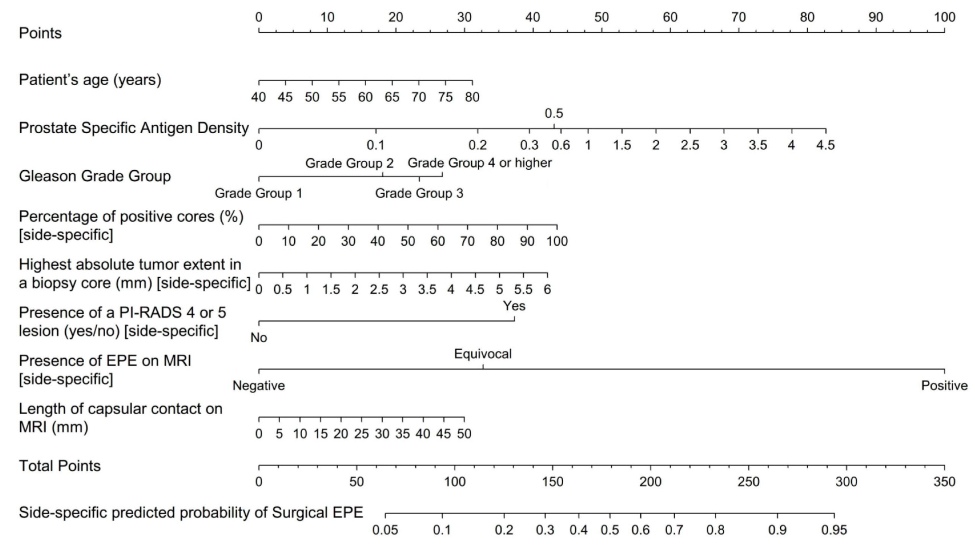
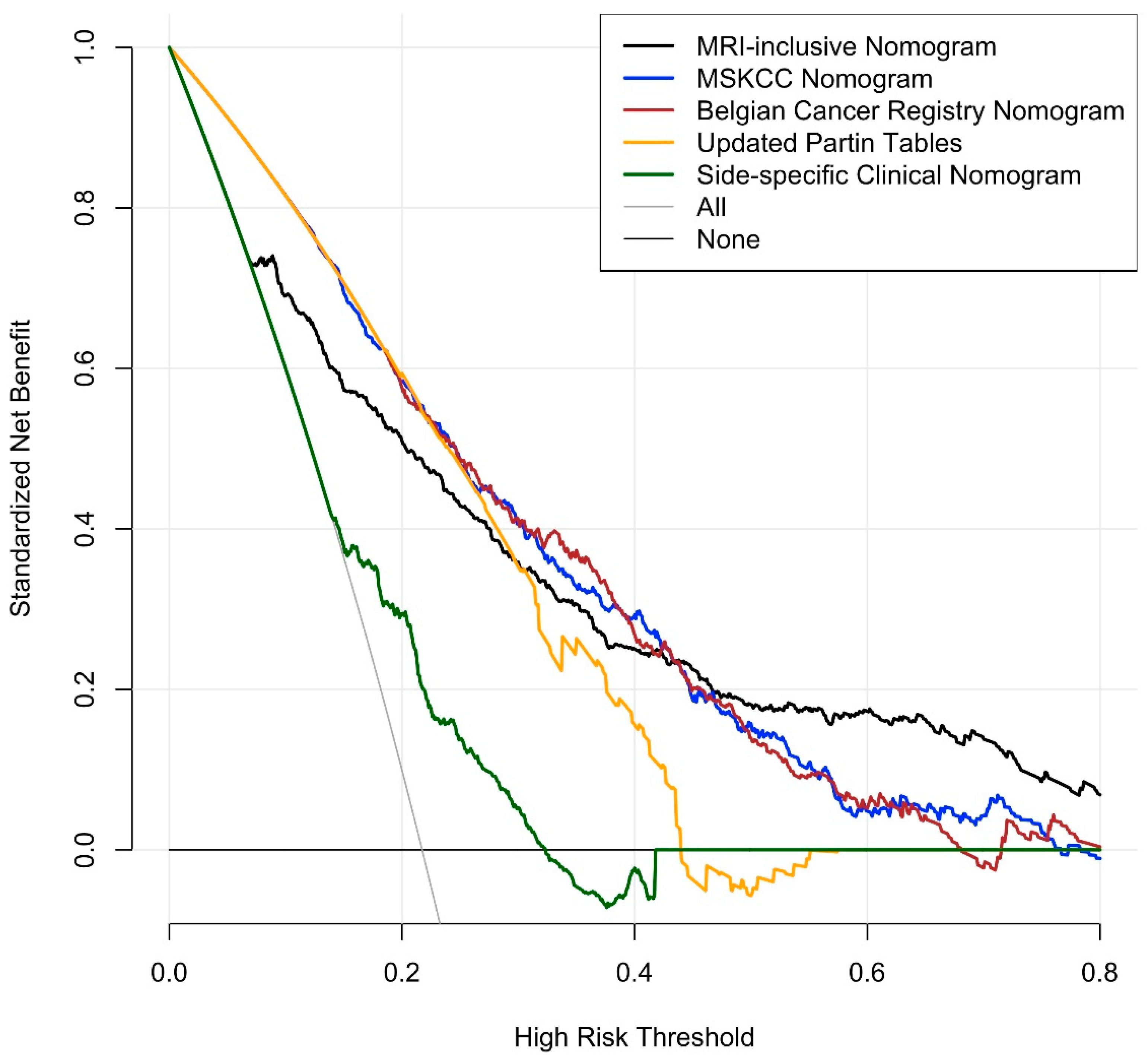
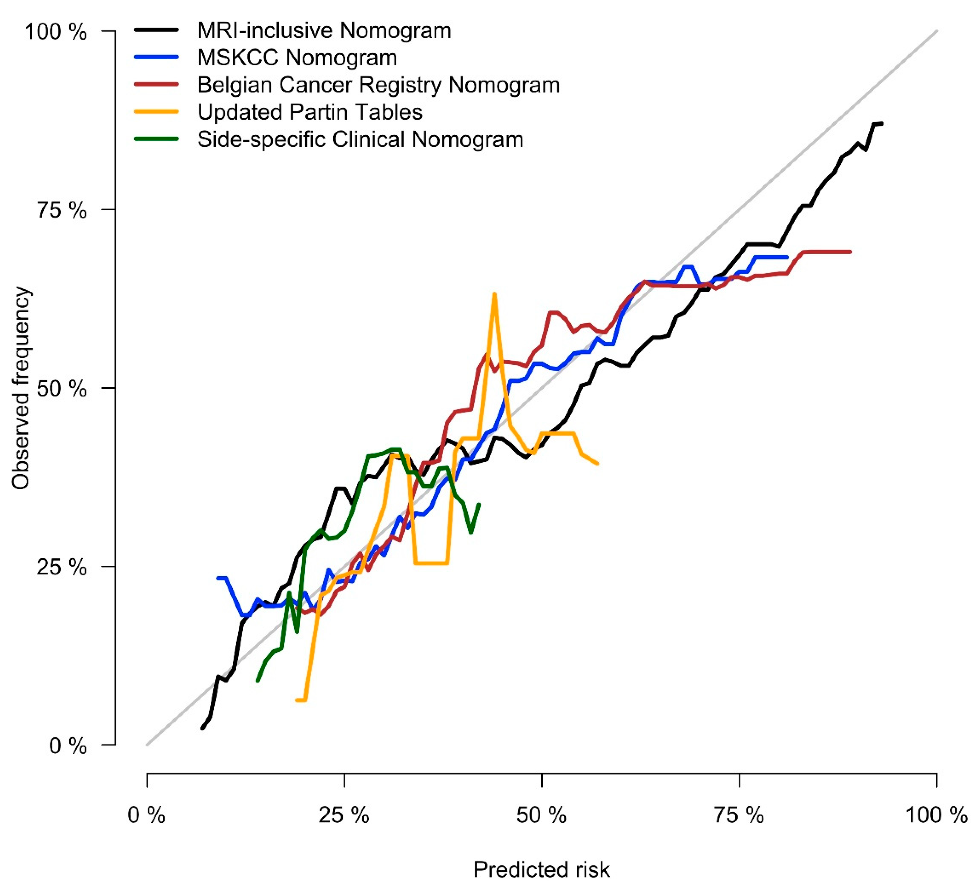
International Multi-Site Initiative to Develop an MRI-Inclusive Nomogram for Side-Specific Prediction of Extraprostatic Extension of Prostate Cancer
by
 , , , , , , , , , , , , , , , and
Andreas G. Wibmer
1,* ,
,
Michael W. Kattan
2,
Francesco Alessandrino
3,
Alexander D. J. Baur
4,
Lars Boesen
5,
Felipe Boschini Franco
3,
David Bonekamp
6,
Riccardo Campa
7,
Hannes Cash
4,8,
Violeta Catalá
9,10,
Sebastien Crouzet
11,
Sounil Dinnoo
12,
James Eastham
13,
Fiona M. Fennessy
3,
Kamyar Ghabili
14,
Markus Hohenfellner
15,
Angelique W. Levi
16,
Xinge Ji
2,
Vibeke Løgager
5,
Daniel J. Margolis
17,
Paul C. Moldovan
11,
Valeria Panebianco
7,
Tobias Penzkofer
4,18,
Philippe Puech
12,
Jan Philipp Radtke
6,15,
Olivier Rouvière
11,19,
Heinz-Peter Schlemmer
6,
Preston C. Sprenkle
14,
Clare M. Tempany
3,
Joan C. Vilanova
20 ,
,
Jeffrey Weinreb
21,
Hedvig Hricak
1 and
Amita Shukla-Dave
1add
Show full author list
1
Department of Radiology, Memorial Sloan Kettering Cancer Center, 1275 York Avenue, New York, NY 10065, USA
2
Department of Quantitative Health Sciences in the Lerner Research Institute, Cleveland Clinic, Cleveland, OH 44195, USA
3
Department of Radiology, Brigham and Women’s Hospital, Harvard Medical School, Boston, MA 02115, USA
4
Charité University Hospital, 10117 Berlin, Germany
5
Herlev Gentofte University Hospital, 2730 Herlev, Denmark
6
DKFZ German Cancer Research Center, 69120 Heidelberg, Germany
7
Department of Radiological Sciences, Oncology & Pathology, Sapienza University of Rome, 00185 Rome, Italy
8
Department of Urology, University Magdeburg, 39120 Magdeburg, Germany
9
Department of Radiology, Fundació Puigvert, 08025 Barcelona, Spain
10
Department of Uro-Radiology, Creu Blanca, 08034 Barcelona, Spain
11
Hospices Civils de Lyon, Hôpital Edouard Herriot, 69003 Lyon, France
12
Genitourinary and Women’s Imaging Departments, Lille University Hospital, 59037 Lille, France
13
Department of Surgery, Memorial Sloan Kettering Cancer Center, New York, NY 10065, USA
14
Department of Urology, Yale School of Medicine, New Haven, CT 06510, USA
15
Department of Urology, University Hospital of Heidelberg, 69120 Heidelberg, Germany
16
Department of Pathology, Yale School of Medicine, New Haven, CT 06510, USA
17
Weill Cornell Medicine, Weill Cornell Imaging, New York-Presbyterian Hospital, New York, NY 10021, USA
18
Berlin Institute of Health (BIH), 10178 Berlin, Germany
19
Faculté de Médecine Lyon Est, Université de Lyon, 69003 Lyon, France
20
Clínica Girona, Institute Catalan of Health-IDI, University of Girona, 17004 Girona, Spain
21
Department of Radiology, Yale School of Medicine, New Haven, CT 06510, USA
*
Author to whom correspondence should be addressed.
Cancers 2021, 13(11), 2627; https://doi.org/10.3390/cancers13112627
Submission received: 31 March 2021
/
Revised: 29 April 2021
/
Accepted: 21 May 2021
/
Published: 27 May 2021
(This article belongs to the Special Issue Transformational Role of Medical Imaging in Oncology)
Simple Summary
For patients with newly diagnosed prostate cancer, it is important to detect tumor growth beyond the prostate, as this can affect a patient’s prognosis, influence management decisions, and alter treatment strategies. It is recognized that on prostate MRI, some instances of extraprostatic tumor growth can be missed. In this study, we merged patient data from multiple hospitals in different countries and developed a type of mathematical formula called “nomogram” that combines MRI findings with other available patient data. The results of our study allow physicians to more accurately diagnose extraprostatic tumor growth by combining clinical, biopsy, and MRI-derived information according to their relative statistical importance.
Abstract
Background: To develop an international, multi-site nomogram for side-specific prediction of extraprostatic extension (EPE) of prostate cancer based on clinical, biopsy, and magnetic resonance imaging- (MRI) derived data. Methods: Ten institutions from the USA and Europe contributed clinical and side-specific biopsy and MRI variables of consecutive patients who underwent prostatectomy. A logistic regression model was used to develop a nomogram for predicting side-specific EPE on prostatectomy specimens. The performance of the statistical model was evaluated by bootstrap resampling and cross validation and compared with the performance of benchmark models that do not incorporate MRI findings. Results: Data from 840 patients were analyzed; pathologic EPE was found in 320/840 (31.8%). The nomogram model included patient age, prostate-specific antigen density, side-specific biopsy data (i.e., Gleason grade group, percent positive cores, tumor extent), and side-specific MRI features (i.e., presence of a PI-RADSv2 4 or 5 lesion, level of suspicion for EPE, length of capsular contact). The area under the receiver operating characteristic curve of the new, MRI-inclusive model (0.828, 95% confidence limits: 0.805, 0.852) was significantly higher than that of any of the benchmark models (p < 0.001 for all). Conclusions: In an international, multi-site study, we developed an MRI-inclusive nomogram for the side-specific prediction of EPE of prostate cancer that demonstrated significantly greater accuracy than clinical benchmark models.