Review

Renal Tumors of Childhood—A Histopathologic Pattern-Based Diagnostic Approach

by
Ariadne H.A.G. Ooms
1,2,†,
Gordan M. Vujanić
3,†,
Ellen D’Hooghe
4,
Paola Collini
5,
Aurore L’Herminé-Coulomb
6,
Christian Vokuhl
7,
Norbert Graf
8,
Marry M. van den Heuvel-Eibrink
1 and
Ronald R. de Krijger
1,9,*
1
Princess Máxima Center for pediatric oncology, 3584 CS Utrecht, The Netherlands
2
Pathan B.V., 3045 PM Rotterdam, The Netherlands
3
Department of Pathology, Sidra Medicine, Doha 0000, Qatar
4
Department of Pathology, Oslo University Hospital, Rikshospitalet, 0372 Oslo, Norway
5
Department of Diagnostic Pathology and Laboratory Medicine, Fondazione IRCCS Istituto Nazionale dei Tumori, 20133 Milan, Italy
6
Sorbonne Université, Department of Pathology, Hôpital Armand Trousseau, Hopitaux Universitaires Est Parisien, 75012 Paris, France
7
Section of Pediatric Pathology, Department of Pathology, University Hospital Bonn, 53127 Bonn, Germany
8
Department of Pediatric Oncology & Hematology, Saarland University, D-66421 Homburg, Germany
9
Department of Pathology, University Medical Center Utrecht, 3584 CX Utrecht, The Netherlands
*
Author to whom correspondence should be addressed.
These authors contributed equally to this paper as first authors.
Cancers 2020, 12(3), 729; https://doi.org/10.3390/cancers12030729
Submission received: 2 February 2020 / Revised: 4 March 2020 / Accepted: 7 March 2020 / Published: 19 March 2020
(This article belongs to the Special Issue Pathogenesis and Diagnosis of Genitourinary Cancer)

Abstract

:
Renal tumors comprise approximately 7% of all malignant pediatric tumors. This is a highly heterogeneous group of tumors, each with its own therapeutic management, outcome, and association with germline predispositions. Histopathology is the key in establishing the correct diagnosis, and therefore pathologists with expertise in pediatric oncology are needed for dealing with these rare tumors. While each tumor shows different histologic features, they do have considerable overlap in cell type and histologic pattern, making the diagnosis difficult to establish, if based on routine histology alone. To this end, ancillary techniques, such as immunohistochemistry and molecular analysis, can be of great importance for the correct diagnosis, resulting in appropriate treatment. To use ancillary techniques cost-effectively, we propose a pattern-based approach and provide recommendations to aid in deciding which panel of antibodies, supplemented by molecular characterization of a subset of genes, are required.

1. Introduction

In Europe, each year about 1000 children are diagnosed with a renal tumor, thereby comprising approximately 7% of all pediatric malignant tumors. Approximately 90% of pediatric renal tumors comprise Wilms tumor (WT), the remainder consisting of clear cell sarcoma of the kidney (CCSK), malignant rhabdoid tumor of the kidney (MRTK), renal cell carcinoma (RCC), congenital mesoblastic nephroma (CMN), and other rare tumors (Table 1) [1]. They represent a very heterogeneous group, which includes a spectrum of tumors from those with low malignant potential (e.g., CMN) to highly aggressive tumors (e.g., MRTK) [2,3], requiring different treatment, and showing remarkable intra-tumor histologic and genetic heterogeneity [4,5]. In addition, it has been recognized that an increasing number of pediatric renal tumors have been associated with specific syndromes or diseases (Table S1) [6], which highlights the importance of precise tumor diagnosis, not only for appropriate treatment, but also for genetic counseling.
While imaging techniques are continuously improving, they are still unable to reliably diagnose and distinguish the most relevant pathology based risk groups, although some new techniques (like MRI-Diffusion Weighted Imaging) might be helpful in the near future [7]. The clinical characteristics of children with renal tumors are similar, although some (like age distribution) may be helpful in the differential diagnosis [8].
In nearly all tumors, the final diagnosis and classification of clinically relevant (sub)types still relies on histopathologic examination. Updated pathology guidelines for handling renal tumors have been published recently by the Renal Tumor Study Group of the International Society of Paediatric Oncology/Société Internationale D’Oncologie Pédiatrique (SIOP-RTSG) in the SIOP-RTSG UMBRELLA 2016 protocol [9]. These guidelines recommend that tumors should be extensively sampled, to allow diagnostic classification, staging, and to capture tumor heterogeneity [9]. Difficulties in achieving the correct histologic classification may arise due to the ability of tumors to display a variety of overlapping histologic patterns. This stresses the need for adequate tumor sampling, since it can reveal other important histopathologic clues. Biopsies, performed in selected cases only, might pose even more difficulties, due to the limited availability of material [10]. Finally, in some cases where abdominal tumors are so large that the kidney and adjacent organs are unrecognizable, even non-renal tumors have to be included into the differential diagnosis. In many cases, additional ancillary techniques are needed to establish the final diagnosis. Since clinically relevant discrepancies between the diagnosis and/or staging of institutional pathologists and central pathology review persist [11,12], rapid central pathology review is now mandatory in both the Children’s Oncology Group (COG) and SIOP studies, in order to establish the correct diagnosis and stage within a clinically relevant timeframe and start appropriate treatment.
In this review, to aid in the diagnostic process, we use a pattern-based diagnostic approach for the most common pediatric renal tumors, by discussing pattern-based differential diagnoses and in providing recommendations for the use of ancillary techniques.

2. A Pattern-Based Approach to the Use of Ancillary Techniques in Pediatric Renal Tumors

In general, there are three major histologic patterns which are the basis for the differential diagnoses of any tumor, including pediatric renal tumors: an epithelial, a mesenchymal, and an undifferentiated pattern (Table 2, Table 3 and Table 4). Most tumors harbor histological clues to the diagnosis, although these can be present only focally. In WTs, all three patterns are often present, but many WTs show only one or two patterns (Figure 1, Figure 2 and Figure 3), or show the other patterns only focally. This underlines the reasons why extensive sampling is required, in order to capture all tumor features [9]. The most common pediatric renal tumors have been described and reviewed extensively elsewhere [3,13,14,15,16], so we only briefly recapitulated their main histologic features (Table S2). Since some tumors harbor a variety of histologic patterns, they appear in more than one of these categories.

2.1. Epithelial Pattern
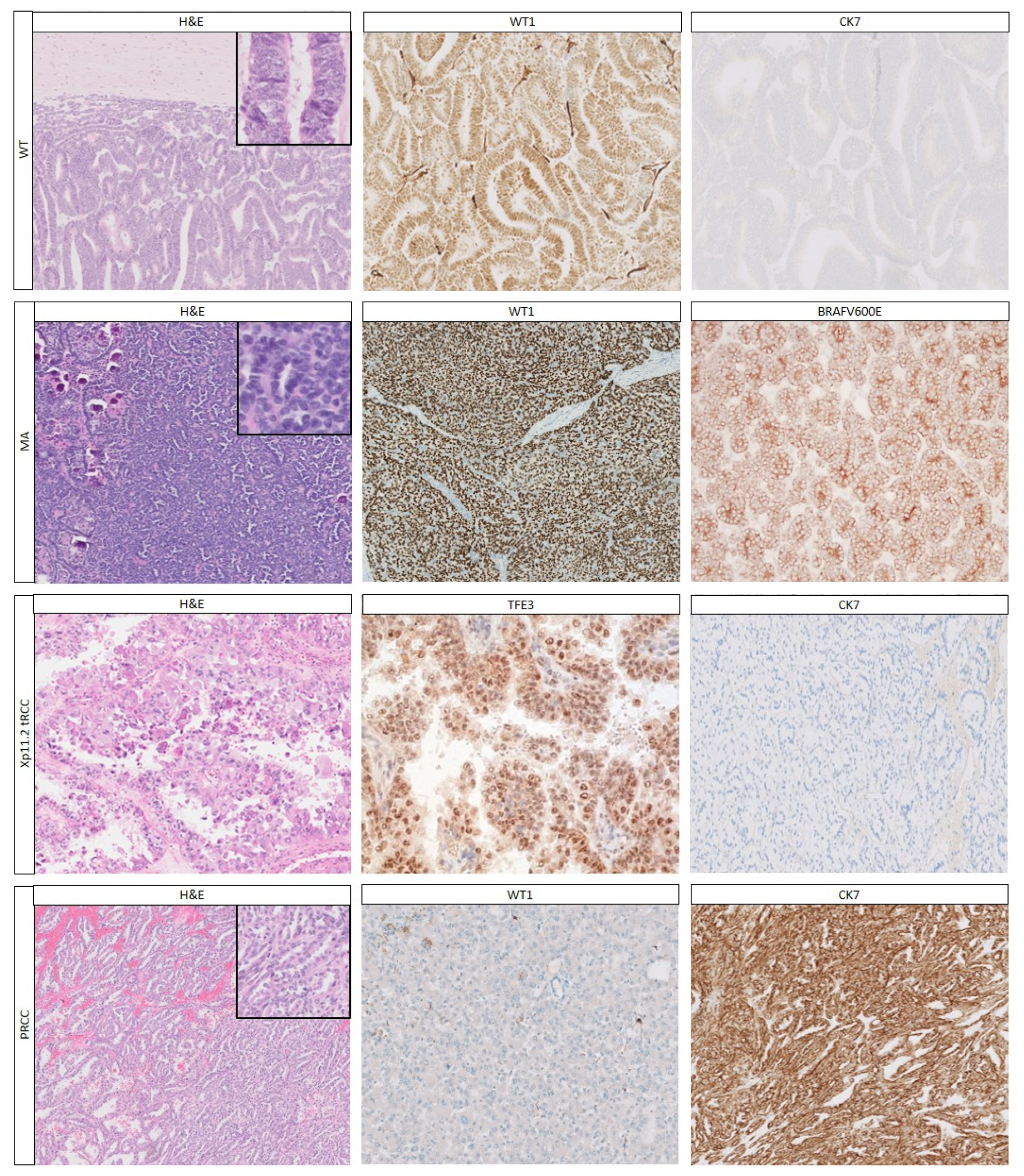
2.1.1. Histology

Tumors demonstrating a pure epithelial pattern include epithelial type WT, hyperplastic perilobar nephrogenic rests, RCC and metanephric adenoma (MA), with WT being by far the most common one.
Epithelial WTs (Figure 1) usually occur in younger children (median age 15 months), and about 80% of cases are stage I [54]. The tumor consists of epithelial structures, which may demonstrate the whole spectrum of nephrogenesis, varying from poorly and moderately differentiated to mature tubules and glomeruli-like structures. Tumor cells are often columnar with elongated, hyperchromatic and crowded nuclei, inconspicuous nucleoli, and scant cytoplasm. Mitoses are often easily identified. For WTs that consist entirely of poorly and moderately differentiated tubules, the diagnosis is more difficult, and tumors, such as papillary RCC and MA, have to be excluded.
If nephrogenic rests (NRs) are identified, the diagnosis of WT can be reliably established since NRs are virtually never associated with non-WTs [55]. NRs are foci of embryonal cells that persist after 36 weeks of gestation and they are considered precursors of WTs [56]. NRs accompany WTs in approximately 40% in unilateral WTs and in >90% of bilateral WTs [56,57]. NRs are subdivided into two main subtypes: perilobar nephrogenic rests (PLNRs) and intralobar nephrogenic rests (ILNRs). PLNRs are confined to the periphery of the renal lobe, while ILNRs can be found anywhere within the renal lobe [56,58]. Nephroblastomatosis is defined as the “diffuse or multifocal presence of NRs” [56]. Hyperplastic NRs can be difficult to distinguish from WTs, as there are no histologic, immunohistochemical or molecular features that are helpful, so the diagnosis is commonly based on experts’ consensus. The presence of a fibrous capsule between the lesion and renal parenchyma may be helpful in diagnosing a WT in patients treated with primary surgery, but not in those treated with preoperative chemotherapy, where a pseudocapsule is often found around NRs (GMV, personal observation).
RCC is the most important tumor in the differential diagnosis of epithelial WT, particularly in older children [27]. Overall, RCCs account for only ~2–4% of pediatric renal tumors, but this increases to >50% in children older than 12 years [27]. The most prevalent RCC types in adulthood (e.g., clear cell RCC, papillary RCC) are less common in childhood [27], but it is papillary RCC (PRCC) which needs to be considered in children in the differential diagnosis of epithelial WTs, due to the overlapping histologic architecture and cell type. PRCCs can be separated into Type 1 and Type 2. Type 1 is composed of small cuboidal cells with scant pale cytoplasm arranged as single layers on papillae, tubules and glomerulus-like structures, and often contain foamy macrophages and psammoma bodies, in contrast to WTs. Type 2 PRCCs show papillae lined with pseudostratified cells, abundant eosinophilic to occasionally clear cytoplasm, and atypical nuclei with prominent nucleoli, which is not a common feature in WT. PRCCs usually have no pseudocapsule, and lack a blastemal and stromal component.
In children, the Microphtalmia-associated Transcription factor family translocation RCC (MiT-RCC) is the most common RCC subtype (42% of all pediatric RCCs), comprised of Xp11.2 tRCC (~90% of cases) and t(6;11) RCC [27]. Since these tumors have distinct histologic characteristics (Table S2), they are usually not difficult to diagnose and distinguish from WT, but the characteristic features can be present only focally, and may thus, be easily overlooked [14].
In addition, there are some RCC subtypes, which are generally rare, with very few cases reported in children. Still, they are important to recognize, since some of them are associated with germline mutations or syndromes, including Anaplastic Lymphoma Kinase (ALK)-rearranged-RCC (ALK-RCC), Succinate Dehydrogenase-deficient renal cell carcinomas (SDH-RCC), and Hereditary Leiomyomatosis and Renal Cell Carcinoma-RCC (HLRCC-RCC) [59]. Most of these tumors show characteristic histologic features (Table S2), although these can be present only focally, and immunohistochemistry and molecular analysis will aid in establishing the correct diagnosis.
Metanephric adenoma is another tumor which can be difficult to distinguish from WTs and RCCs, especially in older children. Histologically, MAs consist of small, uniform closely packed tubules and papilla (Figure 1). Features which can aid in distinguishing them from WTs are the absence of a pseudocapsule, sharp demarcation from the renal parenchyma, the presence of small uniform cells with scant cytoplasm, bland nuclei, fine chromatin, absence of atypia, psammoma bodies, and rare mitoses (in epithelial WTs mitoses are more common) [17,60]. Nevertheless, in a recent report, tumors with histologic and molecular overlap between MA and epithelial WTs were described [61].
Other types of RCC that are more common in adulthood and rare in children are extensively described in literature and not further described here [62,63,64,65]. Finally, in addition to tumors that are genuinely epithelial, there are tumors that can have an epithelioid appearance, such as MRTKs, CCSKs (see below), and epithelioid angiomyolipoma. However, these tumors have other features (such as fat and/or aberrant vessels in epithelioid angiomyolipomas), which distinguish them from tumors with a genuine epithelial pattern.

2.1.2. Immunohistochemistry

Immunohistochemistry using a small panel of immunohistochemical markers (Table 2) including WT1, pan-cytokeratin (pan-CK), and CK7 is helpful in reaching a definite diagnosis in most cases. Nevertheless, immunohistochemistry does not reliably differentiate between NRs and WTs.
In pure epithelial WTs, WT1 is positive in 80–90% of WTs, while all RCC types are negative for WT1 [19,40]. Translocation RCCs are often only weakly positive for keratins, but strongly positive for melanocytic markers (TFEB-tRCCs more commonly and more diffusely than TFE3-tRCCs). Xp11.2 RCC and t(6;11) RCCs show nuclear TFE3 (Figure 1), and TFEB positivity, respectively [14,66]. Notably, a diagnostic pitfall in TFE3 positive RCCs, is that the rare ALK-RCCs also stain positive [67]. Depending on the RCC subtype, further immunohistochemical stains (e.g., CK7, AMACR, CD10, vimentin, cathepsin K) will help establish the diagnosis and rule out WT [20,22,29,68].
If both WT1 and pan-CK are positive, the main differential diagnosis remains between epithelial WT and MA. The majority (~90%) of MAs carry a BRAF mutation (Table 5), and the most common one (BRAFV600E) can be demonstrated by immunohistochemistry (see also below). Interestingly, a recent study showed BRAFV600E staining in both MA and epithelial WTs with overlapping features [61]. Additionally, diffuse CD57 staining favors the diagnosis of MA, but does not rule out WT [20], whereas CK7 negativity of both WTs and MAs distinguishes them from PRCC [17,20].
In general, all other tumors which can show an epithelioid pattern (MRTKs, CCSKs, epithelioid angiomyolipomas) are negative for WT1 [40]. To further distinguish between these tumors, the panel of antibodies can be extended (Table 6), for example with PAX8 for the distinction of TFE-tRCCs from epithelioid angiomyolipomas, since the latter are also positive for melanocytic markers and angiomyolipomas may also have Xp11 (TFE3-related) translocations (next sections; Table 7).

2.1.3. Genetics

Genetic analysis plays a minor role in the identification of WTs, since >50 driver mutations are known in WTs (Table 5), but the most commonly mutated genes may be analyzed to exclude other entities [69]. Interestingly, TRIM28 mutations have been associated with a specific subset of purely epithelial WTs [70].
Type 1 PRCCs show MET alterations in 81%, and 25% of type 2 PRCCs harbor CDKN2A genomic abnormalities [71]. MiT-RCCs harbor translocations that lead to fusions with different genes: Xp11.2 tRCCs harbor translocations of the Transcription Factor E3 (TFE3) gene with different fusion partners (Table 7), and t(6;11) RCC fuses Transcription Factor EB (TFEB) to MALAT1. Since some antibodies can be technically challenging and sensitive to suboptimal fixation (e.g., TFE3 and TFEB) [72], Fluorescent in situ hybridization (FISH) break apart probes detecting the TFE3 [73], TFEB [74], and ALK translocation [67,75,76] can be used (Table 7), although false negative results have been reported.

2.2. Mesenchymal Pattern

2.2.1. Histology

Mesenchymal tumors generally consist of spindle cells arranged in different patterns, such as short or long bundles, fascicles or storiform architecture. WTs can consist (nearly) completely of mesenchymal cells (Figure 2), and thorough sampling can help revealing important histologic clues to the diagnosis of WTs (such as the finding of NRs or small foci of other WT components). CCSK, CMN and metanephric stromal tumor (MST) are the most important differential diagnostic considerations. In rare cases, sarcomas (e.g., synovial sarcoma), inflammatory myofibroblastic tumors, and RCCs with sarcomatous differentiation enter the differential diagnosis.
Histologically, different patterns exist in CCSKs, leading to a substantial number of misdiagnoses [38]. Nevertheless, approximately 90% of CCSKs show, at least focally, the classic pattern with bland ovoid cells with monomorphous nuclei with finely dispersed chromatin and small nucleoli, which are arranged in cords and nests separated by a chicken-wire vasculature (Figure 2) [13,38].
In very young children, CMN is another important consideration. It encompasses three histologic variants: classic, cellular, and mixed, containing areas of both cellular and classic histology (Table S2). The classic type consists of bland spindle cells organized in an interlacing fascicular pattern, embedded in collagenous stroma blending into the pre-existent renal parenchyma, with entrapment of normal renal parenchyma. The cellular type has a higher cellularity of ovoid cells with less cytoplasm, organized in solid sheets, and with a higher proliferation rate, and a rather clear border towards the renal parenchyma [3].
MST is a rare tumor, which histologically resembles CMN, but occurs at an older age than CMN. It shows some characteristic histologic features, including alternating cellularity, a scalloped border with the renal parenchyma, perivascular cuffing, and angiodysplasia (Table S2; Figure 2) [42].
When the most common renal tumors showing a mesenchymal pattern are excluded, some very rare tumors, such as primary renal sarcomas need to be considered, especially in older children [77]. Interestingly, primary renal synovial sarcomas are usually monophasic [77]. Although, rare in the kidney, inflammatory myofibroblastic tumor is another diagnostic consideration.
Finally, in teenagers and young adults, sarcomatous dedifferentiation of RCC types (such as clear cell RCC) is a phenomenon to consider in the differential diagnosis. Interestingly, in these spindle cell areas, immunoreactivity for markers normally seen in the original tumor type is often lost, whereas most cases show abnormal p53 immunoreactivity suggestive of a TP53 mutation [78]. Additional sampling in search for the original tumor type is warranted.

2.2.2. Immunohistochemistry

Although, many renal tumors with a mesenchymal pattern lack a characteristic immunohistochemical profile (Table 3), a panel of markers can be of added value to differentiate the tumors (Table 3, Table 6). In the stromal component of WTs, nuclear staining for WT1 is often weak or absent. CCSKs show a distinctive immunohistochemical profile, with strong CyclinD1, BCL6 corepressor (BCOR), and NGFR staining, and negative WT1 staining (Figure 2; Table 3) [31,32,33,34,39]. CMNs have a rather non-specific immunohistochemical pattern (Figure 2; Table 3), although pan-Trk staining can be used to diagnose cellular CMNs harboring the ETV6-NTRK3 translocation (see below) [79]. CMNs may show variable CyclinD1 positivity [31,32], and in very rare cases even WT1 positivity [40]. The majority of MSTs are BRAFV600E (~65%) and CD34 positive (Figure 2), while the other tumors are usually negative [18,35,42,80]. The rare primary renal synovial sarcomas are positive for CD99, vimentin and TLE1, and often demonstrate at least focal expression of cytokeratins and EMA, but can be negative or only weakly positive for INI1 [81]. Furthermore, nearly all stromal WTs and synovial sarcomas show strong BCL-2 staining, whereas approximately half of CCSK and all CMNs are negative [35].

2.2.3. Genetics

If histologic and immunohistochemical features are insufficient to reach a final diagnosis, molecular analysis can help in the distinction of some mesenchymal tumors (Table 5 and Table 7). As mentioned in previous sections, many driver genes are involved in WTs (Table 5), which may be included in a targeted sequencing panel. In CCSKs, ~85% harbor the BCOR-internal tandem duplication (BCOR-ITD) of exon 15, and ~10% show the t(10;17) leading to YWHAE-NUTM2 fusion. These two molecular abnormalities are mutually exclusive and can be demonstrated by FISH and/or sequencing techniques [82,83]. Rare CCSKs harbor the BCOR-CCNB3 fusion [39]. The recently reported EGFR-ITDs rearrangements in all CMN types, together with the previously known t(12;15) (ETV6-NTRK3 translocation) in cellular CMNs help to confirm the diagnosis of CMN [84,85]. BRAF V600e mutations in MSTs can be identified by immunohistochemistry or sequencing [86]. A PCR study on 22 CMNs (including 10 cellular CMNs) showed no BRAF V600e mutations [80]. In one study 2 out of 3 cellular CMN which showed no ETV6 translocation, showed different rearrangements in the BRAF oncogene on WGS [84]. The diagnosis of synovial sarcoma can be established by demonstrating the characteristic translocation t(X;18)(p11.2;q11.2), present in >90% of cases [87].

2.3. Undifferentiated Pattern

2.3.1. Histology

Some pediatric renal tumors have an undifferentiated “small round blue cell” appearance, such as blastemal WTs, MRTKs, as well as tumors that rarely involve or infiltrate the kidney, such as (poorly differentiated/undifferentiated) neuroblastoma, desmoplastic small round cell tumor (DSRCT), and Ewing sarcoma (EWS) (Table S2). In addition, lymphoma should always be considered and excluded. Other tumors described above (e.g., CCSK) occasionally show an undifferentiated appearance.
The blastemal WT component consists of undifferentiated, small blue round cells with a high cellularity of round to ovoid cells with a high nuclear–cytoplasmic ratio, and brisk mitotic activity (Figure 3). Blastema often shows different histologic patterns, such as nodular, trabecular, diffuse, and serpentine [88].
MRTKs are aggressive infiltrating tumors with a very poor prognosis, often presenting in patients younger than two years of age [2,15]. They are highly cellular, non-encapsulated tumors composed of infiltrating sheets of non-cohesive cells with a large eccentric nucleus, and abundant eosinophilic cytoplasm (Figure 3) (Table S2) [15]. However, MRTKs may show different patterns, which sometimes lead to difficulties in achieving a correct diagnosis on routine H&E staining only [15].
Neuroblastomas often originate in the adrenal gland, but very rarely in the kidney. Clinically, most neuroblastomas can be suspected by elevated levels of catecholamines in urine and serum. Histologically, the nuclei of the undifferentiated cells show the characteristic coarse ‘salt and pepper’ chromatin. Neuroblastomas lack a pseudocapsule, and often show neuroblasts exhibiting variable degrees of differentiation up to ganglion cells and neuropil, which is the basis for histologic classification [89].
DSRCTs have been rarely described in the kidney [90]. Histologically, they consist of solid nests of round to oval cells and show necrosis and occasional pseudoglandular-/rosette formation [44,91]. Intriguingly, renal DSRCTs show no desmoplasia, making their histologic diagnosis even more difficult [90].
Primary renal sarcomas rarely occur, and are histologically characterized by undifferentiated morphology. EWS can originate in the kidney or develop in the retroperitoneum with infiltration of the kidney [45,92]. These tumors consist of small round to oval cells with scanty cytoplasm, molding, high mitotic activity, and with condensation around blood vessels [45]. Two newly described sarcomas, very rarely seen in children in the kidney, are the CIC-DUX4 translocation sarcoma and the BCOR-CCNB3 translocation sarcomas [93]. Both tumors often show a spectrum of round cells to more epithelioid and spindle cells, often present at least focally [93,94]. The CIC-DUX4 sarcomas behave highly aggressively, often metastasizing early in the disease course [93,94].
Renal medullary carcinoma (RMC) and collecting duct carcinoma (CDC) may show significant histologic overlap with MRTKs. Helpful diagnostic clues are the presence of a reticular or cribriform pattern and finding of sickle cells in RMC [95], while dysplastic changes in the adjacent tubules and a dense inflammatory infiltrate suggest CDC [95,96].

2.3.2. Immunohistochemistry

The differential diagnosis of “small blue round cell tumors” of the kidney can be difficult, but most of these tumors can be classified using ancillary techniques (Table 4 and Table 5). The blastemal component of WTs demonstrates nuclear WT1 positivity in 80% of cases [17,46]. DSRCTs are also WT1 positive [44], but only with the C-terminal clone of WT1, while WTs are positive, with both the N-terminal and C-terminal clone [40,44,47,48]. DSRCTs also show dotlike cytokeratin and desmin positivity [16,44]. Blastemal type WTs and neuroblastomas are both positive for CD56, but WT1 is negative in neuroblastoma [16,40,47], while synaptophysin, chromogranin A, NB84, and PHOX2B are positive in neuroblastoma [49,53]. Furthermore, WTs are PAX8 positive, while neuroblastoma, DSRCT and sarcoma are PAX8 negative [34]. MRTKs can be rather easily distinguished from all other pediatric renal tumors by their complete loss of INI1 staining (Figure 3) and PAX8 negativity.
However, RMCs can also show near complete loss of INI1 immunoreactivity, but are PAX8 positive [16,52,96,97]. In EWS, CD99 (diffuse strong membranous) and cyclinD1 (nuclear), in combination with lack of reactivity to WT1, discriminates it from WT and almost all other tumors [16,51]. Of note, CIC-DUX4 translocation sarcomas have been reported to show WT1 staining in >90%, while BCOR-translocation sarcomas were reported to show strong staining of BCOR, BCL-2, cyclinD1, and TLE1 staining in most cases (each 80–90%) [93,94]. Further differentiation of undifferentiated round cell sarcomas needs molecular studies (below).

2.3.3. Genetics

In difficult cases, next generation (targeted) sequencing can be performed including genes frequently mutated or altered in WTs (e.g., WTX, WT1, CTNNB1, SIX1, SIX2, miRNA processing genes) and neuroblastomas (PHOXB2, ALK, ARID1A/B genes) (Table 5). Notably, MYCN can show alterations (mutations as well as gains) in both WT and neuroblastoma [98].
MRTKs are characterized by inactivating mutations in the chromatin-remodeling complex members INI1 (also referred to as SMARCB1; 95%) or SMARCA4 (5%), or show deletions of 22q11 (on which INI1 is located) [99,100,101]. Therefore, sequencing of INI1 and SMARCA4 is helpful in establishing a diagnosis of MRTK. Notably, when patients with a MRTK have a synchronous or metachronous rhabdoid tumor affecting other organs (including the central nervous system, referred to as Atypical Teratoid/Rhabdoid Tumor (AT/RT)), it is highly suggestive of the rhabdoid tumor predisposition syndrome [102].
The diagnosis of DSRCT is based on the demonstration of the characteristic t(11;22)(p13;q12) leading to EWS-WT1 fusion [90]. In contrast, EWS show a wide variety of translocations involving the EWS gene (Table 5 and Table 7), most commonly t(11;22)(q24;q12) resulting in the EWS-FLI1 fusion [45,92]. In EWS-negative round cell sarcomas, CIC-DUX4 gene fusion (resulting from either t(4;19) or t(10;19)), is the most common genetic abnormality detected [94]. The remainder includes BCOR-CCNB3 translocation sarcomas, harboring the same translocation reported in some CCSKs, suggesting that they might be in the same spectrum [93,103].

2.4. Biphasic Tumors

In the differential diagnosis of WTs with stromal and epithelial components, Metanephric adenofibroma (MAF) needs to be considered, despite its rarity. MAFs show features of both MST and MA (as described above), including positive nuclear staining for WT1 in the epithelial component, and positive CD34 staining in the stromal component [104,105]. Furthermore, approximately 50% of MAFs carry the BRAF V600E mutation, although rare WTs may also harbor a BRAF mutation [18,61,106].

3. Future Perspectives

In addition to Hematoxylin and Eosin (H&E)-based diagnosis, with the aid of immunohistochemistry and targeted molecular analysis, there are now several developments, which may revolutionize tumor diagnosis in the near future. First of all, genome-wide molecular methods, including whole exome sequencing, RNA sequencing, and methylation arrays, allow unprecedented molecular classification of tumors, as has been shown for brain tumors and renal cell carcinoma [107,108]. While tumor heterogeneity, which is undoubtedly present in Wilms tumors, might be an obstacle to these genome wide molecular methods, liquid biopsy holds the promise that it would be able to circumvent this problem, as cell-free DNA in the blood might be representative of the entire tumor [109]. Another strategy, by which pediatric renal tumors might be classified, is based on recent advancements in the field of radiology, especially the addition of diffusion-weighted imaging to standard magnetic resonance imaging, with which apparent diffusion coefficient metrics may predict the composition of renal tumors, especially that of WTs [110]. Finally, through digitalization of traditional H&E slides, so-called whole slide images can now be analyzed by machine learning techniques. This has already resulted in automated recognition of specific tissue components, such as lymph node metastases in breast cancer [7]. It is certainly conceivable that such automated recognition will also lead to computer-assisted classification of tumor types, including pediatric renal tumors.

4. Conclusions

The differential diagnosis of pediatric renal tumors can be challenging if based solely on H&E staining, due to the presence of similar cell types and overlapping histologic patterns across these tumors. Extensive sampling may reveal the presence of distinctive histologic features, allowing a diagnosis or narrowing the differential diagnosis. However, the use of ancillary techniques is necessary in many cases. Many ancillary techniques are integrated now in daily pathology practice and provide the pathologist with tools to achieve the correct diagnosis, which contributes to multidisciplinary decision-making regarding the appropriate treatment and counselling for each child with a renal tumor. Since there is a large repertoire of immunostainings and genetic aberrations, the challenge lies in choosing a restricted panel of ancillary techniques, which results in an appropriate diagnosis in an acceptable time frame and at an acceptable cost. In this review, we used a pattern-based approach to discuss the differential diagnosis, and provided recommendations for panels of ancillary techniques. The focused use of ancillary techniques will likely result in fewer unclassified cases, and lead to an increase in children diagnosed with germline mutations. This will result in patients benefitting from receiving adequate therapies and counseling.

Supplementary Materials

The following are available online at https://www.mdpi.com/2072-6694/12/3/729/s1, Table S1: Syndromes associated with the most common pediatric renal tumors; Table S2: Histopathological, immunohistochemical and genetic aberrations useful in the differential diagnosis of the most common pediatric renal tumors.

Author Contributions

Writing—original draft preparation, A.H.A.G.O., G.M.V., R.R.d.K.; writing—review and editing by all authors. All authors have read and agreed to the published version of the manuscript.

Funding

This research received no external funding.

Conflicts of Interest

The authors declare no conflict of interest.

References

  1. Pastore, G.; Znaor, A.; Spreafico, F.; Graf, N.; Pritchard-Jones, K.; Steliarova-Foucher, E. Malignant Renal Tumours Incidence and Survival in European Children (1978–1997): Report from the Automated Childhood Cancer Information System Project. Eur. J. Cancer 2006, 42, 2103–2114. [Google Scholar] [CrossRef] [PubMed]
  2. Van den Heuvel-Eibrink, M.M.; van Tinteren, H.; Rehorst, H.; Coulombe, A.; Patte, C.; de Camargo, B.; de Kraker, J.; Leuschner, I.; Lugtenberg, R.; Pritchard-Jones, K.; et al. Malignant Rhabdoid Tumours of the Kidney (MRTK), Registered on Recent SIOP Protocols from 1993 to 2005: A Report of the SIOP Renal Tumour Study Group. Pediatr. Blood Cancer 2011, 56, 733–737. [Google Scholar] [CrossRef]
  3. Gooskens, S.L.; Houwing, M.E.; Vujanić, G.M.; Dome, J.S.; Diertens, T.; Coulomb-l’Hermine, A.; Godzinski, J.; Pritchard-Jones, K.; Graf, N.; van den Heuvel-Eibrink, M.M. Congenital Mesoblastic Nephroma 50 Years after Its Recognition: A Narrative Review. Pediatr. Blood Cancer 2017, 64, e26437. [Google Scholar] [CrossRef] [PubMed]
  4. Wegert, J.; Vokuhl, C.; Ziegler, B.; Ernestus, K.; Leuschner, I.; Furtwangler, R.; Graf, N.; Gessler, M. Tp53 Alterations in Wilms Tumour Represent Progression Events with Strong Intratumour Heterogeneity that Are Closely Linked but Not Limited to Anaplasia. J. Pathol. Clin. Res. 2017, 3, 234–248. [Google Scholar] [CrossRef] [PubMed] [Green Version]
  5. Cresswell, G.D.; Apps, J.R.; Chagtai, T.; Mifsud, B.; Bentley, C.C.; Maschietto, M.; Popov, S.D.; Weeks, M.E.; Olsen, Ø.E.; Sebire, N.J.; et al. Intra-Tumor Genetic Heterogeneity in Wilms Tumor: Clonal Evolution and Clinical Implications. EBioMedicine 2016, 9, 120–129. [Google Scholar] [CrossRef] [PubMed] [Green Version]
  6. Scott, R.H.; Stiller, C.A.; Walker, L.; Rahman, N. Syndromes and Constitutional Chromosomal Abnormalities Associated with Wilms Tumour. J. Med. Genet. 2006, 43, 705–715. [Google Scholar] [CrossRef]
  7. Littooij, A.S.; Nikkels, P.G.; Hulsbergen-van de Kaa, C.A.; van de Ven, C.P.; van den Heuvel-Eibrink, M.M.; Olsen, Ø.E. Apparent Diffusion Coefficient as It Relates to Histopathology Findings in Post-Chemotherapy Nephroblastoma: A Feasibility Study. Pediatr. Radiol. 2017, 47, 1608–1614. [Google Scholar] [CrossRef] [Green Version]
  8. Van den Heuvel-Eibrink, M.M.; Grundy, P.; Graf, N.; Pritchard-Jones, K.; Bergeron, C.; Patte, C.; van Tinteren, H.; Rey, A.; Langford, C.; Anderson, J.R.; et al. Characteristics and Survival of 750 Children Diagnosed with a Renal Tumor in the First Seven Months of Life: A Collaborative Study by the SIOP/GPOH/SFOP, NWTSG, and UKCCSG Wilms Tumor Study Groups. Pediatr. Blood Cancer 2008, 50, 1130–1134. [Google Scholar] [CrossRef]
  9. Vujanić, G.M.; Gessler, M.; Ooms, A.H.A.G.; Collini, P.; Coulomb-l’Hermine, A.; D’Hooghe, E.; de Krijger, R.R.; Perotti, D.; Pritchard-Jones, K.; Vokuhl, C.; et al. The UMBRELLA SIOP-RTSG 2016 Wilms Tumour Pathology and Molecular Biology Protocol. Nat. Rev. Urol. 2018, 15, 693–701. [Google Scholar] [CrossRef] [Green Version]
  10. Jackson, T.J.; Williams, R.D.; Brok, J.; Chowdhury, T.; Ronghe, M.; Powis, M.; Pritchard-Jones, K.; Vujanić, G.M.; Children’s Cancer and Leukaemia Group (CCLG) Renal Tumours Group. The Diagnostic Accuracy and Clinical Utility of Pediatric Renal Tumor Biopsy: Report of the UK Experience in the SIOP UK WT 2001 Trial. Pediatr. Blood Cancer 2019, 66, e27627. [Google Scholar]
  11. Vujanić, G.M.; Sandstedt, B.; Kelsey, A.; Sebire, N.J. Central Pathology Review in Multicenter Trials and Studies: Lessons from the Nephroblastoma Trials. Cancer 2009, 115, 1977–1983. [Google Scholar] [CrossRef]
  12. Perlman, E.J. Pediatric Renal Tumors: Practical Updates for the Pathologist. Pediatr. Dev. Pathol. 2005, 8, 320–338. [Google Scholar] [CrossRef]
  13. Gooskens, S.L.; Furtwangler, R.; Vujanić, G.M.; Dome, J.S.; Graf, N.; van den Heuvel-Eibrink, M.M. Clear Cell Sarcoma of the Kidney: A Review. Eur. J. Cancer 2012, 48, 2219–2226. [Google Scholar] [CrossRef]
  14. Inamura, K. Translocation Renal Cell Carcinoma: An Update on Clinicopathological and Molecular Features. Cancers 2017, 9, 111. [Google Scholar] [CrossRef]
  15. Weeks, D.A.; Beckwith, J.B.; Mierau, G.W.; Luckey, D.W. Rhabdoid Tumor of Kidney. A Report of 111 Cases from the National Wilms’ Tumor Study Pathology Center. Am. J. Surg. Pathol. 1989, 13, 439–458. [Google Scholar] [CrossRef] [PubMed]
  16. Picarsic, J.; Reyes-Mugica, M. Phenotype and Immunophenotype of the Most Common Pediatric Tumors. Appl. Immunohistochem. Mol. Morphol. 2015, 23, 313–326. [Google Scholar] [CrossRef] [PubMed]
  17. Muir, T.E.; Cheville, J.C.; Lager, D.J. Metanephric Adenoma, Nephrogenic Rests, and Wilms’ Tumor: A Histologic and Immunophenotypic Comparison. Am. J. Surg. Pathol. 2001, 25, 1290–1296. [Google Scholar] [CrossRef] [PubMed]
  18. Chami, R.; Yin, M.; Marrano, P.; Teerapakpinyo, C.; Shuangshoti, S.; Thorner, P.S. BRAF Mutations in Pediatric Metanephric Tumors. Hum. Pathol. 2015, 46, 1153–1161. [Google Scholar] [CrossRef]
  19. Charles, A.K.; Mall, S.; Watson, J.; Berry, P.J. Expression of the Wilms’ Tumour Gene WT1 in the Developing Human and in Paediatric Renal Tumours: An Immunohistochemical Study. Mol. Pathol. 1997, 50, 138–144. [Google Scholar] [CrossRef] [Green Version]
  20. Kinney, S.N.; Eble, J.N.; Hes, O.; Williamson, S.R.; Grignon, D.J.; Wang, M.; Zhang, S.; Baldrige, L.A.; Martignoni, G.; Brunelli, M.; et al. Metanephric Adenoma: The Utility of Immunohistochemical and Cytogenetic Analyses in Differential Diagnosis, including Solid Variant Papillary Renal Cell Carcinoma and Epithelial-Predominant Nephroblastoma. Mod. Pathol. 2015, 28, 1236–1248. [Google Scholar] [CrossRef]
  21. Sun, Z.; Kan, S.; Zhang, L.; Zhang, Y.; Jing, H.; Huang, G.; Yu, Q.; Wu, J. Immunohistochemical Phenotype and Molecular Pathological Characteristics of Metanephric Adenoma. Int. J. Clin. Exp. Pathol. 2015, 8, 6031–6036. [Google Scholar]
  22. Truong, L.D.; Shen, S.S. Immunohistochemical Diagnosis of Renal Neoplasms. Arch. Pathol. Lab. Med. 2011, 135, 92–109. [Google Scholar] [PubMed]
  23. Vasei, M.; Moch, H.; Mousavi, A.; Kajbafzadeh, A.M.; Sauter, G. Immunohistochemical Profiling of Wilms Tumor: A Tissue Microarray Study. Appl. Immunohistochem. Mol. Morphol. 2008, 16, 128–134. [Google Scholar] [CrossRef] [PubMed]
  24. Argani, P.; Lal, P.; Hutchinson, B.; Lui, M.Y.; Reuter, V.E.; Ladanyi, M. Aberrant Nuclear Immunoreactivity for TFE3 in Neoplasms with TFE3 Gene Fusions: A Sensitive and Specific Immunohistochemical Assay. Am. J. Surg. Pathol. 2003, 27, 750–761. [Google Scholar] [CrossRef] [PubMed]
  25. Calio, A.; Eble, J.N.; Hes, O.; Martignoni, G.; Harari, S.E.; Williamson, S.R.; Brunelli, M.; Osunkoya, A.O.; Wang, L.; Comperat, E.; et al. Distinct Clinicopathological Features in Metanephric Adenoma Harboring Braf Mutation. Oncotarget 2017, 8, 54096–54105. [Google Scholar] [CrossRef] [Green Version]
  26. Udager, A.M.; Pan, J.; Magers, M.J.; Palapattu, G.S.; Morgan, T.M.; Montgomery, J.S.; Weizer, A.Z.; Hafex, K.S.; Miller, D.C.; Wolf, J.S., Jr.; et al. Molecular and Immunohistochemical Characterization Reveals Novel BRAF Mutations in Metanephric Adenoma. Am. J. Surg. Pathol. 2015, 39, 549–557. [Google Scholar] [CrossRef] [Green Version]
  27. Cajaiba, M.M.; Dyer, L.M.; Geller, J.I.; Jennings, L.J.; George, D.; Kirschmann, D.; Rohan, S.M.; Cost, N.G.; Khanna, G.; Mullen, E.A.; et al. The Classification of Pediatric and Young Adult Renal Cell Carcinomas Registered on the Children’s Oncology Group (COG) Protocol AREN03B2 after Focused Genetic Testing. Cancer 2018, 124, 3381–3389. [Google Scholar] [CrossRef] [Green Version]
  28. Argani, P.; Hicks, J.; De Marzo, A.M.; Albadine, R.; Illei, P.B.; Ladanyi, M.; Reuter, V.E.; Netto, G.J. Xp11 Translocation Renal Cell Carcinoma (RCC): Extended Immunohistochemical Profile Emphasizing Novel RCC Markers. Am. J. Surg. Pathol. 2010, 34, 1295–1303. [Google Scholar] [CrossRef] [Green Version]
  29. Martignoni, G.; Gobbo, S.; Camparo, P.; Brunelli, M.; Munari, E.; Segala, D.; Pea, M.; Bonetti, F.; Illei, P.B.; Netto, G.J.; et al. Differential Expression of Cathepsin K in Neoplasms Harboring TFE3 Gene Fusions. Mod. Pathol. 2011, 24, 1313–1319. [Google Scholar] [CrossRef] [Green Version]
  30. Gandhi, J.S.; Malik, F.; Amin, M.B.; Argani, P.; Bahrami, A. MiT Family Translocation Renal Cell Carcinomas: A 15th Anniversary Update. Histol. Histopathol. 2020, 35, 125–136. [Google Scholar]
  31. Jet Aw, S.; Hong Kuick, C.; Hwee Yong, M.; Wen Quan Lian, D.; Wang, S.; Liang Loh, A.H.; Ling, S.; Lian Peh, G.; Yen Soh, S.; Pheng Loh, A.H.; et al. Novel Karyotypes and Cyclin D1 Immunoreactivity in Clear Cell Sarcoma of the Kidney. Pediatr. Dev. Pathol. 2015, 18, 297–304. [Google Scholar]
  32. Mirkovic, J.; Calicchio, M.; Fletcher, C.D.; Perez-Atayde, A.R. Diffuse and Strong Cyclin D1 Immunoreactivity in Clear Cell Sarcoma of the Kidney. Histopathology 2015, 67, 306–312. [Google Scholar] [CrossRef] [PubMed]
  33. Argani, P.; Pawel, B.; Szabo, S.; Reyes-Mugica, M.; Timmons, C.; Antonescu, C.R. Diffuse Strong BCOR Immunoreactivity Is a Sensitive and Specific Marker for Clear Cell Sarcoma of the Kidney (CCSK) in Pediatric Renal Neoplasia. Am. J. Surg. Pathol. 2018, 42, 1128–1131. [Google Scholar] [CrossRef] [PubMed]
  34. Arva, N.C.; Bonadio, J.; Perlman, E.J.; Cajaiba, M.M. Diagnostic Utility of PAX8, PAX2, and NGFR Immunohistochemical Expression in Pediatric Renal Tumors. Appl. Immunohistochem. Mol. Morphol. 2018, 26, 721–726. [Google Scholar] [CrossRef] [PubMed]
  35. Shao, L.; Hill, D.A.; Perlman, E.J. Expression of WT-1, BCL-2, and CD34 by Primary Renal Spindle Cell Tumors in Children. Pediatr. Dev. Pathol. 2004, 7, 577–582. [Google Scholar] [CrossRef]
  36. Hoot, A.C.; Russo, P.; Judkins, A.R.; Perlman, E.J.; Biegel, J.A. Immunohistochemical Analysis of HSNF5/INI1 Distinguishes Renal and Extra-Renal Malignant Rhabdoid Tumors from Other Pediatric Soft Tissue Tumors. Am. J. Surg. Pathol. 2004, 28, 1485–1491. [Google Scholar] [CrossRef]
  37. Satoh, F.; Tsutsumi, Y.; Yokoyama, S.; Osamura, R.Y. Comparative Immunohistochemical Analysis of Developing Kidneys, Nephroblastomas and Related Tumors: Considerations on Their Histogenesis. Pathol. Int. 2000, 50, 458–471. [Google Scholar] [CrossRef]
  38. Argani, P.; Perlman, E.J.; Breslow, N.E.; Browning, N.G.; Green, D.M.; D’Angio, G.J.; Beckwith, J.B. Clear Cell Sarcoma of the Kidney: A Review of 351 Cases from the National Wilms Tumor Study Group Pathology Center. Am. J. Surg. Pathol. 2000, 24, 4–18. [Google Scholar] [CrossRef]
  39. Wong, M.K.; Ng, C.C.Y.; Kuick, C.H.; Aw, S.J.; Rajasegaran, V.; Lim, J.Q.; Sudhanshi, J.; Loh, E.; Yin, M.; Ma, J.; et al. Clear Cell Sarcomas of Kidney Are Characterized by BCOR Gene Abnormalities Including Exon 15 Internal Tandem Duplications and BCOR-CCNB3 Gene Fusion. Histopathology 2017, 72, 320–329. [Google Scholar] [CrossRef] [Green Version]
  40. Salvatorelli, L.; Parenti, R.; Leone, G.; Musumeci, G.; Vasquez, E.; Magro, G. Wilms Tumor 1 (WT1) Protein: Diagnostic Utility in Pediatric Tumors. Acta Histochem. 2015, 117, 367–378. [Google Scholar] [CrossRef]
  41. El Demellawy, D.; Cundiff, C.A.; Nasr, A.; Ozolek, J.A.; Elawabdeh, N.; Caltharp, S.A.; Masoudian, P.; Sullivan, K.J.; de Nanassy, J.; Shehata, B.M. Congenital Mesoblastic Nephroma: A Study of 19 Cases Using Immunohistochemistry and ETRV6-NTRK3 Fusion Gene Rearrangement. Pathology 2016, 48, 47–50. [Google Scholar] [CrossRef]
  42. Argani, P.; Beckwith, J.B. Metanephric Stromal Tumor: Report of 31 Cases of a Distinctive Pediatric Renal Neoplasm. Am. J. Surg. Pathol. 2000, 24, 917–926. [Google Scholar] [CrossRef] [PubMed]
  43. Yao, D.W.; Qu, F.; Hu, S.W.; Zheng, J.Y.; Wang, J.M.; Zhu, X.Y.; Li, J.; Guo, H.Q. Metanephric Adenofibroma in a 10-Year-Old Boy: Report of a Case and Review of the Literature. Int. J. Clin. Exp. Pathol. 2015, 8, 3250–3256. [Google Scholar] [PubMed]
  44. Arnold, M.A.; Schoenfield, L.; Limketkai, B.N.; Arnold, C.A. Diagnostic Pitfalls of Differentiating Desmoplastic Small Round Cell Tumor (DSRCT) from Wilms Tumor (WT): Overlapping Morphologic and Immunohistochemical Features. Am. J. Surg. Pathol. 2014, 38, 1220–1226. [Google Scholar] [CrossRef] [PubMed]
  45. Jimenez, R.E.; Folpe, A.L.; Lapham, R.L.; Ro, J.Y.; O’Shea, P.A.; Weiss, S.W.; Amin, M.B. Primary Ewing’s Sarcoma/Primitive Neuroectodermal Tumor of the Kidney: A Clinicopathologic and Immunohistochemical Analysis of 11 Cases. Am. J. Surg. Pathol. 2002, 26, 320–327. [Google Scholar] [CrossRef]
  46. Ramani, P.; Cowell, J.K. The Expression Pattern of Wilms’ Tumour Gene (WT1) Product in Normal Tissues and Paediatric Renal Tumours. J. Pathol. 1996, 179, 162–168. [Google Scholar] [CrossRef]
  47. Magro, G.; Longo, F.R.; Angelico, G.; Spadola, S.; Amore, F.F.; Salvatorelli, L. Immunohistochemistry as Potential Diagnostic Pitfall in the Most Common Solid Tumors of Children and Adolescents. Acta Histochem. 2015, 117, 397–414. [Google Scholar] [CrossRef]
  48. Barnoud, R.; Sabourin, J.C.; Pasquier, D.; Ranchere, D.; Bailly, C.; Terrier-Lacombe, M.J.; Pasquier, B. Immunohistochemical Expression of WT1 by Desmoplastic Small Round Cell Tumor: A Comparative Study with Other Small Round Cell Tumors. Am. J. Surg. Pathol. 2000, 24, 830–836. [Google Scholar] [CrossRef]
  49. Thomas, J.O.; Nijjar, J.; Turley, H.; Micklem, K.; Gatter, K.C. NB84: A New Monoclonal Antibody for the Recognition of Neuroblastoma in Routinely Processed Material. J. Pathol. 1991, 163, 69–75. [Google Scholar] [CrossRef]
  50. Hung, Y.P.; Lee, J.P.; Bellizzi, A.M.; Hornick, J.L. PHOX2b Reliably Distinguishes Neuroblastoma among Small Round Blue Cell Tumours. Histopathology 2017, 71, 786–794. [Google Scholar] [CrossRef]
  51. Magro, G.; Salvatorelli, L.; Alaggio, R.; D’Agata, V.; Nicoletti, F.; Di Cataldo, A.; Parenti, R. Diagnostic Utility of Cyclin D1 in the Diagnosis of Small Round Blue Cell Tumors in Children and Adolescents. Hum. Pathol. 2017, 60, 58–65. [Google Scholar] [CrossRef]
  52. Agaimy, A. The Expanding Family of SMARCB1 (INI1)-Deficient Neoplasia: Implications of Phenotypic, Biological, and Molecular Heterogeneity. Adv. Anat. Pathol. 2014, 21, 394–410. [Google Scholar] [CrossRef]
  53. Hachitanda, Y.; Tsuneyoshi, M.; Enjoji, M. Expression of Pan-Neuroendocrine Proteins in 53 Neuroblastic Tumors. An Immunohistochemical Study with Neuron-Specific Enolase, Chromogranin, and Synaptophysin. Arch. Pathol. Lab. Med. 1989, 113, 381–384. [Google Scholar] [PubMed]
  54. Verschuur, A.C.; Vujanić, G.M.; Van Tinteren, H.; Jones, K.P.; de Kraker, J.; Sandstedt, B. Stromal and Epithelial Predominant Wilms Tumours Have an Excellent Outcome: The SIOP 93 01 Experience. Pediatr. Blood Cancer 2010, 55, 233–238. [Google Scholar] [CrossRef] [PubMed]
  55. Vujanić, G.M.; Sandstedt, B. The Pathology of Wilms’ Tumour (Nephroblastoma): The International Society of Paediatric Oncology Approach. J. Clin. Pathol. 2010, 63, 102–109. [Google Scholar] [CrossRef] [PubMed]
  56. Beckwith, J.B.; Kiviat, N.B.; Bonadio, J.F. Nephrogenic Rests, Nephroblastomatosis, and the Pathogenesis of Wilms’ Tumor. Pediatr. Pathol. 1990, 10, 1–36. [Google Scholar] [CrossRef] [PubMed]
  57. Vujanić, G.M.; Apps, J.R.; Moroz, V.; Ceroni, F.; Williams, R.D.; Sebire, N.J.; Pritchard-Jones, K. Nephrogenic Rests in Wilms Tumors Treated with Preoperative Chemotherapy: The UK SIOP Wilms Tumor 2001 Trial Experience. Pediatr. Blood Cancer 2017, 64, e26547. [Google Scholar] [CrossRef]
  58. Beckwith, J.B. Precursor Lesions of Wilms Tumor: Clinical and Biological Implications. Med. Pediatr. Oncol. 1993, 21, 158–168. [Google Scholar] [CrossRef]
  59. Schmidt, L.S.; Linehan, W.M. Genetic Predisposition to Kidney Cancer. Semin. Oncol. 2016, 43, 566–574. [Google Scholar] [CrossRef] [Green Version]
  60. Spaner, S.J.; Yu, Y.; Cook, A.J.; Boag, G. Pediatric Metanephric Adenoma: Case Report and Review of the Literature. Int. Urol. Nephrol. 2014, 46, 677–680. [Google Scholar] [CrossRef]
  61. Wobker, S.E.; Matoso, A.; Pratilas, C.A.; Mangray, S.; Zheng, G.; Lin, M.T.; Debeljak, M.; Epstein, J.I.; Argani, P. Metanephric Adenoma-Epithelial Wilms Tumor Overlap Lesions: An Analysis of Braf Status. Am. J. Surg. Pathol. 2019, 43, 1157–1169. [Google Scholar] [CrossRef]
  62. Moch, H.H.P.; Ulbright, T.M.; Reuter, V.E. World Health Organization Classification of Tumors of the Urinary System and Male Genital Organs; WHO: Geneva, Switzerland, 2016. [Google Scholar]
  63. Kuroda, N.; Toi, M.; Hiroi, M.; Enzan, H. Review of Chromophobe Renal Cell Carcinoma with Focus on Clinical and Pathobiological Aspects. Histol. Histopathol. 2003, 18, 165–171. [Google Scholar] [PubMed]
  64. Kuroda, N.; Toi, M.; Hiroi, M.; Enzan, H. Review of Papillary Renal Cell Carcinoma with Focus on Clinical and Pathobiological Aspects. Histol. Histopathol. 2003, 18, 487–494. [Google Scholar] [PubMed]
  65. Lopez, J.I. Renal Tumors with Clear Cells. A Review. Pathol. Res. Pract. 2013, 209, 137–146. [Google Scholar] [CrossRef] [PubMed]
  66. Smith, N.E.; Illei, P.B.; Allaf, M.; Gonzalez, N.; Morris, K.; Hicks, J.; Demarzo, A.; Reuter, V.E.; Amin, M.B.; Epstein, J.I.; et al. t(6;11) Renal Cell Carcinoma (RCC): Expanded Immunohistochemical Profile Emphasizing Novel RCC Markers and Report of 10 New Genetically Confirmed Cases. Am. J. Surg. Pathol. 2014, 38, 604–614. [Google Scholar] [CrossRef] [PubMed] [Green Version]
  67. Thorner, P.S.; Shago, M.; Marrano, P.; Shaikh, F.; Somers, G.R. TFE3-Positive Renal Cell Carcinomas Are Not Always Xp11 Translocation Carcinomas: Report of a Case with a TPM3-ALK Translocation. Pathol. Res. Pract. 2016, 212, 937–942. [Google Scholar] [CrossRef] [PubMed]
  68. Zhou, M.; Roma, A.; Magi-Galluzzi, C. The Usefulness of Immunohistochemical Markers in the Differential Diagnosis of Renal Neoplasms. Clin. Lab. Med. 2005, 25, 247–257. [Google Scholar] [CrossRef]
  69. Gadd, S.; Huff, V.; Walz, A.L.; Ooms, A.H.A.G.; Armstrong, A.E.; Gerhard, D.S.; Smith, M.A.; Auvil, J.M.G.; Meerzaman, D.; Chen, Q.R.; et al. A Children’s Oncology Group and Target Initiative Exploring the Genetic Landscape of Wilms Tumor. Nat. Genet. 2017, 49, 1487–1494. [Google Scholar] [CrossRef] [Green Version]
  70. Armstrong, A.E.; Gadd, S.; Huff, V.; Gerhard, D.S.; Dome, J.S.; Perlman, E.J. A Unique Subset of Low-Risk Wilms Tumors Is Characterized by Loss of Function of TRIM28 (KAP1), a Gene Critical in Early Renal Development: A Children’s Oncology Group Study. PLoS ONE 2018, 13, e0208936. [Google Scholar] [CrossRef] [Green Version]
  71. Linehan, W.M.; Spellman, P.T.; Ricketts, C.J.; Creighton, C.J.; Fei, S.S.; Davis, C.; Wheeler, D.A.; Murray, B.A.; Schmidt, L.; Vocke, C.D.; et al. Comprehensive Molecular Characterization of Papillary Renal-Cell Carcinoma. N. Engl. J. Med. 2016, 374, 135–145. [Google Scholar]
  72. Green, W.M.; Yonescu, R.; Morsberger, L.; Morris, K.; Netto, G.J.; Epstein, J.I.; Illei, P.B.; Allaf, M.; Ladanyi, M.; Griffin, C.A.; et al. Utilization of a TFE3 Break-Apart Fish Assay in a Renal Tumor Consultation Service. Am. J. Surg. Pathol. 2013, 37, 1150–1163. [Google Scholar] [CrossRef]
  73. Pradhan, D.; Roy, S.; Quiroga-Garza, G.; Cieply, K.; Mahaffey, A.L.; Bastacky, S.; Dhir, R.; Parwani, A.V. Validation and Utilization of a TFE3 Break-Apart Fish Assay for Xp11.2 Translocation Renal Cell Carcinoma and Alveolar Soft Part Sarcoma. Diagn. Pathol. 2015, 10, 179. [Google Scholar] [CrossRef] [PubMed] [Green Version]
  74. Argani, P.; Yonescu, R.; Morsberger, L.; Morris, K.; Netto, G.J.; Smith, N.; Gonzalez, N.; Illei, P.B.; Ladanyi, M.; Griffin, C.A. Molecular Confirmation of t(6;11)(P21;Q12) Renal Cell Carcinoma in Archival Paraffin-Embedded Material Using a Break-Apart TFEb FISH Assay Expands Its Clinicopathologic Spectrum. Am. J. Surg. Pathol. 2012, 36, 1516–1526. [Google Scholar] [CrossRef] [PubMed] [Green Version]
  75. Yu, W.; Wang, Y.; Jiang, Y.; Zhang, W.; Li, Y. Genetic Analysis and Clinicopathological Features of ALK-Rearranged Renal Cell Carcinoma in a Large Series of Resected Chinese Renal Cell Carcinoma Patients and Literature Review. Histopathology 2017, 71, 53–62. [Google Scholar] [CrossRef]
  76. Debelenko, L.V.; Raimondi, S.C.; Daw, N.; Shivakumar, B.R.; Huang, D.; Nelson, M.; Bridge, J.A. Renal Cell Carcinoma with Novel VCL-ALK Fusion: New Representative of ALK-Associated Tumor Spectrum. Mod. Pathol. 2011, 24, 430–442. [Google Scholar] [CrossRef] [PubMed] [Green Version]
  77. Schoolmeester, J.K.; Cheville, J.C.; Folpe, A.L. Synovial Sarcoma of the Kidney: A Clinicopathologic, Immunohistochemical, and Molecular Genetic Study of 16 Cases. Am. J. Surg. Pathol. 2014, 38, 60–65. [Google Scholar] [CrossRef] [PubMed]
  78. Bi, M.; Zhao, S.; Said, J.W.; Merino, M.J.; Adeniran, A.J.; Xie, Z.; Nawaf, C.B.; Choi, J.; Belldegrun, A.S.; Pantuck, A.J.; et al. Genomic Characterization of Sarcomatoid Transformation in Clear Cell Renal Cell Carcinoma. Proc. Natl. Acad. Sci. USA 2016, 113, 2170–2175. [Google Scholar] [CrossRef] [Green Version]
  79. Rudzinski, E.R.; Lockwood, C.M.; Stohr, B.A.; Vargas, S.O.; Sheridan, R.; Black, J.O.; Rajaram, V.; Laetsch, T.W.; Davis, J.L. Pan-Trk Immunohistochemistry Identifies NTRK Rearrangements in Pediatric Mesenchymal Tumors. Am. J. Surg. Pathol. 2018, 42, 927–935. [Google Scholar] [CrossRef]
  80. Marsden, L.; Jennings, L.J.; Gadd, S.; Yu, M.; Perlman, E.J.; Cajaiba, M.M. BRAF Exon 15 Mutations in Pediatric Renal Stromal Tumors: Prevalence in Metanephric Stromal Tumors. Hum. Pathol. 2017, 60, 32–36. [Google Scholar] [CrossRef]
  81. Yang, L.; Wang, K.; Hong, L.; Wang, Y.; Li, X. The Value of Immunohistochemistry in Diagnosing Primary Renal Synovial Sarcoma: A Case Report and Literature Review. Int. Surg. 2012, 97, 177–181. [Google Scholar] [CrossRef] [Green Version]
  82. Karlsson, J.; Valind, A.; Gisselsson, D. BCOR Internal Tandem Duplication and YWHAE-NUTMB2b/E Fusion Are Mutually Exclusive Events in Clear Cell Sarcoma of the Kidney. Genes Chromosom. Cancer 2016, 55, 120–123. [Google Scholar] [CrossRef]
  83. Kenny, C.; Bausenwein, S.; Lazaro, A.; Furtwangler, R.; Gooskens, S.L.; van den Heuvel Eibrink, M.; Vokuhl, C.; Leuschner, I.; Graf, N.; Gessler, M.; et al. Mutually Exclusive BCOR Internal Tandem Duplications and YWHAE-NUTM2 Fusions in Clear Cell Sarcoma of Kidney: Not the Full Story. J. Pathol. 2016, 238, 617–620. [Google Scholar] [CrossRef] [PubMed]
  84. Wegert, J.; Vokuhl, C.; Collord, G.; Del Castillo Velasco-Herrera, M.; Farndon, S.J.; Guzzo, C.; Jorgensen, M.; Anderson, J.; Slater, O.; Duncan, C.; et al. Recurrent Intragenic Rearrangements of EGFR and BRAF in Soft Tissue Tumors of Infants. Nat. Commun. 2018, 9, 2378. [Google Scholar] [CrossRef] [PubMed] [Green Version]
  85. Vokuhl, C.; Nourkami-Tutdibi, N.; Furtwangler, R.; Gessler, M.; Graf, N.; Leuschner, I. ETV6-NTRK3 in Congenital Mesoblastic Nephroma: A Report of the SIOP/GPOH Nephroblastoma Study. Pediatr. Blood Cancer 2018, 65, e26925. [Google Scholar] [CrossRef] [PubMed]
  86. Argani, P.; Lee, J.; Netto, G.J.; Zheng, G.; Tseh-Lin, M.; Park, B.H. Frequent BRAF V600e Mutations in Metanephric Stromal Tumor. Am. J. Surg. Pathol. 2016, 40, 719–722. [Google Scholar] [CrossRef]
  87. Tan, Y.S.; Ng, L.G.; Yip, S.K.; Tay, M.H.; Lim, A.S.; Tien, S.L.; Cheng, L.; Tan, P.H. Synovial Sarcoma of the Kidney: A Report of 4 Cases with Pathologic Appraisal and Differential Diagnostic Review. Anal. Quant. Cytol. Histol. 2010, 32, 239–245. [Google Scholar]
  88. Murphy, W.M.; Grignon, D.J.; Perlman, E.J. Afip Atlas of Tumor Pathology Series 4; Tumors of the Kidney, Bladder, and Related Urinary Structures. 1; The American Registry of Pathology: Washington, DC, USA, 2004; pp. 10–28. [Google Scholar]
  89. Goto, S.; Umehara, S.; Gerbing, R.B.; Stram, D.O.; Brodeur, G.M.; Seeger, R.C.; Lukens, J.N.; Matthay, K.K.; Shimada, H. Histopathology (International Neuroblastoma Pathology Classification) and MYCN Status in Patients with Peripheral Neuroblastic Tumors: A Report from the Children’s Cancer Group. Cancer 2001, 92, 2699–2708. [Google Scholar] [CrossRef]
  90. Wang, L.L.; Perlman, E.J.; Vujanić, G.M.; Zuppan, C.; Brundler, M.A.; Cheung, C.R.; Calicchio, M.L.; Dubois, S.; Cendron, M.; Murata-Collins, J.L.; et al. Desmoplastic Small Round Cell Tumor of the Kidney in Childhood. Am. J. Surg. Pathol. 2007, 31, 576–584. [Google Scholar] [CrossRef]
  91. Mora, J.; Modak, S.; Cheung, N.K.; Meyers, P.; de Alava, E.; Kushner, B.; Magnan, H.; Tirado, O.M.; Laquaglia, M.; Ladanyi, M.; et al. Desmoplastic Small Round Cell Tumor 20 Years after Its Discovery. Future Oncol. 2015, 11, 1071–1081. [Google Scholar] [CrossRef]
  92. Murugan, P.; Rao, P.; Tamboli, P.; Czerniak, B.; Guo, C.C. Primary Ewing Sarcoma/Primitive Neuroectodermal Tumor of the Kidney: A Clinicopathologic Study of 23 Cases. Pathol. Oncol. Res. 2018, 24, 153–159. [Google Scholar] [CrossRef]
  93. Kao, Y.C.; Owosho, A.A.; Sung, Y.S.; Zhang, L.; Fujisawa, Y.; Lee, J.C.; Wexler, L.; Argani, P.; Swanson, D.; Dickson, B.C.; et al. BCOR-CCNB3 Fusion Positive Sarcomas: A Clinicopathologic and Molecular Analysis of 36 Cases with Comparison to Morphologic Spectrum and Clinical Behavior of Other Round Cell Sarcomas. Am. J. Surg. Pathol. 2018, 42, 604–615. [Google Scholar] [CrossRef]
  94. Antonescu, C.R.; Owosho, A.A.; Zhang, L.; Chen, S.; Deniz, K.; Huryn, J.M.; Kao, Y.C.; Huang, S.C.; Singer, S.; Tap, W.; et al. Sarcomas with CIC-Rearrangements Are a Distinct Pathologic Entity with Aggressive Outcome: A Clinicopathologic and Molecular Study of 115 Cases. Am. J. Surg. Pathol. 2017, 41, 941–949. [Google Scholar] [CrossRef] [PubMed]
  95. Ohe, C.; Smith, S.C.; Sirohi, D.; Divatia, M.; de Peralta-Venturina, M.; Paner, G.P.; Agaimy, A.; Amin, M.B.; Argani, P.; Chen, Y.B.; et al. Reappraisal of Morphologic Differences between Renal Medullary Carcinoma, Collecting Duct Carcinoma, and Fumarate Hydratase-Deficient Renal Cell Carcinoma. Am. J. Surg. Pathol. 2018, 42, 279–292. [Google Scholar] [CrossRef] [PubMed]
  96. Gupta, R.; Billis, A.; Shah, R.B.; Moch, H.; Osunkoya, A.O.; Jochum, W.; Hes, O.; Bacchi, C.E.; de Castro, M.G.; Hansel, D.E.; et al. Carcinoma of the Collecting Ducts of Bellini and Renal Medullary Carcinoma: Clinicopathologic Analysis of 52 Cases of Rare Aggressive Subtypes of Renal Cell Carcinoma with a Focus on Their Interrelationship. Am. J. Surg. Pathol. 2012, 36, 1265–1278. [Google Scholar] [CrossRef] [PubMed]
  97. Seo, A.N.; Yoon, G.; Ro, J.Y. Clinicopathologic and Molecular Pathology of Collecting Duct Carcinoma and Related Renal Cell Carcinomas. Adv. Anat. Pathol. 2017, 24, 65–77. [Google Scholar] [CrossRef] [PubMed]
  98. Williams, R.D.; Chagtai, T.; Alcaide-German, M.; Apps, J.; Wegert, J.; Popov, S.; Vujanić, G.; van Tinteren, H.; van den Heuvel-Eibrink, M.M.; Kool, M.; et al. Multiple Mechanisms of MYCN Dysregulation in Wilms Tumour. Oncotarget 2015, 6, 7232–7243. [Google Scholar] [CrossRef] [Green Version]
  99. Kalimuthu, S.N.; Chetty, R. Gene of the Month: SMARCB1. J. Clin. Pathol. 2016, 69, 484–489. [Google Scholar] [CrossRef] [Green Version]
  100. Wang, X.; Haswell, J.R.; Roberts, C.W. Molecular Pathways: SWI/SNF (BAF) Complexes Are Frequently Mutated in Cancer—Mechanisms and Potential Therapeutic Insights. Clin. Cancer Res. 2014, 20, 21–27. [Google Scholar] [CrossRef] [Green Version]
  101. Wang, X.; Lee, R.S.; Alver, B.H.; Haswell, J.R.; Wang, S.; Mieczkowski, J.; Drier, Y.; Gillespie, S.M.; Archer, T.C.; Wu, J.N.; et al. SMARCB1-Mediated SWI/SNF Complex Function Is Essential for Enhancer Regulation. Nat. Genet. 2017, 49, 289–295. [Google Scholar] [CrossRef]
  102. Sredni, S.T.; Tomita, T. Rhabdoid Tumor Predisposition Syndrome. Pediatr. Dev. Pathol. 2015, 18, 49–58. [Google Scholar] [CrossRef]
  103. Argani, P.; Kao, Y.C.; Zhang, L.; Bacchi, C.; Matoso, A.; Alaggio, R.; Epstein, J.I.; Antonescu, C.R. Primary Renal Sarcomas with BCOR-CCNB3 Gene Fusion: A Report of 2 Cases Showing Histologic Overlap with Clear Cell Sarcoma of Kidney, Suggesting Further Link between BCOR-Related Sarcomas of the Kidney and Soft Tissues. Am. J. Surg. Pathol. 2017, 41, 1702–1712. [Google Scholar] [CrossRef]
  104. Raj, P.; Khanolkar, A.; Sarin, Y.K. Metanephric Adenofibroma Masquerading as Wilms’ Tumor. APSP J. Case Rep. 2016, 7, 37. [Google Scholar] [CrossRef] [PubMed]
  105. Arroyo, M.R.; Green, D.M.; Perlman, E.J.; Beckwith, J.B.; Argani, P. The Spectrum of Metanephric Adenofibroma and Related Lesions: Clinicopathologic Study of 25 Cases from the National Wilms Tumor Study Group Pathology Center. Am. J. Surg. Pathol. 2001, 25, 433–444. [Google Scholar] [CrossRef] [PubMed]
  106. Mangray, S.; Breese, V.; Jackson, C.L.; Lombardo, K.; Taliano, R.; Resnick, M.; Yakirevich, E. Application of BRAF V600e Mutation Analysis for the Diagnosis of Metanephric Adenofibroma. Am. J. Surg. Pathol. 2015, 39, 1301–1304. [Google Scholar] [CrossRef] [PubMed]
  107. Capper, D.; Jones, D.T.W.; Sill, M.; Hovestadt, V.; Schrimpf, D.; Sturm, D.; Koelsche, C.; Sahm, F.; Chavez, L.; Reuss, D.E.; et al. DNA-methylation-based Classification of Central Nervous System Tumours. Nature 2018, 555, 469–474. [Google Scholar] [CrossRef] [PubMed]
  108. Chen, F.; Zhang, Y.; Şenbabaoğlu, Y.; Ciriello, G.; Yang, L.; Reznik, E.; Shuch, B.; Micevic, G.; De Velasco, G.; Shinbrot, E.; et al. Multilevel Genomics-based Taxonomy of Renal Cell Carcinoma. Cell Rep. 2016, 14, 2476–2489. [Google Scholar] [CrossRef] [PubMed] [Green Version]
  109. Van Paemel, R.; Vlug, R.; de Preter, K.; van Roy, N.; Speleman, F.; Willems, L.; Lammens, T.; Laureys, G.; Schleiermacher, G.; Tytgat, G.A.M.; et al. The Pitfalls and Promise of Liquid Biopsies for Diagnosing and Treating Solid Tumors in Children: A Review. Eur. J. Pediatr. 2020, 179, 191–202. [Google Scholar] [CrossRef] [Green Version]
  110. Litjens, G.; Bandi, P.; Ehteshami Bejnordi, B.; Geessink, O.; Balkenhol, M.; Bult, P.; Halilovic, A.; Hermsen, M.; van de Loo, R.; Vogels, R.; et al. 1399 H&E stained Sentinel Lymph Node Sections of Breast Cancer Patients: The CAMYLEON Dataset. Gigascience 2018, 7, giy065. [Google Scholar]
Figure 1. The most common pediatric renal tumors with an epithelial pattern and their most important immunohistochemical staining. Epithelial Wilms tumor (WT) showing epithelial structures which shows characteristic strong nuclear staining for WT1 as well as lack of staining for CK7. The classical histology of metanephric adenoma (MA) consists of closely packed mature tubules and papillary structures as well as psammoma calcifications, which shows strong nuclear staining for WT1, but also strong staining for BRAFV600E corresponding to the BRAF mutation present in ~80–90% of MAs. Xp11.2 translocation renal cell carcinomas (tRCCs) typically show large epithelioid cells with copious amounts of eosinophilic cytoplasm and a large eosinophilic nucleolus; these tumors show nuclear TFE3 staining and most cases show absent or decreased staining for cytokeratins, such as the negative cytokeratin 7 (CK7) shown here. Papillary renal cell carcinoma (PRCC) with typical papillary structures, often with rather small monotonous nuclei, with lack of reactivity to WT1, but strong staining for CK7. H&E and immunohistochemical stainings at 10× magnification; inset at 40× magnification.
Cancers 12 00729 g001
Figure 2. The most common pediatric renal tumors with a stromal pattern and their most important immunohistochemical stainings. The stromal type WT consists of spindle cells, which often shows more patchy or focal staining for WT-1 and they lack staining for BCL6 corepressor (BCOR). The classical pattern of a clear cell sarcoma of the kidney (CCSK) has a fine vasculature surrounding nests of rather monotonous cells (“chicken wire” vasculature), and the characteristic strong nuclear staining for BCOR as well as CyclinD1. A mixed type congenital mesoblastic nephroma (CMN) with hypercellular areas resembling the cellular subtype and less cellular areas resembling the classic subtype; the immunohistochemical pattern is rather indistinct, including lack of staining for WT1 and CyclinD1. Metanephric stromal tumor (MST), showing the formation of concentric rings of stroma surrounding some of the epithelial tubules (referred to as “onion skinning”); the stromal component of metanephric tumors lacks WT1 staining, but stains strongly with CD34. H&E and immunohistochemical stainings at 10× magnification; inset at 40× magnification.
Cancers 12 00729 g002
Figure 3. The most common pediatric tumors with an undifferentiated pattern and their most important immunohistochemical stainings. Wilms tumor (WT) with undifferentiated blastema shows strong nuclear staining for INI1 as well as for WT1. Malignant rhabdoid tumors of the kidney (MRTKs) often show large cells with large eccentric nuclei, characteristic loss of INI1 staining and a significant percentage of MRTKs do not stain (or only focally) for muscle markers such as desmin. The rare desmoplastic small round cell tumor (DSRCT), depicted in the third row, highly resembles blastemal WTs specifically with staining for WT1 (only C-terminal clone; not shown) and MRTKs, but with strong staining for INI1 and desmin in all cells. Ewing sarcoma shows INI1 staining and strong membranous CD99. When tumors are large and infiltrate the kidney, tumors such as neuroblastoma (NB) enter the differential diagnosis. As depicted in the last row, these also show a small blue round cell phenotype, without loss of INI1, but with strong staining for neuroendocrine markers such as chromogranin A. H&E and immunohistochemical stainings at 10× magnification; inset at 40× magnification.
Cancers 12 00729 g003
Table 1. Distribution of pediatric renal tumors in children <15 year of age according to the National Registry Childhood Tumors (UK).
Tumor TypePercentage (%)
WT88.0
CCSK3.6
MRTK3.0
CMN3.0
RCC1.8
Others0.6
WT = Wilms tumor; CCSK = clear cell sarcoma of the kidney; MRTK = malignant rhabdoid tumor of the kidney; CMN = congenital mesoblastic nephroma; RCC = renal cell carcinoma.
Table 2. The immunohistochemical characteristics of the most common pediatric renal tumors with an epithelial pattern.
Epithelial PatternImmunohistochemistryRef
WT1 (n)AMACRPankeratinCK7TFE3TFEBMelanocytic markersBRAF V600E
WT++−−+/−−−−−−−−−−−[16,17,18,19,20,21,22,23,24,25]
NR+NA+−/+ *NANANA[17,18]
MA+++/−−/+ *NANANA+[17,18,20,21,22,25,26]
Xp11.2 tRCC−−++−/+ *++−−−/+−−[14,24,26,27,28,29,30]
t(6;11) RCC−−+−−++−−[14,24,26,27,28,29,30]
PRCC−−+++++−−−−−−−−[17,20,21,26,27,28]
WT = Wilms tumor; NR = nephrogenic rest; MA = metanephric adenoma; RCC = renal cell carcinoma; tRCC = translocation associated RCC; PRCC = papillary RCC; n = nuclear; NA = not available; * = if positive, often only focally; scoring: ++ >95% positive cases; + 76–95% positive cases; +/− 51–75% positive cases; −/+ 26–50% positive cases; − 5–25% positive cases; −− <5% positive cases.
Table 3. The immunohistochemical characteristics of the most common pediatric renal tumors with a mesenchymal pattern.
Mesenchymal PatternImmunohistochemistryNotesRef
WT1 (n)NGFRBCORCyclin D1INI1CD34BCL-2
WT+/−−/+ *++−/++Rhabdomyoblastic cells desmin +[16,23,31,32,33,34,35,36,37]
CCSK−−+++++++−−−/+ [16,31,32,33,34,35,36,37,38,39]
Cellular/Mixed CMN−−− *− *+/++++−−−− [16,19,31,32,33,34,35,36,37,40,41]
Classic CMN−−−−− *+/++++−−−− [16,19,31,34,35,36,37,40,41]
MST+/−− *NA++++NABRAFV600E +/−[33,34,42,43]
WT = Wilms tumor; CCSK = clear cell sarcoma of the kidney; CMN = congenital mesoblastic nephroma; MST = metanephric stromal tumor; n = nuclear; c = cytoplasmic; var = variable; m = membranous staining; NA = not available; * = if positive, often only focally. Scoring: ++ >95% positive cases; + 76–95% positive cases; +/− 51–75% positive cases; −/+ 26–50% positive cases; − 5–25% positive cases; −− <5% positive cases.
Table 4. The immunohistochemical characteristics of the most common pediatric tumors with an undifferentiated pattern.
Undifferentiated PatternImmunohistochemistryNotesRef
WT1 (n)INI1NGFRKeratinCD99NEDesminPAX8NB84Cyclin D1
WT+ a++-+/−−−++−− [16,17,23,31,34,36,40,44,45,46,47,48,49,50,51]
MRTK
(c +/−)
−−− *+/− (d) *+/−NA+/− c−−NA++ [15,16,31,34,36,40,46,47,48,52]
NB+++−−++−−−−+++PHOXB2 + in >90%[16,31,40,47,48,50,51,53]
DSRCT+ b++−−+ (d)++ (d)−−−/+ [16,34,36,40,44,47,48,50,51]
EWS−−++− **++ (m)−−−−−/+++Fli1+ in >85%[16,36,45,47,48,49,50,51]
WT = Wilms Tumor; MRTK = malignant rhabdoid tumor of the kidney; NB = neuroblastoma; DSRCT = desmoplastic small round cell tumor; EWS = Ewing sarcoma; n = nuclear; NE markers = neuroendocrine markers; m = membranous staining; d = dotlike paranuclear staining; c = cytoplasmic; a = both N-terminal and C-terminal clone; b = C-terminal clone only; if positive, often only focally; * rarely diffuse positive staining; scoring: ++ >95% positive cases; + 76–95% positive cases; +/− 51–75% positive cases; −/+ 26–50% positive cases; − 5–25% positive cases; −− <5% positive cases.
Table 5. The most common genetic aberrations useful in the differential diagnoses of pediatric renal tumors.
Tumor TypeSubtypeGenetic Aberrations Useful in DiagnosticsTechniques
WT ~35% WT1, CTNNB1, WTXNGS
~10% SIX1/2NGS
~15% microRNA processing genes (DROSHA, DGCR8, DICER1)NGS
~ 2.5% TRIM28 mutations (up to 90% in pure epithelial WT)NGS
~5% TP53 mutations (Anaplastic WT)IHC, NGS
Less common mutations: FXBW7, MYCN, BCOR, MLLT1NGS
CCSK Somatic BCOR-ITD (85–100%)IHC, FISH
t(10;17) (~10%)FISH
Rare: BCOR-CCNB3 translocationFISH, NGS
MRTK ~95% biallelic inactivation INI1 (SMARCB1)
~5% SMARCA4 mutations
IHC, NGS, NGS
CMNClassic~57% EGFR-ITDNGS
Cellular70–80% t(12;15)(p13;q25)FISH
12% EGFR-ITDNGS
Few cases reported with BRAF-ID rearrangementsNGS
Mixedt(12;15)(p13;q25)FISH, NGS
82% EGFR-ITDNGS
RCCt(6;11) tRCCTranslocations involving TFEBIHC (TFEB), FISH
Xp11.2 tRCCTranslocations involving TFE3IHC (TFE3), FISH
PRCCType 1: 81% MET alterationsNGS
Type 2: 25% CDKN2A alterationsNGS
ALK-RCCTranslocations involving ALKFISH
HLRCC-RCCFH mutationsIHC, NGS
SDH-related RCCSDHB mutationsIHC, NGS
Metanephric tumorsMA~90% BRAFV600E mutationIHC, NGS
MST~65% BRAFV600E mutationIHC, NGS
MAF~50% BRAFV600E mutationIHC, NGS
Neuroblastoma Mainly in high risk: MYCN amplification (18–38%)FISH, NGS
Mutations in a variety of genes (e.g., MYCN)NGS
EWS Translocations involving EWSFISH
DSRCT >95% t(11;22)(p13;q11.2 or q12)FISH
WT = Wilms tumor; CCSK = clear cell sarcoma of the kidney; MRTK = malignant rhabdoid tumor of the kidney; CMN = congenital mesoblastic nephroma; RCC = renal cell carcinoma; tRCC = translocation associated renal cell carcinoma; ALK-RCC = anaplastic lymphoma kinase-rearranged RCC; HLRCC-RCC = hereditary leiomyomatosis renal cell carcinoma - RCC; SDH-related -RCC = succinate dehydrogenase related renal cell carcinoma; MA = metanephric adenoma; MST = metanephric stromal tumor; MAF = metanephric adenofibroma; EWS = Ewing sarcoma; DSRCT = desmoplastic small blue round cell tumor; IHC = immunohistochemistry; FISH = fluorescence in situ hybridization; NGS = next generation sequencing; ITD = internal tandem duplication; ID = internal deletion.
Table 6. Recommended immunohistochemical panels in the differential diagnosis of pediatric renal tumors.
PanelsImmunohistochemical Stains Recommended in PanelAdditional Stains Which Can be Useful
Epithelial patternWT1CK7PAX8INI-1
Pan-cytokeratinTFE3 *BCOR *CD10
Melanocytic markersTFEB *Cathepsin KVimentin
AMACRBRAFV600E *ALKCyclinD1
2SC/FH
Mesenchymal patternWT1BCL-2INI1pan-Trk *
CD34NGFR *CD99TLE1 *
Cyclin D1BCOR *BRAFV600E *
Undifferentiated patternWT1Neuroendocrine markersCD56BCOR *
INI1Cyclin D1CD99PHOX2B *
KeratinsNB84 *CD45
DesminNGFR *
In bold the most important immunohistochemical stains per pattern which will distinguish the most important tumors in the differential diagnosis of each pattern; the additional markers can be used as further support for the differential diagnosis as described in the text and Table 2, Table 3 and Table 4; * if available.
Table 7. Translocations present in the most common pediatric renal tumors which can be demonstrated by fluorescent in situ hybridization (FISH).
Tumor TypeTranslocationFusionFISH Probe
Xp11.2 tRCCt(X;17)(p11.2;q25)TFE3-ASPLTFE3 break-apart
t(X;1)(p11.2;q21)TFE3-PRCC
t(X;1)(p11.2;p34)TFE3-PSF
(X;X)(p11.2;q12)TFE3-NonO
t(X;17)(p11.2;q23)TFE3-CLRC
t(6;11) tRCCt(6;11)(p21;q12)TFEB-MALAT1TFEB break-apart
CCSKt(10;17)(q22;p13)YWHAE-NUTM2B/EYWHAE or NUTM2 break-apart
inv(X)(p11.4;p11.22)BCOR-CCNB3BCOR-CCNB3 fusion
CMNt(12;15) (p13;q25)ETV6-NTRK3ETV6 or NTRK3 break-apart; ETV6-NTRK3 fusion
DSRCTt(11;22)(q13;q12)WT1-EWSEWS break-apart
EWS-rearranged sarcomast(11;22)(q13;q12)WT1-EWSEWS break-apart
t(11;22)((q24;q12)FLI-1-EWS
t(21;22)(q12q12)ERG-EWS
t(7;22)(p22;q12)ETV1-EWS
t(17;22)(q12;q12)E1AF-EWS
t(2;22)(q33;q12)FEV-EWS
EWS-negative sarcomast(4;19) or t(10;19CIC-DUX4CIC or DUX4 break-apart
inv(X)(p11.4;p11.22)BCOR-CCNB3BCOR-CCNB3 fusion
ALK-RCCt(2;10)(p23;q22)VCL-ALKALK break-apart
t(1;2)(q25;p23)TPM3-ALK
t(1;2)(p32;p23)HOOK-ALK
FISH = fluorescence in situ hybridization; tRCC = translocation associated renal cell carcinoma; CCSK = clear cell sarcoma of the kidney; CMN = congenital mesoblastic nephroma; DSRCT = desmoplastic small blue round cell; EWS = Ewing Sarcoma; ALK-RCC: anaplastic lymphoma kinase-rearranged RCC.

