The Nucleoshuttling of the ATM Protein: A Unified Model to Describe the Individual Response to High- and Low-Dose of Radiation?
Abstract
1. Introduction
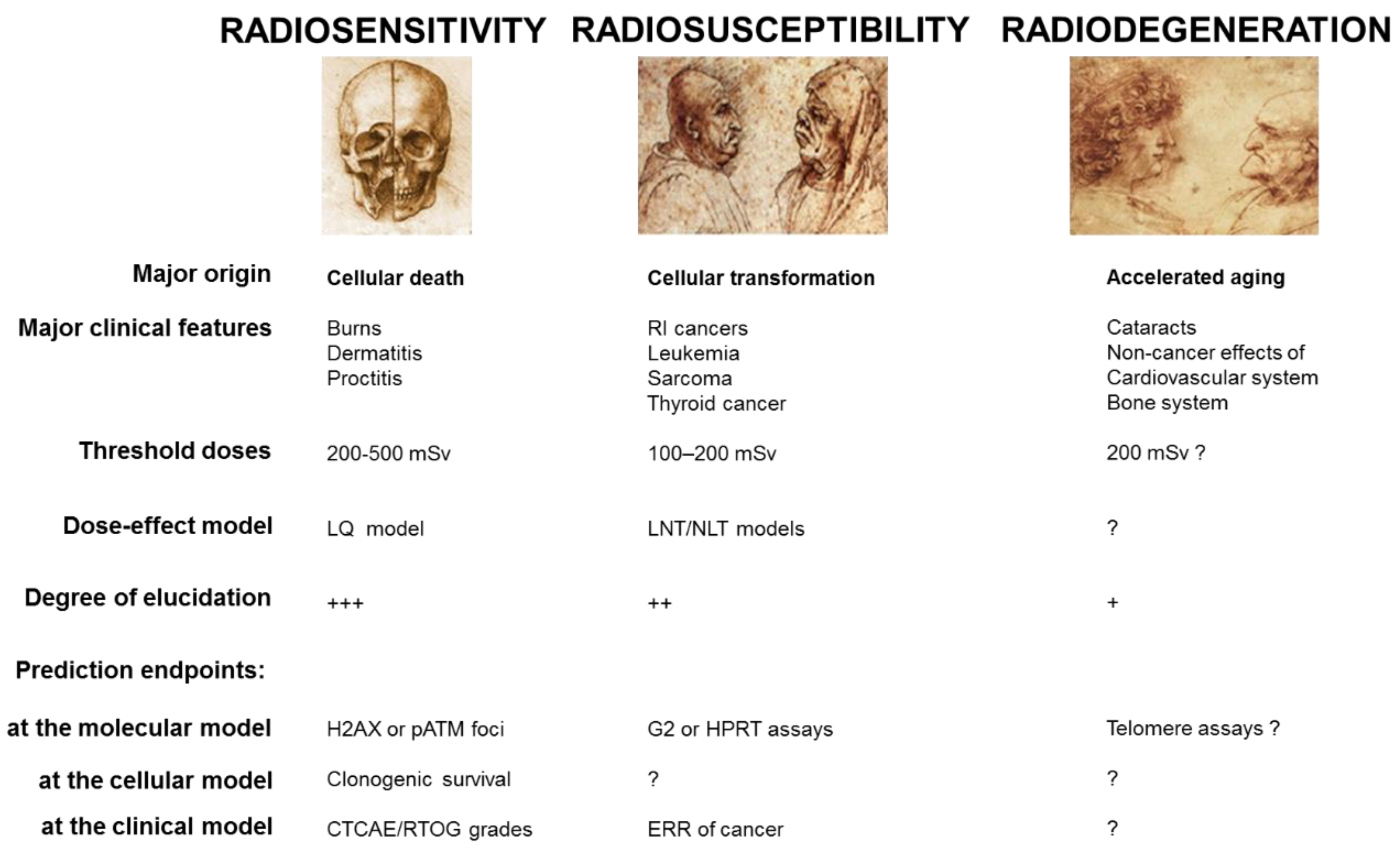
- Radiosensitivity responses, i.e., adverse tissue events, are non-cancer effects, attributable to cell death. First reported by Giezel, Voigt, Albers-Schönberg, and Bouchacourt [6,7,8], detailed descriptions of radiodermatitis and RI reactions to other irradiated organs have progressively led to the definition of consensual severity scales [2,9], like the Common Terminology Criteria for Adverse Events (CTCAE) [10] and the Radiation Therapy Oncology Group (RTOG) [11] scales. These two scales classify RI tissue reactions in six grades (grade 0: no event; grade 5: death), for each organ and independently of the early/late nature of the reactions. The CTCAE or RTOG severity grades are the most reliable endpoints to quantify clinical radiosensitivity. On a biological scale, the quantification of radiosensitivity is dependent on the whole knowledge of RI cell death. The only consensual endpoint to quantify cellular radiosensitivity is clonogenic cell survival, which obeys the empirical linear-quadratic (LQ) model [12,13,14]. However, the cell survival assays are too time-consuming to be applicable in routine. Lastly, while the skin burns and other RI tissue reactions were described earlier, it is noteworthy that the term “radiosensitivity” appeared for the first time in 1907 [15].
- Radiosusceptibility responses, i.e., RI cancers, are non-toxic effects attributable to cell transformation and genomic instability. First reported in 1902 [16], and revealed to the public by the story of the radium dial painters [17], RI cancers have been significantly documented by the reports of Hiroshima survivors [18,19,20]. To date, the most reliable endpoints to quantify the risk of RI cancers is the excess relative risk ratio (ERR) or any related endpoints from epidemiology [21]. However, the statistical robustness of these endpoints is strongly dependent on the size of the cohorts studied. To describe the ERR as a function of radiation dose, two major models were proposed: the linear non-threshold (LNT) and the non-linear threshold (NLT) models. The relevance of these two empirical models is still the source of controversy [22,23,24]. On a biological scale, the quantification of radiosusceptibility is dependent on basic knowledge about carcinogenicity mechanisms. To date, the numbers of G2 chromosomal aberrations [25] and hypoxanthine phosphoribosyltransferase (HRPT) mutations frequency [26] may be considered as the most specific endpoints of the RI cellular transformation but are not consensual. Lastly, while the first RI cancers were described earlier, it is noteworthy that the term “radiosusceptibility” was proposed for the first time in 2016, to avoid any confusion with the use of “radiosensitivity” [15].
- Radiodegeneration responses, i.e., non-cancer effects, are non-cancer effects attributable to mechanisms related to accelerated aging [18,27]. First reported in 1903 in humans, RI cataracts are the most frequent radiodegeneration response [28]. RI cardiovascular effects, first reported in 1932, also belong to this category [29]. Like for RI cancers, the estimation of the incidence of RI radiodegeneration effects is limited by the lack of specific epidemiological data. Similarly, on a biological scale, the quantification of radiodegeneration is dependent on basic knowledge of senescence and aging mechanisms. Telomere length and telomerase activity are frequently cited as the most specific endpoints to describe aging [2,30]. Lastly, it is noteworthy that the term “radiodegeneration” was proposed for the first time in 2016, in order to distinguish syndromes associated with cancer proneness and those associated with aging [15].
2. A Survey of Human Radiosensitivity
2.1. The Different Clinical Features of Radiosensitivity
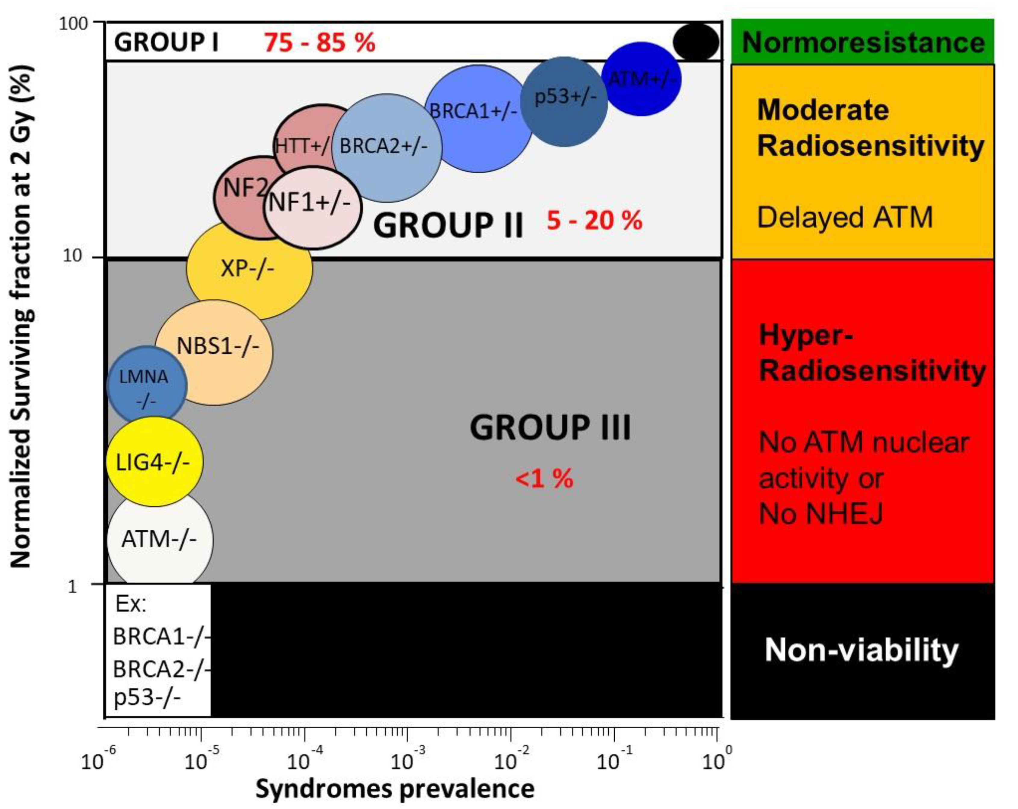
- Hyper-radiosensitivity: The most hyper-radiosensitive cells (SF2 ranging from 1–10%) derive from leukemia/lymphoma patients suffering from homozygous mutations of the Ataxia Telangiectasia Mutated (ATM) gene (the highest hyper-radiosensitivity observed in humans) and homozygous mutations of the ligase IV (LIG4) gene (only one case reported) who succumbed after radiotherapy or homozygous mutations of the Nijmegen Breakage Syndrome (NBS1) gene. Furthermore, the mutations of lamina A (LMNA) derived from patients suffering from the progeroid Hutchinson–Gilford syndrome belong to this group [2,33,39,41,42]. The cumulative incidence of these syndromes does not exceed 1%: they represent, therefore, a minority of patients, whose symptoms are mostly detectable in pediatrics. On the biological scale, all these mutations result in the loss of protein function and lead to a strong inhibition of DNA double-strand breaks (DSB) recognition or repair [2,33,39,41,42].
- Moderate radiosensitivity: SF2 ranging from 10–50% corresponds to a moderate sensitivity, such as that observed in genetic syndromes associated with high cancer proneness, like Fanconi anemia (FANC mutations), Bloom’s syndrome (BLM mutations), and neurofibromatosis (NF1 mutations). Another subset of genetic syndromes belonging to this subcategory gathers aging and/or neurodegenerative diseases like Cockayne syndrome (CS mutations) or Huntington’s disease (HTT mutations) [2,39]. Such moderate radiosensitivities do not correspond to fatal reactions after radiotherapy but to morbidity reactions (i.e., CTCAE/RTOG severity grade ranging from 2 to 4). The cumulative incidence of the cases of moderate radiosensitivity represents the majority of patients who showed significant post-radiotherapy tissue reactions [2]. At the biological scale, all these mutations do not necessarily result in the loss of protein function but lead to a relative inhibition of DSB repair and signaling. Furthermore, it is noteworthy that some heterozygous mutations are associated with an overexpression of the mutated protein, like with Li Fraumeni syndrome (heterozygous p53 mutations) [43].
- Normosensitivity (or radioresistance): SF2 ranging from 50–70%, even up to 80% for some tumors, corresponds to individuals considered “radioresistant”, who do not suffer from cancer (with the notable exception of occupational cancers) and who do not show any secondary effects after radiotherapy (CTCAE/RTOG grade 0) [2]. Normosensitivity is often defined by historical cell lines, for which patient follow-up is well characterized. However, normosensitive controls are difficult to obtain since a patient may or may not show post-radiotherapy tissue reactions, according to the radiotherapy modality and the way of delivering the dose [35].
2.2. The Major Approaches to Predict Radiosensitivity and Their Limits
- Assays based on cellular death: while SF2 is one of the best parameters to quantify cellular radiosensitivity [39], clonogenic cell survival assays are too time-consuming to predict radiosensitivity in routine. Assays based only on a particular cell death are not robust enough statistically to reliably predict radiobiology [32,48]. For example, assays based on apoptosis are irrelevant for predicting the radiosensitivity of fibroblasts that do not show this type of cell death. Furthermore, when applied on lymphocytes, apoptotic assays provide an inverse correlation between apoptotic yield and clinical radiosensitivity (the higher the apoptotic yield, the more radioresistant the patient is) which is not in agreement with the current models and needs further investigation [2,49].
- Assays based on cytogenetics: yields of unrepaired chromosomes, and especially micronuclei, have been quantitatively correlated with radiosensitivity [2,50]. However, the ranges of unrepaired chromosomes and of micronuclei are too small (0–12% and 0–25% per 100 cells, respectively) to reflect moderate radiosensitivity. The predictive power of cytogenetic endpoints is therefore limited [35].
- Assays based on DSB repair: while there is a quantitative correlation between unrepaired DSB and SF2, such a correlation does not make it possible to predict the intermediate radiosensitivity, for the same reasons evoked above with cytogenetics: the yield of unrepaired DSB ranges between 0 and 8 while SF2 varies from 1–70% [35,40].
- Genomics: as evoked above, the boolean nature (yes/no) of the DNA sequence endpoints cannot account for any dose-function. For example, any endpoint from genomics cannot provide biological interpretation of the LQ model. Conversely, genomics data are very useful for identifying gene mutations and new syndromes associated with radiosensitivity [51].
3. ATM, a Nucleocytoplasmic Protein Upstream of the Molecular Response to Radiation
3.1. ATM, a Nucleocytoplasmic Protein Early Activated after Irradiation
3.2. ATM and the Other Serine/Threonine Kinases Involved in the DNA Damage Recognition
3.3. A Crucial Observation Raising Basic Questions about the Role of ATM
4. The RIANS Model: A Solid Basis for Predicting Radiosensitivity
4.1. Major Principles of the RIANS Model
4.2. A Reliable Prediction of Individual Radiosensitivity
4.3. Three Groups of Human Radiosensitivity
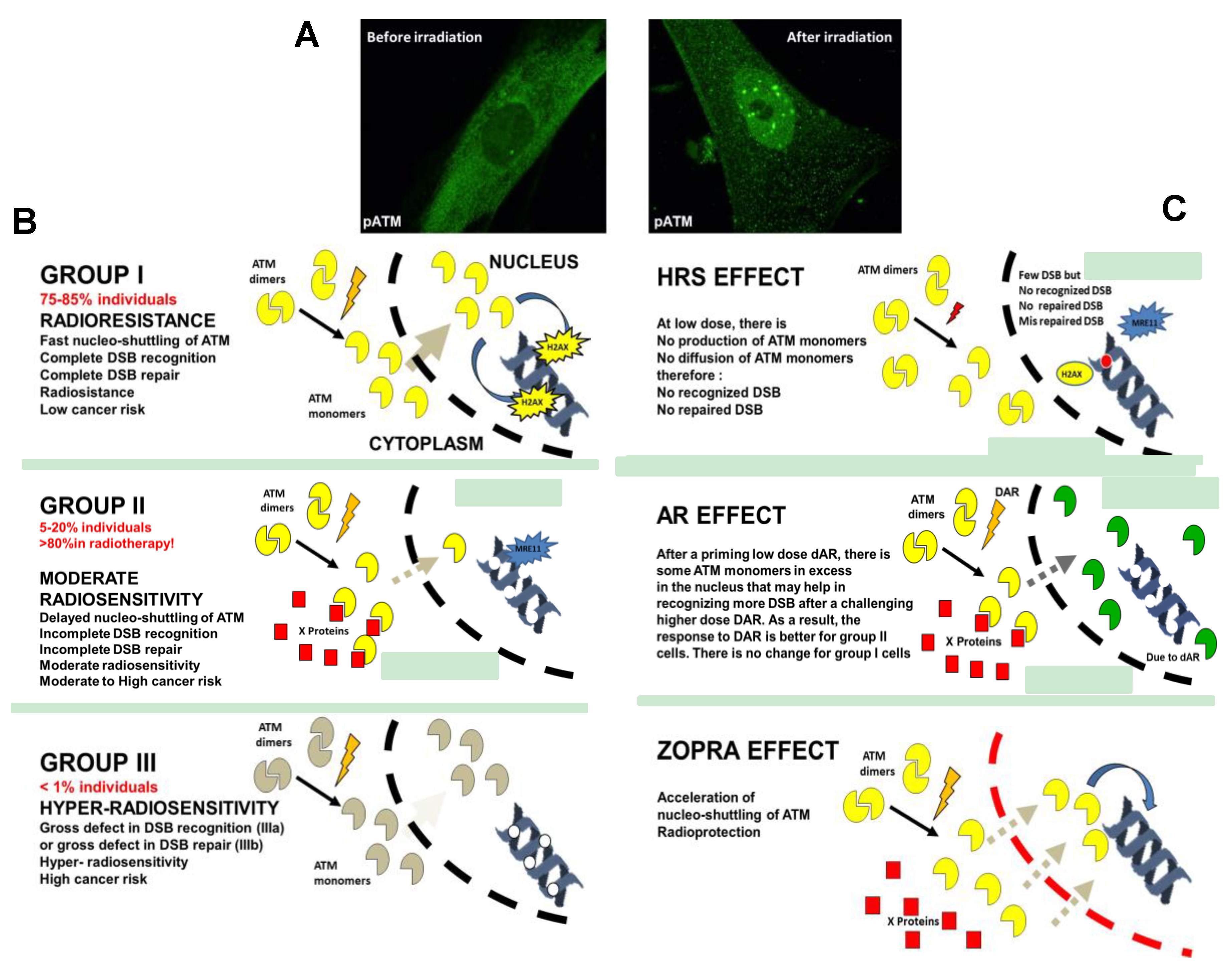
- Group I (about 75–85% of the whole population) represents the normosensitive (radioresistant) patients with a rapid RIANS after 2 Gy, and a low risk of post-radiotherapy tissue reaction and cancer;
- Group II (about 5–20% of the whole population) represents the patients who elicit a delay in the RIANS because of the sequestration of ATM in cytoplasm due to the formation of new cytoplasmic ATM substrates or their overexpression. These patients are moderately radiosensitive and susceptible to either cancer or to neurodegenerative disease;
4.4. Radiosensitivity Caused by Mutated Cytoplasmic Proteins
5. A Unified Model to Describe the Response to High- and Low-Dose of Radiation?
5.1. A New Biological Interpretation of the LQ Model
5.2. A Relevant Explanation for the Hyper-Radiosensitivity of the Low Dose Phenomenon?
5.3. A Relevant Explanation for the Adaptive Response?
- The dAR dose triggers the production of DSB and ATM monomers that diffuse in the nucleus. At this stage, the irradiation conditions are similar to HRS;
- The period of time ΔtAR favors the accumulation of the ATM monomers in the nucleus. However, if ΔtAR is too long, the number of ATM monomers will be reduced because of the limited activity half-time of the ATM protein. If ΔtAR is too short, the accumulation of active ATM monomers in the nucleus will be reduced;
- The challenging DAR dose triggers the production of a high number of ATM monomers. However, the excess of remaining ATM monomers induced by dAR and still active in nucleus after dAR + ΔtAR will facilitate the biological response to the RI DSB induced by DAR: the effect of dAR + ΔtAR + DAR is therefore lower than that of the challenging DAR dose alone.
5.4. Statins and Bisphosphonates: A New Approach of Radiological Protection?
6. Other Applications of the RIANS Model
7. Conclusions
Author Contributions
Funding
Conflicts of Interest
References
- Domina, E.A.; Philchenkov, A.; Dubrovska, A. Individual Response to Ionizing Radiation and Personalized Radiotherapy. Crit. Rev. Oncog. 2018, 23, 69–92. [Google Scholar] [CrossRef] [PubMed]
- Foray, N.; Bourguignon, M.; Hamada, N. Individual response to ionizing radiation. Mutat. Res. Rev. 2016, 770, 369–386. [Google Scholar] [CrossRef] [PubMed]
- Pauwels, E.K.; Foray, N.; Bourguignon, M.H. Breast Cancer Induced by X-Ray Mammography Screening? A Review Based on Recent Understanding of Low-Dose Radiobiology. Med. Princ. Pract. 2016, 25, 101–109. [Google Scholar] [CrossRef] [PubMed]
- Hatch, M.; Cardis, E. Somatic health effects of Chernobyl: 30 years on. Eur. J. Epidemiol. 2017, 32, 1047–1054. [Google Scholar] [CrossRef] [PubMed]
- Roentgen, W. Über eine neue Art von Strahlen. Vorläufige Mitteilung. Aus den Sitzungsberichten der Würzburger Physik.-medic. Gesellschaft Würzburg 1895, S137–S147. [Google Scholar]
- Serwer, D.P. The rise of radiation protection: Science, In Medicine and Technology in Society, 1896–1935; Princeton University Press: Princeton, NJ, USA, 1976. [Google Scholar]
- Albers-Schönberg, H. Über die Benadlung des Lupus und des chronischen Ekzems mit Röntgenstrahlen. Fortschr. Rôntgenstr. 1898, 2, 20–29. [Google Scholar]
- Bouchacourt, L. Sur la différence de sensibilité aux rayons de Roentgen de la peau des différents sujets, et, sur le même sujet des différents régions du corps. Sciences 1911, 942–947. [Google Scholar]
- Morère, J.-F.; Mornex, F.; Soulières, D. Thérapeutique du Cancer; Springer: Paris, France, 2011. [Google Scholar]
- Trotti, A.; Colevas, A.D.; Setser, A.; Rusch, V.; Jaques, D.; Budach, V.; Langer, C.; Murphy, B.; Cumberlin, R.; Coleman, C.N.; et al. CTCAE v3.0: Development of a comprehensive grading system for the adverse effects of cancer treatment. Semin. Radiat. Oncol. 2003, 13, 176–181. [Google Scholar] [CrossRef]
- Cox, J.D.; Stetz, J.; Pajak, T.F. Toxicity criteria of the Radiation Therapy Oncology Group (RTOG) and the European Organization for Research and Treatment of Cancer (EORTC). Int. J. Radiat. Oncol. Biol. Phys. 1995, 31, 1341–1346. [Google Scholar] [CrossRef]
- Kellerer, A.M.; Rossi, H.H. The theory of dual radiation action. Curr. Top. Radiat. Res. 1972, 8, 85–158. [Google Scholar]
- Chadwick, K.H.; Leenhouts, H.P. A molecular theory of cell survival. Phys. Med. Biol. 1973, 13, 78–87. [Google Scholar] [CrossRef]
- Bodgi, L.; Canet, A.; Pujo-Menjouet, L.; Lesne, A.; Victor, J.M.; Foray, N. Mathematical models of radiation action on living cells: From the target theory to the modern approaches. A historical and critical review. J. Theor. Biol. 2016, 394, 93–101. [Google Scholar] [CrossRef] [PubMed]
- Britel, M.; Bourguignon, M.; Foray, N. Radiosensitivity: A term with various meanings at the origin of numerous confusions. A semantic analysis. Int. J. Radiat. Oncol. Biol. 2018, 94, 503–512. [Google Scholar] [CrossRef] [PubMed]
- Frieben, A. Cancroid des rechten Handrückens. Deutsche Med. Wochenschr. 1902, 28, 335. [Google Scholar]
- Gunderman, R.B.; Gonda, A.S. Radium girls. Radiology 2015, 274, 314–318. [Google Scholar] [CrossRef] [PubMed]
- Preston, D.L.; Shimizu, Y.; Pierce, D.A.; Suyama, A.; Mabuchi, K. Studies of mortality of atomic bomb survivors. Report 13: Solid cancer and noncancer disease mortality: 1950–1997. 2003. Radiat. Res. 2012, 178, AV146–AV172. [Google Scholar] [CrossRef]
- Preston, D.L.; Shimizu, Y.; Pierce, D.A.; Suyama, A.; Mabuchi, K. Studies of mortality of atomic bomb survivors. Report 13: Solid cancer and noncancer disease mortality: 1950–1997. Radiat. Res. 2003, 160, 381–407. [Google Scholar] [CrossRef]
- Ozasa, K.; Shimizu, Y.; Suyama, A.; Kasagi, F.; Soda, M.; Grant, E.J.; Sakata, R.; Sugiyama, H.; Kodama, K. Studies of the mortality of atomic bomb survivors, Report 14, 1950-2003: An overview of cancer and noncancer diseases. Radiat. Res. 2012, 177, 229–243. [Google Scholar] [CrossRef]
- Lee, W.C. Excess relative risk as an effect measure in case-control studies of rare diseases. PLoS ONE 2014, 10, e0121141. [Google Scholar] [CrossRef]
- Calabrese, E.J.; Shamoun, D.Y.; Hanekamp, J.C. The Integration of LNT and Hormesis for Cancer Risk Assessment Optimizes Public Health Protection. Health Phys. 2016, 110, 256–259. [Google Scholar] [CrossRef]
- Tubiana, M. Dose-effect relationship and estimation of the carcinogenic effects of low doses of ionizing radiation: The joint report of the Academie des Sciences (Paris) and of the Academie Nationale de Medecine. Int. J. Radiat. Oncol. Biol. Phys. 2005, 63, 317–319. [Google Scholar] [CrossRef] [PubMed]
- Boice, J.D., Jr. The linear nonthreshold (LNT) model as used in radiation protection: An NCRP update. Int. J. Radiat. Oncol. Biol. 2017, 93, 1079–1092. [Google Scholar] [CrossRef] [PubMed]
- Baria, K.; Warren, C.; Roberts, S.A.; West, C.M.; Scott, D. Chromosomal radiosensitivity as a marker of predisposition to common cancers? Br. J. Cancer 2001, 84, 892–896. [Google Scholar] [CrossRef] [PubMed]
- Compton, P.J.; Hooper, K.; Smith, M.T. Human somatic mutation assays as biomarkers of carcinogenesis. Environ. Health Perspect. 1991, 94, 135–141. [Google Scholar] [CrossRef] [PubMed]
- ICRP. ICRP statement on tissue reactions and early and late effects of radiation in normal tissues and organs-threshold doses for tissue reactions in a radiation protection context. Publication 118. Ann. ICRP 2012, 41, 1–322. [Google Scholar] [CrossRef] [PubMed]
- Rollins, W. Notes on x-light. The effect of x-light on the crystalline lens. Boston Med. Surg. J. 1903, 148, 364–365. [Google Scholar] [CrossRef]
- Desjardins, A.U. Action of roentgen rays and radium on the heart and lungs. Am. J. Roentgenol. 1932, 27, 149–176. [Google Scholar]
- Kong, C.M.; Lee, X.W.; Wang, X. Telomere shortening in human diseases. FEBS J. 2013, 280, 3180–3193. [Google Scholar] [CrossRef]
- Brooks, A.L.; Hoel, D.G.; Preston, R.J. The role of dose rate in radiation cancer risk: Evaluating the effect of dose rate at the molecular, cellular and tissue levels using key events in critical pathways following exposure to low LET radiation. Int. J. Radiat. Oncol. Biol. 2016, 92, 405–426. [Google Scholar] [CrossRef]
- Vogin, G.; Bastogne, T.; Bodgi, L.; Gillet-Daubin, J.; Canet, A.; Pereira, S.; Foray, N. The Phosphorylated ATM Immunofluorescence Assay: A High-performance Radiosensitivity Assay to Predict Postradiation Therapy Overreactions. Int. J. Radiat. Oncol. Biol. Phys. 2018, 101, 690–693. [Google Scholar] [CrossRef]
- Varela, I.; Pereira, S.; Ugalde, A.P.; Navarro, C.L.; Suarez, M.F.; Cau, P.; Cadinanos, J.; Osorio, F.G.; Foray, N.; Cobo, J.; et al. Combined treatment with statins and aminobisphosphonates extends longevity in a mouse model of human premature aging. Nat. Med. 2008, 14, 767–772. [Google Scholar] [CrossRef] [PubMed]
- Ferlazzo, M.L.; Sonzogni, L.; Granzotto, A.; Bodgi, L.; Lartin, O.; Devic, C.; Vogin, G.; Pereira, S.; Foray, N. Mutations of the Huntington’s Disease Protein Impact on the ATM-Dependent Signaling and Repair Pathways of the Radiation-Induced DNA Double-Strand Breaks: Corrective Effect of Statins and Bisphosphonates. Mol. Neurobiol. 2014, 49, 1200–1211. [Google Scholar] [CrossRef] [PubMed]
- Granzotto, A.; Benadjaoud, M.A.; Vogin, G.; Devic, C.; Ferlazzo, M.L.; Bodgi, L.; Pereira, S.; Sonzogni, L.; Forcheron, F.; Viau, M.; et al. Influence of Nucleoshuttling of the ATM Protein in the Healthy Tissues Response to Radiation Therapy: Toward a Molecular Classification of Human Radiosensitivity. Int. J. Radiat. Oncol. Biol. Phys. 2016, 94, 450–460. [Google Scholar] [CrossRef] [PubMed]
- Health Public Agency. Human Radiosensitivity; Report RCE 21; Public Health England: London, UK, 2013.
- Fertil, B.; Malaise, E.P. Inherent cellular radiosensitivity as a basic concept for human tumor radiotherapy. Int. J. Radiat. Oncol. Biol. Phys. 1981, 7, 621–629. [Google Scholar] [CrossRef]
- Arlett, C.F.; Harcourt, S.A. Survey of radiosensitivity in a variety of human cell strains. Cancer Res. 1980, 40, 926–932. [Google Scholar] [PubMed]
- Deschavanne, P.J.; Fertil, B. A review of human cell radiosensitivity in vitro. Int. J. Radiat. Oncol. Biol. Phys. 1996, 34, 251–266. [Google Scholar] [CrossRef]
- Joubert, A.; Zimmerman, K.M.; Bencokova, Z.; Gastaldo, J.; Rénier, W.; Chavaudra, N.; Favaudon, V.; Arlett, C.; Foray, N. DNA double-strand break repair defects in syndromes associated with acute radiation response: At least two different assays to predict intrinsic radiosensitivity? Int. J. Radiat. Oncol. Biol. 2008, 84, 1–19. [Google Scholar] [CrossRef]
- Badie, C.; Iliakis, G.; Foray, N.; Alsbeih, G.; Pantellias, G.E.; Okayasu, R.; Cheong, N.; Russell, N.S.; Begg, A.C.; Arlett, C.F.; et al. Defective repair of DNA double-strand breaks and chromosome damage in fibroblasts from a radiosensitive leukemia patient. Cancer Res. 1995, 55, 1232–1234. [Google Scholar]
- Taalman, R.D.; Jaspers, N.G.; Scheres, J.M.; de Wit, J.; Hustinx, T.W. Hypersensitivity to ionizing radiation, in vitro, in a new chromosomal breakage disorder, the Nijmegen Breakage Syndrome. Mutat. Res. 1983, 112, 23–32. [Google Scholar] [CrossRef]
- Foray, N.; Randrianarison, V.; Marot, D.; Perricaudet, M.; Lenoir, G.; Feunteun, J. Gamma-rays-induced death of human cells carrying mutations of BRCA1 or BRCA2. Oncogene 1999, 18, 7334–7342. [Google Scholar] [CrossRef][Green Version]
- Bencokova, Z.; Devic, C.; Ferlazzo, M.L.; Granzotto, A.; Sonzogni, L.; Burlet, S.F.; Viau, M.; Bodgi, L.; Bachelet, J.T.; Combemale, P.; et al. Radiobiological characterization of neurofibromatosis type I: The neurofibromin protein impacts on the ATM-dependent DNA damage repair and signaling pathway. Mol. Neurobiol. 2018, in press. [Google Scholar]
- Ferlazzo, M.L.; Bach-Tobdji, M.K.E.; Djerad, A.; Sonzogni, L.; Burlet, S.F.; Devic, C.; Granzotto, A.; Bodgi, L.; Djeffal-Kerrar, A.; Foray, N. Radiobiological characterization of tuberous sclerosis: A delay in the nucleo-shuttling of ATM may be responsible for radiosensitivity. Mol. Neurobiol. 2017, 55, 4973–4983. [Google Scholar] [CrossRef] [PubMed]
- Huo, Y.K.; Wang, Z.; Hong, J.H.; Chessa, L.; McBride, W.H.; Perlman, S.L.; Gatti, R.A. Radiosensitivity of ataxia-telangiectasia, X-linked agammaglobulinemia, and related syndromes using a modified colony survival assay. Cancer Res. 1994, 54, 2544–2547. [Google Scholar] [PubMed]
- Nove, J.; Tarone, R.E.; Little, J.B.; Robbins, J.H. Radiation sensitivity of fibroblast strains from patients with Usher’s syndrome, Duchenne muscular dystrophy, and Huntington’s disease. Mutat. Res. 1987, 184, 29–38. [Google Scholar] [CrossRef]
- Ferlazzo, M.L.; Bourguignon, M.; Foray, N. Functional Assays for Individual Radiosensitivity: A Critical Review. Semin. Radiat. Oncol. 2017, 27, 310–315. [Google Scholar] [CrossRef] [PubMed]
- Pereira, S.; Bodgi, L.; Duclos, M.; Canet, A.; Ferlazzo, M.L.; Devic, C.; Granzotto, A.; Deneuve, S.; Vogin, G.; Foray, N. The time is coming to compare radiosensitivity predictive assays by their scientific relevance and statistical performances. Int. J. Radiat. Oncol. Biol. Phys. 2018, 101, 491–492. [Google Scholar] [CrossRef] [PubMed]
- Cornforth, M.N.; Bedford, J.S. A quantitative comparison of potentially lethal damage repair and the rejoining of interphase chromosome breaks in low passage normal human fibroblasts. Radiat. Res. 1987, 111, 385–405. [Google Scholar] [CrossRef] [PubMed]
- Rosenstein, B.S.; West, C.M.; Bentzen, S.M.; Alsner, J.; Andreassen, C.N.; Azria, D.; Barnett, G.C.; Baumann, M.; Burnet, N.; Chang-Claude, J.; et al. Radiogenomics: Radiobiology enters the era of big data and team science. Int. J. Radiat. Oncol. Biol. Phys. 2014, 89, 709–713. [Google Scholar] [CrossRef]
- Taylor, A.M.; Harnden, D.G.; Arlett, C.F.; Harcourt, S.A.; Lehmann, A.R.; Stevens, S.; Bridges, B.A. Ataxia telangiectasia: A human mutation with abnormal radiation sensitivity. Nature 1975, 258, 427–429. [Google Scholar] [CrossRef]
- Foray, N.; Marot, D.; Gabriel, A.; Randrianarison, V.; Carr, A.M.; Perricaudet, M.; Ashworth, A.; Jeggo, P. A subset of ATM- and ATR-dependent phosphorylation events requires the BRCA1 protein. EMBO J. 2003, 22, 2860–2871. [Google Scholar] [CrossRef]
- Bakkenist, C.J.; Kastan, M.B. DNA damage activates ATM through intermolecular autophosphorylation and dimer dissociation. Nature 2003, 421, 499–506. [Google Scholar] [CrossRef] [PubMed]
- Oka, A.; Takashima, S. Expression of the ataxia-telangiectasia gene (ATM) product in human cerebellar neurons during development. Neurosci. Lett. 1998, 252, 195–198. [Google Scholar] [CrossRef]
- Lim, D.S.; Kirsch, D.G.; Canman, C.E.; Ahn, J.H.; Ziv, Y.; Newman, L.S.; Darnell, R.B.; Shiloh, Y.; Kastan, M.B. ATM binds to beta-adaptin in cytoplasmic vesicles. Proc. Natl. Acad. Sci. USA 1998, 95, 10146–10151. [Google Scholar] [CrossRef] [PubMed]
- Alexander, A.; Cai, S.L.; Kim, J.; Nanez, A.; Sahin, M.; MacLean, K.H.; Inoki, K.; Guan, K.L.; Shen, J.; Person, M.D.; et al. ATM signals to TSC2 in the cytoplasm to regulate mTORC1 in response to ROS. Proc. Natl. Acad. Sci. USA 2010, 107, 4153–4158. [Google Scholar] [CrossRef] [PubMed]
- Barlow, C.; Ribaut-Barassin, C.; Zwingman, T.A.; Pope, A.J.; Brown, K.D.; Owens, J.W.; Larson, D.; Harrington, E.A.; Haeberle, A.M.; Mariani, J.; et al. ATM is a cytoplasmic protein in mouse brain required to prevent lysosomal accumulation. Proc. Natl. Acad. Sci. USA 2000, 97, 871–876. [Google Scholar] [CrossRef] [PubMed]
- Boehrs, J.K.; He, J.; Halaby, M.J.; Yang, D.Q. Constitutive expression and cytoplasmic compartmentalization of ATM protein in differentiated human neuron-like SH-SY5Y cells. J. Neurochem. 2007, 100, 337–345. [Google Scholar] [CrossRef] [PubMed]
- Hinz, M.; Stilmann, M.; Arslan, S.C.; Khanna, K.K.; Dittmar, G.; Scheidereit, C. A cytoplasmic ATM-TRAF6-cIAP1 module links nuclear DNA damage signaling to ubiquitin-mediated NF-kappaB activation. Mol. Cell 2010, 40, 63–74. [Google Scholar] [CrossRef] [PubMed]
- Li, J.; Han, Y.R.; Plummer, M.R.; Herrup, K. Cytoplasmic ATM in neurons modulates synaptic function. Curr. Biol. 2009, 19, 2091–2096. [Google Scholar] [CrossRef]
- Yang, D.Q.; Halaby, M.J.; Li, Y.; Hibma, J.C.; Burn, P. Cytoplasmic ATM protein kinase: An emerging therapeutic target for diabetes, cancer and neuronal degeneration. Drug Discov. Today 2011, 16, 332–338. [Google Scholar] [CrossRef]
- Rothkamm, K.; Lobrich, M. Evidence for a lack of DNA double-strand break repair in human cells exposed to very low x-ray doses. Proc. Natl. Acad. Sci. USA 2003, 100, 5057–5062. [Google Scholar] [CrossRef]
- Stiff, T.; O’Driscoll, M.; Rief, N.; Iwabuchi, K.; Lobrich, M.; Jeggo, P.A. ATM and DNA-PK function redundantly to phosphorylate H2AX after exposure to ionizing radiation. Cancer Res. 2004, 64, 2390–2396. [Google Scholar] [CrossRef] [PubMed]
- Burdak-Rothkamm, S.; Short, S.C.; Folkard, M.; Rothkamm, K.; Prise, K.M. ATR-dependent radiation-induced gamma H2AX foci in bystander primary human astrocytes and glioma cells. Oncogene 2007, 26, 993–1002. [Google Scholar] [CrossRef] [PubMed]
- O’Driscoll, M.; Ruiz-Perez, V.L.; Woods, C.G.; Jeggo, P.A.; Goodship, J.A. A splicing mutation affecting expression of ataxia-telangiectasia and Rad3-related protein (ATR) results in Seckel syndrome. Nat. Genet. 2003, 33, 497–501. [Google Scholar] [CrossRef] [PubMed]
- O’Driscoll, M.; Gennery, A.R.; Seidel, J.; Concannon, P.; Jeggo, P.A. An overview of three new disorders associated with genetic instability: LIG4 syndrome, RS-SCID and ATR-Seckel syndrome. DNA Repair 2004, 3, 1227–1235. [Google Scholar] [CrossRef] [PubMed]
- van der Burg, M.; Ijspeert, H.; Verkaik, N.S.; Turul, T.; Wiegant, W.W.; Morotomi-Yano, K.; Mari, P.O.; Tezcan, I.; Chen, D.J.; Zdzienicka, M.Z.; et al. A DNA-PKcs mutation in a radiosensitive T-B- SCID patient inhibits Artemis activation and nonhomologous end-joining. J. Clin. Invest. 2009, 119, 91–98. [Google Scholar] [CrossRef]
- Chavaudra, N.; Bourhis, J.; Foray, N. Quantified relationship between cellular radiosensitivity, DNA repair defects and chromatin relaxation: A study of 19 human tumour cell lines from different origin. Radiother. Oncol. 2004, 73, 373–382. [Google Scholar] [CrossRef]
- Markova, E.; Schultz, N.; Belyaev, I.Y. Kinetics and dose-response of residual 53BP1/gamma-H2AX foci: Co-localization, relationship with DSB repair and clonogenic survival. Int. J. Radiat. Oncol. Biol. 2007, 83, 319–329. [Google Scholar] [CrossRef]
- Bodgi, L.; Foray, N. The nucleo-shuttling of the ATM protein as a basis for a novel theory of radiation response: Resolution of the linear-quadratic model. Int. J. Radiat. Oncol. Biol. 2016, 92, 117–131. [Google Scholar] [CrossRef]
- Guo, Z.; Kozlov, S.; Lavin, M.F.; Person, M.D.; Paull, T.T. ATM activation by oxidative stress. Science 2010, 330, 517–521. [Google Scholar] [CrossRef]
- Paull, T.T. Mechanisms of ATM Activation. Ann. Rev. Biochem. 2015, 84, 711–734. [Google Scholar] [CrossRef]
- Maalouf, M.; Granzotto, A.; Devic, C.; Bodgi, L.; Ferlazzo, M.; Peaucelle, C.; Bajard, M.; Giraud, J.Y.; Balosso, J.; Herault, J.; et al. Influence of Linear Energy Transfer on the Nucleo-shuttling of the ATM Protein: A Novel Biological Interpretation Relevant for Particles and Radiation. Int. J. Radiat. Oncol. Biol. Phys. 2019, 103, 709–718. [Google Scholar] [CrossRef] [PubMed]
- Petersen, L.F.; Klimowicz, A.C.; Otsuka, S.; Elegbede, A.A.; Petrillo, S.K.; Williamson, T.; Williamson, C.T.; Konno, M.; Lees-Miller, S.P.; Hao, D.; et al. Loss of tumour-specific ATM protein expression is an independent prognostic factor in early resected NSCLC. Oncotarget 2017, 8, 38326–38336. [Google Scholar] [CrossRef]
- Belkacemi, Y.; Colson-Durand, L.; Granzotto, A.; Husheng, S.; To, N.H.; Majdoul, S.; Guet, S.; Herve, M.L.; Fonteneau, G.; Diana, C.; et al. The Henri Mondor Procedure of Morbidity and Mortality Review Meetings: Prospective Registration of Clinical, Dosimetric, and Individual Radiosensitivity Data of Patients With Severe Radiation Toxicity. Int. J. Radiat. Oncol. Biol. Phys. 2016, 96, 629–636. [Google Scholar] [CrossRef] [PubMed]
- Pereira, S.; Bodgi, L.; Duclos, M.; Canet, A.; Ferlazzo, M.L.; Devic, C.; Granzotto, A.; Deneuve, S.; Vogin, G.; Foray, N. Fast and binary assay for predicting radiosensitivity based on the nucleoshuttling of ATM protein: Development, validation and performances. Int. J. Radiat. Oncol. Biol. Phys. 2018, 100, 353–360. [Google Scholar] [CrossRef] [PubMed]
- Bodgi, L.; Foray, N. Effets biologiques des radiations ionisantes. Une théorie basée sur le transit cyto-nucléaire de la protéine ATM. Applications à la radiothérapie; Editions Universitaires Européennes: Sarrebruck, Germany, 2016. [Google Scholar]
- Bodgi, L.; Granzotto, A.; Devic, C.; Vogin, G.; Lesne, A.; Bottollier-Depois, J.F.; Victor, J.M.; Maalouf, M.; Fares, G.; Foray, N. A single formula to describe radiation-induced protein relocalization: Towards a mathematical definition of individual radiosensitivity. J. Theor. Biol. 2013, 333, 135–145. [Google Scholar] [CrossRef] [PubMed]
- Ferlazzo, M.L.; Foray, N. Huntington Disease: A Disease of DNA Methylation or DNA Breaks? Am. J. Pathol. 2016, 186, 1750–1753. [Google Scholar] [CrossRef][Green Version]
- Lambin, P.; Marples, B.; Fertil, B.; Malaise, E.P.; Joiner, M.C. Hypersensitivity of a human tumour cell line to very low radiation doses. Int. J. Radiat. Oncol. Biol. 1993, 63, 639–650. [Google Scholar] [CrossRef]
- Marples, B.; Joiner, M.C. The response of Chinese hamster V79 cells to low radiation doses: Evidence of enhanced sensitivity of the whole cell population. Radiat. Res. 1993, 133, 41–51. [Google Scholar] [CrossRef]
- Joiner, M.C.; Marples, B.; Lambin, P.; Short, S.C.; Turesson, I. Low-dose hypersensitivity: Current status and possible mechanisms. Int. J. Radiat. Oncol. Biol. Phys. 2001, 49, 379–389. [Google Scholar] [CrossRef]
- Thomas, C.; Martin, J.; Devic, C.; Diserbo, M.; Thariat, J.; Foray, N. Impact of dose-rate on the low-dose hyper-radiosensitivity and induced radioresistance (HRS/IRR) response. Int. J. Radiat. Oncol. Biol. 2013, 89, 813–822. [Google Scholar] [CrossRef]
- Xue, L.; Yu, D.; Furusawa, Y.; Cao, J.; Okayasu, R.; Fan, S. ATM-dependent hyper-radiosensitivity in mammalian cells irradiated by heavy ions. Int. J. Radiat. Oncol. Biol. Phys. 2009, 75, 235–243. [Google Scholar] [CrossRef] [PubMed]
- Joiner, M.C.; Lambin, P.; Marples, B. Adaptive response and induced resistance. Comptes Rendus de l’Académie des Sciences Series III Sciences de la Vie 1999, 322, 167–175. [Google Scholar] [CrossRef]
- Calabrese, E.J.; Bachmann, K.A.; Bailer, A.J.; Bolger, P.M.; Borak, J.; Cai, L.; Cedergreen, N.; Cherian, M.G.; Chiueh, C.C.; Clarkson, T.W.; et al. Biological stress response terminology: Integrating the concepts of adaptive response and preconditioning stress within a hormetic dose-response framework. Toxicol. Appl. Pharmacol. 2007, 222, 122–128. [Google Scholar] [CrossRef] [PubMed]
- Feinendegen, L. Quantification of adaptive protection following low-dose irradiation. Health Phys. 2016, 110, 276–280. [Google Scholar] [CrossRef] [PubMed]
- Joiner, M.C.; Lambin, P.; Malaise, E.P.; Robson, T.; Arrand, J.E.; Skov, K.A.; Marples, B. Hypersensitivity to very-low single radiation doses: Its relationship to the adaptive response and induced radioresistance. Mutat. Res. 1996, 358, 171–183. [Google Scholar] [CrossRef]
- Ryan, L.A.; Seymour, C.B.; Joiner, M.C.; Mothersill, C.E. Radiation-induced adaptive response is not seen in cell lines showing a bystander effect but is seen in lines showing HRS/IRR response. Int. J. Radiat. Oncol. Biol. 2009, 85, 87–95. [Google Scholar] [CrossRef] [PubMed]
- Marples, B.; Joiner, M.C. The elimination of low-dose hypersensitivity in Chinese hamster V79-379A cells by pretreatment with X rays or hydrogen peroxide. Radiat. Res. 1995, 141, 160–169. [Google Scholar] [CrossRef]
- Devic, C.; Ferlazzo, M.L.; Foray, N. Influence of Individual Radiosensitivity on the Adaptive Response Phenomenon: Toward a Mechanistic Explanation Based on the Nucleo-Shuttling of ATM Protein. Dose Response 2018, 16, 1–11. [Google Scholar] [CrossRef]
- Weiss, J.F.; Landauer, M.R. History and development of radiation-protective agents. Int. J. Radiat. Oncol. Biol. 2009, 85, 539–573. [Google Scholar] [CrossRef]
- Galiullina, L.F.; Aganova, O.V.; Latfullin, I.A.; Musabirova, G.S.; Aganov, A.V.; Klochkov, V.V. Interaction of different statins with model membranes by NMR data. Biochim. Biophys. Acta Biomembr. 2017, 1859, 295–300. [Google Scholar] [CrossRef]


| Radiosensitivity of the Patients | CTCAE/RTOG Grade | SF2 (%) | γH2AX Foci at 10 min Post-Irradiation | pATM Foci at 10 min Post-Irradiation | γH2AX foci at 24 h Post-Irradiation |
|---|---|---|---|---|---|
| Group I | 0 | 50–70 | 70–80 | 30–40 | 0–2 |
| Group II | 0–4 | 10–50 | 10–70 | 10–30 | 2–8 |
| Group III | 5 | 1–10 | IIIa2: 0–5 IIIb2: 70–80 | IIIa: 0 IIIb: 30–40 | IIIa: 0–5 IIIb: 30–40 |
© 2019 by the authors. Licensee MDPI, Basel, Switzerland. This article is an open access article distributed under the terms and conditions of the Creative Commons Attribution (CC BY) license (http://creativecommons.org/licenses/by/4.0/).
Share and Cite
Berthel, E.; Foray, N.; Ferlazzo, M.L. The Nucleoshuttling of the ATM Protein: A Unified Model to Describe the Individual Response to High- and Low-Dose of Radiation? Cancers 2019, 11, 905. https://doi.org/10.3390/cancers11070905
Berthel E, Foray N, Ferlazzo ML. The Nucleoshuttling of the ATM Protein: A Unified Model to Describe the Individual Response to High- and Low-Dose of Radiation? Cancers. 2019; 11(7):905. https://doi.org/10.3390/cancers11070905
Chicago/Turabian StyleBerthel, Elise, Nicolas Foray, and Mélanie L. Ferlazzo. 2019. "The Nucleoshuttling of the ATM Protein: A Unified Model to Describe the Individual Response to High- and Low-Dose of Radiation?" Cancers 11, no. 7: 905. https://doi.org/10.3390/cancers11070905
APA StyleBerthel, E., Foray, N., & Ferlazzo, M. L. (2019). The Nucleoshuttling of the ATM Protein: A Unified Model to Describe the Individual Response to High- and Low-Dose of Radiation? Cancers, 11(7), 905. https://doi.org/10.3390/cancers11070905


