Review

Microfluidic and Paper-Based Recombinase Polymerase Amplification Systems for Decentralized Diagnostics and Biosurveillance


Department of Engineering Science, National Cheng Kung University, Tainan 701, Taiwan
*
Author to whom correspondence should be addressed.
These authors contributed equally to this work.
Micromachines 2026, 17(7), 825; https://doi.org/10.3390/mi17070825
Submission received: 23 June 2026 / Revised: 5 July 2026 / Accepted: 6 July 2026 / Published: 10 July 2026
(This article belongs to the Special Issue Microfluidics in Biomedical Research)

Abstract

Recombinase polymerase amplification (RPA) has become a central amplification strategy for decentralized molecular diagnostics because it operates rapidly at mild temperatures and requires far less thermal control than PCR. Its analytical value increases substantially when paired with microfluidic and paper-based platforms, where sample handling, reagent delivery, amplification, and signal readout can be organized within compact, low-power, and field-compatible formats. This review examines recent progress in microfluidic and paper-based RPA systems across biomedical diagnostics, food safety testing, environmental monitoring, and One Health biosurveillance. Particular attention is given to integrated device architectures, including centrifugal chips, capillary-driven platforms, microfluidic paper-based analysis devices (μPADs), electrochemical biosensors, CRISPR-assisted assays, digital microfluidic systems, and sample-to-answer cartridges. Biomedical applications now span respiratory viruses, reproductive and emerging infections, bacterial and parasitic diseases, pharmacogenomic markers, and cancer-related biomarkers. RPA-enabled platforms are moving steadily into food safety and environmental surveillance, covering pathogen detection, seafood and dairy monitoring, agricultural disease control, antimicrobial-resistance tracking, and airborne pathogen screening. At the same time, the field is shifting toward more intelligent diagnostic formats. Smartphone imaging, artificial intelligence (AI)-assisted interpretation, digital partitioning, cloud connectivity, and automated quality control are increasingly being built into rapid testing workflows, giving these systems greater portability, consistency, and decision-making value. Despite this progress, practical deployment still depends on robust sample preparation, multiplex stability, quantitative reliability, reagent storage, scalable fabrication, and regulatory validation. Continued convergence of RPA chemistry with microfluidics, paper devices, CRISPR recognition, electrochemical readout, and data-assisted interpretation is expected to support more robust and accessible molecular diagnostic workflows.

1. Introduction

Molecular diagnostics now occupies a central position in healthcare, food safety control, and environmental surveillance, largely because nucleic acid targets can be identified with high specificity and speed. Since the advent of the polymerase chain reaction (PCR), amplification-based assays have reshaped the detection of infectious agents, antimicrobial resistance markers, foodborne pathogens, and clinically relevant genetic signatures for precision medicine. Despite its central role in nucleic acid analysis, conventional PCR still relies on thermal cyclers, skilled operators, and laboratory-based workflows. Such dependence limits its use outside centralized facilities, particularly in resource-limited settings, point-of-care testing, and field surveillance, where timely decisions require diagnostic platforms that are compact, low-power, robust, and simple to operate [1,2,3,4,5,6].
PCR and recombinase polymerase amplification (RPA) occupy distinct positions in biomedical molecular testing. PCR remains the method most often chosen when clinical confirmation, quantitative measurement, and standardized laboratory reporting are required, largely because thermal cycling gives the reaction tight procedural control and a long record of validated performance. The same requirement for precise temperature cycling, specialized instruments, and laboratory operation also makes PCR less convenient for decentralized use. RPA addresses a different need. Amplifying nucleic acids at a mild, nearly constant temperature lowers the demand for thermal hardware and fits more readily into portable microfluidic chips, paper-based assays, and point-of-care devices. In practical terms, PCR is still the stronger option for laboratory confirmation and quantitative monitoring, whereas RPA is better matched to rapid screening, field testing, and biomedical diagnostics in settings with limited infrastructure. Table 1 summarizes these complementary roles, focusing on reaction conditions, instrument requirements, assay speed, and biomedical suitability.
Isothermal nucleic acid amplification has therefore gained considerable attention as a practical route beyond instrument-intensive PCR. Representative strategies include loop-mediated isothermal amplification (LAMP), nucleic acid sequence-based amplification (NASBA), helicase-dependent amplification (HDA), recombinase-aided amplification (RAA), and RPA [7,8,9,10]. Among these methods, RPA is particularly attractive because amplification proceeds efficiently at mild temperatures, typically 37–42 °C, without complex thermal control. Its short reaction time, modest hardware demand, and PCR-comparable analytical sensitivity make RPA well suited to decentralized molecular testing. Accordingly, RPA has become an important foundation for next-generation diagnostic systems designed for point-of-care and field-deployable applications [11,12].

1.1. Principle and Advantages of RPA

Unlike PCR, RPA does not depend on repeated heating and cooling to separate DNA strands. The reaction instead uses recombinase–primer complexes to probe double-stranded DNA for homologous sequences and to insert primers directly into the target region. The exposed single strand is protected by SSBs, while strand-displacing polymerase extends the invading primer from the opened duplex. At a constant temperature, repeated primer invasion followed by polymerase extension progressively enriches the target sequence, producing amplicons at levels suitable for detection [11,12].
RPA has become a favored choice in isothermal amplification because it brings together three practical qualities that are difficult to achieve in a single assay: speed, mild thermal operation, and compatibility with imperfect samples. In many reports, amplification is completed within 10–30 min, giving RPA a clear advantage when rapid molecular readout is required [11,12]. Its reaction temperature is low enough to ease power consumption and thermal-control demands, which is particularly useful for handheld, battery-powered, or field-deployable diagnostic devices. Equally important, RPA can maintain reliable amplification in the presence of inhibitors commonly carried over from clinical specimens, food-derived matrices, and environmental samples [13,14]. This balance of rapid amplification, simple heating, and matrix tolerance has made RPA especially well-suited to field molecular diagnostics, environmental surveillance, and home-based testing platforms [15,16].
Recent review articles further emphasize the expanding role of RPA in decentralized molecular testing. Analyses covering paper-based biosensors, nucleic acid point-of-care testing systems, and integrated microfluidic amplification platforms repeatedly identify RPA as a particularly suitable amplification chemistry for rapid, low-infrastructure diagnostics [16,17,18]. Its combination of speed, low thermal demand, and compatibility with portable formats has positioned RPA as a key amplification strategy for next-generation molecular diagnostic systems.

1.2. Integration of RPA with Microfluidic Technologies

While RPA simplifies nucleic acid amplification, practical deployment often requires integration with sample preparation, fluid handling, and signal detection systems. Microfluidic technology provides an ideal platform for achieving this integration because it enables precise manipulation of small fluid volumes, automated reagent delivery, reduced reagent consumption, and miniaturization of analytical workflows [19,20].
During the past decade, microfluidic implementation of RPA has expanded well beyond simple reaction chambers. Early platforms were shaped largely by continuous-flow microchannels and centrifugal disks, which provided controlled sample transport, reagent metering, and reaction timing in small-volume formats [21]. Device concepts later became more diverse and increasingly application-driven. Capillary-driven devices offered a practical way to move liquids without external pumps, thereby making RPA assays easier to operate in resource-limited settings [22]. Self-powered platforms made the device concept more practical for field testing, since fewer external components were required during operation [23]. In parallel, multiplex pathogen-detection chips broadened the analytical capacity of RPA, allowing several targets to be screened simultaneously within a single microfluidic layout [24,25]. More recent designs show a clear shift toward deployable diagnostic systems, particularly integrated respiratory-virus testing platforms [26] and automated sample-to-answer cartridges, in which sample processing, amplification, and signal readout are coordinated within one continuous workflow [27].
The pairing of microfluidics with RPA has changed the practical form of nucleic acid testing. Rather than treating extraction, amplification, and detection as separate laboratory steps, many recent devices place these functions inside a single disposable format, with fluid routing and reaction timing handled on-chip. This design logic has been discussed in several reviews of microfluidic nucleic acid testing, where RPA is frequently highlighted for lowering assay complexity and making molecular diagnostics more portable and accessible outside conventional laboratory settings [28,29]. CRISPR-integrated microfluidic systems have added a further layer of analytical capability. In these devices, microfluidic operation, isothermal amplification, and CRISPR-based recognition work together to improve specificity, support multiplex pathogen testing, and reduce operator intervention [30,31]. RPA is therefore no longer used only as a fast amplification step; in many recent devices, it now functions as part of a coordinated molecular testing workflow.

1.3. Emergence of Paper-Based RPA Diagnostics

Progress in microfluidic RPA diagnostics has been accompanied by a parallel shift toward paper-based analytical formats. Paper is not merely a passive support; its low cost, simple fabrication, biodegradability, scalable manufacture, and capillary-driven flow provide a practical basis for decentralized molecular testing [17,18]. These features are especially valuable when diagnostic systems must remain inexpensive, lightweight, and usable without pumps or complex peripheral equipment. Recent reviews have noted the increasing integration of nucleic acid amplification with paper-based biosensors [16]. RPA is especially compatible with this format because it proceeds rapidly under mild thermal conditions and requires little supporting instrumentation. Paper–RPA systems therefore provide a simple route to portable molecular testing, particularly for low-resource environments and home-based diagnostics [15].
Paper-based RPA has progressed from simple amplification–lateral-flow formats to more integrated diagnostic designs. Early platforms mainly coupled RPA with strip-based detection, offering a low-instrumentation route for molecular readout. Newer systems add functional modules that address sample preparation, fluid control, and matrix interference. This transition is evident in several recent design strategies. Some platforms introduce nucleic acid enrichment upstream of RPA to improve target availability before amplification [14]. Origami-inspired paper devices exploit folded geometries to control how liquids move across the substrate, allowing multistep reactions to be arranged in a compact format [32]. Vibration-assisted amplification [33] and inhibitor-removal components [34] address a different but equally important issue: maintaining reliable amplification when complex sample matrices interfere with enzyme activity.
Paper-based CRISPR–RPA devices have further expanded this platform by combining rapid amplification with sequence-specific CRISPR recognition. As a result, pathogen signals can be read visually with high sensitivity, while the assay remains largely free from complex instrumentation [35,36]. Together, these advances make paper-based RPA a practical route toward affordable molecular diagnostics for field use, low-resource settings, and home-based testing [16].
Table 2 sets representative paper-based RPA systems beside one another, comparing substrate choice, target analyte, readout strategy, assay duration, detection limit, and practical strengths. This side-by-side comparison highlights the diversity of paper-supported formats, ranging from lateral-flow strips and origami membranes to wax-gated μPADs, valve-integrated paper devices, and surface-engineered platforms. It also shows that the practical value of paper-based RPA extends beyond low-cost fabrication. In many cases, the substrate itself contributes to sample handling, reagent storage, capillary transport, reaction compartmentalization, or user-friendly signal readout.

1.4. Digital Microfluidics and Quantitative RPA Analysis

Conventional RPA is highly useful for qualitative detection, but obtaining a reliable quantitative readout is less direct. In a bulk reaction, amplification proceeds continuously, so the measured signal represents the collective reaction kinetics rather than discrete amplification events. Digital microfluidics offers a more countable format by splitting the sample into many isolated compartments, with each chamber operating as an independent amplification unit [44,45].
SlipChip technology offered one of the earliest demonstrations of compartmentalized microreactors for digital nucleic acid analysis [44]. Once the sample is distributed into discrete microreactors, positive and negative reaction chambers can be counted directly, allowing absolute nucleic acid quantification without relying on calibration curves. Later digital microfluidic systems carried this compartmentalized strategy further, using improved device layouts to handle more reactions in parallel, reduce manual operation, and make the workflow easier to automate [45].
Recent digital RPA systems have begun to combine automated droplet generation, compartment-based quantification, and high-throughput image analysis within a single analytical workflow [46,47]. By converting bulk amplification into many discrete reaction events, these platforms improve sensitivity for low-abundance targets and provide a clearer basis for quantitative readout. This digital format also fits naturally with automated image processing and machine-learning-assisted interpretation. Reaction images can be treated as structured datasets, allowing more consistent counting, classification, and quantitative biosensing than manual inspection alone [48,49]. In this way, digital RPA adds both analytical sensitivity and a data-driven layer to molecular diagnostic platforms.

1.5. Intelligent and Connected RPA Diagnostic Systems

Artificial intelligence (AI), smartphone imaging, cloud connectivity, and digital health infrastructure are starting to transform the design and interpretation of RPA diagnostics [15]. In several recent systems, smartphones are no longer used only as simple cameras; they support image acquisition, signal extraction, result interpretation, and data transmission to cloud-based platforms [50]. Deep-learning models have also been coupled with microfluidic diagnostic devices to reduce subjective readout differences and improve classification accuracy [51]. Automated digital microfluidics also shows a similar trend, with machine-learning tools being employed for droplet identification, signal quantification, and quality-control decisions [52,53].
AI-assisted molecular diagnostics are moving beyond single-test interpretation. When assay results pass from smartphones to cloud-linked health platforms, molecular testing begins to serve a wider surveillance role, supporting epidemiological monitoring, outbreak mapping, and population-scale disease assessment [54]. In this model, molecular tests no longer function only as isolated diagnostic events; they also become data-generating elements within healthcare and biosurveillance networks.

1.6. Scope of This Review

RPA diagnostics no longer belong to amplification chemistry alone. Microfluidic fluid control, paper-based device engineering, CRISPR-based target recognition, digital microfluidic partitioning, and AI-assisted signal interpretation have shaped their recent development. Each layer adds a different capability: simpler handling, lower-cost fabrication, higher specificity, quantitative readout, or more objective data analysis. As these elements come together, RPA-based platforms are finding roles not only in healthcare but also in food safety, environmental monitoring, and One Health surveillance [11,17,52]. Such breadth also creates a need for a more organized assessment of the field. The literature now spans conventional chips, paper devices, digital assays, CRISPR-coupled formats, smartphone readout, and AI-enabled analytical workflows. Microfluidic and paper-based RPA now reaches beyond benchtop amplification, taking shape as practical molecular assays for clinics, food-safety testing, environmental monitoring, and intelligent detection. This review maps that trajectory, then considers the stubborn gaps—sample handling, contamination, quantitative readout, scalable manufacture, commercialization, and wider use in decentralized diagnostics.
The adjacent domains raised by this comment were not overlooked; they were placed where their technical logic best fits the structure of the review. Veterinary testing, aquaculture surveillance, plant-pathogen screening, antimicrobial-resistance monitoring, and genetically modified crop detection are best placed within environmental and One Health biosurveillance, as these settings share the same field-facing pressures: irregular sampling conditions, matrix interference, biosafety concerns, and fragmented reporting routes [4,6,11,12,41]. Precision medicine, pharmacogenomic assays, mutation profiling, methylation analysis, and selected forensic applications fall more appropriately under biomedical diagnostics, where the analytical targets are mainly human-derived nucleic acids and performance evaluation is shaped by clinical validation requirements [8,9,10,13]. Biosecurity pathogen detection and outbreak preparedness also align with biosurveillance, particularly for portable RPA assays aimed at high-consequence or low-abundance targets [28,39,55]. Artificial intelligence, smartphone imaging, and cloud interpretation serve instead as enabling layers rather than separate biological domains [15,51,52,53,54]. This arrangement preserves a focused discussion around the most mature evidence for microfluidic and paper-based RPA systems while leaving room for adjacent fields as sample-to-answer platforms become stronger and more reliable.

2. Biomedical Applications

Biomedical diagnostics now demands molecular assays that can leave centralized laboratories without losing analytical reliability. This need is clear in infectious disease testing, antimicrobial resistance monitoring, cancer screening, and personalized medicine, where timely nucleic acid information can guide clinical action. Thermal cyclers, trained operation, and relatively long workflows continue to shape the use of PCR and RT-qPCR, which remain strong laboratory standards. Microfluidic and paper-based RPA systems offer a more practical route for near-patient testing, combining rapid amplification at mild temperatures with compact device formats and flexible signal readout [56,57]. The amplification step has also become the foundation for building versatility in the platform, incorporating CRISPR recognition, electrochemical sensing, digital microfluidics, smartphone imaging, and AI-assisted analysis. In this form, RPA works less as a single reaction and more as a diagnostic engine for clinical screening, infectious disease detection, biomarker analysis, and personalized medicine.

2.1. Respiratory Viral Infections

Microfluidic RPA provides a practical route for respiratory-virus diagnostics by combining rapid amplification, simplified handling, and compact chip-based readout. Recent platforms have supported SARS-CoV-2 aerosol monitoring; multiplex detection of SARS-CoV-2, RSV, influenza viruses, and parainfluenza viruses; and influenza subtyping from respiratory samples [19,37,56,57,58]. Reported assays typically finish within 10–45 min, with sensitivities around 10–20 copies/µL. This performance makes microfluidic RPA useful not only for decentralized diagnosis but also for environmental monitoring and faster recognition of respiratory viral coinfections.
In the work of Li et al. [55], SARS-CoV-2 aerosols were monitored using a microfluidic RPA cartridge that brought together cyclone-based air sampling, chitosan-assisted RNA enrichment, and in situ tetra-primer amplification. The same cartridge was applied to aerosols, nasopharyngeal swabs, saliva, exhaled breath condensates, and air-outlet swabs. Fluorescence signals became readable within 25 min, and the detection limit reached 20 copies/mL. Its higher positivity in low-biomass and warm aerosol samples indicates value for noninvasive screening and environmental early warning.
For multiplex respiratory diagnosis, Liu et al. [26] developed MAPnavi, a sealed microfluidic platform that links nested RPA with CRISPR/Cas12a fluorescence readout. Within 40 min, the assay detected SARS-CoV-2 targets, RSV, influenza A/B, 2009 H1N1, H3N2, and an internal control. Analytical sensitivity reached 1 copy/µL, and testing of 171 nasopharyngeal samples showed strong agreement with reference methods. MAPnavi therefore extends microfluidic RPA beyond single-virus confirmation, offering a closed and contamination-controlled format for syndromic respiratory surveillance.
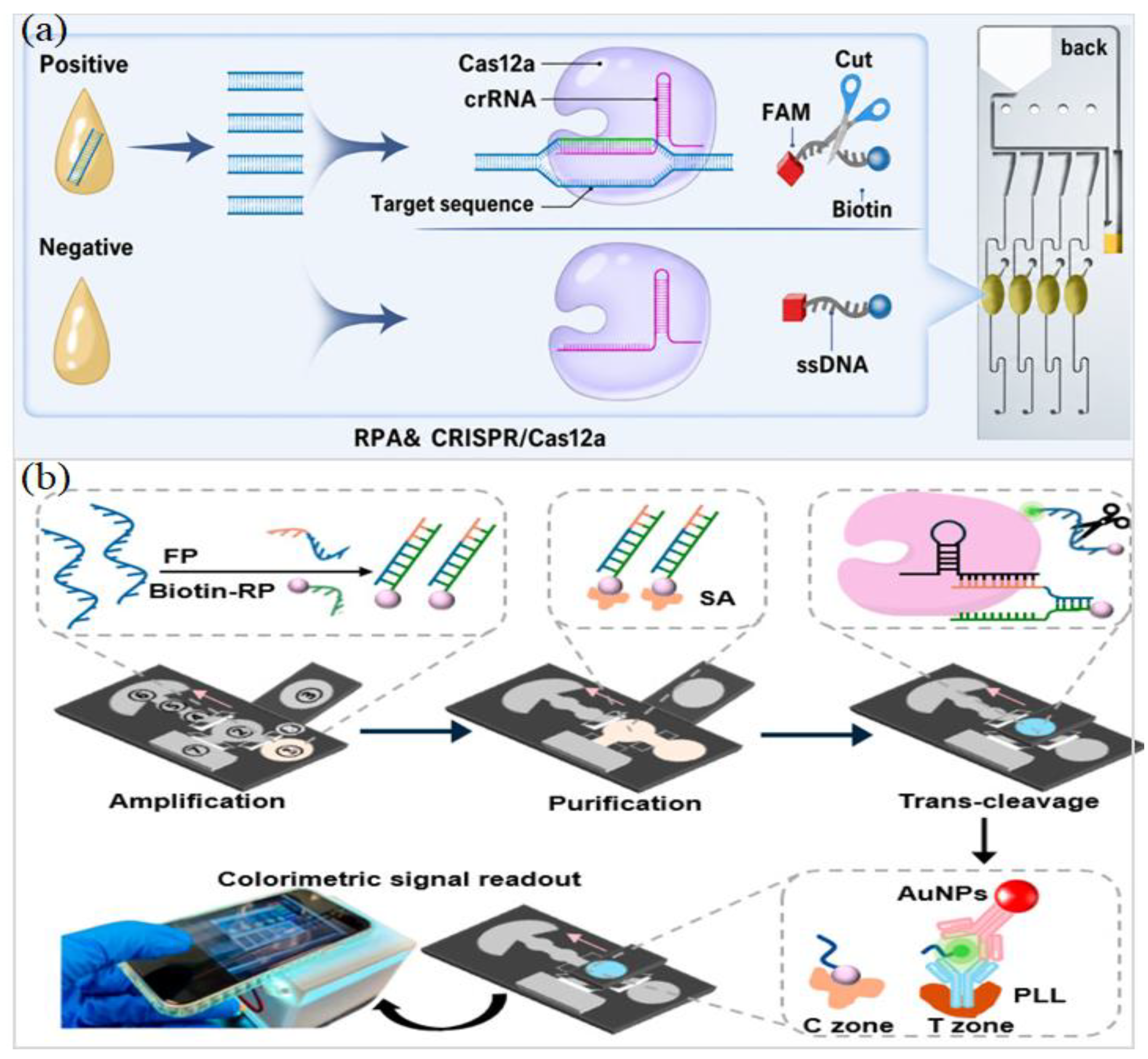
Li et al. [59] described a passively driven RT-RPA-CRISPR/Cas12a chip for respiratory virus screening in infrastructure-limited settings. The assay detected influenza A/B, human parainfluenza virus, and SARS-CoV-2, as shown in Figure 1a. Its workflow remained deliberately simple: 10 min for sample preparation, 35 min for the chip reaction, and colloidal-gold lateral-flow readout after RT-RPA–Cas12a recognition. Without fluorescence optics, the assay detected targets at approximately 10 copies/µL and achieved clinical sensitivity and specificity of 98.44% and 100%, respectively. Capillary/gravity-driven transport and lyophilized on-chip reagents further reduced dependence on pumps, optical readers, and skilled operation.
Lu et al. [60] demonstrated rapid influenza subtyping using a PMMA centrifugal RPA chip covering H1N1, H2N2, H3N2, H5N1, H7N9, and influenza B. The assay used extracted respiratory RNA, operated at 42 °C, and produced results within 10 min. Sensitivity was retained at low viral copy numbers, and results from all 180 clinical samples matched those obtained by RT-PCR. Collectively, these studies show that microfluidic RPA is no longer limited to rapid amplification chemistry; it is becoming a complete diagnostic architecture for respiratory-virus screening, subtyping, and surveillance.

2.2. Reproductive, Blood-Borne, and Emerging Viral Infections

RPA-enabled microfluidic and paper-based platforms are reshaping diagnostics for reproductive, blood-borne, and emerging viruses by coupling mild isothermal amplification to diverse, field-compatible readouts, including CRISPR, electrochemical, colorimetric, lateral-flow, and droplet formats. Across HPV, sexually transmitted pathogens, MPXV, WNV, and hCMV, these systems shorten workflows, reduce equipment dependence, and improve access to decentralized testing in real-world settings [61,62,63,64,65].
Xu et al. [66] described a cervical-swab workflow that integrates multiplex RPA, Cas12a reporting, and microfluidic spatial coding to distinguish nine HPV genotypes covered by the 9-valent vaccine. Genotype assignment relies on position rather than multiple dyes: a single fluorophore is used while reactions are distributed across a 30-well starburst chip, completing subtyping in approximately 40 min. After amplification, signals extended from blank to 1 × 10⁻¹⁶ M, with a detection limit of 2.67 × 10⁻¹⁹ M (0.26 aM). In a 100-specimen clinical evaluation, sensitivity reached 97.8% and specificity 98.1%.
For HPV16/18 genotyping, Zhao et al. [67] used a dual-droplet CRISPR/Cas12a microdevice in which green and red droplets served as separate reaction compartments for target discrimination. The assay reached approximately 10−18 M for HPV16, corresponding to about one copy per reaction, with a turnaround time of approximately 30 min. Across 20 clinical samples, sensitivity was 92.3%, and specificity was 100%. Physical compartmentalization suppresses crosstalk while preserving multiplex capability, making the format well suited to compact HPV genotyping.
Complementary HPV implementations further illustrate the breadth of RPA-enabled formats. Zhang et al. [36] designed a μPAD for HPV16 E7 dsDNA that integrates lyophilized RPA/CRISPR reagents with AuNP–anti-FAM colorimetry for naked-eye readout, achieving 100 pM detection within 60 min, as shown in Figure 1b. Nakowong et al. [68] paired RPA with a methylene-blue electrochemical genosensor to detect HPV16 in cervical tissue samples; the assay read down to 0.23 copies/μL and matched PCR across 40 specimens.
Sexually transmitted and emerging viral infections have also benefited from streamlined RPA workflows. A hand-warmer-heated paper device reported by Chu et al. [38] integrates nucleic-acid isolation, RPA, and lateral-flow readout for Neisseria gonorrhoeae in urine. The assay was completed in 30–35 min and detected 10 DNA copies or 1 CFU/mL, improving field accessibility relative to tube-based RPA and PCR. Hwang et al. [69] established RT-RPA–Cas12a DETECTR formats for West Nile virus lineages 1a and 2, with 102 RNA copies for the two-step and filter-assisted formats and 103 copies for the one-step format. Together, these examples show that the clinical value of RPA is greatest when amplification is embedded in devices that manage transport, partition reactions, stabilize reagents, and translate recognition into readable optical, electrochemical, or lateral-flow signals.

2.3. Tuberculosis, Bacterial, and Parasitic Infectious Diseases

Microfluidic RPA systems now extend from tuberculosis (TB) and bacterial infection testing to malaria genotyping [70,71]. Their practical value lies in short assay times, low-temperature amplification, compact fluid handling, and visually or digitally readable signals that reduce dependence on culture, microscopy, and centralized PCR. Saikaew et al. [72] introduced TB-GoldDx as a visual tuberculosis assay built on IS6110-specific RPA and unmodified AuNP colorimetry. Clinical-isolate and sputum-derived DNA provide a more clinically relevant benchmark for tuberculosis screening than template-only experiments. After amplification at 39 °C for 20 min, salt-induced AuNP aggregation produced a distinct color change, with a detection limit of 0.001 ng H37Rv DNA (≈210 bacilli per 25 µL reaction). Across the 100-strain set, sensitivity was 95.83%, and specificity remained at 100%. In sputum, sensitivity was 81.43% overall, rising to 95.45% in AFB 3+ specimens. This low-instrument format is well suited to screening, whereas low-burden sputum remains best interpreted alongside confirmatory testing.
For broad bacterial diagnosis, Xie et al. [73] reported MiND-DMF, a digital microfluidic platform integrating DNA extraction, RPA, and CRISPR detection for P. aeruginosa, H. influenzae, K. pneumoniae, E. coli, S. aureus, and MRSA. A broad spectrum of clinical matrices—including blood, urine, bronchoalveolar lavage fluid, nasal secretions, appendiceal pus, and otorrhea—was compatible with the chip. Run at 37 °C, the workflow finished in ≤55 min and reached an LOD of 100 CFU/mL; sensitivity was 100% and specificity 98–100% in 50 clinical samples. A key strength is the shift from manual microbiology toward an automated, sample-to-answer identification workflow, which streamlines handling and can inform earlier, evidence-based antibiotic selection.
For urinary tract infection (UTI)-associated bacteria, Chen et al. [74] described an autosampling microfluidic assay for five major pathogens (E. coli, K. pneumoniae, A. baumannii, E. faecium, and E. faecalis). Syringe filtration was used to concentrate urine and related clinical fluids; rapid lysis followed, and the lysate was subsequently vacuum-partitioned inside a PDMS chip. Analytical sensitivity reached 5 DNA copies per reaction and 102 CFU/mL in urine; the complete workflow closed in 55 min. In a 299-specimen study, agreement with culture corresponded to 91.1% sensitivity and 100% specificity. Xiu et al. [75] illustrated parasitic diagnostics by coupling dried-blood-spot DNA with a microfluidic RPA–CRISPR readout for malaria screening and Plasmodium genotyping. Five species were resolved with universal primers and species-specific crRNAs. The two-step format detected 10 copies per reaction, whereas the one-pot chip achieved ~102 copies/µL. Clinical testing reported 98.41% sensitivity and 92.86% specificity, with 90.91% concordance relative to PCR sequencing.

2.4. Precision Medicine and Cancer Biomarkers

RPA workflows designed for precision diagnostics now do more than amplify a target and report an endpoint signal. Genotypes, mutations, and methylation states can be read from buccal swabs and tumor-derived DNA using fluorescence, lateral-flow, electrochemical, or CRISPR outputs, enabling faster, more portable alternatives to sequencing, PCR, and bisulfite-dependent assays [76,77,78,79].
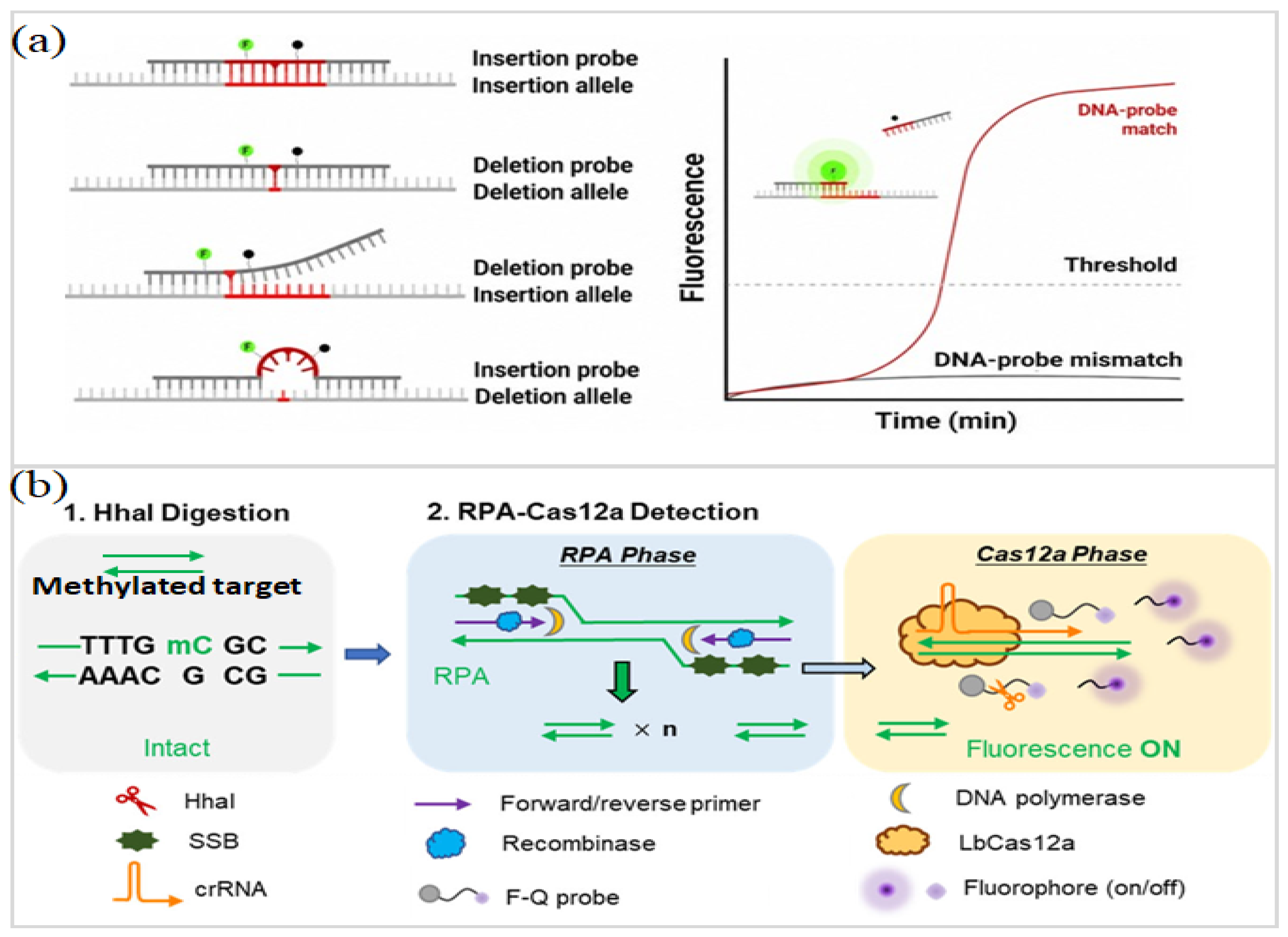
De Keyzer et al. [80] reported a real-time RPA–exo probe fluorescence assay for insertion–deletion genotyping using reference DNA and purified blood-derived DNA, as shown in Figure 2a. The reaction proceeded isothermally at 39°C and generated genotypes within 20 min, reaching 97.07% accuracy with 1 ng input, reliable profiling at 250 pg, and partial recovery at 31 pg. By replacing thermal cycling and electrophoretic separation with low-temperature probe readout, this format provides a credible path toward portable genotyping beyond PCR–capillary electrophoresis and massively parallel sequencing.
In a related study, Pang et al. [81] processed buccal-swab lysates with a smartphone-controlled duplex RPA–LFD assay strengthened by locked nucleic acid probes. The readout was completed within 25 min, and both HLA-B*58:01 and β-globin were detected at 10 copies/µL. Across 15 clinical samples, the assay showed full concordance with PCR-SBT. The two studies together point to a clear trend toward portable HLA screening before prescribing allopurinol, with internal controls helping to reduce false-negative calls and improve pharmacogenomic decision-making.
Thoeny et al. [82] interrogated three PIK3CA hotspot mutations by coupling bio-tin-dCTP–labeled RPA to a screen-printed gold-electrode array with electrochemical immunoreadout. RPA alone reached a detection limit of 100 copies/µL; after electrochemical transduction, the thresholds shifted to 229 copies/µL with asymmetric ssDNA and 224 copies/µL with λ-exonuclease–derived ssDNA. Wild-type blocking oligonucleotides gave the assay a cleaner separation between mutant and wild-type alleles, allowing reliable genotype assignment when the mutant fraction was above approximately 20%. Evidence from plasma ctDNA and primary tumor specimens is still scarce, leaving clinical validation as an important next step.
Figure 2. (a) Schematic of allele-specific RPA-exo probe design for insertion–deletion genotyping and fluorescence-based discrimination of matched and mismatched DNA–probe hybrids. (Reprinted from [80]). (b) Schematic illustration of a methylation-sensitive restriction endonuclease-assisted RPA–CRISPR/Cas12a method for methylation detection. (Reprinted from [83]).
Micromachines 17 00825 g002
For epigenetic screening, Lu et al. [83] reported an HhaI-assisted glycerol-enhanced RPA–CRISPR/Cas12a assay for a CNE2-specific methylation locus associated with nasopharyngeal carcinoma, as shown in Figure 2b. HhaI digestion removed unmethylated DNA, leaving methylated targets intact for RPA–Cas12a fluorescence detection. The method achieved a detection limit of 100 aM, a linear range of 100 aM–100 fM, and methylation-level discrimination down to 0.1%. Cell-line testing separated CNE2 from NP69, although clinical validation remains essential.
Overall, these studies position RPA as a practical engine for decentralized precision diagnostics. Its strongest role is not to replace sequencing or PCR in all settings but to bring selected genotyping, mutation, and methylation tests closer to prescribing decisions, early screening, and resource-limited clinical workflows. Table 3 collates representative RPA-enabled platforms, organized by target, amplification conditions, sensing chemistry, readout modality, and analytical performance. The side-by-side view illustrates how integrated modules—such as CRISPR, electrochemical transduction, or multiplexed designs—shorten workflows while retaining clinically meaningful sensitivity.

2.5. Challenges and Prospects in Biomedical RPA Diagnostics

Biomedical RPA diagnostics are moving beyond “fast amplification” toward sealed, automated formats that deliver interpretation-ready outputs. Among current directions, CRISPR-coupled RPA is especially consequential: Cas12a/Cas13a recognition adds sequence-level verification to low-temperature amplification, tightening specificity for multiplex respiratory panels, HPV subtyping, and bacterial pathogen detection [26,66,84,85,86,87]. Spatially encoded microfluidic designs extend this logic by partitioning targets into predefined reaction addresses, enabling high-plex identification with straightforward optical readout rather than increasingly complex dye and probe combinations [66].
Decentralized formats are gaining momentum. Smartphone-controlled disks, hand-driven centrifugal chips, paper-based assays, and AI-assisted image decoding collectively reduce reliance on centralized laboratories [51,81]. R-CHIP is a notable example of this change because it combines RPA, CRISPR readout, manual actuation, smartphone imaging, and ResNet-based classification for high-risk HPV screening [51]. In parallel, LNA-enhanced duplex RPA for HLA-B*58:01 extends the approach toward pharmacogenomic decision support [81], while self-driven 3D-printed chips with glucose-meter readout suggest an inexpensive route to outbreak-site testing [88].
Because clinical matrices contain inhibitors, variable viscosity, low target abundance, and background signals, sample preparation continues to dictate assay performance and remains a persistent vulnerability in sample-to-answer systems [84,86]. Multiplex RPA hinges on disciplined primer stoichiometry and sequence-level vetting, since even minor imbalances can seed primer-dimer artifacts, promote cross-reactivity, and elevate mismatch-driven false positives [66,81]. The most translatable systems will combine robust front-end extraction, lyophilized shelf-stable reagents, sealed cartridges, reliable valving, controlled heating, automated quality checks, interpretation-ready analytics, and secure data transfer into clinical reporting workflows [51,84,87,88].

3. Food Safety Applications

Food safety sits at the intersection of public health and supply-chain resilience, with hundreds of millions of illnesses each year and costs that ripple across production, processing, and distribution. Hazards are diverse—foodborne bacteria and viruses, spoilage consortia, antimicrobial-resistant organisms, and deliberate adulteration—so rapid checks near the point of risk are essential. Routine surveillance, however, still relies heavily on culture, ELISA, PCR, and sequencing, which typically demand laboratory infrastructure, specialized personnel, and multi-hour timelines [11,89]. Microfluidic and paper-based RPA platforms narrow this gap: mild-temperature isothermal amplification pairs naturally with miniaturized fluid handling and portable readouts, bringing molecular verification into field and factory settings. Recent systems further integrate CRISPR confirmation, smartphone imaging, electrochemical transduction, digital microfluidics, and automated front-end preparation, broadening both throughput and target scope for foodborne surveillance [12,35,90].

3.1. Detection of Foodborne Bacterial Pathogens

Culture-first confirmation is steadily giving way to RPA-enabled devices that unify sample handling, amplification, and portable readout within a single workflow. Recent polymeric, electrochemical, and microfluidic formats compress time-to-answer from days to minutes while maintaining sensitivity in matrix-rich foods, including poultry, leafy vegetables, and milk [90,91].
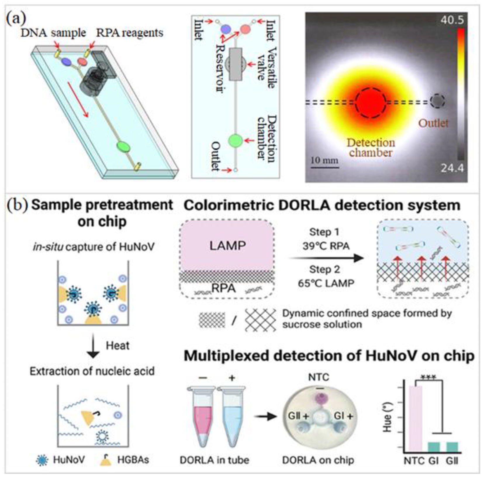
Chen et al. [92] demonstrated a hybrid paper–polymer microfluidic assay for Campylobacter jejuni in which DNA is captured by a dipstick, amplified by RPA for 20 min, and read on a lateral-flow strip. Using the poultry-associated hipO locus as the marker, the assay achieved limits of 46 CFU/mL after cellulose-paper extraction and 460 CFU/mL in the fully integrated format. In chicken meat, 101–102 CFU/g required 5–10 h enrichment, whereas loads above 102 CFU/g were verified directly, compressing culture confirmation (~6–8 days) into a field-ready workflow. Chen et al. [32] translated Salmonella enterica detection into a polyethersulfone (PES)-based origami microfluidic format, in which dipstick nucleic-acid extraction feeds directly into RPA and lateral-flow visualization. A visible readout was obtained within 20 min, with 100% specificity and a detection limit of 260 CFU/mL. After 6 h of enrichment culture, a contamination level of 58 CFU/mL was detected in lettuce, chicken breast, and milk. The choice of PES was central: compared with chromatography paper, it better preserved amplification efficiency and thereby supported a more sensitive sample-to-answer workflow. Complementarily, Jin et al. [50] introduced a PDMS microfluidic biosensing system with an integrated valve for mixing and flow control, coupled to real-time smartphone fluorescence detection of S. Typhimurium (as shown in Figure 3a); the assay reached 1.0 × 10² copies μL−1 within 30 min, approaching qPCR-level sensitivity while avoiding thermal cycling and bulky optics.
Electrochemical CRISPR–RPA formats are increasingly being adapted for quantitative monitoring of foodborne bacteria. Guo et al. [93] constructed an MWCNTs–Au-modified screen-printed carbon electrode for S. aureus quantification. In culture medium, the sensor showed a linear response over 1.04 × 101–1.04 × 10⁸ CFU/mL and reached a detection limit of 3 CFU/mL. The tests in milk further confirmed matrix applicability across 1.07 × 101–1.07 × 10⁷ CFU/mL. Guo et al. [94] further refined this strategy using MoS2–Au electrodes, retaining the 3 CFU/mL detection limit while extending the working range to 1.06 × 101–1.06 × 10⁸ CFU/mL, together with strong selectivity in milk. Chen et al. [95] subsequently introduced a one-pot RPA–CRISPR/Cas12a electrochemical lateral-flow strip for Salmonella, reaching 3.84 CFU/mL by electrochemical readout and 384 CFU/mL by visual inspection, while limiting aerosol contamination associated with tube opening. Taken together, these studies show that foodborne bacterial RPA diagnostics are shifting toward closed, matrix-tolerant, and user-readable formats. The next critical step is multicenter validation using naturally contaminated foods and harmonized quantitative thresholds.

3.2. Viral and Multiplex Detection of Foodborne Pathogens

Food-safety diagnostics based on RPA are gradually extending beyond single-bacterium assays toward viral monitoring and multiplex pathogen screening [35,96]. Compact analytical formats that combine microfluidics or paper substrates with CRISPR, LAMP, SERS, electrochemical transduction, and smartphone-assisted interpretation are shaping this shift. Such integration gives the assay a more field-oriented character: shorter turnaround, lighter instrumentation, and less reliance on centralized PCR facilities or culture-based confirmation.
For viral diagnostics, Chen et al. [97] developed a fully integrated microfluidic RPA–LAMP system for simultaneous human norovirus (HuNoV) GI and GII detection in fecal samples, as shown in Figure 3b. The design brought histo-blood group antigen (HBGA)-based viral capture, nucleic-acid extraction, and sucrose-mediated confinement into the same chip before visual reporting. A staged reaction with RPA at 39 °C and then LAMP at 65 °C kept amplification sealed and minimized carryover. The assay resolved GI and GII at 10 and 1 copy/μL, respectively, and produced clinical readouts that tracked closely with RT-qPCR. Yin et al. [39] adapted norovirus detection to a lyophilized paper-based RT-RPA–CRISPR/Cas12a assay. The system was validated with tap water, lettuce, and oyster matrices over a range of 100–108 copies/µL with a detection limit of 1 copy/µL. Smartphone-readable fluorescence signals were obtained within 45 min, and assay activity was preserved after 8 weeks of room-temperature storage. The method also detected six oyster samples that had been confirmed positive by RT-qPCR. This format is especially attractive for field surveillance, where cold-chain storage, benchtop optical readers, and experienced personnel are often unavailable.
Figure 3. (a) Schematic illustration of the microfluidic RPA chip for Salmonella detection and thermal infrared image of the detection chamber. (Reprinted from [50]). (b) Schematic representation of the fully integrated microfluidic platform for multiplexed detection of HuNoV GI and GII in fecal samples, based on the dynamic confined-space one-pot RPA–LAMP system (DORLA), *** means p < 0.001 (n = 3). (Reprinted from [97]).
Micromachines 17 00825 g003
Multiplex bacterial detection has advanced in parallel. Shang et al. [98] broadened multiplex RPA diagnostics with an integrated, smartphone-assisted micro-platform targeting eight foodborne bacteria: Salmonella Typhimurium, S. aureus, B. cereus, P. aeruginosa, E. coli O157, C. sakazakii, L. monocytogenes, and V. parahaemolyticus. In this microfluidic platform, bacterial DNA was captured on a membrane and then amplified with preloaded, freeze-dried real-time RPA reagents, while smartphone fluorescence imaging served as the readout; the full workflow was completed within 60 min. Culture assays showed limits of 103 CFU/mL for six targets, whereas B. cereus and C. sakazakii required 104 CFU/mL. The method remained applicable to water, milk, fish, and chicken samples, although matrix interference shifted some detection limits upward. Han et al. [99] addressed a practical barrier in on-site testing by introducing a finger-actuated modular pressurizing-pump microfluidic device for milk analysis. The device detected E. coli O157, Salmonella spp., and L. monocytogenes with limits approaching 1 CFU/mL. These studies move food-safety testing closer to field use, with portable and multiplexed readouts replacing laboratory confirmation. Broader adoption is now limited less by analytical sensitivity than by sample cleanup, matrix tolerance, and validation using naturally contaminated foods.

3.3. Aquatic and Dairy Product Safety Monitoring

RPA is redefining aquatic and dairy safety monitoring by moving rapid molecular testing into complex food matrices. Coupled with microfluidics, μPADs, CRISPR/Cas12a, electrochemical sensing, smartphone fluorescence, and test strips, these assays now support seafood pathogen screening, species authentication, and milk-quality surveillance [93,99,100].
For seafood-associated Vibrio parahaemolyticus, Fang et al. [101] developed a centrifugal microfluidic biosensor that coupled one-pot RPA–CRISPR/Cas12a amplification with in-run standard-curve calibration. Using shrimp, mackerel, oyster, and field-collected shrimp samples as target matrices, the system achieved a detection limit of 6.08 copies/μL, a 100–104 copies/μL quantitative range, and qPCR-comparable performance while shortening quantitative analysis from culture-scale timelines to approximately 1.5 h. Chen et al. [40] introduced a foldable μPAD for rapid Vibrio parahaemolyticus screening in seawater and seafood, using micro-vibration-assisted mixing to improve assay performance. Using air-dried RPA reagents together with 40 °C amplification and smartphone fluorescence imaging, the device achieved 102 CFU/mL in 20 min. Results from 36 seafood samples purchased from local markets closely paralleled those from routine culture analysis.
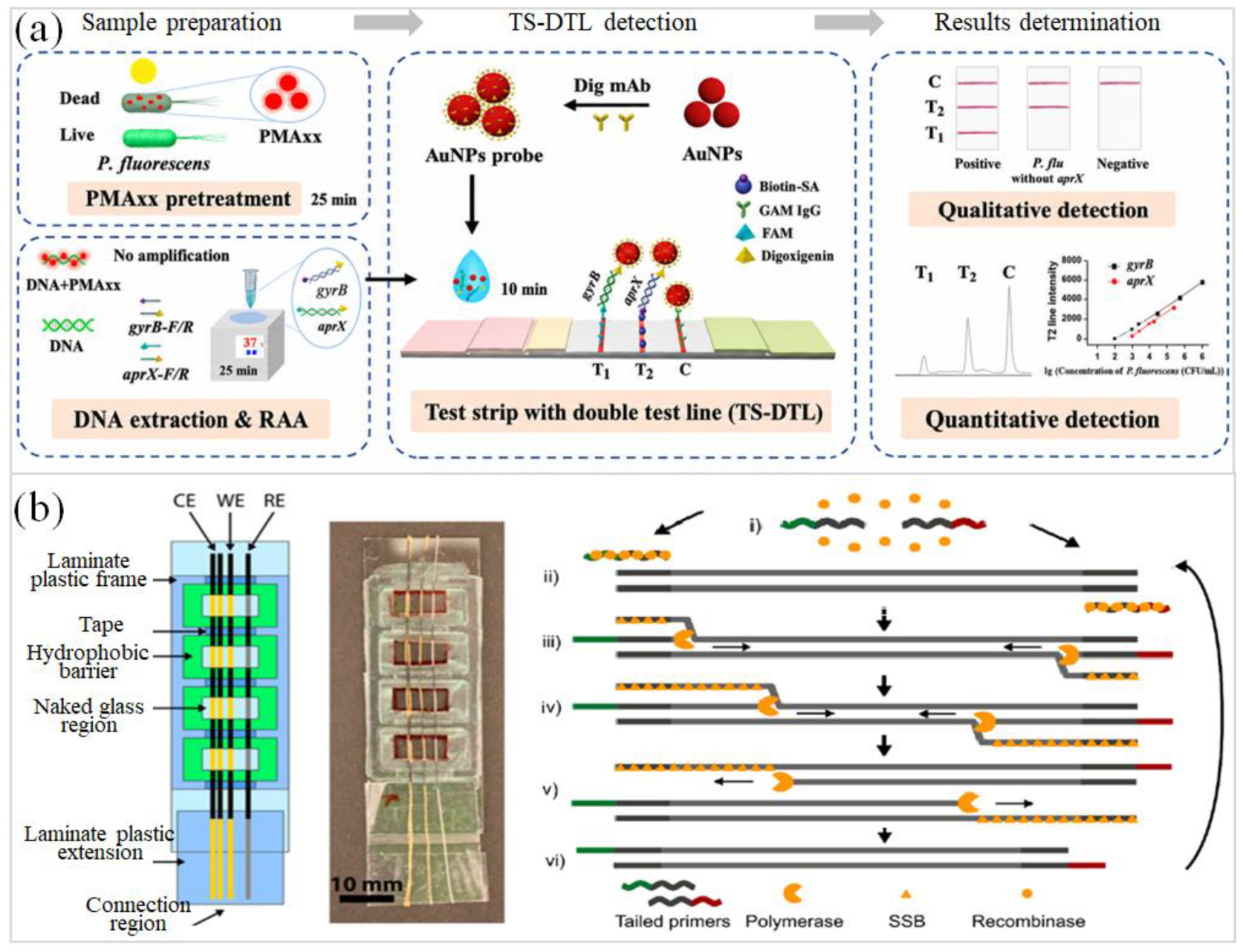
Seafood authentication shows how microfluidic RPA can move beyond pathogen screening. Xu et al. [102] applied a real-time eight-sample chip to 141 commercial seafood products, targeting Atlantic cod, sablefish, and toothfish. The chip processed eight samples in parallel, completed RPA within 20 min, reached 10 copies/μL plasmid or 103 fg/μL genomic DNA, and exposed a 31.91% label–identity mismatch. In dairy monitoring, Guo et al. [94] described electrochemical RPA–CRISPR/Cas12a biosensors for Staphylococcus aureus analysis in milk, using either MWCNTs–Au or MoS2–Au electrode interfaces to convert Cas12a-mediated reporter cleavage into a decrease in DPV current. Both systems reached a detection limit of 3 CFU/mL and produced linear responses across 101–108 CFU/mL-scale bacterial concentrations. With the modified electrodes, milk-derived background effects were restrained, and bacterial quantification remained more stable. Zhang et al. [103] described milk spoilage monitoring with a dual-test-line strip based on recombinase-aided amplification (RAA) for viable Pseudomonas fluorescens targeting gyrB and aprX, as shown in Figure 4a. Naked-eye detection reached 50 and 250 CFU/mL, while quantitative limits in milk were 37 and 233 CFU/mL, respectively; results from spiked milk showed full agreement with culture. Overall, the main bottleneck now lies in cleanup, reagent stability, matrix tolerance, and validation in naturally contaminated aquatic and dairy products.

3.4. Electrochemical Food Safety Biosensors

Electrochemical signal measurement helps move RPA-based food safety assays from instrument-dependent analysis toward portable testing with quantitative output. These platforms convert nucleic-acid recognition into measurable current responses instead of using fluorescence imaging, gel electrophoresis, or centralized PCR systems [93,94]. Recent studies have paired RPA with CRISPR/Cas chemistry, nanostructured electrodes, paper or thread supports, and microfluidic formats, enabling rapid analysis of bacterial pathogens, seafood hazards, toxic microalgae, and GMO markers. The same concept is particularly valuable for genetically modified organism (GMO) traceability, where isothermal amplification and nanomaterial-modified transducers can support sensitive screening directly at the point of need [89,95].
Figure 4. (a) Schematic illustration of recombinase-aided amplification coupled with a dual-test-line strip for P. fluorescens detection. (Reprinted from [103]). (b) Schematic diagram of gold-coated thread-based multi-target electrochemical biosensors for Ostreopsis cf. ovata and Ostreopsis cf. siamensis detection and the tailed-primer RPA (i–vi) cycle. (Reprinted from [104]).
Micromachines 17 00825 g004
Electrochemical coupling brings a practical readout to aquatic food surveillance. In Xu et al. [102], fish-associated Vibrio parahaemolyticus was detected using an RPA-mediated E-CRISPR biosensor built around the tdh and trh loci. RPA first enriched the target sequences; target-activated Cas12a then cleaved ssDNA reporters fixed on the electrode, translating sequence recognition into an electrochemical current change. The assay produced a linear response over 101–106 CFU/mL and reached a detection limit of 32 CFU/mL. Hanze et al. [104] employed gold-coated thread electrodes to read RPA-amplified toxic microalgal DNA electrochemically, as shown in Figure 4b. More than a sensing interface, this roll-to-roll-compatible design points to a scalable route for multi-target surveillance in aquatic environments. Dong et al. [105] advanced electrochemical CRISPR detection with an immobilization-free Cas13a format. Following RPA preamplification, the assay reached 30 zM sensitivity; in a multi-chamber chip, it resolved six pathogens within 50 min. Overall, electrochemical RPA biosensors are shifting toward compact, multiplex, matrix-aware formats; remaining hurdles lie in electrode reproducibility, simpler sample cleanup, and validation with naturally contaminated foods. Table 4 compares representative RPA-based food safety biosensors, emphasizing sensing chemistry, readout strategy, analytical range, and detection limits.

3.5. Challenges and Future Perspectives in Food Safety Testing

Microfluidic RPA systems have advanced rapidly, but their use in food safety testing still faces several barriers before routine deployment becomes realistic. Among them, sample complexity remains one of the most persistent obstacles. Food matrices are inherently heterogeneous, often containing fats, proteins, polysaccharides, salts, pigments, and other endogenous inhibitors that can compromise nucleic-acid recovery, disrupt microfluidic transport, or reduce amplification efficiency [11,12]. Reliable field use therefore calls for pretreatment modules that are both simple and matrix-aware. Filtration, concentration, inhibitor removal, nucleic-acid capture, and reagent-compatible extraction should be built into a single disposable workflow rather than treated as separate preparation steps. A second limitation is quantification. Many RPA platforms still provide endpoint color or fluorescence signals, which are useful for screening but less suitable for risk assessment, regulatory thresholds, or contamination-level tracking. Digital microfluidics, real-time fluorescence, and electrochemical transduction may help close this gap by improving signal normalization, dynamic range, and user-independent interpretation [19,89]. Third, multiplex detection must move beyond placing several reactions side by side. Reducing cross-reactivity and false-positive signals will require improved primer design, CRISPR-assisted target discrimination, compartmentalized reactions, and automated fluidic control [35,50,95]. In general, microfluidic and paper-based RPA technologies are already faster, easier to use, and more portable than culture, PCR, and ELISA. Their broader adoption will depend on sealed cartridges, lyophilized reagents, smartphone- or AI-assisted readout, scalable fabrication, and validation using naturally contaminated foods.

4. Environmental Monitoring Applications

Environmental monitoring is now a crucial aspect of public health, agricultural protection, ecosystem stewardship, and early warning against emerging infections. Air, water, soil, crops, wildlife, and aquaculture environments can all act as reservoirs or transmission routes for pathogenic microorganisms. Conventional surveillance still depends largely on culture, PCR, or sequencing, approaches that remain accurate but slow, instrument-intensive, and tied to centralized laboratories; such constraints can delay intervention during outbreaks or contamination events [11,12]. Microfluidic and paper-based RPA platforms offer a more deployable alternative. Coupling RPA with compact fluidic layouts brings molecular testing directly to the sampling site, allowing rapid screening of environmental pathogens, crop-associated diseases, antimicrobial resistance genes, and zoonotic agents without extensive laboratory infrastructure [106,107]. Selectivity, operational control, and field reliability can be improved when RPA is coupled with CRISPR verification, electrochemical readout, or cartridge-level workflow integration.

4.1. Agricultural and Aquatic Environmental Biosurveillance

Agricultural and aquatic biosurveillance has shifted from laboratory-bound confirmation toward portable RPA-enabled systems. Recent microfluidic and paper-based platforms support airborne spore monitoring, forestry pathogen detection, shrimp disease surveillance, and aquatic pathogen quantification, offering faster operation, lower infrastructure demand, and stronger field relevance [11,108].
Yang et al. [106] used airborne spores as early sampling targets and incorporated this strategy into a microfluidic nucleic acid detection chip for rice smut surveillance. The platform links spore capture, on-chip germination, RPA, and lateral-flow dipstick detection into a compact testing route. A key step is the short germination stage: once the spores develop into delicate mycelial structures, DNA release becomes possible without liquid-nitrogen grinding or hands-on extraction. Within 1 × 10²–1 × 10⁵ CFU/mL, the assay generated a visual sample-in–answer-out response, making it a practical field-oriented option compared with microscopy, staining, or PCR-based laboratory analysis. For field forest protection, Sun et al. [109] developed a portable microfluidic device for detecting pinewood nematodes in pine dust. The system combines boiling pyrolysis, RPA amplification, fluid metering, mixing, temperature control, and gold immunochromatographic strip detection and can obtain visual results within 40 min. Its main value is rapid field warning, not wide-range quantification, as it avoids Baermann extraction and morphology-based identification in routine nematode testing.
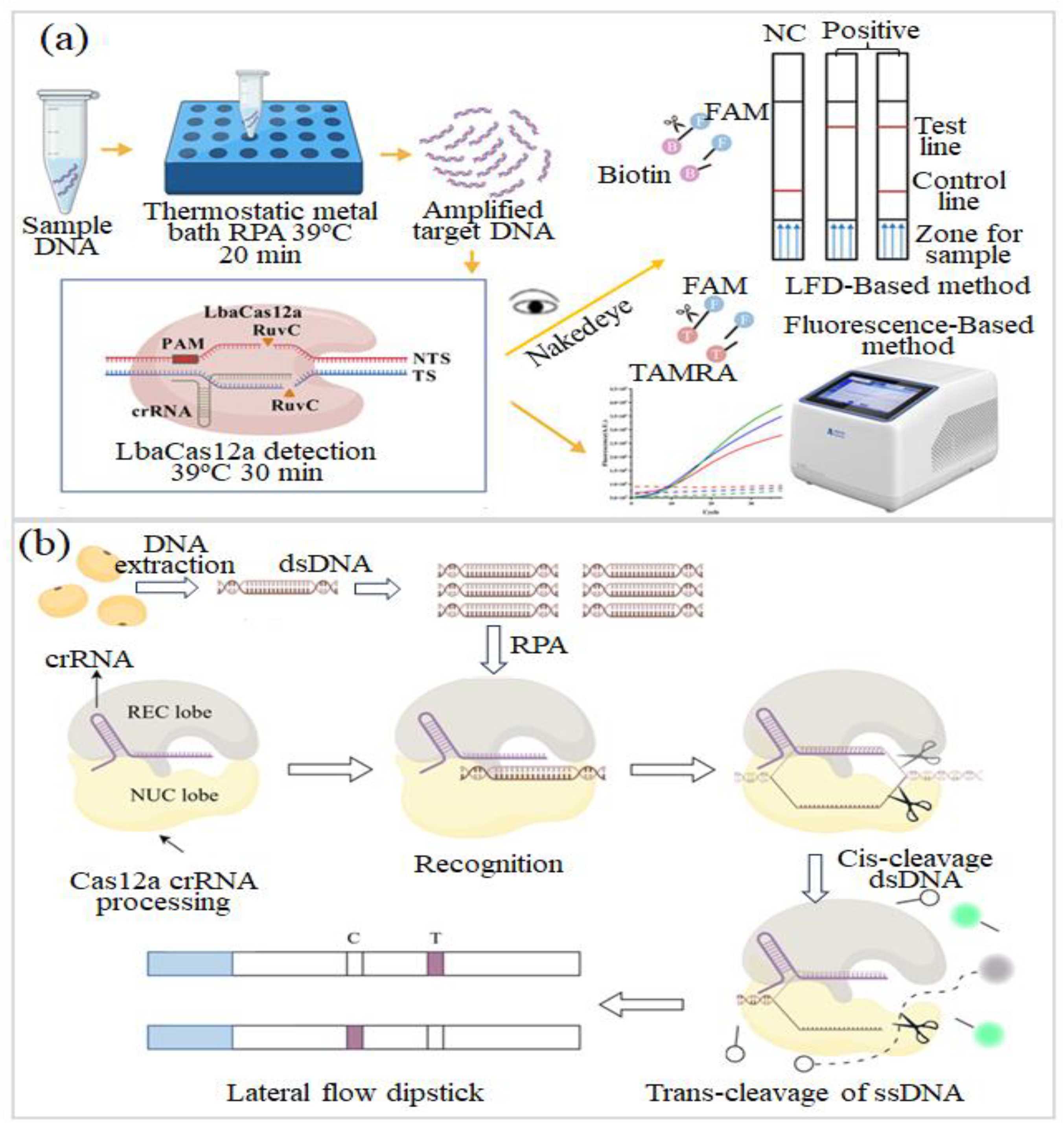
Aquaculture applications show a stronger movement toward multiplex and quantitative surveillance. Li et al. [110] developed a centrifugal microfluidic chip for shrimp pathogens using field shrimp samples and parallel real-time fluorogenic RPA for Vibrio spp. (VAHPND), white spot syndrome virus (WSSV), infectious hypodermal and hematopoietic necrosis virus (IHHNV), shrimp hemocyte iridescent virus (SHIV), and Enterocytozoon hepatopenaei (EHP). Six genetic markers were analyzed from one sample input; 5 μL reaction chambers operated at 39 °C for 20 min, with a detection limit of 10 copies/μL, a coefficient of variation below 0.10, and clinical sensitivity/specificity of 96.4%/100% compared with PCR plus sequencing. For coastal harmful algal bloom surveillance, Yu et al. [111] treated Chrysotila dentata as a field-level signal rather than a laboratory-only target. Their RPA–CRISPR/LbaCas12a–LFD strip (Figure 5a) ran at 39 °C and produced readable bands in 60 min, down to 5 × 10⁻⁶ pg/μL DNA or 2.82 × 101 cells/mL, shortening the path beyond microscopy, qPCR, and sequencing. Fang et al. [101] further improved field quantification by embedding one-pot RPA–CRISPR/Cas12a chemistry into a closed centrifugal chip with in-run standard-curve calibration. Using shrimp, mackerel, oyster, and field shrimp samples, the system reached 6.08 copies/μL, covered 100–104 copies/μL, and showed qPCR-comparable performance. Collectively, these platforms move biosurveillance beyond qualitative readouts toward automated, multiplex, calibration-aware analysis; future progress depends on rugged preparation, stable reagents, matrix tolerance, and real-world validation.

4.2. Environmental Pathogen and One Health Biosurveillance

Environmental pathogen and One Health biosurveillance now form a common analytical ground for public health, animal production, food systems, and ecosystem protection. RPA-enabled microfluidic and biosensing platforms strengthen this ground by linking aerosol, livestock, agricultural, and antimicrobial-resistance monitoring to field-ready molecular evidence [52,107,112]. The result is not only faster detection but also earlier access to information where risk begins to emerge.
Li et al. [55] treated air as a surveillance matrix rather than a passive exposure route. Their Siam/iCASA cartridge joined portable cyclone sampling, chitosan-modified quartz-filter RNA capture, and in situ tetra-primer RPA for SARS-CoV-2 aerosols from isolation wards and public-relevant spaces. Within 25 min, the assay reached 20 copies/mL, detected RNA in 52.2% of ward aerosols, and outperformed a commercial RT-PCR kit. Feng et al. [113] applied the same aerosol-surveillance rationale to influenza subtyping, pairing one-step RT-RPA with a sealed centrifugal microfluidic chip for public-area air samples. Using only 6.25 µL, the device amplified H1N1-HA, H1N1-NA, IVB-HA, and IVB-NA within 25 min. Detection limits reached 102 copies/mL for H1N1-HA and 101 copies/mL for the other targets, with 100% specificity against common respiratory viruses and 96.15–100% agreement with off-chip qRT-RPA. Compared with conventional two-step RT amplification or centralized RT-PCR, the sealed chip format reduced aerosol-contamination risk while enabling subtype-level environmental screening.
At the animal-facing end of One Health surveillance, rapid outbreak control remains essential. Sun et al. [41] reported a surface-engineered μPAD for automated RPA detection of African swine fever virus (ASFV) and pseudorabies virus (PRV) in animal body fluids. Oxygen plasma/PVP treatment improved the capillary movement of viscous RPA reagents, cutting reagent-transfer time from 82 to 27 s. The resulting sample-in–answer-out device detected both ASFV and PRV at 10 copies/µL, with readouts completed in 11.5 and 14.5 min, respectively. Wu et al. [114] brought virulent-strain duck plague virus (DPV) testing closer to farm use by designing a biosensor for duck throat swabs. The assay combines RPA–CRISPR/Cas12a recognition with catalytic hairpin assembly (CHA)-assisted fluorescence amplification, reaching 0.02 fg/µL within 40 min and matching PCR-based clinical diagnoses. Such systems move veterinary diagnostics away from delayed laboratory confirmation and closer to farm-level outbreak containment.
Environmental AMR tracking is another central One Health priority. Skiba et al. [115] reported a foil-based electrochemical genosensor for unpurified asymmetric PCR amplicons of the vanB vancomycin-resistance gene, reaching below 50 nM with 100% specificity and stable microfluidic operation at 2.6–15 µL/min. Tao et al. [116] used RPA–CRISPR/Cas12a to verify the genetically modified soybean cultivar “Zhonghuang 6106” in seeds, young shoots, and blind field-like samples, with lateral-flow and fluorescence readouts available in parallel (Figure 5b). The assay reached 10 copies/µL in approximately 60 min and retained high specificity against mixed genetically modified (GM) maize, rice, cotton, rapeseed, and soybean materials. By moving GM identification into a portable assay format, this method reduces dependence on qPCR-based laboratory supervision for crop screening. Collectively, these works indicate a clear transition from instrument-heavy surveillance toward portable, matrix-aware molecular systems. The remaining challenge is no longer amplification chemistry alone but robust sampling, inhibition tolerance, quantitative calibration, reagent storage, and validation in naturally contaminated environments. Table 5 summarizes representative RPA-based environmental and One Health biosurveillance platforms, highlighting targets, assay conditions, sensing formats, and analytical performance.
Figure 5. (a) Schematic representation of the working principle of the RPA–CRISPR/LbaCas12a–LFD system for visual detection of Chrysotila dentata. (Reprinted from [111]). (b) Schematic illustration of the RPA–CRISPR detection principle for rapid field visualization of genetically modified soybean ‘Zhonghuang 6106’. (Reprinted from [116]).
Micromachines 17 00825 g005

4.3. Emerging Trends in Environmental RPA Biosurveillance

Environmental RPA diagnostics are entering a stage defined by integration, specificity, and networked surveillance. A first clear direction is the maturation of sample-to-answer platforms, where sample collection, pretreatment, nucleic acid extraction, amplification, and signal readout are housed within one compact workflow [106,110]. Such architectures reduce operator-dependent variability and make environmental testing more reproducible under field conditions. A second direction is the rapid expansion of CRISPR-assisted RPA. Cas-based recognition strengthens sequence-level specificity while remaining compatible with fluorescence, electrochemical, colorimetric, lateral-flow, and SERS readouts, thereby widening the analytical choices for complex water, food, animal, and agricultural matrices [41,100,114]. A third movement is toward autonomous and connected biosurveillance. Wireless transmission, cloud analytics, smartphone interfaces, and AI-supported interpretation may convert isolated tests into spatially resolved warning systems [108]. In the longer term, the convergence of RPA, environmental DNA analysis, One Health surveillance, and intelligent sensing could shift environmental monitoring from periodic confirmation to continuous risk tracking, supporting earlier disease control, ecosystem protection, food security, and public health preparedness.

4.4. Challenges and Future Perspectives in Environmental Monitoring

Environmental RPA starts from a less orderly specimen than most clinical or food-testing assays [11,106]. Targets must be recovered from open matrices—air, water, soil, plant tissue, animal fluids, and aquaculture samples—where weak nucleic-acid traces are often patchy and mixed with dispersed cells, extracellular genetic material, humic matter, salts, polysaccharides, pigments, and suspended debris. Each component may compromise nucleic-acid recovery, fluid transport, enzymatic amplification, or signal readout [12,108]. In this setting, sampling is not a preparatory step but a central part of the assay. Even highly sensitive amplification chemistry offers little practical value when sparse targets cannot first be captured, concentrated, and purified from large or variable environmental volumes.
Quantification is another unresolved issue. In environmental surveillance, a positive signal does not always translate directly into infection risk, organism viability, or outbreak severity. Residual DNA, nonviable cells, aerosol dilution, seasonal variation, and uneven spatial distribution can all complicate interpretation [107,108,111]. Field systems therefore need calibration strategies, internal controls, inhibition checks, and reference thresholds that are meaningful for each sample type. High-copy amplicons generated by RPA can generate carryover risk during outdoor or near-source testing, and closed cartridges and contamination-aware workflows are equally important.
Practical deployment also depends on field robustness. Reagents should be tolerant to temperature variations, humidity, transport, and delayed use. The device should remain practical in the field, with low power demand, simple operation, and little need for supporting equipment. For One Health and biosecurity applications, validation should extend beyond spiked samples to naturally contaminated environments, multisite testing, seasonal sampling, and agreement with accepted laboratory methods. Future environmental RPA platforms should therefore combine rugged sampling, inhibitor-tolerant preparation, stable reagent storage, quantitative or semi-quantitative readout, and secure data reporting. Such requirements are essential for moving environmental monitoring from isolated proof-of-concept assays toward reliable early-warning systems.

5. Intelligent, Digital, and AI-Enabled RPA Diagnostic Systems

Microfluidic and paper-based RPA systems have broadened access to molecular diagnostics, but field deployment now depends on more than amplification sensitivity. Reliable interpretation, automation, multiplexing, quantification, and real-time data handling have become central design requirements. Accordingly, RPA platforms are embedding digital sensing, automated image analysis, cloud connectivity, and machine learning to reduce operator dependence and strengthen analytical consistency. This shift is moving point-of-care testing toward intelligent diagnostic networks capable of autonomous operation, data-guided decisions, personalized care, and population-level surveillance [15,108].

5.1. Smartphone- and AI-Assisted RPA Diagnostics

Smartphone- and AI-assisted RPA diagnostics are reshaping molecular testing from instrument-dependent assays into portable, guided, and interpretation-ready platforms. Recent designs combine microfluidics, fluorescence or lateral-flow readout, CRISPR recognition, digital partitioning, and machine learning to improve field operation, quantitative reliability, and decentralized decision-making [117,118].
Liu et al. [15] described paper-based home nucleic acid testing as a practical route toward decentralized diagnostics. In this setting, μPADs offer inexpensive fluid handling and can be paired with intelligent signal interpretation, while broader adoption still depends on more efficient extraction, stable amplification chemistry, and standardized readout criteria. Jin et al. [50] translated Salmonella Typhimurium detection into a smartphone-assisted microfluidic RPA format with fluorescence readout. Purified bacterial DNA was used as input, and an integrated valve was used to route reagents and mix them on demand within the chip. The assay covered 1.0–1.0 × 10⁵ copies/µL and reached a detection limit of 1.0 × 10² copies/µL within 30 min. By replacing 1- to 2-day culture workflows and avoiding benchtop qPCR instrumentation, this format brings rapid pathogen screening closer to field use.
Smartphone-guided platforms have also opened a practical route for applying RPA to pharmacogenomic screening. Pang et al. [81] reported a sealed microfluidic wafer for HLA-B*58:01 genotyping from raw oral swab samples. The platform combines an amplification arrest mutagenesis system and locked nucleic acid technology to enhance double-stranded RPA, β-globin internal control, on-chip lateral flow readings, and smartphone-guided operation. Both targets were detected at 10 copies/μL in 25 min, with all 15 buccal swab samples giving results consistent with sequencing-based typing. For clinical screening, Xu et al. [51] presented R-CHIP, a hand-powered RPA–CRISPR microfluidic platform for high-risk HPV detection, as shown in Figure 6a. In clinical samples, the system detected HPV-16 at 10−17 M and HPV-18 at 10−18 M, delivered sample-to-result analysis within 1 h, and showed strong concordance with multiplex PCR across 300 tests. A smartphone microimaging module, supported by a ResNet-18 classifier, further enabled automated result interpretation, highlighting the value of AI-assisted readout for reducing observer bias and extending screening to community-level settings.

5.2. Digital, Quantitative, and Automated Microfluidic RPA Platforms

Digital, quantitative, and automated microfluidic RPA platforms are strengthening decentralized nucleic-acid testing by bringing reaction partitioning, closed-cartridge operation, programmable fluid handling, and objective readout into the same analytical workflow [42,43,119]. Digital and automated microfluidic RPA platforms address two long-standing weaknesses of conventional RPA: limited quantification and operator-dependent workflow control.
Digital and semi-digital RPA formats are strengthening quantitative molecular diagnostics. Liu et al. [120] developed a photocurable polysiloxane chamber-array chip for E. coli and S. aureus nucleic acid detection, using rapid molding and straightforward sample partitioning to cover 0–5000 copies/μL with a sensitivity of 1 copy/μL. Yang et al. [121] used light-triggered reaction control to adapt digital RPA for detecting clinical oral-pathogen DNA. Photocleavable caged primers kept amplification silent during sample partitioning and triggered the reaction only after digitization, limiting pre-amplification bias and enabling single-molecule analysis across 5–3 × 10⁴ copies/μL within 40 min. Huang et al. [122] used droplet-counting RPA–CRISPR/Cas12a for the detection of methylated FOXD3 with a detection limit of 5 copies/µL and the ability to distinguish methylation fractions as low as 0.01%. This work demonstrates the potential of a partitioned readout for RPA-based assays to detect rare biomarkers.
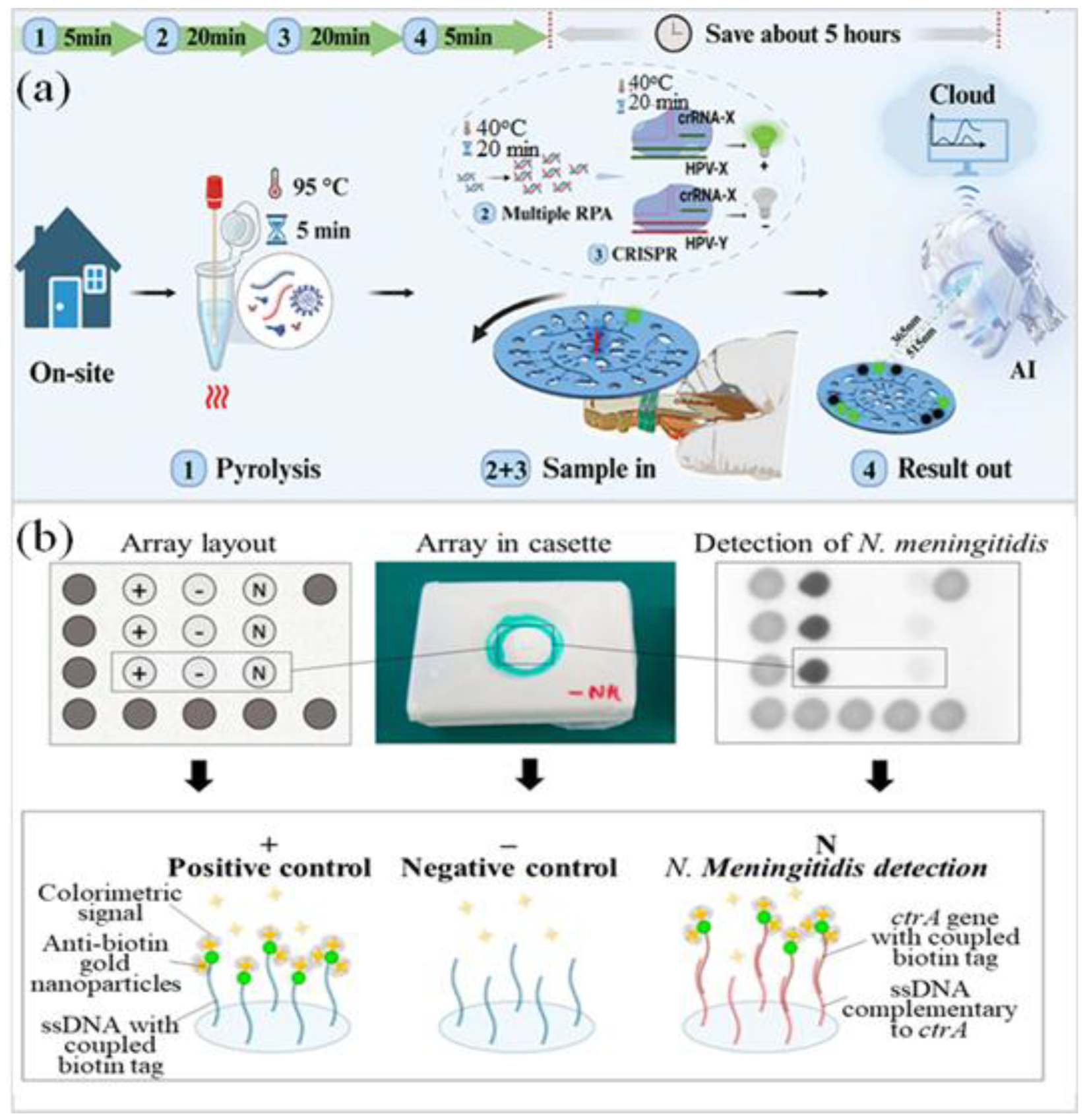
With automation, these systems are closer to practical deployment. Kim et al. [86] reported a disposable cartridge and automated multi-diagnostic platform for nasopharyngeal respiratory samples integrating virus capture, wash, thermal lysis, RT-RPA, and CRISPR-Cas12a fluorescence detection. The platform detected H1N1, H3N2, influenza B, and SARS-CoV-2 within 35–50 min, with on-cartridge detection limit of 1, 100, 10, and 100 copies/µL, respectively. Urrutia Iturritza et al. [87] showed how open-source robotics can streamline RPA-based detection of Neisseria meningitidis in a cerebrospinal fluid (CSF)-like matrix (as shown in Figure 6b). Magnetic-bead DNA isolation, RPA, exonuclease digestion, and paper microarray readout were assembled into a single automated sequence, bringing the total assay time down to 120 min. For user-facing diagnostics, Zhang et al. [53] presented a hand-driven spatially encoded microfluidic device integrating RPA–CRISPR/LFA chemistry with AI-assisted mobile interpretation for HPV and respiratory virus diagnostics. The workflow is compatible with cervical or vaginal swabs and respiratory specimens, supporting user-facing molecular testing outside conventional laboratory settings. The detection limit for HPV plasmids was found to be as low as 1 × 10⁻¹⁸ M, and clinical evaluation of 140 samples yielded 98.57% accuracy. The subjectivity in interpreting the results was further reduced by YOLOv8-assisted image analysis. Collectively, these platforms show that the next stage of RPA diagnostics will depend less on amplification speed alone and more on synchronized initiation, reproducible partitioning, closed automation, matrix-compatible preparation, and objective digital interpretation. Table 6 summarizes intelligent and AI-enabled RPA platforms, highlighting detection models, analytical sensitivity, automation strategy, and practical diagnostic effects.

5.3. Connected RPA Diagnostics and Intelligent Healthcare Systems

The next stage of RPA diagnostics will depend more on the effectiveness of assays in connecting to digital health systems than on amplification performance alone. Cloud computing, wireless communication, and data analytics are now linking microfluidic and paper-based RPA formats with wider healthcare and surveillance infrastructures [15,108]. Automatic result transfer to centralized databases can extend RPA testing from individual diagnosis to real-time outbreak monitoring, hotspot mapping, resource allocation, and public-health response. When coupled with geospatial analysis, these connected workflows could further strengthen population-level tracking during infectious disease events.
At the technical level, the convergence of AI, digital microfluidics, CRISPR recognition, quantitative readout, and cloud connectivity is turning RPA systems into intelligent diagnostic platforms rather than isolated analytical tools [51,53,108,117]. Future formats may combine automated interpretation, predictive analytics, and clinical decision support in portable or home-based devices. Such development could shift molecular diagnostics from centralized laboratories toward patient-centered, digitally connected care while preserving professional oversight and data-driven surveillance capacity.

5.4. Challenges and Future Perspectives in Intelligent RPA Diagnostic Systems

Intelligent, digital, and AI-enabled RPA systems introduce new opportunities, but they also add technical and regulatory demands beyond amplification chemistry [15,108]. Image-based interpretation depends strongly on lighting conditions, camera optics, focal distance, background color, chip alignment, reagent lot, and sample matrix. A model trained under one device setting may not perform equally well across different smartphones, readers, cartridges, or field environments [117,118]. AI-assisted diagnostics gain clinical or field value only when their predictions are backed by solid evidence. This requires representative training data, clearly reported validation, independent testing, and preset criteria for borderline or weak signals.
Digital and automated RPA platforms bring their own engineering burden. Quantitative readout is only as reliable as the partitioning: droplets or chambers must fill evenly, remain sealed, start reactions at nearly the same time, and support defensible calls between positive and negative partitions, especially near the low-copy limit [42,43,119]. Small imperfections—evaporation, leakage, partial filling, spurious amplification, or local thermal drift—can quickly bias the count. Automation removes much of the handwork but adds new places for error: stuck valves, delayed reagent release, bubbles, clogged channels, image-reading mistakes, and software-driven misclassification. For use beyond trained laboratories, internal controls and automatic quality gates should be treated as core design requirements, not optional safeguards.
Connected diagnostics need evaluation beyond the chemistry of the assay. When cloud reporting, wireless transfer, geotagged results, and AI-based interpretation become part of the workflow, the test also inherits concerns over privacy, cybersecurity, audit records, software updates, interoperability, and regulatory review. For clinical translation, validation should span the full operating chain, from biochemical reaction and cartridge performance to algorithm behavior, user interface, data transfer, and final decision logic. Analytical performance alone will not secure adoption. Smartphone readout can bring molecular testing closer to clinics, homes, farms, processing plants, and surveillance sites, but routine use will hinge on the real cost of cartridges, readers, cloud support, and maintenance. The next stage should focus less on connectivity as a feature and more on usable infrastructure: shared benchmark datasets, interpretable outputs, consistent imaging, built-in quality checks, secure data handling, and regulatory routes suited to software-supported molecular diagnostics.

6. Conclusions and Future Outlook

Microfluidic and paper-based RPA technologies have moved isothermal amplification from a bench-level reaction into deployable molecular testing workflows for biomedical diagnosis, food-safety inspection, environmental monitoring, and public-health surveillance. Microfluidic formats provide precise fluid control, reduced reagent consumption, faster reaction handling, and compact operation. Paper-based platforms bring low-cost fabrication, capillary-driven fluid transport, and minimal instrument requirements, making molecular testing more accessible in low-resource and point-of-need settings [11,12,15,17,123]. Across the studies reviewed here, RPA serves not simply as a rapid amplification reaction but as the central engine that coordinates extraction, target recognition, signal generation, and user-facing diagnostic readout.
A major development in this field is the functional coupling of RPA with technologies that improve specificity, quantification, readout, and interpretation. CRISPR recognition strengthens sequence discrimination; digital partitioning supports molecular counting; electrochemical interfaces enable compact quantitative transduction; and smartphone-based imaging improves user-facing readout [31,52,108,124,125]. These combinations have broadened the application space of RPA from pathogen screening to pharmacogenomic testing, cancer-associated biomarker analysis, antimicrobial resistance monitoring, foodborne surveillance, and environmental biosensing [52,76,80,81,82]. The strongest platforms are those in which amplification, sample handling, signal generation, and result interpretation are designed as one workflow rather than assembled as separate steps.
Several barriers still limit routine deployment. Sample preparation remains the most persistent bottleneck, especially for clinical specimens, food matrices, and environmental samples that contain inhibitors, viscous components, low target abundance, or variable background signals [14,126]. Cartridge-based formats that combine extraction, purification, amplification, and readout have begun to address these issues, but stronger robustness across sample types is still needed [87,127]. Multiplex RPA involves more than expanding the primer pool. Reliable performance depends on a careful balance among primer architecture, reaction partitioning, and signal discrimination, since primer competition, biased amplification, and cross-reactivity can readily compromise assay specificity [24,66,70]. Quantification remains equally difficult. Digital RPA and droplet-based strategies improve molecular counting by dividing reactions into smaller compartments, but practical deployment still requires simpler fabrication, lower analytical burden, and workflows that can operate reliably outside highly specialized laboratories [42,43,47,49,52,53].
Translation also depends on manufacturing maturity, regulatory readiness, and a realistic cost structure. Reagent stability is a central issue because RPA enzymes, primers, probes, CRISPR reagents, and reporter chemistries must remain active during storage, shipping, and use outside temperature-controlled laboratories [17,18,108,128]. Lyophilization, humidity protection, sealed reagent compartments, and compatibility between dried reagents and device materials therefore require careful optimization rather than late-stage adjustment. Manufacturing consistency presents a separate challenge. Paper devices are shaped as much by the substrate as by the assay chemistry. Fiber uniformity, wicking rate, surface modification, and reagent placement determine whether liquids reach the reaction zone in a predictable manner. Microfluidic cartridges impose their own manufacturing tolerances. Channel dimensions, sealing strength, valve actuation, chamber volume, and optical or electrochemical coupling must be reproduced from lot to lot; modest deviations can delay amplification, dampen signal readout, or erode quantitative concordance. Quality assessment should therefore extend beyond amplification performance to the device itself, covering lot-to-lot verification, built-in controls, contamination checks, shelf-life testing, user-error analysis, and concordance with accepted reference methods. Regulatory review becomes more demanding once these systems move from research screening toward clinical diagnosis. Analytical sensitivity, specificity, matrix tolerance, reproducibility, stability, usability, and risk management must be demonstrated with well-defined validation protocols. Cost will also shape adoption. A technically elegant cartridge may have limited field value if fabrication, reagent packaging, reader hardware, cold-chain logistics, or per-test pricing remain too high for community clinics, farms, food-processing sites, or environmental surveillance programs. For this reason, practical translation should be considered from the beginning of platform design, with performance, manufacturability, quality assurance, regulatory documentation, and affordability developed as connected requirements rather than separate downstream tasks.
Connected analytical tools will further shape how RPA platforms are used. Machine-learning-assisted image analysis, smartphone interfaces, cloud reporting, and automated quality-control gates can reduce subjective interpretation and support structured data transfer into clinical or surveillance networks [15,51,52,53,54,108]. In parallel, the One Health framework places human diagnostics, veterinary testing, food safety, agricultural monitoring, and environmental surveillance within a shared molecular-warning landscape [52,108]. RPA-based platforms are well suited to this direction because the same amplification principle can be adapted across clinics, farms, food chains, and field sampling sites.
Beyond the application areas emphasized in this review, veterinary outbreak control, antimicrobial-resistance surveillance, forensic identification, plant protection, and biosecurity-oriented monitoring may benefit from the same platform principles: stable reagents, matrix-aware preparation, closed amplification, objective readout, and data-ready reporting. These directions should be evaluated with the same standards applied to clinical and food-safety systems, particularly reproducibility, field robustness, user safety, and agreement with accepted reference methods.
Microfluidic and paper-based RPA technologies have matured into versatile platforms for accessible molecular diagnostics across clinical, food-safety, environmental, and biosurveillance settings. Although challenges related to sample preparation, quantification, standardization, and commercialization remain, ongoing advances in CRISPR diagnostics, digital microfluidics, artificial intelligence, and connected healthcare infrastructures are rapidly accelerating technological maturation. Together, CRISPR-guided recognition, digital microfluidics, electrochemical sensing, smartphone readout, and AI-assisted analysis are pushing RPA diagnostics beyond single-function assays. Their combined use is opening practical routes for deployment in healthcare, precision medicine, food safety, environmental surveillance, and biosurveillance. The next priority is to translate this technological convergence into reproducible devices, validated workflows, and implementation pathways that can be evaluated across clinical, food-safety, and environmental settings.

Author Contributions

Conceptualization, L.-M.F.; data curation, H.-M.W. and S.-Z.L.; formal analysis, H.-M.W. and S.-Z.L.; funding acquisition, L.-M.F.; investigation, H.-M.W. and S.-Z.L.; methodology, H.-M.W. and S.-Z.L.; project administration, L.-M.F.; resources, L.-M.F.; supervision, L.-M.F.; validation, H.-M.W. and S.-Z.L.; visualization, H.-M.W. and S.-Z.L.; writing—original draft, H.-M.W. and S.-Z.L.; writing—review and editing, L.-M.F. All authors have read and agreed to the published version of the manuscript.

Funding

The authors would like to thank the National Science and Technology Council of Taiwan for the financial support of this study under Grant Nos. NSTC 113-2314-B-006-018-MY3, MOST 113-2917-I-006 -004, NSTC 113-2640-B-006-002, and NSTC 114-2314-B-006-048-MY3.

Institutional Review Board Statement

Not applicable.

Informed Consent Statement

Not applicable.

Data Availability Statement

No new data were created or analyzed in this study.

Conflicts of Interest

The authors declare no conflicts of interest. The funders had no role in the design of the study; in the collection, analyses, or interpretation of data; in the writing of the manuscript; or in the decision to publish the results.

References

  1. Huang, Y.; Lu, Y.; Liu, X.; Chai, M.; Yang, L.; Yin, K.; He, J.; Wang, Z.; Zhang, Y.; Yu, Y. A portable all-in-one microfluidic platform integrated with CRISPR-based extraction-free assay for rapid and on-site detection of monkeypox and lumpy skin disease. Sens. Actuators B Chem. 2025, 436, 137612. [Google Scholar] [CrossRef] [Scilit]
  2. Xian, Q.; Zhang, J.; Wong, Y.C.; Gao, Y.; Song, Q.; Xu, N.; Wen, W. Pico-Scale Digital PCR on a Super-Hydrophilic Microarray Chip for Multi-Target Detection. Micromachines 2025, 16, 407. [Google Scholar] [CrossRef] [Scilit] [PubMed]
  3. Marchlewicz, K.; Ziółkowski, R.; Żukowski, K.; Krzemiński, J.; Malinowska, E. Microfluidic System Based on Flexible Structures for Point-of-Care Device Diagnostics with Electrochemical Detection. Biosensors 2025, 15, 483. [Google Scholar] [CrossRef] [Scilit] [PubMed]
  4. Zhao, S.; Guan, X.; Peng, L.; Liu, K.; Zhang, T.; Hu, F. A power-free microfluidic device for the detection of hepatitis B virus in resource-limited settings. Anal. Chim. Acta 2025, 1363, 344154. [Google Scholar] [CrossRef] [Scilit] [PubMed]
  5. Xing, J.; Li, C.; Yang, F. Nucleic acid detection using paper-based microfluidic chips: From amplification to signal readout. Talanta 2026, 309, 130018. [Google Scholar] [CrossRef] [Scilit] [PubMed]
  6. Lin, K.H.; Wang, C.H.; Lin, Y.J.; Huang, W.Y.; Shan, Y.S.; Tsai, H.P.; Lee, G.B. A Portable, Electromagnetically-Driven Microfluidic System for Molecular Diagnosis of SARS-CoV-2 and Influenza A/B Viruses. Sens. Actuators B Chem. 2026, 451, 139324. [Google Scholar] [CrossRef] [Scilit]
  7. De Falco, M.; Colella, S.; Abdelkarim, J.A.Y.; Antonacci, A.; Scognamiglio, V.; De Felice, M. Helicase-dependent isothermal amplification turns biosensing into modern diagnostic technologies. Microchem. J. 2025, 216, 114772. [Google Scholar] [CrossRef] [Scilit]
  8. Das, D.; Masetty, M.; Priye, A. Paper-Based Loop Mediated Isothermal Amplification (LAMP) Platforms: Integrating the Versatility of Paper Microfluidics with Accuracy of Nucleic Acid Amplification Tests. Chemosensors 2023, 11, 163. [Google Scholar] [CrossRef] [Scilit]
  9. Kim, H.S.; Lee, S.S. Isolation of Exosomes from MDA-MB-231 Cells Using a Paddle Screw System and Detection of TNBC-Associated Exosomal miRNAs. Micromachines 2026, 17, 362. [Google Scholar] [CrossRef] [Scilit] [PubMed]
  10. Xu, W.; Luo, X.; Wan, Y.; Dong, X.; Li, X.; Wang, J. Microfluidic-free droplet digital RAA/RT-RAA assisted CRISPR/Cas12a system for the detection of DNA/RNA viruses. Sens. Actuators B Chem. 2025, 422, 136633. [Google Scholar] [CrossRef] [Scilit]
  11. Lu, Y.; Zhang, J.; Lu, X.; Liu, Q. Isothermal nucleic acid amplification based microfluidic “lab-on-a-chip” for the detection of pathogenic bacteria and viruses in agri-foods. Trends Food Sci. Technol. 2024, 148, 104482. [Google Scholar] [CrossRef] [Scilit]
  12. Trinh, T.N.D.; Nam, N.N. Isothermal amplification-based microfluidic devices for detecting foodborne pathogens: A review. Anal. Methods 2024, 16, 1150–1157. [Google Scholar] [CrossRef] [Scilit] [PubMed]
  13. Chondrogiannis, G.; Réu, P.; Hamedi, M.M. Paper-Based Bacterial Lysis Enables Sample-to-Answer Home-based DNA Testing. Adv. Mater. Technol. 2023, 8, 2201004. [Google Scholar]
  14. Mei, J.; Wang, D.; Zhang, Y.; Wu, D.; Cui, J.; Gan, M.; Liu, P. Portable Paper-Based Nucleic Acid Enrichment for Field Testing. Adv. Sci. 2023, 10, 2205217. [Google Scholar] [CrossRef] [Scilit]
  15. Liu, H.; Jia, Y.; Jiang, Y.; Nie, Y.; Hao, R. Paper-Based Microfluidic Chips for At-Home Point-of-Care Nucleic Acid Testing: Applications and Challenges. Diagnostics 2026, 16, 251. [Google Scholar] [CrossRef] [Scilit] [PubMed]
  16. Buchanan, B.C.; Loeffler, R.S.; Liang, R.; Yoon, J.-Y. Capillary flow velocity-based length identification of PCR and RPA products on paper microfluidic chips. Biosens. Bioelectron. 2025, 267, 116861. [Google Scholar] [CrossRef] [Scilit] [PubMed]
  17. Dalgan, S.; Wei, Q. From lab to market: Paper-Based CRISPR diagnostics and commercialization pathways. Adv. Sens. Res. 2025, 4, 23. [Google Scholar] [CrossRef] [Scilit]
  18. Wang, J.; Davidson, J.L.; Kaur, S.; Dextre, A.A.; Ranjbaran, M.; Kamel, M.S.; Athalye, S.M.; Verma, M.S. Paper-Based Biosensors for the Detection of Nucleic Acids from Pathogens. Biosensors 2022, 12, 1094. [Google Scholar] [CrossRef] [Scilit] [PubMed]
  19. Lu, Y.; Hua, M.Z.; Luo, Y.; Lu, X.; Liu, Q. Hybrid paper/PDMS microfluidic device integrated with RNA extraction and recombinase polymerase amplification for detection of norovirus in foods. Appl. Environ. Microbiol. 2024, 90, e01208-24. [Google Scholar] [CrossRef] [Scilit] [PubMed]
  20. Fu, Y.; Chen, X.; Song, W.; Kuang, J.; Wu, W.; Yang, X.; Xia, J.; Liu, L.; Yang, Y.; Ma, S.; et al. Light-Switch Electrochemiluminescence-Driven microfluidic sensor for rapid and sensitive detection of Mpox virus. Chem. Eng. J. 2024, 498, 154930. [Google Scholar] [CrossRef] [Scilit]
  21. Zong, N.; Gao, Y.; Chen, Y.; Luo, X.; Jiang, X. Automated centrifugal microfluidic chip integrating pretreatment and molecular diagnosis for Hepatitis B virus genotyping from whole blood. Anal. Chem. 2022, 94, 5196–5203. [Google Scholar] [CrossRef] [Scilit] [PubMed]
  22. Khamcharoen, W.; Siangproh, W.; Henry, C.S.; Sreamsukcharoenchai, N.; Ratthawongjirakul, P.; Chailapakul, O. Capillary-driven microfluidic device integrating recombinase polymerase amplification for human papillomavirus detection. Sens. Actuators B Chem. 2024, 401, 135016. [Google Scholar] [CrossRef] [Scilit]
  23. Wu, X.; Su, Y.; Xu, Y.; Hu, Q.; Jiao, J.; Ma, J.; Liu, X.; Sun, C.; Guo, W.; Zhou, L. A bioinspired microfluidic nucleic acid sensing platform for rapid and simultaneous screening of viral respiratory infections. ACS Sens. 2025, 10, 4634–4645. [Google Scholar] [CrossRef] [Scilit] [PubMed]
  24. Li, R.; Su, N.; Ren, X.; Sun, X.; Li, W.; Li, Y.; Li, J.; Chen, C.; Wang, H.; Lu, W.; et al. Centrifugal microfluidic-based multiplex recombinase polymerase amplification assay for rapid detection of SARS-CoV-2. iScience 2023, 26, 106245. [Google Scholar] [CrossRef] [Scilit] [PubMed]
  25. Liu, Y.; Yu, G.; Liang, H.; Sun, W.; Zhang, L.; Mauk, M.G.; Li, H.; Chen, L. Detection and identification of SARS-CoV-2 and influenza A based on microfluidic technology. Anal. Methods 2024, 16, 4582–4589. [Google Scholar] [CrossRef] [Scilit] [PubMed]
  26. Liu, J.; Wang, H.; Zhang, L.; Lu, Y.; Wang, X.; Shen, M.; Li, N.; Feng, L.; Jing, J.; Cao, B.; et al. Sensitive and Rapid Diagnosis of Respiratory Virus Coinfection Using a Microfluidic Chip-Powered CRISPR/Cas12a System. Small 2022, 18, 2200854. [Google Scholar] [CrossRef] [Scilit]
  27. Li, B.; Lin, B.; Zeng, W.; Gu, Y.; Zhao, Y.; Liu, P. A fully integrated microfluidic cartridge for rapid and ultrasensitive nucleic acid detection from oropharyngeal swabs. Lab Chip 2025, 25, 454–464. [Google Scholar] [CrossRef] [Scilit] [PubMed]
  28. Dorta-Gorrín, A.; Navas-Méndez, J.; Gozalo-Margüello, M.; Miralles, L.; García-Hevia, L. Detection of SARS-CoV-2 Based on Nucleic Acid Amplification Tests (NAATs) and Its Integration into Nanomedicine and Microfluidic Devices as Point-of-Care Testing (POCT). Int. J. Mol. Sci. 2023, 24, 10233. [Google Scholar] [CrossRef] [Scilit] [PubMed]
  29. Li, Z.; Hua, L.; Xie, L.; Wang, D.; Jiang, X. Automated microfluidic nucleic acid detection platform-integrated RPA-T7-Cas13a for pathogen diagnosis. Anal. Chem. 2023, 95, 6940–6947. [Google Scholar] [CrossRef] [Scilit] [PubMed]
  30. Chen, J.; Lu, Y.; Meng, X.; Chen, D.; Wu, C.; Song, D. Visual and self-contained diagnosis of Salmonella using RPA-CRISPR on a centrifugal force driven microfluidic system. Food Microbiol. 2025, 132, 104844. [Google Scholar] [CrossRef] [Scilit] [PubMed]
  31. Bhattacharjee, G.; Gohil, N.; Khambhati, K.; Gajjar, D.; Abusharha, A.; Singh, V. A paper-based assay for detecting hypervirulent Klebsiella pneumoniae using CRISPR-Cas13a system. Microchem. J. 2024, 203, 110931. [Google Scholar] [CrossRef] [Scilit]
  32. Chen, Y.; Hu, Y.; Lu, X. Polyethersulfone-based microfluidic device integrated with DNA extraction on paper and recombinase polymerase amplification for the detection of Salmonella enterica. ACS Sens. 2023, 8, 2331–2339. [Google Scholar] [CrossRef] [Scilit] [PubMed]
  33. Shimazu, K.N.; Bender, A.T.; Reinhall, P.G.; Posner, J.D. Vibration mixing for enhanced paper-based recombinase polymerase amplification. Lab Chip 2024, 24, 4879–4891. [Google Scholar] [CrossRef] [Scilit] [PubMed]
  34. Chondrogiannis, G.; Toldrà, A.; Hanze, M.; Hamedi, M.M. Paper-based RNase digestion toward viral nucleic acid self-tests. Adv. Mater. Interfaces 2025, 12, 2400553. [Google Scholar]
  35. Zhuang, J.; Zhao, Z.; Lian, K.; Yin, L.; Wang, J.; Man, S.; Liu, G.; Ma, L. SERS-based CRISPR/Cas assay on microfluidic paper analytical devices for supersensitive detection of pathogenic bacteria in foods. Biosens. Bioelectron. 2022, 207, 114167. [Google Scholar] [CrossRef] [Scilit] [PubMed]
  36. Zhang, Z.; Fu, Q.; Wen, T.; Zheng, Y.; Ma, Y.; Liu, S.; Liu, G. Integrated Colorimetric CRISPR/Cas12a Detection of Double-Stranded DNA on Microfluidic Paper-Based Analytical Devices. Biosensors 2026, 16, 32. [Google Scholar] [CrossRef] [Scilit] [PubMed]
  37. Kyung, K.; Lee, H.; Kim, S.-K.; Kim, D.-E. Nucleic Acid Lateral Flow Assay Implemented with Isothermal Gene Amplification of SARS-CoV-2 RNA. Biosensors 2024, 14, 585. [Google Scholar] [CrossRef] [Scilit] [PubMed]
  38. Chu, P.-Y.; Huang, P.-S.; Chen, C.-Y.; Tsai, K.-Y.; Chiu, S.-Y.; Fan, L.-W.; Cheng, Y.-C.; Lin, C.-J.; Hsieh, C.-H.; Wu, M.-H. Development of a point-of-care testing (POCT)-use paper-based device for recombinase polymerase amplification (RPA)-based bioassays: Demonstration of the detection of Neisseria gonorrhoeae. Sens. Actuators Rep. 2025, 9, 100307. [Google Scholar] [CrossRef] [Scilit]
  39. Yin, L.; Wang, L.; Yin, L.; Yang, L.; Gao, B.; Man, S.; Liu, F.; Ma, L. A lyophilized paper-based platform integrating RT-RPA and CRISPR/Cas12a for smartphone-assisted point-of-care detection of norovirus in environmental and food samples. Chem. Eng. J. 2026, 529, 172832. [Google Scholar] [CrossRef] [Scilit]
  40. Chen, L.; Liu, P.; Fu, L.; Zhou, N.; Xiang, J.; Yang, Y.; Sun, H.; Chen, L.; Wang, X.; Wang, X. A vibration-enhanced foldable microfluidic paper-based RPA platform for rapid on-site detection of Vibrio parahaemolyticus. Microchem. J. 2026, 226, 118336. [Google Scholar] [CrossRef] [Scilit]
  41. Sun, J.; Xu, W.; Wang, Y.; Chen, L.; Wang, Y.; Li, T.; Ma, H.; He, M.; Hoettges, K.; Leach, M.; et al. Surface-engineered paper-based microfluidic platform for fully automated RPA detection of swine viral diseases. Sens. Actuators B Chem. 2026, 452, 139384. [Google Scholar] [CrossRef] [Scilit]
  42. Siriyod, N.; Prabowo, M.H.; Cheeveewattanagul, N.; Manopwisedjaroen, K.; Nguitragool, W.; Prachumsri, J.S.; Surareungchai, W.; Rijiravanich, P. Microfluidic paper-based analytical device for point-of-care nucleic acid quantification of malaria. Microchem. J. 2025, 212, 113139. [Google Scholar] [CrossRef] [Scilit]
  43. Liu, S.; Yu, T.; Song, L.; Kalantar-Zadeh, K.; Liu, G. CRISPR/Cas on microfluidic paper-based analytical devices for point-of-care screening of cervical cancer. ACS Sens. 2025, 10, 4569–4579. [Google Scholar] [CrossRef] [Scilit] [PubMed]
  44. Luo, Q.; Yu, Z.; Lyu, W.; Luo, Y.; Xu, L.; Shen, F. Digital Bioassays on the Slipchip Microfluidic Devices. Adv. Sens. Res. 2025, 4, e00030. [Google Scholar] [CrossRef] [Scilit]
  45. Dong, X.; Tang, Z.; Jiang, X.; Fu, Q.; Xu, D.; Zhang, L.; Qiu, X. A highly sensitive, real-time centrifugal microfluidic chip for multiplexed detection based on isothermal amplification. Talanta 2024, 268, 125319. [Google Scholar] [CrossRef] [Scilit]
  46. Wang, N.; Dong, X.; Zhou, Y.; Zhu, R.; Liu, L.; Zhang, L.; Qiu, X. A Low-Cost Handheld Centrifugal Microfluidic System for Multiplexed Visual Detection Based on Isothermal Amplification. Sensors 2024, 24, 5028. [Google Scholar] [CrossRef] [Scilit] [PubMed]
  47. Shu, B.; Yang, J.; Chen, W.; Li, X.; Xue, Y.; Liu, M.; Yin, X.; Xu, L.; Zhang, H.; Qiu, J.; et al. Temperature-programmed microfluidic CRISPR diagnostics enable rapid and automatous point-of-care testing for syphilis. Chem. Eng. J. 2024, 496, 154174. [Google Scholar] [CrossRef] [Scilit]
  48. Su, Y.; Jin, X.; Yang, F.; Liu, X.; Li, F.; Zhao, Q.; Hou, J.; Zhang, S.; Li, H.; Huang, G.; et al. A compact microfluidic platform for rapid multiplex detection of respiratory viruses via centrifugal polar-absorbance spectroscopy. Talanta 2024, 280, 126733. [Google Scholar] [CrossRef] [Scilit] [PubMed]
  49. Feng, J.; Lu, C.; Lin, X.; Kang, L.; Duan, N.; Wang, Z.; Liu, C.; Wu, S. A portable microfluidic chip-based fluorescent and colorimetric aptasensor combining recombinase polymerase amplification for bovine pregnancy-associated glycoproteins detection. Biosens. Bioelectron. 2025, 270, 116981. [Google Scholar] [CrossRef] [Scilit] [PubMed]
  50. Jin, Y.; Wang, J.; Wang, Z.; Xiong, P.; Cheng, J.; Xu, T. An Integrated Microfluidic Biosensing System Based on a Versatile Valve and Recombinase Polymerase Amplification for Rapid and Sensitive Detection of Salmonella typhimurium. Biosensors 2023, 13, 790. [Google Scholar] [CrossRef] [Scilit] [PubMed]
  51. Xu, T.; Zhang, Y.; Li, S.; Dai, C.; Wei, H.; Chen, D.; Zhao, Y.; Liu, H.; Li, D.; Chen, P. Deep learning-enhanced hand-driven microfluidic chip for multiplexed nucleic acid detection based on RPA/CRISPR. Adv. Sci. 2025, 12, 2414918. [Google Scholar] [CrossRef] [Scilit]
  52. Ji, J.; Pang, X.; Chang, C.; Wang, D.; Hu, S.; Fang, Z.; Yu, C.; Mei, Q.; Ma, H. AM-DMF-ddRPA: An all-in-one digital microfluidic platform for rapid and automatic digital nucleic acid analysis. Angew. Chem. Int. Ed. 2025, 64, e202501913. [Google Scholar] [CrossRef] [Scilit]
  53. Zhang, Y.; Chen, D.; Tang, X.; Xu, T.; Li, S.; Zhao, X.; Miao, Z.; Zhang, Y.; Zhou, H.; Li, Y.; et al. Deep learning-enhanced hand-driven spatial encoding microfluidics for multiplexed molecular testing at home. ACS Nano 2025, 19, 25793–25811. [Google Scholar] [CrossRef] [Scilit] [PubMed]
  54. Burrow, D.T.; Heggestad, J.T.; Kinnamon, D.S.; Chilkoti, A. Engineering innovative interfaces for point-of-care diagnostics. Curr. Opin. Colloid Interface Sci. 2023, 66, 101718. [Google Scholar] [CrossRef] [Scilit]
  55. Li, S.; Li, B.; Li, X.; Liu, C.; Qi, X.; Gu, Y.; Lin, B.; Sun, L.; Chen, L.; Han, B.; et al. An ultrasensitive and rapid “sample-to-answer” microsystem for on-site monitoring of SARS-CoV-2 in aerosols using “in situ” tetra-primer recombinase polymerase amplification. Biosens. Bioelectron. 2023, 219, 114816. [Google Scholar] [CrossRef] [Scilit] [PubMed]
  56. Tharakan, S.; Faqah, O.; Asghar, W.; Ilyas, A. Microfluidic Devices for HIV Diagnosis and Monitoring at Point-of-Care (POC) Settings. Biosensors 2022, 12, 949. [Google Scholar] [CrossRef] [Scilit] [PubMed]
  57. Li, J.; Wang, X.; Wang, S.; Zhang, K.; Zhang, Y.; Xu, T.; Chen, D.; Li, Y.; Chen, P.; Liu, B.-F. Ergonomic handheld chip: An instrument-free RPA-CRISPR platform for rapid home self-testing. Biosens. Bioelectron. 2026, 293, 118157. [Google Scholar] [CrossRef] [Scilit] [PubMed]
  58. Huang, Q.; Shan, X.; Cao, R.; Jin, X.; Lin, X.; He, Q.; Zhu, Y.; Fu, R.; Du, W.; Lv, W.; et al. Microfluidic Chip with Two-Stage Isothermal Amplification Method for Highly Sensitive Parallel Detection of SARS-CoV-2 and Measles Virus. Micromachines 2021, 12, 1582. [Google Scholar] [CrossRef] [Scilit] [PubMed]
  59. Li, X.; Guo, J.; Yang, H.; Wu, Y.; Xie, Z.; Li, D. A CRISPR-assisted passive microfluidic chip for rapid, visual detection of multiple respiratory viruses. Sci. Rep. 2026, 16, 2033. [Google Scholar] [CrossRef] [Scilit]
  60. Lu, S.-M.; Wang, J.-L.; Li, Y.; Zhang, L.; Yao, H.-P.; Liang, L.-G. Autonomous microfluidic influenza A/B subtyping system using on-chip multiplex isothermal amplification for field-deployable surveillance. AMB Express 2025, 15, 123. [Google Scholar] [CrossRef] [Scilit] [PubMed]
  61. Chen, H.; Cheng, X.; Wang, Y.; Han, N.; Liu, L.; Wei, H.; Tu, Z.; Gu, Z.; Song, R.; Wang, S. Two-dimensional nanozyme-catalyzed colorimetric CRISPR assay for the microfluidic detection of monkeypox virus. Anal. Chem. 2025, 97, 4407–4415. [Google Scholar] [CrossRef] [Scilit] [PubMed]
  62. Wang, Z.; Yang, F.; Zeng, S.; Sun, R.; Hu, Q.; Du, Y. An integrated valved microfluidic platform for rapid and simultaneous nucleic acid detection. Lab Chip 2026, 26, 507–514. [Google Scholar] [CrossRef] [Scilit] [PubMed]
  63. Chen, J.; Liu, S.; Chen, S.; Mai, J.; Abudukadi, M.; Chen, Y.; Lu, J.; Li, G.; Ge, C. Rapid Visual Detection of Mycoplasma Hominis Using an RPA-CRISPR/Cas12a Assay. Biosensors 2025, 15, 821. [Google Scholar] [CrossRef] [Scilit] [PubMed]
  64. Hua, X.; Xu, D.; Wang, Y.; Yin, Z.; Xiang, Q.; Su, L. Dual recognition probe design combined with asymmetric amplification strategy for rapid electrochemical detection of trace viral RNA. Sens. Actuators B Chem. 2025, 446, 138623. [Google Scholar] [CrossRef] [Scilit]
  65. Huang, B.; Lou, Y.; Zeng, Z.; Kan, X.; Shi, X.; Wu, Y.; Guo, L.; Wang, M.; Huang, X.; Tian, X.; et al. A Cas12a-based fluorescent microfluidic system for rapid on-site human papillomavirus diagnostics. Appl. Microbiol. Biotechnol. 2023, 107, 6287–6297. [Google Scholar] [CrossRef] [Scilit] [PubMed]
  66. Xu, Z.; Chen, D.; Li, T.; Yan, J.; Zhu, J.; He, T.; Hu, R.; Li, Y.; Yang, Y.; Liu, M. Microfluidic space coding for multiplexed nucleic acid detection via CRISPR-Cas12a and recombinase polymerase amplification. Nat. Commun. 2022, 13, 6480. [Google Scholar] [CrossRef] [Scilit] [PubMed]
  67. Zhao, Y.; Chen, D.; Xu, Z.; Li, T.; Zhu, J.; Hu, R.; Xu, G.; Li, Y.; Yang, Y.; Liu, M. Integrating CRISPR-Cas12a into a microfluidic dual-droplet device enables simultaneous detection of HPV16 and HPV18. Anal. Chem. 2023, 95, 3476–3485. [Google Scholar] [CrossRef] [Scilit] [PubMed]
  68. Nakowong, P.; Chatchawal, P.; Chaibun, T.; Boonapatcharoen, N.; Promptmas, C.; Buajeeb, W.; Lee, S.Y.; Jearanaikoon, P.; Lertanantawong, B. Detection of high-risk HPV 16 genotypes in cervical cancers using isothermal DNA amplification with electrochemical genosensor. Talanta 2024, 269, 125495. [Google Scholar] [CrossRef] [Scilit] [PubMed]
  69. Hwang, S.B.; Song, Y.-J.; Park, P.-G. A Novel Diagnostic Tool for West Nile Virus Lineage 1a and 2 Using a CRISPR-Cas12a System. Biosensors 2025, 15, 807. [Google Scholar] [CrossRef] [Scilit] [PubMed]
  70. Ma, Y.; Wang, Y.; Chen, C.; Feng, L.; Shan, J.; Zhang, L.; Ma, X.; Chu, Y.; Wu, H.; Zhou, G. FEN1-aided RPA (FARPA) coupled with autosampling microfluidic chip enables highly multiplexed on-site pathogen screening. Anal. Chem. 2025, 97, 5762–5770. [Google Scholar] [CrossRef] [Scilit] [PubMed]
  71. Wang, Y.; Zhang, W.; Xu, J.; Yan, F.; Mao, M.; Zhang, L.; Jiao, Y.; Xu, X.; Yang, J. Finger driven microfluidic chip based on CRISPR/Cas12a mediated RPA amplification for ultrasensitive and multiplex bacteria detection. Chem. Eng. J. 2026, 539, 176803. [Google Scholar] [CrossRef] [Scilit]
  72. Saikaew, S.; Sangboonruang, S.; Pongsararuk, R.; Srilohasin, P.; Butr-Indr, B.; Intorasoot, S.; Phunpae, P.; Tharinjaroen, C.S.; Arunothong, S.; Panyasit, W.; et al. Gold Nanoparticle-Enhanced Recombinase Polymerase Amplification for Rapid Visual Detection of Mycobacterium tuberculosis. Biosensors 2025, 15, 607. [Google Scholar] [CrossRef] [Scilit] [PubMed]
  73. Xie, R.; Shen, J.; Zhou, L.; Lu, L.; Zhi, A.; Sun, D.; Pei, Y.; Yu, J.; Zeng, L.; Gu, G. Rapid bacterial identification through multiplexed nucleic acid detection on a digital microfluidic platform for enhanced clinical intervention against infections. ACS Sens. 2025, 10, 2520–2530. [Google Scholar] [CrossRef] [Scilit] [PubMed]
  74. Chen, L.; Feng, L.; Wang, Y.; Zhang, J.; Chen, C.; Ma, X.; Wu, H.; Tang, H.; Yue, H.; Zhou, G. An autosampling microfluidic chip enables FEN1-aided recombinase polymerase amplification to rapidly detect multiple urinary tract infection pathogens on-site. Microchim. Acta 2026, 193, 290. [Google Scholar] [CrossRef] [Scilit]
  75. Xiu, L.; Li, H.; Hu, Q.; Zhang, Y.; Chen, S.-B.; Wang, C.; Zhou, X.-N.; Chen, J.-H.; Yin, K. A versatile microfluidic platform for malaria infection screening and Plasmodium species genotyping. eBioMedicine 2023, 98, 104898. [Google Scholar] [CrossRef] [Scilit] [PubMed]
  76. Trinh, T.N.D.; Nguyen, H.A.; Thi, N.P.A.; Ho, T.X.T.; Trinh, K.T.L.; Tran, N.K.S. Integration of Point-of-Care Technology in the Decoding Process of Single Nucleotide Polymorphism for Healthcare Application. Micromachines 2025, 16, 1159. [Google Scholar] [CrossRef] [Scilit] [PubMed]
  77. Li, W.-M.; Ren, X.-D.; Jiang, Y.-Z.; Su, N.; Li, B.-W.; Sun, X.-G.; Li, R.-X.; Lu, W.-P.; Deng, S.-L.; Li, J.; et al. Rapid detection of EGFR mutation in CTCs based on a double spiral microfluidic chip and the real-time RPA method. Anal. Bioanal. Chem. 2023, 415, 3535–3547. [Google Scholar] [CrossRef] [Scilit] [PubMed]
  78. Pang, X.; Lei, Z.; Ji, J.; Fan, L.; Mu, X.; Wang, H.; Dong, W.-F.; Mei, Q. Fully integrated centrifugal microfluidic platform for rapid HLA-B*58:01 allele identification using duplex RPA assay. Biosens. Bioelectron. 2025, 282, 117479. [Google Scholar] [CrossRef] [Scilit] [PubMed]
  79. Chen, Z.; Mao, K.; Meng, H.; Gao, C.; Lv, H.; Li, X.; Zheng, Q.; Yang, Z.; Hamza, I.A.; Tu, C. String-powered microfluidic chip integrating heparin-mediated one-pot RT-RPA/CRISPR-Cas12a for multiplex detection of HFMD viruses. Anal. Chem. 2026, 98, 14315–14329. [Google Scholar] [CrossRef] [Scilit] [PubMed]
  80. De Keyzer, L.; Škevin, S.; Deserranno, K.; Deforce, D.; Van Nieuwerburgh, F. Rapid Forensic DNA Profiling via Real-Time Recombinase Polymerase Amplification of InDel Markers. Biosensors 2026, 16, 106. [Google Scholar] [CrossRef] [Scilit] [PubMed]
  81. Pang, X.; Lei, Z.; Mu, X.; Wu, S.; Dong, W.-F.; Mei, Q. A smartphone-controlled microfluidic platform for rapid, sample-to-answer HLA-B*58:01 genotyping via LNA-enhanced duplex RPA. Chem. Eng. J. 2026, 536, 175031. [Google Scholar] [CrossRef] [Scilit]
  82. Thoeny, V.; Melnik, E.; Huetter, M.; Asadi, M.; Mehrabi, P.; Schalkhammer, T.; Pulverer, W.; Maier, T.; Mutinati, G.C.; Lieberzeit, P.; et al. Recombinase polymerase amplification in combination with electrochemical readout for sensitive and specific detection of PIK3CA point mutations. Anal. Chim. Acta 2023, 1281, 341922. [Google Scholar] [CrossRef] [Scilit] [PubMed]
  83. Lu, Z.; Ye, Z.; Li, P.; Jiang, Y.; Han, S.; Ma, L. An MSRE-Assisted Glycerol-Enhanced RPA-CRISPR/Cas12a Method for Methylation Detection. Biosensors 2024, 14, 608. [Google Scholar] [CrossRef] [Scilit] [PubMed]
  84. Xie, Y.; Li, H.; Chen, F.; Udayakumar, S.; Arora, K.; Chen, H.; Lan, Y.; Hu, Q.; Zhou, X.; Guo, X.; et al. Clustered Regularly Interspaced Short Palindromic Repeats-Based Microfluidic System in Infectious Diseases Diagnosis: Current Status, Challenges, and Perspectives. Adv. Sci. 2022, 9, 2204172. [Google Scholar] [CrossRef] [Scilit]
  85. Xie, Y.; Zong, Z.; Jiang, Q.; Ke, X.; Wu, Z. Seeking Solutions for Inclusively Economic, Rapid, and Safe Molecular Detection of Respiratory Infectious Diseases: Comprehensive Review from Polymerase Chain Reaction Techniques to Amplification-Free Biosensing. Micromachines 2025, 16, 472. [Google Scholar] [CrossRef] [Scilit] [PubMed]
  86. Kim, D.-U.; Choi, S.; Park, S.; Kim, A.Y.; Heo, W.; Wang, Q.; Lim, C.S.; Park, S.J.; Kim, J.; Jung, H.-I. Automated multi-diagnostic platform for respiratory viruses using integrated microfluidic cartridge and RT-RPA/CRISPR detection. Sens. Actuators B Chem. 2026, 458, 139834. [Google Scholar] [CrossRef] [Scilit]
  87. Urrutia Iturritza, M.; Mlotshwa, P.; Gantelius, J.; Alfvén, T.; Loh, E.; Karlsson, J.; Hadjineophytou, C.; Langer, K.; Mitsakakis, K.; Russom, A.; et al. An Automated Versatile Diagnostic Workflow for Infectious Disease Detection in Low-Resource Settings. Micromachines 2024, 15, 708. [Google Scholar] [CrossRef] [Scilit] [PubMed]
  88. Wang, W.; Sun, J.; Wang, T.; Wu, J.; Huang, C.; Wu, S.; Ye, Y.; Peng, Y.; Han, D.; Zhou, H.; et al. A 3D-printed, self-driven microfluidic sensor chip for point-of-care testing (POCT) of norovirus. Biosens. Bioelectron. 2026, 291, 118048. [Google Scholar] [CrossRef] [Scilit]
  89. Kuprešanin, A.; Jarić, S.; Novaković, Z.; Radović, M.; Pavlović, M.; Knežić, T.; Šašić Zorić, L.; Janjušević, L.; Pavlović, Z. Future perspectives of GMO detection in agriculture: Strategies for electrochemical nucleic acid detection. Microchim. Acta 2025, 192, 457. [Google Scholar] [CrossRef] [Scilit]
  90. Zhou, N.; Fan, G.; Yang, N.; Xu, T.; Zhang, Y.; Xu, W.; Shang, Y. A Cascaded RPA-SDA Amplification Strategy on a Sliding Microfluidic Chip for the Ultrasensitive and Rapid Detection of Shigella. Foods 2025, 14, 3891. [Google Scholar] [CrossRef] [Scilit] [PubMed]
  91. Nguyen, V.D.; Sureshkumar, G.; Seo, T.S. Integrated microfluidic device of DNA extraction, recombinase polymerase amplification and micro-capillary electrophoresis for sample-to-answer detection of Salmonella Typhimurium. Sens. Actuators B Chem. 2025, 435, 137625. [Google Scholar] [CrossRef] [Scilit]
  92. Chen, Y.; Hu, Y.; Lu, X. An Integrated Paper Microfluidic Device Based on Isothermal Amplification for Simple Sample-to-Answer Detection of Campylobacter jejuni. Appl. Environ. Microbiol. 2023, 89, e00695-23. [Google Scholar] [CrossRef] [Scilit] [PubMed]
  93. Guo, Y.; Li, C.; Guo, W.; Zhang, X.; Wang, L.; Zhang, W.; Zou, X.; Sun, Z. Advanced electrochemical biosensing toward Staphylococcus aureus based on the RPA-CRISPR/Cas12a system and conductive nanocomposite. J. Agric. Food Chem. 2024, 72, 22918–22925. [Google Scholar]
  94. Guo, Y.; Xu, H.; Wu, Y.; Luo, S.; Hong, Q.; Zhang, X.; Zou, X.; Sun, Z. Conductive MoS2-Au nanocomposite-based electrochemical biosensor for CRISPR/Cas12a-driven Staphylococcus aureus detection. Sens. Actuators B Chem. 2025, 442, 138078. [Google Scholar] [CrossRef] [Scilit]
  95. Chen, Q.; Wang, H.; Xu, H.; Peng, Y.; Yao, B.; Chen, Z.; Yang, J.; Adeloju, S.; Chen, W. One-pot RPA-CRISPR/Cas12a integrated dual-mode electrochemical lateral flow strip for ultrasensitive and precise detection of Salmonella. Biosens. Bioelectron. 2025, 285, 117529. [Google Scholar] [CrossRef] [Scilit] [PubMed]
  96. Guo, Y.; Guo, W.; Wu, Z.; Xu, H.; Zhang, X.; Zou, X.; Sun, Z. A microfluidic chip-based electrochemical biosensor coupled with CRISPR/Cas12a for simultaneous detection of foodborne pathogens. Anal. Chem. 2025, 97, 27266–27275. [Google Scholar] [CrossRef] [Scilit] [PubMed]
  97. Chen, F.; Lyu, C.; Li, Z.; Xiu, L.; Li, H.; Xie, Y.; Cao, R.; Hu, Q.; Yin, K. Fully Integrated Microfluidic Platform for Multiplexed Detection of HuNoV by a Dynamic Confined-Space-Implemented One-Pot RPA-LAMP System. Adv. Sci. 2024, 11, e2306612. [Google Scholar]
  98. Shang, Y.; Xing, G.; Lin, H.; Sun, Y.; Chen, S.; Lin, J.-M. Development of nucleic acid extraction and real-time recombinase polymerase amplification (RPA) assay integrated microfluidic biosensor for multiplex detection of foodborne bacteria. Food Control 2024, 155, 110047. [Google Scholar] [CrossRef] [Scilit]
  99. Han, D.H.; Lee, S.Y.; Kim, Y.; Oh, J.; Park, J.; Park, Y.M.; Kim, S.G.; Kim, T.S.; Park, J.-K. Ultrasensitive detection of multiple foodborne pathogens using CRISPR–Cas12a on a finger-actuated microfluidic device integrated with a modular pressurizing pump. Anal. Chem. 2026, 98, 531–542. [Google Scholar] [PubMed]
  100. Xu, H.; Chen, Q.; Meng, X.; Yan, C.; Yao, B.; Chen, Z.; Wang, Z.; Chen, W. CRISPR/Cas12a-mediated cyclic signal amplification and electrochemical reporting strategy for rapid and accurate sensing of Vibrio parahaemolyticus in aquatic foods. Biosens. Bioelectron. 2025, 277, 117284. [Google Scholar] [CrossRef] [Scilit] [PubMed]
  101. Fang, J.; Chen, Q.; Ran, M.; Chen, R.; Chen, W.; Cui, J.; Wang, J.; Zhong, K.; Shi, L.; Lu, C.; et al. RPA-CRISPR/Cas12a-coupled microfluidic biosensor enabling on-site, sensitive quantification of Vibrio parahaemolyticus. Biosens. Bioelectron. 2026, 296, 118327. [Google Scholar] [CrossRef] [Scilit] [PubMed]
  102. Xu, Z.; Zhang, Z.; Wang, W.; Yi, L.; Shang, S.; Chen, F.; Chen, Y.; Zhang, L.; Zhou, Q.; Chen, J. Real-time microfluidic RPA chip for simultaneous identification of Atlantic cod, sablefish, and toothfish. J. Food Compos. Anal. 2025, 148, 108251. [Google Scholar] [CrossRef] [Scilit]
  103. Zhang, G.; Zhang, L.; Ye, J.; Wang, D.; Lu, Y. A Novel Sensitive Recombinase-Aided Amplification Integrated Test Strip for Pseudomonas fluorescens in Milk via Dual Gene Probes. Biosensors 2025, 15, 553. [Google Scholar] [CrossRef] [Scilit] [PubMed]
  104. Hanze, M.; Khaliliazar, S.; Réu, P.; Toldrà, A.; Hamedi, M.M. Toward Continuous Molecular Testing Using Gold-Coated Threads as Multi-Target Electrochemical Biosensors. Biosensors 2023, 13, 844. [Google Scholar] [CrossRef] [Scilit] [PubMed]
  105. Dong, J.; Wu, X.; Hu, Q.; Sun, C.; Li, J.; Song, P.; Su, Y.; Zhou, L. An immobilization-free electrochemical biosensor based on CRISPR/Cas13a and FAM-RNA-MB for simultaneous detection of multiple pathogens. Biosens. Bioelectron. 2023, 241, 115673. [Google Scholar] [CrossRef] [Scilit] [PubMed]
  106. Yang, N.; Ji, Y.; Wang, A.; Tang, J.; Liu, S.; Zhang, X.; Xu, L.; He, Y. An integrated nucleic acid detection method based on a microfluidic chip for collection and culture of rice false smut spores. Lab Chip 2022, 22, 4894–4904. [Google Scholar] [CrossRef] [Scilit] [PubMed]
  107. Sfragano, P.S.; Reynoso, E.C.; Rojas-Ruíz, N.E.; Laschi, S.; Rossi, G.; Buchinger, M.; Torres, E.; Palchetti, I. A microfluidic card-based electrochemical assay for the detection of sulfonamide resistance genes. Talanta 2024, 271, 125718. [Google Scholar] [CrossRef] [Scilit] [PubMed]
  108. Liu, Q.; Qiu, Z.; Yao, M.; Jiao, B.; Zhou, Y.; Li, C.; Liu, H.; Xin, L. Progress of rapid detection technology for aquatic microorganisms: A comprehensive review. Microorganisms 2026, 14, 939. [Google Scholar] [CrossRef] [Scilit] [PubMed]
  109. Sun, Y.Z.; Wang, C.; Wang, D.Z.; Bao, Y.S.; Zhu, L.; Xia, H.M. A portable microfluidic system for Bursaphelenchus xylophilus detection. J. Plant Dis. Prot. 2025, 132, 72. [Google Scholar] [CrossRef] [Scilit]
  110. Li, Q.; Duan, L.; Jin, D.; Chen, Y.; Lou, Y.; Zhou, Q.; Xu, Z.; Chen, F.; Chen, H.; Xu, G.; et al. A real-time fluorogenic recombinase polymerase amplification microfluidic chip (on-chip RPA) for multiple detection of pathogenic microorganisms of penaeid shrimp. Aquaculture 2024, 578, 740017. [Google Scholar] [CrossRef] [Scilit]
  111. Yu, J.; Shen, Y.; Zhang, Q.; Luo, X.; Zong, Y.; Zhou, C.; Huang, H.; Jiang, H. Development of an RPA-CRISPR/LbaCas12a-Lateral Flow Assay for the Visual Detection of Chrysotila dentata (Haptophyta). Microorganisms 2025, 13, 2203. [Google Scholar] [CrossRef] [Scilit] [PubMed]
  112. Xu, X.; Dai, T.; Xiong, Q.; Yang, J.; Zang, J.; Liu, T. Molecular Detection of Phytophthora cinnamomi by RPA-CRISPR/Cas12a-Mediated Isothermal Amplification. Forests 2024, 15, 772. [Google Scholar] [CrossRef] [Scilit]
  113. Feng, X.; Han, S.; Bian, Y.; Wang, C.; Chen, R.; Tang, F.; Zhang, L.; Yang, S.; Jiang, H.; Tao, R.; et al. Rapid differential diagnosis of influenza A and B viruses with RT-RPA centrifugal microfluidic chip. Biotechnol. J. 2025, 20, e70072. [Google Scholar] [CrossRef] [Scilit] [PubMed]
  114. Wu, Y.; Wan, J.; Wang, X.; Shen, Y.; Li, X.; Zhou, W.; Zhu, Y.; Xu, X. A RPA-CRISPR/Cas12a-Powered Catalytic Hairpin Assembly Fluorescence Biosensor for Duck Plague Virus Virulent Strain Detection. Biosensors 2026, 16, 73. [Google Scholar] [CrossRef] [Scilit] [PubMed]
  115. Skiba, A.; Szymczyk-Drozd, A.; Serafin, K.M.; Olszewski, M.; Ferapontova, E.E.; Krzemiński, J.; Prygiel, M.; Polak, M.; Tokarska, K.; Malinowska, E.; et al. Foil-based lab-on-chip electrochemical genosensors for rapid antimicrobial resistance testing. Measurement 2026, 259, 119668. [Google Scholar] [CrossRef] [Scilit]
  116. Tao, R.; Zhang, J.; Meng, L.; Gao, J.; Miao, C.; Liu, W.; Jin, W.; Wan, Y. A Rapid Field-Visualization Detection Platform for Genetically Modified Soybean ‘Zhonghuang 6106’ Based on RPA-CRISPR. Int. J. Mol. Sci. 2025, 26, 108. [Google Scholar]
  117. Kumar, R.; Harilal, S.; Al-Sehemi, A.G.; Pannipara, M.; Mathew, G.E.; Mathew, B. Advancements in COVID-19 testing: An in-depth overview. Curr. Pharm. Biotechnol. 2023, 24, 1122–1148. [Google Scholar] [CrossRef] [Scilit] [PubMed]
  118. Nazerke, K.; Ruslan, A.; Saule, D.; Aida, D.; Svetlana, V. Advances and emerging technologies in the diagnosis of viral infections in pigs: Progress, challenges, and One Health perspectives. Vet. World 2025, 18, 3788–3805. [Google Scholar] [CrossRef] [Scilit] [PubMed]
  119. Nouwairi, R.L.; O’Connell, K.C.; Turiello, R.A.; Cunha, L.L.; Gunnoe, L.M.; Burton, A.C.; Gibiser, R.M.; Straub, M.E.; Landers, J.P. Real-time amplification and high resolution melt analysis on a rapid microfluidic instrument. Anal. Chim. Acta 2025, 1356, 344046. [Google Scholar] [CrossRef] [Scilit] [PubMed]
  120. Liu, Z.; Lu, Z.; Gui, Y.; Liu, K.; Xu, Q.; Wang, L.; Khou, J.; Wei, S.; Fan, H.; Ye, J.; et al. Photocurable Polysiloxane-Based Microfluidic Chip for Convenient Sample Partitioning and Digital Nucleic Acid Assay. IEEE Sens. J. 2026, 26, 16643–16651. [Google Scholar] [CrossRef] [Scilit]
  121. Yang, N.; Yu, Y.; Yang, H.; Sun, Y.; Fan, G.; Wang, S.; An, J.; Liu, W.; Wang, Y.; Han, L. Self-driven digital microfluidic chip integrated with fully-controllable-initiation RPA toward single-molecule-level digital analysis of pathogen DNA. Sens. Actuators B Chem. 2026, 465, 140231. [Google Scholar] [CrossRef] [Scilit]
  122. Huang, Z.; Guo, C.; Yang, X.; Li, H.; Xu, W.; Li, X. Microfluidic-free droplet-based CRISPR/Cas12a assay for semi-digital detection of DNA methylation. Mater. Des. 2026, 266, 116045. [Google Scholar] [CrossRef] [Scilit]
  123. Ngoc, L.T.N.; Lee, Y.-C. Current Trends in RNA Virus Detection via Nucleic Acid Isothermal Amplification-Based Platforms. Biosensors 2024, 14, 97. [Google Scholar] [CrossRef] [Scilit] [PubMed]
  124. Yang, J.W.; Kim, H.; Hyeon, L.-S.; Yoo, J.S.; Kang, S. Development of a Recombinase Polymerase Amplification-Coupled CRISPR/Cas12a Platform for Rapid Detection of Antimicrobial-Resistant Genes in Carbapenem-Resistant Enterobacterales. Biosensors 2024, 14, 536. [Google Scholar] [CrossRef] [Scilit] [PubMed]
  125. Kang, X.; Zhao, C.; Chen, S.; Yang, S.; Zhang, X.; Xue, B.; Li, C.; Wang, S.; Yang, X.; Li, C.; et al. A Novel Approach Using LuxSit-i Enhanced Toehold Switches for the Rapid Detection of Vibrio parahaemolyticus. Biosensors 2024, 14, 637. [Google Scholar] [CrossRef] [Scilit] [PubMed]
  126. Polak, M.; Wiatrzyk, A.; Krysztopa-Grzybowska, K.; Sobiecka, K.; Mosiej, E.; Prygiel, M.; Ziółkowski, R.; Jańczak, D.; Pancer, K.; Skiba, A.; et al. Comparative Evaluation of Rapid Nucleic Acids Extraction Methods for Biosensor-Based Point-of-Care Solutions. Biosensors 2026, 16, 195. [Google Scholar] [CrossRef] [Scilit] [PubMed]
  127. Georgoutsou-Spyridonos, M.; Filippidou, M.; Kaprou, G.D.; Mastellos, D.C.; Chatzandroulis, S.; Tserepi, A. Isothermal Recombinase Polymerase Amplification (RPA) of E. coli gDNA in Commercially Fabricated PCB-Based Microfluidic Platforms. Micromachines 2021, 12, 1387. [Google Scholar] [CrossRef] [Scilit] [PubMed]
  128. Ansah, F.; Hajialyani, M.; Ahmadi, F.; Gu, Y.; Tarım, E.A.; Mauk, M.G.; Awandare, G.A.; Bau, H.H. Self-actuated microfluidic chiplet for two-stage multiplex nucleic acid amplification assay. Lab Chip 2024, 24, 5175–5183. [Google Scholar] [CrossRef] [Scilit] [PubMed]
Figure 1. (a) Schematic illustration of a CRISPR-assisted microfluidic RPA chip for the detection of multiple respiratory viruses. (Reprinted from Ref. [59]). (b) Schematic representation of a μPAD-based platform integrating RPA and colorimetric CRISPR/Cas12a for HPV16 E7 detection. (Reprinted from [36]).
Micromachines 17 00825 g001
Figure 6. (a) Schematic illustration of a deep learning-enhanced hand-driven microfluidic chip and workflow for HR-HPV detection. (Reprinted from [51]). (b) Schematic illustration of an automated diagnostic workflow for vertical-flow microarray detection of N. meningitidis in low-resource settings. (Reprinted from [87]).
Micromachines 17 00825 g006
Table 1. Comparison of RPA and PCR for biomedical molecular diagnostics.
CategoryRPAPCR
PrincipleIsothermal amplification using recombinase, primers, SSB proteins, and strand-displacing polymerase.Thermal cycling amplification through denaturation, annealing, and extension.
TemperatureMild, nearly constant temperature, usually 37–42 °C.Requires repeated cycling, typically 90–95 °C, 50–70 °C, and 70–75 °C.
Assay timeRapid; often completed within 10–30 min.Slower in conventional formats; micro-PCR can shorten cycling time.
InstrumentationSimple heaters or portable readers are usually sufficient.Requires precise thermal cycling and stable temperature control.
Biomedical useSuitable for point-of-care testing, emergency screening, field diagnosis, and low-resource settings.Suitable for confirmatory diagnosis, viral-load monitoring, genotyping, and regulated clinical testing.
AdvantagesFast, low-power, portable, and compatible with paper-based or microfluidic devices.Reliable, highly validated, quantitative, specific, and broadly accepted in clinical laboratories.
LimitationsQuantification remains less mature; primer design, non-specific amplification, and contamination require careful control.Instrument-dependent, less portable, and less convenient for decentralized testing.
Best scenarioRapid screening when speed, simplicity, and accessibility are priorities.Laboratory confirmation when accuracy, standardization, and traceability are essential.
Overall rolePractical amplification chemistry for decentralized molecular diagnostics.Benchmark method for high-confidence biomedical nucleic acid testing.
Table 2. Representative paper-based RPA devices summarized by substrate type, target analyte, detection strategy, assay time, limit of detection, and practical advantages.
Ref.Substrate Type/Device FormatTarget AnalyteDetection StrategyAssay TimeLODPractical Advantages
[32]Origami microfluidic deviceSalmonella entericaDipstick extraction, RPA, and lateral-flow visualization20 min260 CFU/mL; 58 CFU/mL after 6 h enrichmentIntegrates extraction, amplification, and visual readout in a foldable PES format with enhanced RPA compatibility.
[35]μPADSalmonella TyphimuriumRPA–Cas12a with SERS readout~45 min4.04 CFU/mL Combines paper fluidics, CRISPR specificity, and Raman enhancement for sensitive food-matrix detection.
[36]μPAD with lyophilized reagentsHPV16 E7 dsDNARPA–Cas12a, colorimetric readout60 min100 pMEnables instrument-free visual screening, with dried reagents supporting portable, storage-stable deployment.
[37]Nucleic-acid lateral-flow assaySARS-CoV-2 RdRp RNART-RPA with lateral-flow readout30 min4.1 copies/µLDelivers rapid strip-based visual detection without electrophoresis or fluorescence instrumentation.
[38]Hand-warmer-heated paper deviceNeisseria gonorrhoeaeRPA, lateral-flow readout30–35 min10 DNA copies or 1 CFU/mLUses low-cost heating and paper processing for field-ready STI testing in resource-limited settings.
[39]Lyophilized paper-based platformNorovirus RNART-RPA with fluorescence readout45 min1 copy/µLRetains room-temperature activity and enables testing across water, lettuce, and oyster matrices.
[40]Foldable μPADVibrio parahaemolyticusRPA reagents, fluorescence imaging20 min102 CFU/mLImproves on-paper reaction mixing, reduces equipment demand, and supports rapid seafood and seawater screening.
[41]Surface-engineered μPADAfrican swine fever virus RPA with fluorescence readout14.5 min for PRV10 copies/µLAccelerates viscous RPA flow via plasma/PVP treatment, enabling rapid veterinary outbreak screening.
[42]Wax-gated RPA-PADPlasmodium falciparumRPA with scanner or smartphone image analysis~35 min28 parasites/mLEnables low-resource malaria quantification with inexpensive paper handling and image-assisted interpretation.
[43]Valve-integrated μPADHPV16 E7 geneRPA–CRISPR/Cas12a fluorescence detection35 min1 fMCombines fluidic control, amplification, and CRISPR readout for instrument-light cervical cancer screening.
LOD: Limit of detection; PES: Polyethersulfone; STI: Sexually transmitted infection.
Table 3. Overview of representative RPA-based biomedical diagnostic platforms by target, amplification conditions/time, sensing chemistry, readout mode, and analytical performance.
Ref. and YearsDetection TargetRPA Condition/TimeActive Substance/Sensing ElementDetection ModeDetection Range and LOD
Kyung et al., 2024 [37]SARS-CoV-2 RdRp RNATwo-step RT-RPA: reverse transcription at 42 °C for 15 min, followed by RPA at 38 °C for 15 minTailed RT-RPA primers, AuNP–reporter probeColorimetric readoutR: 400 to 0.01 copies/µL;
LOD: 4.1 copies/µL
Huang et al., 2021 [58]SARS-CoV-2 and measles virus RNA First-stage RPA at 37 °C for 10 min, followed by second-stage LAMP at 65 °C for 50 min RPA primers, LAMP primers, fluorescence readout reagentFluorescence detectionR: 104 to 101 copies;
LOD: 10 copies.
40 nasopharyngeal samples
Chen et al., 2025 [61]Monkeypox virus DNASingle-step RPA–CRISPR/Cas13a at 37 °C for 30 minGO@Pt 2D nanozyme, FAM–ssRNA–biotin reporterColorimetric CRISPR assayR: -
LOD: 1 copy/µL
40 clinical samples
Wang et al., 2026 [62]HPV16 and HPV18 plasmidsRPA at 37 °C for 20 min, followed by Cas12a reaction at 37 °C for 20 minLyophilized RPA mix, Cas12a/crRNA complexLateral-flow visual readoutR: -
LOD: 20 copies/reaction
Chen et al., 2025 [63]Mycoplasma hominis 16S rRNARPA at 37 °C for 20 min, followed by CRISPR/Cas12a at 37 °C for 30 minCas12a/crRNA, bio-dig reporter probeVisual and grayscale readoutR: 105 to 1 copies/µL
LOD: 2 copies/reaction
18 clinical samples
Hua et al., 2026 [64]SARS-CoV-2 ssRNART-aRPA (asymmetric), 42 °C; 10 min; sample-to-result ~20 minDRCP-based ECNA sensing layerElectrochemical ECNA biosensorR: 30 pM–20 nM; LOD: 12.4 pM
Ma et al., 2025 [70]6 viruses: FLUAV, FLUBV, HPIV-1, HPIV-2, HPIV-3, and SARS-CoV-2 FARPA-chip at 42 °C for 20 min, followed by 95 °C for 1 min and 60 °C for 10 minUniversal RPA primers, FEN1 invasive reaction probesMultiplex fluorescence detectionTested at 5, 10, 50, and 200 RNA copies/target;
LOD: 10 copies of each target
Wang et al., 2026 [71]Staphylococcus aureus, Escherichia coli, and Listeria monocytogenesBacterial capture/RPA at 37 °C for 15 min; Cas12a reaction at 37 °C for 40 min; enzyme stopped at 60 °C for 5 minMXene-Fe@Apt nanosheets, bacterial aptamers, released activator DNA, RPA reagentsFluorescence assayR: 50–106 CFU/mL for S. aureus;
50–106 CFU/mL for E. coli;
102–106 CFU/mL for L. monocytogenes.
LODs: 29, 16, and 76 CFU/mL
Chen et al., 2026 [79]HFMD-relevant enteroviruses EV-A71, CV-A16, CV-A6, CV-A10Integrated one-pot RT-RPA/CRISPR maintained at ~ 39 °C during a 1 hCas12a/crRNA + FAM-BHQ ssDNA reporterFluorescenceEvaluated at 10 aM, 100 aM, 1 fM;
LOD: 10 aM
DRCP: Dual recognition sequences-containing capture probe; FARPA: Fen1-aided RPA; HFMD: Hand, foot, and mouth disease; ECNA: Electrochemical nucleic acid.
Table 4. Representative RPA-based food safety diagnostic platforms summarized by detection target, amplification condition/time, sensing element, readout mode, dynamic range, and limit of detection.
Ref. and YearsDetection TargetRPA Condition/TimeActive Substance/Sensing ElementDetection ModeDetection Range and LOD
Zhuang et al., 2022 [35]Salmonella typhimuriumRPA-Cas12a-μPAD about 45 minCRISPR/Cas12a, crRNA, linker ssDNARaman spectrometric readoutR: 1–108 CFU/mL; LOD: 3.72 CFU/mL in milk and 4.04 CFU/mL in meat
Zhou et al., 2025 [90]Shigella; ipaH gene RPA optimized at 39 °C for 25 min; SDA at 58 °C for 10 minG4/Nt.BstNBI-engineered RPA primer; Bst 2.0 polymeraseVisual colorimetric readoutTube assay LOD 3 × 10⁻³ ng/µL; on-chip LOD 3.5 × 10⁻⁴ ng/µL
Nguyen et al., 2025 [91]Salmonella Typhimurium; invA geneRPA at 37 °C for 50 min; full process 75 minFilter-paper DNA capture; freeze-dried RPA powderCapillary electrophoresisR: 102–107 CFU/mL; LOD 103 CFU/mL
Guo et al., 2024 [93]Staphylococcus aureusRPA: 30 min; CRISPR/Cas12a cleavage on electrode: 37 °C for 30 minCRISPR/Cas12a, target-specific crRNA, MB-labeled hairpin DNAElectrochemical biosensorPure culture: 1.04 × 101–1.04 × 10⁸ CFU/mL, LOD 3 CFU/mL; milk: 1.07 × 101–1.07 × 10⁷ CFU/mL
Chen et al., 2025 [95]SalmonellaRPA at 41 °C for 90 min; eLFS visual readout 10 minCRISPR/Cas12a, crRNA, FITC-ssDNA-Bio probeElectrochemical lateral flow stripR: 3.84–3.84 × 10⁷ CFU/mL;
LOD 1.96 CFU/mL
Guo et al., 2025 [96]Staphylococcus aureus and SalmonellaRPA was performed at 37 °C for 30 minCRISPR/Cas12a, target-specific crRNA, Fc-labeled ssDNA probesElectrochemical biosensorS. aureus: 1.06 × 101–1.06 × 10⁷ CFU/mL, LOD 3 CFU/mL; Salmonella: 1.04 × 101–1.04 × 10⁷ CFU/mL, LOD 3 CFU/mL
Xu et al., 2025 [100]Vibrio parahaemolyticusRPA: 37 °C for 30 min, followed by 80 °C for 10 min protein inactivationCRISPR/Cas12a, tdh-/trh-specific crRNAElectrochemical biosensorR:101–106 CFU/mL; LOD 10.6 CFU/mL
Hanze et al., 2023 [104]DNA from toxic microalgaeRPA using TwistAmp Basic Kit; 37 °C for 30 minThiolated DNA capture probes forming SAMs, HRP-tagged reporter probeElectrochemical biosensor1 pM synthetic DNA about 1500 microalgal cells/reaction
Dong et al., 2023 [105]Six pathogen targetsTotal 50 min at 37 ± 1 °CCRISPR/Cas13a, target-specific crRNAElectrochemical/optical CRISPR biosensorLOD down to 30 zM within 45 min
Six pathogen targets (Yersinia pestis, Francisella tularensis, Chlamydia psittaci, Burkholderia mallei, Burkholderia pseudomallei, and Brucella melitensis).
Table 5. Representative RPA-based platforms for environmental monitoring and One Health biosurveillance, summarized by detection target, amplification condition/time, sensing element, readout mode, analytical range, and detection limit.
Ref. and YearsDetection TargetRPA Condition/TimeActive Substance/Sensing ElementDetection ModeDetection Range and LOD
Sun et al., 2026 [41]ASFV and PRVRPA at 42 °C; optimized readout times: 11.5 minRPA reagents, heating module, fluorescence probeFluorescence detectionR: 0–104 copies/µL; LOD: 10 copies/µL
Ji et al., 2025 [52]Influenza A virus and influenza B virusRPA on AM-DMF chip at 39 °C for 25 minAI droplet navigation, RPA primers, and exo-probesFluorescence detectionR: 101–104 copies/µL; LoD: 6.03 copies/µL
Xu et al., 2025 [100]Vibrio parahaemolyticus in aquatic foodsRPA at 37 °C for 30 min, followed by 80 °C for 10 min protein inactivationCRISPR/Cas12a, tdh-/trh-specific crRNAElectrochemical biosensorR: 101–106 CFU/mL; LOD: 10.6 CFU/mL
Yang et al., 2022 [106]Ustilaginoidea virensRPA-LFD performed at 37 °C for 20 minRPA primers/probe, lateral flow dipstickVisual test/control-line readoutR: 102–105 CFU/mL
LOD: 102 CFU/mL
Sfragano et al., 2024 [107]Sulfonamide-resistance genes sul1 and sul4 from E. coliRPA at 37 °C for 20 minCapture probe/signaling probe sandwich Electrochemical biosensorR: 0.1–10 nmol/L; LOD: 44.2 pmol/L
Sun et al., 2025 [109]Bursaphelenchus xylophilusBoiling lysis at 95 °C for 10 min; RPA at 39 °C for 15 minPortable microfluidic system, boiling lysis unitImmunochromatographic strip readoutPositive strip results within ~40 min
Li et al., 2024 [110]VAHPND, WSSV, IHHNV, SHIV, and EHPOn-chip RPA in 5 μL chambers at 39 °C for 20 minPre-immobilized RPA primers, fluorogenic exo probesCentrifugal microfluidic multiplex detectionR: 1–106 copies/μL; LOD:10 copies/μL
Xu et al., 2024 [112]Phytophthora cinnamomi genomicRPA at 37 °C for 10 minCRISPR/Cas12a, Pcinn204169-specific RPA primersFluorescence detectionR: 10 ng to 10 fg; LOD: 10 pg genomic DNA
Feng et al., 2025 [113]Influenza A H1N1 and Influenza B virusAmplification at 42 °C and completed within 25 minPreloaded target-specific primers and fluorescent probesFluorescence detectionR: 106–100 copies/mL; LOD: 102 copies/mL
Wu et al., 2026 [114]Virulent duck plague virus strainRPA–CRISPR/Cas12a reaction at 37 °C for 30 minRPA primers, CRISPR/Cas12a–crRNA recognition Fluorescence detectionR: 10−1–105 fg/µL; LOD: 0.02 fg/µL
Tao et al., 2025 [116]GM soybean “Zhonghuang 6106”RPA optimized at 39 °C for 20 minCRISPR/Cas12a, event-specific RPA primers, crRNAVisual lateral-flow dipstickR: 1–20,000 copies/µL; LOD 10 copies/µL
AM-DMF: Active-matrix digital microfluidic; ASFV: African swine fever virus; EHP: Enterocytozoon hepatopenaei; IHHNV: Infectious hypodermal and hematopoietic necrosis virus; SHIV: Shrimp hemocyte iridescent virus; WSSV: White spot syndrome virus.
Table 6. Representative intelligent, digital, smartphone-assisted, and AI-enabled RPA diagnostic platforms for decentralized molecular testing.
Ref. and YearsDetection ModelTarget and LODRPA Application Method/TimeUses and Effects
Siriyod et al. (2025) [42]Image-assisted RPA-PAD with wax-gate controlPlasmodium falciparum; LOD: 28 parasites/mL by scanner, 46 parasites/mL by smartphoneSmartphone/scanner-assisted solid-phase RPA-PAD; ~35 min
Enhances low-resource malaria diagnostic access
Supports visual and smartphone readout
Reduces lateral-flow strip cost burden
Liu et al. (2025) [43]Valve-integrated μPAD fluorescence RPA–CRISPR platformHPV16 E7 gene; LOD: 1 fM with circular reporterIntelligent μPAD RPA–CRISPR/Cas12a; 35 min
Enables sensitive cervical cancer screening
Integrates amplification and CRISPR detection
Reduces dependence on bulky instruments
Jin et al. (2023) [50]Smartphone-assisted real-time fluorescence microfluidic biosensorSalmonella typhimurium DNA; LOD: 1.0 × 10² copies/µLIntelligent microfluidic RPA with versatile valve; 30 min
Supports on-site Salmonella screening workflows
Improves mixing and flow control
Reduces dependence on benchtop qPCR
Xu et al. (2025) [51]AI-enabled hand-driven R-CHIP platformHR-HPV; LOD: 10−17 M for HPV-16, 10−18 M for HPV-18AI-enabled RPA–CRISPR with ResNet-18 readout; <1 h
Supports community-level HR-HPV screening programs
Automates mobile fluorescence image classification
Shortens PCR-centered diagnostic workflows substantially
Zhang et al. (2025) [53]AI-enhanced MACRO home molecular testing systemHPV subtypes, SARS-CoV-2, influenza A/B; attomolar sensitivityAI-enabled spatial-encoding RPA–CRISPR/LFA; ≤60 min
Enables privacy-preserving home molecular testing
Reduces extraction and heating requirements
Enables AI-guided result interpretation
Pang et al. (2026) [81]Smartphone-controlled centrifugal microfluidic LFA platformHLA-B*58:01 and β-globin; LOD: 10 copies/µLIntelligent LNA-enhanced duplex RPA; <25 min
Supports pharmacogenomic allopurinol risk screening
Combines genotyping with internal control
Avoids bulky fluorescence optical modules
Urrutia Iturritza et al. (2024) [87]Open-source robotic automated diagnostic workflowNeisseria meningitidis ctrA in CSF matrix; formal LOD: NRAutomated robotic RPA workflow with paper microarray; <2 h
Reduces manual pipetting variability risks
Increases accessible modular workflow automation
Supports low-resource infectious disease testing
Nouwairi et al. (2025) [119]Programmable microfluidic real-time amplification–HRM instrumentGAPDH/HeLa gDNA; formal LOD: NR; dilution series to 0.001–10 ng/µLProgrammable microfluidic RPA plus HRM; 20 min + <4 min
Identifies false-positive RPA signals
Avoids electrophoresis or sequencing confirmation
Supports rapid point-of-need genomic analysis
CSF: Cerebrospinal fluid; LFA: Lateral flow assay; LNA: Locked nucleic acid; MACRO: Artificial intelligence-enhanced hand-driven microfluidic system; PAD: Paper-based analytical device.
Disclaimer/Publisher’s Note: The statements, opinions and data contained in all publications are solely those of the individual author(s) and contributor(s) and not of MDPI and/or the editor(s). MDPI and/or the editor(s) disclaim responsibility for any injury to people or property resulting from any ideas, methods, instructions or products referred to in the content.