

Electrochemical Assays for the Determination of Antidiabetic Drugs—A Review


Abstract
1. Introduction
2. Electrochemical Measurements of Antidiabetic Drugs
2.1. Insulin
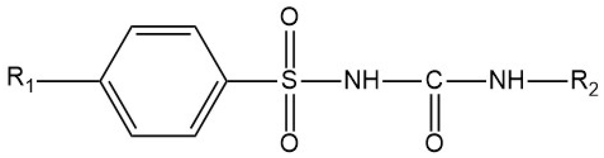
2.2. Sulfonylurea Class of Antidiabetic Agents
2.2.1. Gliclazide
2.2.2. Glipizide
2.2.3. Glibenclamide
2.2.4. Glimepiride
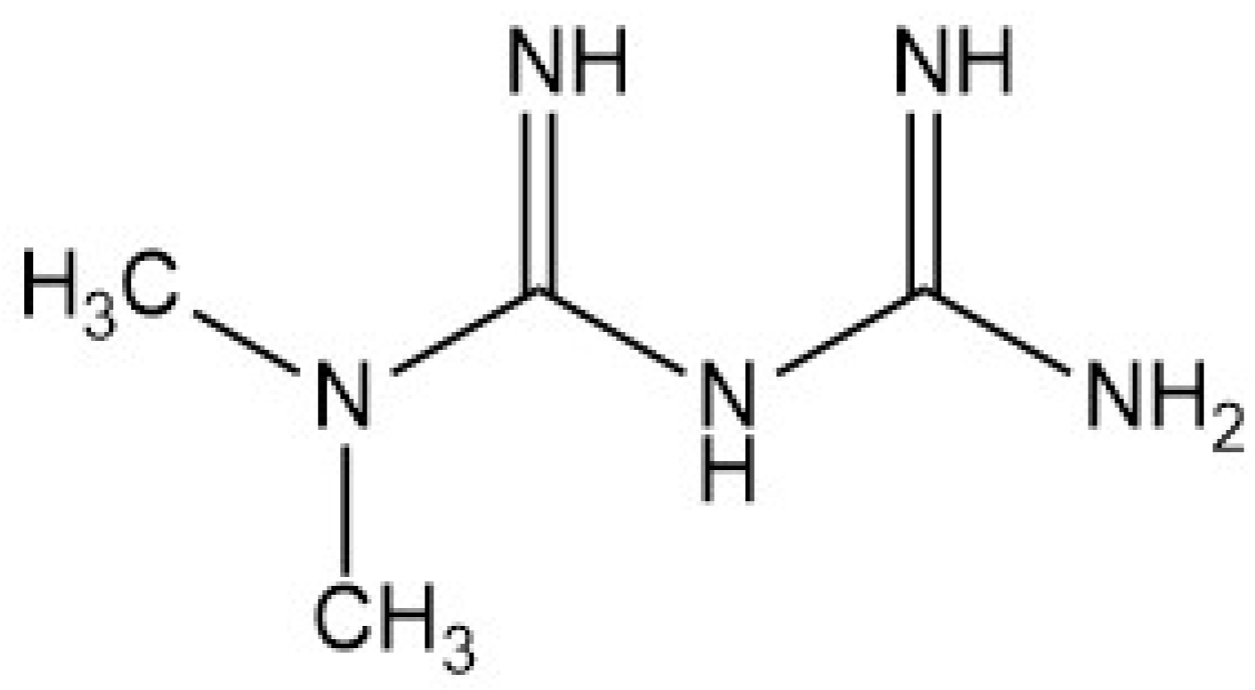
2.3. Metformin
2.4. Dipeptidyl Peptidase-4 Inhibitor
2.4.1. Sitagliptin
2.4.2. Linagliptin
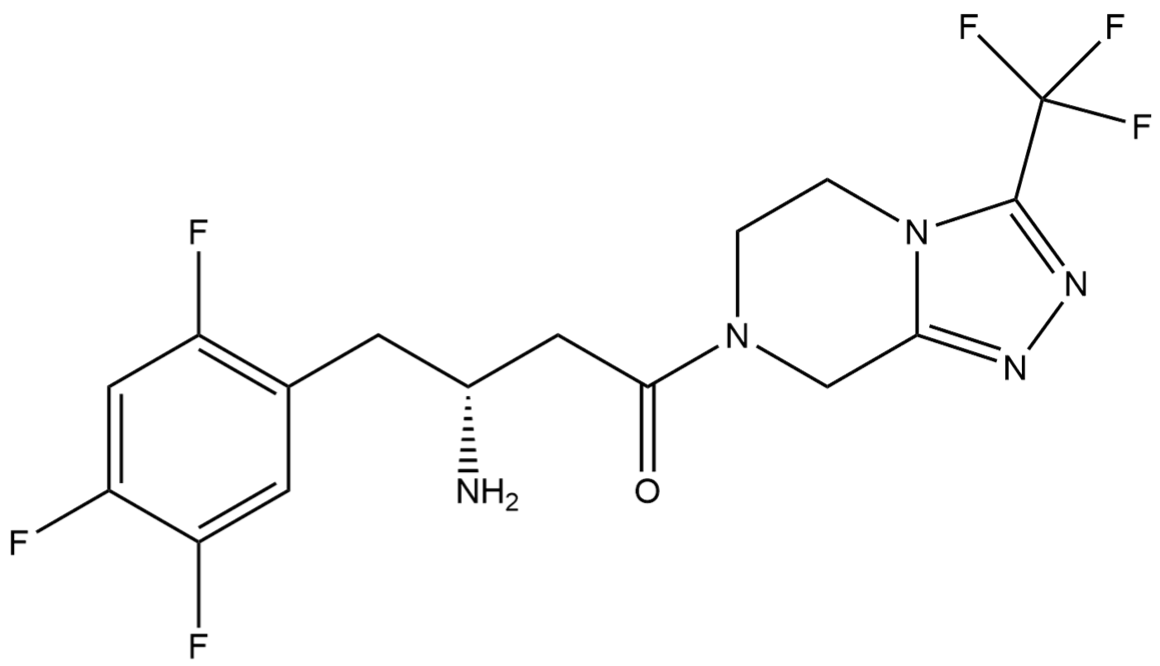
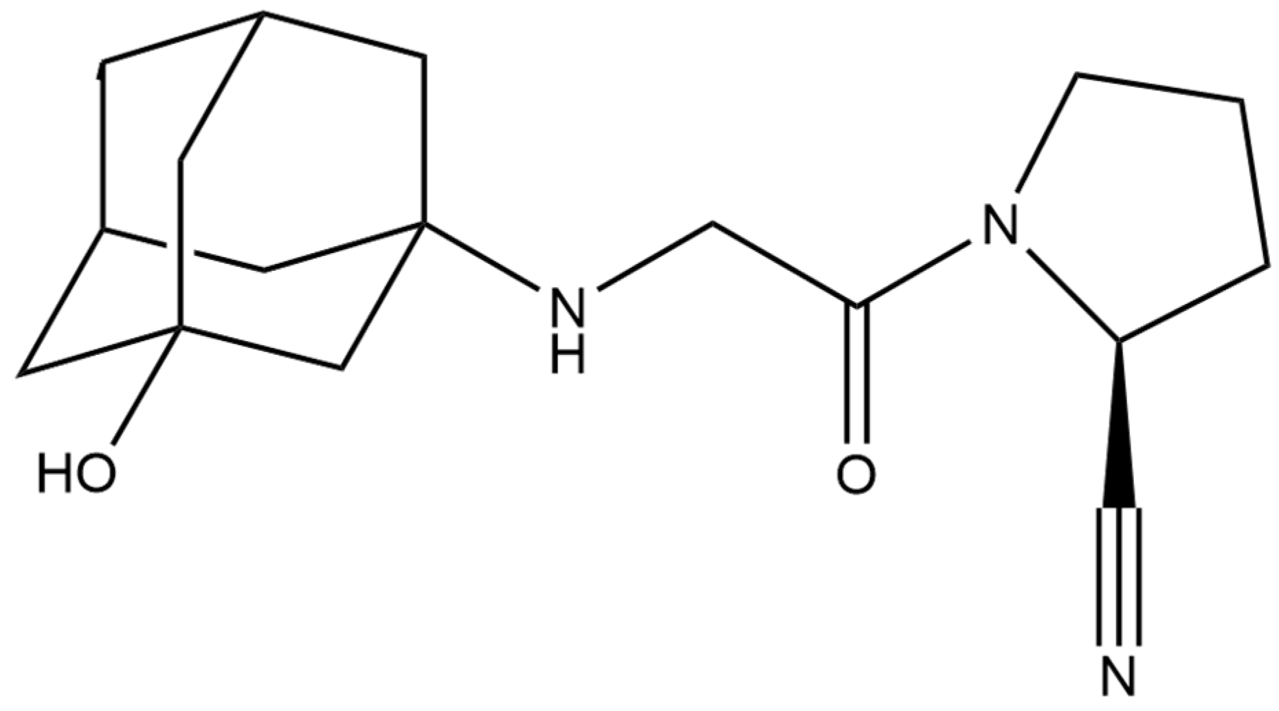
2.4.3. Vildagliptin
2.5. Thiazolidinedione Derivatives
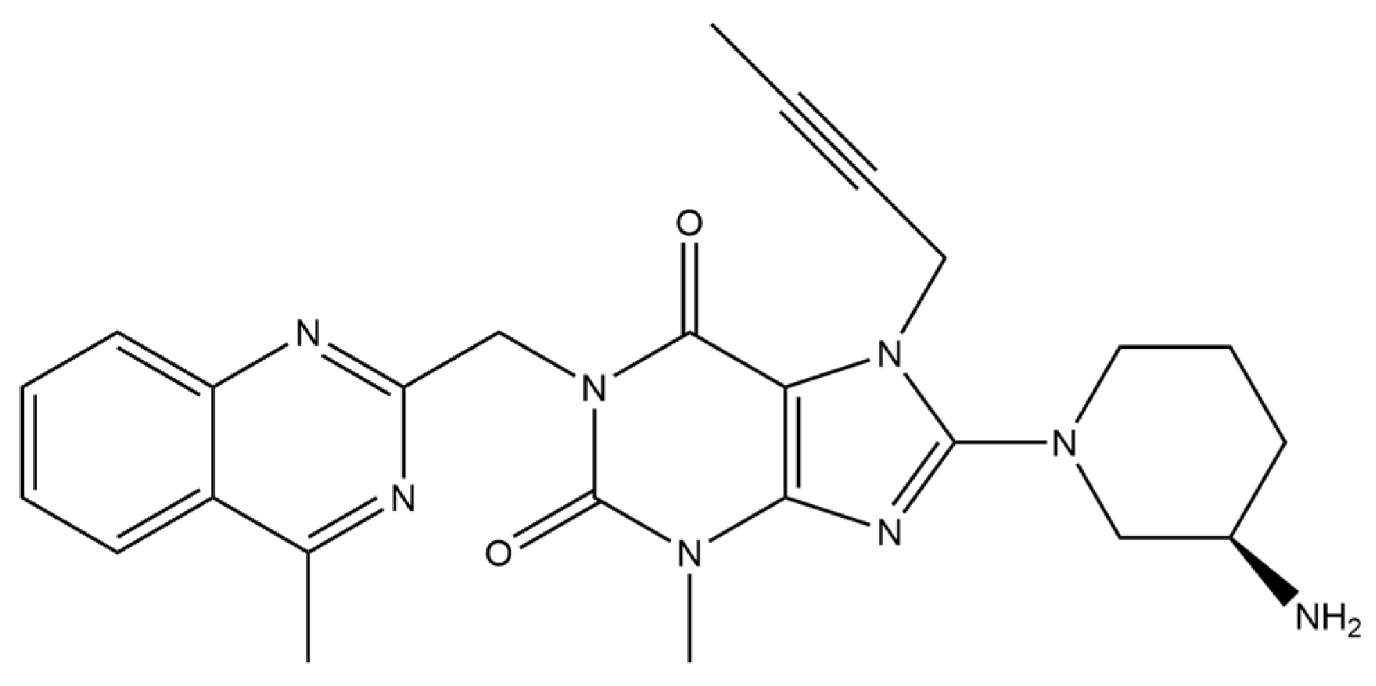
2.5.1. Pioglitazone
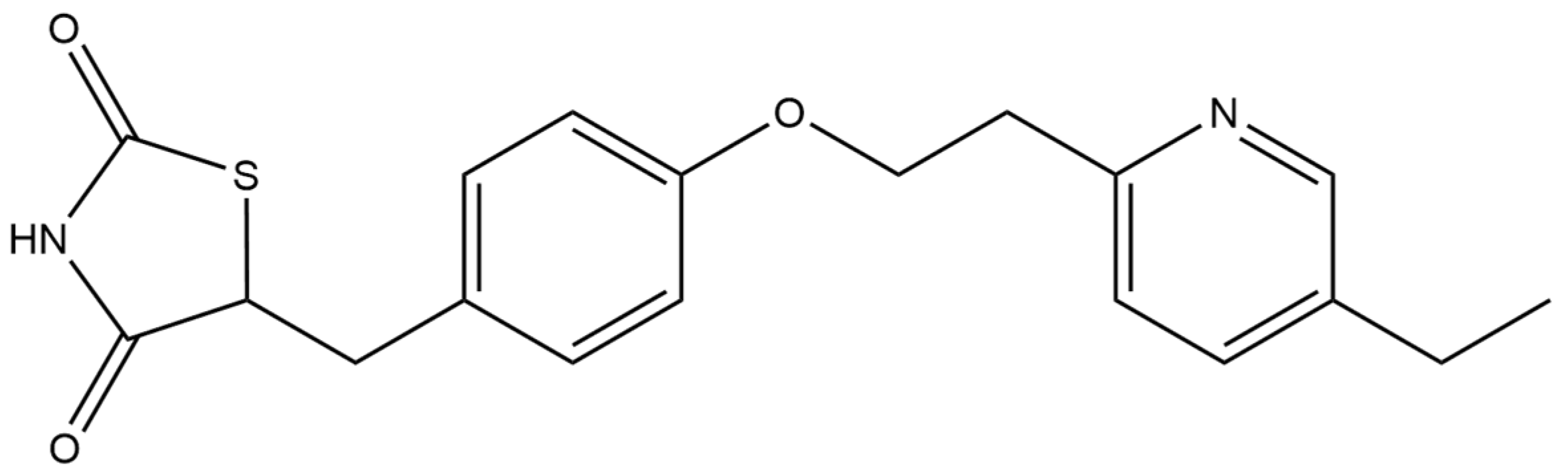
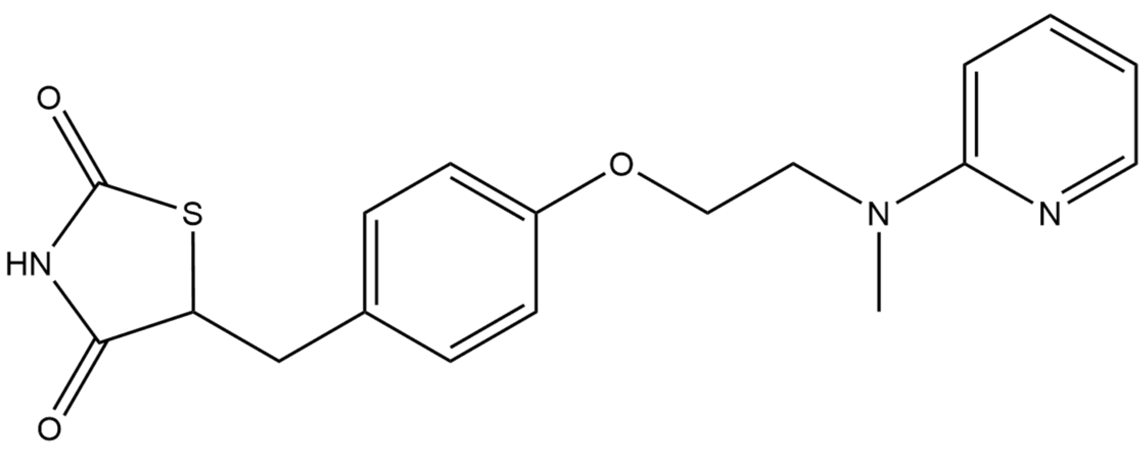
2.5.2. Rosiglitazone
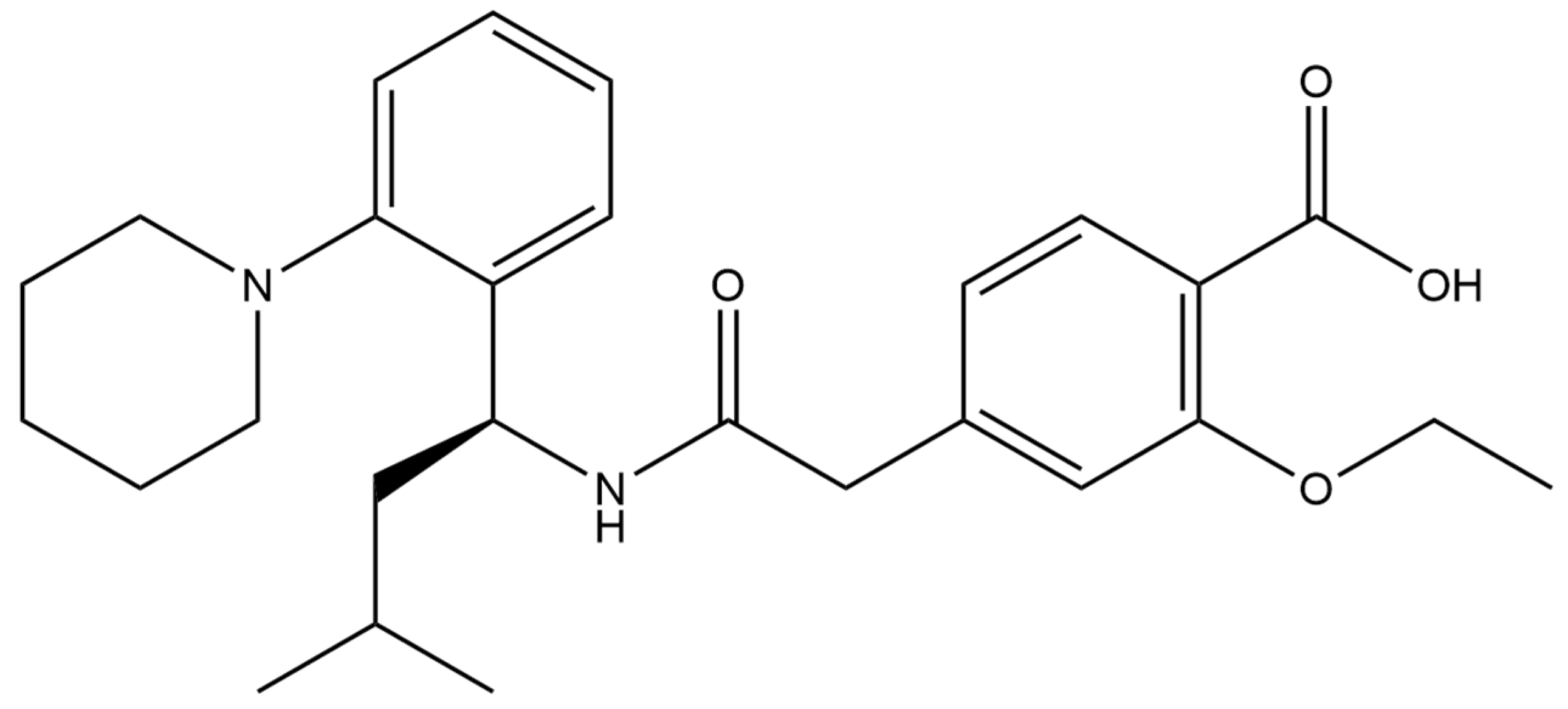
2.6. Repaglinide
3. Conclusions
Author Contributions
Funding
Institutional Review Board Statement
Informed Consent Statement
Data Availability Statement
Conflicts of Interest
References
- World Health Organization. WHO Diabetes Report; World Health Organization: Geneva, Switzerland, 2016. [Google Scholar]
- Sarwar, N.; Gao, P.; Seshasai, S.R.; Gobin, R.; Kaptoge, S.D.A. Diabetes Mellitus, Fasting Blood Glucose Concentration, and Risk of Vascular Disease: A Collaborative Meta-Analysis of 102 Prospective Studies. Emerging Risk Factors Collaboration. Lancet 2010, 26, 2215–2222. [Google Scholar]
- Global Burden of Disease Study 2019. Results. Institute for Health Metrics and Evaluation. Available online: https://vizhub.healthdata.org/gbd-results/ (accessed on 14 October 2023).
- Jean-Marie, E. Diagnosis and Classification of Diabetes Mellitus, 2nd ed.; Elsevier Inc.: Amsterdam, The Netherlands, 2018; ISBN 9780128122006. [Google Scholar]
- Kim, W.; Egan, J.M. The Role of Incretins in Glucose Homeostasis and Diabetes Treatment. Pharmacol. Rev. 2008, 60, 470–512. [Google Scholar] [CrossRef] [PubMed]
- Mogensen, C.E. New Treatment Guidelines for a Patient with Diabetes and Hypertension. J. Hypertens. Suppl. 2003, 21, S25–S30. [Google Scholar] [PubMed]
- Marín-Peñalver, J.J.; Martín-Timón, I.; Sevillano-Collantes, C.; Cañizo-Gómez, F.J. del Update on the Treatment of Type 2 Diabetes Mellitus. World J. Diabetes 2016, 7, 354. [Google Scholar] [CrossRef] [PubMed]
- Robertson, R.P. Diabetes Type 2 (Noninsulin-Dependent Diabetes Mellitus); Elsevier: Amsterdam, The Netherlands, 2016; Volume 2, ISBN 9780128093245. [Google Scholar]
- Blonde, L. Current Antihyperglycemic Treatment Guidelines and Algorithms for Patients with Type 2 Diabetes Mellitus. Am. J. Med. 2010, 123, S12–S18. [Google Scholar] [CrossRef] [PubMed]
- Kampmann, U. Gestational Diabetes: A Clinical Update. World J. Diabetes 2015, 6, 1065. [Google Scholar] [CrossRef] [PubMed]
- Catalano, P.M. Trying to Understand Gestational Diabetes. Diabet. Med. 2014, 31, 273–281. [Google Scholar] [CrossRef]
- McIntyre, H.D.; Catalano, P.; Zhang, C.; Desoye, G.; Mathiesen, E.R.; Damm, P. Gestational Diabetes Mellitus. Nat. Rev. Dis. Prim. 2019, 5, 47. [Google Scholar] [CrossRef]
- Coustan, D.R. Gestational Diabetes Mellitus. Clin. Chem. 2013, 59, 1310–1321. [Google Scholar] [CrossRef]
- Born, G.V.R.; Cuatrecasas, P.; Arbor, A.; Ganten, D.; Herken, H.; Melmon, K.L. Oral Antidiabetics; Springer: Berlin/Heidelberg, Germany, 1990; ISBN 3540212426. [Google Scholar]
- Rahman, M.S.; Hossain, K.S.; Das, S.; Kundu, S.; Adegoke, E.O.; Rahman, M.A.; Hannan, M.A.; Uddin, M.J.; Pang, M.G. Role of Insulin in Health and Disease: An Update. Int. J. Mol. Sci. 2021, 22, 6403. [Google Scholar] [CrossRef]
- Gutch, M.; Kumar, S.; Razi, S.M.; Gupta, K.; Gupta, A. Assessment of Insulin Sensitivity/Resistance. Indian J. Endocrinol. Metab. 2015, 19, 160–164. [Google Scholar] [CrossRef] [PubMed]
- Wilcox, G. Insulin and Insulin Resistance. Clin. Biochem. Rev. 2005, 26, 19–39. [Google Scholar] [CrossRef] [PubMed]
- Cheatham, B.; Kahn, C.R. Insulin Action and the Insulin Signaling Network*. Endocr. Rev. 1995, 16, 117–142. [Google Scholar] [CrossRef] [PubMed]
- Boucher, J.; Kleinridders, A.; Kahn, C.R. Insulin Receptor Signaling in Normal. Cold Spring Harb. Perspect. Biol. 2014, 6, a009191. [Google Scholar] [CrossRef] [PubMed]
- Petersen, M.C.; Shulman, G.I. Mechanisms of Insulin Action and Insulin Resistance. Physiol. Rev. 2018, 98, 2133–2223. [Google Scholar] [CrossRef] [PubMed]
- Arvinte, A.; Westermann, A.C.; Sesay, A.M.; Virtanen, V. Electrocatalytic Oxidation and Determination of Insulin at CNT-Nickel-Cobalt Oxide Modified Electrode. Sens. Actuators B Chem. 2010, 150, 756–763. [Google Scholar] [CrossRef]
- Pikulski, M.; Gorski, W. Iridium-Based Electrocatalytic Systems for the Determination of Insulin. Anal. Chem. 2000, 72, 2696–2702. [Google Scholar] [CrossRef] [PubMed]
- Salimi, A.; Noorbakhash, A.; Sharifi, E.; Semnani, A. Highly Sensitive Sensor for Picomolar Detection of Insulin at Physiological PH, Using GC Electrode Modified with Guanine and Electrodeposited Nickel Oxide Nanoparticles. Biosens. Bioelectron. 2008, 24, 792–798. [Google Scholar] [CrossRef]
- Yu, Y.; Guo, M.; Yuan, M.; Liu, W.; Hu, J. Nickel Nanoparticle-Modified Electrode for Ultra-Sensitive Electrochemical Detection of Insulin. Biosens. Bioelectron. 2016, 77, 215–219. [Google Scholar] [CrossRef]
- Zidarič, T.; Majer, D.; Maver, T.; Finšgar, M.; Maver, U. The Development of an Electropolymerized, Molecularly Imprinted Polymer (MIP) Sensor for Insulin Determination Using Single-Drop Analysis. Analyst 2023, 148, 1102–1115. [Google Scholar] [CrossRef]
- Yagati, A.K.; Choi, Y.; Park, J.; Choi, J.W.; Jun, H.S.; Cho, S. Silver Nanoflower-Reduced Graphene Oxide Composite Based Micro-Disk Electrode for Insulin Detection in Serum. Biosens. Bioelectron. 2016, 80, 307–314. [Google Scholar] [CrossRef] [PubMed]
- Habibi, E.; Omidinia, E.; Heidari, H.; Fazli, M. Flow Injection Amperometric Detection of Insulin at Cobalt Hydroxide Nanoparticles Modified Carbon Ceramic Electrode. Anal. Biochem. 2016, 495, 37–41. [Google Scholar] [CrossRef] [PubMed]
- Salimi, A.; Pourbeyram, S.; Haddadzadeh, H. Sol-Gel Derived Carbon Ceramic Composite Electrode Containing a Ruthenium Complex for Amperometric Detection of Insulin at Physiological PH. J. Electroanal. Chem. 2003, 542, 39–49. [Google Scholar] [CrossRef]
- Wang, J.; Zhang, X. Needle-Type Dual Microsensor for the Simultaneous Monitoring of Glucose and Insulin. Anal. Chem. 2001, 73, 844–847. [Google Scholar] [CrossRef] [PubMed]
- Wang, J.; Tangkuaram, T.; Loyprasert, S.; Vazquez-Alvarez, T.; Veerasai, W.; Kanatharana, P.; Thavarungkul, P. Electrocatalytic Detection of Insulin at RuOx/Carbon Nanotube-Modified Carbon Electrodes. Anal. Chim. Acta 2007, 581, 1–6. [Google Scholar] [CrossRef] [PubMed]
- Rafiee, B.; Fakhari, A.R. Electrocatalytic Oxidation and Determination of Insulin at Nickel Oxide Nanoparticles-Multiwalled Carbon Nanotube Modified Screen Printed Electrode. Biosens. Bioelectron. 2013, 46, 130–135. [Google Scholar] [CrossRef] [PubMed]
- Salimi, A.; Mohamadi, L.; Hallaj, R.; Soltanian, S. Electrooxidation of Insulin at Silicon Carbide Nanoparticles Modified Glassy Carbon Electrode. Electrochem. Commun. 2009, 11, 1116–1119. [Google Scholar] [CrossRef]
- Jaafariasl, M.; Shams, E.; Amini, M.K. Silica Gel Modified Carbon Paste Electrode for Electrochemical Detection of Insulin. Electrochim. Acta 2011, 56, 4390–4395. [Google Scholar] [CrossRef]
- Cheng, L.; Pacey, G.E.; Cox, J.A. Carbon Electrodes Modified with Ruthenium Metallodendrimer Multilayers for the Mediated Oxidation of Methionine and Insulin at Physiological PH. Anal. Chem. 2001, 73, 5606–5610. [Google Scholar] [CrossRef]
- Zhang, M.; Mullens, C.; Gorski, W. Insulin Oxidation and Determination at Carbon Electrodes. Anal. Chem. 2005, 77, 6396–6401. [Google Scholar] [CrossRef]
- Lu, L.; Liang, L.; Xie, Y.; Tang, K.; Wan, Z.; Chen, S. A Nickel Nanoparticle/Carbon Nanotube-Modified Carbon Fiber Microelectrode for Sensitive Insulin Detection. J. Solid State Electrochem. 2018, 22, 825–833. [Google Scholar] [CrossRef]
- Amini, N.; Gholivand, M.B.; Shamsipur, M. Electrocatalytic Determination of Traces of Insulin Using a Novel Silica Nanoparticles-Nafion Modified Glassy Carbon Electrode. J. Electroanal. Chem. 2014, 714–715, 70–75. [Google Scholar] [CrossRef]
- Wang, J.; Musameh, M. Electrochemical Detection of Trace Insulin at Carbon-Nanotube-Modified Electrodes. Anal. Chim. Acta 2004, 511, 33–36. [Google Scholar] [CrossRef]
- Noorbakhsh, A.; Alnajar, A.I.K. Antifouling Properties of Reduced Graphene Oxide Nanosheets for Highly Sensitive Determination of Insulin. Microchem. J. 2016, 129, 310–317. [Google Scholar] [CrossRef]
- Martínez-Periñán, E.; Revenga-Parra, M.; Gennari, M.; Pariente, F.; Mas-Ballesté, R.; Zamora, F.; Lorenzo, E. Insulin Sensor Based on Nanoparticle-Decorated Multiwalled Carbon Nanotubes Modified Electrodes. Sens. Actuators B Chem. 2016, 222, 331–338. [Google Scholar] [CrossRef]
- Kouchakinejad, S.; Babaee, S.; Roshani, F.; Kouchakinejad, R.; Shirmohammadi, N.; Kaki, S. The Performance of the New Modified Pencil Graphite Electrode in Quantifying of Insulin. Chem. Phys. Lett. 2020, 759, 137987. [Google Scholar] [CrossRef]
- Businova, P.; Prasek, J.; Chomoucka, J.; Drbohlavova, J.; Pekarek, J.; Hrdy, R.; Hubalek, J. Voltammetric Sensor for Direct Insulin Detection. In Proceedings of the Procedia Engineering; Elsevier Ltd.: Amsterdam, The Netherlands, 2012; Volume 47, pp. 1235–1238. [Google Scholar]
- Salimi, A.; Hallaj, R. Cobalt Oxide Nanostructure-Modified Glassy Carbon Electrode as a Highly Sensitive Flow Injection Amperometric Sensor for the Picomolar Detection of Insulin. J. Solid State Electrochem. 2012, 16, 1239–1246. [Google Scholar] [CrossRef]
- Shepa, J.; Šišoláková, I.; Vojtko, M.; Trnková, L.; Nagy, G.; Maskaľová, I.; Oriňak, A.; Oriňaková, R. Nio Nanoparticles for Electrochemical Insulin Detection. Sensors 2021, 21, 5063. [Google Scholar] [CrossRef]
- Garcia Cruz, A.; Haq, I.; Cowen, T.; Di Masi, S.; Trivedi, S.; Alanazi, K.; Piletska, E.; Mujahid, A.; Piletsky, S.A. Design and Fabrication of a Smart Sensor Using in Silico Epitope Mapping and Electro-Responsive Imprinted Polymer Nanoparticles for Determination of Insulin Levels in Human Plasma. Biosens. Bioelectron. 2020, 169, 112536. [Google Scholar] [CrossRef]
- Asadpour, F.; Mazloum-Ardakani, M.; Hoseynidokht, F.; Moshtaghioun, S.M. In Situ Monitoring of Gating Approach on Mesoporous Silica Nanoparticles Thin-Film Generated by the EASA Method for Electrochemical Detection of Insulin. Biosens. Bioelectron. 2021, 180, 113124. [Google Scholar] [CrossRef]
- Kouhdareh, J.; Karimi-Nami, R.; Keypour, H.; Rabiei, K.; Alavinia, S.; Saremi, S.G.; Noroozi, M. Synthesis of a Au/Au NPs-PPy/l-CYs/ZIF-8 Nanocomposite Electrode for Voltammetric Determination of Insulin in Human Blood. RSC Adv. 2023, 13, 24474–24486. [Google Scholar] [CrossRef] [PubMed]
- Cho, I.H.; Choi, K.J.; Choi, J.; Lee, K.; Ly, S.Y. Trace Assay of Insulin in a Pharmacy Drug with a Paste Electrode. Amino Acids 2022, 55, 1279–1284. [Google Scholar] [CrossRef] [PubMed]
- Lin, Y.; Hu, L.; Li, L.; Wang, K. Facile Synthesis of Nickel Hydroxide-Graphene Nanocomposites for Insulin Detection with Enhanced Electro-Oxidation Properties. RSC Adv. 2014, 4, 46208–46213. [Google Scholar] [CrossRef]
- Ensafi, A.A.; Khoddami, E.; Rezaei, B. Aptamer@Au-o-Phenylenediamine Modified Pencil Graphite Electrode: A New Selective Electrochemical Impedance Biosensor for the Determination of Insulin. Colloids Surf. B Biointerfaces 2017, 159, 47–53. [Google Scholar] [CrossRef] [PubMed]
- Kaur, A.; Verma, N. Electrochemical Biosensor for Monitoring Insulin in Normal Individuals and Diabetic Mellitus Patients. Eur. J. Exp. Biol. 2012, 2, 389–395. [Google Scholar]
- Müller, G. The Molecular Mechanism of the Insulin-Mimetic/Sensitizing Activity of the Antidiabetic Sulfonylurea Drug Amaryl. Mol. Med. 2000, 6, 907–933. [Google Scholar] [CrossRef] [PubMed]
- Van Staa, T.; Abenhaim, L.; Monette, J. Rates of Hypoglycemia in Users of Sulfonylureas. J. Clin. Epidemiol. 1997, 50, 735–741. [Google Scholar] [CrossRef] [PubMed]
- Radi, A.; El Ries, M.A.; Bekhiet, G.E. Electrochemical Oxidation of the Hypoglycaemic Drug Gliclazide. Anal. Lett. 1999, 32, 1603–1612. [Google Scholar] [CrossRef]
- Hrichi, H.; Louhaichi, M.R.; Monser, L.; Adhoum, N. Gliclazide Voltammetric Sensor Based on Electropolymerized Molecularly Imprinted Polypyrrole Film onto Glassy Carbon Electrode. Sens. Actuators B Chem. 2014, 204, 42–49. [Google Scholar] [CrossRef]
- Ganjali, M.R.; Salimi, H.; Tajik, S.; Beitollahi, H.; Badiei, A.; Ziarani, G.M. Electrochemical Determination of Gliclazide on Magnetic Coreshell Fe3O4@SiO2 Functionalized Multiwall Carbon Nanotubes Modified Glassy Carbon Electrode. Int. J. Electrochem. Sci. 2017, 12, 8868–8877. [Google Scholar] [CrossRef]
- Pourtaheri, E.; Taher, M.A.; Ali, G.A.M.; Agarwal, S.; Gupta, V.K. Electrochemical Detection of Gliclazide and Glibenclamide on ZnIn2S4 Nanoparticles-Modified Carbon Ionic Liquid Electrode. J. Mol. Liq. 2019, 289, 111141. [Google Scholar] [CrossRef]
- Jahani, P.M.; Tajik, S.; Beitollahi, H.; Mohammadi, S.; Aflatoonian, M.R. Voltammetric Detection of Gliclazide and Glibenclamide with Graphite Screen-Printed Electrode Modified with Nanopetal-Structured MoWS2. Res. Chem. Intermed. 2020, 46, 837–852. [Google Scholar] [CrossRef]
- Tajik, S.; Mohammadi, S.Z.; Beitollahi, H. Sensitive Determination of Gliclazide in Tablets and Urine Using Modified Screen-Printed Electrodes. Port. Electrochim. Acta 2021, 39, 265–275. [Google Scholar] [CrossRef]
- Mohammadi, S.Z.; Tajik, S.; Tashakkorian, H.; Zhang, K.; Van Le, Q.; Saeidi, S.; Jang, H.W.; Shokouhimehr, M.; Peng, W. Voltammetric Determination of Antidiabetic Drug Gliclazide in the Presence of Glibenclamide in Real Samples. Int. J. Electrochem. Sci. 2020, 15, 8595–8611. [Google Scholar] [CrossRef]
- Ghoneim, E.M.; El-Attar, M.A.; Hammam, E.; Khashaba, P.Y. Stripping Voltammetric Quantification of the Anti-Diabetic Drug Glipizide in Bulk Form and Pharmaceutical Formulation. J. Pharm. Biomed. Anal. 2007, 43, 1465–1469. [Google Scholar] [CrossRef] [PubMed]
- Radi, A.; Wahdan, T.; El-Ghany, N.A. Electrochemical Oxidation of the Hypoglycaemic Drug Glipizide on Carbon Paste Electrode and Its Determination in Tablet Dosage Form. Chem. Anal. 2004, 49, 627–633. [Google Scholar]
- Radi, A.E.M.; Eissa, S.H. Voltammetric and Spectrophotometric Studies on the Inclusion Complex of Glipizide with β-Cyclodextrin. Eurasian J. Anal. Chem. 2011, 6, 13–21. [Google Scholar]
- Tyszczuk, K.; Korolczuk, M. In-Situ Plated Lead Film Electrode for Determination of Glipizide in Pharmaceutical Formulation and Human Urine. Chem. Anal. 2009, 54, 31–41. [Google Scholar]
- El-Attar, M.A.; Ghoneim, M.M. Electro-Reduction, Quantification and Pharmacokinetic Studies for the Anti-Hyperglycemic Drug Glibenclamide Using Stripping Voltammetric Method: Development and Validation. Egypt. J. Chem. 2021, 64, 2013–2024. [Google Scholar] [CrossRef]
- Radi, A. Voltammetric Study of Glibenclamide at Carbon Paste and Sephadex-Modified Carbon Paste Electrodes. Anal. Bioanal. Chem. 2004, 378, 822–826. [Google Scholar] [CrossRef]
- Radi, A.E.M.; Eissa, S.H. Electrochemical Study of Glimepiride and Its Complexation with β-Cyclodextrin. Collect. Czechoslov. Chem. Commun. 2011, 76, 13–25. [Google Scholar] [CrossRef][Green Version]
- Badawy, W.A.; El-Ries, M.A.; Mahdi, I.M. Electrochemical Determination of Some Antidiabetic Drugs for Type 2 Diabetic Patients. Talanta 2010, 82, 106–112. [Google Scholar] [CrossRef] [PubMed]
- Ali, M.N.M.; Mashkor, M.S. Cyclic Voltametric Determination of Anti Diabetic Drug (Amaryl) in Pharmaceuticals Formulation by Using Different Carbon Electrodes. Eurasia J. Biosci. 2020, 14, 7291–7295. [Google Scholar]
- Suslu, I.; Altinoz, S. Determination of Glimepiride in Pharmaceutical Formulations by Square Wave Voltammetric Method. Curr. Anal. Chem. 2011, 7, 333–340. [Google Scholar] [CrossRef]
- Salma, A.T.; Nawal, A.; Hanan, A.H. Kinetic and Spectrophotometry Methods for Determination of Two Hypoglycemic Drugs, Pioglitazone Hydrochloride and Glimepiride in Their Pharmaceutical Formulations. Res. J. Chem. Environ. 2011, 15, 963–972. [Google Scholar]
- Ahmed, S.A.; Gaber, A.A.A.; Rahim, A.M.A. Elaboration of Impregnated Guava Seed by Cu(II) for Fixing Glimepiride Using Square-Wave Adsorptive Stripping Voltammetry. J. Mater. Environ. Sci. 2019, 10, 163–169. [Google Scholar]
- Al-Rawi, A.M.; Agha, Y.Y.; Al-Hafidh, A.Z.; Al-Taee, A.T. Electrochemical Behavior of Valsartan, Glimepiride and Their Interaction with Each Other Using Square Wave Voltammetry. Rafidain J. Sci. 2019, 28, 64–75. [Google Scholar] [CrossRef]
- Rena, G.; Hardie, D.G.; Pearson, E.R. The Mechanisms of Action of Metformin. Diabetologia 2017, 60, 1577–1585. [Google Scholar] [CrossRef]
- Sattarahmady, N.; Heli, H.; Faramarzi, F. Nickel Oxide Nanotubes-Carbon Microparticles/Nafion Nanocomposite for the Electrooxidation and Sensitive Detection of Metformin. Talanta 2010, 82, 1126–1135. [Google Scholar] [CrossRef]
- Górska, A.; Paczosa-Bator, B.; Wyrwa, J.; Piech, R. New Electrochemical Sensor of Prolonged Application for Metformin Determination Based on Hydrated Ruthenium Dioxide-Carbon Black-Nafion Modified Glassy Carbon Electrode. Electroanalysis 2020, 32, 1875–1884. [Google Scholar] [CrossRef]
- Gholivand, M.B.; Shamsipur, M.; Paimard, G.; Feyzi, M.; Jafari, F. Synthesis of Fe-Cu/TiO2 Nanostructure and Its Use in Construction of a Sensitive and Selective Sensor for Metformin Determination. Mater. Sci. Eng. C 2014, 42, 791–798. [Google Scholar] [CrossRef] [PubMed]
- Gholivand, M.B.; Mohammadi-Behzad, L. Differential Pulse Voltammetric Determination of Metformin Using Copper-Loaded Activated Charcoal Modified Electrode. Anal. Biochem. 2013, 438, 53–60. [Google Scholar] [CrossRef] [PubMed]
- Mirzajani, R.; Karimi, S. Preparation of Γ-Fe2O3/Hydroxyapatite/Cu(II) Magnetic Nanocomposite and Its Application for Electrochemical Detection of Metformin in Urine and Pharmaceutical Samples. Sens. Actuators B Chem. 2018, 270, 405–416. [Google Scholar] [CrossRef]
- Dehdashtian, S.; Gholivand, M.B.; Shamsipur, M.; Karimi, Z. A Nano Sized Functionalized Mesoporous Silica Modified Carbon Paste Electrode as a Novel, Simple, Robust and Selective Anti-Diabetic Metformin Sensor. Sens. Actuators B Chem. 2015, 221, 807–815. [Google Scholar] [CrossRef]
- Attia, A.K.; Salem, W.M.; Mohamed, M.A. Voltammetric Assay of Metformin Hydrochloride Using Pyrogallol Modified Carbon Paste Electrode. Acta Chim. Slov. 2015, 62, 588–594. [Google Scholar] [CrossRef] [PubMed]
- Tian, X.J.; Song, J.F.; Luan, X.J.; Wang, Y.Y.; Shi, Q.Z. Determination of Metformin Based on Amplification of Its Voltammetric Response by a Combination of Molecular Wire and Carbon Nanotubes. Anal. Bioanal. Chem. 2006, 386, 2081–2086. [Google Scholar] [CrossRef] [PubMed]
- Hassasi, S.; Hassaninejad-Darzi, S.K.; Vahid, A. Production of Copper-Graphene Nanocomposite as a Voltammetric Sensor for Determination of Anti-Diabetic Metformin Using Response Surface Methodology. Microchem. J. 2022, 172, 106877. [Google Scholar] [CrossRef]
- Narang, J.; Malhotra, N.; Singhal, C.; Bhatia, R.; Kathuria, V.; Jain, M. Graphene Nanoflakes on Transparent Glass Electrode Sensor for Electrochemical Sensing of Anti-Diabetic Drug. Bioprocess Biosyst. Eng. 2017, 40, 537–548. [Google Scholar] [CrossRef]
- Nezhadali, A.; Khalili, Z. Computer-Aided Study and Multivariate Optimization of Nanomolar Metformin Hydrochloride Analysis Using Molecularly Imprinted Polymer Electrochemical Sensor Based on Silver Nanoparticles. J. Mater. Sci. Mater. Electron. 2021, 32, 27171–27183. [Google Scholar] [CrossRef]
- Skrzypek, S.; Mirčeski, V.; Ciesielski, W.; Sokołowski, A.; Zakrzewski, R. Direct Determination of Metformin in Urine by Adsorptive Catalytic Square-Wave Voltammetry. J. Pharm. Biomed. Anal. 2007, 45, 275–281. [Google Scholar] [CrossRef]
- Tian, X.J.; Song, J.F. Catalytic Action of Copper (II) Ion on Electrochemical Oxidation of Metformine and Voltammetric Determination of Metformine in Pharmaceuticals. J. Pharm. Biomed. Anal. 2007, 44, 1192–1196. [Google Scholar] [CrossRef]
- Machini, W.B.S.; Fernandes, I.P.G.; Oliveira-Brett, A.M. Antidiabetic Drug Metformin Oxidation and in Situ Interaction with DsDNA Using a DsDNA-Electrochemical Biosensor. Electroanalysis 2019, 31, 1977–1987. [Google Scholar] [CrossRef]
- Pathak, R.; Bridgeman, M.B. Dipeptidyl Peptidase-4 (DPP-4) Inhibitors In the Management of Diabetes. Pharm. Ther. 2010, 35, 509–513. [Google Scholar]
- Ahrén, B.; Schweizer, A.; Dejager, S.; Villhauer, E.B.; Dunning, B.E.; Foley, J.E. Mechanisms of Action of the Dipeptidyl Peptidase-4 Inhibitor Vildagliptin in Humans. Diabetes Obes. Metab. 2011, 13, 775–783. [Google Scholar] [CrossRef]
- Aschner, P.; Kipnes, M.S.; Lunceford, J.K.; Sanchez, M.; Mickel, C.; Williams-Herman, D.E.; Sitagliptin Study 021 Group. Effect of the Dipeptidyl Peptidase-4 Inhibitor Sitagliptin as Monotherapy on Glycemic Control in Patients With Type 2 Diabetes. Diabetes Care 2006, 29, 2632–2637. [Google Scholar] [CrossRef]
- Deacon, C.F.; Lebovitz, H.E. Comparative Review of Dipeptidyl Peptidase-4 Inhibitors and Sulphonylureas. Diabetes Obes. Metab. 2016, 18, 333–347. [Google Scholar] [CrossRef]
- Baetta, R.; Corsini, A. Pharmacology of Dipeptidyl Peptidase-4 Inhibitors. Drugs 2011, 71, 1441–1467. [Google Scholar] [CrossRef]
- Mulvihill, E.E.; Drucker, D.J. Pharmacology, Physiology, and Mechanisms of Action of Dipeptidyl Peptidase-4 Inhibitors. Endocr. Rev. 2014, 35, 992–1019. [Google Scholar] [CrossRef] [PubMed]
- Thornberry, N.A.; Gallwitz, B. Mechanism of Action of Inhibitors of Dipeptidyl-Peptidase-4 (DPP-4). Best Pract. Res. Clin. Endocrinol. Metab. 2009, 23, 479–486. [Google Scholar] [CrossRef] [PubMed]
- Deacon, C.F. Dipeptidyl Peptidase 4 Inhibitors in the Treatment of Type 2 Diabetes Mellitus. Nat. Rev. Endocrinol. 2020, 16, 642–653. [Google Scholar] [CrossRef] [PubMed]
- Lyseng-Williamson, K.A. Sitagliptin. Drugs 2007, 67, 587–597. [Google Scholar] [CrossRef] [PubMed]
- Scott, L.J. Sitagliptin: A Review in Type 2 Diabetes. Drugs 2017, 77, 209–224. [Google Scholar] [CrossRef] [PubMed]
- Green, J.B.; Bethel, M.A.; Armstrong, P.W.; Buse, J.B.; Engel, S.S.; Garg, J.; Josse, R.; Kaufman, K.D.; Koglin, J.; Korn, S.; et al. Effect of Sitagliptin on Cardiovascular Outcomes in Type 2 Diabetes. N. Engl. J. Med. 2015, 373, 232–242. [Google Scholar] [CrossRef] [PubMed]
- Drucker, D.; Easley, C.; Kirkpatrick, P. Sitagliptin. Nat. Rev. Drug Discov. 2007, 6, 109–111. [Google Scholar] [CrossRef] [PubMed]
- Haq, I.; Alanazi, K.; Czulak, J.; Di Masi, S.; Piletska, E.; Mujahid, A.; Hussain, T.; Piletsky, S.A.; Garcia-Cruz, A. Determination of Sitagliptin in Human Plasma Using a Smart Electrochemical Sensor Based on Electroactive Molecularly Imprinted Nanoparticles. Nanoscale Adv. 2021, 3, 4276–4285. [Google Scholar] [CrossRef] [PubMed]
- Górska, A.; Paczosa-Bator, B.; Piech, R. Highly Sensitive Adsorptive Stripping Voltammetric Method for Sitagliptin Determination on Renewable Amalgam Film Electrode. J. Electrochem. Soc. 2020, 167, 136510. [Google Scholar] [CrossRef]
- El Sayed Sayed, M.; Abdel-Tawab, M.A.-H.; Elwy, H.M.; Fahmy, H.M.; El Nashar, R.M. Application of Molecularly Imprinted Poly-Itaconic/Multiwalled Carbon Nanotubes for Selective and Sensitive Electrochemical Detection of Linagliptin. J. Electrochem. Soc. 2022, 169, 056504. [Google Scholar] [CrossRef]
- Haredy, A.M.; Derayea, S.M.; Gahlan, A.A.; Omar, M.A.; Saleh, G.A. Graphene Oxide Modified Glassy Carbon Electrode for Determination of Linagliptin in Dosage Form, Biological Fluids, and Rats’ Feces Using Square Wave Voltammetry. Arab. J. Chem. 2022, 15, 103663. [Google Scholar] [CrossRef]
- Gandomi, F.; Marzi Khosrowshahi, E.; Sohouli, E.; Aghaei, M.; Saleh Mohammadnia, M.; Naghian, E.; Rahimi-Nasrabadi, M. Linagliptin Electrochemical Sensor Based on Carbon Nitride-β-Cyclodextrin Nanocomposite as a Modifier. J. Electroanal. Chem. 2020, 876, 114697. [Google Scholar] [CrossRef]
- Gahlan, A.A.; Derayea, S.M.; Omar, M.A.; Saleh, G.A.; Haredy, A.M. Square Wave Anodic Voltammetric Determination of Antidiabetic Drug Linagliptin in the Dosage Form and Biological Fluids by Microparticles Copper Pencil Graphite Electrode. Egypt. J. Chem. 2021, 64, 3121–3130. [Google Scholar] [CrossRef]
- Naggar, A.H.; Saleh, G.A.; Omar, M.A.; Haredy, A.M.; Derayea, S.M. Square-Wave Adsorptive Anodic Stripping Voltammetric Determination of Antidiabetic Drug Linagliptin in Pharmaceutical Formulations and Biological Fluids Using a Pencil Graphite Electrode. Anal. Sci. 2020, 36, 1031–1038. [Google Scholar] [CrossRef] [PubMed]
- Ateş, A.K.; Çelikkan, H.; Erk, N. Voltammetric Determination of Linagliptin in Bulk and Plasma Sample Using an Electrochemical Sensor Based on L-Cysteine Modified 1T-MoS2 Nanosheets. Microchem. J. 2021, 167, 106308. [Google Scholar] [CrossRef]
- El-Shal, M.A.; Azab, S.M.; Hendawy, H.A.M. A Facile Nano-Iron Oxide Sensor for the Electrochemical Detection of the Anti-Diabetic Drug Linagliptin in the Presence of Glucose and Metformin. Bull. Natl. Res. Cent. 2019, 43, 95. [Google Scholar] [CrossRef]
- Topkaya, S.N.; Kaya, H.O.; Cetin, A.E. Electrochemical Detection of Linagliptin and Its Interaction with Dna. Turk. J. Pharm. Sci. 2021, 18, 645–651. [Google Scholar] [CrossRef] [PubMed]
- Baezzat, M.R.; Shojaei, F. Electrochemical Sensor Based on GCE Modified with E-RGO/Poly (B-CD)/Magnetic ZIF-67 Nanocomposite for the Measurement of Linagliptin. Diam. Relat. Mater. 2021, 114, 108345. [Google Scholar] [CrossRef]
- Rizk, M.; Attia, A.K.; Mohamed, H.Y.; Elshahed, M.S. Validated Voltammetric Method for the Simultaneous Determination of Anti-Diabetic Drugs, Linagliptin and Empagliflozin in Bulk, Pharmaceutical Dosage Forms and Biological Fluids. Electroanalysis 2020, 32, 1737–1753. [Google Scholar] [CrossRef]
- Altunkaynak, Y.; Önal, G.; Levent, A. Application of Boron-Doped Diamond Electrode for Rapid and Sensitive Voltammetric Detection of Vildagliptin in Anionic Surfactant Medium. Mon. Fur. Chem. 2023, 154, 181–190. [Google Scholar] [CrossRef]
- Altunkaynak, Y.; Yavuz, Ö.; Levent, A. Firstly Electrochemical Examination of Vildagliptin at Disposable Graphite Sensor: Sensitive Determination in Drugs and Human Urine by Square-Wave Voltammetry. Microchem. J. 2021, 170, 106653. [Google Scholar] [CrossRef]
- Fadr, M.; Amro, A.N.; Ben Aoun, S.; Ratrout, S.; Asfour, F. Voltammetric Determination of Glycopyrrolate in a Pharmaceutical Formulation. Turk. J. Chem. 2018, 42, 1736–1743. [Google Scholar] [CrossRef]
- Hassanein, A.M.; Moharram, Y.I.; Ebied, S.E.; Sadek, M.E.; Khamis, A.A.A. Voltammetric Assay of Vildagliptin Drug as Vildagliptin-Cu2+ Complex and Its Biological Applications. J. Appl. Electrochem. 2022, 52, 1491–1511. [Google Scholar] [CrossRef]
- Hassanein, A.M.; Moharram, Y.I.; Sadek, M.E.; Khamis, A.A.A.; Ebied, S.E. Voltammetric Quantitative Analysis of Vildagliptin in Bulk Form and Spiked Human Serum at a Modified Electrode. J. Iran. Chem. Soc. 2023, 20, 1503–1522. [Google Scholar] [CrossRef]
- Tack, C.J.J.; Smits, P. Thiazolidinedione Derivatives in Type 2 Diabetes Mellitus. Neth. J. Med. 2006, 64, 166–174. [Google Scholar] [PubMed]
- Sarafidis, P.A. Thiazolidinedione Derivatives in Diabetes and Cardiovascular Disease: An Update. Fundam. Clin. Pharmacol. 2008, 22, 247–264. [Google Scholar] [CrossRef] [PubMed]
- Madhavan, G.R.; Chakrabarti, R.; Vikramadithyan, R.K.; Mamidi, R.N.V.S.; Balraju, V.; Rajesh, B.M.; Misra, P.; Kumar, S.K.B.; Lohray, B.B.; Lohray, V.B.; et al. Synthesis and Biological Activity of Novel Pyrimidinone Containing Thiazolidinedione Derivatives. Bioorg. Med. Chem. 2002, 10, 2671–2680. [Google Scholar] [CrossRef] [PubMed]
- Sucheta; Tahlan, S.; Verma, P.K. Biological Potential of Thiazolidinedione Derivatives of Synthetic Origin. Chem. Cent. J. 2017, 11, 130. [Google Scholar] [CrossRef] [PubMed]
- Bhattarai, B.R.; Kafle, B.; Hwang, J.S.; Khadka, D.; Lee, S.M.; Kang, J.S.; Ham, S.W.; Han, I.O.; Park, H.; Cho, H. Thiazolidinedione Derivatives as PTP1B Inhibitors with Antihyperglycemic and Antiobesity Effects. Bioorg. Med. Chem. Lett. 2009, 19, 6161–6165. [Google Scholar] [CrossRef] [PubMed]
- Shah, P.; Mudaliar, S. Pioglitazone: Side Effect and Safety Profile. Expert Opin. Drug Saf. 2010, 9, 347–354. [Google Scholar] [CrossRef] [PubMed]
- Gillies, P.S.; Dunn, C.J. Pioglitazone. Drugs 2000, 60, 333–343. [Google Scholar] [CrossRef]
- Lawrence, J.M.; Reckless, J.P.D. Pioglitazone. Int. J. Clin. Pract. 2000, 54, 614–618. [Google Scholar] [CrossRef]
- Ishibashi, M.; Egashira, K.; Hiasa, K.; Inoue, S.; Ni, W.; Zhao, Q.; Usui, M.; Kitamoto, S.; Ichiki, T.; Takeshita, A. Antiinflammatory and Antiarteriosclerotic Effects of Pioglitazone. Hypertension 2002, 40, 687–693. [Google Scholar] [CrossRef]
- Waugh, J.; Keating, G.M.; Plosker, G.L.; Easthope, S.; Robinson, D.M. Pioglitazone. Drugs 2006, 66, 85–109. [Google Scholar] [CrossRef] [PubMed]
- DeFronzo, R.A.; Tripathy, D.; Schwenke, D.C.; Banerji, M.; Bray, G.A.; Buchanan, T.A.; Clement, S.C.; Henry, R.R.; Hodis, H.N.; Kitabchi, A.E.; et al. Pioglitazone for Diabetes Prevention in Impaired Glucose Tolerance. N. Engl. J. Med. 2011, 364, 1104–1115. [Google Scholar] [CrossRef] [PubMed]
- Smith, U. Pioglitazone: Mechanism of Action. Int. J. Clin. Pract. Suppl. 2001, 121, 13–18. [Google Scholar]
- Al-Arfaj, N.A.; Al-Abdulkareem, E.A.; Aly, F.A. A Validated Adsorptive Stripping Voltammetric Determination of Antidiabetic Agent Pioglitazone HCl in Tablets and Biological Fluids. Int. J. Biomed. Sci. 2008, 4, 310–318. [Google Scholar]
- Aǧbaba, U.; Özkan, S.A. Electrochemical Evaluation of Pioglitazone HCl and Its Determination in Pharmaceutical Dosage Forms. Fabad J. Pharm. Sci. 2006, 31, 133–141. [Google Scholar]
- Kumar, A.; Srivastava, S.K.; Srivastava, M.; Prakash, R. Electrochemical Sensing of Pioglitazone Hydrochloride on N-Doped r-GO Modified Commercial Electrodes. Analyst 2021, 146, 3578–3588. [Google Scholar] [CrossRef]
- El-Sherbiny, D.; El-Enany, N.; Belal, F. Voltammetric Determination of Rosiglitazone in Pharmaceutical Preparations and Human Plasma. Anal. Lett. 2008, 41, 806–821. [Google Scholar] [CrossRef]
- Al-Ghamdi, A.F.; Hefnawy, M.M. Electrochemical Determination of Rosiglitazone by Square-Wave Adsorptive Stripping Voltammetry Method. Arab. J. Chem. 2012, 5, 383–389. [Google Scholar] [CrossRef]
- El-Ries, M.A.N.; Mohamed, G.G.; Attia, A.K. Electrochemical Determination of the Antidiabetic Drug Repaglinide. Yakugaku Zasshi 2008, 128, 171–177. [Google Scholar] [CrossRef][Green Version]
- Gumustas, M.; Coskun, G.; Ozkan, S.A. Selective and Sensitive Determination of Repaglinide in Pharmaceuticals by Voltammetric and LC Methods. Rev. Roum. Chim. 2015, 60, 477–490. [Google Scholar]
- Mathad, A.; Korgaonkar, K.; Jaldappagari, S.; Kalanur, S. Ultrasensitive Electrochemical Sensor Based on SnO2 Anchored 3D Porous Reduced Graphene Oxide Nanostructure Produced via Sustainable Green Protocol for Subnanomolar Determination of Anti-Diabetic Drug, Repaglinide. Chemosensors 2023, 11, 50. [Google Scholar] [CrossRef]
- Roushani, M.; Jalilian, Z. Development of Electrochemical Sensor Based on Glassy Carbon Electrode Modified with a Molecularly Imprinted Copolymer and Its Application for Detection of Repaglinide. Electroanalysis 2018, 30, 2712–2718. [Google Scholar] [CrossRef]


| Electrode | Technique | Medium | Linear Range | LOD | Samples | Ref. |
|---|---|---|---|---|---|---|
| CNT-NiCoO2/Nafion | Amperometry | 0.1 M PBS pH 7.5 | 0.1–31.5 µg mL−1 | 0.22 µg mL−1 | - | [21] |
| CHN|CCE | FIA | 0.3 M PBS pH 10 | 0.5–15 nM | 0.11 nM | Human serum | [27] |
| GCE-IrOx | Amperometry | 0.10 mM Na3IrCl6 + 0.20 M HCl pH 7.4 | 0.05–0.50 µM | 20 nM | - | [22] |
| CCE[Ru(bpy) (tpy)CI]PF6 | Amperometry | 0.1 M PBS pH 7.0 | 0.5–850 nM | 0.4 nM | - | [28] |
| CPE/RuOx | FIA | 0.1 M NaCl + 0.05 M phosphate buffer pH 7.4 | 100–1000 nM | 50 nM | - | [29] |
| GCE/RuOx-CNT | FIA | 0.05 M PBS pH 7.4 | 10–800 nM | 1 nM | - | [30] |
| SPE/NiONPs/Nafion-MWCNTs | Amperometry | 0.1 M NaOH pH 13 | 20–260 nM | 6.1 nM | - | [31] |
| GCE/SiC | Amperometry | PBS pH 7.4 | 0.1–0.6 nM | 0.0033 nM | - | [32] |
| Si-CPE | Amperometry | PBS pH 2.0 | 90–1400 pM | 36 pM | - | [33] |
| GCE/RuRDMs | FIA | 0.2 M PBS pH 7.0 | 6–400 nM | 2 nM | - | [34] |
| GCE/CHIT-CNT | Amperometry | PBS pH 7.4 | 0.1–3.0 µM | 30 nM | - | [35] |
| ITO/NiNPs | CV | 0.1 M NaOH | 1–125 nM | 10 nM | Bovine insulin injections | [24] |
| NiNPs/CNTs/CFME | CV | 0.1 M NaOH | 2–20 µM | 270 nM | - | [36] |
| SiO2 NPs-Nafion/GCE | DPV | 0.1 M PBS pH 7.35 | 10–50 nM | 3.1 nM | Injections, skin sweat | [37] |
| CNT/GC | FIA | 0.05 M PBS pH 7.4 | 100–1000 nM | 14 nM | - | [38] |
| GC/rGO | CV | 0.1 M PBS pH 7.4 | 4–640 nM | 350 pM | Human serum | [39] |
| Guanine/NiOx-GC | Amperometry | PBS pH 7.4 | 100 pM–4 µM | 22 pM | - | [23] |
| Ni(OH)2NPs/Nafion-MWCNTs/GC | Amperometry | 0.1 M NaOH | 0–40 µM | 85 nM | Pharmaceuticals, human plasma | [40] |
| PGE/NiNPs/MB | SWV | B-R pH 7.0 | 25–450 nM | 33.17 nM | Human serum | [41] |
| MWNTs/DMF/TFT | CV | 0.05 M PBS pH 7.4 | 250 nM–1.6 µM | 250 nM | - | [42] |
| GCE/CoOx | FIA | PBS pH 9.0 | 100 pM–15 nM | 25 pM | - | [43] |
| SPCE/MWCNT/NiO1.5 | Amperometry | PBS + 0.1 M NaOH | 600 nM–10 µM | 19.6 nM | Bovine blood serum | [44] |
| MIP-SPCE | SWV | 0.01 M PBS pH 7.2 | 20–70 pM | 1.9 pM | Pharmaceutical samples | [25] |
| NanoMIP/SPPE | DPV | 5 mM PBS pH 7.2 | 50–2000 pM | 26 fM | Human plasma | [45] |
| Aptamer/cDNA- MSTF | DPV | 5 mM Fe(CN)6−3/−4 | 10–350 nM | 3 nM | Biological samples | [46] |
| Au/PPy/AuNPs/L-cys/ZIF-8 | SWV | pH 7.0 | 1–60 nM | 1 nM | Pharmaceutical samples, human serum | [47] |
| CNPE/DNA | SWV | 0.1 M NH4H2PO4 pH 4.5 | 0.01–0.1 ng L−1 | 0.004 ng L−1 | Pharmaceutical samples | [48] |
| AgNF/rGO/MDEA | EIS | PBS | 1–1000 ng mL−1 | 70 pg mL−1 | - | [26] |
| GC/Ni(OH)2-GN | CV | 0.1 M NaOH pH 11 | 800–6400 nM | 200 nM | Artificial physiological matrix, spiked human serum | [49] |
| PGE/GNPs/ss-DNA | EIS | PBS pH 7.4 + 10 mM MgCl2 | 1–1000 nM | 50 nM | Plasma and urine | [50] |
| CPE/RBC | Amperometry | PBS pH 7.4 | 0.006–0.09 µM | 0.006 µM | Human serum | [51] |
| Molecule | IUPAC Name | Molar Mass, g mol−1 | Generation | Dose, mg | Structure |
|---|---|---|---|---|---|
| Tolbutamide | 1-butyl-3-(4-methylphenyl)sulfonylurea | 270.35 | I | 500–2000 |  |
| Gliclazide | 1-(3,3a,4,5,6,6a-hexahydro-1H-cyclopenta[c]pyrrol-2-yl)-3-(4-methylphenyl)sulfonylurea | 323.4 | II | 40–320 |  |
| Glipizide | N-[2-[4-(cyclohexylcarbamoylsulfamoyl)phenyl]ethyl]-5-methylpyrazine-2-carboxamide | 445.5 | II | 2.5–20 |  |
| Glibenclamide | 5-chloro-N-[2-[4-(cyclohexylcarbamoylsulfamoyl)phenyl]ethyl]-2-methoxybenzamide | 494 | II | 2.2–15 |  |
| Glimepiride | 4-ethyl-3-methyl-N-[2-[4-[(4-methylcyclohexyl)carbamoylsulfamoyl]phenyl]ethyl]-5-oxo-2H-pyrrole-1-carboxamide | 490.6 | II | 1–6 |  |
| Electrode | Technique | Medium | Linear Range, mol L−1 | LOD, mol L−1 | Samples | Ref. |
|---|---|---|---|---|---|---|
| CPE | DPV | 0.04 M B-R buffer pH 4.0 | 5 × 10⁻⁷–1.25 × 10⁻⁶ | 1 × 10⁻⁷ | Tablets | [54] |
| E-MIP-GCE | DPV | 0.2 M PBS pH 5.0 | 5 × 10⁻¹¹–4 × 10⁻¹⁰ | 1.2 × 10⁻¹¹ | Tablets, urine | [55] |
| Fe3O4@SiO2/MWCNT/GCE | DPV | 0.1 M PBS pH 7.0 | 5 × 10⁻⁶–8.0 × 10⁻⁴ | 2.1 × 10⁻⁶ | Tablets, urine | [56] |
| ZISILCPE | DPV | 0.1 M PBS pH 7.0 | 7.5 × 10⁻⁷–5.0 × 10⁻⁴ | 1.2 × 10⁻⁷ | Pharmaceuticals, urine | [57] |
| Fe3O4NP/SPE | DPV | PBS pH 7.0 | 5.0 × 10⁻⁷–6.0 × 10⁻⁴ | 1 × 10⁻⁷ | Pharmaceuticals, urine | [60] |
| MoWS2/SPE | DPV | 0.1 M PBS pH 7.0 | 6.0 × 10⁻⁸–5.0 × 10⁻⁴ | 1.8 × 10⁻⁸ | Tablets, urine | [58] |
| MCSNP/SPCE | SWV | 0.1 M PBS pH 7.0 | 5.0 × 10⁻⁷–3.0 × 10⁻⁴ | 1 × 10⁻⁷ | Tablets, urine | [59] |
| Electrode | Technique | Medium | Linear Range, mol L−1 | LOD, mol L−1 | Samples | Ref. |
|---|---|---|---|---|---|---|
| CPE | DPV | B-R buffer pH 5.0 | 7.5 × 10⁻⁷–5 × 10⁻⁵ | 2.5 × 10⁻⁵ | pharmaceutical formulation | [62] |
| HMDE | SWAdCSV | B-R buffer pH 6.0 | 5 × 10⁻¹⁰–1 × 10⁻⁸ | 1.5 × 10⁻¹⁰ | pharmaceutical formulation | [61] |
| in situ plated lead film electrode | SWAdS | 0.1 M acetate buffer pH 4.7 | 5 × 10⁻¹⁰–1 × 10⁻⁸ | 2.5 × 10⁻¹⁰ | pharmaceutical formulation, human urine | [64] |
| Electrode | Technique | Medium | Linear Range, mol L−1 | LOD, mol L−1 | Samples | Ref. |
|---|---|---|---|---|---|---|
| SMCPE | DPV | 0.04 M B-R buffer, pH 5.0 | 1.0 × 10⁻⁹–5.0 × 10⁻⁸ | 4 × 10⁻¹⁰ | Tablets, human serum | [66] |
| ZISILCPE | DPV | 0.1 M PBS, pH 7.0 | 1.0 × 10⁻⁶–8.0 × 10⁻⁴ | 8 × 10⁻⁷ | Tablets, urine | [57] |
| HMDE | SWAdCSV | 0.04 M B-R buffer, pH 9.7 | 2 × 10⁻⁸–1 × 10⁻⁶ | 6 × 10⁻⁹ | Diabetic drugs, human serum | [65] |
| Electrode | Technique | Medium | Linear Range | LOD | Samples | Ref. |
|---|---|---|---|---|---|---|
| GCE | DPV | phosphate buffer pH 6.0 | 1 × 10⁻⁵–3.2 × 10⁻⁵ mol L−1 | 2 × 10⁻⁶ mol L−1 | Tablets | [67] |
| CPE | 2 × 10⁻⁶–1.5 × 10⁻⁵ mol L−1 | 7.5 × 10⁻⁷ mol L−1 | ||||
| CPE | DPV | B-R buffer pH 6.0 | 1.5 × 10⁻⁶–3 × 10⁻⁶ mol L−1 | 2 × 10⁻⁷ mol L−1 | Tablets | [68] |
| GCE | 1.5 × 10⁻⁶–4 × 10⁻⁶ mol L−1 | 6 × 10⁻⁷ mol L−1 | ||||
| HMDE | SVW | 0.04 M B-R buffer pH 9.0 | 0.25–7.81 µg mL−1 | 0.09 µg mL−1 | Tablets | [70] |
| HMDE | SWAdCSV | 0.04 M B-R buffer pH 9.0 | 1.0–10.0 µg mL−1 | 2.4 µg mL−1 | Environmental water | [72] |
| HMDE | SWV | phosphate buffer pH 7.0 | - | 3.48 × 10⁻⁸ mol L−1 | n/i | [73] |
| GCE | CV | 0.1 M acetate buffer pH 6.8 | - | 1.7 × 10⁻⁵ mol L−1 | n/i | [69] |
| GRE | - | 3.51 × 10⁻⁵ mol L−1 | n/i |
| Electrode | Technique | Medium | Linear Range | LOD | Samples | Ref. |
|---|---|---|---|---|---|---|
| GNF-PMB/SnO2/F | CV | SPB, pH 7.0, 0.05 M | 0.01–1 mM | 0.1 nM | Urine, plasma | [84] |
| Fe-Cu/TiO2/CPE | SWAdSV | Phosphate buffer, pH 12, 0.1 M | 0.015–75 μM | 3 nM | Pharmaceutics, urine | [77] |
| MIP-AgNPs-PGE | DPV | Britton–Robinson buffer, pH 4.0 | 0.1–10 μM | 6.8 nM | Pharmaceutics, plasma | [85] |
| Cu-AC-CPE | DPV | Phosphate buffer, pH 12, 0.1 M | 0.05–60 μM | 9 nM | Pharmaceutics, urine | [78] |
| γ-Fe2O3@HAp/Cu(II)-CPE | AdDPV | Phosphate buffer, pH 12, 0.1 M | 0.1–80 μM | 0.014 μM | Pharmaceutics, urine | [79] |
| CGMDE | SWV | Acetate buffer pH 4.7, 0.01M | 0.1–2 μM | 0.018 μM | Urine | [86] |
| DPV | 0.1–2 μM | 0.032 μM | - | |||
| LSV | 0.2–2 μM | 0.077 μM | - | |||
| SBA-15-Cu(II)/CPE | DPV | Phosphate buffer, pH 12, 0.1 M | 0.1–65 μM | 0.03 μM | Pharmaceutics, urine, plasma | [80] |
| PYCPE | DPV | Britton–Robinson, pH 2.0 | 0.8–6 μM | 0.0663 μM | Pharmaceutics, urine | [81] |
| MWCNT/PE | LSV | Ammonia buffer, pH 8.9, 0.1M | 0.2–10 μM | 0.067 μM | Pharmaceutics | [87] |
| CB-DHP/GCE | DPV | Acetate buffer, pH 4.5, 0.1M | 2–10 μM | 0.63 μM | Sewage | [88] |
| CuMW/CNT/PE | SSV | Britton–Robinson, pH 7.2 | 0.9–50 μM | 0.65 μM | Pharmaceutics | [82] |
| Cu-G/CPE | DPV | PBS, pH 12, 0.1M | 10.4–1125 μM | 3.4 μM | Pharmaceutics, plasma | [83] |
| CB/RuO2/Nafion- GCE | DPV | Acetate buffer, pH 4.6, 0.05 M | 10–70 μM | 0.7 μM | Pharmaceutics, sewage, river water | [76] |
| n-NC/CPE | chronoamperometry | NaOH, 100 mM | 4–63 μM | 0.45 μM | Plasma, urine, breast milk | [75] |
| Electrode | Technique | Medium | Linear Range | LOD | Samples | Ref. |
|---|---|---|---|---|---|---|
| nanoMIP-SPPE | DPV | PBS buffer, 5 mM, pH 7.2 | 100–2000 pM | 0.06 pM | plasma | [101] |
| Hg(Ag)FE | DPV | Ammonium buffer, 0.025 M, pH 8.2 | 0.02–0.14 μM | 2.6 nM | pharmaceutics | [102] |
| Electrode | Technique | Medium | Linear Range | LOD | Samples | Ref. |
|---|---|---|---|---|---|---|
| GC/GrOx | SWV | 40 mM phosphate buffer, pH 6.5 | 9.45–103.96 ng mL−1 | 4.0 ng mL−1 | Tablet formulations, spiked human urine, plasma, rats’ feces | [104] |
| CPE/gCN-βCD | DPV | 0.1 M phosphate buffer, pH 7.0 | 0.01–50 µM | 3 nM | Blood serum | [105] |
| PGE/Cu microparticles | SWV | 0.04 M B-R buffer pH 4.5 | 47.25–283.53 ng mL−1 | 47.25 ng mL−1 | Spiked urine and plasma sample, tablets | [106] |
| PGE | SWV | Teorell–Stenhagen buffer, pH 5.5, + 0.1 M NaClO4 | 0.24–5.20 μg mL–1 | 0.10 μg mL–1 | Tablets, spiked human urine and plasma | [107] |
| L-cys@MoS2/GCE | DPV | 0.25 M B-R buffer, pH 7.0 | 1.0–153.4 µM | 0.19 µM | Plasma | [108] |
| CPE/Fe2O3NPs | SWV | B-R buffer, pH 7.4 | 0.03–86 μg mL –1 | 8.0 ng mL–1 | Tablets, spiked urine | [109] |
| MWCNT/MIP/CPE | DPV | 0.1 M B-R buffer, pH 8.0 | 1 × 10⁻¹²–1 × 10⁻⁷ M | 1 × 10⁻¹³ M | Tablets, urine, serum | [103] |
| PGE | DPV | 0.5 M acetate buffer, pH 4.8 | 100–600 μg mL−1 | 21.5 μg mL−1 | - | [110] |
| GCE/E-rGO/Poly (β-CD)/magnetic ZIF-67 | DPV | B-R buffer, pH 7.0 | 0.03–200 μM | 0.01 μM | Human plasma and urine | [111] |
| amperometry | 0.02–300 μM | 0.006 μM | - | |||
| Co3O4NPs/MWCNTs/CPE | SWV | B-R buffer, pH 8.0 | 3.98 × 10⁻⁵–1.53 × 10⁻³ M | 1.13 × 10⁻⁵ M | Tablets, urine | [112] |
| Electrode | Technique | Medium | Linear Range | LOD | Samples | Ref. |
|---|---|---|---|---|---|---|
| BDDE | SWV | B–R buffer + 0.5 mM SDS, pH 11.0 | 2.94–55.86 µM | 77.52 nM | Tablets, urine | [113] |
| PGE | SWV | PBS, pH 9.0 | 2.94–49.98 µM | 82 nM | Tablets, urine | [114] |
| Pt | LSV | Phosphate buffer, pH 6.8 | 2–10 mM | 0.241 mM | Tablets | [115] |
| Ca-MMT/CPE | SWV | B–R buffer, pH 7.0 | 4.0–130 nM | 1.19 nmol L−1 | Tablets, cell lines | [116] |
| Ca-MMT/CPE | SWV | B–R buffer, pH 7.0 | 1.0–110 nM | 0.285 nmol L−1 | Tablets, spiked human serum | [117] |
| Electrode | Technique | Medium | Linear Range | LOD | Samples | Ref. |
|---|---|---|---|---|---|---|
| HMDE | SWAdSV | Britton–Robinson buffer, pH 5.0 | 0.01–100 µM | 8.08 nM | Pharmaceutics, urine, serum | [130] |
| GCE | DPV | Britton–Robinson buffer, pH 6.0 | 1.5–3.0 µM | 0.07 µM | Pharmaceutics | [68] |
| CPE | 0.3 µM | |||||
| GCE | DPV | Phosphate buffer, pH 3.16, 0.2 M | 6–100 µM | 1.66 µM | Pharmaceutics | [131] |
| SWV | 1.12 µM | |||||
| Nr-GO/GCE | DPV | PBS, pH 7.0, 0.1 M | 4–40 µM | 67 nM | Synthetic solution | [132] |
| SPGE | 4–60 µM | 29 nM |
| Electrode | Technique | Medium | Linear Range | LOD | Samples | Ref. |
|---|---|---|---|---|---|---|
| DME | DPP | 0.08 M B–R buffer, pH 4.0 | 0.1–16 mg mL−1 | 0.07 mg mL−1 | Tablets, spiked human plasma | [133] |
| DME | DCt | 4–24 mg mL−1 | 0.15 mg mL−1 | Tablets | ||
| HMDE | SWV | B–R buffer, pH 5.0 | 5 × 10⁻⁸–8 × 10⁻⁷ M | 3.2 × 10⁻¹¹ M | Human urine and plasma | [134] |
| CPE | DPV | B–R buffer, pH 5.0 | 1.5–4 µM | 5 × 10⁻⁸ M | Tablets | [68] |
| GCE | 1.5 × 10⁻⁶–4 × 10⁻⁶ M | 1 × 10⁻⁷ M |
| Electrode | Technique | Medium | Linear Range | LOD | Samples | Ref. |
|---|---|---|---|---|---|---|
| CPE | DPV | B-R buffer, pH 6.0 | 8.0 × 10⁻⁷–3.2 × 10⁻⁶ M | 1.348 × 10⁻⁷ M | Tablets | [135] |
| GCE | DPV | B-R buffer, pH 7.0 | 4.0 × 10⁻⁷–4.0 × 10⁻⁶ M | 1.062 × 10⁻⁷ M | Tablets | |
| GCE | DPV | 0.2 M H2SO4 | 1.81–90.58 µg mL−1 | 0.278 µg mL−1 | Tablets | [136] |
| SWV | 0.230 µg mL−1 | Tablets | ||||
| SnO2@p-rGO/GCE | DPV | Phosphate buffer, pH 3.0 | 4.99 × 10⁻⁸–1.83 × 10⁻⁵ M | 9.02 × 10⁻⁹ M | Tablets | [137] |
| SWV | 1.99 × 10⁻⁸–1.45 × 10⁻⁵ M | 0.85 × 10⁻⁹ M | Tablets | |||
| MIP-PoDB/PoPD-GCE | DPV | 5 mM [Fe(CN)6]3−/4− | 0.005–1.0 µM | 1.8 nM | Tablets, blood serum, urine | [138] |
Disclaimer/Publisher’s Note: The statements, opinions and data contained in all publications are solely those of the individual author(s) and contributor(s) and not of MDPI and/or the editor(s). MDPI and/or the editor(s) disclaim responsibility for any injury to people or property resulting from any ideas, methods, instructions or products referred to in the content. |
© 2023 by the authors. Licensee MDPI, Basel, Switzerland. This article is an open access article distributed under the terms and conditions of the Creative Commons Attribution (CC BY) license (https://creativecommons.org/licenses/by/4.0/).
Share and Cite
Fendrych, K.; Górska-Ratusznik, A.; Smajdor, J. Electrochemical Assays for the Determination of Antidiabetic Drugs—A Review. Micromachines 2024, 15, 10. https://doi.org/10.3390/mi15010010
Fendrych K, Górska-Ratusznik A, Smajdor J. Electrochemical Assays for the Determination of Antidiabetic Drugs—A Review. Micromachines. 2024; 15(1):10. https://doi.org/10.3390/mi15010010
Chicago/Turabian StyleFendrych, Katarzyna, Anna Górska-Ratusznik, and Joanna Smajdor. 2024. "Electrochemical Assays for the Determination of Antidiabetic Drugs—A Review" Micromachines 15, no. 1: 10. https://doi.org/10.3390/mi15010010
APA StyleFendrych, K., Górska-Ratusznik, A., & Smajdor, J. (2024). Electrochemical Assays for the Determination of Antidiabetic Drugs—A Review. Micromachines, 15(1), 10. https://doi.org/10.3390/mi15010010