
Application of Deep Learning in Histopathology Images of Breast Cancer: A Review


Abstract
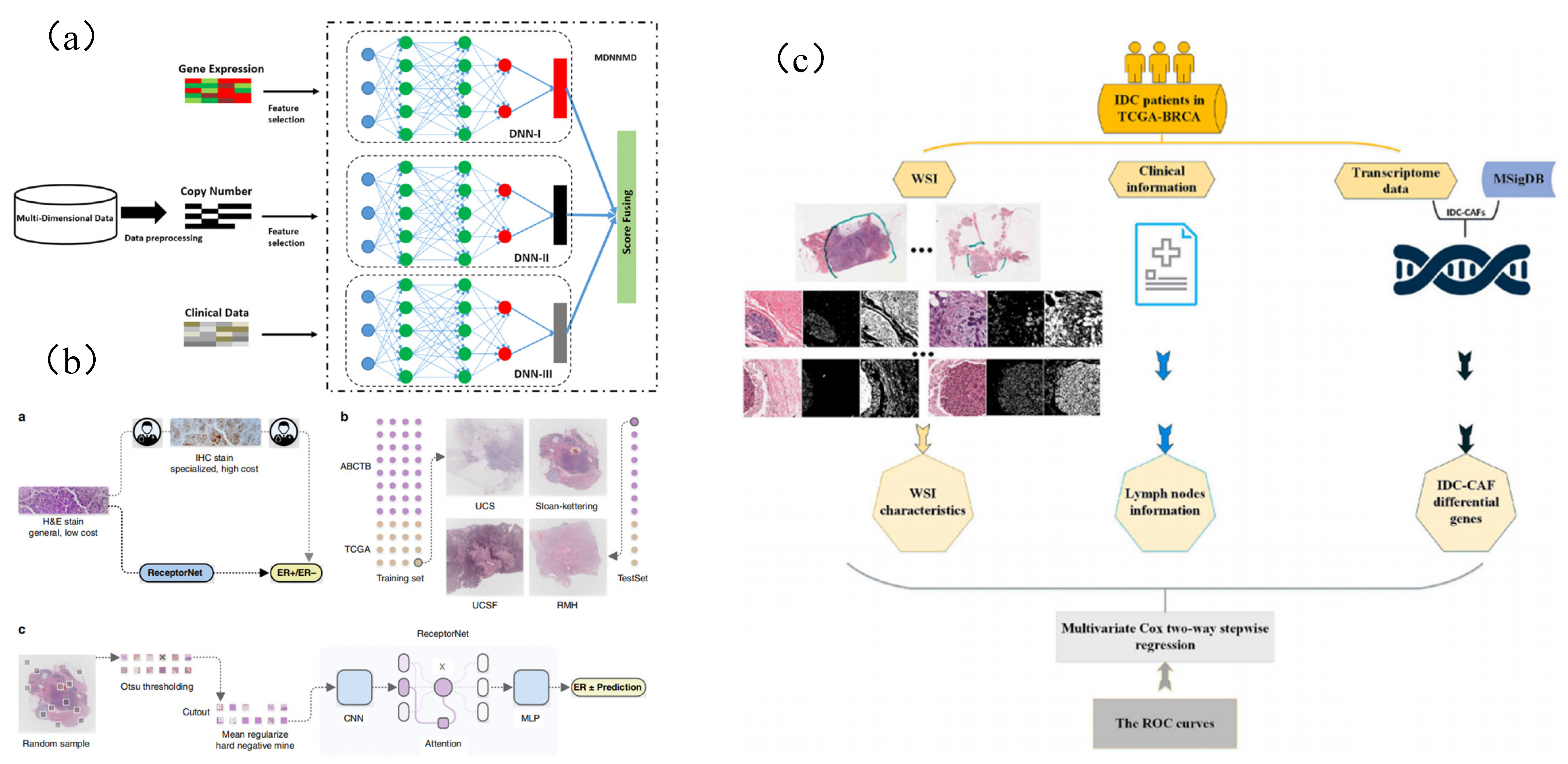
1. Introduction
2. Datasets
3. Methodology
3.1. Detection of Breast Lesions

| Model | Strategy | Advantages | Publication |
|---|---|---|---|
| RNN | Development of decision support systems for pathology | RNN allows neurons in the hidden layer to communicate with each other, storing the previous output as information in the hidden layer | [55] |
| Propose a SmallMitosis framework for the detection of mitotic cells from hematoxylin and eosin (H&E)-stained breast histological images | [56] | ||
| Inception | Histologic identification of tumor cells in lymph nodes | Inception increases the width of the network by pooling each layer with a different convolution to extract features from the previous layer, and by adding a 1*1 convolution after the pooling layer before the 3*3 and 5*5 convolutions, which effectively avoids complex parameters and computational effort | [57] |
| Improve the computer-aided diagnosis method based on deep learning | [58] | ||
| ResNet | Detection of invasive ductal carcinoma in breast histological images and the classification of lymphoma subtypes | The main feature of ResNet is the residual block, the purpose of the residual block is to preserve the characteristics of the parameters before the current layer is trained and to pass these parameters into the subsequent layers together with the trained data | [59] |
| Diagnostic breast cancer whole-slide tissue images | [60] | ||
| Propose an automatic detection method for invasive ductal carcinoma (IDC) based on deep transfer learning technology | [61] | ||
| Propose Mask RCNN, a multi-task deep learning framework for object detection and instance segmentation, to automatically detect mitosis | [62] | ||
| DCNN | Propose an accurate method for detecting the mitotic cells from histopathological slides using a multi-stage deep learning framework | [63] | |
| Present an SSAE for efficient nuclei detection on high-resolution histopathological images of breast cancer | [64] | ||
| Introduce deep learning as a technique to improve the objectivity and efficiency of histopathologic slide analysis | [65,66,67,68,69,70] | ||
| Semi-Supervised Learning | Present a semi-supervised deep learning strategy for breast cancer diagnosis | Semi-supervised learning is to use a large number of unlabeled samples and a small number of labeled samples to train the classifier, solving the problem of insufficient labeled samples | [71,72] |
| YOLO | A fast lesion detection method based on yolo is proposed | Simple structure and fast speed | [50] |
| Faster RCNN | A fast detection method of breast tumor based on Faster RCNN is proposed | Faster RCNN realizes object detection performance with high accuracy through second-order network and Region Proposal Network | [52] |
| Single Shot multibox Detector (SSD) | An automatic detection method of breast cancer lesion based on SSD is proposed | One stage, good at detecting small objects | [53] |
3.2. Segmentation Method of Breast Pathological Image
| Model | Strategy | Advantages | Publication |
|---|---|---|---|
| ResNet | Propose segmentation of limited data using rough image-level tags with performance comparable to fully labeled datasets | The main feature of ResNet is the residual block, the purpose of the residual block is to preserve the characteristics of the parameters before the current layer is trained and to pass these parameters onto the subsequent layers together with the trained data | [84] |
| FCN | Propose a fast segmentation method for breast cancer metastases in pathological images | The FCN replaces the fully connected layer behind the traditional CNN with a convolutional layer so that the output of the network will be a heat map rather than a category; at the same time, the image size is recovered using upsampling in order to address the reduction in image size due to convolution and pooling | [85] |
| Propose an automatic method for detecting mitosis | [86] | ||
| Describe a method to automatically segment nuclei from hematoxylin and eosin (H&E)-stained histopathology data with fully convolutional networks | [87] | ||
| Use annotated datasets to create accurate models | [60] | ||
| Propose a histopathological tissue analysis framework based on deep learning and verifies its universality and model generalization under different data distributions | [88] | ||
| U-Net | Use histopathological images obtained with hematoxylin and eosin staining for biopsy samples for the diagnosis and segmentation of breast cancer | U-Net networks are able to use valid labeled data more effectively from a very small number of training images, relying on data augmentation | [89] |
| Address the task of tissue-level segmentation in intermediate resolution of histopathological breast cancer images | [90] | ||
| Propose a deep learning framework consisting of high-resolution encoder paths, pyramidal pooled bottleneck modules in porous space, and decoders | [91] | ||
| Investigate whether it is possible to further improve the performance of the classifier model at the patch level by integrating multiple extracted histological features into the input image | [92] | ||
| CNN | Improve the performance of current Simple Linear Iterative Clustering (SLIC) algorithm to achieve hyperpixel segmentation of high-dimensional features | [93] | |
| Use a pretrained convolutional neural network (CNN) for segmentation and then another Hybrid-CNN for classification of mitoses | [94] | ||
| Identify a useful cell segmentation approach with histopathological images that uses prominent deep learning algorithms and spatial relationships | [95] | ||
| Propose a framework that combines the effectiveness of attention-based encoder–decoder architecture with an empty space pyramid pool with efficient dimensional convolution (kide-Segnet) | [96] | ||
| Propose a deep learning model for automatic segmentation of complex cores in tissue images by encoder-decoder structure | [97,98] | ||
| Transformer | Transformer-encoded global features improve U-Net segmentation performance | Transformer model can be used to encode the global features of pathological images and can improve the performance of current algorithms in many fields | [79] |
3.3. Disease Classification Based on Breast Pathological Images
| Model | Strategy | Advantages | Publication |
|---|---|---|---|
| Deep Belief Network (DBN) | Propose a new patch-based deep learning method PA-DBN-BC for breast cancer detection and classification in histopathological images | [128] | |
| Deep Neural Network (DNN) | Propose a new feature extractor, Deep Manifold Reservation Autoencoder, for automatic classification of breast cancer histopathological images | Deep Belief Network is a probability generation model, which has important value in the early application of deep learning methods | [129] |
| Generative Adversarial Network (GAN) | Explore whether a deep learning algorithm can learn objective histologic H&E features | GAN can be used as a means of data enhancement to alleviate the problem of insufficient data of pathological images of breast cancer | [103] |
| Visual Geometry Group Network (VGG) | Discuss and compare the task of automatic amplification in breast cancer detection based on multiple classification | VGG is a deep neural network proposed in 2014, which provides rich deep features for early breast pathological image research | [130] |
| Recurrent Neural Network (RNN) | Present a deep learning model to classify hematoxylin¨Ceosin-stained breast biopsy images into four classes | RNN can construct the context between pathological image features and be used for prediction of slide-level diagnostic results | [131] |
| Propose a second-order multi-instance learning approach that stacks adaptive aggregators by attentional mechanisms and recurrent neural networks (RNN) for histopathological image classification | [132] | ||
| Inception | Compare different machine learning methods for classification and evaluation of breast cancer tumors | [133] | |
| Propose a depth model based on computer-aided transfer learning as a binary classifier for breast cancer detection | [134] | ||
| Propose a method for diagnosing breast cancer as benign or malignant in magnification specific binary (MSB) classification | [135] | ||
| Dynamic Convolution Neural Network (DCNN) | Propose an efficient deep convolutional neural network classification model for fast back propagation learning | DCNN can adaptively adjust the convolution kernel parameters according to the input data, enhance the feature expression ability of the model, and specifically solve the tasks related to breast pathological images | [136] |
| Develop a deep learning model biopsy microscopic image cancer network (BMIC_Net) for multiple classification of BC | [137] | ||
| Propose two efficient models based on deep transfer learning to improve the binary and multiclassification systems | [138,139] | ||
| Convolution Neural Network (CNN) | Propose a new deep architecture based on self-integration to leverage semantic information from annotated images and explore information hidden in unlabeled data | [140] | |
| Propose an analysis and synthesis model learning method with novel algorithms and search strategies to classify images more effectively | [141,142,143,144,145,146,147,148,149,150] | ||
| Propose a set of training techniques and use image processing techniques to improve the performance of CNN-based models in breast cancer classification | [143,151,152,153,154,155,156,157] | ||
| Deep residual network (ResNet) | Present a deep neural network which performs representation learning and cell nuclei recognition in an end-to-end manner | [158] | |
| Propose an automatic multiclassification method for breast cancer histopathological images based on metastasis learning | [159] | ||
| Present a method that employs a convolutional neural network for detecting tumor on entire-slide images | [59,130,136,160] | ||
| Propose a breast cancer multiclassification method using a proposed deep learning model | [106,113,137,161,162,163,164,165,166,167,168] |
3.4. Genetic Prediction Based on Deep Learning

| Model | Strategy | Advantages | Publication |
|---|---|---|---|
| Attention mechanism | Weighting different pathological images based on attention mechanism to improve prediction results | Attention mechanism simulates human visual behavior by applying different weights to images. This method can highlight the key areas and non key areas in pathological images, thus improving the prediction results of the model | [181] |
| Based on ResNet and attention mechanism, a method for predicting pathological gene subtypes of breast cancer is proposed | [183] | ||
| KNN and K-means | Use unsupervised clustering method to reduce the workload of manual labeling by pathologists | Unsupervised | [192] |
4. Conclusions and Perspective
Author Contributions
Funding
Data Availability Statement
Conflicts of Interest
Abbreviations
| BC | Breast Cancer |
| CAD | Computer-Aided Diagnosis |
| ReLU | Linear Rectification Function |
| GDC | The Genomic Data Sharing Area |
| NCI | The National Cancer Institute |
| H&E | Hematoxylin and Eosin |
| WSIs | Full-Slide Images |
| CNN | Convolutional Neural Network |
| IDC | The Invasive Ductal Carcinoma |
| Faster R-CNN | Faster Region Convolutional Neural Network |
| SGE | Stacked Generalized Ensemble |
| ROI | Region of Interest |
| DCN | Deep Convolutional Network |
| DCNN | Deep Convolutional Neural Network |
| CRF | Condition Random Field |
| SLIC | Simple Linear Iterative Clustering |
| PBC | Patch-based Classifier |
| OPOD | One Patch in One Decision |
| APOD | All Patches in One Decision |
| MuDeRN | Multicategory Classification of Breast Histopathological |
| Images Using Deep Residual Networks | |
| IRRCNN | Inception Recurrent Residual Convolutional Neural Network |
| ResNet | Residual Network |
| MSB | Magnification Specific Binary |
| BMIC_Net | Biopsy Microscopic Image Cancer Network |
References
- Hoon Tan, P.; Ellis, I.; Allison, K.; Brogi, E.; Fox, S.B.; Lakhani, S.; Lazar, A.J.; Morris, E.A.; Sahin, A.; Salgado, R.; et al. The 2019 World Health Organization classification of tumours of the breast. Histopathology 2020, 77, 181–185. [Google Scholar] [CrossRef] [PubMed]
- Ferlay, J.; Colombet, M.; Soerjomataram, I.; Parkin, D.M.; Piñeros, M.; Znaor, A.; Bray, F. Cancer statistics for the year 2020: An overview. Int. J. Cancer 2021, 149, 778–789. [Google Scholar] [CrossRef] [PubMed]
- Abels, E.; Pantanowitz, L.; Aeffner, F.; Zarella, M.D.; Kozlowski, C. Computational Pathology Definitions, Best Practices, and Recommendations for Regulatory Guidance: A White Paper from the Digital Pathology Association. J. Pathol. 2019, 249, 286–294. [Google Scholar] [CrossRef] [PubMed]
- Veta, M.; Pluim, J.P.; Van Diest, P.J.; Viergever, M.A. Breast cancer histopathology image analysis: A review. IEEE Trans. Biomed. Eng. 2014, 61, 1400–1411. [Google Scholar] [CrossRef] [PubMed]
- Niazi, M.K.K.; Parwani, A.V.; Gurcan, M.N. Digital pathology and artificial intelligence. Lancet Oncol. 2019, 20, e253–e261. [Google Scholar] [CrossRef]
- Yaffe, M.J. Emergence of “Big Data” and Its Potential and Current Limitations in Medical Imaging. Semin. Nucl. Med. 2019, 49, 94–104. [Google Scholar] [CrossRef]
- Jang, H.-J.; Cho, K.-O. Applications of deep learning for the analysis of medical data. Arch. Pharmacal Res. 2019, 42, 492–504. [Google Scholar] [CrossRef]
- Srinidhi, C.L.; Ciga, O.; Martel, A.L. Deep neural network models for computational histopathology: A survey. Med. Image Anal. 2019, 67, 101813. [Google Scholar] [CrossRef]
- Wang, C.W.; Khalil, M.A.; Firdi, N.P. A Survey on Deep Learning for Precision Oncology. Diagnostics 2022, 12, 1489. [Google Scholar] [CrossRef] [PubMed]
- Robertson, S.; Azizpour, H.; Smith, K.; Hartman, J. Digital image analysis in breast pathology-from image processing techniques to artificial intelligence. Transl. Res. 2018, 194, 19–35. [Google Scholar] [CrossRef]
- Gao, J.; Jiang, Q.; Zhou, B.; Chen, D. Convolutional neural networks for computer-aided detection or diagnosis in medical image analysis: An overview. Math. Biosci. Eng. 2019, 16, 6536–6561. [Google Scholar] [CrossRef] [PubMed]
- Suri, J.S.; Biswas, M.; Kuppili, V.; Saba, L.; Edla, D.R.; Suri, H.S.; Cuadrado-Godia, E.; Laird, J.R.; Marinhoe, R.T.; Sanches, J.M.; et al. State-of-the-art review on deep learning in medical imaging. Front.-Biosci.-Landmark 2019, 24, 392–426. [Google Scholar] [CrossRef] [PubMed]
- Krithiga, R.; Geetha, P. Breast Cancer Detection, Segmentation and Classification on Histopathology Images Analysis: A Systematic Review. Arch. Comput. Methods Eng. 2020, 24, 392–426. [Google Scholar] [CrossRef]
- Debelee, T.G.; Schwenker, F.; Ibenthal, A.; Yohannes, D. Survey of deep learning in breast cancer image analysis. Evolving Systems 2020, 11, 143–163. [Google Scholar] [CrossRef]
- Jannesari, M.; Habibzadeh, M.; Aboulkheyr, H.; Khosravi, P.; Elemento, O.; Totonchi, M.; Hajirasouliha, I. Breast Cancer Histopathological Image Classification: A Deep Learning Approach. In Proceedings of the 2018 IEEE International Conference on Bioinformatics and Biomedicine (BIBM), Madrid, Spain, 3–6 December 2018; pp. 2405–2412. [Google Scholar]
- Wang, X.; Chen, H.; Gan, C.; Lin, H.; Dou, Q.; Tsougenis, E.; Huang, Q.; Cai, M.; Heng, P.-A. Weakly Supervised Deep Learning for Whole Slide Lung Cancer Image Analysis. IEEE Trans. Cybern. 2020, 50, 3950–3962. [Google Scholar] [CrossRef]
- Hinton, G.E.; Salakhutdinov, R.R. Reducing the dimensionality of data with neural networks. Science 2006, 313, 504–507. [Google Scholar] [CrossRef]
- Krizhevsky, A.; Sutskever, I.; Hinton, G.E. Imagenet classification with deep convolutional neural networks. Commun. ACM 2017, 60, 84–90. [Google Scholar] [CrossRef]
- Sherstinsky, A. Fundamentals of recurrent neural network (RNN) and long short-term memory (LSTM) network. Phys. D Nonlinear Phenom. 2020, 404, 132306. [Google Scholar] [CrossRef]
- Creswell, A.; White, T.; Dumoulin, V.; Arulkumaran, K.; Sengupta, B.; Bharath, A.A. Generative adversarial networks: An overview. IEEE Signal Process. Mag. 2018, 35, 53–65. [Google Scholar] [CrossRef]
- Shamshad, F.; Khan, S.; Zamir, S.W.; Khan, M.H.; Hayat, M.; Khan, F.S.; Fu, H. Transformers in medical imaging: A survey. arXiv 2022, arXiv:2201.09873. [Google Scholar]
- Dimitriou, N.; Arandjelović, O.; Caie, P.D. Deep Learning for Whole Slide Image Analysis: An Overview. Front. Med. 2019, 6, 264. [Google Scholar] [CrossRef] [PubMed]
- LeCun, Y.; Boser, B.; Denker, J.; Henderson, D.; Howard, R.; Hubbard, W.; Jackel, L. Handwritten digit recognition with a back-propagation network. Adv. Neural Inf. Process. Syst. 1989, 2. Available online: https://proceedings.neurips.cc/paper/1989/file/53c3bce66e43be4f209556518c2fcb54-Paper.pdf (accessed on 1 December 2022).
- AlEisa, H.N.; Touiti, W.; Ali ALHussan, A.; Ben Aoun, N.; Ejbali, R.; Zaied, M.; Saadia, A. Breast Cancer Classification Using FCN and Beta Wavelet Autoencoder. Comput. Intell. Neurosci. 2022, 2022, 8044887. [Google Scholar] [CrossRef] [PubMed]
- Ronneberger, O.; Fischer, P.; Brox, T. U-net: Convolutional networks for biomedical image segmentation. In Proceedings of the International Conference on Medical Image Computing and Computer-Assisted Intervention, Munich, Germany, 5–9 October 2015; pp. 234–241. [Google Scholar]
- Xie, S.; Tu, Z. Holistically-nested edge detection. In Proceedings of the IEEE International Conference on Computer Vision, Santiago, Chile, 7–13 December 2015; pp. 1395–1403. [Google Scholar]
- Rampun, A.; López-Linares, K.; Morrow, P.J.; Scotney, B.W.; Wang, H.; Ocaña, I.G.; Maclair, G.; Zwiggelaar, R.; Ballester, M.A.G.; Macía, I. Breast pectoral muscle segmentation in mammograms using a modified holistically-nested edge detection network. Med. Image Anal. 2019, 57, 1–17. [Google Scholar] [CrossRef]
- Alom, M.Z.; Aspiras, T.; Taha, T.M.; Bowen, T.; Asari, V.K. MitosisNet: End-to-End Mitotic Cell Detection by Multi-Task Learning. IEEE Access 2020, 99, 1. [Google Scholar] [CrossRef]
- Toaar, M.; Zkurt, K.B.; Ergen, B.; Cmert, Z. BreastNet: A novel convolutional neural network model through histopathological images for the diagnosis of breast cancer. Phys. A Statal Mech. Its Appl. 2019, 545, 123592. [Google Scholar]
- Cui, Z.; Su, F.; Li, Y.; Yang, D. Circulating tumour cells as prognosis predictive markers of neoadjuvant chemotherapy-treated breast cancer patients. J. Chemother. 2020, 32, 304–309. [Google Scholar] [CrossRef]
- Khosravi, P.; Kazemi, E.; Imielinski, M.; Elemento, O.; Hajirasouliha, I. Deep Convolutional Neural Networks Enable Discrimination of Heterogeneous Digital Pathology Images. Ebiomedicine 2018, 27, 317–328. [Google Scholar] [CrossRef]
- Feng, M.; Deng, Y.; Yang, L.; Jing, Q.; Bu, H. Automated quantitative analysis of Ki-67 staining and HE images recognition and registration based on whole tissue sections in breast carcinoma. Diagn. Pathol. 2020, 15, 65. [Google Scholar] [CrossRef]
- Akbar, S.; Peikari, M.; Salama, S.; Panah, A.Y.; Nofech-Mozes, S.; Martel, A.L. Automated and Manual Quantification of Tumour Cellularity in Digital Slides for Tumour Burden Assessment. Sci. Rep. 2019, 9, 14099. [Google Scholar] [CrossRef]
- Lin, H.; Chen, H.; Graham, S.; Dou, Q.; Rajpoot, N.; Heng, P.A. Fast ScanNet: Fast and Dense Analysis of Multi-Gigapixel Whole-Slide Images for Cancer Metastasis Detection. IEEE Trans. Med. Imaging 2019, 38, 1948–1958. [Google Scholar] [CrossRef]
- Jimenez, G.; Racoceanu, D. Deep Learning for Semantic Segmentation vs. Classification in Computational Pathology: Application to Mitosis Analysis in Breast Cancer Grading. Front. Bioeng. Biotechnol. 2019, 7, 145. [Google Scholar] [CrossRef] [PubMed]
- Wahab, N.; Khan, A.; Lee, Y.S. Two-phase deep convolutional neural network for reducing class skewness in histopathological images based breast cancer detection. Comput. Biol. Med. 2017, 85, 86–97. [Google Scholar] [CrossRef] [PubMed]
- Mahmood, T.; Arsalan, M.; Owais, M.; Lee, M.B.; Park, K.R. Artificial Intelligence-Based Mitosis Detection in Breast Cancer Histopathology Images Using Faster R-CNN and Deep CNNs. J. Clin. Med. 2020, 9, 749. [Google Scholar] [CrossRef] [PubMed]
- Kumar, D.; Batra, U. Classification of Invasive Ductal Carcinoma from histopathology breast cancer images using Stacked Generalized Ensemble. J. Intell. Fuzzy Syst. 2021, 40, 4919–4934. [Google Scholar] [CrossRef]
- Sigirci, I.O.; Albayrak, A.; Bilgin, G. Detection of mitotic cells in breast cancer histopathological images using deep versus handcrafted features. Multimed. Tools Appl. 2021, 81, 13179–13202. [Google Scholar] [CrossRef]
- Zeiser, F.A.; da Costa, C.A.; de Oliveira Ramos, G.; Bohn, H.C.; Santos, I.; Roehe, A.V. DeepBatch: A hybrid deep learning model for interpretable diagnosis of breast cancer in whole-slide images. Expert Syst. Appl. 2021, 185, 115586. [Google Scholar] [CrossRef]
- Krithiga, R.; Geetha, P. Deep learning based breast cancer detection and classification using fuzzy merging techniques. Mach. Vis. Appl. 2020, 31, 63. [Google Scholar] [CrossRef]
- Zhao, Z.Q.; Zheng, P.; Xu, S.T.; Wu, X. Object detection with deep learning: A review. IEEE Trans. Neural Netw. Learn. Syst. 2019, 30, 3212–3232. [Google Scholar] [CrossRef]
- Liu, Y.; Gadepalli, K.; Norouzi, M.; Dahl, G.E.; Kohlberger, T.; Boyko, A.; Venugopalan, S.; Timofeev, A.; Nelson, P.Q.; Corrado, G.S.; et al. Detecting cancer metastases on gigapixel pathology images. arXiv 2017, arXiv:1703.02442. [Google Scholar]
- George, K.; Sankaran, P.; Joseph, P.K. Computer assisted recognition of breast cancer in biopsy images via fusion of nucleus-guided deep convolutional features. Comput. Methods Programs Biomed. 2020, 194, 105531. [Google Scholar] [CrossRef] [PubMed]
- Chen, H.; Dou, Q.; Wang, X.; Qin, J.; Heng, P. Mitosis detection in breast cancer histology images via deep cascaded networks. In Proceedings of the Thirtieth AAAI Conference on Artificial Intelligence, Phoenix, AZ, USA, 12–17 February 2016. [Google Scholar]
- Bardou, D.; Zhang, K.; Ahmad, S.M. Classification of Breast Cancer Based on Histology Images Using Convolutional Neural Networks. IEEE Access 2018, 6, 24680–24693. [Google Scholar] [CrossRef]
- Redmon, J.; Divvala, S.; Girshick, R.; Farhadi, A. You only look once: Unified, real-time object detection. In Proceedings of the IEEE Conference on Computer Vision and Pattern Recognition, Las Vegas, NV, USA, 27–30 June 2016; pp. 779–788. [Google Scholar]
- Girshick, R. Fast r-cnn. In Proceedings of the IEEE International Conference on Computer Vision, Santiago, Chile, 7–13 December 2015; pp. 1440–1448. [Google Scholar]
- Liu, W.; Anguelov, D.; Erhan, D.; Szegedy, C.; Reed, S.; Fu, C.Y.; Berg, A.C. Ssd: Single shot multibox detector. Eur. Conf. Comput. Vis. 2016, 9905, 21–37. [Google Scholar]
- Lu, Y.; Zhang, J.; Liu, X.; Zhang, Z.; Li, W.; Zhou, X.; Li, R. Prediction of breast cancer metastasis by deep learning pathology. IET Image Process. 2022. [Google Scholar] [CrossRef]
- Huang, H.; Feng, X.; Jiang, J.; Chen, P.; Zhou, S. Mask RCNN algorithm for nuclei detection on breast cancer histopathological images. Int. J. Imaging Syst. Technol. 2022, 32, 209–217. [Google Scholar] [CrossRef]
- Harrison, P.; Park, K. Tumor Detection In Breast Histopathological Images Using Faster R-CNN. In Proceedings of the 2021 International Symposium on Medical Robotics (ISMR), Atlanta, GA, USA, 17–19 November 2021; pp. 1–7. [Google Scholar]
- Yamaguchi, M.; Sasaki, T.; Uemura, K.; Tajima, Y.; Kato, S.; Takagi, K.; Yamazaki, Y.; Saito-Koyama, R.; Inoue, C.; Kawaguchi, K.; et al. Automatic breast carcinoma detection in histopathological micrographs based on Single Shot Multibox Detector. J. Pathol. Inform. 2022, 13, 100147. [Google Scholar] [CrossRef]
- Zorgani, A.; Mohamed, M.; Mehmood, I.; Ugail, H. Deep yolo-based detection of breast cancer mitotic-cells in histopathological images. In Proceedings of the International Conference on Medical Imaging and Computer-Aided Diagnosis, Birmingham, UK, 25–26 March 2021; pp. 335–342. [Google Scholar]
- Campanella, G.; Hanna, M.G.; Geneslaw, L.; Miraflor, A.; Silva, V.W.K.; Busam, K.J.; Brogi, E.; Reuter, V.E.; Klimstra, D.S.; Fuchs, T.J. Clinical-grade computational pathology using weakly supervised deep learning on whole slide images. Nat. Med. 2019, 25, 1301–1309. [Google Scholar] [CrossRef]
- Kausar, T.; Wang, M.; Ashraf, M.A.; Kausar, A. SmallMitosis: Small Size Mitotic Cells Detection in Breast Histopathology Images. IEEE Access 2021, 9, 905–922. [Google Scholar] [CrossRef]
- Liu, Y.; Kohlberger, T.; Norouzi, M.; Dahl, G.E.; Smith, J.L.; Mohtashamian, A.; Olson, N.; Peng, L.H.; Hipp, J.D.; Stumpe, M.C. Artificial Intelligence-Based Breast Cancer Nodal Metastasis Detection. Arch. Pathol. Lab. Med. 2019, 143, 859–868. [Google Scholar] [CrossRef]
- Ma, X.; Liu, H.; Niu, Y.; Zhang, C.; Liu, D. Improvement of Whole-Slide Pathological Image Recognition Method Based on Deep Learning. Int. Symp. Comput. Intell. Des. 2018, 2, 269–272. [Google Scholar]
- Brancati, N.; De Pietro, G.; Frucci, M.; Riccio, D. A Deep Learning Approach for Breast Invasive Ductal Carcinoma Detection and Lymphoma Multi-Classification in Histological Images. IEEE Access 2019, 7, 44709–44720. [Google Scholar] [CrossRef]
- Amgad, M.; Elfandy, H.; Hussein, H.; A Atteya, L.; Elsebaie, M.A.T.; Elnasr, L.S.A.; A Sakr, R.; E Salem, H.S.; Ismail, A.F.; Saad, A.; et al. Structured crowdsourcing enables convolutional segmentation of histology images. Bioinformatics 2019, 35, 3461–3467. [Google Scholar] [CrossRef] [PubMed]
- Celik, Y.; Talo, M.; Yildirim, O.; Karabatak, M.; Acharya, U.R. Automated invasive ductal carcinoma detection based using deep transfer learning with whole-slide images. Pattern Recognit. Lett. 2020, 133, 232–239. [Google Scholar] [CrossRef]
- Sebai, M.; Wang, X.; Wang, T. MaskMitosis: A deep learning framework for fully supervised, weakly supervised, and unsupervised mitosis detection in histopathology images. Med. Biol. Eng. Comput. 2020, 58, 1603–1623. [Google Scholar] [CrossRef] [PubMed]
- Li, C.; Wang, X.; Liu, W.; Latecki, L.J. DeepMitosis: Mitosis detection via deep detection, verification and segmentation networks. Med. Image Anal. 2018, 45, 121–133. [Google Scholar] [CrossRef]
- Xu, J.; Xiang, L.; Liu, Q.; Gilmore, H.; Wu, J.; Tang, J.; Madabhushi, A. Stacked Sparse Autoencoder (SSAE) for Nuclei Detection on Breast Cancer Histopathology Images. IEEE Trans. Med. Imaging 2016, 35, 119–130. [Google Scholar] [CrossRef]
- Couture, H.D.; Williams, L.A.; Geradts, J.; Nyante, S.J.; Butler, E.N.; Marron, J.S.; Perou, C.M.; Troester, M.A.; Niethammer, M. Image analysis with deep learning to predict breast cancer grade, ER status, histologic subtype, and intrinsic subtype. NPJ Breast Cancer 2018, 4, 30. [Google Scholar] [CrossRef]
- Bejnordi, B.E.; Mullooly, M.; Pfeiffer, R.M.; Fan, S.; Vacek, P.M.; Weaver, D.L.; Herschorn, S.; Brinton, L.A.; Van Ginneken, B.; Karssemeijer, N.; et al. Using deep convolutional neural networks to identify and classify tumor-associated stroma in diagnostic breast biopsies. Mod. Pathol. 2018, 31, 1502–1512. [Google Scholar] [CrossRef]
- Litjens, G.; Sánchez, C.I.; Timofeeva, N.; Hermsen, M.; Nagtegaal, I.; Kovacs, I.; Hulsbergen-van de Kaa, C.; Bult, P.; Van Ginneken, B.; Van Der Laak, J. Deep learning as a tool for increased accuracy and efficiency of histopathological diagnosis. Sci. Rep. 2016, 6, 26286. [Google Scholar] [CrossRef]
- Stanitsas, P.; Cherian, A.; Li, X.; Truskinovsky, A.; Morellas, V.; Papanikolopoulos, N. Evaluation of feature descriptors for cancerous tissue recognition. In Proceedings of the International Conference on Pattern Recognition, Cancun, Mexico, 4–8 December 2016. [Google Scholar]
- BenTaieb, A.; Hamarneh, G. Predicting Cancer with a Recurrent Visual Attention Model for Histopathology Images. Med. Image Comput. Comput. Assist. Interv.-Miccai 2018, 110712018, 129–137. [Google Scholar]
- Saha, M.; Chakraborty, C.; Racoceanu, D. Efficient deep learning model for mitosis detection using breast histopathology images. Comput. Med. Imaging Graph. 2018, 64, 29–40. [Google Scholar] [CrossRef] [PubMed]
- Sun, W.; Tseng, T.-L.; Zhang, J.; Qian, W. Enhancing deep convolutional neural network scheme for breast cancer diagnosis with unlabeled data. Comput. Med. Imaging Graph. 2017, 57, 4–9. [Google Scholar] [CrossRef]
- Xiao, Y.; Wu, J.; Lin, Z.; Zhao, X. A semi-supervised deep learning method based on stacked sparse auto-encoder for cancer prediction using RNA-seq data. Comput. Methods Programs Biomed. 2018, 166, 99–105. [Google Scholar] [CrossRef] [PubMed]
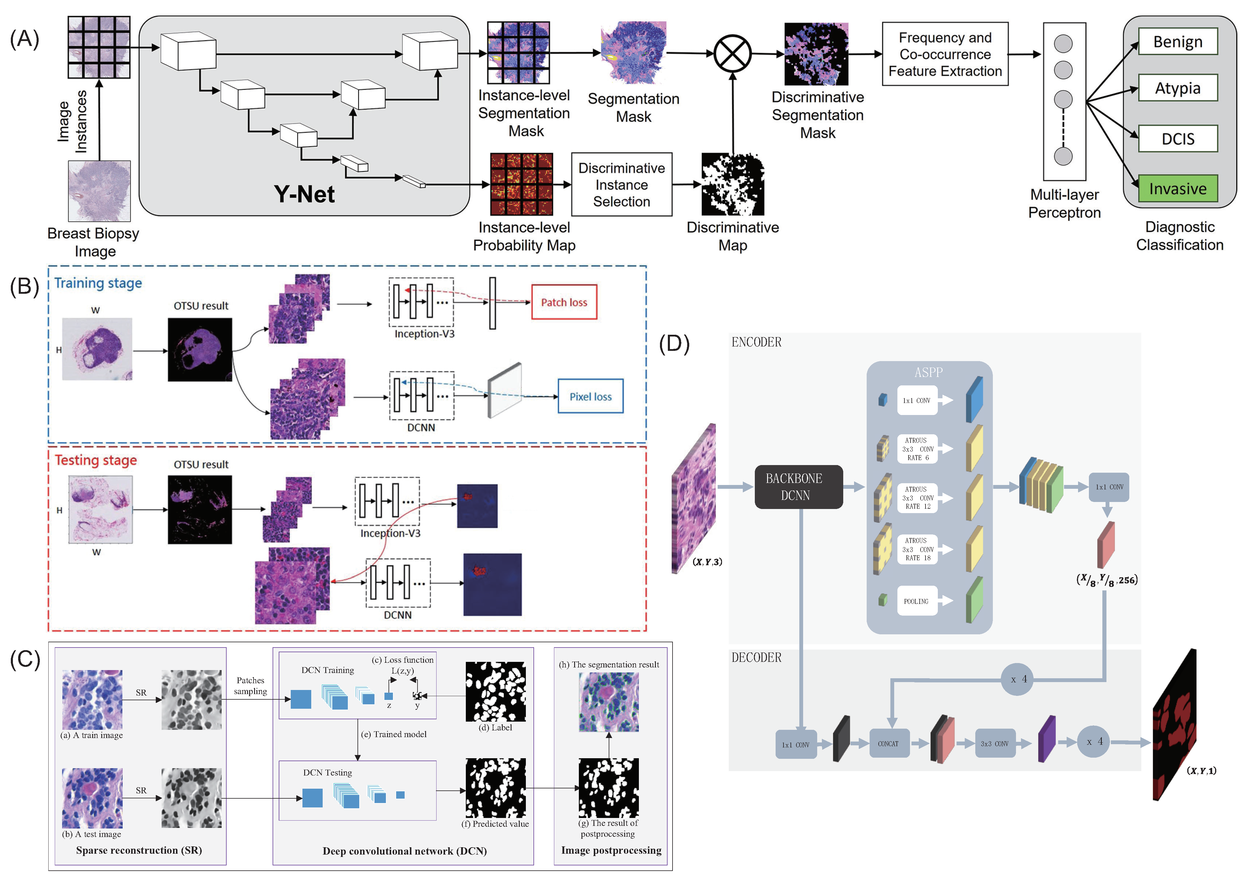
- Mehta, S.; Mercan, E.; Bartlett, J.; Weaver, D.; Elmore, J.G.; Shapiro, L. Y-Net: Joint Segmentation and Classification for Diagnosis of Breast Biopsy Images. In Proceedings of the Medical Image Computing and Computer Assisted Intervention—Miccai 2018, Pt Ii. Lecture Notes in Computer Science, Granada, Spain, 16–20 September 2018; Volume 110712018, pp. 893–901. [Google Scholar]
- Guo, Z.; Liu, H.; Ni, H.; Wang, X.; Qian, Y. Publisher Correction: A Fast and Refined Cancer Regions Segmentation Framework in Whole-slide Breast Pathological Images. Sci. Rep. 2020, 10, 8591. [Google Scholar] [CrossRef] [PubMed]
- Pan, X.; Li, L.; Yang, H.; Liu, Z.; Yang, J.; Zhao, L.; Fan, Y. Accurate segmentation of nuclei in pathological images via sparse reconstruction and deep convolutional networks. Neurocomputing 2017, 229, 88–99. [Google Scholar] [CrossRef]
- Priego-Torres, B.M.; Sanchez-Morillo, D.; Fernandez-Granero, M.A.; Garcia-Rojo, M. Automatic segmentation of whole-slide H&E stained breast histopathology images using a deep convolutional neural network architecture. Expert Syst. Appl. 2020, 151, 113387. [Google Scholar]
- Nguyen, C.; Asad, Z.; Deng, R.; Huo, Y. Evaluating transformer-based semantic segmentation networks for pathological image segmentation. Med. Imaging 2022 Image Process. 2022, 12032, 942–947. [Google Scholar]
- Li, Z.; Li, Y.; Li, Q.; Zhang, Y.; Wang, P.; Guo, D.; Lu, L.; Jin, D.; Hong, Q. LViT: Language meets vision transformer in medical image segmentation. arXiv 2022, arXiv:2206.14718. [Google Scholar]
- Diao, S.; Tang, L.; He, J.; Zhao, H.; Luo, W.; Xie, Y.; Qin, W. Automatic Computer-Aided Histopathologic Segmentation for Nasopharyngeal Carcinoma Using Transformer Framework. In Proceedings of the International Workshop on Computational Mathematics Modeling in Cancer Analysis, Singapore, 18 September 2022; pp. 141–149. [Google Scholar]
- Saleck, M.M.; El Moutaouakkil, A.; Rmili, M. Semi-automatic segmentation of breast masses in mammogram images. In Proceedings of the International Conference on Pattern Recognition and Artificial Intelligence, Beijing, China, 20–24 August 2018; pp. 59–62. [Google Scholar]
- Zhai, D.; Hu, B.; Gong, X.; Zou, H.; Luo, J. ASS-GAN: Asymmetric semi-supervised GAN for breast ultrasound image segmentation. Neurocomputing 2022, 493, 204–216. [Google Scholar] [CrossRef]
- Veeraraghavan, H.; Dashevsky, B.Z.; Onishi, N.; Sadinski, M.; Morris, E.; Deasy, J.; Sutton, E.J. Appearance constrained semi-automatic segmentation from DCE-MRI is reproducible and feasible for breast cancer radiomics: A feasibility study. Sci. Rep. 2018, 8, 4838. [Google Scholar] [CrossRef]
- Lai, Z.; Wang, C.; Oliveira, L.C.; Dugger, B.N.; Cheung, S.-C.; Chuah, C.-N. Joint Semi-supervised and Active Learning for Segmentation of Gigapixel Pathology Images with Cost-Effective Labeling. In Proceedings of the IEEE/CVF International Conference on Computer Vision, Montreal, BC, Canada, 11–17 October 2021; pp. 591–600. [Google Scholar]
- Ciga, O.; Martel, A.L. Learning to segment images with classification labels. Med. Image Anal. 2021, 68, 101912. [Google Scholar] [CrossRef] [PubMed]
- Khalil, M.-A.; Lee, Y.-C.; Lien, H.-C.; Jeng, Y.-M.; Wang, C.-W. Fast Segmentation of Metastatic Foci in H&E Whole-Slide Images for Breast Cancer Diagnosis. Diagnostics 2022, 12, 990. [Google Scholar] [PubMed]
- Li, C.; Wang, X.; Liu, W.; Latecki, L.J.; Wang, B.; Huang, J. Weakly Supervised Mitosis Detection in Breast Histopathology Images using Concentric Loss. Med. Image Anal. 2019, 53, 165–178. [Google Scholar] [CrossRef] [PubMed]
- Naylor, P.; Laé, M.; Reyal, F.; Walter, T. Nuclei segmentation in histopathology images using deep neural networks. In Proceedings of the 2017 IEEE 14th International Symposium on Biomedical Imaging (ISBI 2017), Melbourne, VIC, Australia, 18–21 April 2017. [Google Scholar]
- Khened, M.; Kori, A.; Rajkumar, H.; Krishnamurthi, G.; Srinivasan, B. A generalized deep learning framework for whole-slide image segmentation and analysis. Sci. Rep. 2021, 11, 11579. [Google Scholar] [CrossRef] [PubMed]
- Naylor, P.; Lae, M.; Reyal, F.; Walter, T. Segmentation of Nuclei in Histopathology Images by Deep Regression of the Distance Map. IEEE Trans. Med. Imaging 2018, 38, 448–459. [Google Scholar] [CrossRef]
- Mejbri, S.; Franchet, C.; Ismat-Ara, R.; Mothe, J.; Brousset, P.; Faure, E. Deep Analysis of CNN Settings for New Cancer Whole-slide Histological Images Segmentation: The Case of Small Training Sets. In Proceedings of the 6th International Conference on Bioimaging, Prague, Czech Republic, 22–24 February 2019. [Google Scholar]
- Chanchal, A.K.; Lal, S.; Kini, J. High-resolution deep transferred ASPPU-Net for nuclei segmentation of histopathology images. Int. J. Comput. Assist. Radiol. Surg. 2021, 16, 2159–2175. [Google Scholar] [CrossRef]
- Jin, Y.W.; Jia, S.; Ashraf, A.B.; Hu, P. Integrative Data Augmentation with U-Net Segmentation Masks Improves Detection of Lymph Node Metastases in Breast Cancer Patients. Cancers 2020, 12, 2934. [Google Scholar] [CrossRef]
- Zhou, J.; Ruan, J.; Wu, C.; Ye, G.; Zhu, Z.; Yue, J.; Zhang, Y. Superpixel Segmentation of Breast Cancer Pathology Images Based on Features Extracted from the Autoencoder. In Proceedings of the 2019 IEEE 11th International Conference on Communication Software and Networks, Chongqing, China, 12–15 June 2019; pp. 366–370. [Google Scholar]
- Noorul, W.; Asifullah, K.; Soo, L.Y. Transfer learning based deep CNN for segmentation and detection of mitoses in breast cancer histopathological images. Microscopy 2019, 3, 216–233. [Google Scholar]
- Hatipoglu, N.; Bilgin, G. Cell segmentation in histopathological images with deep learning algorithms by utilizing spatial relationships. Med. Biol. Eng. Comput. 2017, 55, 1829–1848. [Google Scholar] [CrossRef]
- Aatresh, A.A.; Yatgiri, R.P.; Chanchal, A.K.; Kumar, A.; Ravi, A.; Das, D.; Bs, R.; Lal, S.; Kini, J. Efficient deep learning architecture with dimension-wise pyramid pooling for nuclei segmentation of histopathology images. Comput. Med. Imaging Graph. 2021, 93, 101975. [Google Scholar] [CrossRef]
- Chanchal, A.K.; Kumar, A.; Lal, S.; Kini, J. Efficient and robust deep learning architecture for segmentation of kidney and breast histopathology images. Comput. Electr. Eng. 2021, 92, 107177. [Google Scholar] [CrossRef]
- van Rijthoven, M.; Balkenhol, M.; Silina, K.; van der Laak, J.; Ciompi, F. HookNet: Multi-resolution convolutional neural networks for semantic segmentation in histopathology whole-slide images. Med. Image Anal. 2021, 68, 101890. [Google Scholar] [CrossRef] [PubMed]
- Ghanem, N.M.; Attallah, O.; Anwar, F.; Ismail, M.A. AUTO-BREAST: A fully automated pipeline for breast cancer diagnosis using AI technology. Artif. Intell. Cancer Diagn. Progn. 2022, 6, 1–24. [Google Scholar]
- Karthiga, R.; Narasimhan, K. Automated diagnosis of breast cancer using wavelet based entropy features. In Proceedings of the 2018 Second International Conference on Electronics, Communication and Aerospace Technology (ICECA), Coimbatore, India, 29–31 March 2018; pp. 274–279. [Google Scholar]
- Anwar, F.; Attallah, O.; Ghanem, N.; Ismail, M.A. Automatic breast cancer classification from histopathological images. In Proceedings of the 2019 International Conference on Advances in the Emerging Computing Technologies (AECT), Al Madinah Al Munawwarah, Saudi Arabia, 10 February 2020; pp. 1–6. [Google Scholar]
- Spanhol, F.A.; Oliveira, L.S.; Petitjean, C.; Heutte, L. Breast cancer histopathological image classification using convolutional neural networks. In Proceedings of the 2016 International Joint Conference on Neural Networks (IJCNN), Vancouver, BC, Canada, 24–29 July 2016; pp. 2560–2567. [Google Scholar]
- Han, Z.; Wei, B.; Zheng, Y.; Yin, Y.; Li, K.; Li, S. Breast cancer multi-classification from histopathological images with structured deep learning model. Sci. Rep. 2017, 7, 4172. [Google Scholar] [CrossRef] [PubMed]
- Kahya, M.A.; Al-Hayani, W.; Algamal, Z.Y. Classification of breast cancer histopathology images based on adaptive sparse support vector machine. J. Appl. Math. Bioinform. 2017, 7, 49. [Google Scholar]
- Spanhol, F.A.; Oliveira, L.S.; Cavalin, P.R.; Petitjean, C.; Heutte, L. Deep features for breast cancer histopathological image classification. In Proceedings of the IEEE International Conference on Systems, Man, and Cybernetics (SMC), Banff, AB, Canada, 5–8 October 2017; Volume 71, pp. 1868–1873. [Google Scholar]
- Bayramoglu, N.; Kannala, J.; Heikkila, J. Deep learning for magnification independent breast cancer histopathology image classification. In Proceedings of the 2016 23rd International Conference on Pattern Recognition (ICPR), Cancun, Mexico, 4–8 December 2016; pp. 2440–2445. [Google Scholar]
- Attallah, O.; Anwar, F.; Ghanem, N.M.; Ismail, M.A. Histo-CADx: Duo cascaded fusion stages for breast cancer diagnosis from histopathological images. PeerJ Comput. Sci. 2021, 7, e493. [Google Scholar] [CrossRef]
- Nahid, A.A.; Kong, Y. Histopathological breast-image classification using local and frequency domains by convolutional neural network. Information 2018, 9, 19. [Google Scholar] [CrossRef]
- Sudharshan, P.J.; Petitjean, C.; Spanhol, F.; Oliveira, L.E.; Heutte, L.; Honeine, P. Multiple instance learning for histopathological breast cancer image classification. Expert Syst. Appl. 2019, 117, 103–111. [Google Scholar] [CrossRef]
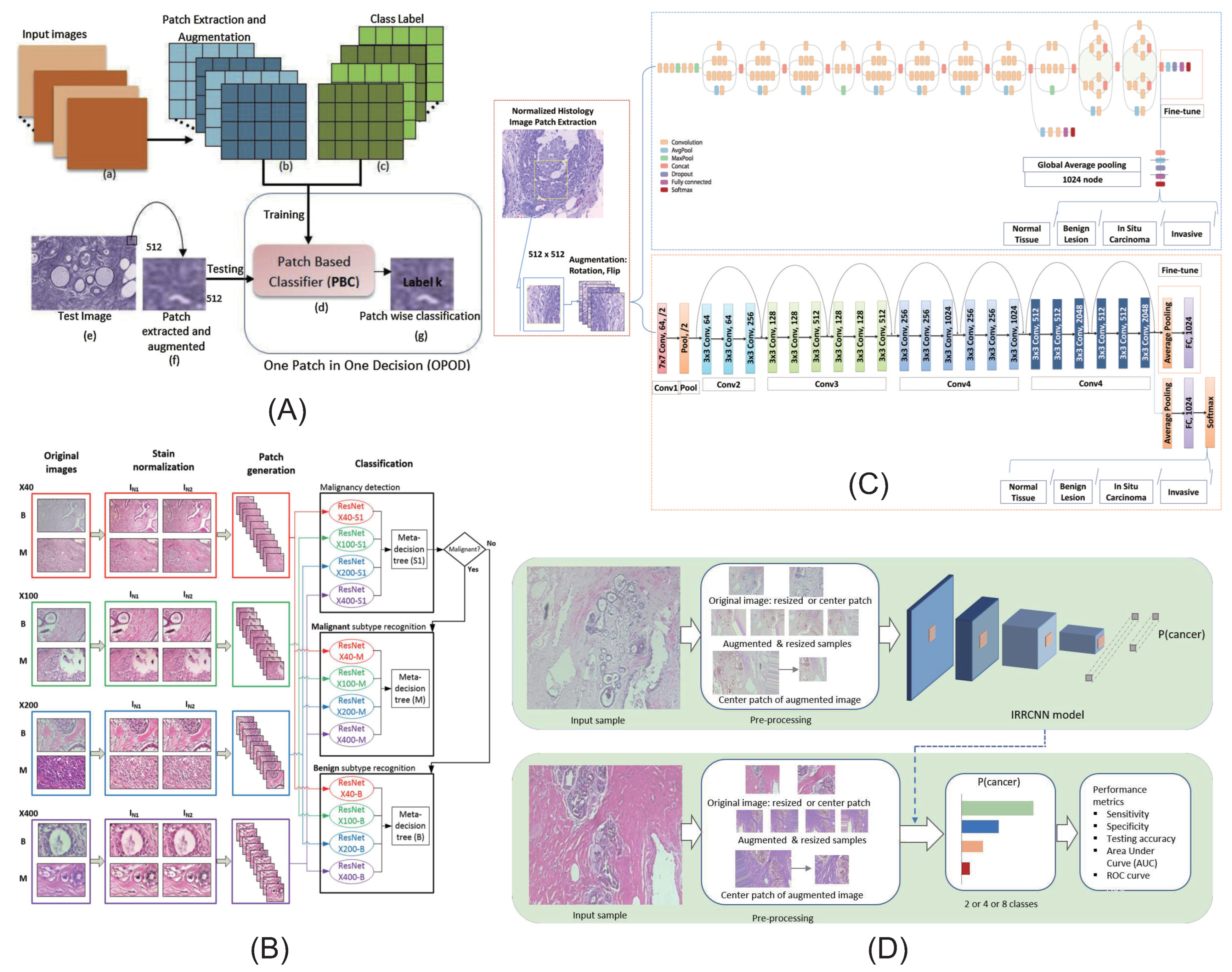
- Roy, K.; Banik, D.; Bhattacharjee, D.; Nasipuri, M. Patch-based system for Classification of Breast Histology images using deep learning. Comput. Med. Imaging Graph. 2019, 71, 90–103. [Google Scholar] [CrossRef]
- Gandomkar, Z.; Brennan, P.C.; Mello-Thoms, C. MuDeRN: Multi-category classification of breast histopathological image using deep residual networks. Artif. Intell. Med. 2018, 88, 14–24. [Google Scholar] [CrossRef]
- Vesal, S.; Ravikumar, N.; Davari, A.; Ellmann, S.; Maier, A. Classification of Breast Cancer Histology Images Using Transfer Learning. In International Conference Image Analysis and Recognition; Springer: Cham, Switzerland, 2018; pp. 812–819. [Google Scholar]
- Alom, M.Z.; Yakopcic, C.; Nasrin, M.S.; Taha, T.M.; Asari, V.K. Breast Cancer Classification from Histopathological Images with Inception Recurrent Residual Convolutional Neural Network. J. Digit. Imaging 2019, 32, 605–617. [Google Scholar] [CrossRef] [PubMed]
- Dai, Y.; Gao, Y.; Liu, F. Transmed: Transformers advance multi-modal medical image classification. Diagnostics 2021, 11, 1384. [Google Scholar] [CrossRef] [PubMed]
- Almalik, F.; Yaqub, M.; Nandakumar, K. Self-Ensembling Vision Transformer (SEViT) for Robust Medical Image Classification. In Proceedings of the International Conference on Medical Image Computing and Computer-Assisted Intervention, Singapore, 18–22 September 2022; pp. 376–386. [Google Scholar]
- Karimi, D.; Vasylechko, S.D.; Gholipour, A. Convolution-free medical image segmentation using transformers. In Proceedings of the International Conference on Medical Image Computing and Computer-Assisted Intervention, Strasbourg, France, 27 September–1 October 2021; pp. 78–88. [Google Scholar]
- Chen, J.; He, Y.; Frey, E.C.; Li, Y.; Du, Y. Vit-v-net: Vision transformer for unsupervised volumetric medical image registration. arXiv 2021, arXiv:2104.06468. [Google Scholar]
- Yu, S.; Ma, K.; Bi, Q.; Bian, C.; Ning, M.; He, N.; Li, Y.; Liu, H.; Zheng, Y. Mil-vt: Multiple instance learning enhanced vision transformer for fundus image classification. In Proceedings of the International Conference on Medical Image Computing and Computer-Assisted Intervention, Strasbourg, France, 27 September–1 October 2021; pp. 45–54. [Google Scholar]
- Alotaibi, A.; Alafif, T.; Alkhilaiwi, F.; Alatawi, Y.; Althobaiti, H.; Alrefaei, A.; Hawsawi, Y.M.; Nguyen, T. ViT-DeiT: An Ensemble Model for Breast Cancer Histopathological Images Classification. arXiv 2022, arXiv:2211.00749. [Google Scholar]
- Shao, Z.; Bian, H.; Chen, Y.; Wang, Y.; Zhang, J.; Ji, X. Transmil: Transformer based correlated multiple instance learning for whole slide image classification. Adv. Neural Inf. Process. Syst. 2021, 34, 2136–2147. [Google Scholar]
- Chen, R.J.; Lu, M.Y.; Weng, W.H.; Chen, T.Y.; Williamson, D.F.; Manz, T.; Shady, M.; Mahmood, F. Multimodal co-attention transformer for survival prediction in gigapixel whole slide images. In Proceedings of the IEEE/CVF International Conference on Computer Vision, Montreal, QC, Canada, 10–17 October 2021; pp. 4015–4025. [Google Scholar]
- Chen, H.; Li, C.; Wang, G.; Li, X.; Rahaman, M.M.; Sun, H.; Hu, W.; Li, Y.; Liu, W.; Sun, C.; et al. GasHis-Transformer: A multi-scale visual transformer approach for gastric histopathological image detection. Pattern Recognit. 2022, 130, 108827. [Google Scholar] [CrossRef]
- He, Z.; Lin, M.; Xu, Z.; Yao, Z.; Chen, H.; Alhudhaif, A.; Alenezi, F. Deconv-transformer (DecT): A histopathological image classification model for breast cancer based on color deconvolution and transformer architecture. Inf. Sci. 2022, 608, 1093–1112. [Google Scholar] [CrossRef]
- Zou, Y.; Chen, S.; Sun, Q.; Liu, B.; Zhang, J. DCET-Net: Dual-Stream Convolution Expanded Transformer for Breast Cancer Histopathological Image Classification. In Proceedings of the IEEE International Conference on Bioinformatics and Biomedicine (BIBM), Houston, TX, USA, 9–12 December 2021; pp. 1235–1240. [Google Scholar]
- Anupama, M.A.; Sowmya, V.; Soman, K.P. Breast cancer classification using capsule network with preprocessed histology images. In Proceedings of the International conference on communication and signal processing (ICCSP), Melmaruvathur, Tamil Nadu, India, 4–6 April 2019; pp. 143–147. [Google Scholar]
- Wang, P.; Wang, J.; Li, Y.; Li, P.; Li, L.; Jiang, M. Automatic classification of breast cancer histopathological images based on deep feature fusion and enhanced routing. Biomed. Signal Process. Control 2021, 65, 102341. [Google Scholar] [CrossRef]
- R, I.T.A. Convolutional capsule network for classification of breast cancer histology images. In Proceedings of the International Conference Image Analysis and Recognition, Waterloo, ON, Canada, 27–29 August 2018; pp. 853–860. [Google Scholar]
- Hirra, I.; Ahmad, M.; Hussain, A.; Ashraf, M.U.; Saeed, I.A.; Qadri, S.F.; Alghamdi, A.M.; Alfakeeh, A.S. Breast Cancer Classification From Histopathological Images Using Patch-Based Deep Learning Modeling. IEEE Access 2021, 9, 24273–24287. [Google Scholar] [CrossRef]
- Feng, Y.; Zhang, L.; Mo, J. Deep Manifold Preserving Autoencoder for Classifying Breast Cancer Histopathological Images. IEEE-ACM Trans. Comput. Biol. Bioinform. 2020, 17, 91–101. [Google Scholar] [CrossRef]
- Sharma, S.; Mehra, R. Conventional Machine Learning and Deep Learning Approach for Multi-Classification of Breast Cancer Histopathology Images-a Comparative Insight. J. Digit. Imaging 2020, 33, 632–654. [Google Scholar] [CrossRef] [PubMed]
- Yao, H.; Zhang, X.; Zhou, X.; Liu, S. Parallel Structure Deep Neural Network Using CNN and RNN with an Attention Mechanism for Breast Cancer Histology Image Classification. Cancers 2019, 11, 1901. [Google Scholar] [CrossRef] [PubMed]
- Wang, Q.; Zou, Y.; Zhang, J.; Liu, B. Second-order multi-instance learning model for whole slide image classification. Phys. Med. Biol. 2021, 66, 145006. [Google Scholar] [CrossRef] [PubMed]
- Yadavendra; Chand, S. A comparative study of breast cancer tumor classification by classical machine learning methods and deep learning method. Mach. Vis. Appl. 2020, 31, 46. [Google Scholar] [CrossRef]
- Tembhurne, J.V.; Hazarika, A.; Diwan, T. BrC-MCDLM: Breast Cancer detection using Multi-Channel deep learning model. Multimed. Tools Appl. 2021, 80, 31647–31670. [Google Scholar] [CrossRef]
- Alkassar, S.; Jebur, B.A.; Abdullah, M.A.M.; Al-Khalidy, J.H.; Chambers, J.A. Going deeper: Magnification-invariant approach for breast cancer classification using histopathological images. IET Comput. Vis. 2021, 15, 151–164. [Google Scholar] [CrossRef]
- Burcak, K.C.; Baykan, O.K.; Uguz, H. A new deep convolutional neural network model for classifying breast cancer histopathological images and the hyperparameter optimisation of the proposed model. J. Supercomput. 2021, 77, 973–989. [Google Scholar] [CrossRef]
- Murtaza, G.; Shuib, L.; Mujtaba, G.; Raza, G. Breast Cancer Multi-classification through Deep Neural Network and Hierarchical Classification Approach. Multimed. Tools Appl. 2020, 79, 15481–15511. [Google Scholar] [CrossRef]
- Yari, Y.; Nguyen, T.V.; Nguyen, H.T. Deep Learning Applied for Histological Diagnosis of Breast Cancer. IEEE Access 2020, 8, 162432–162448. [Google Scholar] [CrossRef]
- Elmannai, H.; Hamdi, M.; AlGarni, A. Deep Learning Models Combining for Breast Cancer Histopathology Image Classification. Int. J. Comput. Intell. Syst. 2021, 14, 1003–1013. [Google Scholar] [CrossRef]
- Shi, X.; Su, H.; Xing, F.; Liang, Y.; Qu, G.; Yang, L. Graph temporal ensembling based semi-supervised convolutional neural network with noisy labels for histopathology image analysis. Med. Image Anal. 2020, 60, 101624. [Google Scholar] [CrossRef]
- Oyelade, O.N.; Ezugwu, A.E. A bioinspired neural architecture search based convolutional neural network for breast cancer detection using histopathology images. Sci. Rep. 2021, 11, 19940. [Google Scholar] [CrossRef] [PubMed]
- Rana, P.; Gupta, P.K.; Sharma, V. A Novel Deep Learning-based Whale Optimization Algorithm for Prediction of Breast Cancer. Braz. Arch. Biol. Technol. 2021, 64, 1–16. [Google Scholar] [CrossRef]
- Li, X.; Monga, V.; Rao, U.K.A. Analysis-Synthesis Learning With Shared Features: Algorithms for Histology Image Classification. IEEE Trans. Biomed. Eng. 2020, 67, 1061–1073. [Google Scholar] [CrossRef] [PubMed]
- George, K.; Faziludeen, S.; Sankaran, P.; Joseph, P.K. Breast cancer detection from biopsy images using nucleus guided transfer learning and belief based fusion. Comput. Biol. Med. 2020, 124, 103954. [Google Scholar] [CrossRef] [PubMed]
- Liu, W.; Juhas, M.; Zhang, Y. Fine-Grained Breast Cancer Classification With Bilinear Convolutional Neural Networks (BCNNs). Front. Genet. 2020, 11, 547327. [Google Scholar] [CrossRef]
- Lin, C.-J.; Jeng, S.-Y.; Lee, C.-L. Hyperparameter Optimization of Deep Learning Networks for Classification of Breast Histopathology Images. Sensors Mater. 2021, 33, 315–325. [Google Scholar] [CrossRef]
- George Melekoodappattu, J.; Sahaya Dhas, A.; Kumar, B.K.; Adarsh, K.S. Malignancy detection on mammograms by integrating modified convolutional neural network classifier and texture features. Int. J. Imaging Syst. Technol. 2021, 32, 564–574. [Google Scholar] [CrossRef]
- Sohail, A.; Khan, A.; Nisar, H.; Tabassum, S.; Zameer, A. Mitotic nuclei analysis in breast cancer histopathology images using deep ensemble classifier. Med. Image Anal. 2021, 72, 102121. [Google Scholar] [CrossRef]
- Arya, N.; Saha, S. Multi-modal advanced deep learning architectures for breast cancer survival prediction. Knowl.-Based Syst. 2021, 221, 106965. [Google Scholar] [CrossRef]
- Lin, C.-J.; Jeng, S.-Y. Optimization of Deep Learning Network Parameters Using Uniform Experimental Design for Breast Cancer Histopathological Image Classification. Diagnostics 2020, 10, 662. [Google Scholar] [CrossRef] [PubMed]
- Yamlome, P.; Akwaboah, A.D.; Marz, A.; Deo, M. Convolutional Neural Network Based Breast Cancer Histopathology Image Classification. In Proceedings of the 42nd Annual International Conference of the IEEE-Engineering-in-Medicine-and-Biology-Society (EMBC), Montreal, QC, Canada, 20–24 July 2020. [Google Scholar]
- Mercan, C.; Aygunes, B.; Aksoy, S.; Mercan, E.; Shapiro, L.G.; Weaver, D.L.; Elmore, J.G. Deep Feature Representations for Variable-Sized Regions of Interest in Breast Histopathology. IEEE J. Biomed. Health Inform. 2021, 25, 2041–2049. [Google Scholar] [CrossRef] [PubMed]
- Pattarone, G.; Acion, L.; Simian, M.; Iarussi, E. Learning deep features for dead and living breast cancer cell classification without staining. Sci. Rep. 2021, 11, 1–10. [Google Scholar]
- Li, G.; Li, C.; Wu, G.; Ji, D.; Zhang, H. Multi-View Attention-Guided Multiple Instance Detection Network for Interpretable Breast Cancer Histopathological Image Diagnosis. IEEE Access 2021, 9, 79671–79684. [Google Scholar] [CrossRef]
- Zormpas-Petridis, K.; Noguera, R.; Ivankovic, D.K.; Roxanis, I.; Jamin, Y.; Yuan, Y. SuperHistopath: A Deep Learning Pipeline for Mapping Tumor Heterogeneity on Low-Resolution Whole-Slide Digital Histopathology Images. Front. Oncol. 2021, 10, 586292. [Google Scholar] [CrossRef]
- Aresta, G.; Araújo, T.; Kwok, S.; Chennamsetty, S.S.; Safwan, M.; Alex, V.; Marami, B.; Prastawa, M.; Chan, M.; Donovan, M.; et al. BACH: Grand Challenge on Breast Cancer Histology Images. Med. Image Anal. 2018, 56, 122–139. [Google Scholar] [CrossRef]
- Baris, G.; Selim, A.; Ezgi, M.; Shapiro, L.G.; Weaver, D.L.; Elmore, J.G. Detection and classification of cancer in whole slide breast histopathology images using deep convolutional networks. Pattern Recognit. 2018, 84, S0031320318302577. [Google Scholar]
- Feng, Y.; Zhang, L.; Yi, Z. Breast cancer cell nuclei classification in histopathology images using deep neural networks. Int. J. Comput. Assist. Radiol. Surg. 2018, 13, 179–191. [Google Scholar] [CrossRef]
- Boumaraf, S.; Liu, X.; Zheng, Z.; Ma, X.; Ferkous, C. A new transfer learning based approach to magnification dependent and independent classification of breast cancer in histopathological images. Biomed. Signal Process. Control 2021, 63, 102192. [Google Scholar] [CrossRef]
- Rawat, R.R.; Ortega, I.; Roy, P.; Sha, F.; Shibata, D.; Ruderman, D.; Agus, D.B. Deep learned tissue “fingerprints” classify breast cancers by ER/PR/Her2 status from H&E images. Sci. Rep. 2020, 10, 7275. [Google Scholar]
- Kate, V.; Shukla, P. Multiple Classifier Framework System for Fast Sequential Prediction of Breast Cancer using Deep Learning Models. In Proceedings of the 2019 IEEE 16th India Council International Conference (INDICON), Rajkot, Gujarat, 13–15 December 2019. [Google Scholar]
- Man, Y.; Yao, H. Automatic Breast Cancer Grading of Histological Images using Dilated Residual Network. In Proceedings of the 2019 11th International Conference on Bioinformatics and Biomedical Technology, Stockholm, Sweden, 29–31 May 2019. [Google Scholar]
- Li, Y.; Xie, X.; Shen, L.; Liu, S. Reversed Active Learning based Atrous DenseNet for Pathological Image Classification. BMC Bioinform. 2018, 20, 445. [Google Scholar]
- Qi, Q.; Li, Y.; Wang, J.; Zheng, H.; Huang, Y.; Ding, X.; Rohde, G.K. Label-Efficient Breast Cancer Histopathological Image Classification. Biomedical and Health Informatics. IEEE J. Biomed. Health Inform. 2019, 23, 2108–2116. [Google Scholar] [CrossRef] [PubMed]
- Kang, J.H.; Krause, S.; Tobin, H.; Mammoto, A.; Kanapathipillai, M.; Ingber, D.E. A combined micromagnetic-microfluidic device for rapid capture and culture of rare circulating tumor cells. Lab Chip 2012, 12, 2175–2181. [Google Scholar] [CrossRef] [PubMed]
- Cruz-Roa, A.; Basavanhally, A.; González, F.; Gilmore, H.; Feldman, M.; Ganesan, S.; Shih, N.; Tomaszewski, J.; Madabhushi, A. Automatic detection of invasive ductal carcinoma in whole slide images with convolutional neural networks. Proc. SPIE–Int. Soc. Opt. Eng. 2014, 9041, 139–144. [Google Scholar]
- Araújo, T.; Aresta, G.; Castro, E.; Rouco, J.; Aguiar, P.; Eloy, C.; Polónia, A.; Campilho, A. Classification of breast cancer histology images using Convolutional Neural Networks. PLoS ONE 2017, 12, e0177544. [Google Scholar] [CrossRef]
- Cruz-Roa, A.; Gilmore, H.; Basavanhally, A.; Feldman, M.; Ganesan, S.; Shih, N.N.; Tomaszewski, J.; González, F.A.; Madabhushi, A. Accurate and reproducible invasive breast cancer detection in whole-slide images: A Deep Learning approach for quantifying tumor extent. Sci. Rep. 2017, 7, 46450. [Google Scholar] [CrossRef]
- Karthik, R.; Menaka, R.; Siddharth, M.V. Classification of breast cancer from histopathology images using an ensemble of deep multiscale networks. Biocybern. Biomed. Eng. 2022, 42, 963–976. [Google Scholar] [CrossRef]
- Hao, Y.; Zhang, L.; Qiao, S.; Bai, Y.; Cheng, R.; Xue, H.; Hou, Y.; Zhang, W.; Zhang, G. Breast cancer histopathological images classification based on deep semantic features and gray level co-occurrence matrix. PLoS ONE 2022, 17, e0267955. [Google Scholar] [CrossRef]
- Van De Vijver, M.J.; He, Y.D.; Van’t Veer, L.J.; Dai, H.; Hart, A.A.; Voskuil, D.W.; Schreiber, G.J.; Peterse, J.L.; Roberts, C.; Marton, M.J.; et al. A gene-expression signature as a predictor of survival in breast cancer. N. Engl. J. Med. 2002, 347, 1999–2009. [Google Scholar] [CrossRef]
- Khademi, M.; Nedialkov, N.S. Probabilistic graphical models and deep belief networks for prognosis of breast cancer. In Proceedings of the 2015 IEEE 14th International Conference on Machine Learning and Applications (ICMLA), Miami, FL, USA, 9–11 December 2015; pp. 727–732. [Google Scholar]
- Lee, E.S.; Son, D.S.; Kim, S.H.; Lee, J.; Jo, J.; Han, J.; Kim, H.; Lee, H.J.; Choi, H.Y.; Jung, Y.; et al. Prediction of recurrence-free survival in postoperative non–small cell lung cancer patients by using an integrated model of clinical information and gene expression. Clin. Cancer Res. 2008, 14, 7397–7404. [Google Scholar] [CrossRef]
- Stone, P.C.; Lund, S. Predicting prognosis in patients with advanced cancer. Ann. Oncol. 2007, 18, 971–976. [Google Scholar] [CrossRef] [PubMed]
- Martin, L.R.; Williams, S.L.; Haskard, K.B.; DiMatteo, M.R. The challenge of patient adherence. Ther. Clin. Risk Manag. 2005, 1, 189. [Google Scholar] [PubMed]
- Wang, Y.; Klijn, J.G.; Zhang, Y.; Sieuwerts, A.M.; Look, M.P.; Yang, F.; Talantov, D.; Timmermans, M.; Meijer-van Gelder, M.E.; Yu, J.; et al. Gene-expression profiles to predict distant metastasis of lymph-node-negative primary breast cancer. Lancet 2005, 365, 671–679. [Google Scholar] [CrossRef] [PubMed]
- Sun, Y.; Goodison, S.; Li, J.; Liu, L.; Farmerie, W. Improved breast cancer prognosis through the combination of clinical and genetic markers. Bioinformatics 2007, 23, 30–37. [Google Scholar] [CrossRef] [PubMed]
- Gevaert, O.; Smet, F.D.; Timmerman, D.; Moreau, Y.; Moor, B.D. Predicting the prognosis of breast cancer by integrating clinical and microarray data with Bayesian networks. Bioinformatics 2006, 22, e184–e190. [Google Scholar] [CrossRef] [PubMed]
- Xu, X.; Zhang, Y.; Zou, L.; Wang, M.; Li, A. A gene signature for breast cancer prognosis using support vector machine. In Proceedings of the 2012 5th International Conference on Biomedical Engineering and Informatics, Chongqing, China, 16–18 October 2012; pp. 928–931. [Google Scholar]
- Nguyen, C.; Wang, Y.; Nguyen, H.N. Random forest classifier combined with feature selection for breast cancer diagnosis and prognostic. J. Biomed. Sci. Eng. 2013, 6, 31887. [Google Scholar] [CrossRef]
- Qu, H.; Zhou, M.; Yan, Z.; Wang, H.; Rustgi, V.K.; Zhang, S.; Gevaert, O.; Metaxas, D.N. Genetic mutation and biological pathway prediction based on whole slide images in breast carcinoma using deep learning. NPJ Precis. Oncol. 2021, 5, 87. [Google Scholar] [CrossRef]
- Wang, X.; Zou, C.; Zhang, Y.; Li, X.; Wang, C.; Ke, F.; Chen, J.; Wang, W.; Wang, D.; Xu, X.; et al. Prediction of BRCA gene mutation in breast cancer based on deep learning and histopathology images. Front. Genet. 2021, 12, 1147. [Google Scholar] [CrossRef]
- He, B.; Bergenstråhle, L.; Stenbeck, L.; Abid, A.; Andersson, A.; Borg, Å.; Maaskola, J.; Lundeberg, J.; Zou, J. Integrating spatial gene expression and breast tumour morphology via deep learning. Nat. Biomed. Eng. 2020, 4, 827–834. [Google Scholar] [CrossRef]
- Wang, F.; Han, J. Multimodal biometric authentication based on score level fusion using support vector machine. Opto-Electron. Rev. 2009, 17, 59–64. [Google Scholar] [CrossRef]
- Jain, A.K.; Ross, A. Multibiometric systems. Commun. ACM 2004, 47, 30–40. [Google Scholar] [CrossRef]
- Sun, D.; Wang, M.; Li, A. A multimodal deep neural network for human breast cancer prognosis prediction by integrating multi-dimensional data. IEEE/ACM Trans. Comput. Biol. Bioinform. 2018, 16, 841–850. [Google Scholar] [CrossRef] [PubMed]
- Petkov, V.I.; Miller, D.P.; Howlader, N.; Gliner, N.; Howe, W.; Schussler, N.; Cronin, K.; Baehner, F.L.; Cress, R.; Deapen, D.; et al. Breast-cancer-specific mortality in patients treated based on the 21-gene assay: A SEER population-based study. NPJ Breast Cancer 2016, 2, 1–9. [Google Scholar] [CrossRef] [PubMed]
- Lin, Z.; He, Y.; Qiu, C.; Yu, Q.; Huang, H.; Zhang, Y.; Li, W.; Qiu, T.; Li, X. A multi-omics signature to predict the prognosis of invasive ductal carcinoma of the breast. Comput. Biol. Med. 2022, 2022, 106291. [Google Scholar] [CrossRef] [PubMed]
- Abbet, C.; Zlobec, I.; Bozorgtabar, B.; Thiran, J.P. Divide-and-rule: Self-supervised learning for survival analysis in colorectal cancer. In Proceedings of the International Conference on Medical Image Computing and Computer-Assisted Intervention, Lima, Peru, 4–8 October 2020; pp. 480–489. [Google Scholar]
- Dooley, A.E.; Tong, L.; Deshpande, S.R.; Wang, M.D. Prediction of heart transplant rejection using histopathological whole-slide imaging. In Proceedings of the 2018 IEEE EMBS International Conference on Biomedical & Health Informatics (BHI), Las Vegas, NV, USA, 4–7 March 2018; pp. 251–254. [Google Scholar]
- Zhu, Y.; Tong, L.; Deshpande, S.R.; Wang, M.D. Improved prediction on heart transplant rejection using convolutional autoencoder and multiple instance learning on whole-slide imaging. In Proceedings of the 2019 IEEE EMBS International Conference on Biomedical & Health Informatics (BHI), Chicago, IL, USA, 19–22 May 2019; pp. 1–4. [Google Scholar]
- Chen, Z.; Li, X.; Yang, M.; Zhang, H.; Xu, X.S. Optimization of deep learning models for the prediction of gene mutations using unsupervised clustering. J. Pathol. Clin. Res. 2022, 9, 3–17. [Google Scholar] [CrossRef]


| Name | Data Details | |
|---|---|---|
| BreakHis | benign | 2480 |
| (https://web.inf.ufpr.br/vri/databases/breast-cancer-histopathological-database-breakhis/, accessed on 1 December 2022) | malignant | 5429 |
| TCIA | malignant | 549 |
| (https://www.cancerimagingarchive.net/, accessed on 1 December 2022) | ||
| GDC Data Portal | malignant | 9114 |
| (https://gdc.cancer.gov/access-data/gdc-data-portal, accessed on 1 December 2022) | ||
| Sklearn.datasets | benign | 357 |
| (https://scikit-learn.org/stable/modules/generated/sklearn.datasets.load_breast_cancer.html, accessed on 1 December 2022) | malignant | 212 |
| BACH (ICIAR 2018 Grand Challenge) | normal | 100 |
| (https://iciar2018-challenge.grand-challenge.org/, accessed on 1 December 2022) | benign | 100 |
| malignant | 200 | |
| Camelyon16 | normal | 160 |
| (https://camelyon16.grand-challenge.org/, accessed on 1 December 2022) | malignant | 240 |
| Camelyon17 | normal | 160 |
| (https://camelyon17.grand-challenge.org/, accessed on 1 December 2022) | malignant | 1240 |
Publisher’s Note: MDPI stays neutral with regard to jurisdictional claims in published maps and institutional affiliations. |
© 2022 by the authors. Licensee MDPI, Basel, Switzerland. This article is an open access article distributed under the terms and conditions of the Creative Commons Attribution (CC BY) license (https://creativecommons.org/licenses/by/4.0/).
Share and Cite
Zhao, Y.; Zhang, J.; Hu, D.; Qu, H.; Tian, Y.; Cui, X. Application of Deep Learning in Histopathology Images of Breast Cancer: A Review. Micromachines 2022, 13, 2197. https://doi.org/10.3390/mi13122197
Zhao Y, Zhang J, Hu D, Qu H, Tian Y, Cui X. Application of Deep Learning in Histopathology Images of Breast Cancer: A Review. Micromachines. 2022; 13(12):2197. https://doi.org/10.3390/mi13122197
Chicago/Turabian StyleZhao, Yue, Jie Zhang, Dayu Hu, Hui Qu, Ye Tian, and Xiaoyu Cui. 2022. "Application of Deep Learning in Histopathology Images of Breast Cancer: A Review" Micromachines 13, no. 12: 2197. https://doi.org/10.3390/mi13122197
APA StyleZhao, Y., Zhang, J., Hu, D., Qu, H., Tian, Y., & Cui, X. (2022). Application of Deep Learning in Histopathology Images of Breast Cancer: A Review. Micromachines, 13(12), 2197. https://doi.org/10.3390/mi13122197