Targeting Uremic Toxins in Chronic Kidney Disease: Current Challenges and Emerging Therapeutic Strategies

 , and
, and 
Abstract
1. Introduction
2. Classification of Uremic Toxins
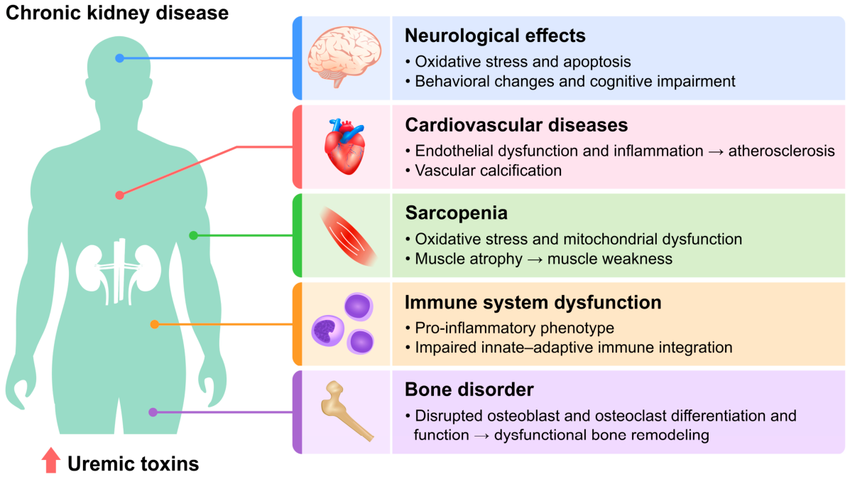
3. Pathophysiological Mechanisms
3.1. Uremic Toxins Induce Endothelial Dysfunction
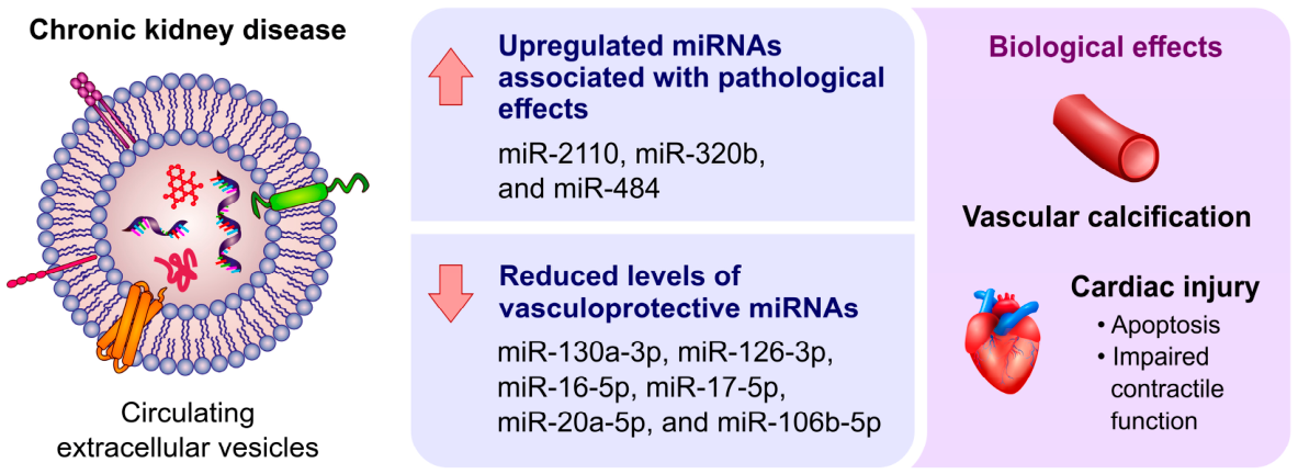
3.2. Extracellular Vesicles Formed in the Uremic Environment
3.3. Bone Disorders
3.4. Neurological Effects Caused by Uremic Toxicity
3.5. Immune System Dysfunction
3.6. Uremic Toxins Promote Sarcopenia
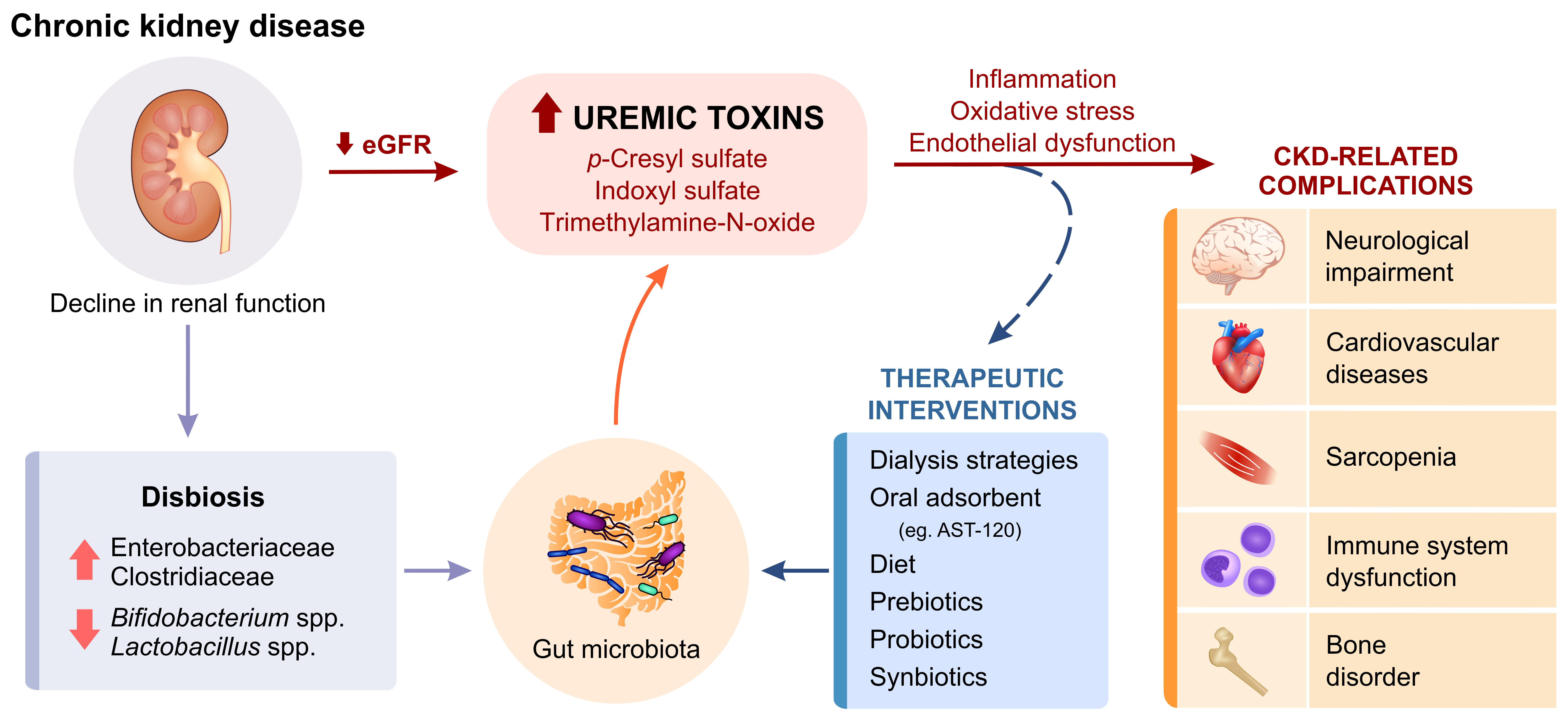
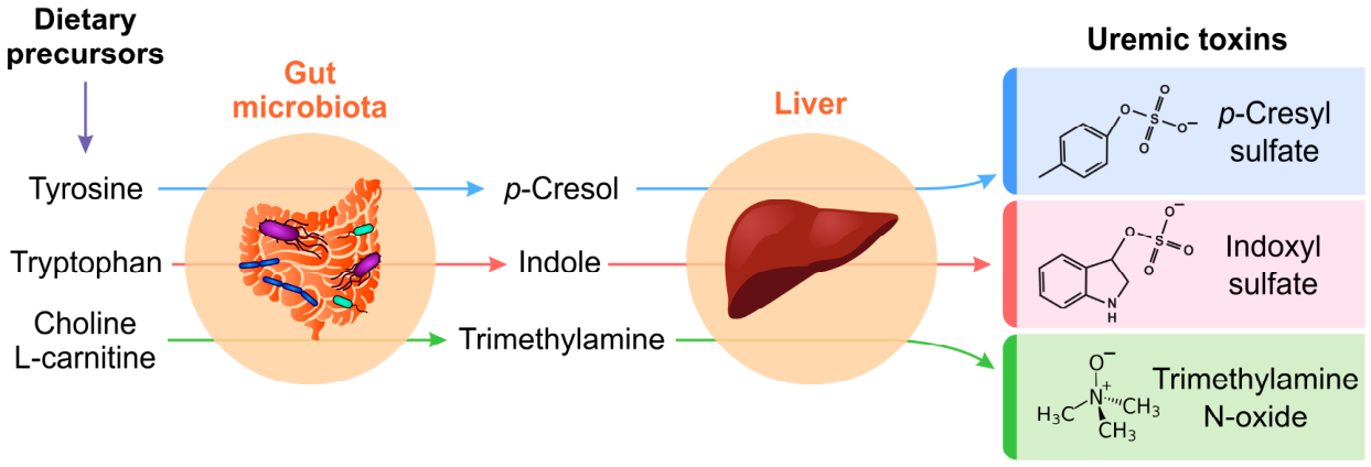
4. Gut Microbiota and Toxin Generation
5. Therapeutic Strategies
6. Future Perspectives
7. Conclusions
Author Contributions
Funding
Institutional Review Board Statement
Informed Consent Statement
Data Availability Statement
Conflicts of Interest
Abbreviations
| ADMA | Asymmetric dimethylarginine |
| AGEs | Advanced glycation end products |
| CKD | Chronic kidney disease |
| CMPF | 3-carboxy-4-methyl-5-propyl-2-furanpropionic acid |
| EMT | Epithelial-to-mesenchymal transition |
| eNOS | Endothelial nitric oxide synthase |
| FFAs | Free fatty acids |
| FGF | Fibroblast growth factor |
| HA | Hippuric acid |
| HD | Hemodialysis |
| HDF | Hemodiafiltration |
| HMW | High molecular weight |
| IAA | Indole-3-acetic acid |
| ICAM-1 | Intercellular adhesion molecule 1 |
| IL-6 | Interleukin-6 |
| IS | Indoxyl sulfate |
| KA | Kynurenic acid |
| KLF | Kruppel-like factor |
| LPS | Lipopolysaccharide |
| MCO | Medium cut-off |
| MCP-1 | Monocyte chemoattractant protein-1 |
| NO | Nitric oxide |
| NF-κB | Nuclear factor kappa B |
| PBUTs | Protein-bound uremic toxins |
| PCG | P-cresyl glucuronide |
| PCS | P-cresyl sulfate |
| PD | Peritoneal dialysis |
| PTH | Parathyroid hormone |
| RCT | Randomized controlled trials |
| ROS | Reactive oxygen species |
| SCFA | Short-chain fatty acid |
| TGF-β | Transforming growth factor-β |
| TMA | Trimethylamine |
| TMAO | Trimethylamine-N-oxide |
| TNF-α | Tumor necrosis factor-alpha |
| VCAM-1 | Vascular cell adhesion molecule 1 |
| VLPD | Very low-protein diet |
| ZO-1 | Zonula occludens-1 |
References
- Chesnaye, N.C.; Ortiz, A.; Zoccali, C.; Stel, V.S.; Jager, K.J. The Impact of Population Ageing on the Burden of Chronic Kidney Disease. Nat. Rev. Nephrol. 2024, 20, 569–585. [Google Scholar] [CrossRef] [PubMed]
- Ortiz, A.; Lees, J.S.; Torra, R.; Stel, V.S.; Kramer, A.; Mark, P.B. The Updated Global Burden of Chronic Kidney Disease: One Death Every 20 Seconds. Nephrol. Dial. Transplant. 2026, gfag040. [Google Scholar] [CrossRef] [PubMed]
- Vanholder, R.; De Smet, R.; Glorieux, G.; Argilés, A.; Baurmeister, U.; Brunet, P.; Clark, W.; Cohen, G.; De Deyn, P.P.; Deppisch, R.; et al. Review on Uremic Toxins: Classification, Concentration, and Interindividual Variability. Kidney Int. 2003, 63, 1934–1943. [Google Scholar] [CrossRef] [PubMed]
- Vanholder, R.; Schepers, E.; Pletinck, A.; Nagler, E.V.; Glorieux, G. The Uremic Toxicity of Indoxyl Sulfate and P-Cresyl Sulfate: A Systematic Review. J. Am. Soc. Nephrol. 2014, 25, 1897–1907. [Google Scholar] [CrossRef] [PubMed]
- Rosner, M.H.; Reis, T.; Husain-Syed, F.; Vanholder, R.; Hutchison, C.; Stenvinkel, P.; Blankestijn, P.J.; Cozzolino, M.; Juillard, L.; Kashani, K.; et al. Classification of Uremic Toxins and Their Role in Kidney Failure. Clin. J. Am. Soc. Nephrol. 2021, 16, 1918–1928. [Google Scholar] [CrossRef] [PubMed]
- Zhao, B.; Hu, X.; Wang, W.; Zhou, Y. Cardiorenal Syndrome: Clinical Diagnosis, Molecular Mechanisms and Therapeutic Strategies. Acta Pharmacol. Sin. 2025, 46, 1539–1555. [Google Scholar] [CrossRef] [PubMed]
- Go, A.S.; Chertow, G.M.; Fan, D.; McCulloch, C.E.; Hsu, C. Chronic Kidney Disease and the Risks of Death, Cardiovascular Events, and Hospitalization. N. Engl. J. Med. 2004, 351, 1296–1305. [Google Scholar] [CrossRef] [PubMed]
- Vondenhoff, S.; Schunk, S.J.; Noels, H. Increased Cardiovascular Risk in Patients with Chronic Kidney Disease. Herz 2024, 49, 95–104. [Google Scholar] [CrossRef] [PubMed]
- Abbad, L.; Esteve, E.; Chatziantoniou, C. Advances and Challenges in Kidney Fibrosis Therapeutics. Nat. Rev. Nephrol. 2025, 21, 314–329. [Google Scholar] [CrossRef] [PubMed]
- Liu, H.; Xiang, X.; Shi, C.; Guo, J.; Ran, T.; Lin, J.; Dong, F.; Yang, J.; Miao, H. Oxidative Stress and Inflammation in Renal Fibrosis: Novel Molecular Mechanisms and Therapeutic Targets. Chem. Biol. Interact. 2025, 421, 111784. [Google Scholar] [CrossRef] [PubMed]
- Liu, Y. Kidney Fibrosis: Fundamental Questions, Challenges, and Perspectives. Integr. Med. Nephrol. Androl. 2024, 11, e24-00027. [Google Scholar] [CrossRef]
- Zhao, H.; Guo, J. Macrophages in Focus: Key Drivers and Therapeutic Opportunities in Diabetic Kidney Disease. Int. J. Biol. Sci. 2025, 21, 4647–4662. [Google Scholar] [CrossRef] [PubMed]
- Cantero-Navarro, E.; Rayego-Mateos, S.; Orejudo, M.; Tejedor-Santamaria, L.; Tejera-Muñoz, A.; Sanz, A.B.; Marquez-Exposito, L.; Marchant, V.; Santos-Sanchez, L.; Egido, J.; et al. Role of Macrophages and Related Cytokines in Kidney Disease. Front. Med. 2021, 8, 688060. [Google Scholar] [CrossRef] [PubMed]
- Evenepoel, P.; Poesen, R.; Meijers, B. The Gut–Kidney Axis. Pediatr. Nephrol. 2017, 32, 2005–2014. [Google Scholar] [CrossRef] [PubMed]
- Vanholder, R.; Glorieux, G.; De Smet, R.; Lameire, N. New Insights in Uremic Toxins. Kidney Int. 2003, 63, S6–S10. [Google Scholar] [CrossRef] [PubMed]
- Corradi, V.; Caprara, C.; Barzon, E.; Mattarollo, C.; Zanetti, F.; Ferrari, F.; Husain-Syed, F.; Giavarina, D.; Ronco, C.; Zanella, M. A Possible Role of P-Cresyl Sulfate and Indoxyl Sulfate as Biomarkers in the Prediction of Renal Function According to the GFR (G) Categories. Integr. Med. Nephrol. Androl. 2024, 11, e24-00002. [Google Scholar] [CrossRef]
- Poesen, R.; Windey, K.; Neven, E.; Kuypers, D.; De Preter, V.; Augustijns, P.; D’Haese, P.; Evenepoel, P.; Verbeke, K.; Meijers, B. The Influence of CKD on Colonic Microbial Metabolism. J. Am. Soc. Nephrol. 2016, 27, 1389–1399. [Google Scholar] [CrossRef] [PubMed]
- Maciel, R.; Cunha, R.; Busato, V.; Franco, C.; Gregório, P.; Dolenga, C.; Nakao, L.; Massy, Z.; Boullier, A.; Pecoits-Filho, R.; et al. Uremia Impacts VE-Cadherin and ZO-1 Expression in Human Endothelial Cell-to-Cell Junctions. Toxins 2018, 10, 404. [Google Scholar] [CrossRef] [PubMed]
- da Cunha, R.S.; Santos, A.F.; Barreto, F.C.; Stinghen, A.E.M. How Do Uremic Toxins Affect the Endothelium? Toxins 2020, 12, 412. [Google Scholar] [CrossRef] [PubMed]
- Favretto, G.; da Cunha, R.S.; Flores Santos, A.; Leitolis, A.; Schiefer, E.M.; Gregório, P.C.; Franco, C.R.C.; Massy, Z.; Dalboni, M.A.; Stinghen, A.E.M. Uremic Endothelial-Derived Extracellular Vesicles: Mechanisms of Formation and Their Role in Cell Adhesion, Cell Migration, Inflammation, and Oxidative Stress. Toxicol. Lett. 2021, 347, 12–22. [Google Scholar] [CrossRef] [PubMed]
- Wang, Z.; Klipfell, E.; Bennett, B.J.; Koeth, R.; Levison, B.S.; DuGar, B.; Feldstein, A.E.; Britt, E.B.; Fu, X.; Chung, Y.-M.; et al. Gut Flora Metabolism of Phosphatidylcholine Promotes Cardiovascular Disease. Nature 2011, 472, 57–63. [Google Scholar] [CrossRef] [PubMed]
- Budoff, M.J.; De Oliveira Otto, M.C.; Li, X.S.; Lee, Y.; Wang, M.; Lai, H.T.M.; Lemaitre, R.N.; Pratt, A.; Tang, W.H.W.; Psaty, B.M.; et al. Trimethylamine-N-Oxide (TMAO) and Risk of Incident Cardiovascular Events in the Multi Ethnic Study of Atherosclerosis. Sci. Rep. 2025, 15, 23362. [Google Scholar] [CrossRef] [PubMed]
- Xu, C.; Snelson, M.; Marques, F.Z. Cardiovascular–Kidney–Metabolic Syndrome through the Lens of Gut-derived Uremic Toxins. Gut Microbes 2026, 18, 2685906. [Google Scholar] [CrossRef] [PubMed]
- Dhondt, A.; Vanholder, R.; Van Biesen, W.; Lameire, N. The Removal of Uremic Toxins. Kidney Int. 2000, 58, S47–S59. [Google Scholar] [CrossRef] [PubMed]
- Glorieux, G.; Burtey, S.; Evenepoel, P.; Jankowski, J.; Koppe, L.; Masereeuw, R.; Vanholder, R. A Guide to Uraemic Toxicity. Nat. Rev. Nephrol. 2026, 22, 50–68. [Google Scholar] [PubMed]
- Ho, Y.; Soliman, K.M.; Cheungpasitporn, W.; Wathanavasin, W.; Daoud, A.; Kamal, A.I.; Szamosfalvi, B.; Fülöp, T. A Bibliometric Analysis of Publications in Uremic Toxins From 1991 to 2024. Ther. Apher. Dial. 2026, 1–14. [Google Scholar] [CrossRef] [PubMed]
- Falconi, C.A.; Junho, C.V.D.C.; Fogaça-Ruiz, F.; Vernier, I.C.S.; da Cunha, R.S.; Stinghen, A.E.M.; Carneiro-Ramos, M.S. Uremic Toxins: An Alarming Danger Concerning the Cardiovascular System. Front. Physiol. 2021, 12, 114460. [Google Scholar] [CrossRef] [PubMed]
- Shafi, T.; Hostetter, T.H.; Meyer, T.W.; Hwang, S.; Hai, X.; Melamed, M.L.; Banerjee, T.; Coresh, J.; Powe, N.R. Serum Asymmetric and Symmetric Dimethylarginine and Morbidity and Mortality in Hemodialysis Patients. Am. J. Kidney Dis. 2017, 70, 48–58. [Google Scholar] [CrossRef] [PubMed]
- Barreto, F.C.; Barreto, D.V.; Liabeuf, S.; Meert, N.; Glorieux, G.; Temmar, M.; Choukroun, G.; Vanholder, R.; Massy, Z.A. Serum Indoxyl Sulfate Is Associated with Vascular Disease and Mortality in Chronic Kidney Disease Patients. Clin. J. Am. Soc. Nephrol. 2009, 4, 1551–1558. [Google Scholar] [CrossRef] [PubMed]
- Lin, C.-J.; Pan, C.-F.; Chuang, C.-K.; Sun, F.-J.; Wang, D.-J.; Chen, H.-H.; Liu, H.-L.; Wu, C.-J. P-Cresyl Sulfate Is a Valuable Predictor of Clinical Outcomes in Pre-ESRD Patients. BioMed Res. Int. 2014, 2014, 526932. [Google Scholar] [CrossRef] [PubMed]
- Zhou, Z.; Jin, H.; Ju, H.; Sun, M.; Chen, H.; Li, L. Circulating Trimethylamine-N-Oxide and Risk of All-Cause and Cardiovascular Mortality in Patients with Chronic Kidney Disease: A Systematic Review and Meta-Analysis. Front. Med. 2022, 9, 828343. [Google Scholar] [CrossRef] [PubMed]
- Zwaenepoel, B.; De Backer, T.; Glorieux, G.; Verbeke, F. Predictive Value of Protein-Bound Uremic Toxins for Heart Failure in Patients with Chronic Kidney Disease. ESC Heart Fail. 2024, 11, 466–474. [Google Scholar] [CrossRef] [PubMed]
- Tumur, Z.; Shimizu, H.; Enomoto, A.; Miyazaki, H.; Niwa, T. Indoxyl Sulfate Upregulates Expression of ICAM-1 and MCP-1 by Oxidative Stress-Induced NF-ĸB Activation. Am. J. Nephrol. 2010, 31, 435–441. [Google Scholar] [CrossRef] [PubMed]
- Favretto, G.; Souza, L.M.; Gregório, P.C.; Cunha, R.S.; Maciel, R.A.P.; Sassaki, G.L.; Toledo, M.G.; Pecoits-Filho, R.; Souza, W.M.; Stinghen, A.E.M. Role of Organic Anion Transporters in the Uptake of Protein-Bound Uremic Toxins by Human Endothelial Cells and Monocyte Chemoattractant Protein-1 Expression. J. Vasc. Res. 2017, 54, 170–179. [Google Scholar] [CrossRef] [PubMed]
- Gregório, P.C.; Favretto, G.; Sassaki, G.L.; Cunha, R.S.; Becker-Finco, A.; Pecoits-Filho, R.; Souza, W.M.; Barreto, F.C.; Stinghen, A.E.M. Sevelamer Reduces Endothelial Inflammatory Response to Advanced Glycation End Products. Clin. Kidney J. 2018, 11, 89–98. [Google Scholar] [CrossRef] [PubMed]
- Ma, G.; Pan, B.; Chen, Y.; Guo, C.; Zhao, M.; Zheng, L.; Chen, B. Trimethylamine N-Oxide in Atherogenesis: Impairing Endothelial Self-Repair Capacity and Enhancing Monocyte Adhesion. Biosci. Rep. 2017, 37, BSR20160244. [Google Scholar] [CrossRef] [PubMed]
- Short, S.A.P.; Wilkinson, K.; Long, D.L.; Crews, D.C.; Gutierrez, O.M.; Irvin, M.R.; Wheeler, M.; Cushman, M.; Cheung, K.L. Endothelial Dysfunction Biomarkers and CKD Incidence in the REGARDS Cohort. Kidney Int. Rep. 2024, 9, 2016–2027. [Google Scholar] [CrossRef] [PubMed]
- Masai, N.; Tatebe, J.; Yoshino, G.; Morita, T. Indoxyl Sulfate Stimulates Monocyte Chemoattractant Protein-1 Expression in Human Umbilical Vein Endothelial Cells by Inducing Oxidative Stress through Activation of the NADPH Oxidase-Nuclear Factor-κB Pathway. Circ. J. 2010, 74, 2216–2224. [Google Scholar] [CrossRef] [PubMed]
- Marinčić Žagar, A.; Kolobarić, N.; Šušnjara, P.; Mihaljević, J.; Mihaljević, Z.; Drenjančević, I. The Role of Endothelial Glycocalyx in the Pathophysiology of Chronic Kidney Disease and Hypertension: From Molecular Mechanisms to Clinical Biomarkers. Life 2026, 16, 642. [Google Scholar] [CrossRef] [PubMed]
- Gondouin, B.; Cerini, C.; Dou, L.; Sallée, M.; Duval-Sabatier, A.; Pletinck, A.; Calaf, R.; Lacroix, R.; Jourde-Chiche, N.; Poitevin, S.; et al. Indolic Uremic Solutes Increase Tissue Factor Production in Endothelial Cells by the Aryl Hydrocarbon Receptor Pathway. Kidney Int. 2013, 84, 733–744. [Google Scholar] [CrossRef] [PubMed]
- Liew, H.; Roberts, M.A.; Pope, A.; McMahon, L.P. Endothelial Glycocalyx Damage in Kidney Disease Correlates with Uraemic Toxins and Endothelial Dysfunction. BMC Nephrol. 2021, 22, 21. [Google Scholar] [CrossRef] [PubMed]
- Valera, G.; Figuer, A.; Caro, J.; Yuste, C.; Morales, E.; Ceprián, N.; Bodega, G.; Ramírez, R.; Alique, M.; Carracedo, J. Plasma Glycocalyx Pattern: A Mirror of Endothelial Damage in Chronic Kidney Disease. Clin. Kidney J. 2023, 16, 1278–1287. [Google Scholar] [CrossRef] [PubMed]
- Amaral Bueno Azevedo, C.; Miniskiskosky, G.; Guimarães Del Amo, M.V.; Povala Theisen, V.; Cavichiolo Franco, C.R.; Stafim Da Cunha, R.; Marques Stinghen, A.E. Trimethylamine N-Oxide (TMAO) Disrupts Endothelial Junction Integrity through VE-Cadherin Tyr658 Phosphorylation in Vitro. Arch. Toxicol. 2026, 100, 3063–3072. [Google Scholar] [CrossRef] [PubMed]
- Chen, S.C.; Huang, S.Y.; Wu, C.C.; Hsu, C.F. P-Cresylsulfate, the Protein-Bound Uremic Toxin, Increased Endothelial Permeability Partly Mediated by Src-Induced Phosphorylation of VE-Cadherin. Toxins 2020, 12, 62. [Google Scholar] [CrossRef] [PubMed]
- Assefa, E.G.; Yan, Q.; Gezahegn, S.B.; Salissou, M.T.M.; He, S.; Wu, N.; Zuo, X.; Ying, C. Role of Resveratrol on Indoxyl Sulfate-Induced Endothelial Hyperpermeability via Aryl Hydrocarbon Receptor (AHR)/Src-Dependent Pathway. Oxid. Med. Cell. Longev. 2019, 2019, 5847040. [Google Scholar] [CrossRef] [PubMed]
- Stinghen, A.E.M.; Chillon, J.-M.; Massy, Z.A.; Boullier, A. Differential Effects of Indoxyl Sulfate and Inorganic Phosphate in a Murine Cerebral Endothelial Cell Line (bEnd.3). Toxins 2014, 6, 1742–1760. [Google Scholar] [CrossRef] [PubMed]
- Watanabe, H.; Miyamoto, Y.; Enoki, Y.; Ishima, Y.; Kadowaki, D.; Kotani, S.; Nakajima, M.; Tanaka, M.; Matsushita, K.; Mori, Y.; et al. P-Cresyl Sulfate, a Uremic Toxin, Causes Vascular Endothelial and Smooth Muscle Cell Damages by Inducing Oxidative Stress. Pharmacol. Res. Perspect. 2015, 3, e00092. [Google Scholar] [CrossRef] [PubMed]
- Kajimoto, H.; Kai, H.; Aoki, H.; Yasuoka, S.; Anegawa, T.; Aoki, Y.; Ueda, S.; Okuda, S.; Imaizumi, T. Inhibition of ENOS Phosphorylation Mediates Endothelial Dysfunction in Renal Failure: New Effect of Asymmetric Dimethylarginine. Kidney Int. 2012, 81, 762–768. [Google Scholar] [CrossRef] [PubMed]
- Shang, F.; Wang, S.-C.; Hsu, C.-Y.; Miao, Y.; Martin, M.; Yin, Y.; Wu, C.-C.; Wang, Y.-T.; Wu, G.; Chien, S.; et al. MicroRNA-92a Mediates Endothelial Dysfunction in CKD. J. Am. Soc. Nephrol. 2017, 28, 3251–3261. [Google Scholar] [CrossRef] [PubMed]
- Saum, K.; Campos, B.; Celdran-Bonafonte, D.; Nayak, L.; Sangwung, P.; Thakar, C.; Roy-Chaudhury, P.; Owens, A.P. Uremic Advanced Glycation End Products and Protein-Bound Solutes Induce Endothelial Dysfunction Through Suppression of Krüppel-Like Factor 2. J. Am. Heart Assoc. 2018, 7, e007566. [Google Scholar] [CrossRef] [PubMed]
- Wang, D.; Liu, J.; Wang, J.; Feng, Y. Resveratrol Protects against Uremic Serum-Induced Endothelial Cell Injury by Activating the FUS/KLF2/FBXW7 Signaling Pathway. Appl. Biol. Chem. 2025, 68, 24. [Google Scholar] [CrossRef]
- Dabravolski, S.A.; Sukhorukov, V.N.; Kalmykov, V.A.; Grechko, A.V.; Shakhpazyan, N.K.; Orekhov, A.N. The Role of KLF2 in the Regulation of Atherosclerosis Development and Potential Use of KLF2-Targeted Therapy. Biomedicines 2022, 10, 254. [Google Scholar] [CrossRef] [PubMed]
- Li, X.; Raisinghani, N.; Gallinat, A.; Santos-Gallego, C.G.; Zhang, S.; La Salvia, S.; Yoon, S.; Yavuz, H.; Phan, A.; Shao, A.; et al. Circulating Extracellular Vesicles in the Pathogenesis of Heart Failure in Patients with Chronic Kidney Disease. Circulation 2026, 153, 94–114. [Google Scholar] [CrossRef] [PubMed]
- Behrens, F.; Holle, J.; Chen, C.Y.; Ginsbach, L.F.; Krause, B.C.; Bruning, U.; Kriegel, F.L.; Kaiser, T.; Szijártó, I.A.; Anandakumar, H.; et al. Circulating Extracellular Vesicles as Putative Mediators of Cardiovascular Disease in Paediatric Chronic Kidney Disease. J. Extracell. Vesicles 2025, 14, e70062. [Google Scholar] [CrossRef] [PubMed]
- Zietzer, A.; Steffen, E.; Niepmann, S.; Düsing, P.; Hosen, M.R.; Liu, W.; Jamme, P.; Al-Kassou, B.; Goody, P.R.; Zimmer, S.; et al. MicroRNA-Mediated Vascular Intercellular Communication Is Altered in Chronic Kidney Disease. Cardiovasc. Res. 2020, 118, 316–333. [Google Scholar] [CrossRef] [PubMed]
- Koide, T.; Mandai, S.; Kitaoka, R.; Matsuki, H.; Chiga, M.; Yamamoto, K.; Yoshioka, K.; Yagi, Y.; Suzuki, S.; Fujiki, T.; et al. Circulating Extracellular Vesicle-Propagated MicroRNA Signature as a Vascular Calcification Factor in Chronic Kidney Disease. Circ. Res. 2023, 132, 415–431. [Google Scholar] [CrossRef] [PubMed]
- Salera, D.; Merkel, N.; Bellasi, A.; de Borst, M.H. Pathophysiology of Chronic Kidney Disease–Mineral Bone Disorder (CKD-MBD): From Adaptive to Maladaptive Mineral Homeostasis. Clin. Kidney J. 2025, 18, i3–i14. [Google Scholar] [CrossRef] [PubMed]
- Watanabe, K.; Tominari, T.; Hirata, M.; Matsumoto, C.; Hirata, J.; Murphy, G.; Nagase, H.; Miyaura, C.; Inada, M. Indoxyl Sulfate, a Uremic Toxin in Chronic Kidney Disease, Suppresses Both Bone Formation and Bone Resorption. FEBS Open Bio 2017, 7, 1178–1185. [Google Scholar] [CrossRef] [PubMed]
- Iwasaki, Y.; Yamato, H.; Fukagawa, M. Treatment with Pravastatin Attenuates Oxidative Stress and Protects Osteoblast Cell Viability From Indoxyl Sulfate. Ther. Apher. Dial. 2011, 15, 151–155. [Google Scholar] [CrossRef] [PubMed]
- Kim, Y.-H.; Kwak, K.-A.; Gil, H.-W.; Song, H.-Y.; Hong, S.-Y. Indoxyl Sulfate Promotes Apoptosis in Cultured Osteoblast Cells. BMC Pharmacol. Toxicol. 2013, 14, 60. [Google Scholar] [CrossRef] [PubMed]
- Nii-Kono, T.; Iwasaki, Y.; Uchida, M.; Fujieda, A.; Hosokawa, A.; Motojima, M.; Yamato, H.; Kurokawa, K.; Fukagawa, M. Indoxyl Sulfate Induces Skeletal Resistance to Parathyroid Hormone in Cultured Osteoblastic Cells. Kidney Int. 2007, 71, 738–743. [Google Scholar] [CrossRef] [PubMed]
- Tanaka, H.; Iwasaki, Y.; Yamato, H.; Mori, Y.; Komaba, H.; Watanabe, H.; Maruyama, T.; Fukagawa, M. P-Cresyl Sulfate Induces Osteoblast Dysfunction through Activating JNK and P38 MAPK Pathways. Bone 2013, 56, 347–354. [Google Scholar] [CrossRef] [PubMed]
- Wu, Q.; Lan, J.; Wan, Z.; Liang, Y.; Nie, K.; Zhou, J.; Ji, B.; Zhang, Q.; Jiang, Z.; Huang, X.; et al. Parallel Metabolomic and Transcriptomic Profiling Reveals the Cytotoxic Mechanism of Protein-Bound Uremic Toxin p-Cresol Sulfate via Disruption of Glutathione and Glycerophospholipid Metabolism in MC3T3-E1 Osteoblasts. BMC Nephrol. 2026, 27, 369. [Google Scholar] [CrossRef] [PubMed]
- Liabeuf, S.; Pepin, M.; Franssen, C.F.M.; Viggiano, D.; Carriazo, S.; Gansevoort, R.T.; Gesualdo, L.; Hafez, G.; Malyszko, J.; Mayer, C.; et al. Chronic Kidney Disease and Neurological Disorders: Are Uraemic Toxins the Missing Piece of the Puzzle? Nephrol. Dial. Transplant. 2021, 37, ii33–ii44. [Google Scholar] [CrossRef] [PubMed]
- Faucher, Q.; van der Made, T.K.; De Lange, E.; Masereeuw, R. Blood-Brain Barrier Perturbations by Uremic Toxins: Key Contributors in Chronic Kidney Disease-Induced Neurological Disorders? Eur. J. Pharm. Sci. 2023, 187, 106462. [Google Scholar] [CrossRef] [PubMed]
- Lin, Y.-T.; Wu, P.-H.; Tsai, Y.-C.; Hsu, Y.-L.; Wang, H.Y.; Kuo, M.-C.; Kuo, P.-L.; Hwang, S.-J. Indoxyl Sulfate Induces Apoptosis through Oxidative Stress and Mitogen-Activated Protein Kinase Signaling Pathway Inhibition in Human Astrocytes. J. Clin. Med. 2019, 8, 191. [Google Scholar] [CrossRef] [PubMed]
- Adesso, S.; Magnus, T.; Cuzzocrea, S.; Campolo, M.; Rissiek, B.; Paciello, O.; Autore, G.; Pinto, A.; Marzocco, S. Indoxyl Sulfate Affects Glial Function Increasing Oxidative Stress and Neuroinflammation in Chronic Kidney Disease: Interaction between Astrocytes and Microglia. Front. Pharmacol. 2017, 8, 370. [Google Scholar] [CrossRef] [PubMed]
- Watanabe, K.; Sato, E.; Mishima, E.; Watanabe, M.; Abe, T.; Takahashi, N.; Nakayama, M. Effect of Uremic Toxins on Hippocampal Cell Damage: Analysis in Vitro and in Rat Model of Chronic Kidney Disease. Heliyon 2021, 7, e06221. [Google Scholar] [CrossRef] [PubMed]
- Karbowska, M.; Hermanowicz, J.M.; Tankiewicz-Kwedlo, A.; Kalaska, B.; Kaminski, T.W.; Nosek, K.; Wisniewska, R.J.; Pawlak, D. Neurobehavioral Effects of Uremic Toxin–Indoxyl Sulfate in the Rat Model. Sci. Rep. 2020, 10, 9483. [Google Scholar] [CrossRef] [PubMed]
- Sun, C.-Y.; Li, J.-R.; Wang, Y.-Y.; Lin, S.-Y.; Ou, Y.-C.; Lin, C.-J.; Wang, J.-D.; Liao, S.-L.; Chen, C.-J. P-Cresol Sulfate Caused Behavior Disorders and Neurodegeneration in Mice with Unilateral Nephrectomy Involving Oxidative Stress and Neuroinflammation. Int. J. Mol. Sci. 2020, 21, 6687. [Google Scholar] [CrossRef] [PubMed]
- Wakamatsu, T.; Yamamoto, S.; Yoshida, S.; Narita, I. Indoxyl Sulfate-Induced Macrophage Toxicity and Therapeutic Strategies in Uremic Atherosclerosis. Toxins 2024, 16, 254. [Google Scholar] [CrossRef] [PubMed]
- Adesso, S.; Popolo, A.; Bianco, G.; Sorrentino, R.; Pinto, A.; Autore, G.; Marzocco, S. The Uremic Toxin Indoxyl Sulphate Enhances Macrophage Response to LPS. PLoS ONE 2013, 8, e76778. [Google Scholar] [CrossRef] [PubMed]
- Azevedo, M.L.V.; Bonan, N.B.; Dias, G.; Brehm, F.; Steiner, T.M.; Souza, W.M.; Stinghen, A.E.M.; Barreto, F.C.; Elifio-Esposito, S.; Pecoits-Filho, R.; et al. P-Cresyl Sulfate Affects the Oxidative Burst, Phagocytosis Process, and Antigen Presentation of Monocyte-Derived Macrophages. Toxicol. Lett. 2016, 263, 1–5. [Google Scholar] [CrossRef] [PubMed]
- Kim, H.Y.; Yoo, T.-H.; Hwang, Y.; Lee, G.H.; Kim, B.; Jang, J.; Yu, H.T.; Kim, M.C.; Cho, J.-Y.; Lee, C.J.; et al. Indoxyl Sulfate (IS)-Mediated Immune Dysfunction Provokes Endothelial Damage in Patients with End-Stage Renal Disease (ESRD). Sci. Rep. 2017, 7, 3057. [Google Scholar] [CrossRef] [PubMed]
- Sato, E.; Mori, T.; Mishima, E.; Suzuki, A.; Sugawara, S.; Kurasawa, N.; Saigusa, D.; Miura, D.; Morikawa-Ichinose, T.; Saito, R.; et al. Metabolic Alterations by Indoxyl Sulfate in Skeletal Muscle Induce Uremic Sarcopenia in Chronic Kidney Disease. Sci. Rep. 2016, 6, 36618. [Google Scholar] [CrossRef] [PubMed]
- Enoki, Y.; Watanabe, H.; Arake, R.; Sugimoto, R.; Imafuku, T.; Tominaga, Y.; Ishima, Y.; Kotani, S.; Nakajima, M.; Tanaka, M.; et al. Indoxyl Sulfate Potentiates Skeletal Muscle Atrophy by Inducing the Oxidative Stress-Mediated Expression of Myostatin and Atrogin-1. Sci. Rep. 2016, 6, 32084. [Google Scholar] [CrossRef] [PubMed]
- Rodrigues, G.G.C.; Dellê, H.; Brito, R.B.O.; Cardoso, V.O.; Fernandes, K.P.S.; Mesquita-Ferrari, R.A.; Cunha, R.S.; Stinghen, A.E.M.; Dalboni, M.A.; Barreto, F.C. Indoxyl Sulfate Contributes to Uremic Sarcopenia by Inducing Apoptosis in Myoblasts. Arch. Med. Res. 2020, 51, 21–29. [Google Scholar] [CrossRef] [PubMed]
- Koeth, R.A.; Wang, Z.; Levison, B.S.; Buffa, J.A.; Org, E.; Sheehy, B.T.; Britt, E.B.; Fu, X.; Wu, Y.; Li, L.; et al. Intestinal Microbiota Metabolism of L-Carnitine, a Nutrient in Red Meat, Promotes Atherosclerosis. Nat. Med. 2013, 19, 576–585. [Google Scholar] [CrossRef] [PubMed]
- Wang, Z.; Bergeron, N.; Levison, B.S.; Li, X.S.; Chiu, S.; Jia, X.; Koeth, R.A.; Li, L.; Wu, Y.; Tang, W.H.W.; et al. Impact of Chronic Dietary Red Meat, White Meat, or Non-Meat Protein on Trimethylamine N-Oxide Metabolism and Renal Excretion in Healthy Men and Women. Eur. Heart J. 2019, 40, 583–594. [Google Scholar] [CrossRef] [PubMed]
- Scarpioni, R.; Ricardi, M.; Albertazzi, V.; De Amicis, S.; Rastelli, F.; Zerbini, L. Dialysis-Related Amyloidosis: Challenges and Solutions. Int. J. Nephrol. Renov. Dis. 2016, 9, 319–328. [Google Scholar] [CrossRef] [PubMed]
- Brinck, J.E.; Laursen, M.F.; Pedersen, M.; Carey, M.R.; Procházková, N.; Jørgensen, M.S.; Mortensen, M.S.; Lauritzen, L.; Pedersen, O.; Roager, H.M.; et al. PH Regulates Gut Bacterial Tryptophan Metabolism. npj Biofilms Microbiomes 2026, 12, 72. [Google Scholar] [CrossRef] [PubMed]
- Poesen, R.; Evenepoel, P.; de Loor, H.; Kuypers, D.; Augustijns, P.; Meijers, B. Metabolism, Protein Binding, and Renal Clearance of Microbiota-Derived p-Cresol in Patients with CKD. Clin. J. Am. Soc. Nephrol. 2016, 11, 1136–1144. [Google Scholar] [CrossRef] [PubMed]
- Han, H.; Chen, Y.; Zhu, J.; Ni, J.; Sun, J.; Zhang, R. Atorvastatin Attenuates P-Cresyl Sulfate-Induced Atherogenesis and Plaque Instability in ApoE Knockout Mice. Mol. Med. Rep. 2016, 14, 3122–3128. [Google Scholar] [CrossRef] [PubMed][Green Version]
- Han, H.; Chen, Y.; Zhu, Z.; Su, X.; Ni, J.; Du, R.; Zhang, R.; Jin, W. P-Cresyl Sulfate Promotes the Formation of Atherosclerotic Lesions and Induces Plaque Instability by Targeting Vascular Smooth Muscle Cells. Front. Med. 2016, 10, 320–329. [Google Scholar] [CrossRef] [PubMed]
- Jing, Y.J.; Ni, J.W.; Ding, F.H.; Fang, Y.H.; Wang, X.Q.; Wang, H.B.; Chen, X.N.; Chen, N.; Zhan, W.W.; Lu, L.; et al. P-Cresyl Sulfate Is Associated with Carotid Arteriosclerosis in Hemodialysis Patients and Promotes Atherogenesis in ApoE−/− Mice. Kidney Int. 2016, 89, 439–449. [Google Scholar] [CrossRef] [PubMed]
- Chen, Y.-Y.; Chen, D.-Q.; Chen, L.; Liu, J.-R.; Vaziri, N.D.; Guo, Y.; Zhao, Y.-Y. Microbiome–Metabolome Reveals the Contribution of Gut–Kidney Axis on Kidney Disease. J. Transl. Med. 2019, 17, 5. [Google Scholar] [CrossRef] [PubMed]
- James, P.; Stanford, J.; Dixit, O.V.A.; Nicdao, M.A.; McWhinney, B.; Sud, K.; Ryan, M.; Read, S.; Ahlenstiel, G.; Lambert, K.; et al. Associations Between Uraemic Toxins and Gut Microbiota in Adults Initiating Peritoneal Dialysis. Toxins 2025, 17, 334. [Google Scholar] [CrossRef] [PubMed]
- Sun, C.-Y.; Chang, S.-C.; Wu, M.-S. Uremic Toxins Induce Kidney Fibrosis by Activating Intrarenal Renin–Angiotensin–Aldosterone System Associated Epithelial-to-Mesenchymal Transition. PLoS ONE 2012, 7, e34026. [Google Scholar] [CrossRef] [PubMed]
- Guan, R.; Peng, J.; Gu, Q.; Xu, J.; Zhang, J.; Adamu, H.; Yang, Q.; Wang, L.; Cao, G. The Gut-Kidney Axis in Chronic Kidney Disease: A Vicious Cycle of Microbial Dysbiosis and Uremic Toxin Accumulation. Front. Immunol. 2026, 17, 1801912. [Google Scholar] [CrossRef] [PubMed]
- Suliman, I.L.; Panculescu, F.G.; Fasie, D.; Cimpineanu, B.; Alexandru, A.; Gafar, N.; Popescu, S.; Nitu, T.S.; Enache, F.-D.; Chisnoiu, T.; et al. Gut Microbiome in Patients with Chronic Kidney Disease Stages 4 and 5: A Systematic Literature Review. Int. J. Mol. Sci. 2025, 26, 10706. [Google Scholar] [CrossRef] [PubMed]
- Beker, B.M.; Colombo, I.; Gonzalez-Torres, H.; Musso, C.G. Decreasing Microbiota-Derived Uremic Toxins to Improve CKD Outcomes. Clin. Kidney J. 2022, 15, 2214–2219. [Google Scholar] [CrossRef] [PubMed]
- Rysz, J.; Franczyk, B.; Ławiński, J.; Olszewski, R.; Ciałkowska-Rysz, A.; Gluba-Brzózka, A. The Impact of CKD on Uremic Toxins and Gut Microbiota. Toxins 2021, 13, 252. [Google Scholar] [CrossRef] [PubMed]
- Gryp, T.; Vanholder, R.; Vaneechoutte, M.; Glorieux, G. P-Cresyl Sulfate. Toxins 2017, 9, 52. [Google Scholar] [CrossRef] [PubMed]
- Lin, C.J.; Wu, V.; Wu, P.C.; Wu, C.J. Meta-Analysis of the Associations of p-Cresyl Sulfate (PCS) and Indoxyl Sulfate (IS) with Cardiovascular Events and All-Cause Mortality in Patients with Chronic Renal Failure. PLoS ONE 2015, 10, e0132589. [Google Scholar] [CrossRef] [PubMed]
- Tsuji, K.; Uchida, N.; Nakanoh, H.; Fukushima, K.; Haraguchi, S.; Kitamura, S.; Wada, J. The Gut–Kidney Axis in Chronic Kidney Diseases. Diagnostics 2024, 15, 21. [Google Scholar] [CrossRef] [PubMed]
- Pan, S.; Zhao, D.; Duan, S.; Chen, X. The Role of Gut-Dependent Molecule Trimethylamine N-Oxide as a Novel Target for the Treatment of Chronic Kidney Disease. Int. Urol. Nephrol. 2023, 55, 1747–1756. [Google Scholar] [CrossRef] [PubMed]
- Tang, W.H.W.; Wang, Z.; Kennedy, D.J.; Wu, Y.; Buffa, J.A.; Agatisa-Boyle, B.; Li, X.S.; Levison, B.S.; Hazen, S.L. Gut Microbiota-Dependent Trimethylamine N -Oxide (TMAO) Pathway Contributes to Both Development of Renal Insufficiency and Mortality Risk in Chronic Kidney Disease. Circ. Res. 2015, 116, 448–455. [Google Scholar] [CrossRef] [PubMed]
- Vaziri, N.D. CKD Impairs Barrier Function and Alters Microbial Flora of the Intestine: A Major Link to Inflammation and Uremic Toxicity. Curr. Opin. Nephrol. Hypertens. 2012, 21, 587–592. [Google Scholar] [CrossRef] [PubMed]
- Yokoo, K.; Yamamoto, Y.; Suzuki, T. Ammonia Impairs Tight Junction Barriers by Inducing Mitochondrial Dysfunction in Caco-2 Cells. FASEB J. 2021, 35, e21854. [Google Scholar] [CrossRef] [PubMed]
- Anders, H.-J.; Andersen, K.; Stecher, B. The Intestinal Microbiota, a Leaky Gut, and Abnormal Immunity in Kidney Disease. Kidney Int. 2013, 83, 1010–1016. [Google Scholar] [CrossRef] [PubMed]
- Honda, H.; Qureshi, A.R.; Heimbürger, O.; Barany, P.; Wang, K.; Pecoits-Filho, R.; Stenvinkel, P.; Lindholm, B. Serum Albumin, C-Reactive Protein, Interleukin 6, and Fetuin a as Predictors of Malnutrition, Cardiovascular Disease, and Mortality in Patients with ESRD. Am. J. Kidney Dis. 2006, 47, 139–148. [Google Scholar] [CrossRef] [PubMed]
- Rossi, M.; Johnson, D.W.; Xu, H.; Carrero, J.J.; Pascoe, E.; French, C.; Campbell, K.L. Dietary Protein-Fiber Ratio Associates with Circulating Levels of Indoxyl Sulfate and p-Cresyl Sulfate in Chronic Kidney Disease Patients. Nutr. Metab. Cardiovasc. Dis. 2015, 25, 860–865. [Google Scholar] [CrossRef] [PubMed]
- Patel, K.P.; Luo, F.J.G.; Plummer, N.S.; Hostetter, T.H.; Meyer, T.W. The Production of P-Cresol Sulfate and Indoxyl Sulfate in Vegetarians versus Omnivores. Clin. J. Am. Soc. Nephrol. 2012, 7, 982–988. [Google Scholar] [CrossRef] [PubMed]
- Czaja-Stolc, S.; Potrykus, M.; Ruszkowski, J.; Styburski, D.; Dębska-Ślizień, A.; Małgorzewicz, S. The Associations between Nutrition and Circulating Gut Microbiota-Derived Uremic Toxins in Patients Undergoing Kidney Replacement Therapy: An Observational, Cross-Sectional Study. Clin. Nutr. ESPEN 2025, 65, 105–114. [Google Scholar] [CrossRef] [PubMed]
- Wang, H.; Ainiwaer, A.; Song, Y.; Qin, L.; Peng, A.; Bao, H.; Qin, H. Perturbed Gut Microbiome and Fecal and Serum Metabolomes Are Associated with Chronic Kidney Disease Severity. Microbiome 2023, 11, 3. [Google Scholar] [CrossRef] [PubMed]
- Fernandes, A.L.F.; Borges, N.A.; Black, A.P.; dos Anjos, J.; da Silva, G.S.; Nakao, L.S.; Mafra, D. Dietary Intake of Tyrosine and Phenylalanine, and p-Cresyl Sulfate Plasma Levels in Non-Dialyzed Patients with Chronic Kidney Disease. Braz. J. Nephrol. 2020, 42, 307–314. [Google Scholar] [CrossRef] [PubMed]
- McFarlane, C.; Krishnasamy, R.; Stanton, T.; Savill, E.; Snelson, M.; Mihala, G.; Morrison, M.; Johnson, D.W.; Campbell, K.L. Diet Quality and Protein-Bound Uraemic Toxins: Investigation of Novel Risk Factors and the Role of Microbiome in Chronic Kidney Disease. J. Ren. Nutr. 2022, 32, 542–551. [Google Scholar] [CrossRef] [PubMed]
- Marzocco, S.; Dal Piaz, F.; Di Micco, L.; Torraca, S.; Sirico, M.L.; Tartaglia, D.; Autore, G.; Di Iorio, B. Very Low Protein Diet Reduces Indoxyl Sulfate Levels in Chronic Kidney Disease. Blood Purif. 2013, 35, 196–201. [Google Scholar] [CrossRef] [PubMed]
- Castillo-Rodriguez, E.; Fernandez-Prado, R.; Esteras, R.; Perez-Gomez, M.V.; Gracia-Iguacel, C.; Fernandez-Fernandez, B.; Kanbay, M.; Tejedor, A.; Lazaro, A.; Ruiz-Ortega, M.; et al. Impact of Altered Intestinal Microbiota on Chronic Kidney Disease Progression. Toxins 2018, 10, 300. [Google Scholar] [CrossRef] [PubMed]
- Chen, L.; Shi, J.; Ma, X.; Shi, D.; Qu, H. Effects of Microbiota-Driven Therapy on Circulating Indoxyl Sulfate and P-Cresyl Sulfate in Patients with Chronic Kidney Disease: A Systematic Review and Meta-Analysis of Randomized Controlled Trials. Adv. Nutr. 2022, 13, 1267–1278. [Google Scholar] [CrossRef] [PubMed]
- Poesen, R.; Evenepoel, P.; de Loor, H.; Delcour, J.A.; Courtin, C.M.; Kuypers, D.; Augustijns, P.; Verbeke, K.; Meijers, B. The Influence of Prebiotic Arabinoxylan Oligosaccharides on Microbiota Derived Uremic Retention Solutes in Patients with Chronic Kidney Disease: A Randomized Controlled Trial. PLoS ONE 2016, 11, e0153893. [Google Scholar] [CrossRef] [PubMed]
- McFarlane, C.; Krishnasamy, R.; Stanton, T.; Savill, E.; Snelson, M.; Mihala, G.; Kelly, J.T.; Morrison, M.; Johnson, D.W.; Campbell, K.L. Synbiotics Easing Renal Failure by Improving Gut Microbiology II (SYNERGY II): A Feasibility Randomized Controlled Trial. Nutrients 2021, 13, 4481. [Google Scholar] [CrossRef] [PubMed]
- Favero, C.; Ortiz, A.; Sanchez-Niño, M.D. Probiotics for Kidney Disease. Clin. Kidney J. 2022, 15, 1981–1986. [Google Scholar] [CrossRef] [PubMed]
- Zhu, H.; Cao, C.; Wu, Z.; Zhang, H.; Sun, Z.; Wang, M.; Xu, H.; Zhao, Z.; Wang, Y.; Pei, G.; et al. The Probiotic L. casei Zhang Slows the Progression of Acute and Chronic Kidney Disease. Cell Metab. 2021, 33, 2091–2093. [Google Scholar] [CrossRef] [PubMed]
- Sun, Z.; Gong, Q.; Wang, Z.; Li, C.; Wang, J.; Yu, J.; Ji, M.; Zhang, D.; Wei, J.; Xie, S.; et al. Microbiota-Gut-Kidney Axis and Targeted Therapeutic Strategies in Kidney Diseases. Int. J. Biol. Sci. 2026, 22, 1142–1161. [Google Scholar] [CrossRef] [PubMed]
- Vanholder, R.; Meert, N.; Van Biesen, W.; Meyer, T.; Hostetter, T.; Dhondt, A.; Eloot, S. Why Do Patients on Peritoneal Dialysis Have Low Blood Levels of Protein-Bound Solutes? Nat. Rev. Nephrol. 2009, 5, 130–131. [Google Scholar] [CrossRef] [PubMed]
- Toth-Manikowski, S.M.; Sirich, T.L.; Meyer, T.W.; Hostetter, T.H.; Hwang, S.; Plummer, N.S.; Hai, X.; Coresh, J.; Powe, N.R.; Shafi, T. Contribution of ‘Clinically Negligible’ Residual Kidney Function to Clearance of Uremic Solutes. Nephrol. Dial. Transplant. 2020, 35, 846–853. [Google Scholar] [CrossRef] [PubMed]
- Meyer, T.W.; Hostetter, T.H. Uremia. N. Engl. J. Med. 2007, 357, 1316–1325. [Google Scholar] [CrossRef] [PubMed]
- Vanholder, R.; Pletinck, A.; Schepers, E.; Glorieux, G. Biochemical and Clinical Impact of Organic Uremic Retention Solutes: A Comprehensive Update. Toxins 2018, 10, 33. [Google Scholar] [CrossRef] [PubMed]
- Battaglia, Y.; Shroff, R.; Meijers, B.; Nistor, I.; Alfano, G.; Franssen, C.; Luyckx, V.; Liakopoulos, V.; Mantovani, A.; Baciga, F.; et al. Haemodiafiltration versus High-Flux Haemodialysis—A Consensus Statement from the EuDial Working Group of the ERA. Nephrol. Dial. Transplant. 2025, 40, 1590–1614. [Google Scholar] [CrossRef] [PubMed]
- Palmer, S.C.; Rabindranath, K.S.; Craig, J.C.; Roderick, P.J.; Locatelli, F.; Strippoli, G.F. High-Flux versus Low-Flux Membranes for End-Stage Kidney Disease. Cochrane Database Syst. Rev. 2012, 2012, CD005016. [Google Scholar] [CrossRef] [PubMed]
- Nistor, I.; Palmer, S.C.; Craig, J.C.; Saglimbene, V.; Vecchio, M.; Covic, A.; Strippoli, G.F.M. Convective Versus Diffusive Dialysis Therapies for Chronic Kidney Failure: An Updated Systematic Review of Randomized Controlled Trials. Am. J. Kidney Dis. 2014, 63, 954–967. [Google Scholar] [CrossRef] [PubMed]
- Ward, R.A.; Daugirdas, J.T. Kinetics of β-2-Microglobulin with Hemodiafiltration and High-Flux Hemodialysis. Clin. J. Am. Soc. Nephrol. 2024, 19, 869–876. [Google Scholar] [CrossRef] [PubMed]
- Cheung, A.K.; Rocco, M.V.; Yan, G.; Leypoldt, J.K.; Levin, N.W.; Greene, T.; Agodoa, L.; Bailey, J.; Beck, G.J.; Clark, W.; et al. Serum β-2 Microglobulin Levels Predict Mortality in Dialysis Patients: Results of the HEMO Study. J. Am. Soc. Nephrol. 2006, 17, 546–555. [Google Scholar] [CrossRef] [PubMed]
- Locatelli, F.; Martin-Malo, A.; Hannedouche, T.; Loureiro, A.; Papadimitriou, M.; Wizemann, V.; Jacobson, S.H.; Czekalski, S.; Ronco, C.; Vanholder, R. Effect of Membrane Permeability on Survival of Hemodialysis Patients. J. Am. Soc. Nephrol. 2009, 20, 645–654. [Google Scholar] [CrossRef] [PubMed]
- Vanholder, R.; Schepers, E.; Pletinck, A.; Neirynck, N.; Glorieux, G. An Update on Protein-Bound Uremic Retention Solutes. J. Ren. Nutr. 2012, 22, 90–94. [Google Scholar] [CrossRef] [PubMed]
- Belmouaz, M.; Bauwens, M.; Hauet, T.; Bossard, V.; Jamet, P.; Joly, F.; Chikhi, E.; Joffrion, S.; Gand, E.; Bridoux, F. Comparison of the Removal of Uraemic Toxins with Medium Cut-off and High-Flux Dialysers: A Randomized Clinical Trial. Nephrol. Dial. Transplant. 2020, 35, 328–335. [Google Scholar] [CrossRef] [PubMed]
- Vega-Vega, O.; Ronco, C.; Martínez-Rueda, A.J. Pushing the Boundaries of Hemodialysis: Innovations in Membranes and Sorbents. Rev. Investig. Clín. 2023, 75, 274–288. [Google Scholar] [CrossRef] [PubMed]
- Morgera, S.; Haase, M.; Kuss, T.; Vargas-Hein, O.; Zuckermann-Becker, H.; Melzer, C.; Krieg, H.; Wegner, B.; Bellomo, R.; Neumayer, H.-H. Pilot Study on the Effects of High Cutoff Hemofiltration on the Need for Norepinephrine in Septic Patients with Acute Renal Failure. Crit. Care Med. 2006, 34, 2099–2104. [Google Scholar] [CrossRef] [PubMed]
- Lukkanalikitkul, E.; Kidkaem, H.; Phonrat, M.; Prathompong, P.; Anutrakulchai, S. A Randomized Trial Comparing Medium Cut-off Membrane Dialyzers with Online Hemodiafiltration for Uremic Toxins Clearance in Hemodialysis Patients. Sci. Rep. 2025, 15, 5467. [Google Scholar] [CrossRef] [PubMed]
- Liabeuf, S.; Desjardins, L.; Massy, Z.A.; Brazier, F.; Westeel, P.F.; Mazouz, H.; Titeca-Beauport, D.; Diouf, M.; Glorieux, G.; Vanholder, R.; et al. Levels of Indoxyl Sulfate in Kidney Transplant Patients, and the Relationship with Hard Outcomes. Circ. J. 2016, 80, 722–730. [Google Scholar] [CrossRef] [PubMed]
- Liabeuf, S.; Cheddani, L.; Massy, Z.A. Uremic Toxins and Clinical Outcomes: The Impact of Kidney Transplantation. Toxins 2018, 10, 229. [Google Scholar] [CrossRef] [PubMed]
- Swarte, J.C.; Douwes, R.M.; Hu, S.; Vich Vila, A.; Eisenga, M.F.; van Londen, M.; Gomes-Neto, A.W.; Weersma, R.K.; Harmsen, H.J.M.; Bakker, S.J.L. Characteristics and Dysbiosis of the Gut Microbiome in Renal Transplant Recipients. J. Clin. Med. 2020, 9, 386. [Google Scholar] [CrossRef] [PubMed]
- McFarlane, C.; Ramos, C.I.; Johnson, D.W.; Campbell, K.L. Prebiotic, Probiotic, and Synbiotic Supplementation in Chronic Kidney Disease: A Systematic Review and Meta-Analysis. J. Ren. Nutr. 2019, 29, 209–220. [Google Scholar] [CrossRef] [PubMed]
- Sumida, K.; Lau, W.L.; Kovesdy, C.P.; Kalantar-Zadeh, K.; Kalantar-Zadeh, K. Microbiome Modulation as a Novel Therapeutic Approach in Chronic Kidney Disease. Curr. Opin. Nephrol. Hypertens. 2021, 30, 75–84. [Google Scholar] [CrossRef] [PubMed]
- Niwa, T. Targeting Protein-Bound Uremic Toxins in Chronic Kidney Disease. Expert Opin. Ther. Targets 2013, 17, 1287–1301. [Google Scholar] [CrossRef] [PubMed]
- Schulman, G.; Berl, T.; Beck, G.J.; Remuzzi, G.; Ritz, E.; Shimizu, M.; Kikuchi, M.; Shobu, Y. Risk Factors for Progression of Chronic Kidney Disease in the EPPIC Trials and the Effect of AST-120. Clin. Exp. Nephrol. 2018, 22, 299–308. [Google Scholar] [CrossRef] [PubMed]
- Su, P.Y.; Lee, Y.H.; Kuo, L.N.; Chen, Y.C.; Chen, C.; Kang, Y.N.; Chang, E.H. Efficacy of AST-120 for Patients with Chronic Kidney Disease: A Network Meta-Analysis of Randomized Controlled Trials. Front. Pharmacol. 2021, 12, 676345. [Google Scholar] [CrossRef] [PubMed]
- Takkavatakarn, K.; Puapatanakul, P.; Phannajit, J.; Sukkumme, W.; Chariyavilaskul, P.; Sitticharoenchai, P.; Leelahavanichkul, A.; Katavetin, P.; Praditpornsilpa, K.; Eiam-Ong, S.; et al. Protein-Bound Uremic Toxins Lowering Effect of Sevelamer in Pre-Dialysis Chronic Kidney Disease Patients with Hyperphosphatemia: A Randomized Controlled Trial. Toxins 2021, 13, 688. [Google Scholar] [CrossRef] [PubMed]
- De Smet, R.; Thermote, F.; Lameire, N.; Vanholder, R. Sevelamer Hydrochloride (Renagel (R)) Adsorbs the Uremic Compound Indoxyl Sulfate. J. Am. Soc. Nephrol. 2003, 14, 206. [Google Scholar]
- Bennis, Y.; Cluet, Y.; Titeca-Beauport, D.; El Esper, N.; Ureña, P.; Bodeau, S.; Combe, C.; Dussol, B.; Fouque, D.; Choukroun, G.; et al. The Effect of Sevelamer on Serum Levels of Gut-Derived Uremic Toxins: Results from In Vitro Experiments and A Multicenter, Double-Blind, Placebo-Controlled, Randomized Clinical Trial. Toxins 2019, 11, 279. [Google Scholar] [CrossRef] [PubMed]
- Yamamoto, S.; Ito, T.; Sato, M.; Goto, S.; Kazama, J.J.; Gejyo, F.; Narita, I. Adsorption of Protein-Bound Uremic Toxins Using Activated Carbon through Direct Hemoperfusion in Vitro. Blood Purif. 2019, 48, 215–222. [Google Scholar] [CrossRef] [PubMed]
- Deltombe, O.; Van Biesen, W.; Glorieux, G.; Massy, Z.; Dhondt, A.; Eloot, S. Exploring Protein Binding of Uremic Toxins in Patients with Different Stages of Chronic Kidney Disease and during Hemodialysis. Toxins 2015, 7, 3933–3946. [Google Scholar] [CrossRef] [PubMed]
- Maheshwari, V.; Thijssen, S.; Tao, X.; Fuertinger, D.H.; Kappel, F.; Kotanko, P. In Silico Comparison of Protein-Bound Uremic Toxin Removal by Hemodialysis, Hemodiafiltration, Membrane Adsorption, and Binding Competition. Sci. Rep. 2019, 9, 909. [Google Scholar] [CrossRef] [PubMed]
- Madero, M.; Cano, K.B.; Campos, I.; Tao, X.; Maheshwari, V.; Brown, J.; Cornejo, B.; Handelman, G.; Thijssen, S.; Kotanko, P. Removal of Protein-Bound Uremic Toxins during Hemodialysis Using a Binding Competitor. Clin. J. Am. Soc. Nephrol. 2019, 14, 394–402. [Google Scholar] [CrossRef] [PubMed]
- Raillon, L.-A.; Florens, N.; Payelle, F.; Martin, M.; Soulère, L.; Yi, D.; Simon-Onfroy, Z.; Chambert, S.; Legras, M.; Chardon, L.; et al. Medium Chain Fatty Acids Are Potent Binding Competitors to Improve Protein-Bound Uremic Toxin Clearance during Hemodialysis. Kidney Int. 2025, 108, 411–426. [Google Scholar] [CrossRef] [PubMed]
- Shi, Y.; Tian, H.; Wang, Y.; Shen, Y.; Zhu, Q.; Ding, F. Improved Dialysis Removal of Protein-Bound Uraemic Toxins with a Combined Displacement and Adsorption Technique. Blood Purif. 2022, 51, 548–558. [Google Scholar] [CrossRef] [PubMed]
- Vanholder, R.; Glorieux, G.; Argiles, A.; Burtey, S.; Cohen, G.; Duranton, F.; Koppe, L.; Massy, Z.A.; Ortiz, A.; Masereeuw, R.; et al. Metabolomics to Identify Unclassified Uremic Toxins: A Comprehensive Literature Review. Kidney Med. 2025, 7, 100955. [Google Scholar] [CrossRef] [PubMed]
- Li, Y.; Taherkhani, S.; Saidov, K.; Matniyozova, M. Integrative Multi-Omics Profiling for Early Diagnosis, Stratification and Personalized Management of Chronic Kidney Disease: A New Paradigm. Clin. Exp. Med. 2025, 26, 94. [Google Scholar] [CrossRef] [PubMed]



| Group | Main Prototypes |
|---|---|
| Small water-soluble molecules | Urea, uric acid, creatinine, trimethylamine-N-oxide (TMAO), asymmetric dimethylarginine (ADMA), guanidine, guanidinosuccinic acid |
| Middle molecules | β2-microglobulin, parathyroid hormone (PTH), leptin, adiponectin, basic fibroblast growth factor (FGF-2), cystatin C, hyaluronic acid, endothelin |
| Protein-bound molecules | Indoxyl sulfate (IS), indole-3-acetic acid (IAA), p-cresyl sulfate (PCS), p-cresyl glucuronide (PCG), hippuric acid (HA), kynurenic acid (KA), 3-carboxy-4-methyl-5-propyl-2-furanpropionic acid (CMPF), homocysteine, methylglyoxal |
| Uremic Toxin | Source | Removal by Dialytic Therapies | Main Biological Effects |
|---|---|---|---|
| Trimethylamine-N-oxide (TMAO) | Choline and carnitine metabolism by gut microbiota [78,79] | Moderate | Atherosclerosis, cardiovascular risk [21,22] |
| Asymmetric dimethylarginine (ADMA) | Endogenous metabolism | Moderate | Reduced NO bioavailability, endothelial dysfunction [48] |
| β2-microglobulin | Endogenous protein turnover | Moderate | Dialysis-related amyloidosis, inflammation [80] |
| Indoxyl sulfate (IS) | Tryptophan metabolism by gut microbiota [81] | Very low | Oxidative stress, endothelial dysfunction [33,38] |
| p-Cresyl sulfate (PCS) | Tyrosine and phenylalanine metabolism by gut microbiota [82] | Very low | Inflammation, vascular injury, atherosclerosis [83,84,85] |
| Dietary Comparison | Study Design | Main Findings on IS and PCS | Reference |
|---|---|---|---|
| Vegetarian vs. omnivore diet | Cross-sectional study | Vegetarians exhibited significantly lower circulating free PCS and IS levels compared with omnivores. | Patel et al. (2012) [103] |
| Dietary protein-to-fiber ratio | Cross-sectional study, n = 40 (CKD) | A higher protein-to-fiber ratio was independently associated with higher serum IS and PCS, beyond the effect of protein or fiber intake alone. | Rossi et al. (2015) [102] |
| Dietary intake of tyrosine and phenylalanine (protein precursors) | Cross-sectional study, n = 27 (non-dialysis CKD) | Tyrosine and phenylalanine intake positively correlated with plasma PCS (r = 0.58 and r = 0.53, respectively; p < 0.01), independent of eGFR and age. | Fernandes et al. (2020) [106] |
| Dietary fiber intake and plant-based diet quality | Cross-sectional study, n = 68 (CKD) | Higher dietary fiber intake was associated with lower total IS; a healthier plant-based diet index was associated with lower free PCS. | McFarlane et al. (2022) [107] |
| Very low-protein diet (VLPD) vs. free diet | Interventional study | VLPD significantly reduced serum IS levels compared with a free diet. | Marzocco et al. (2013) [108] |
| Therapy | Small Water-Soluble Toxins | Middle Molecules | Protein-Bound Uremic Toxins | Main Advantages | Main Limitations | Typical Clinical Indications |
|---|---|---|---|---|---|---|
| Conventional HD | Excellent | Limited | Poor | Widely available; efficient small-solute removal | Limited PBUT and middle-molecule clearance | Standard chronic dialysis |
| Peritoneal dialysis | Good | Moderate | Poor | Better preservation of residual kidney function | Limited PBUT removal; lower efficiency for large molecules | Home dialysis; patients with preserved residual kidney function |
| High-flux HD | Excellent | Better than low-flux HD | Poor | Improved β2-microglobulin removal | No consistent survival benefit demonstrated | Standard chronic dialysis |
| Online HDF | Excellent | Excellent | Limited | Superior middle-molecule clearance through convection | Requires high blood flow and ultrapure water; inconsistent RCT results for mortality | Selected chronic HD patients |
| Expanded HD (MCO membrane) | Excellent | Excellent | Limited | Improved clearance of large middle molecules without significant albumin loss | Limited PBUT removal; long-term clinical benefit remains uncertain | Alternative when HDF is unavailable |
| High cut-off membrane | Excellent | Excellent | Limited | Very high permeability for large molecules | Clinically relevant albumin loss | Acute kidney injury, myeloma cast nephropathy, selected inflammatory conditions |
Disclaimer/Publisher’s Note: The statements, opinions and data contained in all publications are solely those of the individual author(s) and contributor(s) and not of MDPI and/or the editor(s). MDPI and/or the editor(s) disclaim responsibility for any injury to people or property resulting from any ideas, methods, instructions or products referred to in the content. |
© 2026 by the authors. Licensee MDPI, Basel, Switzerland. This article is an open access article distributed under the terms and conditions of the Creative Commons Attribution (CC BY) license.
Share and Cite
da Cunha, R.S.; Gregório, P.C.; Morais, A.C.C.; Barreto, F.C.; Stinghen, A.E.M. Targeting Uremic Toxins in Chronic Kidney Disease: Current Challenges and Emerging Therapeutic Strategies. Toxins 2026, 18, 311. https://doi.org/10.3390/toxins18070311
da Cunha RS, Gregório PC, Morais ACC, Barreto FC, Stinghen AEM. Targeting Uremic Toxins in Chronic Kidney Disease: Current Challenges and Emerging Therapeutic Strategies. Toxins. 2026; 18(7):311. https://doi.org/10.3390/toxins18070311
Chicago/Turabian Styleda Cunha, Regiane Stafim, Paulo Cézar Gregório, Amanda Carina Coelho Morais, Fellype Carvalho Barreto, and Andréa Emilia Marques Stinghen. 2026. "Targeting Uremic Toxins in Chronic Kidney Disease: Current Challenges and Emerging Therapeutic Strategies" Toxins 18, no. 7: 311. https://doi.org/10.3390/toxins18070311
APA Styleda Cunha, R. S., Gregório, P. C., Morais, A. C. C., Barreto, F. C., & Stinghen, A. E. M. (2026). Targeting Uremic Toxins in Chronic Kidney Disease: Current Challenges and Emerging Therapeutic Strategies. Toxins, 18(7), 311. https://doi.org/10.3390/toxins18070311