1. Introduction
Peanut (
Arachis hypogaea L.) is a globally widespread and crucial oilseed and economic crop, holding significant importance in agricultural production and food security [
1,
2]. Its growth cycle is regulated by multiple factors, including variety, climate, soil, and cultivation management. Peanut kernels are rich in oils, proteins, vitamins, and minerals, serving not only as a vital dietary component but also being extensively used in food processing, the oil industry, and feed production. According to the Food and Agriculture Organization (FAO) of the United Nations, the global cultivated area for peanuts reached approximately 31 million hectares with a total production of about 54 million tons in 2022, highlighting its significant role in alleviating vegetable oil and protein demands and supporting rural economies [
3].
However, peanuts are highly susceptible to infection by
Aspergillus flavus during field growth, harvest, and storage.
A. flavus is a common saprophytic fungus widely distributed in soil, air, and plant residues, capable of rapid proliferation and infection of peanut pods under suitable temperature and humidity conditions. This fungus not only causes yield losses but, more critically, produces highly toxic aflatoxins. Among these, aflatoxin B1 (AFB1) is classified as a Group I human carcinogen by the International Agency for Research on Cancer [
4]. Long-term dietary exposure to AFB1 is significantly associated with an increased risk of hepatocellular carcinoma, posing a serious public health burden, particularly in parts of Africa and Asia [
5]. Global annual economic losses in agriculture and health due to aflatoxin contamination amount to tens of billions of US dollars [
6].
The root system functions as a critical interface for plant–soil interactions, and the rhizosphere microbiome is the core microecosystem governing peanut growth, nutrient acquisition, and resistance to biotic stress [
7,
8,
9]. For peanuts—a legume highly dependent on rhizosphere symbiosis—a stable microbial community is indispensable to agronomic performance. It facilitates the mobilization of soil nitrogen, phosphorus, and potassium for plant uptake [
10], synthesizes growth-promoting metabolites such as indole-3-acetic acid and cytokinins [
11], and primes systemic resistance against major soil-borne pathogens, including
Fusarium oxysporum f. sp.
arachidis (
fusarium wilt),
Pythium aphanidermatum (root rot), and
A. flavus (pod rot and aflatoxin contamination) [
12,
13,
14]. The disruption of this microbial equilibrium can lead to substantial yield penalties and significant deterioration of kernel quality [
15]. For instance, disturbances to rhizosphere bacterial communities have been associated with yield reductions ranging from 10% to 93% under different cropping systems [
16], while shifts in specific beneficial taxa such as
Bradyrhizobium and
Streptomyces can impair soil nutrient cycling and subsequently affect peanut productivity [
17]. Furthermore, continuous cropping practices that destabilize the microbial balance have been shown to decrease enzyme activities (e.g., L-leucine aminopeptidase) and increase the abundance of fungal pathogens, directly contributing to yield decline [
18]. These microbial perturbations not only reduce pod yield but also compromise kernel quality by altering key metabolic pathways and nutrient accumulation [
19].
A. flavus infection induces profound rhizosphere perturbations in peanut, including reduced microbial diversity, altered community structure, and repressed expression of plant defense- and nutrient cycling-related functional genes [
20]. Beyond these rhizosphere-level effects, the pathogen exerts direct pathogenic damage through pod rot, seed deterioration, and substantial yield losses under severe infection conditions [
21,
22]. A major concern is the contamination with Group I carcinogenic aflatoxins—including AFB
1, AFB
2, AFG
1, and AFG
2—which pose serious food safety risks [
21]. Additionally,
A. flavus produces cell wall-degrading enzymes and mycotoxins that inhibit seed germination and seedling establishment, further compromising crop productivity [
22]. Most relevant studies to date have focused on direct plant-fungus interactions, with limited attention to rhizosphere microbiome-mediated indirect effects [
22,
23]. Given that bacteria account for the majority of rhizosphere microbial biomass and possess exceptional metabolic diversity, they represent a critical yet understudied component in elucidating the indirect pathogenic mechanisms of
A. flavus [
20,
23].
While
A. flavus is known to cause direct pathological damage through pod rot and aflatoxin contamination [
21,
22], accumulating evidence suggests that its pathogenic success may also involve the manipulation of the rhizosphere microbiome [
20,
23]. However, current understanding of the tripartite interaction among
A. flavus, peanut, and the rhizosphere microbiome remains fundamentally incomplete [
24,
25]. Three critical knowledge gaps persist: (i) the absence of systematic quantification of dynamic rhizosphere bacterial community shifts during both the early colonization and late pathogenic stages of infection, despite bacteria being the most rapid responders to environmental perturbations; (ii) unknown comparative effects of toxigenic versus non-toxigenic
A. flavus strains on rhizosphere bacterial assemblages—a critical comparison given their natural co-existence and potential to exert contrasting selective pressures; and (iii) a lack of causal evidence directly linking bacterial dysbiosis to peanut growth inhibition and yield suppression within the context of
A. flavus pathogenesis.
To address these gaps, this study focuses on the rhizosphere bacterial community as an entry point for deciphering the indirect mechanisms of
A. flavus pathogenicity. Specifically, we aim to: (1) assess the temporal effects of
A. flavus infection on peanut growth parameters at 7 and 60 days post-inoculation; (2) characterize the dynamic changes in rhizosphere bacterial diversity and community composition in response to infection; (3) compare the strain-specific effects of two
A. flavus isolates (CGMCC 3.4408 and LNZW 23) on the rhizosphere microbiome; and (4) elucidate the mechanistic links between shifts in the bacterial community and the suppression of peanut growth. By establishing a foundational understanding of the bacterial response, this study will provide a theoretical framework for the development of microbiome-based biocontrol strategies [
8,
26]. We acknowledge that a holistic understanding of the rhizosphere ecosystem will ultimately require integration of fungal community dynamics, which will be the focus of our subsequent investigations.
3. Discussion
3.1. Temporal Shifts in Peanut Rhizosphere Bacterial Diversity and Community Structure Induced by Aspergillus flavus Infection
This study uncovered a time-dependent disruption of the peanut rhizosphere bacterial microbiome by
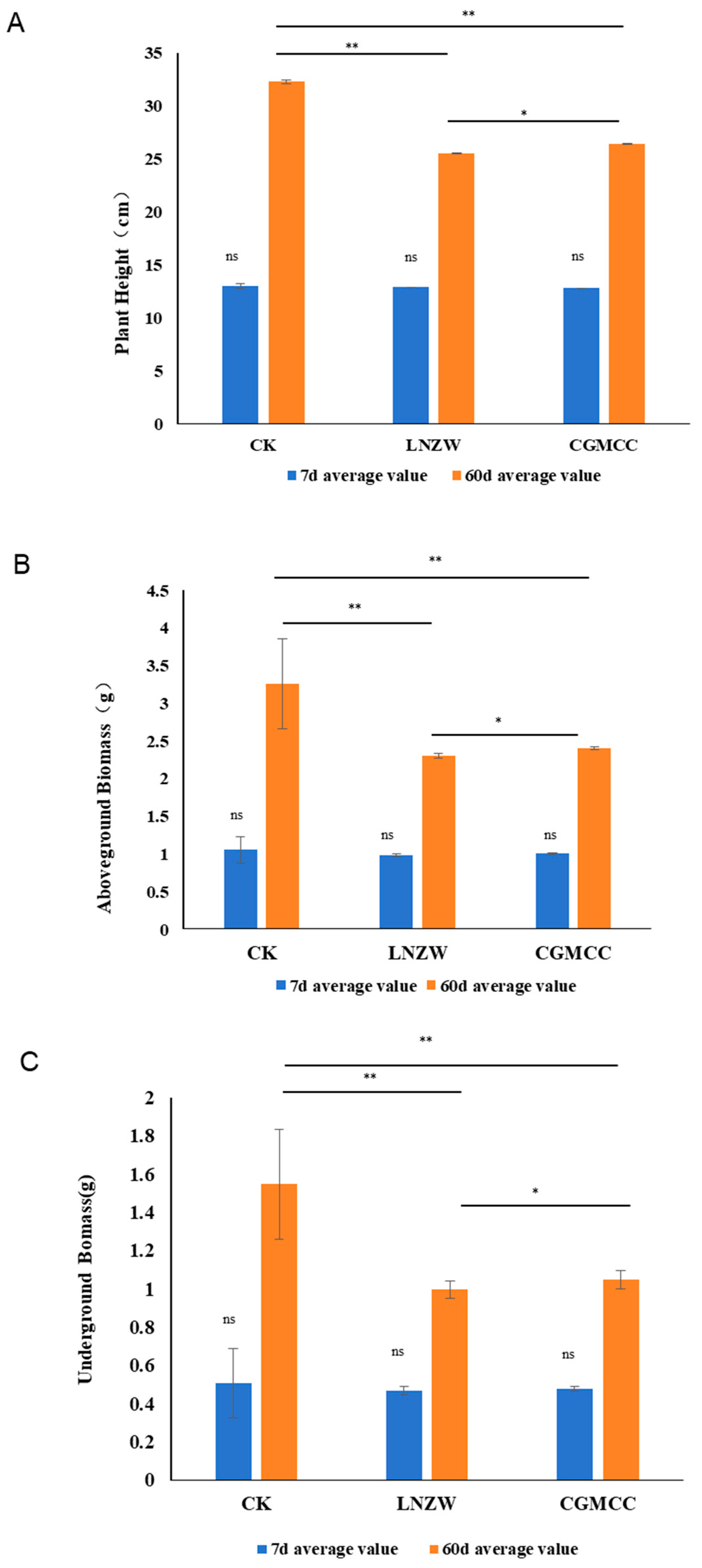
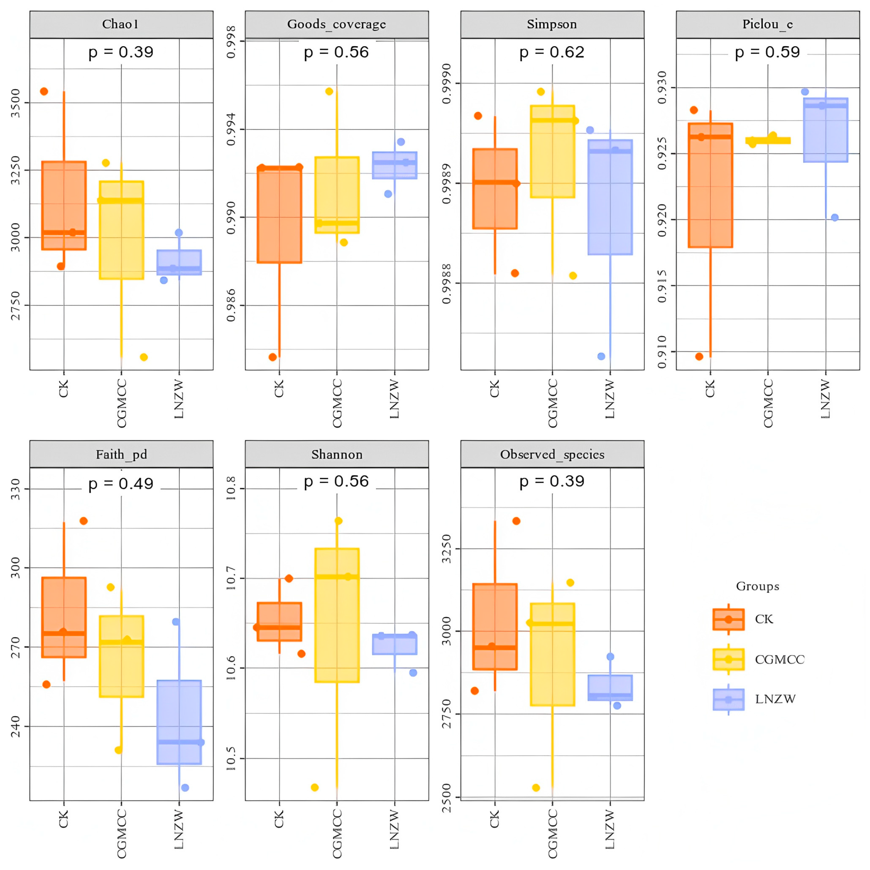
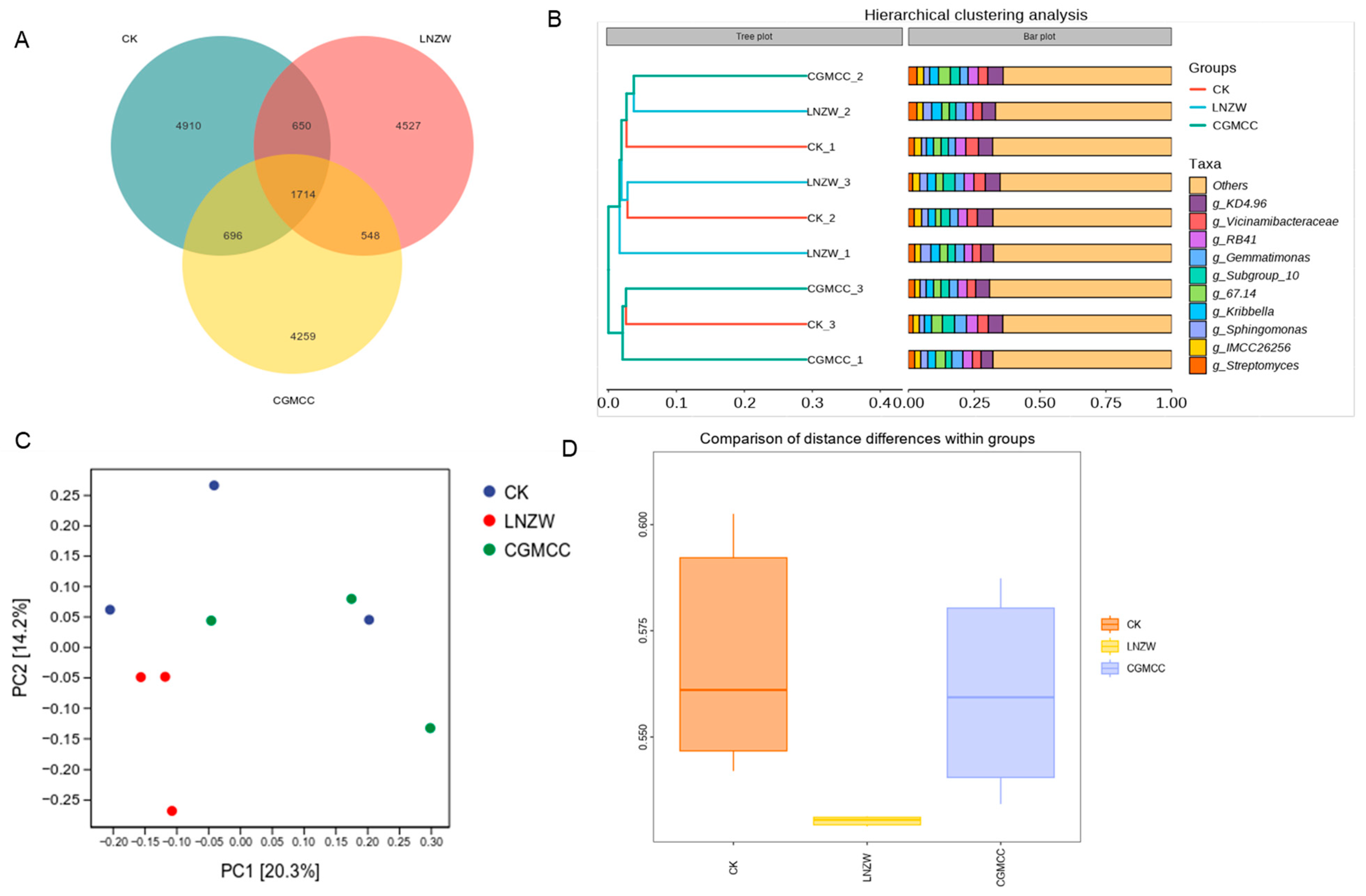
A. flavus infection, with ecological perturbations emerging prior to visible plant growth inhibition. At 7 days post-inoculation (dpi), no significant differences in peanut plant height were observed between inoculated and control groups, yet bacterial species richness (Chao1, Observed species) and phylogenetic diversity (Faith_pd) were already reduced in the rhizosphere of inoculated plants, and the number of unique operational taxonomic units (OTUs) was markedly lower in the
A. flavus-treated groups (LNZW < CGMCC < CK). This early decline in microbial diversity indicates that
A. flavus exerts immediate selective pressure on the rhizosphere bacterial community upon colonization, a phenomenon driven by resource competition and microenvironmental alterations in the rhizosphere niche [
27]. This finding aligns with reports in various plant-pathogen systems where “pathogen infection leads to an early decline in rhizosphere microbial diversity” [
28]. Notably, similar temporal patterns of early microbial diversity loss have been documented in
A. flavus-infected maize and cotton rhizospheres [
29,
30], suggesting that this early microbiome response is a conserved feature across host crops susceptible to
A. flavus.
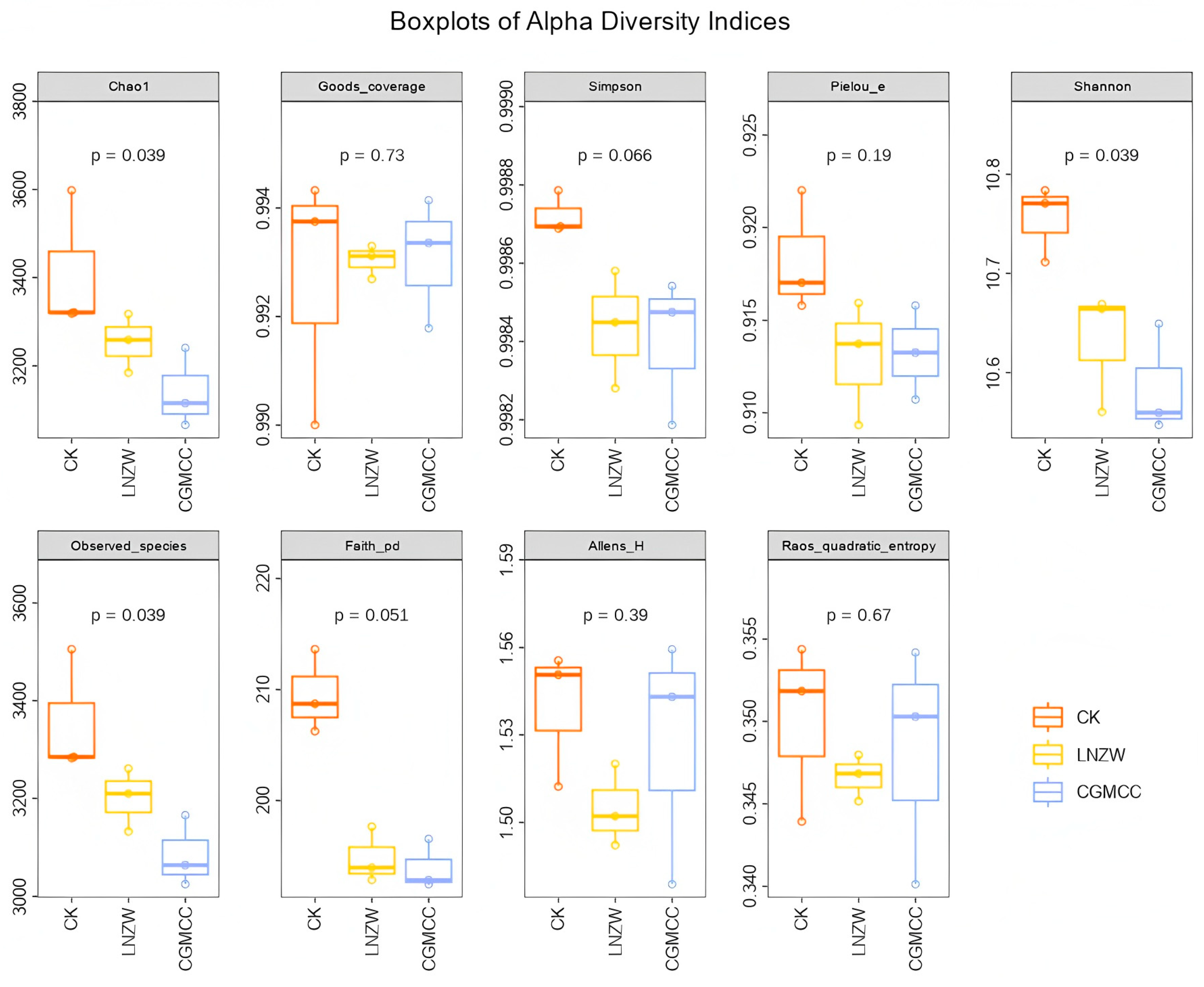
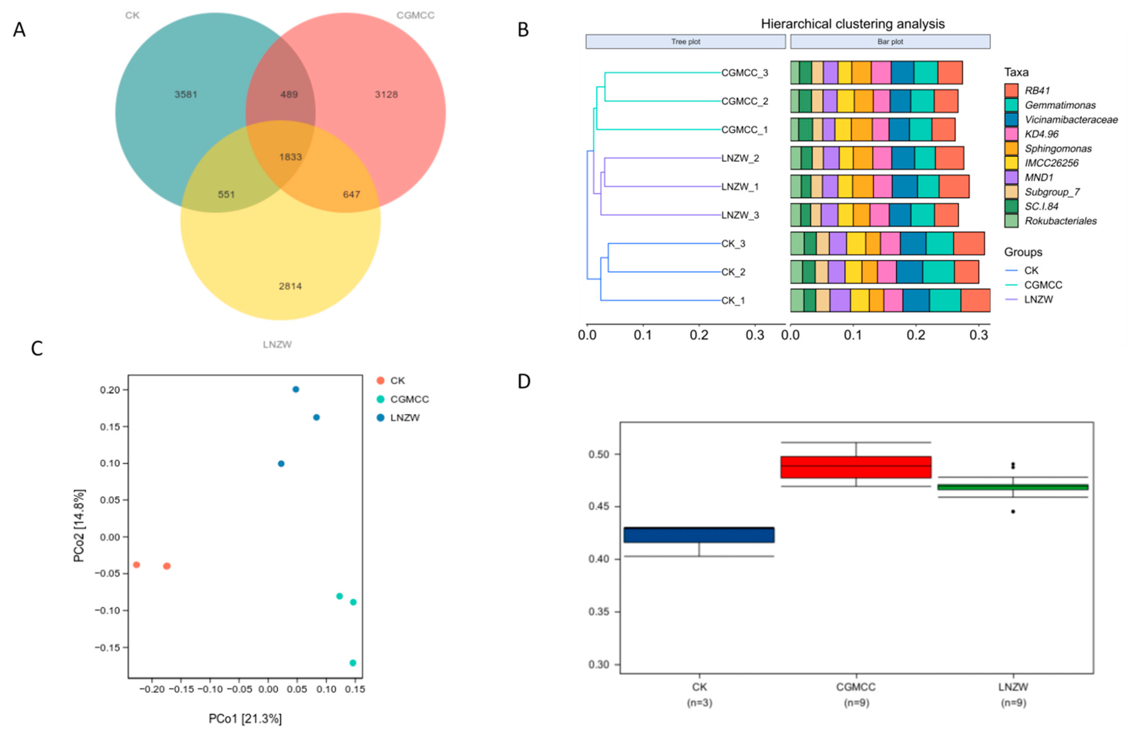
By 60 dpi, the restructuring effect of
A. flavus on the rhizosphere bacterial community became highly pronounced. Beta diversity analysis (PCoA) revealed a clear separation of microbial communities between inoculated and control groups, and intra-group distance analysis confirmed a statistically significant divergence between the two
A. flavus strains (LNZW vs. CGMCC,
p = 0.016). This progressive community differentiation demonstrates that
A. flavus acts not only as a biological filter reducing microbial richness but also as a key driver of rhizosphere community assembly [
29,
30]. The strain-specific effects on microbiome structure highlight that the ecological impact of
A. flavus is dependent on the genetic and physiological traits of the infecting strain, which may be linked to differences in toxin production, colonization rate, or metabolic activity between LNZW (a natural toxigenic strain) and CGMCC (a model strain). Notably, the LNZW group exhibited a slight recovery in microbial richness at 60 dpi compared to CGMCC, suggesting potential adaptive changes in the rhizosphere community in response to long-term infection by the natural toxigenic strain.
The reduction in phylogenetic diversity (Faith_pd) persisted throughout the experimental period in inoculated groups, indicating that
A. flavus infection not only reduces bacterial species number but also disrupts the evolutionary relatedness of the rhizosphere community [
31]. This loss of phylogenetic diversity may impair the functional redundancy of the microbiome, as closely related taxa often share similar ecological functions. In contrast, community evenness (Simpson, Pielou_e indices) showed minor changes across groups, implying that
A. flavus infection primarily affects the composition of the rhizosphere community rather than the relative abundance distribution of dominant taxa [
32].
3.2. Response of Key Rhizosphere Bacterial Taxa to A. flavus Infection and Their Ecological Implications
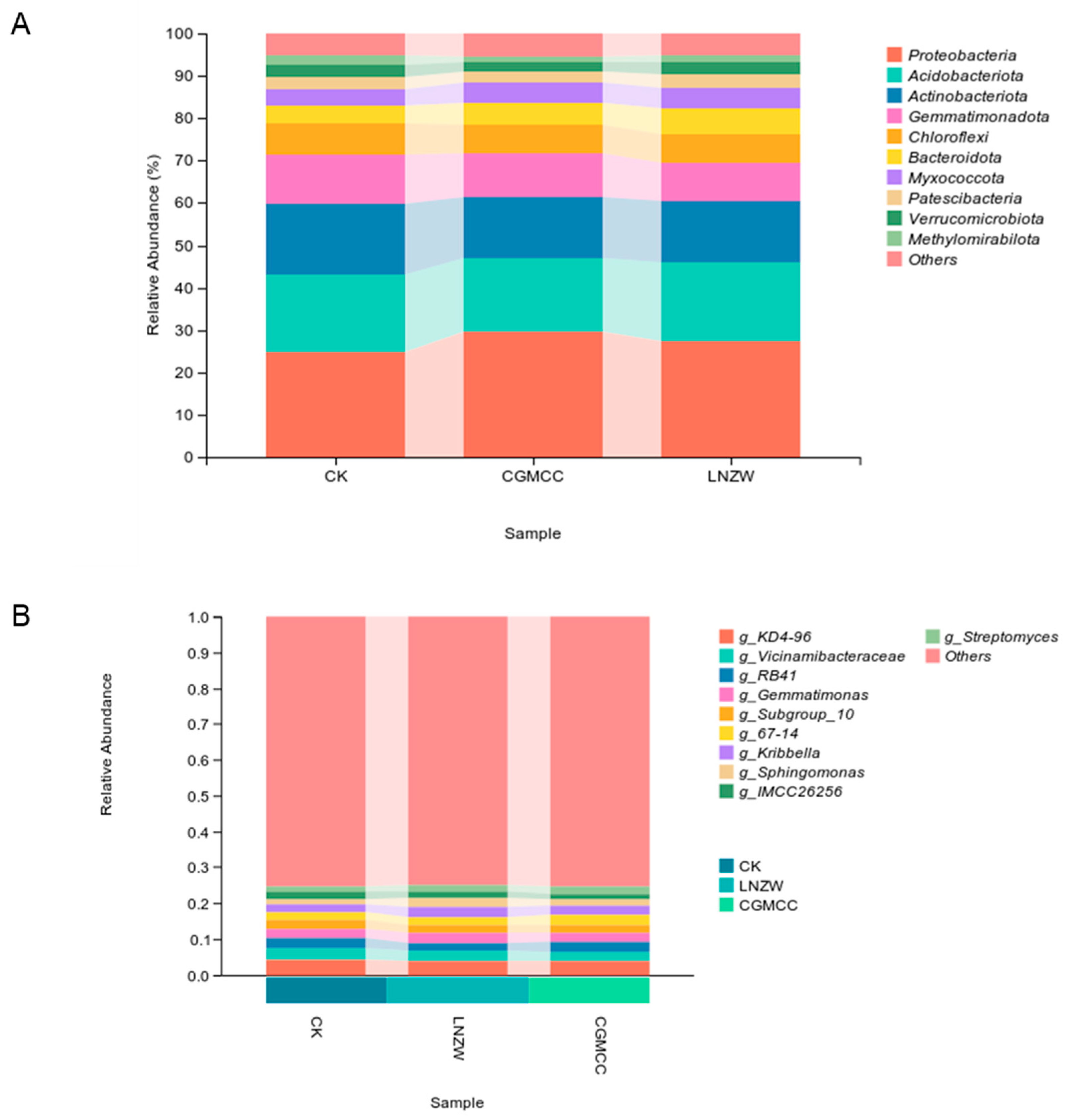
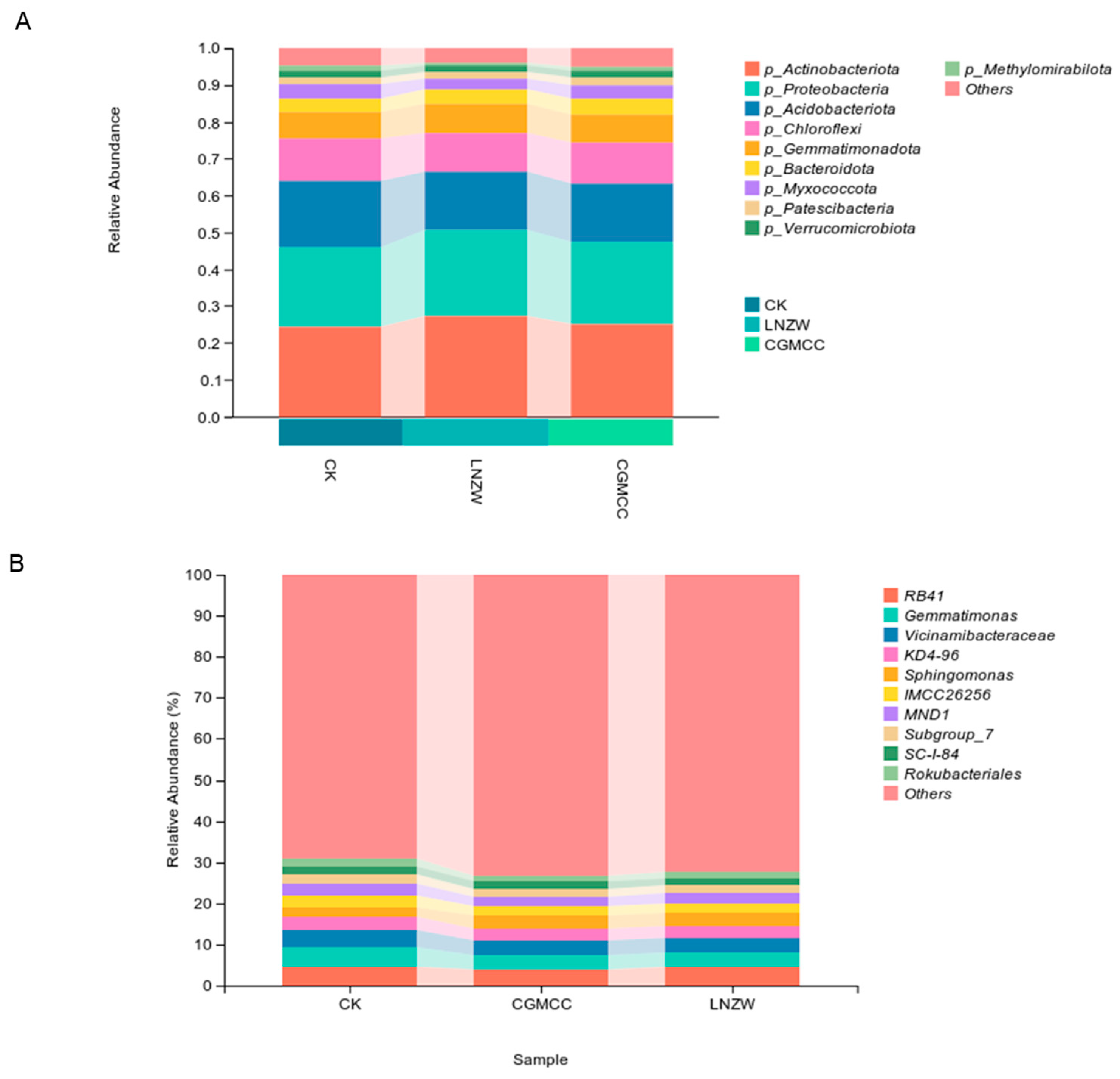
At the phylum level, Proteobacteria, Acidobacteriota, and Actinobacteria constituted the core of the peanut rhizosphere bacterial community, collectively accounting for over 70% of the total relative abundance across all treatments.
A. flavus infection induced a consistent enrichment of Proteobacteria and Actinobacteria at 60 dpi, while reducing the relative abundance of Acidobacteriota and Chloroflexi—an ecological shift closely linked to the stress adaptability of these phyla. Proteobacteria, as typical copiotrophic bacteria, exhibit rapid growth and metabolic versatility, enabling them to exploit the nutrient-rich rhizosphere microenvironment created by
A. flavus colonization and plant stress responses [
31,
32]. The enrichment of Actinobacteria, by contrast, is likely a defensive response of the rhizosphere microbiome: members of this phylum produce a diverse array of antifungal secondary metabolites and hydrolytic enzymes, which may mitigate
A. flavus proliferation and invasion [
33]. In addition, Actinobacteria are highly tolerant to biotic stress, making them dominant taxa in the disturbed rhizosphere environment induced by pathogen infection [
34].
The decline in Acidobacteriota and Chloroflexi abundance in inoculated groups reflects a shift from a stable, oligotrophic rhizosphere ecosystem to a stress-adapted one [
35]. Acidobacteriota are key taxa involved in soil organic matter decomposition and nutrient cycling in low-nutrient environments, and their reduction may impair the rhizosphere’s capacity for nutrient mobilization [
36]. Chloroflexi, a phylum associated with microenvironmental stability, plays an important role in maintaining the structural integrity of the soil microbial community; their decline further exacerbates the dysbiosis of the rhizosphere ecosystem [
37].
At the genus level,
A. flavus infection induced distinct responses in key bacterial taxa, with potential implications for peanut health. The relative abundance of
KD4-96,
Vicinamibacteraceae, and
RB41 decreased significantly at 7 dpi, a trend that either persisted or was reversed at 60 dpi.
RB41, a subgroup of Acidobacteria, is closely involved in soil nitrogen and phosphorus cycling [
36], and its early decline may reduce the nutrient supply capacity of the rhizosphere, indirectly affecting peanut growth.
Vicinamibacteraceae, a genus associated with plant growth promotion, plays a role in regulating root development and enhancing plant stress resistance [
38]; its reduction may weaken the symbiotic relationship between the peanut and its rhizosphere microbiome. Notably, the relative abundance of these genera increased at 60 dpi, which may represent a compensatory adaptive response of the rhizosphere community to long-term
A. flavus stress.
In contrast to the above taxa,
Sphingomonas maintained a relatively stable abundance throughout the infection period, a finding with notable ecological significance.
Sphingomonas is known for its ability to degrade aromatic compounds and mitigate biotic and abiotic stress in plants [
39]; its stability in the infected rhizosphere suggests that this genus may be a core stress-tolerant taxon in the peanut rhizosphere, potentially playing a role in alleviating
A. flavus-induced stress and aflatoxin detoxification. The increase in the relative abundance of
Streptomyces (a genus of Actinobacteria) at 60 dpi further supports the defensive response of the rhizosphere microbiome, as
Streptomyces is a well-characterized producer of antifungal antibiotics and plant growth-promoting substances [
33].
The shift in the rhizosphere community toward fewer dominant genera at 60 dpi (marked by a substantial reduction in the “Others” category) indicates that
A. flavus infection imposes strong selective pressure on the rhizosphere microbiome, favoring the proliferation of stress-tolerant and competitive taxa [
31,
32]. While this selective enrichment may enhance the short-term adaptability of the rhizosphere community to
A. flavus stress, it reduces microbial functional redundancy, making the community more vulnerable to subsequent biotic and abiotic perturbations [
8].
3.3. Mechanistic Links Between Rhizosphere Microbiome Dysbiosis and Peanut Growth Inhibition
The significant inhibition of peanut growth (reduced plant height, shoot and root biomass) at 60 dpi coincided with the structural and compositional dysbiosis of the rhizosphere bacterial microbiome, establishing a clear link between pathogen-induced microbiome changes and plant growth impairment.
A. flavus infection inhibits peanut growth through a dual mechanism of direct pathogenicity and indirect microbiome-mediated effects. Directly,
A. flavus produces aflatoxins and phytotoxic metabolites that damage plant cells, inhibit photosynthesis, and disrupt root nutrient absorption [
33]. Indirectly, the dysbiosis of the rhizosphere microbiome impairs key ecological functions critical for peanut growth and health, amplifying the inhibitory effects of direct infection.
First, the decline in plant-growth-promoting rhizobacteria (PGPR) such as
Bacillus and
Pseudomonas in inoculated groups weakens the rhizosphere’s capacity for plant growth promotion [
40]. PGPR enhances plant growth through the secretion of phytohormones (e.g., indole-3-acetic acid), solubilization of insoluble nutrients (e.g., phosphorus, potassium), and biological nitrogen fixation [
40]; the reduction in these taxa directly limits peanut nutrient acquisition and growth regulation. Second, the disruption of the rhizosphere microbiome impairs the plant’s induced systemic resistance (ISR) [
8]. A stable rhizosphere microbiome is critical for triggering plant ISR against pathogen invasion, and the loss of beneficial taxa reduces the plant’s ability to mount an effective defense response to
A. flavus, leading to increased pathogen colonization and spread [
27,
28]. Third, the decline in nutrient-cycling taxa (e.g.,
RB41, Acidobacteriota) impairs the rhizosphere’s capacity for organic matter decomposition and nutrient mobilization, leading to insufficient supply of nitrogen, phosphorus, and other essential nutrients for peanut growth [
36,
37]. This nutrient limitation further exacerbates the growth inhibition caused by direct
A. flavus infection.
Furthermore, the structural dysbiosis of the rhizosphere microbiome disrupts microbial network interactions, reducing the community’s “immune” capacity to resist pathogen invasion [
41]. In healthy rhizosphere ecosystems, complex microbial interactions form a stable network that inhibits the proliferation of pathogenic microbes through niche competition and microbial antagonism;
A. flavus infection breaks this network, creating ecological niches for further pathogen colonization and leading to a vicious cycle of microbiome dysbiosis and plant growth inhibition [
8]. The strain-specific differences in peanut growth inhibition (LNZW > CGMCC) are consistent with the degree of microbiome dysbiosis induced by the two strains, further confirming that microbiome alterations are a key mediator of
A. flavus-induced growth suppression.
3.4. Limitations of the Study
This study provides novel insights into the impact of A. flavus infection on the peanut rhizosphere bacterial microbiome, but several limitations should be acknowledged, which point to directions for future research:
Experimental scale and conditions: The study was conducted under controlled pot experiment conditions with sterilized soil, which cannot fully simulate the complex biotic and abiotic interactions in natural field soils (e.g., indigenous microbial communities, soil texture, climate factors, and crop rotation systems). The sterilization of soil eliminates the native microbiome, which may alter the response of the rhizosphere community to
A. flavus infection and the plant-microbe-pathogen interaction dynamics [
8,
27]. Field validation trials under natural soil conditions are therefore necessary to verify the findings of this study.
Focus on bacterial communities only: The study exclusively analyzed the rhizosphere bacterial community and did not investigate other key components of the rhizosphere microbiome, including fungi, archaea, and protists. Fungi (e.g., mycorrhizal fungi) and archaea play important roles in rhizosphere nutrient cycling and plant stress resistance, and their interactions with bacteria and
A. flavus may shape the rhizosphere ecosystem response to infection. In addition,
A. flavus itself is a fungal pathogen, and its interaction with the rhizosphere fungal community may be a key driver of microbiome restructuring. Recent studies have shown that manipulating bacterial quorum sensing can significantly shape the structure of the citrus mycobiome, highlighting the importance of investigating interkingdom interactions in the peanut rhizosphere under
A. flavus stress [
42].
Lack of functional and metabolic analysis: The study characterized the rhizosphere bacterial community based on 16S rRNA gene sequencing, which provides information on community composition and diversity but limited insights into microbial functional activity and metabolic potential.
A. flavus infection likely alters the expression of functional genes related to nutrient cycling, stress response, and antimicrobial activity in the rhizosphere microbiome [
27,
33]. Integrating multi-omics technologies (metagenomics, metatranscriptomics, metabolomics) is necessary to elucidate the functional responses and metabolic interaction networks of the rhizosphere microbiome under
A. flavus infection. For example, multi-omics approaches have successfully revealed how beneficial microorganisms like Penicillium baileys W2 inhibit
A. flavus and promote peanut growth by disrupting membrane permeability and altering the rhizosphere microbial community structure [
43]. Similarly, integrated transcriptomics, metabolomics, and microbiomics have been used to uncover plant defense mechanisms against soil-borne pathogens [
44].
No analysis of aflatoxin content and microbial detoxification: The study focused on plant growth and microbiome changes but did not quantify aflatoxin content in the rhizosphere soil or peanut tissues, nor did it investigate the aflatoxin detoxification capacity of the rhizosphere microbiome. Some rhizosphere bacteria (e.g.,
Sphingomonas,
Streptomyces) are known to degrade aflatoxins [
34,
39], and their activity in the infected rhizosphere may be a key factor in mitigating aflatoxin contamination and plant stress. Recent research has isolated novel aflatoxin-degrading bacteria from peanut rhizospheric soil, such as
Pseudomonas qingdaonensis [
45], and demonstrated that various
Pseudomonas isolates can effectively control
A. flavus and aflatoxin B1 production through different enzymatic or non-enzymatic mechanisms [
46]. Furthermore,
Achromobacter xylosoxidans from the peanut rhizosphere has been shown to inhibit
A. flavus development and aflatoxin synthesis by inducing apoptosis through targeting the cell membrane [
47].
Short experimental time scale: The study sampled only at 7 and 60 dpi, providing a snapshot of microbiome changes during the peanut growth period. A more frequent sampling regime (e.g., 14, 28, 42 dpi) would enable a more detailed characterization of the dynamic trajectory of rhizosphere microbiome restructuring in response to A. flavus infection, and help identify critical time points for microbiome intervention.
No investigation of microbial inoculation as a mitigation strategy: The study established a link between
A. flavus-induced microbiome dysbiosis and peanut growth inhibition, but did not test potential mitigation strategies (e.g., inoculation with beneficial rhizobacteria or synthetic microbial communities). Given the role of PGPR and antifungal taxa in plant growth promotion and disease resistance [
40,
41], testing the efficacy of microbial inoculation to restore the rhizosphere microbiome and alleviate
A. flavus infection is a critical next step. Research in other crops has demonstrated the effectiveness of synthetic microbial communities (SynComs) in mitigating soil-borne diseases. For instance, SynComs have been shown to rescue strawberries from soil-borne diseases by enhancing soil functional microbial abundance and multifunctionality [
48], and well-designed SynComs have proven effective in controlling fire blight disease in apples [
49]. Additionally, studies on plant growth-promoting bacteria have revealed complex interkingdom signaling pathways, such as the “root ROS-microbial IAA-root DNA methylation” pathway, that mediate plant-microbe interactions under stress conditions [
50].
3.5. Future Research Directions
Based on the findings and limitations of this study, future research should focus on the following directions:
Conduct field experiments under natural soil conditions to verify the time-dependent and strain-specific effects of A. flavus on the peanut rhizosphere microbiome, and investigate the influence of environmental factors (e.g., soil type, temperature, humidity) on these effects.
Characterize the entire rhizosphere microbiome (bacteria, fungi, archaea, protists) using high-throughput sequencing technologies, and analyze the interkingdom interactions between these microbial groups and A. flavus to elucidate the holistic response of the rhizosphere ecosystem to infection.
Integrate multi-omics approaches to investigate the functional and metabolic changes in the rhizosphere microbiome under A. flavus infection, and identify key functional genes and metabolic pathways associated with aflatoxin detoxification, plant growth promotion, and disease resistance.
Quantify aflatoxin content in the rhizosphere and peanut tissues, and screen for aflatoxin-degrading rhizosphere bacteria with high activity; investigate the potential of these bacteria for biocontrol of A. flavus and aflatoxin contamination.
Construct synthetic microbial communities (SynComs) based on core stress-tolerant and beneficial taxa (e.g., Sphingomonas, Streptomyces, PGPR), and test their efficacy in restoring the rhizosphere microbiome, enhancing peanut resistance to A. flavus, and reducing aflatoxin contamination.
Investigate the interaction between peanut genotypes and the rhizosphere microbiome in response to A. flavus infection, and breed peanut varieties with the ability to recruit beneficial rhizobacteria and form a disease-resistant microbiome.
4. Conclusions
This study demonstrates that infection by the aflatoxigenic fungus A. flavus significantly impairs peanut plant growth and substantially restructures the associated rhizosphere bacterial community. Key findings reveal that while no growth inhibition was observed at 7 days post-inoculation (dpi), a pronounced reduction in plant height, shoot biomass, and root biomass occurred by 60 dpi. Concurrently, high-throughput sequencing analysis showed a clear reduction in bacterial alpha diversity (species richness and phylogenetic diversity) in the rhizosphere of infected plants, particularly at the early infection stage. Beta diversity analysis confirmed a significant and progressive divergence in microbial community structure between infected and control plants over time, with distinct effects also noted between the two A. flavus strains used.
At the compositional level, infection enriched for bacterial phyla such as Proteobacteria and Actinobacteria, while reducing the relative abundance of others like Acidobacteriota. Specific genera, including some putative beneficial taxa, were suppressed. These collective alterations suggest that A. flavus influences plant health through a dual mechanism: direct pathogenic effects and indirect modulation of the root-associated microbiome, potentially disrupting its supportive functions.
In summary, our work establishes a tangible link between A. flavus infection, peanut growth suppression, and rhizosphere microbiome dysbiosis. This insight underscores the importance of microbial community stability in plant-pathogen interactions and provides a foundational framework for developing microbiome-based strategies to enhance crop resilience and mitigate aflatoxin contamination in agroecosystems.
5. Materials and Methods
5.1. Experimental Materials
Peanut variety selection: The peanut (Arachis hypogaea L.) cultivar ‘Baisha’ was provided by the Peanut Research Institute, Liaoning Academy of Agricultural Sciences, Shenyang, China. This variety is characterized by strong growth vigor, broad adaptability, and stable yield, and its response to A. flavus infection is considered typical.
Aspergillus flavus strain CGMCC 3.4408 was obtained from the China General Microbiological Culture Collection Center (CGMCC).
Aspergillus flavus strain LNZW 23 was isolated from peanut rhizosphere soil in Zhangwu County, Liaoning Province, China, by the Oil Crops Research Institute, Chinese Academy of Agricultural Sciences. LNZW 23 is a laboratory code with no public culture collection number.
Culture medium: Potato Dextrose Agar (PDA) was employed for the activation, cultivation, and preservation of A. flavus. The medium was prepared according to the following formulation: 200 g of potatoes, 20 g of glucose, 15–20 g of agar, and 1000 mL of distilled water. For preparation, potatoes were peeled, diced, and boiled in distilled water for 30 min. The resulting infusion was then filtered through gauze to obtain the potato extract. Glucose and agar (Both purchased from China National Pharmaceutical Group Chemical Reagent Co., Ltd., Shanghai, China) were subsequently added to the filtrate, and the mixture was heated with continuous stirring until all components were completely dissolved. The medium was then dispensed into appropriate vessels and sterilized by autoclaving at 121 °C for 20 min.
Reagents: The OMEGA E.Z.N.A. Soil DNA Kit (Bao Bioengineering Co., Ltd., Dalian, China) was used for total microbial DNA extraction from peanut rhizosphere soil. The PCR primers used in this study were synthesized by Shenggong Biotechnology Co., Ltd. (Shanghai, China). Universal primers 27F (5′-AGAGTTTGATCMTGGCTCAG-3′) and 1492R (5′-TGGYTACCTTGTTACGACTT-3′) were employed to amplify the nearly full-length bacterial 16S rRNA gene (V1–V9 regions). Additionally, routine reagents including absolute ethanol, chloroform, isoamyl alcohol, Tris-HCl, and EDTA (all analytical grade) were purchased from China National Pharmaceutical Group Chemical Reagent Co., Ltd. (Shanghai, China).
5.2. Experimental Design
A controlled pot experiment was conducted in a climate chamber to investigate the effects of A. flavus infection on peanut plants. The experiment consisted of three groups: a treatment group (inoculated with two A. flavus strains: one group was inoculated with the A. flavus strain CGMCC 3.4408, referred to as Group CGMCC, while the other group was inoculated with LNZW 23, abbreviated as Group LNZW) and a control group (mock-inoculated with an equivalent volume of sterile water), each comprising ten biological replicates.
Peanut seeds were surface-sterilized with 75% ethanol (Sinopharm Chemical Reagent Co., Ltd., Shanghai, China) for 1 min, rinsed 2–3 times with sterile distilled water, and placed on moist sterile filter paper for germination in an incubator at 28 °C for 2–3 days. On May 20, uniformly germinated seeds with emerged radicles were selected and sown in pots (two seeds per pot). Each pot (15 cm diameter) was filled with 1.5 kg of field soil (loam; pH 6.8; organic matter content 18 g/kg), which had been sterilized by autoclaving at 121 °C for 2 h. After emergence, seedlings were thinned to one robust plant per pot (
Figure S1). The pots were maintained in a climate chamber under natural light and temperature conditions, with regular and quantitative watering to ensure consistent soil moisture throughout the experiment.
At the pegging stage (15 July), plants in the treatment group were inoculated by injecting an A. flavus spore suspension into the rhizosphere soil. To prepare the inoculum, preserved A. flavus strains were cultured on potato dextrose agar (PDA) plates at 28 °C for 7–10 days until abundant conidial production. Conidia were harvested by rinsing the colony surface with sterile distilled water, and the spore suspension was adjusted to a concentration of 1 × 10⁷ spores/mL. Using a sterile syringe, 10 mL of this suspension was injected into the rhizosphere soil of each treatment plant. Control plants received an equal volume of sterile water injected in the same manner.
Throughout the experimental period, plants were maintained under natural cultivation conditions with regular and quantitative watering to maintain uniform soil moisture.
5.3. Sample Collection and Processing
Samples were taken at 7 and 60 days (dpi) after vaccination. Collect rhizosphere soil using the shaking method. Specifically, use sterile scissors to remove the aboveground part of the peanut plant, and then gently remove the entire root system and attached soil from the flowerpot. After discarding loose and attached large soil blocks, carefully brush off the soil tightly attached to the root surface (defined as the 0–4 mm area) with a sterile brush and collect it as rhizosphere soil. The rhizosphere soil of each plant is considered an independent biological replica, placed in a separate sterile self-sealing bag, and immediately transported to the laboratory on ice. Fresh rhizosphere soil samples were rapidly stored at −80 °C for subsequent soil microbial metagenomic DNA extraction and high-throughput sequencing analysis.
5.4. Microbial Diversity Analysis
High-throughput sequencing was employed to analyze the diversity of the peanut root-associated microbiome. First, total microbial DNA was extracted from rhizosphere soil samples using the OMEGA E.Z.N.A. Soil DNA Kit according to the manufacturer’s instructions. DNA integrity was verified by 1% agarose gel electrophoresis, and concentration/purity were determined using a NanoDrop 2000 spectrophotometer (Changzhou Meikuanda Electronic Appliance Sales Co., Ltd., Changzhou, China) to ensure quality for downstream applications.
Using the extracted DNA as a template, PCR amplification was performed with the aforementioned primers targeting the bacterial 16S rRNA gene. The PCR reaction mixture (25 μL) contained: 12.5 μL of 2× Taq PCR Master Mix, 0.5 μL each of forward and reverse primers (10 μmol/L), 1 μL of DNA template, and 10.5 μL of ddH2O. The PCR conditions were: initial denaturation at 95 °C for 5 min; 35 cycles of 95 °C for 30 s, 55 °C for 30 s, and 72 °C for 30 s; final extension at 72 °C for 10 min.
PCR products were examined by agarose gel electrophoresis. Target bands were excised and purified using the Axygen DNA Gel Extraction Kit (Corning, Shanghai, China). The purified amplicons were subjected to high-throughput sequencing on an Illumina MiSeq platform, performed by Nanjing Paisennuo Biotechnology Co., Ltd. (Nanjing, China).
Raw sequencing data underwent quality control to remove low-quality reads, adapter sequences, and chimeras. High-quality sequences were clustered into operational taxonomic units (OTUs) at a 97% similarity threshold using Quantitative Insights Into Microbial Ecology (QIIME, v2022.11, QIIME 2 Development Team, Boulder, CO, USA) software. Taxonomic annotation of each OTU was performed by comparison against the Ribosomal Database Project (RDP, v11.4, Michigan State University, Michigan, USA) database to determine microbial identities.
Microbial diversity indices, including the Shannon index, Simpson index, Ace index, and Chao1 index, were calculated. The Shannon and Simpson indices assess community diversity (higher values indicate greater diversity), while the Ace and Chao1 indices estimate species richness (higher values indicate greater richness). Community structure differences among treatment and control groups were visualized using Principal Component Analysis (PCA), Principal Coordinates Analysis (PCoA), and Non-metric Multidimensional Scaling (NMDS). Graphs were generated using OriginPro (v2023b, OriginLab Corporation, Northampton, MA, USA).
5.5. Statistical Analysis
Experimental data were initially collated and summarized using Microsoft Excel 2019 (16.0.10356.20006) to calculate means and standard deviations. Significance testing was performed using Excel 2019 software. Differences in microbial diversity indices, richness indices, and relative abundances of microbial taxa between treatment and control groups were compared using one-way analysis of variance (ANOVA). Differences were considered statistically significant at p < 0.05.